Embed Size (px)
Citation preview
1
Pathophysiology, diagnosis and management of nephrogenic diabetes insipidus
Detlef Bockenhauer and Daniel G. Bichet University College London Institute of Child Health, 30 Guilford Street, London, WC1N 1EH, UK (D.B.). Departments of Medicine and Molecular and Integrative Physiology, Université de Montréal, Research Center, Hôpital du Sacré-Coeur de Montréal, 5400 Boulevard Gouin Ouest, Montréal, H4J 1C5 Canada (D.G.B.). Correspondence to: D.G.B. [email protected]
Key points
Nephrogenic diabetes insipidus (NDI) is caused by inability of the kidneys to
concentrate urine by reabsorption of water in the collecting duct
NDI can be inherited (X-linked or autosomal) or acquired, most commonly as
a result of lithium treatment
Management of primary forms of NDI focuses on dietary modification to
reduce osmotic load and pharmacological treatment with prostaglandin
synthesis inhibitors and thiazide diuretics
With appropriate treatment, complications of NDI, such as failure to thrive
and mental retardation resulting from repeated hypernatraemic dehydration,
can be avoided
New treatment approaches for congenital NDI have been tested in animal
models, but efficacy in patients has not yet been confirmed
2
Abstract
Healthy kidneys maintain fluid and electrolyte homoeostasis by adjusting urine
volume and composition according to physiological needs. The final urine
composition is determined in the last tubular segment: the collecting duct. Water
permeability in the collecting duct is regulated by arginine vasopressin (AVP).
Secretion of AVP from the neurohypophysis is regulated by a complex signalling
network that involves osmosensors, barosensors and volume sensors. AVP
facilitates aquaporin (AQP)-mediated water reabsorption via activation of the
vasopressin V2 receptor (AVPR2) in the collecting duct, thus enabling
concentration of urine. In nephrogenic diabetes insipidus (NDI), inability of the
kidneys to respond to AVP results in functional AQP deficiency. Consequently,
affected patients have constant diuresis, resulting in large volumes of dilute
urine. Primary forms of NDI result from mutations in the genes that encode the
key proteins AVPR2 and AQP2, whereas secondary forms are associated with
biochemical abnormalities, obstructive uropathy or the use of certain
medications, particularly lithium. Treatment of the disease is informed by
identification of the underlying cause. Here we review the clinical and diagnostic
aspects of NDI, the various aetiologies, current treatment options and potential
future developments.
[H1] Introduction
Nephrogenic diabetes insipidus (NDI) results from failure of the kidneys to
concentrate urine. Patients with this disease typically produce around 12 l of
urine per day. Assuming a bladder volume of 500 ml, this volume of urine will
necessitate voiding approximately once per hour throughout the day and night.
3
Moreover urinary fluid losses need to be replenished by constant fluid intake.
The disease has a substantial impact on quality of life as sleep is frequently
interrupted and patients must plan their daily activities with logistics for
frequent voiding and access to drinking water.
Primary and secondary forms of NDI exist. Paediatricians typically see
patients with primary inherited forms of the disease, mostly boys presenting in
the first year of life with failure-to-thrive and vomiting. By contrast, in adults
acquired NDI is much more common than primary NDI. Medical treatment can
ameliorate the symptoms of NDI and as understanding of the underlying
pathophysiology improves, new potential treatment options arise. In this Review
we consider the clinical, diagnostic and therapeutic aspects of NDI in the context
of the physiology of renal water handling to enable a molecular understanding of
the disease. We also discuss recent insights into the regulation of water
permeability and their consequences with respect to diagnosis and new
treatment options.
[H1] Physiology of urine concentration
Healthy adult kidneys produce approximately 180 l of primary glomerular
filtrate per day.1 The vast majority of this filtrate is reabsorbed in the proximal
tubule, which is freely permeable to water owing to the constitutive expression
of aquaporin (AQP)-1 water channels.2 As solutes are reabsorbed in the proximal
tubule, water follows passively along the osmotic gradient. The remaining urine
is thus still isotonic when it enters the loop of Henle, the key segment for
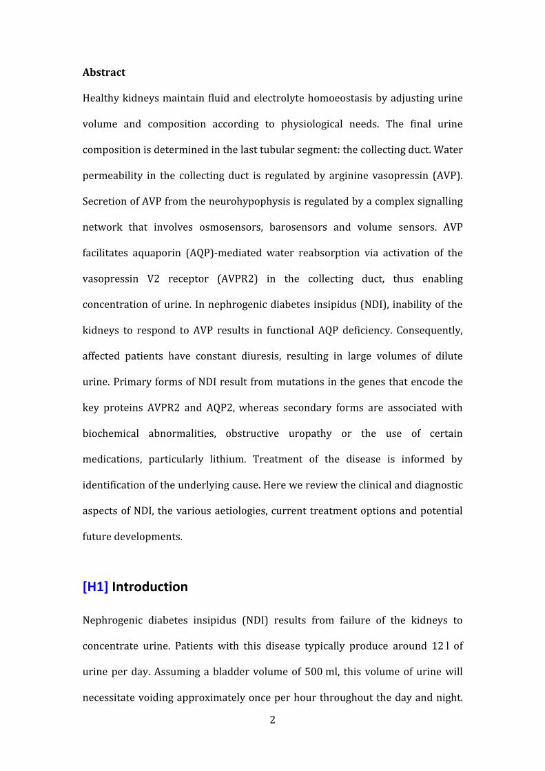
counter-current concentration (Figure 1). Urine concentration begins in the thin
descending limb (TDL). Mechanisms of concentration include AQP1-mediated
4
exit of water into the medullary interstitium.3 However, data from several rodent
models show that Aqp1 expression is mainly restricted to the first 60% of the
TDL rather than to the deeper papillary parts in which the steepest part of the
osmotic gradient is generated.4 Concentration of the tubular fluid in these
innermost parts of the TDL might occur via passive sodium influx,5 but no
substantial sodium permeability has been observed.6 Thus, the exact mechanism
of urinary concentration in the innermost part of the medulla remains to be
elucidated.
Urine subsequently enters the thick ascending limb (TAL, also known as the
diluting segment), which is impermeable to water but actively removes sodium
chloride via the co-transporter solute carrier family 12 member 1 (SLC12A1,
also known as NKCC2), thereby diluting the urine.7 The accumulation of solutes
in the interstitium generates the driving force for the removal of water from the
TDL (in long-looped nephrons) and the entry of sodium chloride (in short-looped
nephrons), completing the counter-current multiplier.
Further removal of sodium chloride occurs in the distal convoluted tubule via
SLC12A3 (also known as NCC). At entry into the arginine vasopressin (AVP)-
sensitive connecting tubules and collecting ducts, urine osmolality is typically
around 50–100 mOsm/kg. The final osmolality of the urine is solely dependent
on the availability of water channels. If these channels are present, water exits
the tubule following the interstitial concentration gradient and the urine is
concentrated. If no water channels are present, dilute urine will be excreted.
5
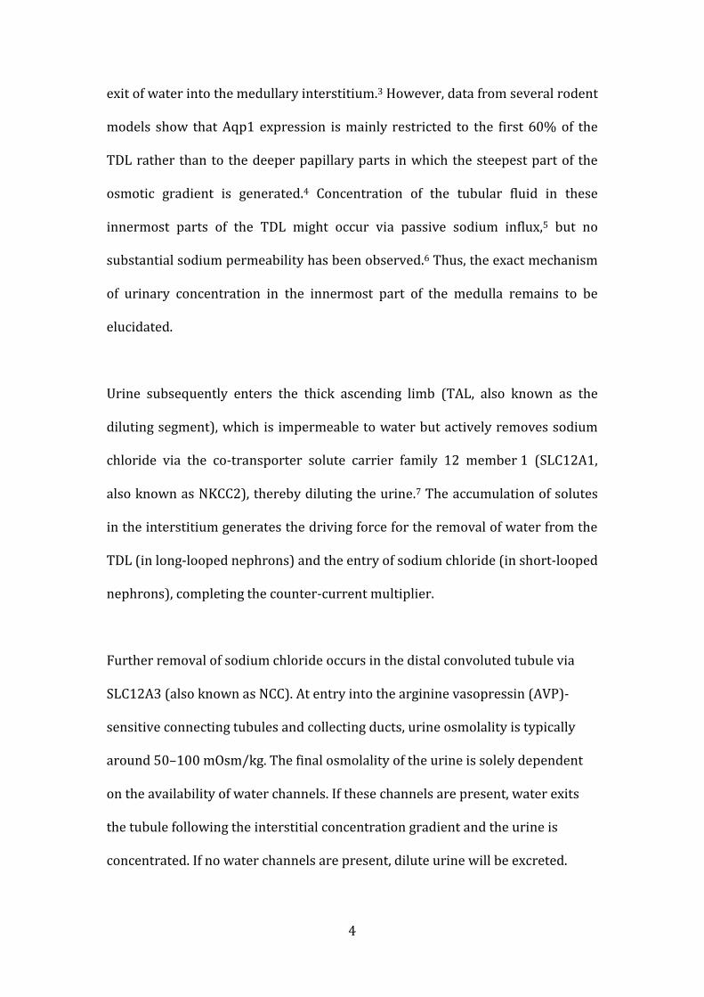
[H1] Vasopressin-regulated water permeability
Vasopressin secreted by hypothalamic neurons in response to changes in soma
volume (Figure 2) is the key regulator of the water permeability of the collecting
ducts. The key cell type that mediates this regulated water permeability is the
principal cell, which constitutively expresses AQP3 and AQP4 on the basolateral
side.2 The water permeability of the apical membranes of the principal cell is
determined by the availability of AQP2 water channels. In the absence of AVP
signalling, these channels are located in intracellular vesicles and no apical water
permeability exists.8 Binding of AVP to the vasopressin V2 receptor (AVPR2)
initiates a signalling cascade that ultimately leads to the insertion of AQP2
channels into the apical membrane(Figure 3). AVPR2 is a G-protein-coupled
receptor and binding of AVP leads to the release of stimulatory G-protein, which
in turn activates the neighbouring adenylyl cyclase.9 The resulting increase in
levels of cAMP activates protein kinase A (PKA).
The cytoplasmic carboxy-terminal tail of AQP2 has five canonical PKA-
phosphorylation sites: Thr244, Ser256, Ser261, Ser264 and Thr269 (Ser269 in
mice).10 Upon administration of the vasopressin analogue 1-desamino-8-D-
arginine vasopressin (DDAVP), phosphorylation of AQP2 at Ser256, Ser264 and
Ser269 is abundantly increased, whereas phosphorylation at Ser261 is
decreased;10 specific phosphorylation of these residues might serve distinct
physiological roles. In a mammalian cell line (LLC-PK1) transiently transfected
with AQP2, phosphorylation at Ser256 was essential for trafficking of the water
channel to the plasma membrane,11 whereas in Xenopus oocytes,
phosphorylation of at least three subunits of the AQP2 tetramer at Ser256 was
6
required for redistribution from storage vesicles to the apical plasma
membrane.12 The critical role of Ser256 in AQP2 function was confirmed by the
identification of mutations that impair phosphorylation at this site in patients
with NDI.13, 14 In vitro studies demonstrated the failure of such mutants to reach
the apical membrane.13, 14
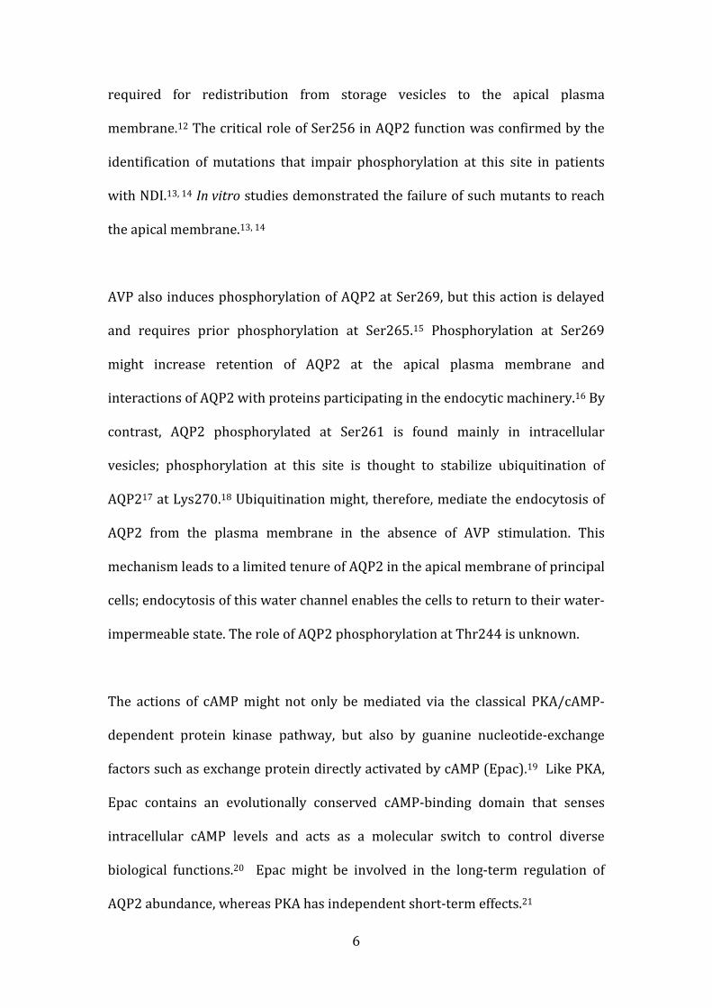
AVP also induces phosphorylation of AQP2 at Ser269, but this action is delayed
and requires prior phosphorylation at Ser265.15 Phosphorylation at Ser269
might increase retention of AQP2 at the apical plasma membrane and
interactions of AQP2 with proteins participating in the endocytic machinery.16 By
contrast, AQP2 phosphorylated at Ser261 is found mainly in intracellular
vesicles; phosphorylation at this site is thought to stabilize ubiquitination of
AQP217 at Lys270.18 Ubiquitination might, therefore, mediate the endocytosis of
AQP2 from the plasma membrane in the absence of AVP stimulation. This
mechanism leads to a limited tenure of AQP2 in the apical membrane of principal
cells; endocytosis of this water channel enables the cells to return to their water-
impermeable state. The role of AQP2 phosphorylation at Thr244 is unknown.
The actions of cAMP might not only be mediated via the classical PKA/cAMP-
dependent protein kinase pathway, but also by guanine nucleotide-exchange
factors such as exchange protein directly activated by cAMP (Epac).19 Like PKA,
Epac contains an evolutionally conserved cAMP-binding domain that senses
intracellular cAMP levels and acts as a molecular switch to control diverse
biological functions.20 Epac might be involved in the long-term regulation of
AQP2 abundance, whereas PKA has independent short-term effects.21
7
In addition to an acute role in AQP2 localization, AVP-mediated increases in
cAMP promote AQP2 expression via PKA-mediated phosphorylation of the cAMP
responsive element binding protein, which subsequently stimulates
transcription from the AQP2 promoter.22-24 The change in localization of AQP2
occurs within minutes, whereas the increase in transcription requires hours to
take effect.25,26
[H1] Congenital NDI
In 1992 the AVPR2 gene that encodes the AVP2 receptor was cloned and
mutations in this gene were identified in patients with X-linked NDI (Figure 4).27-
30 The AQP2 gene was cloned in 199331, 32 and in 1994 mutations in AQP2 were
found to underlie autosomal recessive NDI.33 The discovery of these two key
genes—AVPR2 and AQP2—has enabled genetic testing of affected patients.
Mutations in either of these genes are identified in almost all patients with a
clear clinical phenotype of congenital NDI.34, 35
[H2] AVPR2 mutations Approximately 90% of patients with inherited NDI have a mutation in AVPR2.36,
37 As this gene is located on the X-chromosome, AVPR2 mutations have an X-
linked pattern of inheritance; consequently the majority of patients with NDI are
male. Occasionally, female patients with NDI due to AVPR2 mutations have been
described.34, 35, 38-41 In one study, 16 of 64 (25%) female carriers of AVPR2
mutations showed polyuric symptoms and four of these patients (6%) were
diagnosed with complete NDI rather than the milder partial NDI phenotype
described below.34 Skewed X-inactivation is thought to cause symptoms in some
female carriers of AVPR2 mutations, although these symptoms do not necessarily
8
correlate with the X-inactivation patterns observed in leukocytes.42 Presumably,
X-inactivation patterns can differ between tissues, so the pattern in leukocytes
might not reflect that in the kidney. In our experience at Great Ormond Street
Hospital, London, UK, two female relatives in 20 families with AVPR2 mutations
have been diagnosed with complete NDI (unpublished data). One of these
women was the index case in her family, but not all female carriers have been
systematically investigated. Partial NDI symptoms were observed in two other
female carriers in one family.43
A few AVPR2 mutations have been identified recurrently owing to a founder
effect. For example, the Hopewell mutation (Trp71X)—derived from an Ulster
Scot immigrant who arrived in Halifax, Canada on the ship Hopewell in 1761—
has been identified in >40 North American patients.44, 45 Another recurrent
mutation is the Cannon mutation (Leu312X), which is named after a large
pedigree with ≥38 affected patients in Utah.45 Most patients, however, have
individual mutations; to date >250 different AVPR2 mutation have been
described in >300 families (46 and our unpublished data). Interestingly, most of
these mutations are missense and, when assessed in vitro, seem to encode
functional but misfolded receptors, which are retained and degraded in the
endoplasmic reticulum (ER).47
[H3] AQP2 mutations
Approximately 10% of cases of congenital NDI are due to loss-of-function
mutations in the AQP2 gene located on chromosome 12.45,34, 48 These mutations
are typically inherited in an autosomal recessive fashion, although a few
mutations have been described to cause autosomal dominant disease.13, 49-51
9
AQP2 is a homotetramer; four subunits assemble to form the water channel.
Dominant mutations typically affect amino acids in the C-terminus, leading to
aberrant trafficking; mutant AQP2 subunits are able to oligomerize with wild-
type protein to form tetramers, but exert a dominant-negative effect on
trafficking of these assembled tetramers.52 Interestingly, specific mutations
direct AQP2 trafficking to distinct cellular compartments, such as the Golgi
complex,13 late endosomes and lysosomes49 or the basolateral membrane.51, 53
These findings have provided insights into the intracellular trafficking motifs of
AQP2.
[H2] Epidemiology
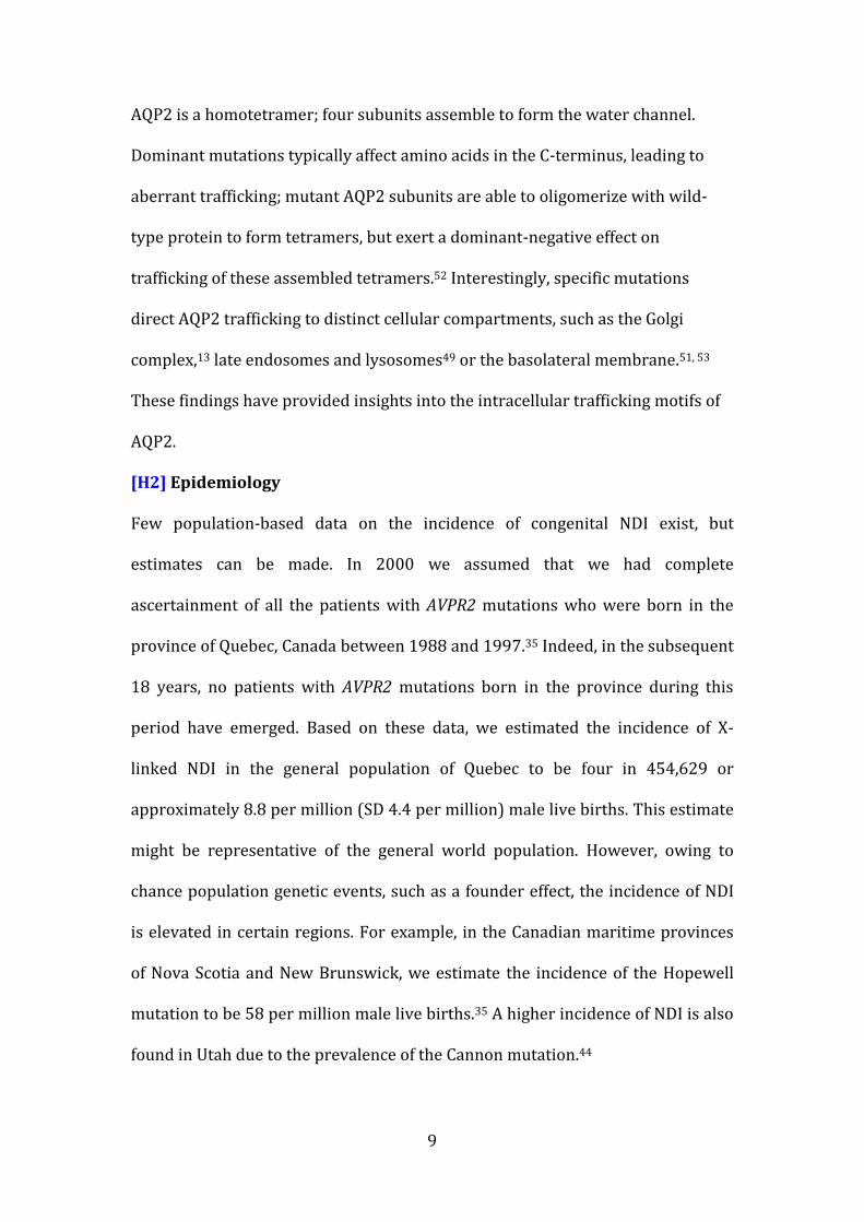
Few population-based data on the incidence of congenital NDI exist, but
estimates can be made. In 2000 we assumed that we had complete
ascertainment of all the patients with AVPR2 mutations who were born in the
province of Quebec, Canada between 1988 and 1997.35 Indeed, in the subsequent
18 years, no patients with AVPR2 mutations born in the province during this
period have emerged. Based on these data, we estimated the incidence of X-
linked NDI in the general population of Quebec to be four in 454,629 or
approximately 8.8 per million (SD 4.4 per million) male live births. This estimate
might be representative of the general world population. However, owing to
chance population genetic events, such as a founder effect, the incidence of NDI
is elevated in certain regions. For example, in the Canadian maritime provinces
of Nova Scotia and New Brunswick, we estimate the incidence of the Hopewell
mutation to be 58 per million male live births.35 A higher incidence of NDI is also
found in Utah due to the prevalence of the Cannon mutation.44
10
Based on the families referred to us for genetic testing, we estimate that the
incidence of NDI secondary to AQP2 mutations is five to ten times lower than
that of NDI secondary to AVPR2 mutations. The incidence of this type of
congenital NDI might, however, be increased in populations with a high degree of
consanguinity; we have found an over-representation of the Val71Met AQP2
allele in patients of Pakistani descent.54
[H1] Secondary inherited forms of NDI
In addition to primary forms of congenital NDI a few cases of secondary
inherited NDI have been reported.55, 56 These patients have Mendelian diseases
that affect tubular function with NDI as a secondary complication. In some such
patients the NDI symptoms dominate the clinical picture, leading to an initial
misdiagnosis, with the true underlying cause of disease subsequently
identified.55, 57 Most of the primary diseases associated with secondary NDI, such
as Bartter syndrome and apparent mineralocorticoid excess, are associated with
hypokalaemia and hypercalciuria. These associations might provide clues to the
aetiology of NDI, as both hypokalaemia and hypercalciuria are associated with
decreased AQP2 expression.58, 59 Hypercalciuria has been suggested to cause a
urinary concentration defect mediated by the calcium-sensing receptor, which is
expressed on the luminal side of collecting duct cells and is thought to affect
AQP2 trafficking by altering cAMP levels.60, 61
[H1] Acquired NDI
In adult nephrology practice the majority of patients who present with NDI have
acquired this disease, with lithium treatment being the predominant cause.62-64
Reports of NDI among patients treated with lithium vary widely but the
incidence of this adverse effect has been reported to be as high as 85%.63, 65 As
11
0.25–0.77% of the general population aged >65 years are prescribed lithium, it is
not surprising that lithium-induced NDI is fairly commonly.66, 67 Indications for
lithium treatment include bipolar disorder, schizoaffective disorder, depression,
alcoholism and cluster headaches.64 Cessation of lithium therapy can resolve the
symptoms of NDI, but this approach is not an option in most cases because the
beneficial effects of the drug on the psychiatric disorder outweighs the negative
impact of the polyuric complications on quality of life.
In rats, chronic lithium administration (>4 weeks) led to epithelial remodelling
in the collecting duct and a dramatic reduction in the number of principal cells.68
Moreover, in the mouse collecting duct cell line mpkCCD, lithium exposure
decreased Aqp2 abundance independent of cAMP levels.69 Consistent with these
data, decreased urinary excretion of AQP2 has been observed in patients with
lithium-induced NDI.70 The exact mechanism of lithium toxicity on the principal
cell remains to be elucidated, but robust data suggest that lithium exerts its
effect after entering the cell through epithelial sodium channels (ENaCs), which
have a high permeability for lithium.10 Consequently, treatment with ENaC
blockers, such as amiloride, can increase urine osmolality and ameliorate
polyuria in lithium-induced NDI.71-73
Other acquired causes of typically transient NDI include hypercalcaemia,
hypercalciuria and obstructive uropathy. In patients with obstructive uropathy,
NDI is thought to result from direct suppression of AQP2 expression, which
might be mediated by hydrostatic pressure. Animal studies have shown
decreased Aqp2 expression in the setting of bilateral ureteric obstruction,74
12
whereas in unilateral obstruction a marked decrease in Aqp2 is seen only in the
obstructed kidney.75 This down-regulation of Aqp2 persists up to 30 days after
release of obstruction, perhaps explaining the diuresis often observed after
release of severe obstruction in patients.9
[H1] Initial presentation
Most patients with congenital NDI present with failure to thrive during the first
few months of life, whereas those with acquired NDI typically present later in life
with polyuria and or polydipsia. In patients with congenital NDI the antenatal
history is typically normal with no polyhydramnios, which helps to distinguish
NDI from other polyuric disorders, such as Bartter syndrome. Parents often
report that the baby sucks vigorously but vomits shortly afterwards. This
vomiting is thought to occur because intake of large fluid volumes causes gastro-
oesophageal reflux.
NDI is a rare disease so does not feature prominently on the diagnostic radar of
frontline medical staff. Families seeking medical attention for their yet
undiagnosed baby might, therefore, be sent home without further investigations
because the attending professionals are falsely reassured by the large urine
output. Laboratory investigations in such instances would show the typical
picture of hypernatraemia with inappropriately dilute urine, the biochemical
“fingerprint” of diabetes insipidus. If blood tests are obtained and
hypernatraemia is noted without consideration of a urinary concentration
defect, inappropriate treatment with 0.9% saline might be instigated because of
concerns regarding too rapid correction of the hypernatraemia. Unfortunately,
13
given the imbalance between the sodium concentration of the administered fluid
(154 mmol/l) and the urine (usually <10 mmol/l), this approach will result in
even higher plasma sodium concentrations.
[H1] Diagnosis
In patients who present with hypernatraemic dehydration and dilute urine, a
DDAVP test can help to distinguish between cranial diabetes insipidus (which is
normally the result of disease of the hypothalamus or surrounding tissues) and
NDI. In patients with suspected diabetes insipidus who present with polyuria
and normal plasma biochemistries, a water deprivation test might first be
performed to challenge urinary concentration.
[H2] DDAVP test
As DDAVP has high specificity for AVPR2 it can be used to assess the renal
response whilst avoiding AVPR1-mediated vasoconstriction. DDAVP is available
in formulations suitable for oral, intranasal, subcutaneous or intravenous (IV)
administration. After DDAVP administration a urine osmolality >800 mOsm/kg
is usually considered normal and excludes a diagnosis of NDI, whereas a urine
osmolality below plasma osmolality indicates AQP2 deficiency and is consistent
with the diagnosis. Care needs to be taken in the interpretation of intermediate
results, especially in infants, as full urinary concentrating ability develops during
the first year of life.76 Other causes for submaximal values include interstitial
renal disease, use of loop diuretics, partial NDI, washout of the interstitial
medullary concentration gradient or technical failure of the test, for example as a
result of incomplete DDAVP absorption.
14
Various protocols for the DDAVP test exist, but we prefer to use IV
administration to enable the most reliable absorption of the polypeptide.77 The
DDAVP dose (0.3 mcg/kg) used in this test is the same as that used to boost
factor VIII concentration in patients with mild haemophilia or von Willebrand
disease.78, 79 The key risk of the test is the development of hyponatraemia in
patients who respond to DDAVP and keep drinking. The physiological
suppression of thirst from reduced plasma osmolality prevents this complication
in the vast majority of responders, but patients with habitual polydipsia and
babies who keep receiving feeds from their carer are at risk. Strict observation of
fluid balance is required to prevent this potentially serious complication and
patients should only be allowed to receive a fluid volume equivalent to their
ongoing losses.
A key advantage of IV administration is the short half-life of DDAVP in blood,
which necessitates an observation time of only 2 h, thus minimizing the burden
of the test. Other advantages include a low risk of test failure owing to
incomplete absorption and the ability to distinguish between X-linked and
autosomal-recessive forms of NDI, as AVPR2 mediates haemodynamic and
coagulation factor changes in response to high-dose DDAVP.77 This response is
abrogated in patients with defective AVPR2 function, yet retained in those with
mutations in AQP2. The low plasma levels of DDAVP achieved with other forms
of administration make detection of these subtle changes virtually impossible.
[H1] Spectrum of clinical disease
[H2] Mental impairment
15
As with most inherited diseases a wide spectrum of severity of clinical disease is
observed in NDI. The most severe consequences are seen in patients who have
not received adequate treatment and have experienced repeated episodes of
hypernatraemic dehydration, leading to brain damage and impaired mental
development.39, 80, 81 In some such patients, intracranial calcifications can be
observed. These calcifications are thought to result from endothelial cell necrosis
during severe dehydration and thus might represent a radiological correlate to
brain damage from recurrent hyperosmolar stress associated with
hypernatraemic dehydration.82
Fortunately, cases of severe mental impairment in patients with NDI are now
virtually non-existent in countries where modern health care is available.
Nevertheless, impaired school performance and behavioural abnormalities are
often reported among patients with NDI. In a Dutch study, 8 of 17 participants
were diagnosed with attention deficit hyperactivity disorder.83 This diagnosis
might be partly related to the constant craving for water and need to void. Some
data, however, suggest that behavioural abnormalities might be an intrinsic
aspect of the disease. Vasopressin (and oxytocin) act as neurotransmitters that
modulate the autonomic fear response84 and vasopressin has been described to
enhance aggressiveness, anxiety and stress levels in an animal model.85 Raised
vasopressin levels might also potentially affect long-term behaviour in patients
with NDI.
[H2] Flow uropathy
Dilatation of the urinary tract is a recurrently noted complication of NDI and
patients with poor voiding habits are especially at risk.39, 86, 87 In those with
16
hydronephrosis, anatomic causes of obstruction should also be considered. Such
obstructions are potentially remediable and even minor impediments to urine
flow can cause severe dilatation in this polyuric disorder.88 Obstructive
nephropathy can be seen in cranial diabetes insipidus and NDI, and if untreated
can lead to obstructive end-stage renal disease.
[H2] Partial NDI
Some patients with congenital NDI have a mild phenotype; they present after
infancy with normal development, often for the assessment of polyuria or
enuresis, and typically show intermediate urine osmolality after DDAVP
administration (greater than plasma osmolality but <800 mosm/kg). Patients
with partial NDI typically carry mutations that result in partial function of either
AVPR2 43, 89 or AQP2.90, 91 The first mutation to be associated with partial NDI
was the Asp85Asn mutation in AVPR2.89 Interestingly, another missense
mutation, Val88Met, has been associated with partial NDI in some patients and
complete NDI in others.43, 92, 93 The underlying disease mechanism in patients
with these missense mutations is ER retention of the mutant protein, but degree
to which the mutant protein can escape from this retention and traffic to the cell
membrane seems to differ between individual patients.
[H1] Treatment
Current treatment approaches for congenital NDI focus on amelioration of the
symptoms rather than curing the disease. If possible, treatment of acquired NDI
should target the underlying cause, such as relief of urinary obstruction or
amiloride therapy in lithium-associated NDI.73 If such approaches are not
possible, treatment of congenital NDI is comparable to that of primary NDI.
[H2] Management of hypernatraemic dehydration
Most emergency protocols for any patient with hypernatraemic dehydration
17
suggest initial treatment with 0.9% saline, owing to concerns about lowering
plasma sodium levels too rapidly. The situation differs, however, in patients with
NDI because of the ongoing losses of essentially pure water with the urine;
infusion of 0.9% saline will result in excess sodium chloride administration and
thus worsen the hypernatraemia. In these patients, isotonic fluids should be
reserved only for acute intravascular volume expansion in hypovolaemic shock,
which is an exceedingly rare complication, as extracellular fluid volume is
usually sufficiently preserved in hypernatraemia. Thus, patients with NDI should
be treated with hypotonic fluids, either enterally (using water or milk) or if
necessary intravenously (using 5% dextrose in water). Hypotonic fluids must
never be administered as an intravenous bolus; instead, the infusion rate should
only slightly exceed the urine output. The aim is to provide just enough water to
safely normalize plasma sodium concentration at a rate of <0.5 mmol/l per h
(<10–12 mmol/l per day ). The main risk of a rapid decrease in plasma sodium is
cerebral oedema and potentially death.94-97 Consequently, careful fluid balance
and frequent monitoring of clinical state and biochemistries is key to safe
treatment and requires a clinical environment with sufficient experience in the
treatment of complicated electrolyte disorders. Fluids should be taken orally as
soon as feasible, to enable the thirst physiology to properly regulate fluid intake.
As dextrose in water provides no osmotic load (Box 1), urine output can
decrease substantially, highlighting the importance of ongoing monitoring and
fluid balance to avoid rapid swings in plasma sodium concentrations.
[H2] Diet
Dieticians have a key role in the management of patients with NDI, especially in
the first year of life when intake of fluid and calories is coupled. Minimizing the
18
osmotic load (Box 1), whilst providing the recommended caloric and protein
intake to enable normal growth and development is a cornerstone of paediatric
NDI management.
[H2] Thiazide diuretics
The use of diuretics in polyuric disorders seems counterintuitive. The
therapeutic potential of these agents in NDI was discovered more or less
serendipitously, but reported in patients as early as 1905.98 This observation
was confirmed in the 1950s when administration of hydrochlorothiazide was
shown to reduce urine output in experimental animals with diabetes insipidus
by as much as 50%.99 An early study in patients with NDI showed a small
reduction in urine volume with concomitant increases in urine osmolality and
sodium shortly after commencing hydrochlorothiazide treatment.100 After 3 days
of treatment, sodium excretion had decreased to less than baseline, whereas
urine output had decreased to ~50% of baseline. These changes were associated
with reductions in plasma volume and body weight. The researchers astutely
concluded that the initial increase in urine osmolality was related to inhibition of
urinary dilution, mediated by decreased salt reabsorption through the thiazide-
sensitive co-transporter SLC12A3 in the distal tubule, and that the loss of sodium
led to a reduction in plasma volume with subsequent enhanced proximal
reabsorption of the glomerular filtrate, so that less water was presented to the
collecting duct and lost in the urine.
A SLC12A3-independent mechanism of thiazide-mediated reductions in urine
output was proposed based on studies in Slc12a3 knock-out mice with lithium-
induced NDI.101 A marked reduction in urine volume with unchanged urine
19
osmolality was observed in these mice after hydrochlorothiazide administration.
The researchers speculated that this reduction might be mediated by inhibition
of carbonic anhydrase in the proximal tubule, resulting in reduced proximal
sodium uptake and consequently—via tubuloglomerular feedback—to
decreased glomerular filtration. They also found that in mpkCCD cells treated
with lithium, hydrochlorothiazide administration led to an increase in Aqp2
abundance at the apical membrane.101 However, the finding that lithium-treated
and control SLC12A3 knock-out mice had similar urine osmolality after
hydrochlorothiazide administration suggests that this effect is negligible in vivo.
The hypothesis of impaired proximal sodium reabsorption as a mechanism for
reduced urine volume in NDI101 is surprising and in direct contrast to the earlier
hypothesis that enhanced proximal sodium uptake leads to enhanced proximal
water reabsorption.100 To what degree these apparently opposing mechanisms
occur in patients remains to be determined.
[H2] Prostaglandin synthesis inhibitors
An effect of prostaglandins on epithelial water permeability was noted soon after
their discovery.102 With the advent of inhibitors of prostaglandin synthesis, such
as indomethacin, experiments showed a synergistic effect of these new drugs
with AVP.103 An early study in an animal model of diabetes insipidus
demonstrated that indomethacin reduced water diuresis independent of AVP.104
A similar effect of prostaglandin synthesis inhibitors was noted in patients with
NDI.105-108
20
Prostaglandin synthesis inhibitors have become essential components in the
treatment of NDI, particularly in the first years of life when management is the
most complicated. The effect of these drugs can be quite marked when first
initiated. Indeed, hyponatraemic seizures associated with rapid lowering of
plasma sodium levels as a result of commencement of indomethacin and
hydrochlorothiazide have been reported.109 In our experience, urine osmolality
is unchanged after indomethacin treatment, suggesting that the drug acts mainly
by enhancing the proximal reabsorption of salt and water.110
[H1] Future outlook
[H2] Gene discovery
Since the discovery of the two known NDI disease genes, AVPR2 and AQP2, in the
1990s (Figure 4) no additional causative genes have been reported. Mutations in
AVPR2 or AQP2 are not, however, found in all patients. Between 2003 and 2013,
the Montreal laboratory performed genetic testing for NDI in 208 families and
identified causative mutations in AQP2 in 18 families and in AVPR2 in 119
families (D. Bichet unpublished data). In some of the samples, mutations in genes
causing renal disorders with secondary forms of NDI, such as Bartter syndrome
were found, whereas in others, the phenotype was not detailed enough to clearly
establish a diagnosis of NDI.56 In a Japanese study no mutations were identified
in 7 of 62 (11%) families with clinical NDI,34 suggesting that genes other than
AQP2 and AVPR2 might be involved. However the phenotypes of these patients
were poorly described and not further investigated by the referring physicians.
21
In our clinical experience very few patients with a convincing NDI phenotype
have no identified mutation. In our Great Ormond Street cohort, we have
identified three such patients from two families (unpublished data). The results
of our linkage analyses in these families were, however, consistent with the
presence of mutations in AVPR2 or AQP2. Thus, if additional NDI disease genes
are identified, they are likely to be causative only in a small subset of patients.
[H2] Novel treatments
The promise of molecular medicine is that through improved understanding of
specific molecular defects in individual patients, we can develop targeted,
rational treatments.111 Unfortunately, progress has been slow at best. Although
several novel treatment approaches for patients with congenital NDI have been
suggested, little clinical data are available. These novel approaches focus on
restoring AVP signalling upstream of AQP2 so are not applicable to patients with
AQP2 mutations.
[H3] AVPR2 antagonists
A new treatment strategy for NDI focuses on the fact that most patients carry
missense mutations in AVPR2 that result in misfolding of AVPR2 and its
retention in the ER.47 If proper folding is induced using a molecular chaperone,
the mutant receptor can escape the ER, traffic to the cell membrane and provide
normal AVP signalling.
Cell-permeable AVPR2 antagonists are molecular chaperones that fit into the
AVP binding pocket of AVPR2 and induce proper folding of the receptor. In
patients with suitable mutations, these antagonists might provide a new
treatment approach for NDI. The problem with this strategy is that the tighter
22
the AVPR2 antagonists fit into the binding pocket, the better they promote
surface expression of the receptor, but the less likely they are to diffuse off the
receptor and enable normal AVP signalling. Conversely, compounds with low
affinity for AVPR2 are less efficient at promoting surface expression, but are
more likely to diffuse off the receptors that traffic to the cell surface. Antagonists
with intermediate affinity have the best overall efficacy, but one such agent, the
orally active, nonpeptide AVPR1A antagonist SR49059, only moderately reduced
urine volume in five patients with X-linked NDI.112 SR49059 is no longer
provided by the pharmaceutical company because of the possibility of
idiosyncratic increases in liver enzymes (these increases were not observed in
patients with NDI who received the drug) and no further clinical data on this
treatment approach have been published.
[H3] AVPR2 agonists
Cell-permeable agonists that stimulate AVPR2 independent of AVP are another
potential treatment approach for patients with NDI. As these agents do not block
AVPR2 signalling they could potentially be a more effective treatment approach
than AVPR2 antagonists.
[H3] Vasopressin analogues
A mutation-specific treatment strategy has also been suggested for patients with
the Asn321Lys mutation in AVPR2.113 The mutant receptor seems to have normal
cell surface expression in vitro, but severely decreased affinity for vasopressin
and DDAVP. AVP signalling via this receptor could reportedly be rescued using
the vasopressin analogue Val4-desmopressin. Clinical data on the use of this
compound in patients with NDI bearing this mutation have not yet been
reported.
23
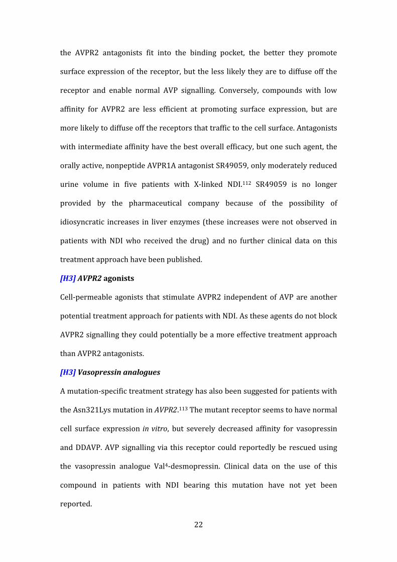
[H3] Prostaglandin receptor agonists Another novel treatment approach for patients with AVPR2 mutations focuses on
enhancing cAMP production in the principal cells independent of AVPR2, for
example via stimulation of prostaglandin E2 receptors coupled to adenylyl
cyclase (Figure 3). Such a strategy would not benefit patients with the autosomal
form of NDI, as AQP2 mutations result in a defect downstream of cAMP
production.
In a mouse model of X-linked diabetes insipidus with conditional deletion of the
Avpr2 gene, administration of a prostaglandin E2 receptor EP4 subtype (EP4
receptor) agonist resulted in an increase in urine concentration.114 The peak
increase in urine concentration (from 150 mOsm/kg to a maximum of
500 mOsm/kg) was short lived (3 h), possibly due to down regulation of EP4
receptors in response to the high osmolality, but with mini-pump constant
infusions a sustained increase in urine osmolality from 200 mOsmol/kg to
300 mOsmol/kg was observed.
Doubling of urine osmolality in patients with NDI would halve urine output (to
~6 l daily in adults) so would constitute successful treatment. In a rat model of
NDI secondary to the administration of the AVPR2 antagonist, butaprost, an EP2
receptor agonist approximately doubled urine osmolality.115 To date, no clinical
data on the use of prostaglandin receptor agonists in patients with NDI have
been reported. Moreover, given the proven efficacy of prostaglandin synthesis
inhibitors in NDI treatment, doubts remain as to whether this approach would be
a viable treatment option.116
24
[H3] Secretin receptor agonists
The secretin receptor is a G-protein coupled receptor expressed in principal
cells. Stimulation of this receptor can increase cAMP levels.117 Infusion of
secretin and fluvastatin resulted in doubling in urine osmolality in mice with
conditional deletion of the Avpr2 gene.118 If appropriate scientific and ethical
permission was obtained, short-term therapy with a combination of secretin and
fluvastatin could potentially be tested in humans with X-linked NDI. Long-term
secretin treatment might be limited by adverse effects of the drug.
[H3] cGMP phosphodiesterase inhibitors
Increased levels of cyclic guanosine monophosphate (cGMP) have been shown to
enhance trafficking of AQP2 to the luminal membrane and induce a short-lived
increase in urine osmolality in Brattleboro rats (a model of cerebral diabetes
insipidus with deficient vasopressin production by the hypothalamus).119 This
finding suggests a potential role of cGMP phosphodiesterase inhibitors, such as
sildenafil, in the treatment of patients with NDI resulting from AVPR2 mutations.
[H3] Gene therapy
Genome editing of somatic tissue or embryos is of considerable recent interest to
correct defective genes in humans but the ethics, especially of embryo editing is
controversial 120 121.. Currently, no such endeavours have reached the clinical
arena. We anticipate that the ethical governance, safety and long term effects of
such editing therapies will be determined possibly within the next two decades
so that patients with hereditary disorders, such as NDI will benefit from them.
[H1] Conclusions
Our understanding of the molecular physiology of urine concentration has
advanced tremendously in the past 25 years. The study of patients with inherited
25
NDI has identified AVPR2 and AQP2 as key proteins required for the regulation
of water permeability in the collecting duct. Other tubular disorders, such as
Bartter syndrome, can present with a secondary form of NDI, which might cause
diagnostic confusion. In adult nephrology practice, the majority of patients with
NDI have an acquired form of the disease owing to lithium treatment. Current
treatment of congenital NDI focuses on dietary modification, thiazides and
prostaglandin synthesis inhibitors. Novel therapies, such as mutation-specific
treatment using molecular chaperones, have been investigated in animal models,
but few data from clinical studies are currently available. The aim of these
treatment approaches is amelioration of the clinical phenotype of NDI. In the
future, gene therapy approaches involving kidney-specific delivery of wild-type
AVPR2 or AQP2 could potentially enable long-term cure of congenital NDI.
1. Stevens, L.A., Coresh, J., Greene, T. & Levey, A.S. Assessing kidney function--measured and estimated glomerular filtration rate. N Engl J Med 354, 2473-83 (2006).
2. Nielsen, S., Marples, D., Frokiaer, J., Knepper, M. & Agre, P. The aquaporin family of water channels in kidney: an update on physiology and pathophysiology of aquaporin-2. Kidney Int 49, 1718-23 (1996).
3. Nielsen, S. et al. Aquaporin-1 water channels in short and long loop descending thin limbs and in descending vasa recta in rat kidney. Am J Physiol 268, F1023-37 (1995).
4. Dantzler, W.H., Layton, A.T., Layton, H.E. & Pannabecker, T.L. Urine-concentrating mechanism in the inner medulla: function of the thin limbs of the loops of Henle. Clin J Am Soc Nephrol 9, 1781-9 (2014).
5. Halperin, M.L., Kamel, K.S. & Oh, M.S. Mechanisms to concentrate the urine: an opinion. Curr Opin Nephrol Hypertens 17, 416-22 (2008).
6. Pannabecker, T.L. Structure and function of the thin limbs of the loop of Henle. Compr Physiol 2, 2063-86 (2012).
7. Obermuller, N., Kunchaparty, S., Ellison, D.H. & Bachmann, S. Expression of the Na-K-2Cl cotransporter by macula densa and thick ascending limb cells of rat and rabbit nephron. J Clin Invest 98, 635-40 (1996).
8. Bichet, D.G., Oksche, A. & Rosenthal, W. Congenital nephrogenic diabetes insipidus. J Am Soc Nephrol 8, 1951-8 (1997).
9. Nielsen, S. et al. Aquaporins in the kidney: from molecules to medicine. Physiol Rev 82, 205-44 (2002).
10. Kortenoeven, M.L. & Fenton, R.A. Renal aquaporins and water balance disorders. Biochim Biophys Acta 1840, 1533-49 (2014).
26
11. Fushimi, K., Sasaki, S. & Marumo, F. Phosphorylation of serine 256 is required for cAMP-dependent regulatory exocytosis of the aquaporin-2 water channel. J Biol Chem 272, 14800-4 (1997).
12. Kamsteeg, E.J., Heijnen, I., van Os, C.H. & Deen, P.M. The subcellular localization of an aquaporin-2 tetramer depends on the stoichiometry of phosphorylated and nonphosphorylated monomers. J Cell Biol 151, 919-30 (2000).
13. Mulders, S.M. et al. An aquaporin-2 water channel mutant which causes autosomal dominant nephrogenic diabetes insipidus is retained in the Golgi complex. J Clin Invest 102, 57-66 (1998).
14. Savelkoul, P.J. et al. p.R254Q mutation in the aquaporin-2 water channel causing dominant nephrogenic diabetes insipidus is due to a lack of arginine vasopressin-induced phosphorylation. Hum Mutat 30, E891-903 (2009).
15. Hoffert, J.D. et al. Vasopressin-stimulated increase in phosphorylation at Ser269 potentiates plasma membrane retention of aquaporin-2. J Biol Chem 283, 24617-27 (2008).
16. Moeller, H.B., Praetorius, J., Rutzler, M.R. & Fenton, R.A. Phosphorylation of aquaporin-2 regulates its endocytosis and protein-protein interactions. Proc Natl Acad Sci U S A 107, 424-9 (2010).
17. Tamma, G., Robben, J.H., Trimpert, C., Boone, M. & Deen, P.M. Regulation of AQP2 localization by S256 and S261 phosphorylation and ubiquitination. Am J Physiol Cell Physiol 300, C636-46 (2011).
18. Kamsteeg, E.J. et al. Short-chain ubiquitination mediates the regulated endocytosis of the aquaporin-2 water channel. Proc Natl Acad Sci U S A 103, 18344-9 (2006).
19. Rehmann, H., Wittinghofer, A. & Bos, J.L. Capturing cyclic nucleotides in action: snapshots from crystallographic studies. Nat Rev Mol Cell Biol 8, 63-73 (2007).
20. Holz, G.G., Kang, G., Harbeck, M., Roe, M.W. & Chepurny, O.G. Cell physiology of cAMP sensor Epac. J Physiol 577, 5-15 (2006).
21. Kortenoeven, M.L. et al. In mpkCCD cells, long-term regulation of aquaporin-2 by vasopressin occurs independent of protein kinase A and CREB but may involve Epac. Am J Physiol Renal Physiol 302, F1395-401 (2012).
22. Hozawa, S., Holtzman, E.J. & Ausiello, D.A. cAMP motifs regulating transcription in the aquaporin 2 gene. Am J Physiol 270, C1695-702 (1996).
23. Matsumura, Y., Uchida, S., Rai, T., Sasaki, S. & Marumo, F. Transcriptional regulation of aquaporin-2 water channel gene by cAMP. J Am Soc Nephrol 8, 861-7 (1997).
24. Yasui, M., Zelenin, S.M., Celsi, G. & Aperia, A. Adenylate cyclase-coupled vasopressin receptor activates AQP2 promoter via a dual effect on CRE and AP1 elements. Am J Physiol 272, F443-50 (1997).
25. Hasler, U. et al. Long term regulation of aquaporin-2 expression in vasopressin-responsive renal collecting duct principal cells. J Biol Chem 277, 10379-86 (2002).
27
26. Terris, J., Ecelbarger, C.A., Nielsen, S. & Knepper, M.A. Long-term regulation of four renal aquaporins in rats. Am J Physiol 271, F414-22 (1996).
27. Lolait, S.J. et al. Cloning and characterization of a vasopressin V2 receptor and possible link to nephrogenic diabetes insipidus. Nature 357, 336-9 (1992).
28. Rosenthal, W. et al. Molecular identification of the gene responsible for congenital nephrogenic diabetes insipidus. Nature 359, 233-5 (1992).
29. van den Ouweland, A.M. et al. Mutations in the vasopressin type 2 receptor gene (AVPR2) associated with nephrogenic diabetes insipidus. Nat Genet 2, 99-102 (1992).
30. Pan, Y., Metzenberg, A., Das, S., Jing, B. & Gitschier, J. Mutations in the V2 vasopressin receptor gene are associated with X-linked nephrogenic diabetes insipidus. Nat Genet 2, 103-6 (1992).
31. Fushimi, K. et al. Cloning and expression of apical membrane water channel of rat kidney collecting tubule. Nature 361, 549-52 (1993).
32. Sasaki, S. et al. Cloning, characterization, and chromosomal mapping of human aquaporin of collecting duct. J Clin Invest 93, 1250-6 (1994).
33. Deen, P.M. et al. Requirement of human renal water channel aquaporin-2 for vasopressin-dependent concentration of urine. Science 264, 92-5 (1994).
34. Sasaki, S., Chiga, M., Kikuchi, E., Rai, T. & Uchida, S. Hereditary nephrogenic diabetes insipidus in Japanese patients: analysis of 78 families and report of 22 new mutations in AVPR2 and AQP2. Clin Exp Nephrol 17, 338-44 (2013).
35. Arthus, M.F. et al. Report of 33 novel AVPR2 mutations and analysis of 117 families with X-linked nephrogenic diabetes insipidus. J Am Soc Nephrol 11, 1044-54 (2000).
36. Sands, J.M. & Bichet, D.G. Nephrogenic diabetes insipidus. Ann Intern Med 144, 186-94 (2006).
37. Wesche, D., Deen, P.M. & Knoers, N.V. Congenital nephrogenic diabetes insipidus: the current state of affairs. Pediatr Nephrol 27, 2183-204 (2012).
38. Bichet, D.G. Nephrogenic diabetes insipidus. Adv Chronic Kidney Dis 13, 96-104 (2006).
39. van Lieburg, A.F., Knoers, N.V. & Monnens, L.A. Clinical presentation and follow-up of 30 patients with congenital nephrogenic diabetes insipidus. J Am Soc Nephrol 10, 1958-64 (1999).
40. Sato, K. et al. A novel mutation in the vasopressin V2 receptor gene in a woman with congenital nephrogenic diabetes insipidus. Intern Med 38, 808-12 (1999).
41. Kinoshita, K. et al. A novel deletion mutation in the arginine vasopressin receptor 2 gene and skewed X chromosome inactivation in a female patient with congenital nephrogenic diabetes insipidus. J Endocrinol Invest 27, 167-70 (2004).
42. Satoh, M., Ogikubo, S. & Yoshizawa-Ogasawara, A. Correlation between clinical phenotypes and X-inactivation patterns in six female carriers with heterozygote vasopressin type 2 receptor gene mutations. Endocr J 55, 277-84 (2008).
28
43. Bockenhauer, D. et al. Vasopressin type 2 receptor V88M mutation: molecular basis of partial and complete nephrogenic diabetes insipidus. Nephron Physiol 114, p1-10 (2010).
44. Bichet, D.G. et al. X-linked nephrogenic diabetes insipidus mutations in North America and the Hopewell hypothesis. J Clin Invest 92, 1262-8 (1993).
45. Fujiwara, T.M. & Bichet, D.G. Molecular biology of hereditary diabetes insipidus. J Am Soc Nephrol 16, 2836-46 (2005).
46. Spanakis, E., Milord, E. & Gragnoli, C. AVPR2 variants and mutations in nephrogenic diabetes insipidus: review and missense mutation significance. J Cell Physiol 217, 605-17 (2008).
47. Morello, J.P. et al. Pharmacological chaperones rescue cell-surface expression and function of misfolded V2 vasopressin receptor mutants. J Clin Invest 105, 887-95 (2000).
48. Duzenli, D. et al. Mutations in the AVPR2, AVP-NPII, and AQP2 genes in Turkish patients with diabetes insipidus. Endocrine 42, 664-9 (2012).
49. Marr, N. et al. Heteroligomerization of an Aquaporin-2 mutant with wild-type Aquaporin-2 and their misrouting to late endosomes/lysosomes explains dominant nephrogenic diabetes insipidus. Hum Mol Genet 11, 779-89 (2002).
50. Kuwahara, M. et al. Three families with autosomal dominant nephrogenic diabetes insipidus caused by aquaporin-2 mutations in the C-terminus. Am J Hum Genet 69, 738-48 (2001).
51. Kamsteeg, E.J. et al. Reversed polarized delivery of an aquaporin-2 mutant causes dominant nephrogenic diabetes insipidus. J Cell Biol 163, 1099-109 (2003).
52. Frick, A. et al. X-ray structure of human aquaporin 2 and its implications for nephrogenic diabetes insipidus and trafficking. Proc Natl Acad Sci U S A 111, 6305-10 (2014).
53. Bichet, D.G. et al. Aquaporin-2: new mutations responsible for autosomal-recessive nephrogenic diabetes insipidus—update and epidemiology. Clin Kidney J 5, 195-202 (2012).
54. Bichet, D.G. et al. Aquaporin-2: new mutations responsible for autosomal-recessive nephrogenic diabetes insipidus—update and epidemiology. Clinical Kidney Journal 5, 195-202 (2012).
55. Bockenhauer, D. et al. Secondary nephrogenic diabetes insipidus as a complication of inherited renal diseases. Nephron Physiol 116, p23-9 (2010).
56. Bockenhauer, D. & Bichet, D.G. Inherited secondary nephrogenic diabetes insipidus: concentrating on humans. Am J Physiol Renal Physiol 304, F1037-42 (2013).
57. Bettinelli, A. et al. Phenotypic variability in Bartter syndrome type I. Pediatr Nephrol 14, 940-5 (2000).
58. Hebert, S.C., Brown, E.M. & Harris, H.W. Role of the Ca(2+)-sensing receptor in divalent mineral ion homeostasis. J Exp Biol 200, 295-302 (1997).
59. Marples, D., Frokiaer, J., Dorup, J., Knepper, M.A. & Nielsen, S. Hypokalemia-induced downregulation of aquaporin-2 water channel
29
expression in rat kidney medulla and cortex. J Clin Invest 97, 1960-8 (1996).
60. Sands, J.M. et al. Apical extracellular calcium/polyvalent cation-sensing receptor regulates vasopressin-elicited water permeability in rat kidney inner medullary collecting duct. J Clin Invest 99, 1399-405 (1997).
61. Earm, J.H. et al. Decreased aquaporin-2 expression and apical plasma membrane delivery in kidney collecting ducts of polyuric hypercalcemic rats. J Am Soc Nephrol 9, 2181-93 (1998).
62. Trepiccione, F. & Christensen, B.M. Lithium-induced nephrogenic diabetes insipidus: new clinical and experimental findings. J Nephrol 23 Suppl 16, S43-8 (2010).
63. Rej, S., Herrmann, N. & Shulman, K. The effects of lithium on renal function in older adults--a systematic review. Journal of geriatric psychiatry and neurology 25, 51-61 (2012).
64. Timmer, R.T. & Sands, J.M. Lithium intoxication. J Am Soc Nephrol 10, 666-74 (1999).
65. Hetmar, O. et al. Lithium: long-term effects on the kidney. I. Renal function in retrospect. Acta Psychiatr Scand 73, 574-81 (1986).
66. Juurlink, D.N. et al. Drug-induced lithium toxicity in the elderly: a population-based study. J Am Geriatr Soc 52, 794-8 (2004).
67. Head, L. & Dening, T. Lithium in the over-65s: who is taking it and who is monitoring it? A survey of older adults on lithium in the Cambridge Mental Health Services catchment area. Int J Geriatr Psychiatry 13, 164-71 (1998).
68. Christensen, B.M. et al. Changes in cellular composition of kidney collecting duct cells in rats with lithium-induced NDI. Am J Physiol Cell Physiol 286, C952-64 (2004).
69. Li, Y., Shaw, S., Kamsteeg, E.J., Vandewalle, A. & Deen, P.M. Development of lithium-induced nephrogenic diabetes insipidus is dissociated from adenylyl cyclase activity. J Am Soc Nephrol 17, 1063-72 (2006).
70. Walker, R.J. et al. Lithium-induced reduction in urinary concentrating ability and urinary aquaporin 2 (AQP2) excretion in healthy volunteers. Kidney Int 67, 291-4 (2005).
71. Kortenoeven, M.L. et al. Amiloride blocks lithium entry through the sodium channel thereby attenuating the resultant nephrogenic diabetes insipidus. Kidney Int 76, 44-53 (2009).
72. Bedford, J.J. et al. Lithium-induced nephrogenic diabetes insipidus: renal effects of amiloride. Clin J Am Soc Nephrol 3, 1324-31 (2008).
73. Batlle, D.C., von Riotte, A.B., Gaviria, M. & Grupp, M. Amelioration of polyuria by amiloride in patients receiving long-term lithium therapy. N Engl J Med 312, 408-14 (1985).
74. Frokiaer, J., Marples, D., Knepper, M.A. & Nielsen, S. Bilateral ureteral obstruction downregulates expression of vasopressin-sensitive AQP-2 water channel in rat kidney. Am J Physiol 270, F657-68 (1996).
75. Frokiaer, J. et al. Downregulation of aquaporin-2 parallels changes in renal water excretion in unilateral ureteral obstruction. Am J Physiol 273, F213-23 (1997).
76. Winberg, J. Determination of Renal Concentration Capacity in Infants and Children without Renal Disease. Acta Paediatrica 48, 318-328 (1958).
30
77. Bichet, D.G. et al. Hemodynamic and coagulation responses to 1-desamino[8-D-arginine] vasopressin in patients with congenital nephrogenic diabetes insipidus. N Engl J Med 318, 881-7 (1988).
78. Mannucci, P.M. Treatment of von Willebrand's Disease. N Engl J Med 351, 683-94 (2004).
79. Kaufmann, J.E. & Vischer, U.M. Cellular mechanisms of the hemostatic effects of desmopressin (DDAVP). J Thromb Haemost 1, 682-9 (2003).
80. Hillman, D.A., Neyzi, O., Porter, P., Cushman, A. & Talbot, N.B. Renal (vasopressin-resistant) diabetes insipidus; definition of the effects of a homeostatic limitation in capacity to conserve water on the physical, intellectual and emotional development of a child. Pediatrics 21, 430-5 (1958).
81. Vest, M., Talbotnb & Crawford, J.D. Hypocaloric dwarfism and hydronephrosis in diabetes insipidus. Am J Dis Child 105, 175-81 (1963).
82. Schofer, O. et al. Nephrogenic diabetes insipidus and intracerebral calcification. Arch Dis Child 65, 885-7 (1990).
83. Hoekstra, J.A., van Lieburg, A.F., Monnens, L.A., Hulstijn-Dirkmaat, G.M. & Knoers, V.V. Cognitive and psychosocial functioning of patients with congenital nephrogenic diabetes insipidus. Am J Med Genet 61, 81-8 (1996).
84. Huber, D., Veinante, P. & Stoop, R. Vasopressin and oxytocin excite distinct neuronal populations in the central amygdala. Science 308, 245-8 (2005).
85. Griebel, G. et al. Anxiolytic- and antidepressant-like effects of the non-peptide vasopressin V1b receptor antagonist, SSR149415, suggest an innovative approach for the treatment of stress-related disorders. Proc Natl Acad Sci U S A 99, 6370-5 (2002).
86. Yoo, T.H. et al. Congenital Nephrogenic Diabetes Insipidus Presented with Bilateral Hydronephrosis: Genetic Analysis of V2R Gene Mutations. Yonsei Med J 47, 126-30 (2006).
87. Stevens, S., Brown, B.D. & McGahan, J.P. Nephrogenic diabetes insipidus: a cause of severe nonobstructive urinary tract dilatation. J Ultrasound Med 14, 543-5 (1995).
88. Jaureguiberry, G. et al. A patient with polyuria and hydronephrosis: question. Pediatric Nephrology 26, 1977-8, 1979-80 (2011).
89. Sadeghi, H., Robertson, G.L., Bichet, D.G., Innamorati, G. & Birnbaumer, M. Biochemical basis of partial nephrogenic diabetes insipidus phenotypes. Mol Endocrinol 11, 1806-13 (1997).
90. Canfield, M.C., Tamarappoo, B.K., Moses, A.M., Verkman, A.S. & Holtzman, E.J. Identification and characterization of aquaporin-2 water channel mutations causing nephrogenic diabetes insipidus with partial vasopressin response. Hum Mol Genet 6, 1865-71 (1997).
91. Guyon, C. et al. Characterization of D150E and G196D aquaporin-2 mutations responsible for nephrogenic diabetes insipidus: importance of a mild phenotype. Am J Physiol Renal Physiol 297, F489-98 (2009).
92. Bichet, D.G. et al. Nature and recurrence of AVPR2 mutations in X-linked nephrogenic diabetes insipidus. Am J Hum Genet 55, 278-86 (1994).
93. Ala, Y. et al. Functional studies of twelve mutant V2 vasopressin receptors related to nephrogenic diabetes insipidus: molecular basis of a mild clinical phenotype. J Am Soc Nephrol 9, 1861-72 (1998).
31
94. Kahn, A., Brachet, E. & Blum, D. Controlled fall in natremia and risk of seizures in hypertonic dehydration. Intensive Care Med 5, 27-31 (1979).
95. Cansick, J., Rees, L., Koffman, G., Van't Hoff, W. & Bockenhauer, D. A fatal case of cerebral oedema with hyponatraemia and massive polyuria after renal transplantation. Pediatric Nephrology 24, 1231-4 (2009).
96. Sterns, R.H. Disorders of plasma sodium - causes, consequences, and correction. N Engl J Med 372, 55-65 (2015).
97. Fang, C. et al. Fluid management of hypernatraemic dehydration to prevent cerebral oedema: a retrospective case control study of 97 children in China. J Paediatr Child Health 46, 301-3 (2010).
98. Meyer, E. Über Diabetes insipidus und andere Polyurien. Deutsch. Arch. klin. Med. 83, 1 (1905).
99. Crawford, J.D. & Kennedy, G.C. Chlorothiazid in diabetes insipidus. Nature 183, 891-2 (1959).
100. Havard, C.W. Thiazide-induced antidiuresis in diabetes insipidus. Proc R Soc Med 58, 1005-7 (1965).
101. Sinke, A.P. et al. Hydrochlorothiazide attenuates lithium-induced nephrogenic diabetes insipidus independently of the sodium-chloride cotransporter. Am J Physiol Renal Physiol 306, F525-33 (2014).
102. Orloff, J., Handler, J.S. & Bergstrom, S. Effect of Prostaglandin (Pge-1) on the Permeability Response of Toad Bladder to Vasopressin, Theophylline and Adenosine 3',5'-Monophosphate. Nature 205, 397-8 (1965).
103. Anderson, R.J., Berl, T., McDonald, K.D. & Schrier, R.W. Evidence for an in vivo antagonism between vasopressin and prostaglandin in the mammalian kidney. J Clin Invest 56, 420-6 (1975).
104. Stoff, J.S., Rosa, R.M., Silva, P. & Epstein, F.H. Indomethacin impairs water diuresis in the DI rat: role of prostaglandins independent of ADH. Am J Physiol 241, F231-7 (1981).
105. Libber, S., Harrison, H. & Spector, D. Treatment of nephrogenic diabetes insipidus with prostaglandin synthesis inhibitors. J Pediatr 108, 305-11 (1986).
106. Monnens, L., Jonkman, A. & Thomas, C. Response to indomethacin and hydrochlorothiazide in nephrogenic diabetes insipidus. Clin Sci (Lond) 66, 709-15 (1984).
107. Monn, E. Prostaglandin synthetase inhibitors in the treatment of nephrogenic diabetes insipidus. Acta Paediatr Scand 70, 39-42 (1981).
108. Usberti, M. et al. Renal prostaglandin E2 in nephrogenic diabetes insipidus: effects of inhibition of prostaglandin synthesis by indomethacin. J Pediatr 97, 476-8 (1980).
109. Boussemart, T., Nsota, J., Martin-Coignard, D. & Champion, G. Nephrogenic diabetes insipidus: treat with caution. Pediatr Nephrol 24, 1761-3 (2009).
110. Bockenhauer, D. et al. Antenatal Bartter's syndrome: why is this not a lethal condition? QJM : monthly journal of the Association of Physicians 101, 927-42 (2008).
111. Bockenhauer, D., Medlar, A.J., Ashton, E., Kleta, R. & Lench, N. Genetic testing in renal disease. Pediatric Nephrology 27, 873-83 (2012).
112. Bernier, V. et al. Pharmacologic chaperones as a potential treatment for X-linked nephrogenic diabetes insipidus. J Am Soc Nephrol 17, 232-43 (2006).
32
113. Erdelyi, L.S. et al. Altered agonist sensitivity of a mutant v2 receptor suggests a novel therapeutic strategy for nephrogenic diabetes insipidus. Mol Endocrinol 28, 634-43 (2014).
114. Li, J.H. et al. A selective EP4 PGE2 receptor agonist alleviates disease in a new mouse model of X-linked nephrogenic diabetes insipidus. J Clin Invest 119, 3115-26 (2009).
115. Olesen, E.T., Rutzler, M.R., Moeller, H.B., Praetorius, H.A. & Fenton, R.A. Vasopressin-independent targeting of aquaporin-2 by selective E-prostanoid receptor agonists alleviates nephrogenic diabetes insipidus. Proc Natl Acad Sci U S A 108, 12949-54 (2011).
116. Bockenhauer, D. & Bichet, D.G. Urinary concentration: different ways to open and close the tap. Pediatr Nephrol 29, 1297-303 (2014).
117. Chu, J.Y. et al. Phenotypes developed in secretin receptor-null mice indicated a role for secretin in regulating renal water reabsorption. Mol Cell Biol 27, 2499-511 (2007).
118. Procino, G. et al. Combination of secretin and fluvastatin ameliorates the polyuria associated with X-linked nephrogenic diabetes insipidus in mice. Kidney Int 86, 127-38 (2014).
119. Bouley, R. et al. Stimulation of AQP2 membrane insertion in renal epithelial cells in vitro and in vivo by the cGMP phosphodiesterase inhibitor sildenafil citrate (Viagra). Am J Physiol Renal Physiol 288, F1103-12 (2005).
120. Ran, F.A. et al. In vivo genome editing using Staphylococcus aureus Cas9. Nature 520, 186-91 (2015).
121. Cyranoski, D. Ethics of embryo editing divides scientists. Nature 519, 272 (2015).
122. Coleman, J. in Clinical Paediatric Dietetics (eds. Shaw, V. & Lawson, M.) (Blackwell Science Ltd, Oxford, 2001).
33
Box 1: Osmotic load
The osmotic load consists of osmotically active substances that need to be
excreted in the urine. The key contributors to the osmotic load are proteins,
which are metabolized to urea (approximately 4 mmol per g of protein) and
salts. A typical western diet contains about 800 m0sm per day. An individual
with a urine osmolality of 800 m0sm/kg only needs 1 l of water to excrete this
osmotic load, whereas a patient with maximal urine osmolality of 100 m0sm/kg
will need at least 8 l of water for excretion. 1 g of table salt providing an osmolar
load of ~36 mOsm (18 mOsm Na and 18 mOsm Cl). Consequently, in a patient
with a urine osmolality of 100 mOsm/kg, each gram of salt ingested will increase
the obligatory urine output by 360 ml. The osmolar load of a diet can be roughly
estimated by multiplying the millimolar amounts of sodium and potassium by
two (to account for the accompanying anions) and adding the amount of protein
in g multiplied by four.122 As metabolism of lipids and sugars does not produce
byproducts that require renal excretion, they do not contribute to the osmotic
load. Diet is the key contributor to the osmotic load, but metabolism of own
proteins during catabolic states also generate urea.
Figure 1: The renal concentration and dilution mechanism. The loop of
Henle forms a counter-current multiplier system that concentrates the urine.
Urine is isotonic when it enters the loop of Henle and hypotonic when it exits
into the collecting duct. The concentration gradient generated in the loop of
Henle is driven by the active reabsorption of NaCl in the thick ascending limb by
the transporter solute carrier family 12 member 1. The mechanism of
concentration in the thin descending limb is not completely resolved, but likely
34
involves passive water efflux and/or NaCl influx. Final concentration of urine
occurs in the collecting duct and depends on the availability of aquaporin 2
water channels. The approximate osmolalities of the tubular fluid (pink boxes)
and interstitial fluid (green boxes) are indicated.
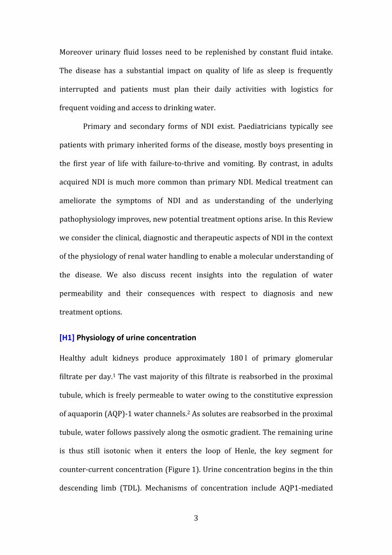
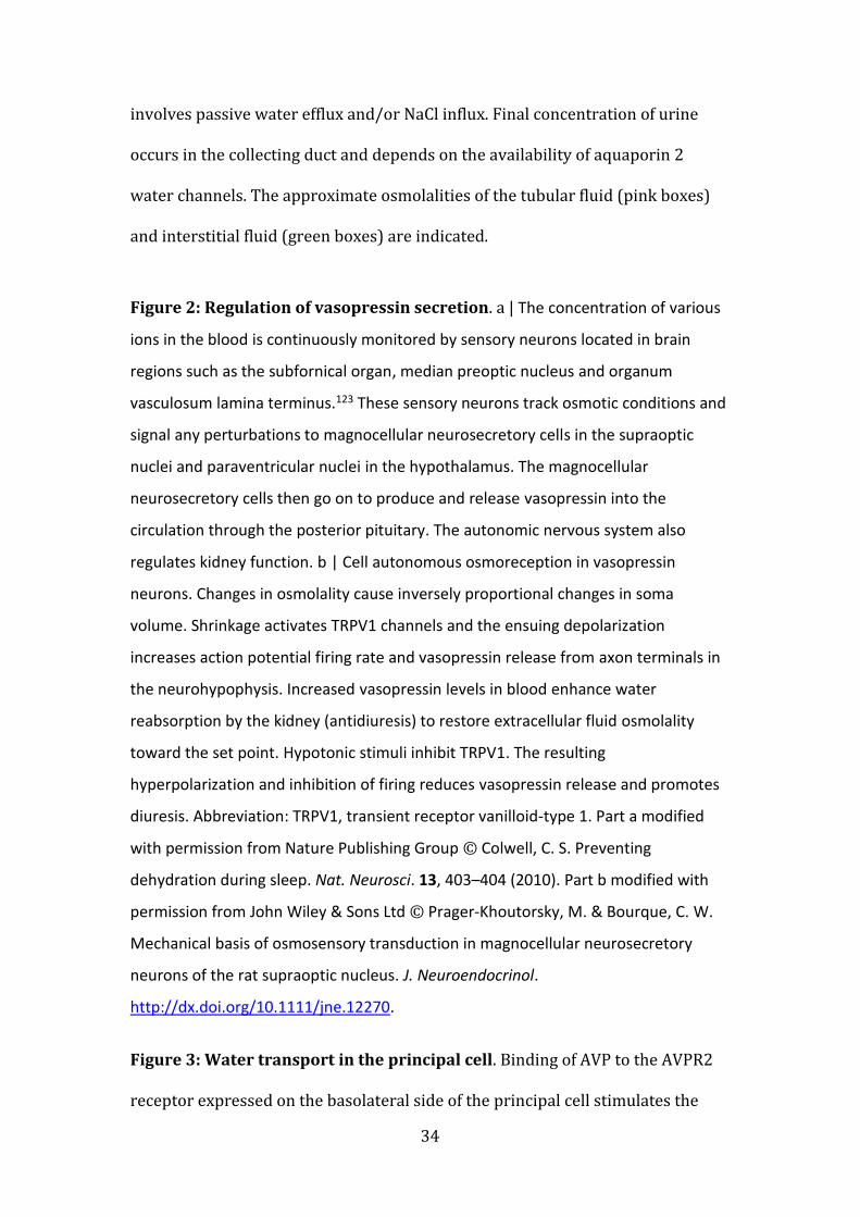
Figure 2: Regulation of vasopressin secretion. a | The concentration of various
ions in the blood is continuously monitored by sensory neurons located in brain
regions such as the subfornical organ, median preoptic nucleus and organum
vasculosum lamina terminus.123 These sensory neurons track osmotic conditions and
signal any perturbations to magnocellular neurosecretory cells in the supraoptic
nuclei and paraventricular nuclei in the hypothalamus. The magnocellular
neurosecretory cells then go on to produce and release vasopressin into the
circulation through the posterior pituitary. The autonomic nervous system also
regulates kidney function. b | Cell autonomous osmoreception in vasopressin
neurons. Changes in osmolality cause inversely proportional changes in soma
volume. Shrinkage activates TRPV1 channels and the ensuing depolarization
increases action potential firing rate and vasopressin release from axon terminals in
the neurohypophysis. Increased vasopressin levels in blood enhance water
reabsorption by the kidney (antidiuresis) to restore extracellular fluid osmolality
toward the set point. Hypotonic stimuli inhibit TRPV1. The resulting
hyperpolarization and inhibition of firing reduces vasopressin release and promotes
diuresis. Abbreviation: TRPV1, transient receptor vanilloid-type 1. Part a modified
with permission from Nature Publishing Group © Colwell, C. S. Preventing
dehydration during sleep. Nat. Neurosci. 13, 403–404 (2010). Part b modified with
permission from John Wiley & Sons Ltd © Prager-Khoutorsky, M. & Bourque, C. W.
Mechanical basis of osmosensory transduction in magnocellular neurosecretory
neurons of the rat supraoptic nucleus. J. Neuroendocrinol.
http://dx.doi.org/10.1111/jne.12270.
Figure 3: Water transport in the principal cell. Binding of AVP to the AVPR2
receptor expressed on the basolateral side of the principal cell stimulates the
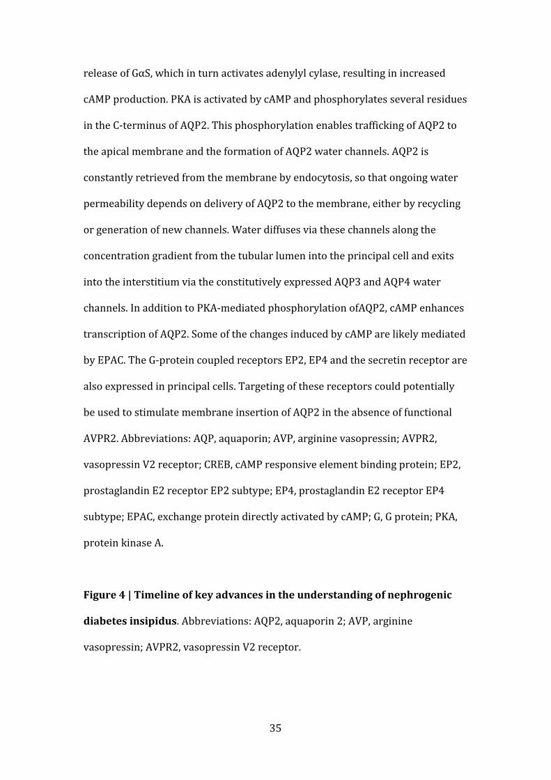
35
release of GαS, which in turn activates adenylyl cylase, resulting in increased
cAMP production. PKA is activated by cAMP and phosphorylates several residues
in the C-terminus of AQP2. This phosphorylation enables trafficking of AQP2 to
the apical membrane and the formation of AQP2 water channels. AQP2 is
constantly retrieved from the membrane by endocytosis, so that ongoing water
permeability depends on delivery of AQP2 to the membrane, either by recycling
or generation of new channels. Water diffuses via these channels along the
concentration gradient from the tubular lumen into the principal cell and exits
into the interstitium via the constitutively expressed AQP3 and AQP4 water
channels. In addition to PKA-mediated phosphorylation ofAQP2, cAMP enhances
transcription of AQP2. Some of the changes induced by cAMP are likely mediated
by EPAC. The G-protein coupled receptors EP2, EP4 and the secretin receptor are
also expressed in principal cells. Targeting of these receptors could potentially
be used to stimulate membrane insertion of AQP2 in the absence of functional
AVPR2. Abbreviations: AQP, aquaporin; AVP, arginine vasopressin; AVPR2,
vasopressin V2 receptor; CREB, cAMP responsive element binding protein; EP2,
prostaglandin E2 receptor EP2 subtype; EP4, prostaglandin E2 receptor EP4
subtype; EPAC, exchange protein directly activated by cAMP; G, G protein; PKA,
protein kinase A.
Figure 4 | Timeline of key advances in the understanding of nephrogenic
diabetes insipidus. Abbreviations: AQP2, aquaporin 2; AVP, arginine
vasopressin; AVPR2, vasopressin V2 receptor.