Embed Size (px)
Citation preview

Pathophysiology & Complications Of Burn
DEFINITION
Burns are wounds produced by various
kinds of agents that cause cutaneous injury and
destruction of underlying tissue.
TYPES OF BURNS
• Thermal injury
– Scald—spillage of hot liquids
– Flame burns
– Flash burns due to exposure of natural gas, alcohol, combustible
liquids
– Contact burns—contact with hot metals/objects/materials
• Electrical injury
• Chemical burns—acid/alkali
• Cold injury—frost bite
• Ionizing radiation
• Sun burns

Classification of Burns
1 Degree 4 (first,second,third,fourth)
2 Thickness 2 ( partial,full thickness)
3 Percentage 3 (mild,moderate severe)

I. DEPENDING ON DEGREE
a. First degree:
Epidermis is red and painful, No
blisters
Heals rapidly in 5-7 days
By epithelialization without
scarring.

b. Second degree:
• Red, painful,with blisters,
• Heals in 14-21 days.
• Superficial burn heals,causing
pigmentation.
• Deep burn heals, causing scarring,
and pigmentation.

• Third degree:
• Charred, painless and insensitive,
• Thrombosis of superficial vessels.
• It requires grafting.
-Eschar
Charred, denatured, insensitive,contracted full
thickness burn.
• These wound must heal by reepithelialisation
from wound edge.

• Fourth degree:
Involves the underlying tissues—muscles, bones.

II. DEPENDING ON THICKNESS OF SKIN INVOLVED
• a. Partial thickness burns:
It is either first or second degree burn which is red
and painful, often with blisters.
• b. Full thickness burns:
It is third degree burns which is charred,
insensitive, deep involving all layers of the
skin.

DEPENDING ON THEPERCENTAGE OF BURNS
Mild:
• Partial thickness burns < 15% in adult or <10% in children.
• Full thickness burns less than 2%.
• Can be treated on outpatient basis.
Moderate:
• Second degree of 15-25% burns (10-20% in children).
• Third degree between 2-10% burns.
• Burns which are not involving eyes, ears, face, hand, feet,perineum.

Major (severe):
• Second degree burns more than 25% in adults, in children more
than 20%.
• All third degree burns of 10% or more.
• Burns involving eyes, ears, feet, hands, perineum.
• All inhalation and electrical burns.
• Burns with fractures or major mechanical trauma.

JACKSON`S THERMAL WOUND THEORY
-Zone of coagulation
Centre area of wound ,where all tissuses are damaged
-Zone of stasis
Surrounds the coagulation area ,some tissues are damaged
-Zone of hyperaemia
Unburned area surrounds the stasis but it is red due to inflammation

ZONE OF COAGULATION(BURNED ZONE)
• The necrotic area of burn where cells have been disrupted
• This tissue is irreversibly damaged at the time of injury

ZONE OF STASIS
• The area immediately surrounding the necrotic zone.
• decreased tissue perfusion.
• can either survive(with good resuscitation) or go on to coagulative
necrosis.
• Associated with vascular damage and vessel leakage
• Thromboxane A2, a potent vasoconstrictor is the main mediator
• Treatment aims to spare this zone to
Prevent it’s turnover to coagulative
necrosis zone

ZONE OF HYPERMEIA
-is characterized by vasodilation from inflammation
surrounding the burn wound
-contains the clearly viable tissue from which the
healing process begins
-not at risk for further necrosis.

PATHOPHYSIOLOGY OF BURNS

EBB PHASE (1ST 24 HOURS AFTER INURY)
• Occurs usually in the first 24 hours
• It’s initial period of hypofunction manifests as:
(a)Hypotension
(b) Low cardiac output
(c)Metabolic acidosis
(d) Hypoventilation
(e) Hyperglycemia
(f) Low oxygen consumption
(g)Inability to thermoregulate
Responds to fluid resuscitation

The flow phase, Resuscitation
• Follows ebb phase and is characterized by gradual increases
in:
(a) Cardiac output
(b) Heart rate
(c) Oxygen consumption
(d) Supranormal increases of temperature

HYPERMETABOLIC STATE
• Hypermetabolic hyperdynamic response peaks in 10-14
days after the injury after which condition slowly recedes
to normal as the burn wounds heal naturally or surgically
closed by applying skin grafting

SYSTEMIC RESPONSE TO BURN
• Metabolic
• Cardiac
• Renal
• Blood
• Immunologic
• Lungs
• GIT
• Edema
• Infections

METABOLIC RESPONSE
They are:
1-Histamine
2-PGS
3-Thromboxane
4-Kinins
5-Serotonin
6Catecholamines
7Oxygen free radicals
8CRF (corticotropin releasing factor)
9-Platelet aggregation factor
10-Angiotensin 2 ,Vasopressin
Inflammatory mediators has systemic response in > 30 % TBSA Burn

HISTAMINE
• Responsible for increased microvascular permeability
seen immediately after burn.
• Released from mast cells in thermal-injured skin
• Its actions are only transient

PROSTAGLANDINS (PGS)
• Potent vasoactive
• Released from burned tissue and inflammatory cells
• Prostaglandin E2(PGE2) and leukotrienes LB4 and LD4
directly and indirectly increase microvascular permeability
• PGE2 is a potent vasodilator, which, when coupled with
the increased microvascular permeability amplifies edema
formation
THROMBOXANE
• Thromboxane A2 (TXA2),thromboxane B2 (TXB2) produced
locally in the burn wound by platelets
• TXA2 is a potent vasoconstrictor
• Decrease blood flow (ischemia) under the burn
• Cause the conversion of a partial-thickness wound to a deeper
full-thickness wound
• Topically applied ibuprofen ( synthesis of prostaglandins and
thromboxanes) decreases both local edema without altering
systemic production

KININS
• Bradykinin is a local mediator of inflammation that
increases venular permeability

SEROTONIN
• Smooth-muscle constrictor of large blood vessels
• Antiserotonin agents such as ketanserin have been
found to decrease peripheral vascular resistance
after burn injury

CATECHOLAMINES
• Cause vasoconstriction
• Reduced capillary pressure may limit edema and induce
interstitial fluid to reabsorb from nonburned skin, skeletal
muscle, and visceral organs in nonresuscitated burn
shock
• Via B-agonist activity, may also partially inhibit increased
capillary permeability induced by histamine and
bradykinin
• It has beneficial effect to reduce edema

OXYGEN RADICALS
• Superoxide anion (O2-), hydrogen peroxide (H2O2), and hydroxyl
ion (OH-) from activated neutrophils
• The hydroxyl ion (OH-) is the most potent and damaging of the
three
• Play an important inflammatory role in all types of shock
• High doses of antioxidant ascorbic acid (vitamin-C) have been found
to be efficacious in reducing fluid needs in burn
• (10 – 20 g per day) of vitc

PLATELET AGGREGATION FACTOR
• increase capillary permeability

ANGIOTENSIN II AND VASOPRESSIN
• Participate in the normal regulation of extracellular fluid volume by
controlling sodium balance and osmolality through renal function
and thirst
• Both are potent vasoconstrictors of terminal arterioles
• Angiotensin II responsible for the selective gut and mucosal
ischemia, which cause translocation of endotoxins and bacteria
and the development of sepsis and even multi-organ failure
• Vasopressin, along with catecholamines responsible for increased
system vascular resistance and left heart afterload, which can occur
in resuscitated burn shock

CORTICOTROPHIN- RELEASING FACTOR (CRF)
• Reduce protein extravasation and edema in burn
• CRF may be is a powerful natural inhibitory mediator of
the acute inflammatory response of the skin to thermal
injury

2- CARDIAC
• Cardiac output decreases due to:
1)Decreased preload induced by fluid shifts
2)Increased systemic vascular resistance caused by
both hypovolemia and systemic catecholamine
release
• Cardiac output normal within 12-18 hours, with
successful resuscitation
• After 24 hours, it may increase up to 2 ½ times the
normal and remain elevated until several months after
the burn is closed

3-RENAL
• Renal blood flow and GFR decrease soon after injury
due to hypovolemia, decreased cardiac output, and
elevated systemic vascular
• Oliguria and antidiuresis develops during 1st 12-24
hours
• Followed by a usually modest diuresis as the capillary
leaks seal, plasma volume normalizes, and cardiac
output increases after successful resuscitation and
coinciding with onset of the postburn hypermetabolic
state, and hyperdynamic circulation

BLOOD
• The red-cell mass decreases due to direct losses
• Immediate, 1-2 hours after, and delayed, 2-7 days postburn, hemolysis
occurs due to damaged cells and increased fragility
• Anemia within 4-7 days is common
• Anemia persists until wound healing occur
• Early mild thrombocytopenia followed by thrombocytosis (2-4x normal)
and elevated fibrinogen, factor V and factor VIII levels commonly by end
of the 1st week
• Persistent thrombocytopenia is associated with poor prognosis --
suspect sepsis

IMMUNOLOGIC
• Mechanical barrier to infection is impaired because of skin
destruction
• Immunoglobulin levels decreased as part of general leak and
leukocyte chemotaxis, phagocytosis, and cytotoxic activity
impaired
• The reticuloendothelial system's depressed bacterial clearance is
due to decreases in opsonic (phagocyte) function
• These changes, together with a non-perfused, bacterially-colonized
eschar overlying a wound full of proteinaceous fluid, put the patient
in a significant risk for infection

EDEMA
• Injured tissue Increases permeability of entire vascular tree with
loss of water, electrolytes and proteins from the vascular
compartment , severe hemoconcentration occurs
• Protein leakage causing hypoproteinemia, increase osmotic
pressure in the interstitial space
• Decreased cell membrane potential cause inward shift of Na+ and
H2O cellular swelling
• In the injured skin, effect maximal 30 min after the burn but
capillary integrity not restored until 8-12 hours after, usually
resolved by 3-5 days
• In non-injured tissues, only mild and transient leaks even for burns
>40% BSA

GIT
• Mucosal atrophy decreased absorption & increased intestinal permeability duto
changes in gut blood flow
increased bacterial translocation and Septicemia
• Occurs within 12 hours of injury
Acute gastric dilatation which occurs in 2-4 days.
Paralytic ileus.
Curling’s ulcer (stress ulcer).
Acute a calculous cholecystitis, acute pancreatitis
Abdominal Compartment syndrome

INFECTIONS
• Streptococci (Beta haemolytic—most common)
• Pseudomonas
• Staphylococci
• Other gram-negative organisms
• Candida albicans

• Burn size greater than 40 % TBSA, 75 % of all deaths are
due to infection
• Causes:
• Burn wound represents a susceptible site for
opportunistic colonization by organisms
• Age, immunosuppressed status, extent of injury, and
depth of burn in combination with microbial factors such
as type and number of organisms, enzyme and toxin
production and motility

• Aggressive early debridement of devitalized and infected tissue plus
catheter related infections are the cornerstone of management of
infections.
• Once an infection is disseminated hematogenously and becomes
established in a burn patient, it is very difficult to eradicate, even
with large does of broad-spectrum antimicrobial therapy
• Time-related changes in the predominant flora of the burn wound
converts bacterial growth from gram-positive to gram-negative
• Treatment with two or more agents is becoming necessary in the
management of these gram-negative invasive infections

SUMMARY OF PATHOPHYSIOLOGY

Complications of Burns• Burn Shock
• Pulmonary complications due to inhalation injury
• Acute Renal Failure
• Infections and Sepsis
• Curling’s ulcer in large burns over 30% usually after 9th day
• Extensive and disabling scarring
• Psychological trauma
• Cancer called Marjolin’s ulcer, may take 21 years to develop

SEPSIS
• Bacteremia:presence of bacteria in the
bloodstream without clinical manifestations
• Sepsis:bacteremia +clinical manifestations
(fever..tachycardia..tachypnea)
• Septic shock: sepsis+ refractory hypotension
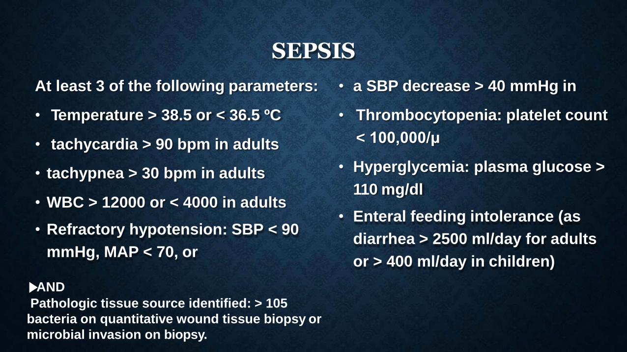
SEPSIS
• tachypnea > 30 bpm in adults
• WBC > 12000 or < 4000 in adults
• Refractory hypotension: SBP < 90
mmHg, MAP < 70, or
At least 3 of the following parameters: • a SBP decrease > 40 mmHg in
• Temperature > 38.5 or < 36.5 ºC •
• tachycardia > 90 bpm in adults
Thrombocytopenia: platelet count
< 100,000/μ
• Hyperglycemia: plasma glucose >
110 mg/dl
• Enteral feeding intolerance (as
diarrhea > 2500 ml/day for adults
or > 400 ml/day in children)
▶AND
Pathologic tissue source identified: > 105
bacteria on quantitative wound tissue biopsy or
microbial invasion on biopsy.

CAUSES OF DEATH
Hypovolaemia (refractory and uncontrolled) and shock
Renal failure
Pulmonary oedema and ARDS
Septicaemia
Multiorgan failure
Acute airway block in head and neck burns