Embed Size (px)
Citation preview
REVIEW Open Access
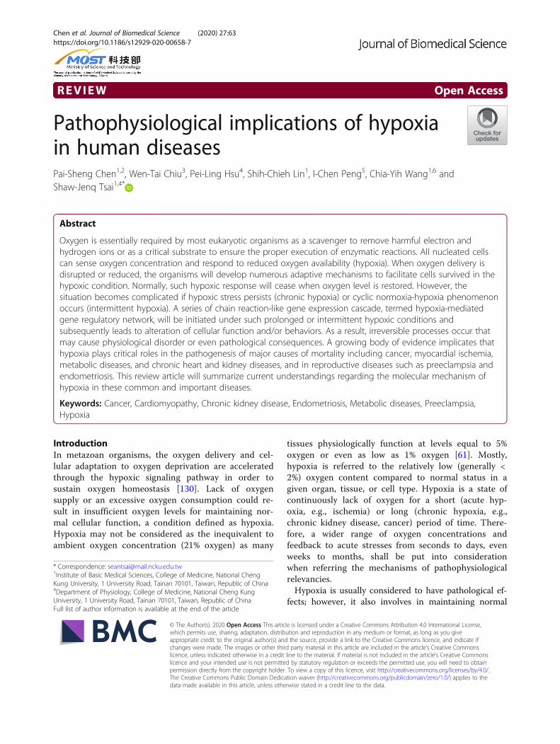
Pathophysiological implications of hypoxiain human diseasesPai-Sheng Chen1,2, Wen-Tai Chiu3, Pei-Ling Hsu4, Shih-Chieh Lin1, I-Chen Peng5, Chia-Yih Wang1,6 andShaw-Jenq Tsai1,4*
Abstract
Oxygen is essentially required by most eukaryotic organisms as a scavenger to remove harmful electron andhydrogen ions or as a critical substrate to ensure the proper execution of enzymatic reactions. All nucleated cellscan sense oxygen concentration and respond to reduced oxygen availability (hypoxia). When oxygen delivery isdisrupted or reduced, the organisms will develop numerous adaptive mechanisms to facilitate cells survived in thehypoxic condition. Normally, such hypoxic response will cease when oxygen level is restored. However, thesituation becomes complicated if hypoxic stress persists (chronic hypoxia) or cyclic normoxia-hypoxia phenomenonoccurs (intermittent hypoxia). A series of chain reaction-like gene expression cascade, termed hypoxia-mediatedgene regulatory network, will be initiated under such prolonged or intermittent hypoxic conditions andsubsequently leads to alteration of cellular function and/or behaviors. As a result, irreversible processes occur thatmay cause physiological disorder or even pathological consequences. A growing body of evidence implicates thathypoxia plays critical roles in the pathogenesis of major causes of mortality including cancer, myocardial ischemia,metabolic diseases, and chronic heart and kidney diseases, and in reproductive diseases such as preeclampsia andendometriosis. This review article will summarize current understandings regarding the molecular mechanism ofhypoxia in these common and important diseases.
Keywords: Cancer, Cardiomyopathy, Chronic kidney disease, Endometriosis, Metabolic diseases, Preeclampsia,Hypoxia
IntroductionIn metazoan organisms, the oxygen delivery and cel-lular adaptation to oxygen deprivation are acceleratedthrough the hypoxic signaling pathway in order tosustain oxygen homeostasis [130]. Lack of oxygensupply or an excessive oxygen consumption could re-sult in insufficient oxygen levels for maintaining nor-mal cellular function, a condition defined as hypoxia.Hypoxia may not be considered as the inequivalent toambient oxygen concentration (21% oxygen) as many
tissues physiologically function at levels equal to 5%oxygen or even as low as 1% oxygen [61]. Mostly,hypoxia is referred to the relatively low (generally <2%) oxygen content compared to normal status in agiven organ, tissue, or cell type. Hypoxia is a state ofcontinuously lack of oxygen for a short (acute hyp-oxia, e.g., ischemia) or long (chronic hypoxia, e.g.,chronic kidney disease, cancer) period of time. There-fore, a wider range of oxygen concentrations andfeedback to acute stresses from seconds to days, evenweeks to months, shall be put into considerationwhen referring the mechanisms of pathophysiologicalrelevancies.Hypoxia is usually considered to have pathological ef-
fects; however, it also involves in maintaining normal
© The Author(s). 2020 Open Access This article is licensed under a Creative Commons Attribution 4.0 International License,which permits use, sharing, adaptation, distribution and reproduction in any medium or format, as long as you giveappropriate credit to the original author(s) and the source, provide a link to the Creative Commons licence, and indicate ifchanges were made. The images or other third party material in this article are included in the article's Creative Commonslicence, unless indicated otherwise in a credit line to the material. If material is not included in the article's Creative Commonslicence and your intended use is not permitted by statutory regulation or exceeds the permitted use, you will need to obtainpermission directly from the copyright holder. To view a copy of this licence, visit http://creativecommons.org/licenses/by/4.0/.The Creative Commons Public Domain Dedication waiver (http://creativecommons.org/publicdomain/zero/1.0/) applies to thedata made available in this article, unless otherwise stated in a credit line to the data.
* Correspondence: [email protected] of Basic Medical Sciences, College of Medicine, National ChengKung University, 1 University Road, Tainan 70101, Taiwan, Republic of China4Department of Physiology, College of Medicine, National Cheng KungUniversity, 1 University Road, Tainan 70101, Taiwan, Republic of ChinaFull list of author information is available at the end of the article
Chen et al. Journal of Biomedical Science (2020) 27:63 https://doi.org/10.1186/s12929-020-00658-7
physiological functions. Taking human as an example,the central and peripheral chemoreceptors sense the re-duction of oxygen tension and send signals to respira-tory center in medulla and pons, where a series ofprocesses are initiated to increase pulmonary ventilationand cardiac output to maintain normal functions of hu-man body. Oxygen exchange occurs in the alveoli oflung with more than 95% of oxygen diffuses into the ca-pillary vessels via the alveolar-capillary exchange systemand then binds to hemoglobin. The oxygenated bloodreturns to left atrium through pulmonary vein. Theheart then pumps out the oxygenated blood from leftventricle to its periphery to maintain the proper functionof every single cell. In this minireview, we will not dis-cuss the physiological effects of hypoxia but focus onpathological impacts of hypoxia in several key humandiseases including cancer, cardiovascular diseases,chronic kidney diseases, metabolic diseases, preeclamp-sia, and endometriosis. Understanding the disease patho-logical process shall help us dissecting the molecularmechanisms of causing the disorders and designing bet-ter therapeutic regimens against them.
Molecular basis of hypoxia in regulating gene expressionOxygen is essentially required by most eukaryotic organ-isms as a scavenger to remove harmful electron andhydrogen ions generated as by-products of mitochon-drial oxidative phosphorylation. At the cellular level,adaptation involves a switch of energy metabolism fromoxidative phosphorylation to anaerobic glycolysis, whichincreases glucose uptake, and expression of stress pro-teins related to cell survival or death [22]. All nucleatedcells can sense oxygen concentration and respond to re-duced oxygen availability in one of two distinctive ways.Alterations of preexisting proteins (such as phosphoryl-ation or changing redox state) primarily occur in re-sponse to acute hypoxia (within minutes) whilealterations in gene expression principally occur in re-sponse to chronic hypoxia (lasting from minutes tohours or longer). The expression of hypoxia responsivegenes is mainly regulated by hypoxia inducible factor(HIF)- or nuclear factor-κB (NF-κB)- dependent man-ners. There are three HIF-αs (HIF-1α, −2α, and -3α)identified thus far [128]. All three HIFs dimerize withconstitutively expressed HIF-1β (also known as arylhydrocarbon nuclear translocator, ARNT) to form a het-erodimeric functional unit. HIF-1α is expressed in most,if not all, human tissues [165] while HIF-2α and HIF-3αare expressed in more restricted tissues and developingstages such as fetal lung or developing vascular endothe-lium [39, 43, 146]. In reflecting to their specific tissueexpression patterns, HIF-1α appears to play a generalrole in transcriptional regulation of all cells in responseto hypoxia whereas HIF-2α and HIF-3α play more
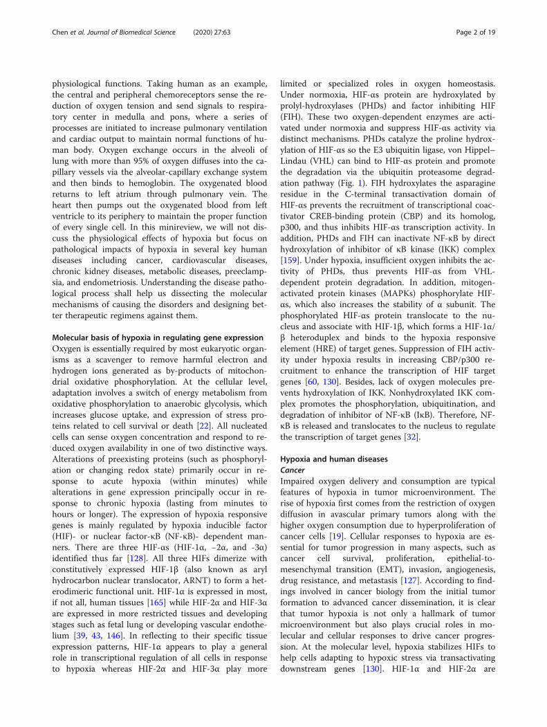
limited or specialized roles in oxygen homeostasis.Under normoxia, HIF-αs protein are hydroxylated byprolyl-hydroxylases (PHDs) and factor inhibiting HIF(FIH). These two oxygen-dependent enzymes are acti-vated under normoxia and suppress HIF-αs activity viadistinct mechanisms. PHDs catalyze the proline hydrox-ylation of HIF-αs so the E3 ubiquitin ligase, von Hippel–Lindau (VHL) can bind to HIF-αs protein and promotethe degradation via the ubiquitin proteasome degrad-ation pathway (Fig. 1). FIH hydroxylates the asparagineresidue in the C-terminal transactivation domain ofHIF-αs prevents the recruitment of transcriptional coac-tivator CREB-binding protein (CBP) and its homolog,p300, and thus inhibits HIF-αs transcription activity. Inaddition, PHDs and FIH can inactivate NF-κB by directhydroxylation of inhibitor of κB kinase (IKK) complex[159]. Under hypoxia, insufficient oxygen inhibits the ac-tivity of PHDs, thus prevents HIF-αs from VHL-dependent protein degradation. In addition, mitogen-activated protein kinases (MAPKs) phosphorylate HIF-αs, which also increases the stability of α subunit. Thephosphorylated HIF-αs protein translocate to the nu-cleus and associate with HIF-1β, which forms a HIF-1α/β heteroduplex and binds to the hypoxia responsiveelement (HRE) of target genes. Suppression of FIH activ-ity under hypoxia results in increasing CBP/p300 re-cruitment to enhance the transcription of HIF targetgenes [60, 130]. Besides, lack of oxygen molecules pre-vents hydroxylation of IKK. Nonhydroxylated IKK com-plex promotes the phosphorylation, ubiquitination, anddegradation of inhibitor of NF-κB (IκB). Therefore, NF-κB is released and translocates to the nucleus to regulatethe transcription of target genes [32].
Hypoxia and human diseasesCancerImpaired oxygen delivery and consumption are typicalfeatures of hypoxia in tumor microenvironment. Therise of hypoxia first comes from the restriction of oxygendiffusion in avascular primary tumors along with thehigher oxygen consumption due to hyperproliferation ofcancer cells [19]. Cellular responses to hypoxia are es-sential for tumor progression in many aspects, such ascancer cell survival, proliferation, epithelial-to-mesenchymal transition (EMT), invasion, angiogenesis,drug resistance, and metastasis [127]. According to find-ings involved in cancer biology from the initial tumorformation to advanced cancer dissemination, it is clearthat tumor hypoxia is not only a hallmark of tumormicroenvironment but also plays crucial roles in mo-lecular and cellular responses to drive cancer progres-sion. At the molecular level, hypoxia stabilizes HIFs tohelp cells adapting to hypoxic stress via transactivatingdownstream genes [130]. HIF-1α and HIF-2α are
Chen et al. Journal of Biomedical Science (2020) 27:63 Page 2 of 19
clinically correlated with advanced stages and poor sur-vival of cancer patients [127]. In addition, the HIF sig-naling is essential for promoting the metastatic ability ofcancer cells [117], suggesting that HIF pathway plays avital role in cancer biology [127]. Furthermore, otherpathways such as the mammalian target of rapamycin(mTOR) and the unfolded protein response (UPR) actcooperatively with HIF to regulate cellular functions[168]. Hypoxia suppresses mTORC1 activity throughmultiple pathways. Prolonged hypoxia causes energystress that activates AMP-activated protein kinase(AMPK), which induces the transcription of regulated indevelopment and DNA damage responses 1 (REDD1).REDD1 suppresses mTOR activity through tuberoussclerosis complex (TSC)1/TSC2-mediated mTOR inacti-vation [20]. Hypoxia can also inhibit mTORC1 activitythrough BCL-2 interacting protein 3 (BNIP3) and thepromyelocytic leukemia (PML) tumor suppressor [168].The suppressed mTORC1 activity results in decreasedEIF4E-binding protein 1 (4E-BP1) dephosphorylationand thus sequesters eukaryotic translation initiation fac-tor 4E (EIF4E) from cap-dependent translation initiation.The hypoxic inhibition of translation initiation is also re-ported to act through enhanced association with EIF4E-transporter (4E-T). Hypoxia induces endoplasmicreticulum (ER) stress sensors PKR-like ER kinase(PERK), inositol-requiring protein 1 (IRE1), and activat-ing transcription factor 6 (ATF6) to induce the UPRpathway. The PERK-mediated phosphorylation of EIF2α
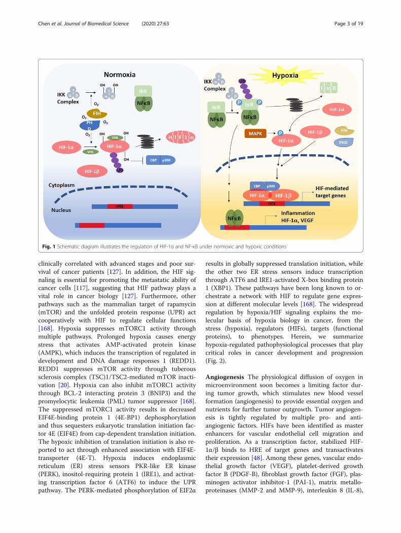
results in globally suppressed translation initiation, whilethe other two ER stress sensors induce transcriptionthrough ATF6 and IRE1-activated X-box binding protein1 (XBP1). These pathways have been long known to or-chestrate a network with HIF to regulate gene expres-sion at different molecular levels [168]. The widespreadregulation by hypoxia/HIF signaling explains the mo-lecular basis of hypoxia biology in cancer, from thestress (hypoxia), regulators (HIFs), targets (functionalproteins), to phenotypes. Herein, we summarizehypoxia-regulated pathophysiological processes that playcritical roles in cancer development and progression(Fig. 2).
Angiogenesis The physiological diffusion of oxygen inmicroenvironment soon becomes a limiting factor dur-ing tumor growth, which stimulates new blood vesselformation (angiogenesis) to provide essential oxygen andnutrients for further tumor outgrowth. Tumor angiogen-esis is tightly regulated by multiple pro- and anti-angiogenic factors. HIFs have been identified as masterenhancers for vascular endothelial cell migration andproliferation. As a transcription factor, stabilized HIF-1α/β binds to HRE of target genes and transactivatestheir expression [48]. Among these genes, vascular endo-thelial growth factor (VEGF), platelet-derived growthfactor B (PDGF-B), fibroblast growth factor (FGF), plas-minogen activator inhibitor-1 (PAI-1), matrix metallo-proteinases (MMP-2 and MMP-9), interleukin 8 (IL-8),
Fig. 1 Schematic diagram illustrates the regulation of HIF-1α and NF-κB under normoxic and hypoxic conditions
Chen et al. Journal of Biomedical Science (2020) 27:63 Page 3 of 19
and angiopoietins (ANG-1 and ANG − 2) are known aspro-angiogenic factors playing crucial roles duringtumor angiogenesis [82, 94]. Moreover, VEGF and PAI-1has also been found to enhance tumor angiogenesisunder the control of HIF-2α [119, 138]. Among thesesecretory factors, VEGF, PDGF-B, FGF, ANG-1, andANG-2 bind to their specific receptors VEGFR2,PDGFRβ, FGFR, TIE-1 and TIE-2, respectively, on vas-cular endothelial cells to activate cell proliferation, mi-gration, tube formation, and vascularization [57, 113,114], while other factors (e.g., MMPs, PAI-1) participatein remodeling extracellular matrix (ECM) and are alsoinvolved in local invasion of cancer cells [111].
Survival advantages from adaptive cellular responsesMalignant tumors tend to exhibit enhanced anaerobic gly-colysis as their energy source. This metabolic shift enablescancer cells adapting to microenvironmental stresses in-cluding hypoxia [98]. Under hypoxia, cancer cell adapt tothe reduced available oxygen and nutrient through upregu-lation of glucose transporters (GLUT1 and GLUT3),carbonic anhydrases (CA9 and CA12), pyruvate dehydro-genase kinases (PDK1 and PDK3), lactate dehydrogenase A(LDHA), phosphoglycerate kinase 1 (PGK-1), and hexoki-nases (HK1 and HK2) to cooperatively modulate a meta-bolic shift from oxidative phosphorylation to anaerobicglycolysis [92, 129]. Epigenetically, histone lysine demethy-lase 3A (KDM3A) removes demethylated histone 3 lysine 9(H3K9me2) from PGK1 promoter to enhance HIF-1α-dependent PGK1 transcription thus facilitate glycolysisunder hypoxia. Under hypoxia, the HIF-1α-dependent
histone demethylase KDM4B induction removes H3K9me3from the promoters of hypoxia-inducible genes involved incell survival [124]. While the nonadaptive precancer/cancercells undergo cell death, the hypoxia-induced anti-apoptosis pathways help other cancer cells survive. Theseevolutionally survived cells tend to express reduced pro-apoptotic factors such as Bcl-2-associated X protein (Bax),Bcl-2 associated agonist of cell death (Bad), and BH3interacting domain death agonist (Bid), and enhanced anti-apoptotic factors such as B-cell lymphoma 2 (Bcl-2) and B-cell lymphoma-extra large (Bcl-xL) [50, 131]. Consequently,both of the poly (ADP-ribose) polymerase (PARP) cleavageand caspase activity are suppressed under hypoxia [40, 65].These adaptive responses, including the metabolic and pro-survival shifts, not only cooperatively maintain the survivalof cancer cell, but may also continually facilitate tumorigen-esis from initial tumor formation to secondary tumorigen-esis after/under treatment.
Epithelial-mesenchymal transition Hypoxia/HIF sig-naling also regulates cellular behaviors, including migra-tion/invasion, intra−/extra-vasation, colonization andtumorigenesis at distant organs, to drive cancer metasta-sis. EMT is a common process which enables noninva-sive (epithelial-like) cancer cells to invade andmetastasize (mesenchymal-like). It is now clear that hyp-oxia enhances the metastatic ability through promotingEMT of cancer cells. Hypoxia/HIF signaling directly fa-cilitates the EMT gene profile to induce the invasivenessof cancer cells [117] through recognition of HREs ofEMT transcription factors zinc finger E-box-binding
Fig. 2 Hypoxia-regulated cancer progression. Hypoxia is a typical feature of tumor microenvironment, which contributes to initial tumorigenesis,induced angiogenesis, drug resistance, and cancer metastasis. The major upstream regulators (gray), functional downstream genes (blue), andresulting cellular consequences (yellow) under the control of hypoxia signaling are indicated
Chen et al. Journal of Biomedical Science (2020) 27:63 Page 4 of 19
homeobox 1 (ZEB1), snail family transcriptional repres-sor 1 (SNAI1), and twist family bHLH transcription fac-tor 1 (TWIST1) genes [154]. Alternatively, HIFmodulates Notch [123], transforming growth factor beta(TGF-β) [29], integrin-linked kinase (ILK) [26], tyrosinekinase receptors (TKRs) [34], Hedgehog [75, 140], AXLreceptor tyrosine kinase [116], lysyl oxidase (LOX) [162],and protein 3-phosphoinositide-dependent protein kin-ase 1 [110] to indirectly facilitate EMT. In addition, his-tone deacetylase 3 (HDAC3) interacts with WD repeatdomain 5 (WDR5) to enhance deacetylation of H3K4Acin the promoters of EMT regulators under hypoxia[174]. These genetic regulations drive the phenotypicshift from epithelial-like (noninvasive) to mesenchymal-like (invasive), which help cancer cell motility during themultistep metastasis process, such as migration/invasionand intra−/extra-vasation.
Cancer stemness and drug resistance Cancer stemcells (CSCs) expressing stem cell markers (e.g.,CD133, CD44) and transcription factors (e.g.,OCT3/4, SOX2, KLF4, c-MYC) have been identifiedto exhibit undifferentiated (stem cell-like) andtumorigenic properties [12]. Hypoxic microenviron-ment also facilitates the maintenance of CSCs [53].It is known that both HIF-1α and HIF-2α inductionactivate OCT4, c-MYCc, SOX2 and enrich the ex-pression of CD133- and CD44-positive cancer cells,along with the enhanced self-renewal functions andtumorigenic potential [4], while several studies pro-posed that cancer stemness is predominately con-trolled by HIF-2α [30, 62]. In addition tomaintaining the unlimited primary tumor growth,CSCs have slow growing rate (quiescent) thus arealso relatively insensitive to chemotherapy targetingproliferative cancer cells [25]. Thus, it is also sug-gested that CSCs within tumors determine thetherapeutic efficacy and cancer prognosis, as thereservoir CSCs result in resistant subpopulationafter chemotherapy and become the cellular sourcesfor continued cancer propagation and recurrence.Irrespective of the involvement of CSCs in cancerrecurrence, hypoxia signaling also activates severaldrug resistant pathways to protect cancer cells[151]. The nature of hypoxic region with poorvascularization may partly limit the diffusion of cir-culating drugs. Furthermore, HIF-1α induces the ex-pression of multidrug resistance (MDR) genes underhypoxia. Expression of MDR gene products, thedrug efflux pump protein ABC transporters, enablescancer cells to pump out intracellular chemothera-peutic drugs, thus reduces the therapeutic effectsand enhances drug resistance [151].
Exosome secretion and priming Exosomes released tomicroenvironment have been known to participate inintercellular communication since these extracellularmicrovesicles containing functional nucleic acids andproteins [100] . Recent studies demonstrated the clinicalsignificance of hypoxic exosome in cancer. First, thecontents of exosome, including proteins and nucleicacids, have significant changes under hypoxia [132] .Many of the proteins and RNAs enriched in hypoxicexosomes are canonical downstream gene products ofhypoxia, such as HIF-1α, MMPs, and LOX [2, 69]. It isalso known that the exosome secretion is enhanced inhypoxic cells to regulate cancer metastasis through theupregulation of small guanosine triphosphatase RAB22A[161]. Exosomes secreted by hypoxic cancer cells con-taining high levels of oncogenic proteins to enhanceEMT, stemness and invasiveness through degradation ofE-cadherin and activation of β-catenin pathway [115].The currently identified extracellular secretion and func-tions of hypoxic exosomes make them as a possible hyp-oxic biomarker and therapeutic target.
Hypoxia regulates miRNA biogenesis Current know-ledge based on the large-scale genomic sequencingprojects illustrated that the protein-coding transcrip-tional output is less than 2% in human genome. Forrecent 20 years, the roles of non-coding RNAs, suchas miRNAs, have been extensively studied and theirroles in cancer progression have been well-documented [134]. Here, we will discuss the regula-tion of hypoxia on miRNA biogenesis. The matur-ation of miRNA is a multistep process involvingseveral protein factors. It is now established that boththe global level of miRNAs and the expression ofmiRNA biogenesis factors (Dicer, Drosha, TARPB2,and DCGR8) are downregulated under hypoxia [10] .The cytoplasmic biogenesis factor Dicer is suppressedby HIF-1α-mediated proteasomal degradation orH3K27me3 demethylases KDM6A/B-dependent epi-genetic modification [70, 156]. Several miRNAs in-cluding miR-103/107, let-7, and miR-630 were alsoreported to target and suppress Dicer expression [96,122, 147]. Moreover, activation of EGFR pathwayunder hypoxia phosphorylates AGO2 and abolishes itsinteraction with Dicer, thus represses miRNA matur-ation and activity [135]. These pathways consequentlyresult in either global or specific miRNA downregula-tion to promote cancer progression. Notably, many ofthe genes regulated by miRNAs are also canonicalhypoxia/HIF signaling downstream genes, such asZEB1, GLUT1, and VEGF, which further suggests thatthe hypoxia-regulated miRNAs act post-transcriptionally and synergistically with canonicalhypoxia pathway of transcriptional regulation.
Chen et al. Journal of Biomedical Science (2020) 27:63 Page 5 of 19
Cardiovascular diseasesCardiovascular diseases are the leading cause of mortal-ity worldwide, representing 1 of every 3 deaths in 2018.Hypoxia is one of the common features in the patho-physiology of a variety of cardiovascular disorders [1].Heart failure with reduced left ventricular systolic func-tion after myocardial infarction causes the insufficientoxygen in the body. Moreover, heart failure with pre-served ejection fraction in patients eventually leads tosystemic and pulmonary hypertension. Pulmonaryhypertension also associates with other hypoxic pulmon-ary diseases, including chronic obstructive pulmonarydisease and obstructive sleep apnea syndrome, and pro-motes inflammation and atherosclerosis [95]. Athero-sclerosis is a chronic inflammatory disease that canincrease the risk of myocardial infarction and stroke.The thickness of arterial wall causes hypoxia in the in-tima, reduces the perfusion of the tissue, and furtherstimulates proatherosclerotic processes, like inflamma-tion, lipid synthesis, and angiogenesis [139].
Roles of NF-κB As mentioned above, hypoxia simultan-eously activates HIF and NF-κB signaling pathways toregulate numerous biological processes (Fig. 1). It shouldbe noted that the crosstalk of HIF-1α and NF-κB playsan important role in ischemic cardiovascular disease.The hypoxia-induced HIF-1 upregulates NF-κB, whichreciprocally activates the transcription of HIF-1α [38].This forms a positive feedback loop to worsen the dis-ease. In addition, NF-κB also induces many other targetgenes such as inflammatory cytokines. The inflammatoryresponse leads to smooth muscle cell activation, result-ing in neointima formation and occlusive plaque. Im-portantly, elevated serum inflammatory marker, such asIL-6 and tumor necrosis factor-α (TNF-α), in patientsare correlated with their prognosis in hypoxic cardiovas-cular disease [76, 103].
Roles of Hif-1α The functional consequences of HIF-1αin cardiomyopathy had been investigated using transaor-tic constriction (TAC) murine models to mimicpressure-overload heart failure human disease. A 3-weekTAC in the mice with cardiomyocyte-specific deletion ofHif1-α fails to induce Vegf expression and neoangiogen-esis. As a result, acute heart failure occurs due to thelack of enough oxygen being delivered to the rapidly in-creasing cardiac muscle cells [125]. Another study usingcardiomyocyte- and endothelial cell-specific Hif1-αknockout mice showed 1-week TAC causes severe heartfailure phenotype. Moreover, the myocardial capillarydensity is decreased in these mice, resulting from mark-edly increased endothelial cell apoptosis [164]. These re-sults implicate the proangiogenic and cardio-protectiveeffect of Hif-1α. Conversely, Krishnan and colleagues
demonstrated that, after TAC, the Hif1-α+/− mice havebetter cardiac function than wild-type mice. They alsofound Hif-1 promotes the expression of peroxisomeproliferator-activated receptor γ (PPARγ), and activateslipid synthesis by engaging glycerolipid and fatty aciduptake genes, and, in turn, induces cell apoptosis [68].These conflicting results suggest that Hif-1 mediates thecomplexity of adaptive responses. Hence, further re-search differentiating the roles of HIF-1 in cardiomyo-cyte is needed in order to acquire a clear picture.
Roles of miRNAs In addition to the transcriptionalregulation by HIF-1α and NF-κB, ischemic/hypoxia alsomodulates cardiofunction via miRNAs at the posttran-scriptional level. Numerous miRNAs had been reportedto be up- or down-regulated in patients with myocardialischemia/reperfusion injury (Table 1). Among them,MiR-22, miR-134, miR-135a, miR-203, miR-144, miR-98,miR-18a, miR-210, miR-340-5p, miR-374a-5p, and miR-1192 exert protective effects in cardiovascular ischemicinjury through downregulating their target genes [37, 41,56, 77, 79, 112, 160, 163, 175, 183, 184]. On the otherhand, a specific set of miRNAs has been linked to car-diac dysfunctions in varied cardiac injury models. Forexample, in intermittent hypoxia-induced myocardialdamage, miR-146a-5p promotes cell death by targetingX-linked inhibitor of apoptosis protein [80] . MiR-327reduces the expression of apoptosis repressor with cas-pase recruitment domain expression, and subsequentlydeteriorates myocardial ischemia/reperfusion injury [78].MiR-429 accelerates ischemia/reperfusion injury by tar-geting mouse protein 25 and decreasing the protectiveeffect of autophagy [189]. These data indicate that miR-NAs do play important roles in regulating cardiovascularfunction after heart injury and imply they might be po-tential molecular targets for diagnosis or treatment ofcardiovascular diseases. Indeed, miRNA-based therapiesusing modified oligonucleotides have been developed intreating different cardiovascular diseases. The criticalrole of miR-34a in cardiac ageing and function made itto be selected as a target for treating myocardial infarc-tion [17, 144]. Targeting miR-34a by locked nucleicacid-modified anti-miR-34a attenuates adverse cardiacremodeling in myocardial infarction- or TAC-inducedcardiac injury [15]. Similarly, anti-miR-92a and anti-miR-132 therapies are also effectively resistant tohypoxia-induced cardiac injury [13, 155]. Most recently,circulating extracellular vesicles-containing miRNAs at-tract great attention as promising biomarkers for earlydetection of cardiovascular diseases [3]. In lights of theserecent advances, future studies focusing on elucidatingkey miRNA diagnostic biomarkers that can be targetedusing mimetics or inhibitors to alleviate ischemic cardio-vascular diseases are warranted.
Chen et al. Journal of Biomedical Science (2020) 27:63 Page 6 of 19
Metabolic diseases
Adipocytes and metabolic diseases Adipose tissue isone of the least metabolically dynamic structure forlipid turnover and can also communicate with othertissues through secretion of adipocyte-derived hor-mones, growth factors, inflammatory cytokines, leptin,adiponectin, signaling lipids, fatty acids, and miRNAspackaged in exosomes [145, 188]. These adipocyte-secreted factors (adipokines) and fatty acids regulatesystemic metabolism and play important roles in thedevelopment of metabolic diseases, such as metabolicsyndrome, ischemic heart disease, stroke, obesity, type2 diabetes mellitus, and cancer [7, 148]. White adipo-cytes in white adipose tissue, which is the most im-portant lipid buffering organ, can either accumulatefatty acids in lipid droplets or supply fatty acids forother tissues determined by the balance between fattyacid synthesis (lipogenesis) and lipid breakdown (lip-olysis and fatty acid β-oxidation) [63]. Conversely,brown adipocytes in brown adipose tissue and beigeadipocytes in beige/brite adipose tissue more fre-quently produce heat through fatty acid β-oxidationin mitochondria (thermogenesis) [31]. Owing to theircontributions to whole-body lipid homeostasis, bothwhite and brown adipose tissues are considered pri-mary targets for the treatment of obesity and type 2diabetes mellitus [23, 28]. In addition, both white andbrown adipose tissues are able to elaborate adipokinesto control nutritional intake, sensitivity to insulin, and
inflammatory processes in other tissues [28, 188].Therefore, lipid metabolism and adipokines secretionin adipose tissue exert an impact on whole-body me-tabolism and are important for the progress of meta-bolic diseases.
Effects of hypoxia on adipocytes Hypoxia is one of themechanisms responsible for the development of metabolicchanges and pro-inflammatory situations of white adiposetissue [150]. In obesity, due to the enlargement of adipo-cytes and increased distance from the vasculature, hypoxiaoccurs within the expanding white adipose tissue in ob/oband dietary obese mice. Accordingly, white adipose tissuesfrom obese people are subjected to intra-adipose tissuehypoxia and characterized by increased HIF-1α expression[67]. In vitro experiments show that HIF-1α and HIF-2αinhibit insulin signaling in both human and murine whiteadipocytes [120]. Moreover, chronic hypoxia has beensuggested to be part of the pathogenic pathways leading toadipose tissue dysfunction [166, 178]. Hypoxia triggers re-active oxygen species (ROS) production, ER stress, inflam-matory responses, angiogenesis, and adipocyte death [66,150, 176, 185]. HIF also regulates the expression of variousadipokine genes, such as increasing leptin, visfatin, apelin,TNF-α, IL-1, IL-6, VEGF, MMP2, MMP9, angiopoietin-like protein-4, macrophage migration inhibitory factor,and PAI-1 expression, while downregulating adiponectinand PPARγ expression in adipocytes [52, 150, 176].Hypoxia alters several key metabolic processes includ-
ing glucose uptake, glycolysis, oxidative metabolism,
Table 1 MicroRNAs involve in hypoxia-mediated human diseases
MicroRNA(s) Change in expression Regulatory gene(s) Disease(s)
miR-18a Up BDNF Myocardial infarction
miR-21 Up PDCD4Spry1
Heart failureHeart failure
miR-22miR-34a
Up SIRT1 Hypertrophy
miR-98 Down DAPK1 Ischemia/reperfusion
miR-134 Up NOS3 Ischemia/reperfusion
miR-135amiR-203
DownDown
PTP1B Ischemia/reperfusionMyocardial infarction
miR-144 Down FOXO1 Ischemia/reperfusion
miR-146a-5p Up XIAP Intermittent hypoxia
miR-210 Up EFNA3PTP1BHIF-3α
Vascular remodeling
miR-327 Up CRD Ischemia/reperfusion
miR-340-5p Down Act1 Ischemia/reperfusion
miR-374a-5p Down MAPK6 Ischemia/reperfusion
miR-429 Down MO25 Ischemia/reperfusion
miR-1192 Down CASP3 Myocardial infarction
Chen et al. Journal of Biomedical Science (2020) 27:63 Page 7 of 19

lipolysis, and lipogenesis in adipocytes. Hypoxia stimu-lates glucose uptake in adipocytes through HIF-1α-upregulated GLUT expression, and increases anaerobicglycolysis and lactate production by induction of glyco-lytic enzymes [121, 167]. Hypoxia also induces rear-rangements of lipid metabolism in adipocytes. Inresponse to hypoxia, extracellular fatty acid uptake is re-duced by inhibition of fatty acid transporters (FATP1and CD36) and transcription factors (PPARγ and C/EBPα) while lipolysis is increased in 3 T3-L1 adipocytes[179]. Another study shows that hypoxia inhibits lipo-genesis by reducing PPARγ and fatty acid synthase(FAS), and induces basal lipolysis in visceral and sub-cutaneous human adipocytes [109]. In addition, hypoxiainhibits adipogenesis and differentiation in 3 T3-L1 adi-pocytes via HIF-1α-dependent upregulation of differenti-ated embryo-chondrocyte expressed gene 1 (DEC1/Stra13) and subsequent repression of PPARγ2 expres-sion [182]. HIF-1α also suppresses expression of genesinvolved in fatty acid β-oxidation by repression of sirtuin2-mediated deacetylation of PPARγ coactivator 1-α(PGC-1α) in white adipocytes [67]. Besides, studies indi-cate that obesity induces hypoxia in brown adipose tis-sue and causes the loss of its thermogenic capacity[149]. Other studies show that hypoxia is a trigger forbrown adipose tissue whitening with diminished β-adrenergic signaling, enlarged lipid droplets, and loss ofmitochondria in the cells [137]. Increased HIF-1α andsuppressed uncoupling protein 1 (UCP1) expressionwith lower fatty acid β-oxidation are observed in hypoxicbrown adipose tissue [136]. Therefore, hypoxia alterslipid metabolism in adipocytes mainly by inhibiting lipo-genesis and decreasing fatty acid β-oxidation.
Effects of hypoxia on other cell types The effects ofhypoxia on lipid metabolism are also studied in othercell types related to metabolic diseases. As fatty acid β-oxidation takes place inside mitochondria and requiresoxygen, fatty acid metabolism has to be modified otherthan energy production under hypoxia. Furthermore, themajor source of cytoplasmic acetyl-CoA from glucoseconverted citrate is prohibited under hypoxia due to theinhibition of the TCA cycle, so alternative sources offatty acid precursors have to be exploited. Uptake ofextracellular fatty acids for triacylglycerol synthesis ispromoted by HIF-1α-induced PPARγ in cardiomyocytesunder hypoxia [68]. Extracellular fatty acid influx andlipid droplet accumulation are enhanced via HIF-1α-mediated induction of fatty acid binding protein 3 and 7(FABP3 and FABP7), while de novo lipogenesis is re-pressed in glioblastoma and breast cancer cells underhypoxia [14]. To maintain certain level of lipogenesisunder hypoxia, production of fatty acid precursors, cit-rate and acetyl-CoA, are supported through reductive
glutamine metabolism in several cancer cells [46, 105]and brown adipocytes [180]. HIF-induced isocitrate de-hydrogenase 1 and 2 (IDH1 and IDH2) contribute to thepreservation of citrate levels via conversion of α-ketoglutarate to isocitrate and its subsequent reductivecarboxylation to produce citrate from glutamine underhypoxia [101]. Adequate fatty acid synthesis is furthersupported by HIF-1-dependent activation of sterol regu-latory element-binding protein 1 (SREBP1), which inturn upregulates the expression of FAS in breast cancercells [45]. Hypoxia also induces lipid droplet accumula-tion by upregulating two enzymes of the triacylglycerolbiosynthesis pathway, acylglycerol-3-phosphate acyl-transferase 2 (AGPAT2) and lipin-1 in different types ofcancer cells [106, 152]. Furthermore, hypoxia-inducedlipid droplet accumulation is accompanied by the inhib-ition of fatty acid β-oxidation though HIF-1 and HIF-2-dependent downregulation of PGC-1α and carnitine pal-mitoyltransferase 1A (CPT1A) in both hepatoma andrenal cell carcinoma cells [35, 90]. Therefore, hypoxiamay result in different adaptation of lipid metabolismdepending on the cell types.
Effects of hypoxia on animal models of obesity Sincehypoxia is shown to inhibit fatty acid β-oxidation as apromoter of obesity and inhibit lipogenesis as a suppres-sor of obesity, several animal studies based on the over-expression or inhibition of HIFs in adipocytes suggestthat HIF activation either promotes or inhibits metabolicdiseases. As a promoter of obesity, mice overexpressingHif-1α in adipocytes have elevated obesity and insulinresistance associated with increased inflammation and fi-brosis [49, 59]. Adipocyte specific Hif-1α or Hif-1βknockout, or inhibition of Hif-1α by inhibitors decreaseobesity and insulin resistance in mice fed with high-fatdiet [58, 74, 142]. In agreement, adipocyte-specific Phd2ablation enhances adiposity in mice under normal chowdiet (low-fat) with lower expression of adipose triglycer-ide lipase (ATGL) and suppresses lipolysis in white adi-pocytes [102]. These obesity-promoting effects can becorrelated with the capacity of Hif-1 to downregulatefatty acid β-oxidation in white and brown adipose tissue[67, 136]. On the other hand, a number of studies haveshown that Hif activation decreases obesity and Hif in-hibition increases obesity indicating hypoxia as a sup-pressor of obesity. Accordingly, transgenic miceoverexpressing an adipose tissue-specific dominantnegative Hif-1α mutant developed severe obesity, insulinresistance, and accumulated enlarged lipid droplets inbrown adipose tissue with decreased mitochondrial bio-genesis after high-fat diet [187]. Another group showsthat transgenic mice with adipose tissue-specific knock-out of Phd2 (for constitutive expression of Hif) are re-sistant to high-fat diet-induced obesity with fewer lipid
Chen et al. Journal of Biomedical Science (2020) 27:63 Page 8 of 19

droplets in white adipose tissue and increased Ucp1 ex-pression in brown adipose tissue [97], while adipocyte-specific knockout of Phd2 induces adiposity in miceunder chow diet (low-fat) [102]. Similarly, phenotypesare also observed in mice globally lacking Fih, which areresistant to high-fat diet-induced weight gain and hep-atic steatosis [186]. Taken together, these studies suggestthat Hif-1α may stimulate the thermogenic functions ofbrown adipose tissue to conquer high-fat diet-inducedobesity, which is contradictory with other findings thatHif-1 suppressed expression of genes involved in fattyacid β-oxidation in white and brown adipose tissue lead-ing to obesity [67, 136].In summary, studies investigating the functions of
HIFs in adipocytes and other metabolic diseases revealedthe conflicting results due to different experimental con-ditions (Fig. 3). The roles of HIFs in these studies werediscovered primarily through the analysis of conditionalHif-knockout mice or through some pharmacologicalHIF inhibitors. These inconsistent consequences mightbe also due to the complexity of metabolic regulationwith the complicated roles of HIFs that extend furtherthan lipid metabolism. Therefore, the actual functions ofHIFs in adipocytes and related metabolic diseases must
be carefully interpreted relative to different physiologicalconditions.
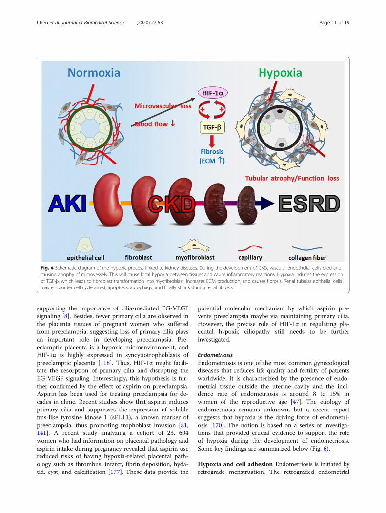
Kidney diseasesAcute kidney injury (AKI) is notorious for its correlationwith the development of chronic kidney disease (CKD).AKI is a rapid failure of kidney function, usually causedby decreased blood flow, toxic exposure, and ureteralobstruction. As the injuries are removed, renal cellsundergo repair process. However, some of the AKI pa-tients fail to fully recover. Instead, CKD starts to de-velop, which is characterized by the progressive loss ofthe kidney function [27]. Events include tubular atrophy,vascular rarefaction, and hypoxia, which lead to renal fi-brosis and functional loss of kidney. Ultimately, CKDwill develop into the end-stage renal disease (ESRD).Today, the only treatments for ESRD are dialysis andkidney transplantation [91]. Kidney fibrosis, includingglomerulosclerosis and tubulointerstitial fibrosis, aphenomenon of excessive ECM deposit and accumula-tion, has been recognized as the hallmark of CKD andthe major pathway leading to ESRD [107]. Fibrosis is theterminal pathway involved in the continuous progressionof CKD and it is a consequence of failed recovery after
Fig. 3 Hypoxia-regulated lipid metabolism related to obesity. Hypoxia is shown as a promoter or a suppressor of obesity by regulating lipidmetabolism. The major changes (increases shown in red; decreases shown in green) involved in fatty acid β-oxidation, extracellular fatty acidinflux, lipolysis, lipogenesis and lipid droplet accumulation under hypoxia for its promoting obesity or anti-obesity effects are summarized
Chen et al. Journal of Biomedical Science (2020) 27:63 Page 9 of 19
kidney damage. When injuries happen, cells in kidneyincluding fibroblasts, tubular epithelial cells, endothelialcells, pericytes, lymphocytes, and macrophages, undergowound healing programming in an attempt to repair tis-sues. Sometimes, the damage is too severe for cells toovercome. Subsequently, they become victims and evenact to fuel the fibrogenesis progressively [18]. As the di-verse origin of cells involved in kidney fibrosis, it is com-plicated to figure out a comprehensive therapy fortreating CKD patients. Today, there is still no effectiveways to prevent CKD from getting worse. Several publi-cations have pointed out that hinder renal fibrosis fromgetting worse is a potential way to prevent AKI-CKDtransition and delay ESRD development [11, 16].
Hypoxia in kidney fibrosis Tubular injury may lead torenal microvascular loss, which restricts downstreamblood flow from glomerular capillaries and contributesto the development of renal hypoxia. Loss of microvas-culature, reduced oxygen dispersion, and metabolic ab-normality of cells in the kidney are the main causes ofthe hypoxic state. The initiation of hypoxia is one of themain causes of AKI, which can increase levels of HIF-1α,followed by the induction of TGF-β signaling. Duringthe process of kidney fibrosis, hypoxia and TGF-β1 sig-naling are excessively upregulated. AKI can be graduallydeveloped into CKD if TGF-β signaling remains hyper-activated for a period of time. Thus, hyperactivation ofTGF-β signaling is responsible for the renal fibrosis andcan also be identified as the hallmark of CKD. Hypoxiais common at the beginning of kidney fibrosis, whichpromotes HIF-1α expression that contributes to both aresult and a cause of renal fibrosis. Hyperactivation ofmyofibroblasts is responsible for kidney fibrosis as theycontinuously produce collagens, fibronectin, and vimen-tin, which contribute to tubulointerstitial fibrosis andlocal tissue hypoxia [72, 143]. Prolonged hypoxia furtherpromotes renal fibrosis by increasing the synthesis oftype I and type IV collagen and inhibiting the expressionof MMP-1 in human renal fibroblasts [108]. By usingkidney-specific Vhl−/− mice, which have a stable expres-sion of Hif-1α in kidney, researchers found that thesetransgenic mice exhibit more severe interstitial fibrosisafter conduct 5/6 nephrectomy [64]. In contrast, intra-peritoneal injection of HIF-1α inhibitor YC-1 protectsunilateral ureter obstruction mice from kidney fibrosisdevelopment [126]. These data indicate that HIF-1α hasa pivotal role in mediating kidney fibrosis (Fig. 4). How-ever, the underlying mechanisms by which HIF-1 accel-erates kidney fibrosis remain unclear.
PreeclampsiaPreeclampsia is a severe gestational complication fea-tured by new onset of high blood pressure after 20 weeks
of gestation along with signs of proteinuria, abnormallyhigh serum creatinine, or damaged liver function [36].Its complications remain a major cause for morbidityand mortality in pregnant women and fetuses. Defectivetrophoblast invasion into the decidualized endometriumleads to poor transformation of uterine spiral arteriesfrom high to low resistant vessels [42]. This deficientvessel remodeling causes a sustained hypoxic environ-ment implicating the development of preeclampsia.Thus, placental ischemia and hypoxia is a major cause ofpreeclampsia.
Primary cilium and preeclampsia Defective tropho-blast invasion leads to placental hypoxia. The tropho-blast invasion is regulated by endocrine gland-derivedvascular endothelial growth factor (EG-VEGF, alsoknown as prokineticin 1) [51]. EG-VEGF induces the ex-pression of MMPs by triggering ERK signaling cascadefor proper trophoblast invasion [157]. The receptor ofEG-VEGF localizes to the primary cilium, a cellular pro-trusion atop from the centrosome (Fig. 5). When EG-VEGF binds to its receptor, ERK signaling is initiatedfrom the base of the primary cilium, and then trans-duced throughout the cytoplasm. Inhibition of EG-VEGF signaling or disruption of primary cilia alleviatestrophoblast invasion in vitro [158]. More importantly,these phenotypes are also observed in pregnant womenwho suffered from preeclampsia. These data suggest theimportant roles of EG-VEGF and primary cilia in pre-venting placental hypoxia.Primary cilium functions as a sensory hub for transdu-
cing environmental chemo- and/or mechano-signalinginto the cells for proper development and differentiation.Growing body of evidence supports the important roleof primary cilium in maintaining embryo development.Interestingly, hypoxia suppresses primary cilium forma-tion [71], and elevated levels of HIF-1α in the humantrophoblasts have been linked to the development ofpreeclampsia [5], suggesting placental hypoxia-activatedHIF-1α plays a key role in the pathogenesis of pre-eclampsia. It has been shown that HIF-1α translocates tothe base of primary cilium for the resorption of primarycilia in the nutrient deprivation model, suggesting itsnon-genomic function in regulating primary cilium for-mation (Fig. 5).
HIF-1α in ciliogenesis Despite the role of hypoxia inpreeclampsia has been demonstrated for long, theunderlying molecular mechanism remains unclear.Trophoblast invasion is triggered by the binding of EG-VEGF to its congenital receptor on the primary cilium,thus inducing downstream ERK signaling for MMPs ex-pression. Disruption of primary cilium inhibits tropho-blast invasion even in the presence of EG-VEGF,
Chen et al. Journal of Biomedical Science (2020) 27:63 Page 10 of 19
supporting the importance of cilia-mediated EG-VEGFsignaling [8]. Besides, fewer primary cilia are observed inthe placenta tissues of pregnant women who sufferedfrom preeclampsia, suggesting loss of primary cilia playsan important role in developing preeclampsia. Pre-eclamptic placenta is a hypoxic microenvironment, andHIF-1α is highly expressed in syncytiotrophoblasts ofpreeclamptic placenta [118]. Thus, HIF-1α might facili-tate the resorption of primary cilia and disrupting theEG-VEGF signaling. Interestingly, this hypothesis is fur-ther confirmed by the effect of aspirin on preeclampsia.Aspirin has been used for treating preeclampsia for de-cades in clinic. Recent studies show that aspirin inducesprimary cilia and suppresses the expression of solublefms-like tyrosine kinase 1 (sFLT1), a known marker ofpreeclampsia, thus promoting trophoblast invasion [81,141]. A recent study analyzing a cohort of 23, 604women who had information on placental pathology andaspirin intake during pregnancy revealed that aspirin usereduced risks of having hypoxia-related placental path-ology such as thrombus, infarct, fibrin deposition, hyda-tid, cyst, and calcification [177]. These data provide the
potential molecular mechanism by which aspirin pre-vents preeclampsia maybe via maintaining primary cilia.However, the precise role of HIF-1α in regulating pla-cental hypoxic ciliopathy still needs to be furtherinvestigated.
EndometriosisEndometriosis is one of the most common gynecologicaldiseases that reduces life quality and fertility of patientsworldwide. It is characterized by the presence of endo-metrial tissue outside the uterine cavity and the inci-dence rate of endometriosis is around 8 to 15% inwomen of the reproductive age [47]. The etiology ofendometriosis remains unknown, but a recent reportsuggests that hypoxia is the driving force of endometri-osis [170]. The notion is based on a series of investiga-tions that provided crucial evidence to support the roleof hypoxia during the development of endometriosis.Some key findings are summarized below (Fig. 6).
Hypoxia and cell adhesion Endometriosis is initiated byretrograde menstruation. The retrograded endometrial
Fig. 4 Schematic diagram of the hypoxic process linked to kidney diseases. During the development of CKD, vascular endothelial cells died andcausing atrophy of microvessels. This will cause local hypoxia between tissues and cause inflammatory reactions. Hypoxia induces the expressionof TGF-β, which leads to fibroblast transformation into myofibroblast, increases ECM production, and causes fibrosis. Renal tubular epithelial cellsmay encounter cell cycle arrest, apoptosis, autophagy, and finally shrink during renal fibrosis
Chen et al. Journal of Biomedical Science (2020) 27:63 Page 11 of 19

tissues need to land on the surface of organs in the peri-toneal cavity and implant in order to survive ectopically.Two challenges that the retrograded cells have to face arehypoxic stress and adhesive ability. Recently, two studiesreported that hypoxia enhances cell adhesive ability of theendometrial stromal cells by inducing the expression ofcell adhesion molecules such as integrin α5, αV, β3, andβ5 via TGF-β1/Smad signaling and anthrax toxin receptor2 (ANTXR2) via HIF-1α-dependent manner [84, 87]. Fur-thermore, hypoxia-induced ANTXR2 expression is medi-ated by downregulation of EZH2 causing epigeneticchange in ANTXR2 locus [84]. Treatment with inhibitorsof TGF β1 receptor and ANTXR2 significantly attenuateshypoxia-induced cell adhesion in normal endometrialstromal cells [84, 87]. Furthermore, ANTXR2 inhibitorcan prevent and reduce endometriotic lesion formation inthe mouse model of endometriosis [84], revealing itstherapeutic potential for endometriosis. Taken together, itis indicated that hypoxia can promote the development ofendometriosis via increasing cell adhesive ability.
Hypoxia and hormone production Previous studieshave revealed that both estrogen (E2) and prostaglandinE2 (PGE2) are crucial factors for the development ofendometriosis [21, 172] and the enzymes control the
rate limiting steps of E2 (aromatase and steroidogenicacute regulatory protein, StAR) and PGE2 (cyclooxygen-ase-2, COX-2) biogenesis are aberrantly expressed inendometriotic stromal cells [9, 55, 153, 173]. It was laterrevealed that hypoxia upregulates both StAR and COX-2expression. Hypoxia represses dual-specificityphosphatase-2 (DUSP2) expression and contributes toincrease COX-2 expression via activation of ERK andp38 signaling pathways [171]. Furthermore, hypoxia alsoinhibits chicken ovalbumin upstream promoter-transcription factor II (COUP-TFII) to de-repress COX-2 expression in endometriotic stromal cells [85]. Finally,recent study has identified that hypoxia promotes YAP1activation, which leads to StAR and COX-2 overexpres-sion [83]. The same study also reported that inhibitionof YAP1 by its inhibitor, verteporfin, not only decreasesE2 and PGE2 production but also causes the regressionof endometriotic lesion in the mouse model of endomet-riosis [83]. These data indicate that retrograded endo-metrial tissues can produce E2 and PGE2 via theassistance of hypoxia to support their growth anddevelopment.
Hypoxia and angiogenesis To sustain the growth ofendometriotic lesion in the hostile peritoneal cavity,
Fig. 5 Potential role of hypoxia in developing preeclampsia. Binding of EG-VEGF to its receptor on the primary cilium activates ERK signaling atthe basal body for proper placentation. Under hypoxic condition, however, HIF-1α translocates to the base of cilia and induces cilia deacetylation,thus leading to ciliary resorption. The hypoxia-induced ciliary defects contribute to the development of preeclampsia
Chen et al. Journal of Biomedical Science (2020) 27:63 Page 12 of 19

new blood vessels must be established to provide oxy-gen and nutrient. Heterograft animal model of endo-metriosis by implanting human eutopic endometriuminto severe combined immunodeficiency mice hasidentified that hypoxia pretreatment for eutopic endo-metrium significantly increases the level of VEGF topromote cell proliferation and angiogenesis in vivo[93]. Critically, numerous studies have demonstratedthe effects of hypoxia on angiogenesis via differentmechanisms in endometriosis. Among them, leptinand VEGF-A are two well-known angiogenic factorsupregulated by hypoxic treatment in normal endomet-rial stromal cells [133, 169]. In addition, hypoxia-induced miR-20a causes prolonged activation of ERKsingling results in increasing several angiogenic factorincluding CYR61 and osteopontin [86]. Furthermore,hypoxia can increase angiogenin and IL-8 expressionby downregulation of COUP-TFII and DUSP2 expres-sion, respectively [44, 54]. More importantly, IL-8 re-ceptor inhibitor, reparixin, and YAP1 inhibitor,veterporfin, inhibit angiogenesis and the growth ofendometriotic lesion in the animal model of endomet-riosis [54, 83]. Taken together, these findings revealthe critical role of hypoxia-induced angiogenesis dur-ing the development of endometriosis.
Hypoxia and autophagy Peritoneal cavity is an unfavor-able microenvironment for the retrograded endometrialtissues due to lack of blood vessel to supply oxygen andnutrients. The study has reported that the higher level ofoxidative stress-induced DNA damage is observed inendometriotic specimen compared to the normal endo-metrial tissues [33]. Furthermore, primary endometrioticstromal cells produce more ROS than normal endomet-rial stromal cells [24]. Therefore, how endometrioticcells survive under this stressful condition is an intri-guing question. Previously, autophagy, a cellular mech-anism to remove or recycle unnecessary or dysfunctionalcomponents to produce energy, has been thought to playcrucial roles in protecting cells from oxidative stress-induced cell apoptosis [99, 104]. Indeed, autophagy is re-ported to be upregulated in the ovarian endometriosis[6, 89] and blocking hypoxia-induced autophagy en-hances apoptosis of endometrial stromal cells [88]. Re-cently, it is further demonstrated that hypoxia-inducedlong non-coding RNA MALAT1 (lncRNA-MALAT1) isinvolved in autophagy to protect cells from apoptosis[88]. However, the underlying mechanism that howlncRNA-MALAT1 regulates hypoxia-induced autophagyis still unclear. Besides autophagy, the change of meta-bolic phenotype in endometriotic lesions may also favor
Fig. 6 Impacts of hypoxia on endometriosis pathogenesis. Shed-off endometrial tissues will immediately suffer hypoxic stress when it retrogradesinto the peritoneal cavity during menstruation. Hypoxia regulates numerous downstream target genes involved in different cellular processesincluding cell survival, metabolism, angiogenesis, E2 and PGE2 production, and cell adhesion to help the development of endometriosis
Chen et al. Journal of Biomedical Science (2020) 27:63 Page 13 of 19

the development of endometriosis. For example, higherlevels of glycolysis-related genes such as pyruvate de-hydrogenase kinase 1 and lactate dehydrogenase A havebeen reported in endometriotic specimens and stromalcells, respectively [73, 181]. Furthermore, hypoxia-upregulated pyruvate dehydrogenase kinase 1 hasprevented cell death induced by H2O2 or low nutrienttreatment [73]. In summary, hypoxia-induced autophagyand change of cell metabolism may help retrogradedendometrial tissues to adapt to the hostile microenviron-ment and favor the development of endometriosis.
Conclusion and perspectiveThe mechanisms of cellular response to hypoxia contrib-ute to stress-induced pathophysiological outcomes. Pre-vious studies established the fundamental concept ofhypoxia biology, while recent advances in molecular andcellular biology such as non-coding RNA and microvesi-cle accelerate us to demonstrate a more complete andcomplex view of the regulatory network under hypoxia.Notably, most of these new findings were found to beclosely integrated in the canonical hypoxia pathway, sug-gesting a fine-tuned cellular machinery with multiplepathways cooperatively, synergistically, or mutual exclu-sively work together to modulate the transduction ofhypoxia signaling. Identification of these new sensors,messengers, and functional modulators not only ad-vances our knowledge of hypoxia biology but also pro-vides insights into the development of potentialdiagnostic and therapeutic approaches.
Abbreviations4E-BP1: EIF4E-binding protein 1; 4E-T: EIF4E-transporter; Act1: Activator 1;AGPAT2: Acylglycerol-3-phosphate acyltransferase 2; AKI: Acute kidney injury;AMPK: AMP-activated protein kinase; ANG: Angiopoietin; ANTX2: Anthraxtoxin receptor 2; ARNT: Aryl hydrocarbon nuclear translocator;ATF6: Activating transcription factor 6; ATGL: Adipose triglyceride lipase; Bcl-2: B-cell lymphoma 2; Bad: Bcl-2 associated agonist of cell death; Bax: Bcl-2-associated X protein; Bcl-xL: B-cell lymphoma-extra large; BDNF: Brain derivedneurotrophic factor; Bid: BH3 interacting domain death agonist; BNIP3: Bcl-2interacting protein 3; C/EBP: CCAAT/enhancer binding protein; CA: Carbonicanhydrase; CASP3: Caspase-3; CBP: CREB-binding protein; CD36: CD36molecule; CKD: Chronic kidney disease; COUP-TFII: Chicken ovalbuminupstream promoter-transcription factor II; COX-2: Cyclooxygenase-2;CPT1A: Carnitine palmitoyltransferase 1A; CRD: Apoptosis repressor withcaspase recruitment domain; CSC: Cancer stem cell; CYR61: Cysteine richangiogenic inducer 61; DAPK1: Death-associated protein kinase 1;DEC1: Differentiated embryo-chondrocyte expressed gene 1; DUSP2: Dual-specificity phosphatase-2; E2: Estrogen; ECM: Extracellular matrix;EFNA3: Ephrin A3; EGFR: Epidermal growth factor receptor; EG-VEGF: Endocrine gland-derived vascular endothelial growth factor;EIF4E: Eukaryotic translation initiation factor 4E; EMT: Epithelial-to-mesenchymal transition; ER: Endoplasmic reticulum; ERK: Extracellular signal-regulated kinase; ESRD: End-stage renal disease; EZH2: Enhancer of zeste 2polycomb repressive complex 2 subunit; FABP: Fatty acid binding protein;FAS: Fatty acid synthase; FATP1: Fatty acid transport protein 1; FGF: Fibroblastgrowth factor; FIH: Factor inhibiting HIF; FOXO1: Forkhead box protein O1;GLUT: Glucose transporter; H3K4Ac: Histone 3 lysine 4; HDAC3: Histonedeacetylase 3; HIF: Hypoxia inducible factor; HK: Hexokinase; HRE: Hypoxiaresponsive element; IDH: Isocitrate dehydrogenase; IKK: Inhibitor of κB kinase;IL-8: Interleukin 8; ILK: Integrin-linked kinase; IRE1: Inositol-requiring protein 1;IκB: Inhibitor of NF-κB; KDM: Histone lysine demethylase; LDHA: Lactate
dehydrogenase A; lncRNA-MALAT1: Long non-coding RNA MALAT1;LOX: Lysyl oxidase; MAP K6: Mitogen-activated protein kinase 6; MMP: Matrixmetalloproteinases; MO25: Mouse protein 25; mTOR: Mammalian target ofrapamycin; NF-κB: Nuclear factor-κB; NOS3: Nitric oxide synthase 3; PAI-1: Plasminogen activator inhibitor-1; PARP: Poly (ADP-ribose) polymerase;PDCD4: Programmed cell death 4; PDGF-B: Platelet-derived growth factor B;PDK: Pyruvate dehydrogenase kinase; PERK: PKR-like ER kinase; PGC-1α: PPARγ coactivator 1-α; PGE2: Prostaglandin E2; PGK-1: Phosphoglyceratekinase 1; PHDs: Prolyl-hydroxylases; PPARγ: Peroxisome proliferator-activatedreceptor γ; PTP1B: Protein tyrosine phosphatase 1B; REDD1: Regulated indevelopment and DNA damage responses 1; RHEB: Ras homolog enriched inbrain; ROS: Reactive oxygen species; sFLT1: Soluble Fms-Like tyrosine kinase1; SIRT1: Sirtuin 1; SNAI1: Snail family transcriptional repressor 1;Spry1: Sprouty 1; SREBP1: Sterol regulatory element-binding protein 1;StAR: Steroidogenic acute regulatory protein; TAC: Transaortic constriction;TGF-β: Transforming growth factor beta; TKRs: Tyrosine kinase receptors; TNF-α: Tumor necrosis factor-α; TWIST1: Twist family bHLH transcription factor 1;UCP1: Uncoupling protein 1; UPR: Unfolded protein response; VEGF: Vascularendothelial growth factor; VEGF-A: Vascular endothelial growth factor A;VHL: E3 ubiquitin ligase von Hippel–Lindau; WDR5: WD repeat domain 5;XBP1: X-box binding protein 1; XIAP: X-linked inhibitor of apoptosis protein;YAP1: Yes Associated Protein 1; ZEB1: Zinc finger E-box-binding homeobox 1
AcknowledgementsThe authors appreciate all the scientists whose works contribute to thisreview article. We also apologize to scientists whose works were notincluded in this review due to space constriction.
Authors’ contributionsSJT conceived the project. All authors wrote the manuscript. Particularcontribution of each section was made by: PSC (cancer), PLH(cardiovascular), ICP (metabolic diseases), WTC (chronic kidney disease), CYW(preeclampsia), and SCL (endometriosis). The author(s) read and approvedthe final manuscript.
FundingThis work was supported by grants from Ministry of Science and Technology(MOST 106-2320-B-006-072 -MY3 and MOST 107-2321-B-006-014) and Na-tional Health Research Institutes, Taiwan (NHRI-EX106-10516BI).
Availability of data and materialsNot applicable.
Ethics approval and consent to participateNot applicable.
Consent for publicationNot applicable.
Competing interestsAll authors declare there is no conflict of interest.
Author details1Institute of Basic Medical Sciences, College of Medicine, National ChengKung University, 1 University Road, Tainan 70101, Taiwan, Republic of China.2Department of Medical Laboratory Science and Biotechnology, College ofMedicine, National Cheng Kung University, 1 University Road, Tainan 70101,Taiwan, Republic of China. 3Department of Biomedical Engineering, Collegeof Engineering, National Cheng Kung University, 1 University Road, Tainan70101, Taiwan, Republic of China. 4Department of Physiology, College ofMedicine, National Cheng Kung University, 1 University Road, Tainan 70101,Taiwan, Republic of China. 5Department of Life Sciences, College ofBioscience and Biotechnology, National Cheng Kung University, 1 UniversityRoad, Tainan 70101, Taiwan, Republic of China. 6Department of Cell Biologyand Anatomy, College of Medicine, National Cheng Kung University, 1University Road, Tainan 70101, Taiwan, Republic of China.
Chen et al. Journal of Biomedical Science (2020) 27:63 Page 14 of 19
Received: 10 February 2020 Accepted: 6 May 2020
References1. Abe H, Semba H, Takeda N. The roles of hypoxia signaling in the
pathogenesis of cardiovascular diseases. J Atheroscler Thromb. 2017;24(9):884–94.
2. Aga M, Bentz GL, Raffa S, Torrisi MR, Kondo S, Wakisaka N, Yoshizaki T,Pagano JS, Shackelford J. Exosomal HIF1α supports invasive potential ofnasopharyngeal carcinoma-associated LMP1-positive exosomes. Oncogene.2014;33(37):4613–22.
3. Aghabozorgi AS, Ahangari N, Eftekhaari TE, Torbati PN, Bahiraee A, EbrahimiR, Pasdar A. Circulating exosomal miRNAs in cardiovascular diseasepathogenesis: new emerging hopes. J Cell Physiol. 2019;234(12):21796–809.
4. Al TW, Dale TP, Al-Jumaily RMK, Forsyth NR. Hypoxia-modified cancer cellmetabolism. Front Cell Dev Biol. 2019;7:4.
5. Albers RE, Kaufman MR, Natale BV, Keoni C, Kulkarni-Datar K, Min S, WilliamsCR, Natale DRC, Brown TL. Trophoblast-specific expression of Hif-1α resultsin preeclampsia-like symptoms and fetal growth restriction. Sci Rep. 2019;9(1):2742.
6. Allavena G, Carrarelli P, Del Bello B, Luisi S, Petraglia F, Maellaro E.Autophagy is upregulated in ovarian endometriosis: a possible interplaywith p53 and heme oxygenase-1. Fertil Steril. 2015;103(5):1244–51.
7. Antuna-Puente B, Feve B, Fellahi S, Bastard JP. Adipokines: the missing linkbetween insulin resistance and obesity. Diabetes Metab. 2008;34(1):2–11.
8. Anvarian Z, Mykytyn K, Mukhopadhyay S, Pedersen LB, Christensen ST.Cellular signalling by primary cilia in development, organ function anddisease. Nat Rev Nephrol. 2019;15(4):199–219.
9. Attar E, Bulun SE. Aromatase and other steroidogenic genes inendometriosis: translational aspects. Hum Reprod Update. 2006;12(1):49–56.
10. Bandara V, Michael MZ, Gleadle JM. Hypoxia represses microRNA biogenesisproteins in breast cancer cells. BMC Cancer. 2014;14:533.
11. Basile DP, Bonventre JV, Mehta R, Nangaku M, Unwin R, Rosner MH, KellumJA, Ronco C, Group A.X.W. Progression after AKI: understanding maladaptiverepair processes to predict and identify therapeutic treatments. J Am SocNephrol. 2016;27(3):687–97.
12. Beck B, Blanpain C. Unravelling cancer stem cell potential. Nat Rev Cancer.2013;13(10):727–38.
13. Bellera N, Barba I, Rodriguez-Sinovas A, Ferret E, Asin MA, Gonzalez-AlujasMT, Perez-Rodon J, Esteves M, Fonseca C, Toran N, Garcia Del Blanco B,Perez A, Garcia-Dorado D. Single intracoronary injection of encapsulatedantagomir-92a promotes angiogenesis and prevents adverse infarctremodeling. J Am Heart Assoc. 2014;3(5):e000946.
14. Bensaad K, Favaro E, Lewis CA, Peck B, Lord S, Collins JM, Pinnick KE,Wigfield S, Buffa FM, Li JL, Zhang Q, Wakelam MJO, Karpe F, Schulze A,Harris AL. Fatty acid uptake and lipid storage induced by HIF-1α contributeto cell growth and survival after hypoxia-reoxygenation. Cell Rep. 2014;9(1):349–65.
15. Bernardo BC, Gao XM, Winbanks CE, Boey EJ, Tham YK, Kiriazis H, GregorevicP, Obad S, Kauppinen S, Du XJ, Lin RC, McMullen JR. Therapeutic inhibitionof the miR-34 family attenuates pathological cardiac remodeling andimproves heart function. Proc Natl Acad Sci U S A. 2012;109(43):17615–20.
16. Bonventre JV. Maladaptive proximal tubule repair: cell cycle arrest. NephronClin Pract. 2014;127(1-4):61–4.
17. Boon RA, Iekushi K, Lechner S, Seeger T, Fischer A, Heydt S, Kaluza D,Treguer K, Carmona G, Bonauer A, Horrevoets AJ, Didier N, Girmatsion Z,Biliczki P, Ehrlich JR, Katus HA, Muller OJ, Potente M, Zeiher AM, HermekingH, Dimmeler S. MicroRNA-34a regulates cardiac ageing and function.Nature. 2013;495(7439):107–10.
18. Boor P, Ostendorf T, Floege J. Renal fibrosis: novel insights into mechanismsand therapeutic targets. Nat Rev Nephrol. 2010;6(11):643–56.
19. Brown JM, Giaccia AJ. The unique physiology of solid tumors: opportunities(and problems) for cancer therapy. Cancer Res. 1998;58(7):1408–16.
20. Brugarolas J, Lei K, Hurley RL, Manning BD, Reiling JH, Hafen E, Witters LA,Ellisen LW, Kaelin WG Jr. Regulation of mTOR function in response tohypoxia by REDD1 and the TSC1/TSC2 tumor suppressor complex. GenesDev. 2004;18(23):2893–904.
21. Bulun SE, Yang S, Fang Z, Gurates B, Tamura M, Sebastian S. Estrogenproduction and metabolism in endometriosis. Ann N Y Acad Sci. 2002;955:75–85.
22. Bunn HF, Poyton RO. Oxygen sensing and molecular adaptation to hypoxia.Physiol Rev. 1996;76(3):839–85.
23. Carpentier AC, Blondin DP, Virtanen KA, Richard D, Haman F, Turcotte EE.Brown adipose tissue energy metabolism in humans. Front Endocrinol(Lausanne). 2018;9:447.
24. Chen C, Zhou Y, Hu C, Wang Y, Yan Z, Li Z, Wu R. Mitochondria andoxidative stress in ovarian endometriosis. Free Radic Biol Med. 2019;136:22–34.
25. Chen W, Dong J, Haiech J, Kilhoffer MC, Zeniou M. Cancer stem cellquiescence and plasticity as major challenges in cancer therapy. Stem CellsInt. 2016;2016:1740936.
26. Chou CC, Chuang HC, Salunke SB, Kulp SK, Chen CS. A novel HIF-1α-integrin-linked kinase regulatory loop that facilitates hypoxia-induced HIF-1αexpression and epithelial-mesenchymal transition in cancer cells.Oncotarget. 2015;6(10):8271–85.
27. Chou YH, Huang TM, Chu TS. Novel insights into acute kidney injury-chronickidney disease continuum and the role of renin-angiotensin system. JFormos Med Assoc. 2017;116(9):652–9.
28. Coelho M, Oliveira T, Fernandes R. Biochemistry of adipose tissue: anendocrine organ. Arch Med Sci. 2013;9(2):191–200.
29. Copple BL. Hypoxia stimulates hepatocyte epithelial to mesenchymaltransition by hypoxia-inducible factor and transforming growth factor-beta-dependent mechanisms. Liver Int. 2010;30(5):669–82.
30. Covello KL, Kehler J, Yu H, Gordan JD, Arsham AM, Hu CJ, Labosky PA,Simon MC, Keith B. HIF-2α regulates Oct-4: effects of hypoxia on stem cellfunction, embryonic development, and tumor growth. Genes Dev. 2006;20(5):557–70.
31. Cypess AM, Lehman S, Williams G, Tal I, Rodman D, Goldfine AB, Kuo FC,Palmer EL, Tseng YH, Doria A, Kolodny GM, Kahn CR. Identification andimportance of brown adipose tissue in adult humans. N Engl J Med. 2009;360(15):1509–17.
32. D'Ignazio L, Rocha S. Hypoxia induced NF-kappaB. Cells. 2016;5(1):e10.33. Dai Y, Lin X, Xu W, Lin X, Huang Q, Shi L, Pan Y, Zhang Y, Zhu Y, Li C, Liu L,
Zhang S. MiR-210-3p protects endometriotic cells from oxidative stress-induced cell cycle arrest by targeting BARD1. Cell Death Dis. 2019;10(2):144.
34. De Bock K, Mazzone M, Carmeliet P. Antiangiogenic therapy, hypoxia, andmetastasis: risky liaisons, or not? Nat Rev Clin Oncol. 2011;8(7):393–404.
35. Du W, Zhang L, Brett-Morris A, Aguila B, Kerner J, Hoppel CL, Puchowicz M,Serra D, Herrero L, Rini BI, Campbell S, Welford SM. HIF drives lipiddeposition and cancer in ccRCC via repression of fatty acid metabolism. NatCommun. 2017;8(1):1769.
36. Duley L. The global impact of pre-eclampsia and eclampsia. SeminPerinatol. 2009;33(3):130–7.
37. Lusha E, Jiang H, Lu Z. MicroRNA-144 attenuates cardiac ischemia/reperfusion injury by targeting FOXO1. Exp Ther Med. 2019;17(3):2152–60.
38. Eltzschig HK, Carmeliet P. Hypoxia and inflammation. N Engl J Med. 2011;364(7):656–65.
39. Ema M, Taya S, Yokotani N, Sogawa K, Matsuda Y, Fujii-Kuriyama Y. A novelbHLH-PAS factor with close sequence similarity to hypoxia-inducible factor1α regulates the VEGF expression and is potentially involved in lung andvascular development. Proc Natl Acad Sci U S A. 1997;94(9):4273–8.
40. Errami Y, Naura AS, Kim H, Ju J, Suzuki Y, El-Bahrawy AH, Ghonim MA,Hemeida RA, Mansy MS, Zhang J, Xu M, Smulson ME, Brim H, BoularesAH. Apoptotic DNA fragmentation may be a cooperative activitybetween caspase-activated deoxyribonuclease and the poly (ADP-ribose)polymerase-regulated DNAS1L3, an endoplasmic reticulum-localizedendonuclease that translocates to the nucleus during apoptosis. J BiolChem. 2013;288(5):3460–8.
41. Fasanaro P, D'Alessandra Y, Di Stefano V, Melchionna R, Romani S, PompilioG, Capogrossi MC, Martelli F. MicroRNA-210 modulates endothelial cellresponse to hypoxia and inhibits the receptor tyrosine kinase ligand Ephrin-A3. J Biol Chem. 2008;283(23):15878–83.
42. Fisher SJ. Why is placentation abnormal in preeclampsia? Am J ObstetGynecol. 2015;213(4 Suppl):S115–22.
43. Flamme I, Frolich T, Risau W. Molecular mechanisms of vasculogenesis andembryonic angiogenesis. J Cell Physiol. 1997;173(2):206–10.
44. Fu JL, Hsiao KY, Lee HC, Li WN, Chang N, Wu MH, Tsai SJ. Suppression ofCOUP-TFII upregulates angiogenin and promotes angiogenesis inendometriosis. Hum Reprod. 2018;33(8):1517–27.
45. Furuta E, Pai SK, Zhan R, Bandyopadhyay S, Watabe M, Mo YY, Hirota S,Hosobe S, Tsukada T, Miura K, Kamada S, Saito K, Iiizumi M, Liu W, Ericsson J,
Chen et al. Journal of Biomedical Science (2020) 27:63 Page 15 of 19
Watabe K. Fatty acid synthase gene is up-regulated by hypoxia viaactivation of Akt and sterol regulatory element binding protein-1. CancerRes. 2008;68(4):1003–11.
46. Gameiro PA, Yang J, Metelo AM, Perez-Carro R, Baker R, Wang Z, Arreola A,Rathmell WK, Olumi A, Lopez-Larrubia P, Stephanopoulos G, Iliopoulos O. Invivo HIF-mediated reductive carboxylation is regulated by citrate levels andsensitizes VHL-deficient cells to glutamine deprivation. Cell Metab. 2013;17(3):372–85.
47. Giudice LC, Kao LC. Endometriosis. Lancet. 2004;364(9447):1789–99.48. Greer SN, Metcalf JL, Wang Y, Ohh M. The updated biology of hypoxia-
inducible factor. EMBO J. 2012;31(11):2448–60.49. Halberg N, Khan T, Trujillo ME, Wernstedt-Asterholm I, Attie AD, Sherwani S,
Wang ZV, Landskroner-Eiger S, Dineen S, Magalang UJ, Brekken RA, SchererPE. Hypoxia-inducible factor 1α induces fibrosis and insulin resistance inwhite adipose tissue. Mol Cell Biol. 2009;29(16):4467–83.
50. Harrison LR, Micha D, Brandenburg M, Simpson KL, Morrow CJ, Denneny O,Hodgkinson C, Yunus Z, Dempsey C, Roberts D, Blackhall F, Makin G, Dive C.Hypoxic human cancer cells are sensitized to BH-3 mimetic-inducedapoptosis via downregulation of the Bcl-2 protein Mcl-1. J Clin Invest. 2011;121(3):1075–87.
51. Hoffmann P, Feige JJ, Alfaidy N. Expression and oxygen regulation ofendocrine gland-derived vascular endothelial growth factor/prokineticin-1and its receptors in human placenta during early pregnancy. Endocrinology.2006;147(4):1675–84.
52. Hosogai N, Fukuhara A, Oshima K, Miyata Y, Tanaka S, Segawa K, FurukawaS, Tochino Y, Komuro R, Matsuda M, Shimomura I. Adipose tissue hypoxia inobesity and its impact on adipocytokine dysregulation. Diabetes. 2007;56(4):901–11.
53. Hou PC, Li YH, Lin SC, Lin SC, Lee JC, Lin BW, Liou JP, Chang JY, Kuo CC, LiuYM, Sun HS, Tsai SJ. Hypoxia-induced downregulation of DUSP-2phosphatase drives colon cancer stemness. Cancer Res. 2017;77(16):4305–16.
54. Hsiao KY, Chang N, Lin SC, Li YH, Wu MH. Inhibition of dual specificityphosphatase-2 by hypoxia promotes interleukin-8-mediated angiogenesis inendometriosis. Hum Reprod. 2014;29(12):2747–55.
55. Hsu CC, Lu CW, Huang BM, Wu MH, Tsai SJ. Cyclic adenosine 3′,5′-monophosphate response element-binding protein and CCAAT/enhancer-binding protein mediate prostaglandin E2-induced steroidogenic acuteregulatory protein expression in endometriotic stromal cells. Am J Pathol.2008;173(2):433–41.
56. Huang ZQ, Xu W, Wu JL, Lu X, Chen XM. MicroRNA-374a protects againstmyocardial ischemia-reperfusion injury in mice by targeting the MAPK6pathway. Life Sci. 2019;232:116619.
57. Javerzat S, Auguste P, Bikfalvi A. The role of fibroblast growth factors invascular development. Trends Mol Med. 2002;8(10):483–9.
58. Jiang C, Kim JH, Li F, Qu A, Gavrilova O, Shah YM, Gonzalez FJ. Hypoxia-inducible factor 1α regulates a SOCS3-STAT3-adiponectin signaltransduction pathway in adipocytes. J Biol Chem. 2013;288(6):3844–57.
59. Jun JC, Devera R, Unnikrishnan D, Shin MK, Bevans-Fonti S, Yao Q, RathoreA, Younas H, Halberg N, Scherer PE, Polotsky VY. Adipose HIF-1α causesobesity by suppressing brown adipose tissue thermogenesis. J Mol Med(Berl). 2017;95(3):287–97.
60. Ke Q, Costa M. Hypoxia-inducible factor-1 (HIF-1). Mol Pharmacol. 2006;70(5):1469–80.
61. Keeley TP, Mann GE. Defining physiological normoxia for improvedtranslation of cell physiology to animal models and humans. Physiol Rev.2019;99(1):161–234.
62. Keith B, Johnson RS, Simon MC. HIF1α and HIF2α: sibling rivalry in hypoxictumour growth and progression. Nat Rev Cancer. 2011;12(1):9–22.
63. Kersten S. Mechanisms of nutritional and hormonal regulation oflipogenesis. EMBO Rep. 2001;2(4):282–6.
64. Kimura K, Iwano M, Higgins DF, Yamaguchi Y, Nakatani K, Harada K, Kubo A,Akai Y, Rankin EB, Neilson EG, Haase VH, Saito Y. Stable expression of HIF-1αin tubular epithelial cells promotes interstitial fibrosis. Am J Physiol RenalPhysiol. 2008;295(4):F1023–9.
65. Kitazumi I, Tsukahara M. Regulation of DNA fragmentation: the role ofcaspases and phosphorylation. FEBS J. 2011;278(3):427–41.
66. Koumenis C, Naczki C, Koritzinsky M, Rastani S, Diehl A, Sonenberg N,Koromilas A, Wouters BG. Regulation of protein synthesis by hypoxia viaactivation of the endoplasmic reticulum kinase PERK and phosphorylationof the translation initiation factor eIF2α. Mol Cell Biol. 2002;22(21):7405–16.
67. Krishnan J, Danzer C, Simka T, Ukropec J, Walter KM, Kumpf S, Mirtschink P,Ukropcova B, Gasperikova D, Pedrazzini T, Krek W. Dietary obesity-associatedHif1α activation in adipocytes restricts fatty acid oxidation and energyexpenditure via suppression of the Sirt2-NAD+ system. Genes Dev. 2012;26(3):259–70.
68. Krishnan J, Suter M, Windak R, Krebs T, Felley A, Montessuit C, Tokarska-Schlattner M, Aasum E, Bogdanova A, Perriard E, Perriard JC, Larsen T,Pedrazzini T, Krek W. Activation of a HIF1α-PPARgamma axis underlies theintegration of glycolytic and lipid anabolic pathways in pathologic cardiachypertrophy. Cell Metab. 2009;9(6):512–24.
69. Kucharzewska P, Christianson HC, Welch JE, Svensson KJ, Fredlund E,Ringner M, Morgelin M, Bourseau-Guilmain E, Bengzon J, Belting M.Exosomes reflect the hypoxic status of glioma cells and mediate hypoxia-dependent activation of vascular cells during tumor development. Proc NatlAcad Sci U S A. 2013;110(18):7312–7.
70. Lai HH, Li JN, Wang MY, Huang HY, Croce CM, Sun HL, Lyu YJ, Kang JW,Chiu CF, Hung MC, Suzuki HI, Chen PS. HIF-1α promotes autophagicproteolysis of dicer and enhances tumor metastasis. J Clin Invest. 2018;128(2):625–43.
71. Lavagnino M, Oslapas AN, Gardner KL, Arnoczky SP. Hypoxia inhibits primarycilia formation and reduces cell-mediated contraction in stress-deprived rattail tendon fascicles. Muscles Ligaments Tendons J. 2016;6(2):193–7.
72. LeBleu VS, Taduri G, O'Connell J, Teng Y, Cooke VG, Woda C, Sugimoto H,Kalluri R. Origin and function of myofibroblasts in kidney fibrosis. Nat Med.2013;19(8):1047–53.
73. Lee HC, Lin SC, Wu MH, Tsai SJ. Induction of pyruvate dehydrogenasekinase 1 by hypoxia alters cellular metabolism and inhibits apoptosis inEndometriotic stromal cells. Reprod Sci. 2019;26(6):734–44.
74. Lee YS, Kim JW, Osborne O, Oh DY, Sasik R, Schenk S, Chen A, Chung H,Murphy A, Watkins SM, Quehenberger O, Johnson RS, Olefsky JM. Increasedadipocyte O2 consumption triggers HIF-1α, causing inflammation andinsulin resistance in obesity. Cell. 2014;157(6):1339–52.
75. Lei J, Ma J, Ma Q, Li X, Liu H, Xu Q, Duan W, Sun Q, Xu J, Wu Z, Wu E.Hedgehog signaling regulates hypoxia induced epithelial to mesenchymaltransition and invasion in pancreatic cancer cells via a ligand-independentmanner. Mol Cancer. 2013;12:66.
76. Levine B, Kalman J, Mayer L, Fillit HM, Packer M. Elevated circulating levelsof tumor necrosis factor in severe chronic heart failure. N Engl J Med. 1990;323(4):236–41.
77. Li D, Zhou J, Yang B, Yu Y. microRNA-340-5p inhibits hypoxia/reoxygenation-induced apoptosis and oxidative stress in cardiomyocytesby regulating the Act1/NF-kappaB pathway. J Cell Biochem. 2019;120(9):14618–27.
78. Li Q, Yang J, Zhang J, Liu XW, Yang CJ, Fan ZX, Wang HB, Yang Y, Zheng T,Yang J. Inhibition of microRNA-327 ameliorates ischemia/reperfusion injury-induced cardiomyocytes apoptosis through targeting apoptosis repressorwith caspase recruitment domain. J Cell Physiol. 2020;235(4):3753–67.
79. Lin B, Feng D, Xu J. Cardioprotective effects of microRNA-18a on acutemyocardial infarction by promoting cardiomyocyte autophagy andsuppressing cellular senescence via brain derived neurotrophic factor. CellBiosci. 2019;9:38.
80. Lin G, Huang J, Chen Q, Chen L, Feng D, Zhang S, Huang X, Huang Y, Lin Q.miR-146a-5p mediates intermittent hypoxia-induced injury in H9c2 cells bytargeting XIAP. Oxidative Med Cell Longev. 2019;6581217:2019.
81. Lin L, Li G, Zhang W, Wang YL, Yang H. Low-dose aspirin reduces hypoxia-induced sFlt1 release via the JNK/AP-1 pathway in human trophoblast andendothelial cells. J Cell Physiol. 2019;234(10):18928–41.
82. Lin SC, Hsiao KY, Chang N, Hou PC, Tsai SJ. Loss of dual-specificityphosphatase-2 promotes angiogenesis and metastasis via up-regulation ofinterleukin-8 in colon cancer. J Pathol. 2017;241(5):638–48.
83. Lin SC, Lee HC, Hou PC, Fu JL, Wu MH, Tsai SJ. Targeting hypoxia-mediatedYAP1 nuclear translocation ameliorates pathogenesis of endometriosiswithout compromising maternal fertility. J Pathol. 2017;242(4):476–87.
84. Lin SC, Lee HC, Hsu CT, Huang YH, Li WN, Hsu PL, Wu MH, Tsai SJ. Targetinganthrax toxin receptor 2 ameliorates endometriosis progression.Theranostics. 2019;9(3):620–32.
85. Lin SC, Li YH, Wu MH, Chang YF, Lee DK, Tsai SY, Tsai MJ, Tsai SJ.Suppression of COUP-TFII by proinflammatory cytokines contributes to thepathogenesis of endometriosis. J Clin Endocrinol Metab. 2014;99(3):e427–37.
86. Lin SC, Wang CC, Wu MH, Yang SH, Li YH, Tsai SJ. Hypoxia-inducedmicroRNA-20a expression increases ERK phosphorylation and angiogenic
Chen et al. Journal of Biomedical Science (2020) 27:63 Page 16 of 19
gene expression in endometriotic stromal cells. J Clin Endocrinol Metab.2012;97(8):e1515–23.
87. Lin X, Dai Y, Xu W, Shi L, Jin X, Li C, Zhou F, Pan Y, Zhang Y, Lin X, Zhang S.Hypoxia promotes ectopic adhesion ability of endometrial stromal cells via TGF-beta1/Smad signaling in endometriosis. Endocrinology. 2018;159(4):1630–41.
88. Liu H, Zhang Z, Xiong W, Zhang L, Du Y, Liu Y, Xiong X. Long non-coding RNA MALAT1 mediates hypoxia-induced pro-survival autophagyof endometrial stromal cells in endometriosis. J Cell Mol Med. 2019;23(1):439–52.
89. Liu H, Zhang Z, Xiong W, Zhang L, Xiong Y, Li N, He H, Du Y, Liu Y.Hypoxia-inducible factor-1α promotes endometrial stromal cells migrationand invasion by upregulating autophagy in endometriosis. Reproduction.2017;153(6):809–20.
90. Liu Y, Ma Z, Zhao C, Wang Y, Wu G, Xiao J, McClain CJ, Li X, Feng W. HIF-1αand HIF-2α are critically involved in hypoxia-induced lipid accumulation inhepatocytes through reducing PGC-1α-mediated fatty acid beta-oxidation.Toxicol Lett. 2014;226(2):117–23.
91. Lovisa S, Zeisberg M, Kalluri R. Partial epithelial-to-mesenchymal transitionand other new mechanisms of kidney fibrosis. Trends Endocrinol Metab.2016;27(10):681–95.
92. Lu CW, Lin SC, Chen KF, Lai YY, Tsai SJ. Induction of pyruvatedehydrogenase kinase-3 by hypoxia-inducible factor-1 promotes metabolicswitch and drug resistance. J Biol Chem. 2008;283(42):28106–14.
93. Lu Z, Zhang W, Jiang S, Zou J, Li Y. Effect of oxygen tensions on theproliferation and angiogenesis of endometriosis heterograft in severecombined immunodeficiency mice. Fertil Steril. 2014;101(2):568–76.
94. Lv X, Li J, Zhang C, Hu T, Li S, He S, Yan H, Tan Y, Lei M, Wen M, Zuo J. Therole of hypoxia-inducible factors in tumor angiogenesis and cellmetabolism. Genes Dis. 2017;4(1):19–24.
95. Ma L, Zhang J, Liu Y. Roles and mechanisms of obstructive sleep apnea-hypopnea syndrome and chronic intermittent hypoxia in atherosclerosis:evidence and prospective. Oxidative Med Cell Longev. 2016;2016:8215082.
96. Martello G, Rosato A, Ferrari F, Manfrin A, Cordenonsi M, Dupont S, Enzo E,Guzzardo V, Rondina M, Spruce T, Parenti AR, Daidone MG, Bicciato S,Piccolo S. A MicroRNA targeting dicer for metastasis control. Cell. 2010;141(7):1195–207.
97. Matsuura H, Ichiki T, Inoue E, Nomura M, Miyazaki R, Hashimoto T, Ikeda J,Takayanagi R, Fong GH, Sunagawa K. Prolyl hydroxylase domain protein 2plays a critical role in diet-induced obesity and glucose intolerance.Circulation. 2013;127(21):2078–87.
98. Maxwell PH, Pugh CW, Ratcliffe PJ. Activation of the HIF pathway in cancer.Curr Opin Genet Dev. 2001;11(3):293–9.
99. Mazure NM, Pouyssegur J. Hypoxia-induced autophagy: cell death or cellsurvival? Curr Opin Cell Biol. 2010;22(2):177–80.
100. Meng W, Hao Y, He C, Li L, Zhu G. Exosome-orchestrated hypoxic tumormicroenvironment. Mol Cancer. 2019;18(1):57.
101. Metallo CM, Gameiro PA, Bell EL, Mattaini KR, Yang J, Hiller K, Jewell CM,Johnson ZR, Irvine DJ, Guarente L, Kelleher JK, Vander Heiden MG, IliopoulosO, Stephanopoulos G. Reductive glutamine metabolism by IDH1 mediateslipogenesis under hypoxia. Nature. 2012;481(7381):380–4.
102. Michailidou Z, Morton NM, Moreno Navarrete JM, West CC, Stewart KJ,Fernandez-Real JM, Schofield CJ, Seckl JR, Ratcliffe PJ. Adipocytepseudohypoxia suppresses lipolysis and facilitates benign adipose tissueexpansion. Diabetes. 2015;64(3):733–45.
103. Miettinen KH, Lassus J, Harjola VP, Siirila-Waris K, Melin J, Punnonen KR,Nieminen MS, Laakso M, Peuhkurinen KJ. Prognostic role of pro- and anti-inflammatory cytokines and their polymorphisms in acute decompensatedheart failure. Eur J Heart Fail. 2008;10(4):396–403.
104. Mizushima N, Komatsu M. Autophagy: renovation of cells and tissues. Cell.2011;147(4):728–41.
105. Mullen AR, Wheaton WW, Jin ES, Chen PH, Sullivan LB, Cheng T, Yang Y,Linehan WM, Chandel NS, DeBerardinis RJ. Reductive carboxylation supportsgrowth in tumour cells with defective mitochondria. Nature. 2011;481(7381):385–8.
106. Mylonis I, Sembongi H, Befani C, Liakos P, Siniossoglou S, Simos G. Hypoxiacauses triglyceride accumulation by HIF-1-mediated stimulation of lipin 1expression. J Cell Sci. 2012;125(Pt 14):3485–93.
107. Nath KA. Tubulointerstitial changes as a major determinant in theprogression of renal damage. Am J Kidney Dis. 1992;20(1):1–17.
108. Norman JT, Clark IM, Garcia PL. Hypoxia promotes fibrogenesis in humanrenal fibroblasts. Kidney Int. 2000;58(6):2351–66.
109. O'Rourke RW, Meyer KA, Gaston G, White AE, Lumeng CN, Marks DL.Hexosamine biosynthesis is a possible mechanism underlying hypoxia'seffects on lipid metabolism in human adipocytes. PLoS One. 2013;8(8):e71165.
110. Papandreou I, Cairns RA, Fontana L, Lim AL, Denko NC. HIF-1 mediatesadaptation to hypoxia by actively downregulating mitochondrial oxygenconsumption. Cell Metab. 2006;3(3):187–97.
111. Pepper MS. Role of the matrix metalloproteinase and plasminogenactivator-plasmin systems in angiogenesis. Arterioscler Thromb Vasc Biol.2001;21(7):1104–17.
112. Planavila A, Dominguez E, Navarro M, Vinciguerra M, Iglesias R, Giralt M,Lope-Piedrafita S, Ruberte J, Villarroya F. Dilated cardiomyopathy andmitochondrial dysfunction in Sirt1-deficient mice: a role for Sirt1-Mef2 inadult heart. J Mol Cell Cardiol. 2012;53(4):521–31.
113. Potente M, Gerhardt H, Carmeliet P. Basic and therapeutic aspects ofangiogenesis. Cell. 2011;146(6):873–87.
114. Raica M, Cimpean AM. Platelet-derived growth factor (PDGF)/PDGFreceptors (PDGFR) axis as target for antitumor and antiangiogenic therapy.Pharmaceuticals (Basel). 2010;3(3):572–99.
115. Ramteke A, Ting H, Agarwal C, Mateen S, Somasagara R, Hussain A, GranerM, Frederick B, Agarwal R, Deep G. Exosomes secreted under hypoxiaenhance invasiveness and stemness of prostate cancer cells by targetingadherens junction molecules. Mol Carcinog. 2015;54(7):554–65.
116. Rankin EB, Fuh KC, Castellini L, Viswanathan K, Finger EC, Diep AN, LaGoryEL, Kariolis MS, Chan A, Lindgren D, Axelson H, Miao YR, Krieg AJ, Giaccia AJ.Direct regulation of GAS6/AXL signaling by HIF promotes renal metastasisthrough SRC and MET. Proc Natl Acad Sci U S A. 2014;111(37):13373–8.
117. Rankin EB, Giaccia AJ. Hypoxic control of metastasis. Science. 2016;352(6282):175–80.
118. Rath G, Aggarwal R, Jawanjal P, Tripathi R, Batra A. HIF-1α and placentalgrowth factor in pregnancies complicated with preeclampsia: a qualitativeand quantitative analysis. J Clin Lab Anal. 2016;30(1):75–83.
119. Raval RR, Lau KW, Tran MGB, Sowter HM, Mandriota SJ, Li JL, Pugh CW,Maxwell PH, Harris AL, Ratcliffe PJ. Contrasting properties of hypoxia-inducible factor 1 (HIF-1) and HIF-2 in von Hippel-Lindau-associated renalcell carcinoma. Mol Cell Biol. 2005;25(13):5675–86.
120. Regazzetti C, Peraldi P, Gremeaux T, Najem-Lendom R, Ben-Sahra I, Cormont M,Bost F, Le Marchand-Brustel Y, Tanti JF, Giorgetti-Peraldi S. Hypoxia decreasesinsulin signaling pathways in adipocytes. Diabetes. 2009;58(1):95–103.
121. Rocha S. Gene regulation under low oxygen: holding your breath fortranscription. Trends Biochem Sci. 2007;32(8):389–97.
122. Rupaimoole R, Ivan C, Yang D, Gharpure KM, Wu SY, Pecot CV, Previs RA,Nagaraja AS, Armaiz-Pena GN, McGuire M, Pradeep S, Mangala LS,Rodriguez-Aguayo C, Huang L, Bar-Eli M, Zhang W, Lopez-Berestein G, CalinGA, Sood AK. Hypoxia-upregulated microRNA-630 targets dicer, leading toincreased tumor progression. Oncogene. 2016;35(33):4312–20.
123. Sahlgren C, Gustafsson MV, Jin S, Poellinger L, Lendahl U. Notch signalingmediates hypoxia-induced tumor cell migration and invasion. Proc NatlAcad Sci U S A. 2008;105(17):6392–7.
124. Salminen A, Kaarniranta K, Kauppinen A. Hypoxia-inducible histone lysinedemethylases: impact on the aging process and age-related diseases. AgingDis. 2016;7(2):180–200.
125. Sano M, Minamino T, Toko H, Miyauchi H, Orimo M, Qin Y, Akazawa H,Tateno K, Kayama Y, Harada M, Shimizu I, Asahara T, Hamada H, TomitaS, Molkentin JD, Zou Y, Komuro I. p53-induced inhibition of Hif-1causes cardiac dysfunction during pressure overload. Nature. 2007;446(7134):444–8.
126. Schinner E, Schramm A, Kees F, Hofmann F, Schlossmann J. The cyclic GMP-dependent protein kinase Iα suppresses kidney fibrosis. Kidney Int. 2013;84(6):1198–206.
127. Schito L, Semenza GL. Hypoxia-inducible factors: master regulators of cancerprogression. Trends Cancer. 2016;2(12):758–70.
128. Semenza GL. HIF-1: mediator of physiological and pathophysiologicalresponses to hypoxia. J Appl Physiol. 2000;88(4):1474–80.
129. Semenza GL. HIF-1: upstream and downstream of cancer metabolism. CurrOpin Genet Dev. 2010;20(1):51–6.
130. Semenza GL. Hypoxia-inducible factors in physiology and medicine. Cell.2012;148(3):399–408.
131. Sermeus A, Genin M, Maincent A, Fransolet M, Notte A, Leclere L, Riquier H,Arnould T, Michiels C. Hypoxia-induced modulation of apoptosis and BCL-2family proteins in different cancer cell types. PLoS One. 2012;7(11):e47519.
Chen et al. Journal of Biomedical Science (2020) 27:63 Page 17 of 19

132. Shao C, Yang F, Miao S, Liu W, Wang C, Shu Y, Shen H. Role of hypoxia-induced exosomes in tumor biology. Mol Cancer. 2018;17(1):120.
133. Sharkey AM, Day K, McPherson A, Malik S, Licence D, Smith SK, Charnock-Jones DS. Vascular endothelial growth factor expression in humanendometrium is regulated by hypoxia. J Clin Endocrinol Metab. 2000;85(1):402–9.
134. Shen G, Li X, Jia YF, Piazza GA, Xi Y. Hypoxia-regulated microRNAs in humancancer. Acta Pharmacol Sin. 2013;34(3):336–41.
135. Shen J, Xia WY, Khotskaya YB, Huo LF, Nakanishi K, Lim SO, Du Y, Wang Y,Chang WC, Chen CH, Hsu JL, Wu Y, Lam YC, James BP, Liu XP, Liu CG, PatelDJ, Hung MC. EGFR modulates microRNA maturation in response tohypoxia through phosphorylation of AGO2. Nature. 2013;497(7449):383–7.
136. Shimizu I, Aprahamian T, Kikuchi R, Shimizu A, Papanicolaou KN,MacLauchlan S, Maruyama S, Walsh K. Vascular rarefaction mediateswhitening of brown fat in obesity. J Clin Invest. 2014;124(5):2099–112.
137. Shimizu I, Walsh K. The whitening of brown fat and its implications forweight management in obesity. Curr Obes Rep. 2015;4(2):224–9.
138. Skuli N, Liu LP, Runge A, Wang T, Yuan LJ, Patel S, Iruela-Arispe L, SimonMC, Keith B. Endothelial deletion of hypoxia-inducible factor-2α (HIF-2α)alters vascular function and tumor angiogenesis. Blood. 2009;114(2):469–77.
139. Sluimer JC, Daemen MJ. Novel concepts in atherogenesis: angiogenesis andhypoxia in atherosclerosis. J Pathol. 2009;218(1):7–29.
140. Spivak-Kroizman TR, Hostetter G, Posner R, Aziz M, Hu C, Demeure MJ, VonHoff D, Hingorani SR, Palculict TB, Izzo J, Kiriakova GM, Abdelmelek M,Bartholomeusz G, James BP, Powis G. Hypoxia triggers hedgehog-mediatedtumor-stromal interactions in pancreatic cancer. Cancer Res. 2013;73(11):3235–47.
141. Su MT, Wang CY, Tsai PY, Chen TY, Tsai HL, Kuo PL. Aspirin enhancestrophoblast invasion and represses soluble fms-like tyrosine kinase 1production: a putative mechanism for preventing preeclampsia. JHypertens. 2019;37(12):2461–9.
142. Sun K, Halberg N, Khan M, Magalang UJ, Scherer PE. Selective inhibition ofhypoxia-inducible factor 1α ameliorates adipose tissue dysfunction. Mol CellBiol. 2013;33(5):904–17.
143. Sun YB, Qu X, Caruana G, Li J. The origin of renal fibroblasts/myofibroblastsand the signals that trigger fibrosis. Differentiation. 2016;92(3):102–7.
144. Tabuchi T, Satoh M, Itoh T, Nakamura M. MicroRNA-34a regulates thelongevity-associated protein SIRT1 in coronary artery disease: effect of statinson SIRT1 and microRNA-34a expression. Clin Sci (Lond). 2012;123(3):161–71.
145. Thomou T, Mori MA, Dreyfuss JM, Konishi M, Sakaguchi M, Wolfrum C, RaoTN, Winnay JN, Garcia-Martin R, Grinspoon SK, Gorden P, Kahn CR. Adipose-derived circulating miRNAs regulate gene expression in other tissues.Nature. 2017;542(7642):450–5.
146. Tian H, McKnight SL, Russell DW. Endothelial PAS domain protein 1 (EPAS1),a transcription factor selectively expressed in endothelial cells. Genes Dev.1997;11(1):72–82.
147. Tokumaru S, Suzuki M, Yamada H, Nagino M, Takahashi T. let-7 regulatesDicer expression and constitutes a negative feedback loop. Carcinogenesis.2008;29(11):2073–7.
148. Trayhurn P. Endocrine and signalling role of adipose tissue: newperspectives on fat. Acta Physiol Scand. 2005;184(4):285–93.
149. Trayhurn P, Alomar SY. Oxygen deprivation and the cellular response tohypoxia in adipocytes - perspectives on white and brown adipose tissues inobesity. Front Endocrinol (Lausanne). 2015;6:19.
150. Trayhurn P, Wang B, Wood IS. Hypoxia and the endocrine and signallingrole of white adipose tissue. Arch Physiol Biochem. 2008;114(4):267–76.
151. Tredan O, Galmarini CM, Patel K, Tannock IF. Drug resistance and the solidtumor microenvironment. J Natl Cancer Inst. 2007;99(19):1441–54.
152. Triantafyllou EA, Georgatsou E, Mylonis I, Simos G, Paraskeva E. Expression ofAGPAT2, an enzyme involved in the glycerophospholipid/triacylglycerolbiosynthesis pathway, is directly regulated by HIF-1 and promotes survivaland etoposide resistance of cancer cells under hypoxia. Biochim BiophysActa Mol Cell Biol Lipids. 2018;1863(9):1142–52.
153. Tsai SJ, Wu MH, Lin CC, Sun HS, Chen HM. Regulation of steroidogenicacute regulatory protein expression and progesterone production inendometriotic stromal cells. J Clin Endocrinol Metab. 2001;86(12):5765–73.
154. Tsai YP, Wu KJ. Hypoxia-regulated target genes implicated in tumormetastasis. J Biomed Sci. 2012;19:102.
155. Ucar A, Gupta SK, Fiedler J, Erikci E, Kardasinski M, Batkai S, Dangwal S,Kumarswamy R, Bang C, Holzmann A, Remke J, Caprio M, Jentzsch C,Engelhardt S, Geisendorf S, Glas C, Hofmann TG, Nessling M, Richter K,
Schiffer M, Carrier L, Napp LC, Bauersachs J, Chowdhury K, Thum T. ThemiRNA-212/132 family regulates both cardiac hypertrophy andcardiomyocyte autophagy. Nat Commun. 2012;3:1078.
156. van den Beucken T, Koch E, Chu K, Rupaimoole R, Prickaerts P, Adriaens M,Voncken JW, Harris AL, Buffa FM, Haider S, Starmans MHW, Yao CQ, Ivan M,Ivan C, Pecot CV, Boutros PC, Sood AK, Koritzinsky M, Wouters BG. Hypoxiapromotes stem cell phenotypes and poor prognosis through epigeneticregulation of DICER. Nat Commun. 2014;5:5203.
157. Wang CY, Tsai HL, Syu JS, Chen TY, Su MT. Primary cilium-regulated EG-VEGFsignaling facilitates trophoblast invasion. J Cell Physiol. 2017;232(6):1467–77.
158. Wang CY, Tsai PY, Chen TY, Tsai HL, Kuo PL, Su MT. Elevated miR-200a andmiR-141 inhibit endocrine gland-derived vascular endothelial growth factorexpression and ciliogenesis in preeclampsia. J Physiol. 2019;597(12):3069–83.
159. Wang GL, Jiang BH, Rue EA, Semenza GL. Hypoxia-inducible factor 1 is abasic-helix-loop-helix-PAS heterodimer regulated by cellular O2 tension.Proc Natl Acad Sci U S A. 1995;92(12):5510–4.
160. Wang S, Cheng Z, Chen X, Xue H. microRNA-135a protects againstmyocardial ischemia-reperfusion injury in rats by targeting protein tyrosinephosphatase 1B. J Cell Biochem. 2019;120(6):10421–33.
161. Wang T, Gilkes DM, Takano N, Xiang L, Luo W, Bishop CJ, Chaturvedi P,Green JJ, Semenza GL. Hypoxia-inducible factors and RAB22A mediateformation of microvesicles that stimulate breast cancer invasion andmetastasis. Proc Natl Acad Sci U S A. 2014;111(31):e3234–42.
162. Wang TH, Hsia SM, Shieh TM. Lysyl oxidase and the tumormicroenvironment. Int J Mol Sci. 2016;18(1):62.
163. Wang Y, Tian MM, Mi CJ, Chen KL, Ji YC, Wang L, Zhang J, Cheng K. Exerciseprotects the heart against myocardial infarction through upregulation ofmiR-1192. Biochem Biophys Res Commun. 2019;521(4):1061–9.
164. Wei H, Bedja D, Koitabashi N, Xing D, Chen J, Fox-Talbot K, Rouf R, Chen S,Steenbergen C, Harmon JW, Dietz HC, Gabrielson KL, Kass DA, Semenza GL.Endothelial expression of hypoxia-inducible factor 1 protects the murineheart and aorta from pressure overload by suppression of TGF-betasignaling. Proc Natl Acad Sci U S A. 2012;109(14):e841–50.
165. Wiener CM, Booth G, Semenza GL. In vivo expression of mRNAsencoding hypoxia-inducible factor 1. Biochem Biophys Res Commun.1996;225(2):485–8.
166. Wood IS, de Heredia FP, Wang B, Trayhurn P. Cellular hypoxia and adiposetissue dysfunction in obesity. Proc Nutr Soc. 2009;68(4):370–7.
167. Wood IS, Wang B, Lorente-Cebrian S, Trayhurn P. Hypoxia increasesexpression of selective facilitative glucose transporters (GLUT) and 2-deoxy-D-glucose uptake in human adipocytes. Biochem Biophys Res Commun.2007;361(2):468–73.
168. Wouters BG, Koritzinsky M. Hypoxia signalling through mTOR and theunfolded protein response in cancer. Nat Rev Cancer. 2008;8(11):851–64.
169. Wu MH, Chen KF, Lin SC, Lgu CW, Tsai SJ. Aberrant expression of leptin inhuman endometriotic stromal cells is induced by elevated levels of hypoxiainducible factor-1α. Am J Pathol. 2007;170(2):590–8.
170. Wu MH, Hsiao KY, Tsai SJ. Hypoxia: the force of endometriosis. J ObstetGynaecol Res. 2019;45(3):532–41.
171. Wu MH, Lin SC, Hsiao KY, Tsai SJ. Hypoxia-inhibited dual-specificityphosphatase-2 expression in endometriotic cells regulates cyclooxygenase-2expression. J Pathol. 2011;225(3):390–400.
172. Wu MH, Lu CW, Chuang PC, Tsai SJ. Prostaglandin E2: the master ofendometriosis? Exp Biol Med (Maywood). 2010;235(6):668–77.
173. Wu MH, Wang CA, Lin CC, Chen LC, Chang WC, Tsai SJ. Distinct regulationof cyclooxygenase-2 by interleukin-1beta in normal and endometrioticstromal cells. J Clin Endocrinol Metab. 2005;90(1):286–95.
174. Wu MZ, Tsai YP, Yang MH, Huang CH, Chang SY, Chang CC, Teng SC, WuKJ. Interplay between HDAC3 and WDR5 is essential for hypoxia-inducedepithelial-mesenchymal transition. Mol Cell. 2011;43(5):811–22.
175. Xiao JM, Wang JJ, Sun LL. Effect of miR-134 against myocardial hypoxia/reoxygenation injury by directly targeting NOS3 and regulating PI3K/Aktpathway. Acta Cir Bras. 2019;34(8):e201900802.
176. Ye J. Emerging role of adipose tissue hypoxia in obesity and insulinresistance. Int J Obes. 2009;33(1):54–66.
177. Ye J, Chen Y, Zhu J, Chen C, Zhu X, Feng L, Ye W, Zhang J. Aspirin useduring pregnancy and hypoxia-related placental pathology. PregnancyHypertens. 2018;14:177–88.
178. Ye J, Gao Z, Yin J, He Q. Hypoxia is a potential risk factor for chronicinflammation and adiponectin reduction in adipose tissue of Ob/Ob anddietary obese mice. Am J Physiol Endocrinol Metab. 2007;293(4):e1118–28.
Chen et al. Journal of Biomedical Science (2020) 27:63 Page 18 of 19
179. Yin J, Gao Z, He Q, Zhou D, Guo Z, Ye J. Role of hypoxia in obesity-induceddisorders of glucose and lipid metabolism in adipose tissue. Am J PhysiolEndocrinol Metab. 2009;296(2):e333–42.
180. Yoo H, Antoniewicz MR, Stephanopoulos G, Kelleher JK. Quantifyingreductive carboxylation flux of glutamine to lipid in a brown adipocyte cellline. J Biol Chem. 2008;283(30):20621–7.
181. Young VJ, Brown JK, Maybin J, Saunders PT, Duncan WC, Horne AW.Transforming growth factor-beta induced Warburg-like metabolicreprogramming may underpin the development of peritonealendometriosis. J Clin Endocrinol Metab. 2014;99(9):3450–9.
182. Yun Z, Maecker HL, Johnson RS, Giaccia AJ. Inhibition of PPAR gamma 2gene expression by the HIF-1-regulated gene DEC1/Stra13: a mechanism forregulation of adipogenesis by hypoxia. Dev Cell. 2002;2(3):331–41.
183. Zhai CL, Tang GM, Qian G, Hu HL, Wang SJ, Yin D, Zhang S. MicroRNA-98attenuates cardiac ischemia-reperfusion injury through inhibiting DAPK1expression. IUBMB Life. 2019;71(2):166–76.
184. Zhang J, Pan J, Yang M, Jin X, Feng J, Wang A, Chen Z. UpregulatingmicroRNA-203 alleviates myocardial remodeling and cell apoptosis throughdownregulating protein tyrosine phosphatase 1B in rats with myocardialinfarction. J Cardiovasc Pharmacol. 2019;74(5):474–81.
185. Zhang L, Ebenezer PJ, Dasuri K, Fernandez-Kim SO, Francis J, Mariappan N,Gao Z, Ye J, Bruce-Keller AJ, Keller JN. Aging is associated with hypoxia andoxidative stress in adipose tissue: implications for adipose function. Am JPhysiol Endocrinol Metab. 2011;301(4):e599–607.
186. Zhang N, Fu Z, Linke S, Chicher J, Gorman JJ, Visk D, Haddad GG, PoellingerL, Peet DJ, Powell F, Johnson RS. The asparaginyl hydroxylase factorinhibiting HIF-1α is an essential regulator of metabolism. Cell Metab. 2010;11(5):364–78.
187. Zhang X, Lam KS, Ye H, Chung SK, Zhou M, Wang Y, Xu A. Adipose tissue-specific inhibition of hypoxia-inducible factor 1α induces obesity andglucose intolerance by impeding energy expenditure in mice. J Biol Chem.2010;285(43):32869–77.
188. Zhong J, Krawczyk SA, Chaerkady R, Huang H, Goel R, Bader JS, Wong GW,Corkey BE, Pandey A. Temporal profiling of the secretome duringadipogenesis in humans. J Proteome Res. 2010;9(10):5228–38.
189. Zhu Q, Hu F. Antagonism of miR-429 ameliorates anoxia/reoxygenationinjury in cardiomyocytes by enhancing MO25/LKB1/AMPK mediatedautophagy. Life Sci. 2019;235:116842.
Publisher’s NoteSpringer Nature remains neutral with regard to jurisdictional claims inpublished maps and institutional affiliations.
Chen et al. Journal of Biomedical Science (2020) 27:63 Page 19 of 19