Embed Size (px)
Citation preview
Food Structure Food Structure
Volume 10 Number 4 Article 5
1-1-1991
Pathology of Turkey Skeletal Muscle: Implications for the Poultry Pathology of Turkey Skeletal Muscle: Implications for the Poultry
Industry Industry
Andrzej A. Sosnicki Oscar Meyer Foods
Barry W. Wilson University of California, Davis
Follow this and additional works at: https://digitalcommons.usu.edu/foodmicrostructure
Recommended Citation Recommended Citation Sosnicki, Andrzej A. and Wilson, Barry W. (1991) "Pathology of Turkey Skeletal Muscle: Implications for the Poultry Industry," Food Structure: Vol. 10 : No. 4 , Article 5. Available at: https://digitalcommons.usu.edu/foodmicrostructure/vol10/iss4/5
This Article is brought to you for free and open access by the Western Dairy Center at DigitalCommons@USU. It has been accepted for inclusion in Food Structure by an authorized administrator of DigitalCommons@USU. For more information, please contact [email protected].
FOOD STRUCTURE, Vol. 10 (1991), pp. 317-326 1 046-705X/91 $3. ()() +. ()() Scannin~ Microscopy International, Chicago (AMF O'Hare), IL 60666 USA
PATHOLOGY OF TURKEY SKELETAL MUSCLE: IMPLICATIONS FOR THE POULTRY INDUSTRY
Andrzej A. Sosnicki 1·* and Barry W. Wilson2
1Research & Development Department Oscar Mayer Foods Corporation, Madison, WI 53707;
2Department of Avian Sciences, University of California, Davis, CA 95616
Abstract
P ocessed turkey meat is one of the fastest growing procucts of the food industry. However, recently there have been some problems associated with the texture, cohesiveness, and juiciness of turkey meat. These changes in quality may be due to growth related alterations in he turkey musculoskeletal system. Abnormalities sue as leg weakness and edema, deep pectoral myopathy ar.d focal myopathy may be associated with the rapid increase in body weight brought about by years of intensive genetic selection. More studies are necessary to understand the hereditary and environmental factors influenc:ng turkey muscle differentiation and growth, abnorm lities and, consequently, meat quality.
Initial paper received April 24, 1991 Manuscript received September 20, 1991 Direct inquiries to A.A. Sosnicki Telephone number: 608 241 3311 Fax number: 608 242 6010
Key Words: Skeletal Muscle, Morphology, Pathology, Rigor Mortis, Meat Quality, Turkey Breeding.
*Address for correspondence Andrzej A. Sosnicki Resea~ch & Development Department Oscar Mayer Foods Corporation 910 Mayer A venue Madis:m, WI 53707
Phone No. 608-241-3311
317
Introduction
Intense genetic selection and continuing achievements in nutrition and management are major contributors to the size of the modern turkey. Breeding stocks have been intensively selected for important economic traits such as body size, and grown on high protein diets. It is possible that the modern turkey may soon reach a weight of 23-25 kg in less than twenty weeks (Ricklefs, 1985; Sell, 1991; Toelle et al., 1991). However, the physiological state of the turkey skeletal muscle has not kept pace with its size. The incidence of leg weakness and edema, deep pectoral myopathy (DPM) and focal myopathy of turkey skeletal muscle may be associated with fast growth of the birds (Ferket and Sell, 1989; Harper and Parker, 1964; Harper et al., 1975, 1983; Hollands et al., 1981; Siller, 1985; Sosnicki et al., 1988a; Sosnicki et al., 1989a; Sutherland, 1974; Swatland, 1985, 1989a,b, 1990; Wilson, 1990). Reduction in feed efficiency and partial or total condemnation of carcasses result in economic losses (Ferket and Sell, 1989; The Merck Veterinary Manual, 1986).
There is also evidence that the alterations of rigor mortis onset in turkey breast muscle and concomitant breast meat quality are affected by the size of the birds, breeding and handling conditions (Barbut et al., 1990; Faraci, 1986; Froning et al., 1978; Ma and Addis, 1973; Mills and Nicoli, 1990; Swatland, 1990; Van Hoof, 1979; Van Hoof and Dezeure-Wallays, 1980).
This review focuses on the abnormalities of growing skeletal muscle in domestic turkeys. We also present hypotheses concerning the relationship of turkey muscle growth to meat quality.
Morphology and Biochemistry of Avian Skeletal Muscle
Ultimately, problems in meat quality are caused by changes in the biochemistry and morphology of the muscles themselves, as well as by post-mortem events. There are two major fiber types, designated "white" and "red" in vertebrate skeletal muscle. The fibers differ in their amounts of sarcoplasmic reticulum (SR),
A.A . Sosnicki and B.W. Wilson
mitochondria, oxidative and glycolytic enzymes and substrates. They also differ in the isoforms of contractile proteins such as myosin light (MLC) and heavy (MHC) chains, speed of contraction, other mechanical properties, and in their innervation pattern.
Avian muscle fibers may be classified into three major s~btypes based on myosin Ca +2-ATPase activity after ac1d (pH 4.35 and 4.6) and alkaline (pH 9.4 and 10.25) preincubations, metabolic enzyme levels, MLC, and. MHC isoforms: fast-contracting and glycolytic, (wh1te, FG or IIW), fast-contracting, oxidative and glycolytic, (white, FOG or IIR), and slow-contracting oxidative, (red, SO or IRA, IRB) (Carpenter et al., 1984; Crow, 1987; Khan, 1979; Sosnicki and Cassens, 1987; Wiscus et al., 1976). Avian FG and FOG fiber types are focally innervated (i.e., a single fiber has only one neuromuscular junction) whereas SO fibers are multiply innervated.
Most skeletal muscles contain different fiber types, typically forming a "mosaic" pattern (Gauthier, 1987). The pectoralis major (superficialis), the major breast muscle of chicken and turkey, is an exception--it contains only "fast" forms of MLC and MHC and has predominantly a glycolytic energy metabolism (Band man et al., 1982; Bandman, 1985; Maruyama and Kanemaki 1991). '
Deep Pectoral Myopathy (DPM)
DPM is a polygenic abnormality of the suprac~ra~oideus (deep pectoralis) muscle, first described by D1ckmson et al., (1968). The affected necrotic muscles usually have a dry stringy texture, a discoloration ranging from light yellow to green to blue, a dehydrated wood-like texture and a gross edematous appearance.
Anatomical and histopathological studies of the subclavian vein and its role in the circulation of the muscle, occlusion experiments, electrical stimulation, and exercise studies established that the myopathy is due to an ischemia brought about by swelling of the muscle during exercise (Hollands et al., 1971; Orr and Riddell, 1977; Siller and Wight, 1978; Siller et al., 1979). The combination of an inelastic muscle fascia and a rigid sternum causes swelling of the supracoracoideus muscle during exercise. The swelling creates an occlusion of cranial and caudal pectoral arteries causing a loss of blood circulation to the muscle mid-region, bringing about its degeneration (Siller, 1985). Selection against the trait, and marketing of birds at ages before the problem appears have dramatically reduced the incidence of DPM.
Focal Myopathy
Recently, there have been problems in the cohesiveness of processed turkey breast meat, reducing its quality and value (Dutson and Carter, 1985; Grey et al., 1986; Grey , 1989; Seeman et al., 1986). Swatland (1990) found that fragmentation along the longitudinal fiber axis is associated with gaping holes on
318
10
Creatine Kinase Versus Weight 0 8
I"')
0 0 Line 28
X 6 • Line 55 • ~ 6. Line 09 > ... Line 91 ~ 4 u <: ...
2
.6. 0
...... 0
5 10 15 KG
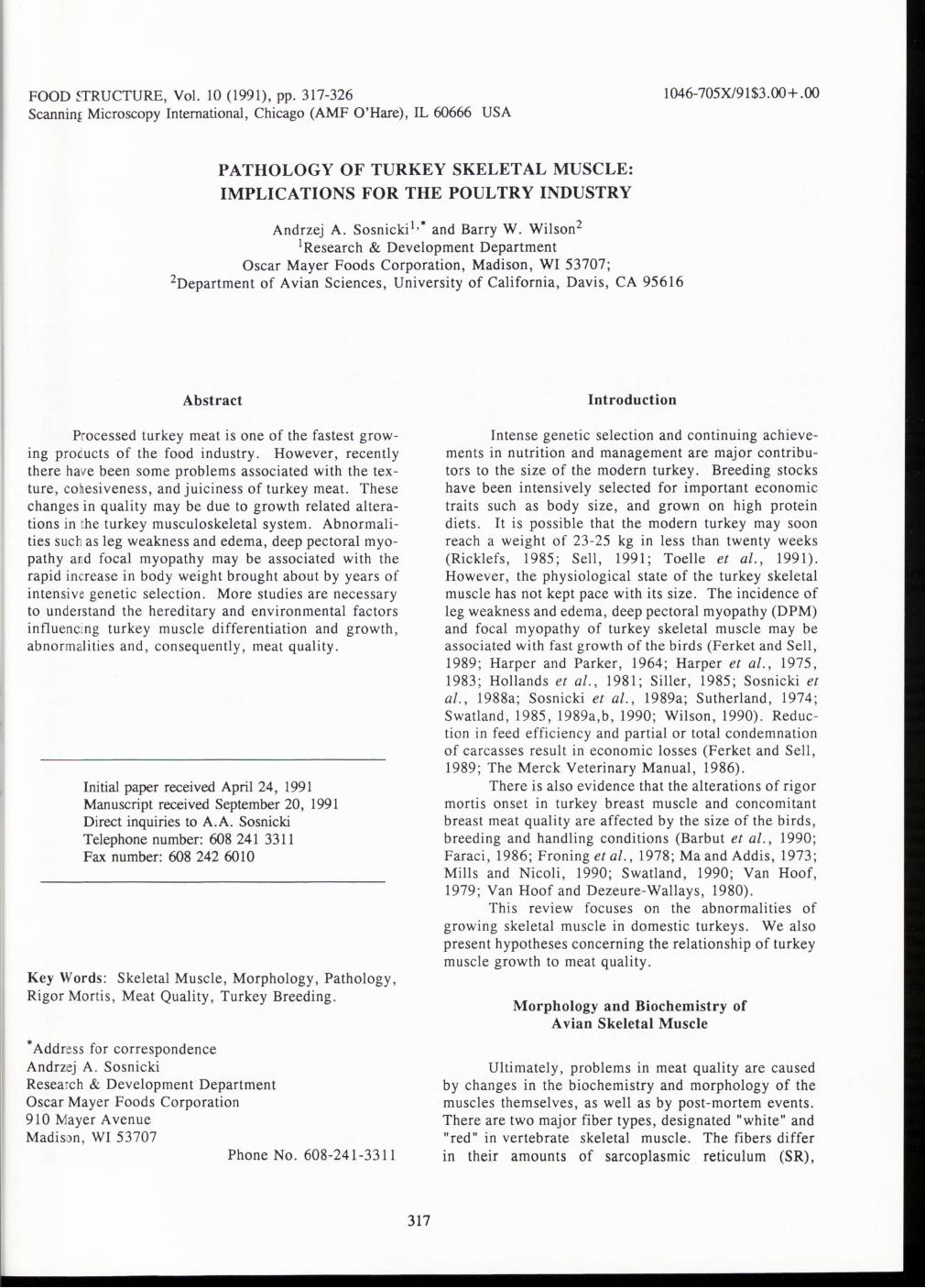
Figure 1. Relationship between plasma creatine kinase activity and body weight of four turkey lines from 12 to 16 weeks of age. Lines 28, 55, and 09 were selected for rapid growth. Line 91 is an unselected primitive line. Values are mean activities expressed as J.'mollmin/ml plasma, Y = 0.932X- 3.54; r = 0.97 (significant at P < 0.01); (adapted from data in Wilson et al., 1990).
the dissected surfaces. He also noted that, "the fragment~tion of turkey rolls may have some relationship to the size of turkeys," and that the outgrowth of breast muscle fibers over supportive connective tissue may also predispose products to fragmentation and poor cohesion. . A focal myopathy may be associated with changes In cohesiveness and juiciness of processed turkey meat. Several degenerative characteristics were detected microscopically in skeletal muscles of marketable turkeys even though the birds had no mobility or postural problems (Sosnicki et al., 1988b, 1989a,b, 1991a,b; Wilson, ~990; Wilson et al., 1990). The degenerative changes mcluded scattered and focal necrosis, hypercontraction of muscle fibers , strong proliferation of connective and fat tissues in the endomysium and perimysium, and infiltration of the necrotic areas by mononuclear cells.
Another significant poor meat quality characteristic--toughening of the turkey pectoralis major muscle--was associated with the presence of giant cells (hyper-contracted), fiber defects, and generally with a larger muscle cell size (Grey et al., 1986; Grey, 1989; Seemann et al., 1986). However, experiments examining a direct relationship between muscle damage and alterations in meat quality have yet to be performed.
Unrelated to DPM, the incidence of muscle degeneration has been associated with rapid growth of turkeys (Wilson et al, 1990; Wilson, 1990). Body weight, muscle histopathology and serum creatine kinase (CK) were studied for three lines of turkeys selected for rapid growth and one slow growing line (Wilson, 1990).
The incidence of damaged muscle; i.e., the presence of large, opaque-rounded, and degenerated fibers and the activity of serum CK correlated with age and growth rate in birds studied from hatching to 20 weeks. The older the birds, and the faster their growth, the greater the muscles damage, and the higher the serum enzyme levels (Figure 1, based on Wilson et al., 1990).
Turkey muscle pathology and meat quality
TABLE 1. Capillary and muscle fiber morphometry of normal and myopathic Biceps femoris (Iliofibularis) and Pectoralis major turkey muscles 1 • 2 • 3
Measurement NORMAL GROUP MYOPATHIC GROUP
Biceps femoris Pectoralis Biceps femoris Pectoralis (n = 23) (n = 23) (n = 7) (n = 7)
Capillaries/mm2 (density) 1,595 ±79a 839+19b 1,054+199b 457±88c
Capillaries surrounding a single fiber 8.51±.2a 6.91±.1b 7.06± .3b 6.01 ± .2c
Fiber area/mm2 .0051 ± .0001 a .0051 ± .0002a . 0052 ± . 0003a .0045 ± .0002b
Capillary: fiber ratio 4.27±.9a 3.43±.050b 3.53±.14b 3.01 ± .09c
Intercapillary distance, mm .03 + .0001 c 05±.003b .04+ .0004b .09± .006a
1 For visualization of capillaries , alkaline phosphatase activity was used. An analysis of the capillary-muscle fiber relationship was made without regard to fiber types. The intercapillary distances were calculating using "Krogh's cylinder model" , radius rk = [1/""'"(1r x capillary density)] is half of the mean intercapillary distance. 2 Results expressed as least square means + SE. 3 Adapted from data in Sosnicki et al., ( 1991 b).
a-c Means within a row with the same superscript are not significantly different (P < 0.05).
Wilson and co-workers suggested that selection for rapid growth in turkeys created muscles that "outgrow their life-support systems," and "bring about muscle damage when coupled with the conditions used to grow turkeys" (Wilson , 1990).
A study by Swatland (1990) of the endomysia! septa in turkey pectoralis muscle, 1 to 15 weeks after hatching , supported the hypothesis that the 35-fold increase in cross-sectional area of the muscle fibers in the first 15 weeks was relatively greater than the growth of endomysia! and perimysial connective tissue, when considered on a two-dimensional basis . However , the relative thickness of connective tissue to muscle fiber diameter in a strain of turkeys not prone to the muscle abnormality or to breast meat quality defects , was not studied.
Sosnicki et al. (1988a,b , 1989a,b, 1991 a,b) studied the pathology of muscles in rapidly growing turkeys of marketable ages . Typical degenerative changes observed in the pectoralis major and iliofibularis (biceps femoris) muscle included granular necrosis with phagocytosis by mononuclear cells of several adjacent muscle fibers or multi-fiber areas , hypercontraction of muscle fibers, infiltration of the endomysia! connective tissue with mononuclear cells , and fatty tissue replacement in the necrotic areas (see Fig. 2, 3 and 4). Necrotic areas usually presented a uniform activity of Ca +2-ATPase, diffuse activity of succinic dehydrogenase (SDH) in muscle fibers and alkaline phosphatase (ALP) in capillaries , and a positive reaction for acid phosphatase in muscle fibers undergoing necrosis or hypercontraction. Electron microscopy showed dilation of the SR, intense
319
Z-line streaming, and the presence of several myeloid and lysosomal dark-bodies in the necrotic areas (see Figure 5 , for example).
Proximate Cause of Focal Myopathy
Our working hypothesis is that the ultimate cause of focal myopathy of turkey muscles is a polygenic, multifactorial imbalance of the growing muscle tissue. One possible proximate cause is muscle microischemia. Alterations in histochemical activity of SDH, myosin Ca +2-ATPase, and ALP , and the ultrastructural pattern of extensive multifocal Z-line streaming observed in turkey muscle are similar to the "relative ischemia" symptoms described in human and laboratory animals (Sosnicki et al., 1991a,b). Ischemic myopathies have been brought about by experimental blockage of the major arteries in rats (Carpenter and Karpati, 1984). The fact that the necrotic regions are scattered in turkey muscle with focal myopathy suggests that such a vascular defect might involve a localized microischemia at the terminal capillary bed level (Sosnicki, 1991a, b).
Evidence for a microvascular defect in turkey skeletal muscle was obtained from a morphometric study of normal and myopathic turkey muscle regions (Sosnicki et al. , 1991 b). Lower values of capillary density and capillary to fiber ratio, and greater intercapillary distances (which is the most important limiting parameter for aerobic capacity of the skeletal muscle) were found in the necrotic regions of the pectoralis major and biceps femoris muscle (Table 1). Whether the capillary morphometry of muscles from slow-growing
A.A. Sosnicki and B.W. Wilson
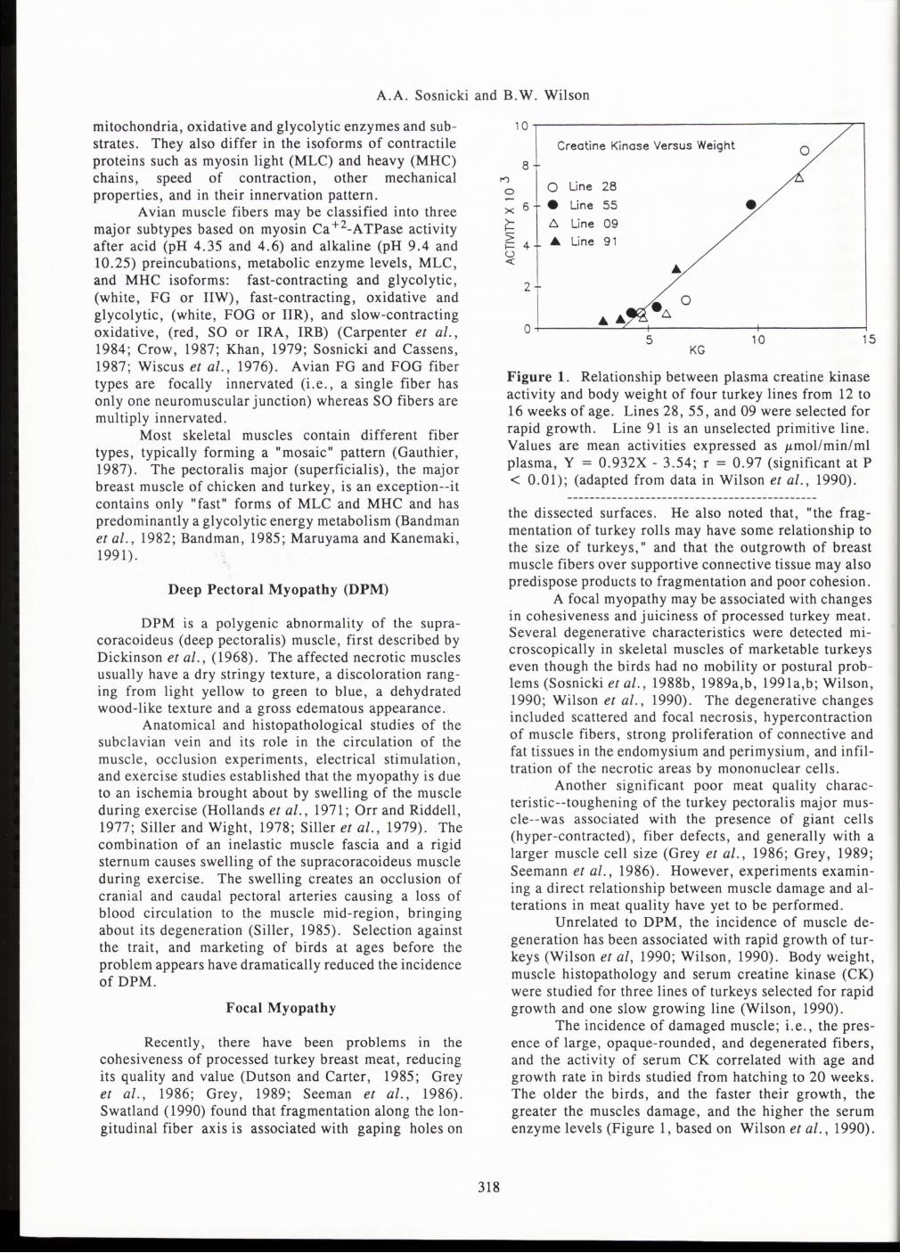
Figure 2. Cross-section showing necrosis with phagocytosis (arrows) of adjacent muscle fibers in turkey pectoralis major muscle. Frozen section stained with modified trichrome. Bar = 50 J.tm.
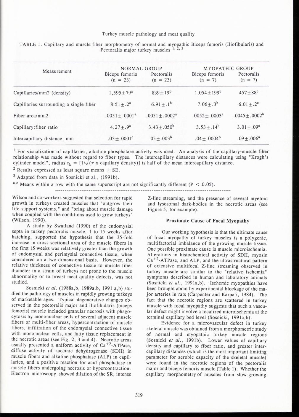
Figure 3. Focal necrosis of muscle tissue: several hypercontracted muscle fibers (small arrows) and infiltration by mononuclear cells (large arrow) are present in turkey pectoralis major muscle. Apparently "empty" (light) areas represent connective tissue replacing spaces resulting from fiber hypercontraction or necrosis. Frozen section stained with hematoxylin and eosin. Bar = 50 J.tm.
turkeys will resemble that of normal regions of the muscles from fast-growing birds is yet to be determined.
Capture Myopathy
The phenomenon of "capture myopathy" may provide a clue to the chain of events linking muscle damage, a rapid rigor mortis and changes in meat quality . Capture myopathy is a syndrome associated with the trapping, handling, and transporting of wild mammalian and avian species. As a result, a high proportion of the animals may be paralyzed and have muscle damage (Spraker et al., 1987). In avian species, this phenomenon has been described in flamingos (Young, 1967), sandhill crane (Wingdingstad et al., 1983), Canada geese
320
(Chalmers and Barrett, 1982), and wild turkeys (Spraker et al., 1987).
The lesion in wild turkey muscle is characterized by multi-focal areas of muscle fibers containing basophilic sarcoplasm, rhabdomyolysis with subsequent phagocytosis by macrophages or loss of striation with marked disruption and fragmentation of myofibrils (Spraker et al., 1987). These authors did not report frequent clinical signs of capture myopathy in wild turkeys, but histopathological subclinical lesions were present in the pectoralis, wing, and thigh muscles. Nevertheless, the findings indicate that wild turkeys are sensitive to stressful conditions, and care should be taken during trapping, handling, and transporting the birds. It is,
Turkey muscle pathology and meat quality
therefore , possible that the domestic turkey may also be predisposed to stress (i.e . , preslaughter handling), prone to muscle damage, alterations in rigor mortis and , consequently , in meat quality (Froning et al ., 1978 ; Mills and Nicoli , 1990; Van Hoof, 1979).
Leg Edema Syndrome
A phenomenon possibly related to capture myopathy is leg edema syndrome, which is characterized by acute necrosis of muscle fibers , shrunken and pyknotic nuclei, infiltration of the walls of blood vessels by mononuclear cells , fiber hypercontraction or proliferation of endomysia! and perimysial connective tissue (Sosnicki et al. , 1988a) . The edematous subcutis which
321
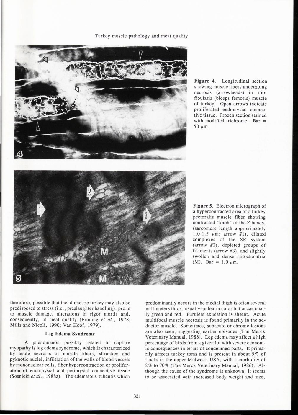
Figure 4. Longitudinal section showing muscle fibers undergoing necrosis (arrowheads) in iliofibularis (biceps femoris) muscle of turkey. Open arrows indicate proliferated endomysia! connective tissue. Frozen section stained with modified trichrome. Bar =
50 J.Lm.
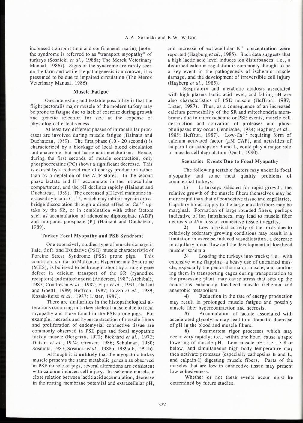
Figure 5. Electron micrograph of a hypercontracted area of a turkey pectoralis muscle fiber showing contracted "knob" of the Z bands , (sarcomere length approximately 1.0-1.5 J.Lm ; arrow #1) , dilated complexes of the SR system (arrow #2) , depleted groups of filaments (arrow #3) , and slightly swollen and dense mitochondria (M). Bar = 1.0 J.Lm .
predominantly occurs in the medial thigh is often several millimeters thick , usually amber in color but occasionally green and red. Purulent exudation is absent. Acute multifocal muscle necrosis is found primarily in the adductor muscle. Sometimes , subacute or chronic lesions are also seen , suggesting earlier episodes (The Merck Veterinary Manual, 1986). Leg edema may affect a high percentage of birds from a given lot with severe economic consequences in terms of condemned parts. It primarily affects turkey toms and is present in about 5% of flocks in the upper Midwest , USA , with a morbidity of 2% to 70% (The Merck Veterinary Manual, 1986). Although the cause of the syndrome is unknown, it seems to be associated with increased body weight and size,
A.A. Sosnicki and B. W. Wilson
increased transport time and confinement rearing [note: the syndrome is referred to as "transport myopathy" of turkeys (Sosnicki et al., 1988a; The Merck Veterinary Manual, 1986)]. Signs of the syndrome are rarely seen on the farm and while the pathogenesis is unknown, it is presumed to be due to impaired circulation (The Merck Veterinary Manual, 1986) .
Muscle Fatigue
One interesting and testable possibility is that the flight pectoralis major muscle of the modern turkey may be prone to fatigue due to lack of exercise during growth and genetic selection for size at the expense of physiological effectiveness.
At least two different phases of intracellular processes are involved during muscle fatigue (Hainaut and Duchateau, 1989). The first phase (10 - 20 seconds) is characterized by a blockage of local blood circulation and anaerobic, but not lactic acid metabolism. Hence, during the first seconds of muscle contraction , only phosphocreatine (PC) shows a significant decrease . This is caused by a reduced rate of energy production rather than by a depletion of the ATP stores. In the second phase lactate and H + accumulate in the intracellular compartment , and the pH declines rapidly (Hainaut and Duchateau, 1989). The decreased pH level maintains increased cytosolic Ca +2 , which may inhibit myosin crossbridge dissociation through a direct effect on Ca +2 uptake by the SR , or in combination with other factors such as accumulation of adenosine diphosphate (ADP) and inorganic phosphate (Pj) (Hainaut and Duchateau , 1989) .
Turkey Focal Myopathy and PSE Syndrome
One extensively studied type of muscle damage is Pale , Soft , and Exudative (PSE) muscle characteristic of Porcine Stress Syndrome (PSS) prone pigs . This condition , similar to Malignant Hyperthermia Syndrome (MHS), is believed to be brought about by a single gene defect in calcium transport of the SR (ryanodine receptors) and mitochondria (Andersen , 1987; Archibals , 1987; Condrescu et al. , 1987; Fujii et al. , 1991 ; Gallant and Goettl , 1989; Heffron , 1987; Iaizzo et al., 1989; Kozak-Reiss et al ., 1987; Lister, 1987).
There are similarities in the histopathological alterations occurring in turkey skeletal muscle due to focal myopathy and those found in the PSE-prone pigs. For example, necrosis and hypercontraction of muscle fibers and proliferation of endomysia! connective tissue are commonly observed in PSE pigs and focal myopathic turkey muscle (Bergman, 1972; Bickhard et al., 1972; Dutson et al., 1974; Greaser, 1986; Schulman, 1980; Sosnicki, 1987; Sosnicki et al., 1988b, 1989a,b, 1991b).
Although it is unlikely that the myopathic turkey muscle presents the same metabolic genesis as observed in PSE muscle of pigs, several alterations are consistent with calcium induced cell injury. In ischemic muscle, a close relation between lactic acid accumulation, decrease in the resting membrane potential and extracellular pH,
322
and increase of extracellular K + concentration were reported (Hagberg et al., 1985). Such data suggests that a high lactic acid level induces ion disturbances; i.e., a disturbed calcium regulation is commonly thought to be a key event in the pathogenesis of ischemic muscle damage, and the development of irreversible cell inj ury (Hagberg et al. , 1985).
Respiratory and metabolic acidosis associated with high plasma lactic acid level, and falling pH are also characteristics of PSE muscle (Heffron, 1987; Lister , 1987). Thus , as a consequence of an increased calcium permeability of the SR and mitochondria membranes due to microischemic or PSE events , muscle cell destruction and activation of proteases and phospholipases may occur (Jennische, 1984; Hagberg et al., 1985 ; Heffron , 1987). Low-ca+ 2 requiring form of calcium activated factor (I-'M CAF), and activities of calpain I or cathepsins B and L , could play a major role in muscle cell degradation (Quali , 1990).
Scenario: Events Due to Focal Myopathy
The following testable factors may underlie focal myopathy and some meat quality problems of commercial turkeys :
1) In turkeys selected for rapid growth , the relative growth of the muscle fibers themselves may be more rapid than that of connective tissue and capillaries. Capillary blood supply to the large muscle fibers may be marginal. Formation of large rounded fibers , perhaps indicative of ion imbalances , may lead to muscle fiber necrosis and/or loss of connective tissue integrity.
2) Low physical activity of the birds due to relatively sedentary growing conditions may result in a limitation in exercise-induced vasodilatation, a decrease in capillary blood flow and the development of localized muscle i schemia.
3) Loading the turkeys into trucks; i.e. , with extensive wing flapping--a heavy use of untrained muscle , especially the pectoralis major muscle , and confining them in transporting cages during transportation to the processing plant, may cause stress that sets up the conditions enhancing localized muscle ischemia and anaerobic metabolism.
4) Reduction in the rate of energy production may result in prolonged muscle fatigue and possibly muscle fiber hypercontraction and necrosis.
5) Accumulation of lactate associated with accelerated glycolysis may lead to a dramatic decrease of pH in the blood and muscle fibers.
6) Postmortem rigor processes which may occur very rapidly; i.e., within one hour , cause a rapid lowering of muscle pH. Low muscle pH; i.e. , 5 .8 or below, and simultaneous high body temperature may then activate proteases (especially cathepsins B and L , and calpain-I) digesting muscle fibers. Parts of the muscles that are low in connective tissue may present low cohesiveness.
Whether or not these events occur must be determined by future studies.
Turkey muscle pathology and meat quality
Conclusions
Siller (1985) , described DPM as a "penalty of successful selection , " a "man-made" disease. It remains to be seen whether focal myopathy or leg edema syndromes may also be results of genetic selection. If this is true one solution to the muscle abnormalities and meat q~ality problems would be to breed turkeys with different muscle properties and better circulation.
The traditional approach of breeding and feeding turkeys to maximize their growth performance may no longer be the best breeding program . A more complex selection strategy predicated on maintaining the physiological state of the muscles may be required. Future research programs should be focused on the complex molecular, neurohormonal, biochemical and morphological alterations occurring during growth of turkey muscle as well as on post mortem metabolism and meat quality . Such comprehensive studies will be important in understanding the hereditary and environmental factors influencing turkey muscle growth and differentiation, abnormalities , and consequently meat quality.
Acknowledgments
We thank Dr. Andrew Milkowski for critically reviewing the manuscript.
References
Andersen E (1987). Selection against PSS by means of blood typing . In: Evaluation and control of meat quality in pigs , Tarrant PV , Eikelenboom G, Monin G (eds.) , Martinus Nijhoff Publishers, pp. 317-327.
Archibals AL (1987). A molecular approach to the porcine stress syndrome. In: Evaluation and control of meat quality in pigs, Tarrant PV , Eikelenboom G, Monin G (eds.) , Martinus Nijhoff Publishers , pp. 343-357.
Bandman E (1985). Myosin isoenzyme transitions in muscle development , maturation and disease . Int. Rev. Cytol. 97: 97-131.
Bandman E, Matsuda R, Strohman RC (1982). Developmental appearance of myosin heavy chain and light chain isoforms "in vivo" and "in vitro" in chicken skeletal muscle. Dev. Biol. 93: 508-514.
Barbut S, McEwen SA , Julian RJ (1990). Turkey downgrading: effect of truck cage location and unloading. Poultry Sci. 69: 1410-1413.
Bergman V (1972). Zur ultrastructur der kapillaren in der skelettmuskulatur des fleischschweines (Ultrastructure of capillaries in the skeletal muscle of pork pigs). Arch. Exp. Vet. Med. 3: 465-475.
Bickhard K, Chevalier H -J, Giese W, Reinhard HJ (1972). Akute Ruckenmuskelnekrose und Belastungsmyopathie beim Schwein (Acute necrosis of the longissimus dorsi muscles and stress myopathy in pigs). Forstchr. Veterinaermed. no. 18, Verlag Paul Parey, Berlin, 27-35.
Carpenter S, Karpati G (1984). Pathology of skeletal muscle. Churchill Livingstone , New York, NY.
323
pp. 592-596. Carpenter CE, Cassens RG, Greaser ML (198~).
The agreement of ATPase with immunology for typmg myofibers of chicken skeletal muscle. Proceedings. 30th European Meeting of Meat Research Workers, Bnstol, England, p. 120 (abstract, available from R. G. Cassens, Univ. Wisconsin, Madison, WI 53706, USA).
Chalmers GA, Barrett MW (1982). Capture myopathy. In: Noninfectious disease in wildlife, Hoff GL, Davis JW (eds.), Iowa State University Press, Ames, lA, pp. 84-94.
Condrescu M, Lopez JR, Medina P, Alamo L (1987). Deficient function of the sarcoplasmic reticul~m in patients susceptible to malignant hyperthermia. Muscle and Nerve 10: 238-241.
Crow MT (1987). The determinants of muscle fiber type during embryonic development. Amer. Zool. 27: 1043-1053.
Dickinson EM, Stevens JO, Helfer DH (1968). A degenerative myopathy in turkeys. In: Proceedings 17th Western Disease Conference, Cooperative Extension , Univ. of California, Davis, p. 7 (abstract).
Dutson RD, Carter A (1985) . Microstructure and biochemistry of avian muscle and its relevance to meat processing industries. Poultry Sci. 64: 1577-1590.
Dutson TR, Pearson AM, Merkel RA (1974). Ultrastructural post-mortem changes in normal and low quality porcine muscle fibers . J. Food Sc~. 39: 32-~7:
Faraci FM (1986). Circulation dunng hypoxia In birds. J. Comp. Biochem. Physiol. 85: 613-620.
Ferket PR, Sell JL (1989). Effect of severity of early protein restriction on large turkey toms. 1. Performance characteristics and leg weakness. Poultry Sci. 68: 676-686.
Froning GW, Babji AS, Mather FB (1978) . The effect of preslaughter temperature, stress , struggle and anesthetization on color and textural characteristics of turkey muscle. Poultry Sci. 57: 630-633.
Fujii J, Otsu K, Zorzato F, De Leon S, Khanna VK, Weiler JE, O'Brien PJ, MacLennan DH (1991). Identification of a mutation in porcine ryanodine receptor associated with Malignant Hyperthermia. Science 53: 448-451.
Gallant EM, Goettl VM (1989). Porcine malignant hyperthermia: halothane effects on force generation in skeletal muscles. Muscle and Nerve 12: 56-63.
Gauthier GF (1987). Vertebrate muscle fiber types and neuronal regulation of myosin expression. Amer. Zool. 27: 1033-1042.
Greaser ML (1986). Conversion of muscle to meat. In: Muscle as Food, Academic Press, pp. 37-102.
Grey TC (1989). Turkey meat texture. In: Recent Advances in Turkey Science. Nixey C, Grey TC (eds.), Butterworths, England, pp. 289-311.
Grey TC, Griffiths NM, Jones JM, Robinson D (1986) . A study of some factors influencing the tenderness of turkey breast meat. Lebensm. Wiss. Techno!. 19: 412-414.
Hagberg H, Jennische E, Haljamae H (1985).
A.A. Sosnicki and B. W. Wilson
Influence of tissue lactic acid and A TP levels on postischemic recovery in rabbit skeletal muscle. Circulatory Shock 16: 363-374.
Hainaut K, Duchateau J (1989). Muscle fatigue, effect of training and disuse. Muscle and Nerve, 12: 660-669.
Harper JA, Parker JE (1964). Hereditary muscular dystrophy in the domestic turkey, Meleagris gallopavo. Poultry Sci., 43: 1326-1327.
Harper JA, Bernier PE, Helper DR, Schmitz JA (1975). Degenerative myopathy of the deep pectoral muscle of the turkey. J. Hered. 66: 352-366.
Harper JA, Bernier PE, Thompson-Cowley LL ( 1983). Early expression of hereditary deep pectoral myopathy in turkeys due to forced wing exercise. Poultry Sci. 62: 2303-2308.
Heffron JJA (1987). Calcium releasing systems in mitochondria and sarcoplasmic reticulum with respect to the aetiology of Malignant Hyperthermia: a review. In: Evaluation and control of meat quality in pigs. Tarrant PV, Eikelenboom G, Monin G (eds.), Martinus Nijhoff Publishers, pp. 17-26.
Hollands KG, Grunder AA, Gavora J, Williams CJ (1971). Creatine phosphokinase as an assay for green muscle disease in turkey. Poultry Sci. 57: 1145-1150.
Hollands KG, Grunder AA, Williams CJ, Gavora JS, Chambers JR, Cave NAG (1981). Degenerative myopathy of meat type poultry: its effect on production traits in chickens and its identification in live turkeys. In: Quality of Poultry Meat, Mulder RWA W, Scheele CW, Veerkamp CH (eds.), Spelderholt Inst. Poultry Res., Beekbergen, Netherlands, pp. 337-344.
Iaizzo PA, Lehmann-Horn F, Taylor SR, Gallant EM (1989). Malignant hyperthermia: effects of halothane on the surface membrane. Muscle and Nerve, 12: 178-183.
Jennische E (1984). Post-ischemic calcification in skeletal muscle . Acta. Path. Microbial. Immunol. Scand. Sec. A. 92: 139-145.
Khan MA (1979). Histochemical and ultrastructural characteristics of a new muscle fiber type in avian striated muscle. Histochem. J. 11: 321-335.
Kozak-Reiss G, Desmoulin F, Canioni P, Cozzone P, Gascard JP, Monin G, Pusel JM, Renou JP, Talmant A (1987). Contraction and metabolism traits in skeletal muscle biopsies from halothane positive pigs as studied by mechanical measurements and 31P NMR. In: Evaluation and control of meat quality in pigs, Tarrant PV, Eikelenboom G, Monin G (eds.), Martinus Nijhoff Publishers, pp. 27-38.
Lister D (1987). The physiology and biochemistry of the Porcine Stress Syndrome. In: Evaluation and control of meat quality in pigs. Tarrant PV, Eikelenboom G, Monin G (eds.), Martinus Nijhoff Publishers. pp. 3-16.
Ma RT-I, Addis PB (1973). The association of struggle during exsanguination to glycolysis, protein solubility and shear in turkey pectoralis muscle. J. Food Sci. 38: 995-997.
324
Maruyama K, Kanemaki N (1991). M.rosin isoforms expression in skeletal muscles of t rkeys at various ages. Poultry Sci. 70: 1748-1757.
Mills DS, Nicoli CJ (1990). Tonic immobility in spent hens after catching and transport. Ve. Record 126: 210-212.
Orr JP, Riddell JR (1977). Investigation of the vascular supply of the pectoralis muscle of the domestic turkey and comparison of experimentally produced infarcts with naturally occurring deep pectoral myopathy. Am. J. Vet. Res. 38: 1237-1242.
Quali A (1990). Meat tenderization : possible causes and mechanisms. A review. J. Muscle Foods 1: 129-165.
Ricklefs ER ( 1985). Modification of growth and development of muscles of poultry. Poultry Sci. 64: 1563-1576.
Schulman A (1980). Exertional myopathy in Finnish Landrace pigs. A survey of the situation and evaluation of different control methods. J. Sci. Agr. Soc. Fin. 52: 102-192.
Seemann G, Jones JM, Griffiths NM , Grey TC (1986). The influence of storage time-temperature on turkey breast meat quality. Arch. Geflugelkde. 50: 149-153.
Sell JL (1991). Continued improvements in turkey performance in 1990. Turkey World, 67(1), 12-16.
Siller WG (1985). Deep pectoral myopathy: A penalty of successful selection for muscle growth. Poultry Sci. 64: 1591-1595.
Siller WG, Wight PAL (1978). The pathology of deep pectoral myopathy of turkeys. Avian Pathol. 7: 583-617.
Siller WG, Wight PAL, Martindale L (1979). Exercise-induced deep pectoral myopathy in broiler fowls and turkeys. Vet. Sci. Commun. 2: 331-336.
Sosnicki AA (1987). Histopathological observation of stress myopathy in M. longissimus in the pig and relationship with meat quality, fattening, and slaughter traits. J. Anim. Sci. 65: 584-596.
Sosnicki AA, Cassens RG (1987). Determination of fiber types in chicken skeletal muscles based on reaction for actomyosin, Ca +2 ,Mg +2-dependent ATPase. Poultry Sci. 67: 973-978.
Sosnicki AA, Cassens RG, Mcintyre DR, Vimini RJ (1988a). Structural alterations in oedematous and apparently normal skeletal muscle of domestic turkey. Avian Pathology 17: 14 7-152.
Sosnicki AA, Cassens RG, Mcintyre DR, Vimini, RJ, Greaser ML (1988b). Characterization of hypercontracted fibers in skeletal muscle of domestic turkey (Meleagris gallopavo). Food Microstructure 7: 147-152.
Sosnicki AA, Cassens RG, Mcintyre DR, Vimini RJ, Greaser ML (1989a). Incidence of microscopically detectable degenerative characteristics in skeletal muscle of turkey. British Poultry Sci. 30: 69-80.
Sosnicki AA, Cassens RG, Vimini RJ, Greaser ML (1989b). Histopathology and morphometry of normal and ischemic muscle in domestic turkey. Poultry
Turkey muscle pathology and meat quality
Sci. 68, Suppl. 1, 139 (abstract). Sosnicki AA, Cassens RG, Vimini RJ, Greaser
ML (1991a). Histopathological and ultrastructural alterations of turkey skeletal muscle. Poultry Sci. 70: 343-348.
Sosnicki AA, Cassens RG, Vimini RJ, Greaser ML (1991b). Distribution of capillaries in normal and ischemic turkey skeletal muscle. Poultry Sci. 70: 349-357.
Spraker TR, Adrian WJ, Lance WR (1987). Capture myopathy in wild turkeys (Meleagris gallopavo) following trapping, handling, and transportation in Colorado. J. Wildlife Dis. 23: 447-453.
Sutherland IR (1974). Hereditary pectoral myopathy in the domestic turkey (Meleagris gallopavo). Canadian Vet. J. 15: 77-80.
Swatland HJ (1985). Growth-related changes in the intracellular distribution of succinate dehydrogenase activity in turkey muscle. Growth 49: 409-416.
Swatland HJ (1989a). Physiology of muscle growth. In: Recent Advances in Turkey Science. Nixey C, Grey TC (eds.), Butterworths, England, pp. 167-182.
Swatland HJ (1989b). Morphometry of pectoral development in turkey breeding stock. Br. Poultry Sci. 30: 785-795.
Swatland HJ (1990). A note on the growth of connective tissues binding turkey muscle fibers together. Can. Inst. Food Sci. Techno!. J. 23:239-241.
The Merck Veterinary Manual (1986) . A handbook of diagnosis, therapy, and disease prevention and control. Sixth Edition, Merck & Co., Inc. , Rahway, NJ, U.S.A., p. 1302.
Toelle VD, Havenstein GB, Nestor KE, Bacon WL (1991). Inheritance of and selected for carcass traits in turkeys. Zootecnica Int. 1: 37-43.
Van Hoof J (1979). Influence of ante- and peri -mortem factors on muscle biochemical and physical characteristics of turkey breast muscle. Vet. Quarterly, 1: 29-36.
Van Hoof J , Dezeure-Wallays B (1980). Breakdown of diphosphate in turkey breast muscle from different meat quality groups. Fleischwirtschaft, 60: 489-451.
Wilson BW (1990). Developmental and maturational aspects of inherited avian myopathies. Proc. Soc. Exp. Biol. Med. 194: 87-96.
Wilson BW, Nieberg PS, Buhr RJ, Shultz FT, Kelly BJS (1990). Turkey muscle growth and focal myopathy. Poultry Sci. 69: 1553-1562.
Wingdingstad RS, Hurley S, Sileo L (1983). Captive myopathy in a free-flying greater sandhill crane (Grus canadensis tabida) from Wisconsin. J. Wildlife Dis. 19: 289-290.
Wiscus KJ, Addis PB, MA RT-I (1976) . Distribution of BR, Alpha-R and Alpha-W fibers in turkey muscles. Poultry Sci. 55: 562-572.
YoungE (1967). Leg paralysis in the greater flamingo and lesser flamingo. International Zoo Yearbook, 7: 226-227.
325
Discussion with Reviewers
S.H. Cohen: What is normal pH level? Authors: Since the reviewer did not specify the time post-mortem; i.e., initial or ultimate pH, and the metabolic type of muscle (SO, FOG or FG), it is difficult to answer this question. For turkey breast muscle (pectoralis superficials, FG), and assuming that the chilling of carcasses starts at about 30 minutes post mortem, we can provide the following information:
1. The pH values measured at about 15-20 minutes post mortem (initial pH) may be as low as 5.60 and as high as 6. 80. On average, normal initial pH ranges between 6.3- 6.6.
2. Ultimate pH values (when completed) also vary considerably. experience, normal pH varies between measured 3-12 hours post mortem.
the rigor is From our
5. 6 - 6.1 if
S.H. Cohen: At what pH level are the lysosomal proteases released? Authors: Of the large number of muscle proteolytic systems described, two deserve highest consideration: 1) acidic lysosomal proteases: (cathepsins D , B, H and L); and 2) two forms of calcium-dependent neutral proteinases: calpain I (CDP-I), and calpain II (CDP-II). Although the mechanism regulating these enzyme activities in vivo and post mortem is still uncertain, the range of muscle pH between 6.4-5. 8 appears to correlate with activities of CDP-1 and cathepsin B and L.
S.H. Cohen: Do you know which proteases have a specificity towards myofibrillar proteins and which affect sarcoplasmic proteins? Authors: Several in vitro studies showed that lysosomal proteases (cathepsins B and L), and calpains (I and II) , both degrade troponin T, troponin I, tropomyosin, C-protein, desmin, titin, and nebulin. Myosin heavy chain, myosin light chains, a-actinin, troponin C and actin also appear to be sensitive to action of cathepsins B and L. Considering sarcomere ultrastructure, myofibrils are extensively fragmented by cathepsins B and L near N2-lines, and at the junction between A and I bands. Degradation of Z-lines and M-lines seem to be the main ultrastructural changes caused by calpains I and II. Cathepsins D and H appear to cause little structural alterations to myofibrils. A group of proteinases designated as "multicatalytic proteinase complex" active at neutral and mildly alkaline pH appear to hydrolyze sarcoplasmic proteins. However, their effect on myofibrillar proteins is not known.
S.H. Cohen: What role is played by CAF? Authors: The high-calcium requiring form of calcim activation factor ( mM -CAF) is maximally active at 1-5 mM free Ca +2 concentration, and at pH 7.0- 8.0. Because in normal skeletal muscle the intracellular free Ca +2 concentration is between 1-10 mM, it is unlikely that mM-CAF can be activated post mortem, or in vivo, even in abnormal muscle. However, it appears that lowcalcium-requiring CAF (J.LM-CAF) can induce post mortem degradation of desmin, troponin T, A-actinin and Z-

A.A. Sosnicki and B. W. Wilson
bands. Interestingly, ,uM-CAF retains activity even at muscle pH of 5.5- 5.8 (optimum activity is at pH 7.5). Thus, if damage to the SR system in turkey muscles occurs in vivo, then the ,uM-CAF may be activated and induce muscle degeneration. Furthermore, a gradual increase of free Ca +2 concentration early post mortem would enhance proteolytic processes.
S.H. Cohen: You discuss the rapid onset of rigor mortis; what time frame do you mean? Authors: Based on our recent studies on ATP and pH depletion in the pectoralis muscle of adult turkey toms, rigor is completed within 3-6 hours post mortem; i.e., ATP level is below 1 ,uM/g of muscle tissue. At the same time interval, the pH of the muscle reaches between 5. 6 - 6. 1. However, in some cases, the rigor can be completed within 30 minutes - 1 hour post mortem. At most turkey processing plants, chilling of carcasses starts about 30 minutes post mortem. Hence, a high rate of glycolysis and a rapid rigor mortis development at prevalent high muscle temperature ( > 30 °C), may cause formation of PSE-like breast meat.
S.H. Cohen: Do you think sarcomere measurement would be of some value? Authors: In our opinion, measurement of sarcomere length in prerigor muscle has little practical application because it is extremely difficult to excise prerigor muscle without causing sarcomere contraction. An acceptable method would be to incise a muscle strip longitudinally, suture it to a wood applicator stick at its in situ length, and cut the muscle strip external to the sutures. However, even with this precaution, it is still very difficult to dissect small muscle pieces (without causing muscle contraction) for sarcomere length measurement by the laser diffraction method. Fixation in any fixative (for muscle histology or electron microscopy) would cause sarcomere shrinkage and further reduction in their length. We think that a muscle strip obtained as described above may be frozen in liquid nitrogen and then sectioned longitudinally in a microtome-cryostat. One would have to avoid compression of the sarcomeres by the microtome knife; i.e., a parallel orientation of muscle fibers to the knife would be required. Finally, 0.02 M EGT A would have to be smeared on the surface of microscopic slides to avoid further contraction of the sarcomeres. However, calculation of sarcomere length from frozen sections is laborious and time consuming.
A. Suzuki: In Figure 3, unstained portions are seen. Do these portions show disappearance of myofibers? Authors: As stated in Fig. 3 caption, the "empty" spaces seen in this figure represent perimysial and endomysia! connective tissue stained lightly yellow with the H&E staining method. These areas illustrate proliferating connective tissue into the "empty" spaces developing probably as a result of fiber hypercontraction (see Fig. 4 showing connective tissue accumulated at the level of intrafascicular termination of hypercontracted and necrotic fibers).
R. Wroblewski: Please provide more information about
326
the methods of estimating capillary density; what type of staining was employed to visualize capillaries! Authors: Our original paper concerning capillary distribution in normal and ischemic turkey muscle contains details of the method used (Sosnicki et al., 1991 b). In addition to the information included with Table 1 brief-
' ly, for visualization of capillaries, alkaline phosphatase (ALP) activity was demonstrated using a modified Gomori method by incubating tissue sections at room temperature for 2.5 hours. Unfortunately, a combination of the ALP and dipeptidil peptidase IV method was not very successful, and the lectin method was not available at the time of the study (J. Histochem. Cytochem. 37(8): 1303-1304, 1989).
An analysis of capillary-muscle fiber relationships was made without regard to fiber types. Again, histochemical method for simultaneous fiber typing and demonstration of capillaries (Histochemistry, 93:3 85-3 87, 1990) was not available at the time of the experiments. Capillaries were identified directly from the sections by viewing at a magnification of 400x. The fields were selected at random except for the hypercontracted fibers and areas where the activity of the ALP was too low to establish an unequivocal identification of capillaries; i.e., in the necrotic areas. All measurements were done with a ZIDAS image analysis system. The capillary density was estimated by counting capillary cross-sections on muscle cross-section (capillaries per square millimeter). For each fiber, the fiber area was determined in square millimeter's and the number of capillaries surrounding each single fiber was counted. The mean fiber area and capillaries surrounding each fiber ~e~e .calculated from the measurements of 100 fibers per md1v1dual muscle per bird. The mean capillary to fiber ~atio was equal to the number of capillaries per fiber; 1. e., the ratio of the number of capillaries per area divided by the number of fibers in a given area. The intercapillary distances were calculated using Krogh's cylinder model (Krogh, J. Physiol (London). 52:409-415, 1929).
R. Wroblewski: Which preincubation, using the A TPase reaction, is used in avian muscles for fiber type estimation? Authors: The classical avian muscle histochemistry paper by Khan (1979) provides detailed information about A TPase preincubation pH's and buffers. Briefly, two acid (pH 4.35 and 4.6) and two alkaline (pH 9.40 and 10.25) are required to identify fiber subtypes. As stated in the text, our method for determination of fiber types in chicken slow, fast and intermediate types of skeletal muscle, based on the reaction for actomyosin, Ca + 2, Mg + 2-dependent A TPase also involved two acid and two alkaline preincubations (Sosnicki and Cassens . ' Poultry Sc1. 67: 973-978, 1987). In addition, several combinations of ATPase histochemistry and immunochemical methods (antibodies specific for fast and slow MLC's and MHC's) have been successfully employed (Bandman, 1985; 1991; Carpenter et al., 1984; Crow, 1987; Guthier, 1987; Maruyama and Kanemaki, 1991).