Embed Size (px)
Citation preview
Pathology of Testicular and Penile Neoplasms
Maurizio ColecchiaEditor
123

Pathology of Testicular and Penile Neoplasms

Maurizio Colecchia Editor
Pathology of Testicular and Penile Neoplasms
ISBN 978-3-319-27615-1 ISBN 978-3-319-27617-5 (eBook) DOI 10.1007/978-3-319-27617-5
Library of Congress Control Number: 2016936098
© Springer International Publishing Switzerland 2016 This work is subject to copyright. All rights are reserved by the Publisher, whether the whole or part of the material is concerned, specifi cally the rights of translation, reprinting, reuse of illustrations, recitation, broadcasting, reproduction on microfi lms or in any other physical way, and transmission or information storage and retrieval, electronic adaptation, computer software, or by similar or dissimilar methodology now known or hereafter developed. The use of general descriptive names, registered names, trademarks, service marks, etc. in this publication does not imply, even in the absence of a specifi c statement, that such names are exempt from the relevant protective laws and regulations and therefore free for general use. The publisher, the authors and the editors are safe to assume that the advice and information in this book are believed to be true and accurate at the date of publication. Neither the publisher nor the authors or the editors give a warranty, express or implied, with respect to the material contained herein or for any errors or omissions that may have been made.
Printed on acid-free paper
This Springer imprint is published by Springer NatureThe registered company is Springer International Publishing AG Switzerland
Editor Maurizio Colecchia Department of Pathology Fondazione IRCCS Istituto dei Tumor Milan , Italy
To Mariangela and Matteo for their patient, silent, and loving support and to my parents

vii
As editor of Pathology of Testicular and Penile Neoplasms, I would like to briefl y explain the inspiration for this book, which came from a sentence I read in the Treasure Gallery of the British Library: “Science depends on the accurate observation, collection and recording of information.” This sentence (attributed to Galileo, the father of scientifi c observation) is the leitmotif in this book on penile and testicular neoplasms – rare tumors which are, however, increasing in frequency and whose epidemiological implications will be illustrated here in light of the most recent knowledge in the fi eld. Congenital testicular lesions, impaired spermatogenesis, cryptorchidism, Klinefelter syndrome, testicular dysgenesis, entities contributing to infertility and their correlation with an increased risk of cancer, and childhood and prepubertal germ cell tumors will be discussed.
The book will also describe the precursor lesions of invasive cancer as well as the staging criteria that provide the clinician with information for the correct treatment of metastatic disease. Furthermore, with the aid of summary tables, it will illustrate the main differential diagnoses between morphological entities. One chapter will be dedicated to the use of intraoperative frozen section analysis, which is increasingly being requested for the diagnosis of ultrasound-detected small testicular lesions.
As a practical handbook for pathologists, this volume will be an aid to the diagnosis of these uncommon tumors and to dealing with the diagnostic pitfalls they often present. The text will provide updates on the new editions of the classifi cation systems for testicular and penile tumors, with a view to achieving the necessary clarity in the terminology used by the pathologist in communicating diagnostic information. It aims to familiarize also less experienced pathologists with the sampling methods for orchiectomy and penectomy specimens and with the appropriate recording of pathology fi ndings according to the guidelines approved by the International Society of Urological Pathology (ISUP). The book is designed to provide practitioners with the tools and knowledge to formulate a complete histopathology report with all the necessary information for the clinician. Its contents refl ect the experience of the authors of the various chapters (11 on testicular cancer, 3 on cancer of the penis) in light of the recent literature, documented by more than 250 photographs; we have scrutinized the currently available information in an attempt to provide valuable updates on the variants of penile carcinoma, HPV-related and unrelated penile tumors, and newly classifi ed testicular neoplasms. The images have not been published before, and I am deeply
Pref ace

viii
grateful to the authors and coauthors of Pathology of Testicular and Penile Neoplasms , who have contributed their personal cases, giving their time and energy to the drafting of the chapters of this book, which I hope you will enjoy reading.
Milan, Italy Maurizio Colecchia
Preface
ix
Many people have helped me get this book into shape. The contributions of Alessia Bertolotti, PhD, in all stages of its development and in the technical preparation of the slides (recutting, immunohistochemical staining, scanning of interesting cases) were irreplaceable. Biagio Paolini, MD, has improved the photomicrographs through refi ned color enhancement. His efforts in standardizing the appearance of the illustrations will surely be appreciated by our readers. I also gratefully acknowledge the work of Alessio Pellegrinelli, MD, who has shown surprising versatility in designing tables and drawing illustrations to make the macroscopic images of Chap. 13 easier to understand.
I thank Drs Bandieramonte, Torelli, and Catanzaro for having contributed with the macroscopic images of penile lesions from their clinical activity.
Special thanks go to Marije de Jager for meticulously correcting the manuscript. I am particularly grateful for her help in uniforming the style across chapters. Thanks go as well to Paolo Castioni and Maria Morelli, who have enthusiastically contributed to this book in various ways. I express my heartfelt appreciation to the library staff of Fondazione IRCCS Istituto Nazionale dei Tumori and in particular to Rossella Ballarini.
Finally, I gratefully remember the teachings of Professor Juan Rosai (hoping they shaped me) that directed me and many other pathologists to a methodical approach in diagnostic pathology.
Maurizio Colecchia
Acknowledgements

xi
Part I Testicular Tumors: Epidemiology
1 Epidemiology of Testicular Cancer . . . . . . . . . . . . . . . . . . . . . . . . . 3 Gemma Gatta and Annalisa Trama
Part II Tumors of the Testis and Paratesticular Structures
2 Congenital Lesions . . . . . . . . . . . . . . . . . . . . . . . . . . . . . . . . . . . . . 21 Manuel Nistal and Pilar González-Peramato
3 Entities Contributing to Infertility and Their Relationship to Oncogenic Risk . . . . . . . . . . . . . . . . . . . . . . . . . . . . . . . . . . . . . . 49 Manuel Nistal and Pilar González-Peramato
4 Germ Cell Tumors of Infancy and Childhood . . . . . . . . . . . . . . . 69 Manuel Nistal and Pilar González-Peramato
5 The Stem Cell Origin and Pathogenetic Routes of Testicular Germ Cell Tumors . . . . . . . . . . . . . . . . . . . . . . . . . . 91 Maurizio Colecchia , Alessia Bertolotti , Biagio Paolini , and Gregor Mikuz
6 Tumors of the Testis . . . . . . . . . . . . . . . . . . . . . . . . . . . . . . . . . . . . 97 Gregor Mikuz and Maurizio Colecchia
7 Prognostic and Predictive Factors in Pathology of the Testis . . . . . . . . . . . . . . . . . . . . . . . . . . . . . . . . . . . . . . . . . . 159 Nicola Nicolai
8 Handling of the Surgical Specimen and Pathology Reporting of Malignant Germ Cell and Sex Cord-Stromal Tumors of the Testis . . . . . . . . . . . . . . . . . . . . . . . 165 Marina Scarpelli , Roberta Mazzucchelli , Matteo Santoni , Valeria Sotte , Alessandro Conti , Antonio Lopez-Beltran , Liang Cheng , and Rodolfo Montironi
9 Cysts and Epithelial Proliferations of the Testicular Collecting System . . . . . . . . . . . . . . . . . . . . . . . 171 Antonio Lopez-Beltran , Rita Canas-Marques , Maria R. Raspollini , Rodolfo Montironi , Marina Scarpelli , and Liang Cheng
Contents
xii
10 Paratesticular Soft Tissue Neoplasms . . . . . . . . . . . . . . . . . . . . . 191 Alessandro Franchi , Raffaella Santi , and Gabriella Nesi
11 Frozen Section in Testicular Pathology . . . . . . . . . . . . . . . . . . . . 203 Maurizio Colecchia and Nicola Nicolai
Part III Pathology of Precancerous Lesions and Tumors of the Penis
12 Precancerous Lesions of the Penis . . . . . . . . . . . . . . . . . . . . . . . . 215 Maurizio Colecchia and Biagio Paolini
13 Tumors of the Penis and Scrotum . . . . . . . . . . . . . . . . . . . . . . . . 229 Maurizio Colecchia , Alessia Bertolotti , Biagio Paolini , and Roberto Salvioni
14 Handling of the Surgical Specimen and Pathology Reporting of Penile Neoplasms . . . . . . . . . . . . . . . . . . . . . . . . . . 275 Rodolfo Montironi , Roberta Mazzucchelli , Matteo Santoni , Valeria Sotte , Antonio Lopez- Beltran , Liang Cheng , and Marina Scarpelli
Index . . . . . . . . . . . . . . . . . . . . . . . . . . . . . . . . . . . . . . . . . . . . . . . . . . . . 281
Contents
xiii
Alessia Bertolotti Department of Pathology , Fondazione IRCCS Istituto Nazionale dei Tumori di Milano , Milan , Italy
Rita Canas-Marques Serviço de Anatomia Patológica , Instituto Português de Oncologia de Lisboa , Lisbon , Portugal
Liang Cheng Department of Pathology and Laboratory Medicine , Indiana University School of Medicine , Indianapolis , IN , USA
Maurizio Colecchia Uropathology Unit, Department of Pathology , Fondazione IRCCS Istituto Nazionale dei Tumori di Milano , Milan , Italy
Alessandro Conti Department of Odontostomatologic and Specialized Clinical Sciences, Section of Urology , Polytechnic University of the Marche Region, School of Medicine, United Hospitals , Ancona , Italy
Alessandro Franchi Division of Pathological Anatomy , University of Florence , Florence , Italy
Gemma Gatta Evaluative Epidemiology , Fondazione IRCCS Istituto Nazionale dei Tumori di Milano , Milan , Italy
Pilar González-Peramato Department of Pathology , Autonoma University of Madrid, Hospital Univ. La Paz. , Madrid , Spain
Antonio Lopez-Beltran Department of Pathology and Surgery , Faculty of Medicine, University of Cordoba , Cordoba , Spain
Roberta Mazzucchelli Pathological Anatomy , Polytechnic University of the Marche Region, School of Medicine, United Hospitals , Ancona , Italy
Gregor Mikuz Institute of Pathology , Medical University of Innsbruck , Innsbruck , Austria
Rodolfo Montironi Pathological Anatomy , Polytechnic University of the Marche Region, School of Medicine, United Hospitals , Ancona , Italy
Gabriella Nesi Division of Pathological Anatomy , University of Florence , Florence , Italy
Nicola Nicolai Testis Surgery Unit , Fondazione IRCCS Istituto Nazionale dei Tumori di Milano , Milan , Italy
Contributors
xiv
Manuel Nistal Department of Anatomy, Histology and Neuroscience , Autonoma University of Madrid , Madrid , Spain
Biagio Paolini Department of Pathology , Fondazione IRCCS Istituto Nazionale dei Tumori di Milano , Milan , Italy
Maria R. Raspollini Division of Histology and Molecular Diagnostics , University Hospital Careggi, University of Florence , Florence , Italy
Roberto Salvioni Urology Unit , Fondazione IRCCS Istituto dei Tumori di Milano , Milan , Italy
Raffaella Santi Division of Pathological Anatomy , University of Florence , Florence , Italy
Matteo Santoni Medical Oncology , Polytechnic University of the Marche Region, School of Medicine, United Hospitals , Ancona , Italy
Marina Scarpelli Pathological Anatomy , Polytechnic University of the Marche Region, School of Medicine, United Hospitals , Ancona , Italy
Valeria Sotte Medical Oncology , Polytechnic University of the Marche Region, School of Medicine, United Hospitals , Ancona , Italy
Annalisa Trama Evaluative Epidemiology , Fondazione IRCCS Istituto Nazionale dei Tumori di Milano , Milan , Italy
Contributors
Part I
Testicular Tumors: Epidemiology
3© Springer International Publishing Switzerland 2016 M. Colecchia (ed.), Pathology of Testicular and Penile Neoplasms, DOI 10.1007/978-3-319-27617-5_1
Epidemiology of Testicular Cancer
Gemma Gatta and Annalisa Trama
Descriptive Epidemiology
Introduction
Although testicular cancers are rare, accounting for only 1 % of all cancers in males, in many countries they are the most common malignancy in young men (15–35 years) [ 1 ]. Approximately 98 % of testicular cancers are germ cell tumors (GCTs); the remaining 2 % are sex cord tumors (including Leydig cell and Sertoli cell tumors), rhabdomyosarcomas, and lymphomas [ 1 ].
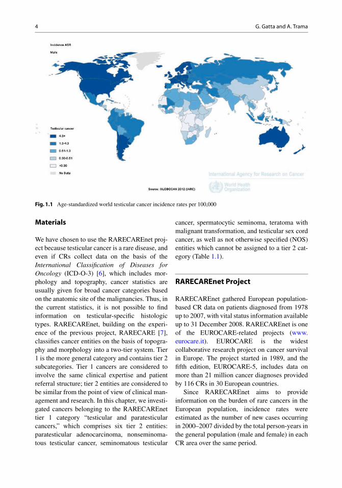
The age-standardized incidence rate for testicular cancers ranges widely across the world. Figure 1.1 shows the incidence of testicular cancers across dif-ferent continents in 2012. White males living in Western industrialized countries, particularly in Northern and Western Europe, had the highest inci-dence rates of testicular tumors (12/100,000 in Denmark, Norway, and Switzerland), whereas the incidence was lowest among black males in Africa (<0.5/100,000 in the majority of African countries). In Australia and New Zealand, the incidence was 7/100,000; in North America (the USA and Canada), 5/100,000; and in South and Central America, 2/100,000, with differences between countries
(Chile, 7/100,000; Uruguay, 6/100,000; Argentina and Costa Rica, 5/100,000; Mexico and Colombia, 3/100,000; Brazil, 2/100,000; and the remaining countries, <1/100,000). In Japan, the incidence was 2/100,000; in Southern, Eastern, and Central Asia, <1/100,000, while it was higher in Western Asia (1.7/100,000) with regional differences (5/100,000 in Israel, 3/100,000 in Georgia, and <1 in Oman, Qatar, Iraq, and Azerbaijan). In China, the incidence was 0.5/100,000 [ 2 ].
The testicular cancer incidence has increased in many Western countries and is still rising in most European countries [ 3 ]. By contrast, the mortality has declined since the mid-1970s; this is attributable to the development of effective treatments that are typically multidisciplinary and centered around platinum-based chemotherapy. However, recent trend analyses (1980–2010) showed a leveling-off of the rates in the most recent calendar years, suggesting that a plateau in mortality has been reached [ 4 ]. In the period 2000–2008, EUROCARE estimated the pooled 5-year relative survival to be 89 % for European testicular cancer patients, with some differences between countries including lower survival in Eastern European countries [ 5 ].
Here, we describe the epidemiology of testicular cancers on the basis of data from European population-based cancer registries (CRs) analyzed in the framework of RARECAREnet, the Information Network on Rare Cancers ( www.rarecarenet.eu ).
G. Gatta (*) • A. Trama Evaluative Epidemiology , Fondazione IRCCS Istituto Nazionale dei Tumori di Milano , Milan , Italy e-mail: [email protected]; [email protected]
1
4
Materials
We have chosen to use the RARECAREnet proj-ect because testicular cancer is a rare disease, and even if CRs collect data on the basis of the International Classifi cation of Diseases for Oncology (ICD-O-3) [ 6 ], which includes mor-phology and topography, cancer statistics are usually given for broad cancer categories based on the anatomic site of the malignancies. Thus, in the current statistics, it is not possible to fi nd information on testicular-specifi c histologic types. RARECAREnet, building on the experi-ence of the previous project, RARECARE [ 7 ], classifi es cancer entities on the basis of topogra-phy and morphology into a two-tier system. Tier 1 is the more general category and contains tier 2 subcategories. Tier 1 cancers are considered to involve the same clinical expertise and patient referral structure; tier 2 entities are considered to be similar from the point of view of clinical man-agement and research. In this chapter, we investi-gated cancers belonging to the RARECAREnet tier 1 category “testicular and paratesticular cancers,” which comprises six tier 2 entities: paratesticular adenocarcinoma, nonseminoma-tous testicular cancer, seminomatous testicular
cancer, spermatocytic seminoma, teratoma with malignant transformation, and testicular sex cord cancer, as well as not otherwise specifi ed (NOS) entities which cannot be assigned to a tier 2 cat-egory (Table 1.1 ).
RARECAREnet Project
RARECAREnet gathered European population- based CR data on patients diagnosed from 1978 up to 2007, with vital status information available up to 31 December 2008. RARECAREnet is one of the EUROCARE-related projects ( www.eurocare.it ). EUROCARE is the widest collaborative research project on cancer survival in Europe. The project started in 1989, and the fi fth edition, EUROCARE-5, includes data on more than 21 million cancer diagnoses provided by 116 CRs in 30 European countries.
Since RARECAREnet aims to provide information on the burden of rare cancers in the European population, incidence rates were estimated as the number of new cases occurring in 2000–2007 divided by the total person-years in the general population (male and female) in each CR area over the same period.
Fig. 1.1 Age-standardized world testicular cancer incidence rates per 100,000
G. Gatta and A. Trama
5
Relative survival is the ratio between the mea-sured survival of patients and the expected sur-vival in the general population for the same region (or country), age, sex, and calendar year. Relative survival accounts for the mortality from causes other than the relevant cancer. Relative survival estimates were obtained using the complete approach [ 8 ], considering cases diagnosed between 2000 and 2007 and followed up to 31 December 2008.
The observed prevalence per 100,000 at the index date 1 January 2008 was estimated by the counting method [ 9 ], using data from incident cases in the period 1993–2007. CRs that started recently do not have records of longer-term can-cer survivors who were diagnosed before the reg-istry started, resulting in underestimation of the prevalence. To account for this problem, RARECARE estimated the complete prevalence using the completeness index method [ 10 ], where
the estimated surviving cases diagnosed prior to 1993 were added to those counted in 1993–2007. Complete prevalence was provided at the index date of 1 January 2008. All data presented were extracted from the RARECAREnet online analysis tool.
Incidence
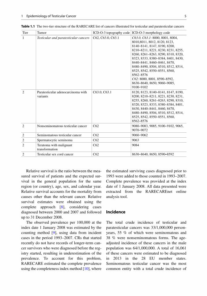
The total crude incidence of testicular and paratesticular cancers was 33/1,000,000 person- years, 55 % of which were seminomatous and 38 % were nonseminomatous forms. The age- adjusted incidence of these cancers in the male population was 64/1,000,000. A total of 16,061 of these cancers were estimated to be diagnosed in 2013 in the 28 EU member states. Seminomatous testicular cancer was the most common entity with a total crude incidence of
Table 1.1 The two-tier structure of the RARECARE list of cancers illustrated for testicular and paratesticular cancers
Tier Tumor ICD-O-3 topography code ICD-O-3 morphology code
1 Testicular and paratesticular cancers C62, C63.0, C63.1 C63.0, C63.1 : 8000, 8001, 8004, 8010,8011, 8012, 8120, 8123, 8140–8141, 8147, 8190, 8200, 8210–8211, 8221, 8230, 8231, 8255, 8260, 8261–8263, 8290, 8310, 8320, 8323, 8333, 8380–8384, 8401, 8430, 8440–8441, 8460–8461, 8470, 8480–8490, 8504, 8510, 8512, 8514, 8525, 8542, 8550–8551, 8560, 8562–8576 C62 : 8000, 8001, 8590–8592, 8630–8640, 8650, 9060–9085, 9100–9102
2 Paratesticular adenocarcinoma with variants
C63.0, C63.1 8120, 8123, 8140–8141, 8147, 8190, 8200, 8210–8211, 8221, 8230, 8231, 8255, 8260, 8261–8263, 8290, 8310, 8320, 8323, 8333, 8380–8384, 8401, 8430, 8440–8441, 8460, 8470, 8480–8490, 8504, 8510, 8512, 8514, 8525, 8542, 8550–8551, 8560, 8562–8576
2 Nonseminomatous testicular cancer C62 9080–9083, 9085, 9100–9102, 9065, 9070–9072
2 Seminomatous testicular cancer C62 9060–9062
2 Spermatocytic seminoma C62 9063
2 Teratoma with malignant transformation
C62 9084
2 Testicular sex cord cancer C62 8630–8640, 8650, 8590 – 8592
1 Epidemiology of Testicular Cancer
6
18/1,000,000, followed by nonseminomatous testicular cancer (13/1,000,000) (Table 1.2 ).
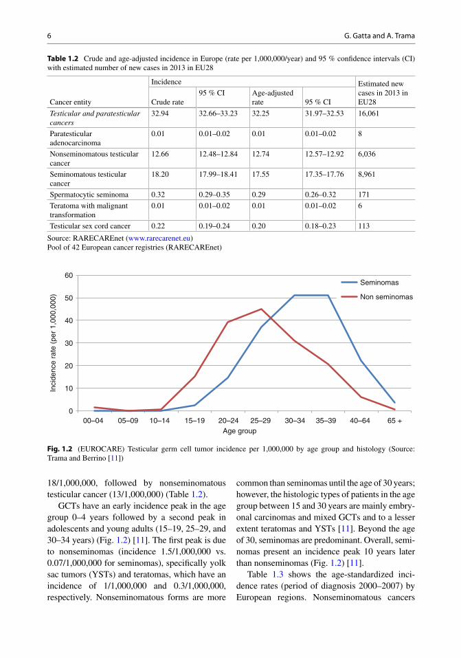
GCTs have an early incidence peak in the age group 0–4 years followed by a second peak in adolescents and young adults (15–19, 25–29, and 30–34 years) (Fig. 1.2 ) [ 11 ]. The fi rst peak is due to nonseminomas (incidence 1.5/1,000,000 vs. 0.07/1,000,000 for seminomas), specifi cally yolk sac tumors (YSTs) and teratomas, which have an incidence of 1/1,000,000 and 0.3/1,000,000, respectively. Nonseminomatous forms are more
common than seminomas until the age of 30 years; however, the histologic types of patients in the age group between 15 and 30 years are mainly embry-onal carcinomas and mixed GCTs and to a lesser extent teratomas and YSTs [ 11 ]. Beyond the age of 30, seminomas are predominant. Overall, semi-nomas present an incidence peak 10 years later than nonseminomas (Fig. 1.2 ) [ 11 ].
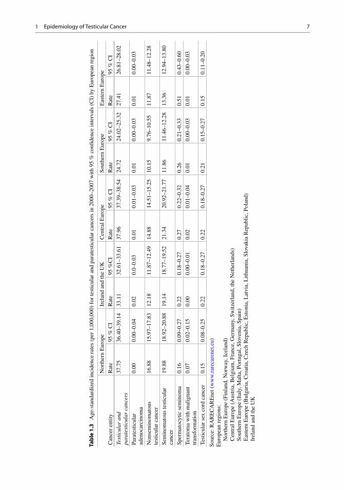
Table 1.3 shows the age-standardized inci-dence rates (period of diagnosis 2000–2007) by European regions. Nonseminomatous cancers
Table 1.2 Crude and age-adjusted incidence in Europe (rate per 1,000,000/year) and 95 % confi dence intervals (CI) with estimated number of new cases in 2013 in EU28
Cancer entity
Incidence Estimated new cases in 2013 in EU28 Crude rate
95 % CI Age-adjusted rate 95 % CI
Testicular and paratesticular cancers
32.94 32.66–33.23 32.25 31.97–32.53 16,061
Paratesticular adenocarcinoma
0.01 0.01–0.02 0.01 0.01–0.02 8
Nonseminomatous testicular cancer
12.66 12.48–12.84 12.74 12.57–12.92 6,036
Seminomatous testicular cancer
18.20 17.99–18.41 17.55 17.35–17.76 8,961
Spermatocytic seminoma 0.32 0.29–0.35 0.29 0.26–0.32 171
Teratoma with malignant transformation
0.01 0.01–0.02 0.01 0.01–0.02 6
Testicular sex cord cancer 0.22 0.19–0.24 0.20 0.18–0.23 113
Source: RARECAREnet ( www.rarecarenet.eu ) Pool of 42 European cancer registries (RARECAREnet)
0
10
20
30
40
50
60
00–04 05–09 10–14 15–19 20–24 25–29 30–34 35–39 40–64 65 +
Seminomas
Non seminomas
Age group
Inci
denc
e ra
te (
per
1,00
0,00
0)
Fig. 1.2 (EUROCARE) Testicular germ cell tumor incidence per 1,000,000 by age group and histology (Source: Trama and Berrino [ 11 ])
G. Gatta and A. Trama
7
Tab
le 1
.3
Age
-sta
ndar
dize
d in
cide
nce
rate
s (p
er 1
,000
,000
) fo
r te
stic
ular
and
par
ates
ticul
ar c
ance
rs in
200
0–20
07 w
ith 9
5 %
con
fi den
ce in
terv
als
(CI)
by
Eur
opea
n re
gion
Can
cer
entit
y
Nor
ther
n E
urop
e Ir
elan
d an
d th
e U
K
Cen
tral
Eur
ope
Sout
hern
Eur
ope
Eas
tern
Eur
ope
Rat
e 95
% C
I R
ate
95 %
CI
Rat
e 95
% C
I R
ate
95 %
CI
Rat
e 95
% C
I
Test
icul
ar a
nd
para
test
icul
ar c
ance
rs
37.7
5 36
.40–
39.1
4 33
.11
32.6
1–33
.61
37.9
6 37
.39–
38.5
4 24
.72
24.0
2–25
.32
27.4
1 26
.81–
28.0
2
Para
test
icul
ar
aden
ocar
cino
ma
0.00
0.
00–0
.04
0.02
0.
0–0.
03
0.01
0.
01–0
.03
0.01
0.
00–0
.03
0.01
0.
00–0
.03
Non
sem
inom
atou
s te
stic
ular
can
cer
16.8
8 15
.97–
17.8
3 12
.18
11.8
7–12
.49
14.8
8 14
.51–
15.2
5 10
.15
9.76
–10.
55
11.8
7 11
.48–
12.2
8
Sem
inom
atou
s te
stic
ular
ca
ncer
19
.88
18.9
2–20
.88
19.1
4 18
.77–
19.5
2 21
.34
20.9
2–21
.77
11.8
6 11
.46–
12.2
8 13
.36
12.9
4–13
.80
Sper
mat
ocyt
ic s
emin
oma
0.16
0.
09–0
.27
0.22
0.
18–0
.27
0.27
0.
22–0
.32
0.26
0.
21–0
.33
0.51
0.
43–0
.60
Tera
tom
a w
ith m
alig
nant
tr
ansf
orm
atio
n 0.
07
0.02
–0.1
5 0.
00
0.00
–0.0
1 0.
02
0.01
–0.0
4 0.
01
0.00
–0.0
3 0.
01
0.00
–0.0
3
Test
icul
ar s
ex c
ord
canc
er
0.15
0.
08–0
.25
0.22
0.
18–0
.27
0.22
0.
18–0
.27
0.21
0.
15–0
.27
0.15
0.
11–0
.20
Sour
ce: R
AR
EC
AR
Ene
t ( w
ww
.rar
ecar
enet
.eu )
E
urop
ean
regi
ons:
Nor
ther
n E
urop
e (F
inla
nd, N
orw
ay, I
cela
nd)
C
entr
al E
urop
e (A
ustr
ia, B
elgi
um, F
ranc
e, G
erm
any,
Sw
itzer
land
, the
Net
herl
ands
)
Sout
hern
Eur
ope
(Ita
ly, M
alta
, Por
tuga
l, Sl
oven
ia, S
pain
)
Eas
tern
Eur
ope
(Bul
gari
a, C
roat
ia, C
zech
Rep
ublic
, Est
onia
, Lat
via,
Lith
uani
a, S
lova
kia
Rep
ublic
, Pol
and)
Irel
and
and
the
UK
1 Epidemiology of Testicular Cancer
8
had the highest incidence (17/1,000,000) in Northern Europe. The incidence of seminoma-tous cancers was highest in Northern and Central Europe (about 20/1,000,000) followed closely by the UK and Ireland (19/1,000,000) and more dis-tantly by Eastern and Southern Europe. The inci-dence rates for the other cancers varied relatively little between regions.
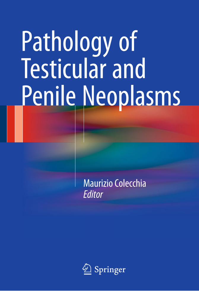
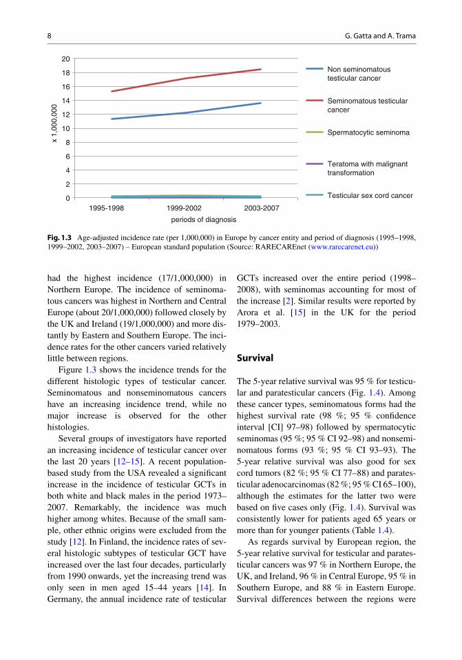
Figure 1.3 shows the incidence trends for the different histologic types of testicular cancer. Seminomatous and nonseminomatous cancers have an increasing incidence trend, while no major increase is observed for the other histologies.
Several groups of investigators have reported an increasing incidence of testicular cancer over the last 20 years [ 12 – 15 ]. A recent population- based study from the USA revealed a signifi cant increase in the incidence of testicular GCTs in both white and black males in the period 1973–2007. Remarkably, the incidence was much higher among whites. Because of the small sam-ple, other ethnic origins were excluded from the study [ 12 ]. In Finland, the incidence rates of sev-eral histologic subtypes of testicular GCT have increased over the last four decades, particularly from 1990 onwards, yet the increasing trend was only seen in men aged 15–44 years [ 14 ]. In Germany, the annual incidence rate of testicular
GCTs increased over the entire period (1998–2008), with seminomas accounting for most of the increase [ 2 ]. Similar results were reported by Arora et al. [ 15 ] in the UK for the period 1979–2003.
Survival
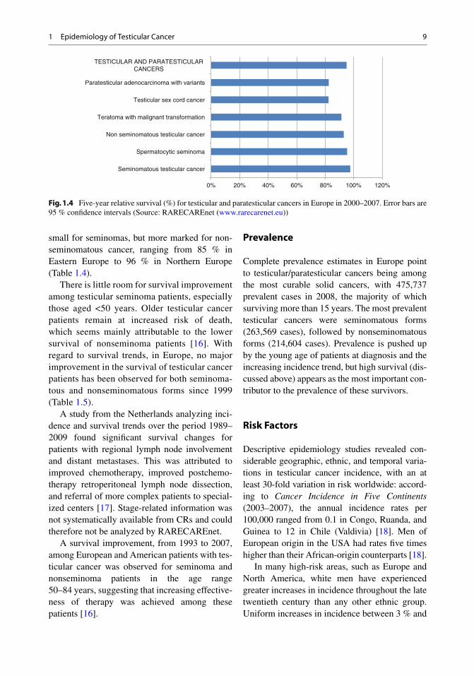
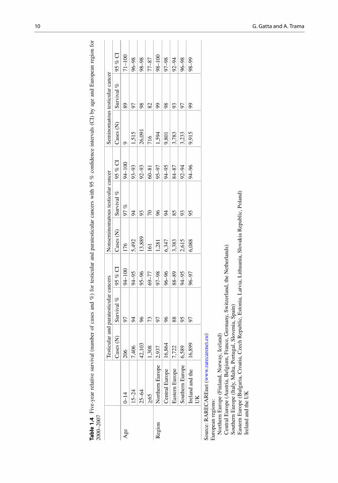
The 5-year relative survival was 95 % for testicu-lar and paratesticular cancers (Fig. 1.4 ). Among these cancer types, seminomatous forms had the highest survival rate (98 %; 95 % confi dence interval [CI] 97–98) followed by spermatocytic seminomas (95 %; 95 % CI 92–98) and nonsemi-nomatous forms (93 %; 95 % CI 93–93). The 5-year relative survival was also good for sex cord tumors (82 %; 95 % CI 77–88) and parates-ticular adenocarcinomas (82 %; 95 % CI 65–100), although the estimates for the latter two were based on fi ve cases only (Fig. 1.4 ). Survival was consistently lower for patients aged 65 years or more than for younger patients (Table 1.4 ).
As regards survival by European region, the 5-year relative survival for testicular and parates-ticular cancers was 97 % in Northern Europe, the UK, and Ireland, 96 % in Central Europe, 95 % in Southern Europe, and 88 % in Eastern Europe. Survival differences between the regions were
0
2
4
6
8
10
12
14
16
18
20
1995-1998 1999-2002 2003-2007
Non seminomatoustesticular cancer
Seminomatous testicularcancer
Spermatocytic seminoma
Teratoma with malignanttransformation
Testicular sex cord cancer
x 1,
000,
000
periods of diagnosis
Fig. 1.3 Age-adjusted incidence rate (per 1,000,000) in Europe by cancer entity and period of diagnosis (1995–1998, 1999–2002, 2003–2007) – European standard population (Source: RARECAREnet ( www.rarecarenet.eu ))
G. Gatta and A. Trama
9
small for seminomas, but more marked for non-seminomatous cancer, ranging from 85 % in Eastern Europe to 96 % in Northern Europe (Table 1.4 ).
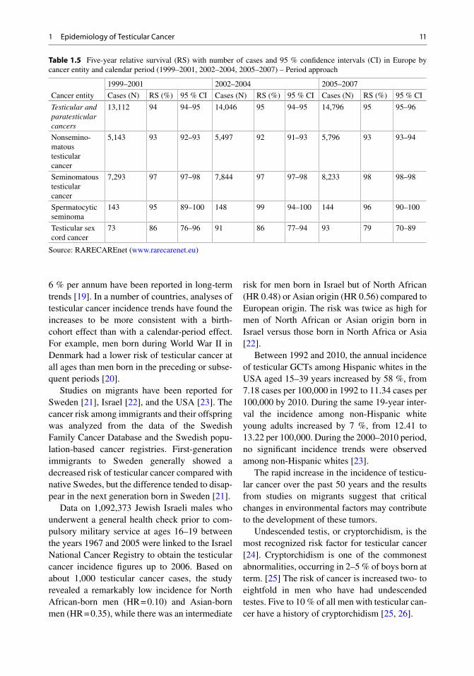
There is little room for survival improvement among testicular seminoma patients, especially those aged <50 years. Older testicular cancer patients remain at increased risk of death, which seems mainly attributable to the lower survival of nonseminoma patients [ 16 ]. With regard to survival trends, in Europe, no major improvement in the survival of testicular cancer patients has been observed for both seminoma-tous and nonseminomatous forms since 1999 (Table 1.5 ).
A study from the Netherlands analyzing inci-dence and survival trends over the period 1989–2009 found signifi cant survival changes for patients with regional lymph node involvement and distant metastases. This was attributed to improved chemotherapy, improved postchemo-therapy retroperitoneal lymph node dissection, and referral of more complex patients to special-ized centers [ 17 ]. Stage-related information was not systematically available from CRs and could therefore not be analyzed by RARECAREnet.
A survival improvement, from 1993 to 2007, among European and American patients with tes-ticular cancer was observed for seminoma and nonseminoma patients in the age range 50–84 years, suggesting that increasing effective-ness of therapy was achieved among these patients [ 16 ].
Prevalence
Complete prevalence estimates in Europe point to testicular/paratesticular cancers being among the most curable solid cancers, with 475,737 prevalent cases in 2008, the majority of which surviving more than 15 years. The most prevalent testicular cancers were seminomatous forms (263,569 cases), followed by nonseminomatous forms (214,604 cases). Prevalence is pushed up by the young age of patients at diagnosis and the increasing incidence trend, but high survival (dis-cussed above) appears as the most important con-tributor to the prevalence of these survivors.
Risk Factors
Descriptive epidemiology studies revealed con-siderable geographic, ethnic, and temporal varia-tions in testicular cancer incidence, with an at least 30-fold variation in risk worldwide: accord-ing to Cancer Incidence in Five Continents (2003–2007), the annual incidence rates per 100,000 ranged from 0.1 in Congo, Ruanda, and Guinea to 12 in Chile (Valdivia) [ 18 ]. Men of European origin in the USA had rates fi ve times higher than their African-origin counterparts [ 18 ].
In many high-risk areas, such as Europe and North America, white men have experienced greater increases in incidence throughout the late twentieth century than any other ethnic group. Uniform increases in incidence between 3 % and
0% 20% 40% 60% 80% 100% 120%
Seminomatous testicular cancer
Spermatocytic seminoma
Non seminomatous testicular cancer
Teratoma with malignant transformation
Testicular sex cord cancer
Paratesticular adenocarcinoma with variants
TESTICULAR AND PARATESTICULARCANCERS
Fig. 1.4 Five-year relative survival (%) for testicular and paratesticular cancers in Europe in 2000–2007. Error bars are 95 % confi dence intervals (Source: RARECAREnet ( www.rarecarenet.eu ))
1 Epidemiology of Testicular Cancer
10
Tab
le 1
.4
Five
-yea
r re
lativ
e su
rviv
al (
num
ber
of c
ases
and
%)
for
test
icul
ar a
nd p
arat
estic
ular
can
cers
with
95
% c
onfi d
ence
int
erva
ls (
CI)
by
age
and
Eur
opea
n re
gion
for
20
00–2
007
Test
icul
ar a
nd p
arat
estic
ular
can
cers
N
onse
min
omat
ous
test
icul
ar c
ance
r Se
min
omat
ous
test
icul
ar c
ance
r
Cas
es (
N)
Surv
ival
%
95 %
CI
Cas
es (
N)
Surv
ival
%
95 %
CI
Cas
es (
N)
Surv
ival
%
95 %
CI
Age
0–
14
206
97
94–1
00
176
97 %
94
–100
9
89
71–1
00
15–2
4 7,
406
94
94–9
5 5,
492
94
93–9
3 1,
515
97
96–9
8
25–6
4 42
,103
96
95
–96
13,8
89
93
92–9
3 26
,091
98
98
–98
≥65
1,30
8 73
69
–77
161
70
60–8
1 71
6 82
77
–87
Reg
ion
Nor
ther
n E
urop
e 2,
937
97
97–9
8 1,
281
96
95–9
7 1,
594
99
98–1
00
Cen
tral
Eur
ope
16,8
64
96
96–9
6 6,
347
94
94–9
5 9,
801
98
97–9
8
Eas
tern
Eur
ope
7,72
2 88
88
–89
3,38
3 85
84
–87
3,78
3 93
92
–94
Sout
hern
Eur
ope
6,58
9 95
94
–95
2,61
5 93
92
–94
3,23
3 97
96
–98
Irel
and
and
the
UK
16
,899
97
96
–97
6,08
8 95
94
–96
9,91
5 99
98
–99
Sour
ce: R
AR
EC
AR
Ene
t ( w
ww
.rar
ecar
enet
.eu )
E
urop
ean
regi
ons:
Nor
ther
n E
urop
e (F
inla
nd, N
orw
ay, I
cela
nd)
C
entr
al E
urop
e (A
ustr
ia, B
elgi
um, F
ranc
e, G
erm
any,
Sw
itzer
land
, the
Net
herl
ands
)
Sout
hern
Eur
ope
(Ita
ly, M
alta
, Por
tuga
l, Sl
oven
ia, S
pain
)
Eas
tern
Eur
ope
(Bul
gari
a, C
roat
ia, C
zech
Rep
ublic
, Est
onia
, Lat
via,
Lith
uani
a, S
lova
kia
Rep
ublic
, Pol
and)
Irel
and
and
the
UK
G. Gatta and A. Trama
11
6 % per annum have been reported in long-term trends [ 19 ]. In a number of countries, analyses of testicular cancer incidence trends have found the increases to be more consistent with a birth- cohort effect than with a calendar-period effect. For example, men born during World War II in Denmark had a lower risk of testicular cancer at all ages than men born in the preceding or subse-quent periods [ 20 ].
Studies on migrants have been reported for Sweden [ 21 ], Israel [ 22 ], and the USA [ 23 ]. The cancer risk among immigrants and their offspring was analyzed from the data of the Swedish Family Cancer Database and the Swedish popu-lation-based cancer registries. First- generation immigrants to Sweden generally showed a decreased risk of testicular cancer compared with native Swedes, but the difference tended to disap-pear in the next generation born in Sweden [ 21 ].
Data on 1,092,373 Jewish Israeli males who underwent a general health check prior to com-pulsory military service at ages 16–19 between the years 1967 and 2005 were linked to the Israel National Cancer Registry to obtain the testicular cancer incidence fi gures up to 2006. Based on about 1,000 testicular cancer cases, the study revealed a remarkably low incidence for North African-born men (HR = 0.10) and Asian- born men (HR = 0.35), while there was an intermediate
risk for men born in Israel but of North African (HR 0.48) or Asian origin (HR 0.56) compared to European origin. The risk was twice as high for men of North African or Asian origin born in Israel versus those born in North Africa or Asia [ 22 ].
Between 1992 and 2010, the annual incidence of testicular GCTs among Hispanic whites in the USA aged 15–39 years increased by 58 %, from 7.18 cases per 100,000 in 1992 to 11.34 cases per 100,000 by 2010. During the same 19-year inter-val the incidence among non-Hispanic white young adults increased by 7 %, from 12.41 to 13.22 per 100,000. During the 2000–2010 period, no signifi cant incidence trends were observed among non-Hispanic whites [ 23 ].
The rapid increase in the incidence of testicu-lar cancer over the past 50 years and the results from studies on migrants suggest that critical changes in environmental factors may contribute to the development of these tumors.
Undescended testis, or cryptorchidism, is the most recognized risk factor for testicular cancer [ 24 ]. Cryptorchidism is one of the commonest abnormalities, occurring in 2–5 % of boys born at term. [ 25 ] The risk of cancer is increased two- to eightfold in men who have had undescended testes. Five to 10 % of all men with testicular can-cer have a history of cryptorchidism [ 25 , 26 ].
Table 1.5 Five-year relative survival (RS) with number of cases and 95 % confi dence intervals (CI) in Europe by cancer entity and calendar period (1999–2001, 2002–2004, 2005–2007) – Period approach
Cancer entity
1999–2001 2002–2004 2005–2007
Cases (N) RS (%) 95 % CI Cases (N) RS (%) 95 % CI Cases (N) RS (%) 95 % CI
Testicular and paratesticular cancers
13,112 94 94–95 14,046 95 94–95 14,796 95 95–96
Nonsemino-matous testicular cancer
5,143 93 92–93 5,497 92 91–93 5,796 93 93–94
Seminomatous testicular cancer
7,293 97 97–98 7,844 97 97–98 8,233 98 98–98
Spermatocytic seminoma
143 95 89–100 148 99 94–100 144 96 90–100
Testicular sex cord cancer
73 86 76–96 91 86 77–94 93 79 70–89
Source: RARECAREnet ( www.rarecarenet.eu )
1 Epidemiology of Testicular Cancer
12
Treatment for undescended testis before puberty can prevent testicular cancer. A cohort of about 20,000 Swedish boys treated for cryptorchi-dism with orchiopexy from 1964 to 1999 and fol-lowed up until the end of 2000 indicates that age at orchiopexy has an effect on the risk of testicular cancer in boys with an undescended testicle. Those treated at 13 years or older had a relative risk of 5.4 compared with the general population, about twice the risk of those treated at younger ages [ 27 ]. In the Swedish study, the risk of testicu-lar cancer increased in the entire cohort regardless of age at surgery, supporting the hypothesis that the risk of testicular cancer is largely determined in utero and also suggesting that puberty is another crucial event in testicular carcinogenesis. Findings from a subsequent meta-analysis further support that early surgery reduces the risk of testicular cancer among cryptorchid men [ 28 ].
Other factors clearly associated with the risk of testicular cancer are prior unilateral testicular cancer and a family history of testicular cancer. From the Swedish Family Cancer Database, it appeared that the incidence ratios for familial risk were almost four- and eightfold increased when the father or a brother had testicular cancer, respectively [ 29 ].
From several studies, it emerges that a previ-ous history of testicular cancer is strongly associ-ated with a second cancer in the contralateral testis. Two studies from the Netherlands and New Zealand reported an increased risk of a contralat-eral tumor in patients who had previously had testicular cancer; the relative risk was as high as 25–28 [ 30 , 31 ]. A large population-based cohort study involving about 30,000 US men estimated a lower risk: testis cancer patients had a 12 times greater risk of developing metachronous contra-lateral testicular cancer than men in the general population [ 32 ].
A systematic review of pre- and perinatal vari-ables in relation to the risk of testicular cancer with a focus on maternal characteristics [ 33 ] revealed evidence for associations with maternal bleeding (odds ratio, OR = 1.33), birth order (fi rst vs. others, OR = 1.08), and sibship size (OR rang-ing from 0.93 to 0.75 for 2, 3, 4 vs. 1). Three large US studies also found cesarean section to be
associated with the risk of testicular cancer (OR = 1.67) [ 33 ]. The meta-analyses indicated no association with maternal age, maternal nausea, maternal hypertension, preeclampsia, and breech delivery. Bleeding is generally the result of a developmental aberration such as abnormal pla-cental differentiation or aggressive implantation, which may be hypothesized to disrupt fetal devel-opment and increase the risk of a prenatal stem-cell progenitor of testicular cancer. Parental subfertility was suggested among the underlying causes of the association with birth order and sib-ship size [ 34 ]. Furthermore, all of the studied vari-ables could theoretically be due to an altered in utero hormonal milieu [ 35 – 38 ] and delayed expo-sure to a communicable agent for the son [ 39 , 40 ].
Another systematic review and meta-analysis of perinatal variables with a focus on characteris-tics of the son provided evidence that low birth weight, low gestational age, inguinal hernia, cryptorchidism, and twinning were associated with a higher risk of testicular cancer [ 24 ]. The ORs ranged between 1.63 and 1.22, with the exclusion of cryptorchidism, where the OR was as high as 4.3. A subsequent Swedish study con-fi rmed that genital malformations were associated with an increased risk of testicular cancer; in addi-tion to cryptorchidism and inguinal hernia, also hypospadias was reported, with an OR of 2.4 [ 41 ]. It should be taken into consideration that some of these factors are interconnected and could argu-ably be proxies of the maternal in utero environ-ment. There is evidence that exposure to maternal hormones, particularly estrogens, is associated with testicular GCT risk (OR = 4.9) [ 42 ].
With the improvements in recent decades of the survival of premature infants, an increased frequency of testis cancers arising among survivors of premature birth may have contributed to an increase in testicular cancer rates.
Exposure to endocrine-disrupting chemicals (EDCs) during the fetal period has been postu-lated to be a risk factor for testicular cancer [ 43 ]. EDCs are able to interact with human endocrine systems such as the system of the sex hormones testosterone and estrogen. They can act either directly or indirectly, for example, by blocking hormone receptors or altering hormone
G. Gatta and A. Trama
13
degradation [ 43 ]. Pentachlorophenol, bisphenol A, and phthalates are examples of known EDCs to which humans are exposed via the environment. In a Swedish study, the concentrations of chlori-nated biphenyls (PCBs), p,p’-dichlorodiphenyldi-chloroethylene, hexachlorobenzene, chlordanes, and polybrominated diphenyl ethers were higher in blood from mothers of testicular cancer patients compared with control mothers [ 44 ]. For the sum of PCBs, an OR of 3.8 was estimated. Most of the patients were born during a period with the high-est concentrations of persistent organic pollutants (POPs) such as PCBs in the ecological system. Since then the concentrations have fallen substan-tially. In Sweden, the large difference in the increase in testicular cancer incidence between 1984–1993 and 1994–2003 supports the hypoth-esis that exposure to certain POPs during the fetal period is of etiologic signifi cance in testicular car-cinogenesis. EDC research will need to assess these chemicals in the most appropriate specimen to ensure accurate EDC exposure assessment dur-ing pregnancy. Currently, there are no studies evaluating the association between placental con-centrations of EDCs and risk of any testicular dys-genesis syndrome (TDS) condition. Furthermore, effect modifi cations of the EDC–TDS associa-tions by potential genetic factors or infl ammatory processes have not been evaluated to date.
Average stature and the incidence of testicular cancer have both been increasing over several generations with strong cohort effects [ 45 ]. A systematic review and meta-analysis analyzed the relation between body size and testicular cancer, but confi rmed the association with stature only, with an OR of 1.13 per 5 cm increase in height. Possible mechanisms to explain the association of stature with testicular cancer risk include childhood nutrition [ 46 ], increased levels of insulin-like growth factor [ 47 , 48 ], and earlier age at puberty in those of greater height. [ 49 , 50 ] As adult stature is largely determined in the period between birth and age 2 years [ 50 ], nutrition at this age may be particularly relevant to the testicular cancer risk [ 51 ].
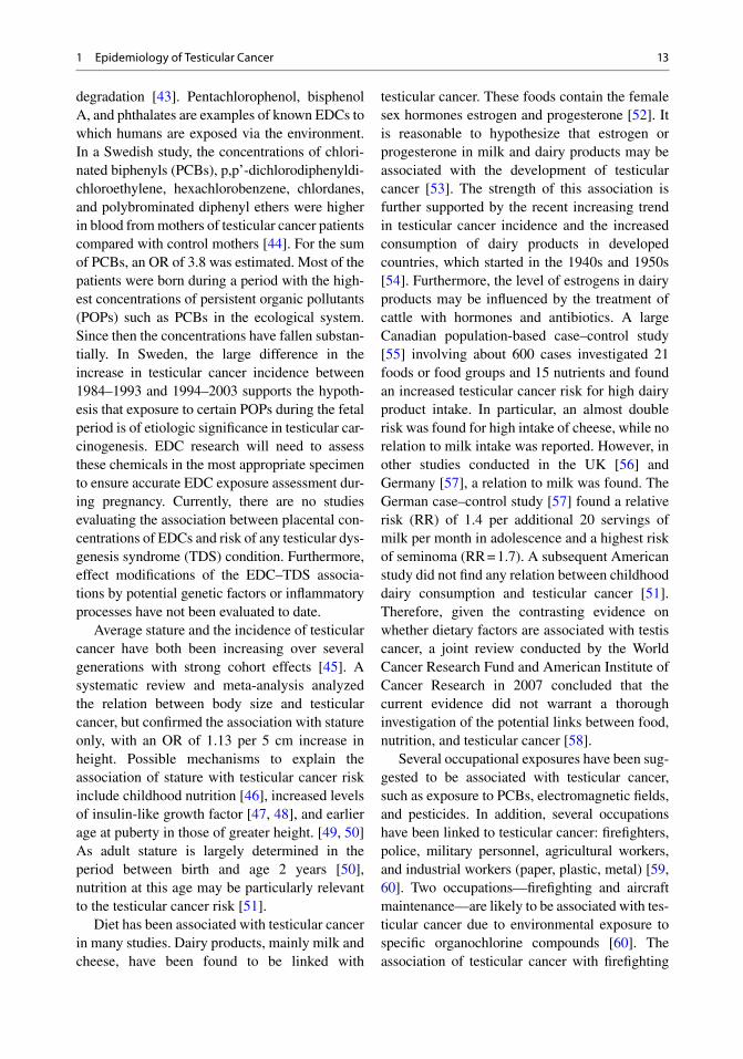
Diet has been associated with testicular cancer in many studies. Dairy products, mainly milk and cheese, have been found to be linked with
testicular cancer. These foods contain the female sex hormones estrogen and progesterone [ 52 ]. It is reasonable to hypothesize that estrogen or progesterone in milk and dairy products may be associated with the development of testicular cancer [ 53 ]. The strength of this association is further supported by the recent increasing trend in testicular cancer incidence and the increased consumption of dairy products in developed countries, which started in the 1940s and 1950s [ 54 ]. Furthermore, the level of estrogens in dairy products may be infl uenced by the treatment of cattle with hormones and antibiotics. A large Canadian population-based case–control study [ 55 ] involving about 600 cases investigated 21 foods or food groups and 15 nutrients and found an increased testicular cancer risk for high dairy product intake. In particular, an almost double risk was found for high intake of cheese, while no relation to milk intake was reported. However, in other studies conducted in the UK [ 56 ] and Germany [ 57 ], a relation to milk was found. The German case–control study [ 57 ] found a relative risk (RR) of 1.4 per additional 20 servings of milk per month in adolescence and a highest risk of seminoma (RR = 1.7). A subsequent American study did not fi nd any relation between childhood dairy consumption and testicular cancer [ 51 ]. Therefore, given the contrasting evidence on whether dietary factors are associated with testis cancer, a joint review conducted by the World Cancer Research Fund and American Institute of Cancer Research in 2007 concluded that the current evidence did not warrant a thorough investigation of the potential links between food, nutrition, and testicular cancer [ 58 ].
Several occupational exposures have been sug-gested to be associated with testicular cancer, such as exposure to PCBs, electromagnetic fi elds, and pesticides. In addition, several occupations have been linked to testicular cancer: fi refi ghters, police, military personnel, agricultural workers, and industrial workers (paper, plastic, metal) [ 59 , 60 ]. Two occupations—fi refi ghting and aircraft maintenance—are likely to be associated with tes-ticular cancer due to environmental exposure to specifi c organochlorine compounds [ 60 ]. The association of testicular cancer with fi refi ghting
1 Epidemiology of Testicular Cancer
14
originates from reports of a statistically signifi -cant increased risk among fi refi ghters in New Zealand [ 61 , 62 ]. This association was supported by subsequent studies in California [ 63 ] and Florida [ 64 ] that also found signifi cantly increased risks of testicular cancer, as well as by a study in Germany that reported a trend toward an increased risk [ 65 ]. The latest review on this topic produced a summary risk estimate of 2 [ 66 ]. The associa-tion of fi refi ghting with the risk of testicular can-cer might be related to exposure to a wide variety of carcinogenic substances, including benzene and polycyclic aromatic hydrocarbons. Mortality due to testicular cancers in a very large cohort of aircraft manufacturing workers was almost dou-ble compared to the general population, possibly due to mixed solvent exposure. An excess risk was also found among pesticide applicators in the USA [ 67 ] and the UK [ 68 ]. A study from Norway [ 69 ] reported an increased incidence of testicular cancer in farmers’ sons, whereas studies from Denmark [ 70 ] and the USA [ 71 ] have not repli-cated this fi nding.
In the early 2000s, molecular epidemiologic fi ndings of serum levels of pesticides have been made available to better assess the environmental exposure. The pesticides that have been most com-monly examined to date are the organochlorine compounds. These are among the oldest pesticides used and are persistent both in the environment and in human adipose tissue [ 72 ]. In addition, organo-chlorine compounds can mimic sex steroid hor-mones and might therefore alter gene expression patterns that are important in urogenital develop-ment and homeostasis [ 73 , 74 ]. Organochlorine pesticides include dichlorodiphenyltrichloroethane (DDT), its most persistent metabolite dichlorodi-phenyldichloroethylene (DDE), and many others. Current evidence suggests that of the organochlo-rine pesticides examined, only DDE and chlor-danes are associated with an increased risk of testicular cancer [ 60 ]. However, the available evi-dence has not permitted the identifi cation of strong and consistent occupational risk factors for testicu-lar cancer. Since testicular cancer occurs mainly in young adults, the potential impact of occupational exposure of mothers to endocrine disruptors during intrauterine development has to be considered
[ 60 , 75 ]. Other exposures – to heat, polyvinyl chlo-ride, nonionizing radiation, heavy metals, agricul-tural work, and PCBs as well as marijuana use – might also have an association with testicular cancer [ 60 ].
Final Remarks
The testicular cancer incidence has increased in the past fi ve decades and is now increasing in some countries and ethnic groups historically at low risk. Cryptorchidism is the condition most evidently related to testicular cancer. Among the potentially modifi able risk factors, studies of the age at surgery for cryptorchidism suggest that treating undescended testes before puberty reduces the risk of testicular cancer.
Cryptorchidism with hypospadias and poor semen quality together make up the TDS; they result from disrupted embryonal programming and gonadal development, possibly caused by exposures of environmental origin [ 75 ]. Several studies were conducted on diet, primarily focused on dietary patterns during early life. Although the evidence is not conclusive, diet especially in early life and puberty is likely to be very important in reducing the risk of many chronic diseases and cancers in adult life. One can further investigate these hypotheses, but from the public health point of view we believe the current state of knowledge is suffi cient to implement programs to contrast the so-called Western diet.
Some occupations are likely to be associated with testicular cancer, and occupational exposure to PVC, PCBs, and organochlorine compounds requires further study. However, exposure to pro-fessional risks in agriculture and environmental exposure may occur in utero and/or act as perina-tal and nonperinatal risk factors. Organochlorine compounds, especially those included in pesti-cides, are widely dispersed among populations and may be responsible for a large proportion of the attributable risk. In this case, the principle of precaution should be considered to promote poli-cies of risk reduction (Table 1.6 ).
The low incidence of testicular cancer often constituted a barrier to conducting adequately powered epidemiologic studies. Collaborative
G. Gatta and A. Trama
15
investigations that pool data from a number of countries are strongly encouraged. International collaborative studies should allow more ade-quately powered epidemiologic studies. In order to provide new ideas and hypotheses to be exam-ined in such studies, a survey was conducted among testicular cancer researchers on new hypotheses concerning the etiology of the malig-nancy [ 76 ]. Hypotheses with the highest plausi-bility ratings were related to prenatal exposures and exposures that might have an effect during pregnancy and in postnatal life. They included factors related to endocrine disruption, DNA damage, infl ammation, and nutrition during preg-nancy. The results of the survey should promote a multidisciplinary debate and enhance new etio-logic research into testicular cancers.
Acknowledgments This chapter arises from the project RARECAREnet which has received funding from the European Union in the framework of the Health Programme.
References
1. Chia VM, Quraishi SM, Devesa SS, Purdue MP, Cook MB, McGlynn KA. International trends in the incidence of testicular cancer, 1973–2002. Cancer Epidemiol Biomarkers Prev. 2010;19:1151–9.
2. Ferlay J, Soerjomataram I, Ervik M, Dikshit R, Eser S, Mathers C, Rebelo M, Parkin DM, Forman D, Bray, F. GLOBOCAN 2012 v1.0, Cancer incidence and mortality worldwide: IARC CancerBase No. 11 [Internet]. Lyon: International Agency for Research on Cancer; 2013. Available from: http://globocan.iarc.fr . Accessed on 29 Dec 2014.
3. Znaor A, Lortet-Tieulent J, Laversanne M, Jemal A, Bray F. International testicular cancer incidence trends: generational transitions in 38 countries 1900–1990. Cancer Causes Control. 2015;26:151–8.
4. Bosetti C, Bertuccio P, Malvezzi M, Levi F, Chatenoud L, Negri E, La Vecchia C. Cancer mortality in Europe, 2005–2009, and an overview of trends since 1980. Ann Oncol. 2013;24:2657–71.
5. Trama A, Foschi R, Larrañagab N, Sant M, Fuentes- Raspall R, Serraino D, Tavilla A, Van Eycken L, Nicolai N, the EUROCARE-5 Working Group. Survival of male genital cancers (prostate, testis and penis) in Europe 1999–2007: results from the Eurocare-5 study. Eur J Cancer. 2015; in press.
6. Percy C, Fritz A, Jack A, Shanmugarathan S, Sobin L, Parkin DM, Whelan S. International Classifi cation of Diseases for Oncology (ICD-O). 3rd ed. Geneva: World Health Organisation; 2000.
7. Gatta G, van der Zwan JM, Casali PG, Siesling S, Dei Tos AP, Kunkler I, Otter R, Licitra L, Mallone S, Tavilla A, Trama A. Capocaccia R; RARECARE working group. rare cancers are not so rare: the rare cancer bur-den in Europe. Eur J Cancer. 2011;47:2493–511.
8. Brenner H, Gefeller O, Hakulinen T. Period analysis for ‘up-to-date’ cancer survival data: theory, empirical evaluation, computational realisation and applications. Eur J Cancer. 2004;40:326–35.
9. Capocaccia R, Colonna M, Corazziari I, De Angelis R, Francisci S, Micheli A, Mugno E, EUROPREVAL Working Group. Measuring cancer prevalence in Europe: the EUROPREVAL project. Ann Oncol. 2002;13:831–9.
10. Capocaccia R, De Angelis R. Estimating the com-pleteness of prevalence based on cancer registry data. Stat Med. 1997;16:425–40.
11. Trama A, Berrino F. Epidemiology of malignant germ cell tumours. The EUROCARE study. In: Nogales FF, Jimenez R, editors. The pathology and biology of germ cell tumours. New York: Springer; 2017 (in print).
12. Stang A, Trabert B, Wentzensen N, Cook MB, Rusner C, Oosterhuis JW, McGlynn KA. Gonadal and extragonadal germ cell tumours in the United States, 1973–2007. Int J Androl. 2012;35:616–25.
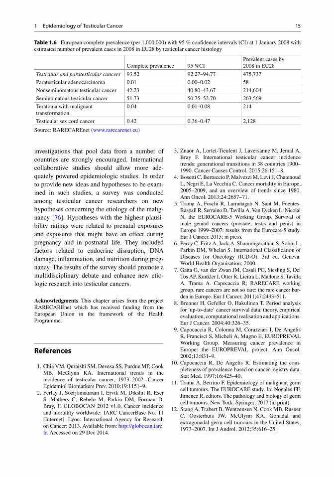
Table 1.6 European complete prevalence (per 1,000,000) with 95 % confi dence intervals (CI) at 1 January 2008 with estimated number of prevalent cases in 2008 in EU28 by testicular cancer histology
Complete prevalence 95 %CI Prevalent cases by 2008 in EU28
Testicular and paratesticular cancers 93.52 92.27–94.77 475,737
Paratesticular adenocarcinoma 0.01 0.00–0.02 58
Nonseminomatous testicular cancer 42.23 40.80–43.67 214,604
Seminomatous testicular cancer 51.73 50.75–52.70 263,569
Teratoma with malignant transformation
0.04 0.01–0.08 214
Testicular sex cord cancer 0.42 0.36–0.47 2,128
Source: RARECAREnet ( www.rarecarenet.eu )
1 Epidemiology of Testicular Cancer
16
13. Rusner C, Trabert B, Katalinic A, Kieschke J, Emrich K, Stang A, Network of German Cancer Registries (GEKID). Incidence patterns and trends of malignant gonadal and extragonadal germ cell tumors in Germany, 1998–2008. Cancer Epidemiol. 2013;37:370–3.
14. Pauniaho SL, Salonen J, Helminen M, Vettenranta K, Heikinheimo M, Heikinheimo O. The incidences of malignant gonadal and extragonadal germ cell tumors in males and females: a population-based study covering over 40 years in Finland. Cancer Causes Control. 2012;23:1921–7.
15. Arora RS, Alston RD, Eden TO, Geraci M, Birch JM. Comparative incidence patterns and trends of gonadal and extragonadal germ cell tumors in England, 1979–2003. Cancer. 2012;118:4290–7.
16. Verhoeven RH, Gondos A, Janssen-Heijnen ML, Saum KU, Brewster DH, Holleczek B, Crocetti E, Rosso S, Hakulinen T, Aareleid T, Brenner H, EUNICE Survival Working Group. Testicular cancer in Europe and the USA: survival still rising among older patients. Ann Oncol. 2013;24:508–13.
17. Verhoeven RH, Karim-Kos HE, Coebergh JW, Brink M, Horenblas S, de Wit R, Kiemeney BA. Markedly increased incidence and improved survival of testicular cancer in the Netherlands. Acta Oncol. 2014;53:342–50.
18. Forman D, Bray F, Brewster DH, Gombe Mbalawa C, Kohler B, Piñeros M, Steliarova-Foucher E, Swaminathan R and Ferlay J, editors. Cancer incidence in fi ve conti-nents, Vol. X (electronic version). Lyon: International Agency for Research on Cancer; 2013. Available from: http://ci5.iarc.fr . Accessed on 20 July 2015.
19. Bray F, Richiardi L, Ekbom A, Forman D, Pukkala E, Cuninkova M, Møller H. Do testicular seminoma and nonseminoma share the same etiology? Evidence from an age-period-cohort analysis of incidence trends in eight European countries. Cancer Epidemiol Biomarkers Prev. 2006;15:652–8.
20. Møller H. Decreased testicular cancer risk in men born in wartime. J Natl Cancer Inst. 1989;81:1668–9.
21. Hemminki K, Li X. Cancer risks in Nordic immigrants and their offspring in Sweden. Eur J Cancer. 2002;38:2428–34.
22. Levine H, Afek A, Shamiss A, Derazne E, Tzur D, Zavdy O, Barchana M, Kark JD. Risk of germ cell testicular cancer according to origin: a migrant cohort study in 1,100,000 Israeli men. Int J Cancer. 2013;132:1878–85.
23. Chien FL, Schwartz SM, Johnson RH. Increase in tes-ticular germ cell tumor incidence among Hispanic adolescents and young adults in the United States. Cancer. 2014;120:2728–34.
24. Cook MB, Akre O, Forman D, Madigan MP, Richiardi L, McGlynn KA. A systematic review and meta- analysis of perinatal variables in relation to the risk of testicular cancer--experiences of the son. Int J Epidemiol. 2010;39:1605–18.
25. Toppari J, Kaleva M. Maldescendus testis. Horm Res. 1999;51:261–9.
26. Dieckmann KP, Pichlmeier U. Clinical epidemiology of testicular germ cell tumors. World J Urol. 2004;22:2–14.
27. Pettersson A, Richiardi L, Nordenskjold A, Kaijser M, Akre O. Age at surgery for undescended testis and risk of testicular cancer. N Engl J Med. 2007;356:1835–41.
28. Banks K, Tuazon E, Berhane K, Koh CJ, De Filippo RE, Chang A, Kim SS, Daneshmand S, Davis-Dao C, Lewinger JP, Bernstein L, Cortessis VK. Cryptorchidism and testicular germ cell tumors: comprehensive meta-analysis reveals that association between these condi-tions diminished over time and is modifi ed by clinical characteristics. Front Endocrinol (Lausanne). 2013;3:182.
29. Hemminki K, Chen B. Familial risks in testicular can-cer as aetiological clues. Int J Androl. 2006;29:205–10.
30. Osterlind A, Berthelsen JG, Abildgaard N, Hansen SO, Hjalgrim H, Johansen B, Munck-Hansen J, Rasmussen LH. Risk of bilateral testicular germ cell cancer in Denmark: 1960–1984. J Natl Cancer Inst. 1991;83:1391–5.
31. Colls BM, Harvey VJ, Skelton L, Thompson PI, Frampton CM. Bilateral testicular germ cell tumours in New Zealand: experience in Auckland and Christchurch 1978–94. J Clin Oncol. 1996;14:2061–5.
32. Fosså SD, Chen J, Schonfeld SJ, McGlynn KA, McMaster ML, Gail MH, Travis LB. Risk of contralat-eral testicular cancer: a population-based study of 29,515 U.S. men. J Natl Cancer Inst. 2005;97:1056–66.
33. Cook MB, Akre O, Forman D, Madigan MP, Richiardi L, McGlynn KA. A systematic review and meta- analysis of perinatal variables in relation to the risk of testicular cancer–experiences of the mother. Int J Epidemiol. 2009;38:1532–42.
34. Richiardi L, Akre O, Lambe M, Granath F, Montgomery SM, Ekbom A. Birth order, sibship size, and risk for germ-cell testicular cancer. Epidemiology. 2004;15:323–9.
35. Bernstein L, Depue RH, Ross RK, Judd HL, Pike MC, Henderson BE. Higher maternal levels of free estradiol in fi rst compared to second pregnancy: early gesta-tional differences. J Natl Cancer Inst. 1986;76:1035–9.
36. Panagiotopoulou K, Katsouyanni K, Petridou E, Garas Y, Tzonou A, Trichopoulos D. Maternal age, parity, and pregnancy estrogens. Cancer Causes Control. 1990;1:119–24.
37. Troisi R, Hoover RN, Thadhani R, Hsieh CC, Sluss P, Ballard-Barbash R, Potischman N. Maternal, prenatal and perinatal characteristics and fi rst trimester maternal serum hormone concentrations. Br J Cancer. 2008;99:1161–4.
38. Arslan AA, Zeleniuch-Jacquotte A, Lukanova A, Afanasyeva Y, Katz J, Levitz M, Del Priore G, Toniolo P. Effects of parity on pregnancy hormonal profi les across ethnic groups with a diverse incidence of breast cancer. Cancer Epidemiol Biomarkers Prev. 2006;15:2123–30.
39. Green MS, Zaaide Y. Sibship size as a risk factor for hepatitis a infection. Am J Epidemiol. 1989;129:800–5.
G. Gatta and A. Trama