Embed Size (px)
Citation preview
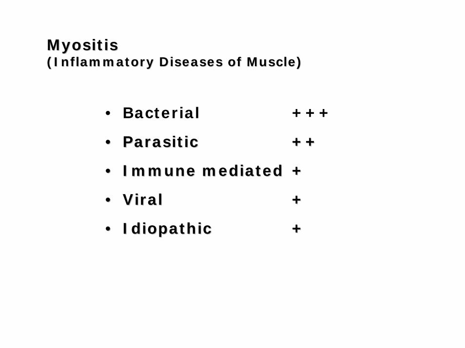
MyositisMyositis(Inflammatory Diseases of Muscle)(Inflammatory Diseases of Muscle)
• Bacterial +++
•• ParasiticParasitic ++++
•• Immune mediatedImmune mediated ++
•• ViralViral ++
•• IdiopathicIdiopathic ++
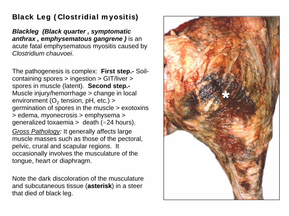
Black Leg (Clostridial Black Leg (Clostridial myositismyositis))
Blackleg (Black quarter , symptomatic anthrax , emphysematous gangrene ) is an acute fatal emphysematous myositis caused by Clostridium chauvoei.
The pathogenesis is complex: First step.- Soil-containing spores > ingestion > GIT/liver > spores in muscle (latent). Second step.-Muscle injury/hemorrhage > change in local environment (O2 tension, pH, etc.) > germination of spores in the muscle > exotoxins> edema, myonecrosis > emphysema > generalized toxaemia > death (.24 hours). Gross Pathology: It generally affects large muscle masses such as those of the pectoral, pelvic, crural and scapular regions. It occasionally involves the musculature of the tongue, heart or diaphragm.
Note the dark discoloration of the musculature and subcutaneous tissue (asteriskasterisk) in a steer that died of black leg.
Black Leg Black Leg
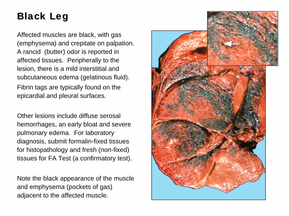
Affected muscles are black, with gas (emphysema) and crepitate on palpation. A rancid (butter) odor is reported in affected tissues. Peripherally to the lesion, there is a mild interstitial and subcutaneous edema (gelatinous fluid). Fibrin tags are typically found on the epicardial and pleural surfaces.
Other lesions include diffuse serosal hemorrhages, an early bloat and severe pulmonary edema. For laboratory diagnosis, submit formalin-fixed tissues for histopathology and fresh (non-fixed) tissues for FA Test (a confirmatory test).
Note the black appearance of the muscle and emphysema (pockets of gas) adjacent to the affected muscle.
Black Leg Black Leg
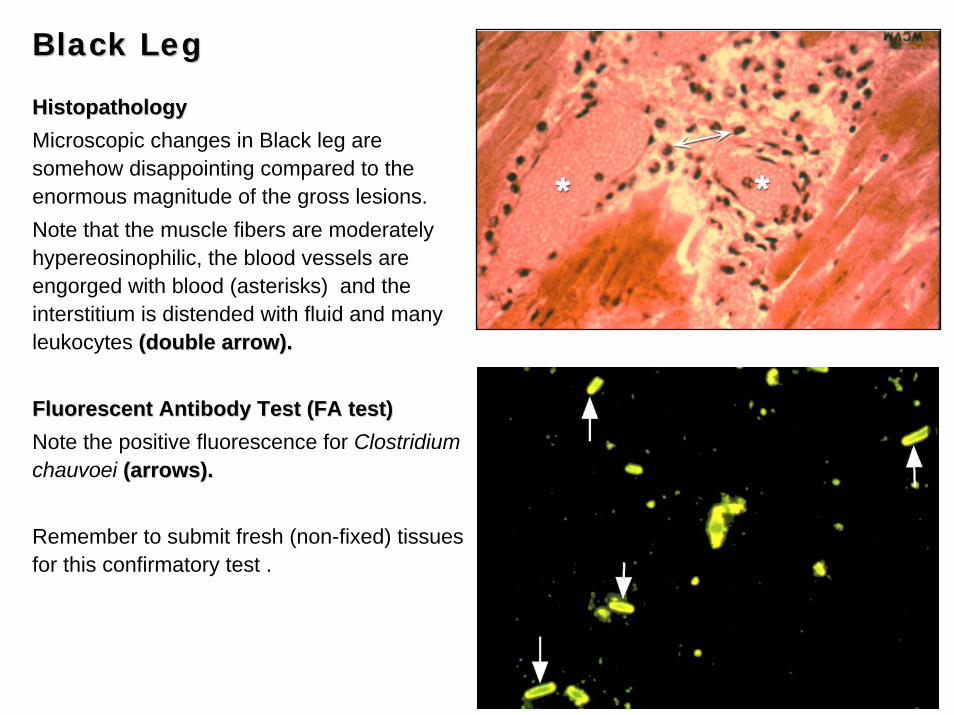
HistopathologyHistopathologyMicroscopic changes in Black leg are somehow disappointing compared to the enormous magnitude of the gross lesions. Note that the muscle fibers are moderately hypereosinophilic, the blood vessels are engorged with blood (asterisks) and the interstitium is distended with fluid and many leukocytes (double arrow).(double arrow).
Fluorescent Antibody Test (FA test)Fluorescent Antibody Test (FA test)Note the positive fluorescence for Clostridium chauvoei (arrows).(arrows).
Remember to submit fresh (non-fixed) tissues for this confirmatory test .
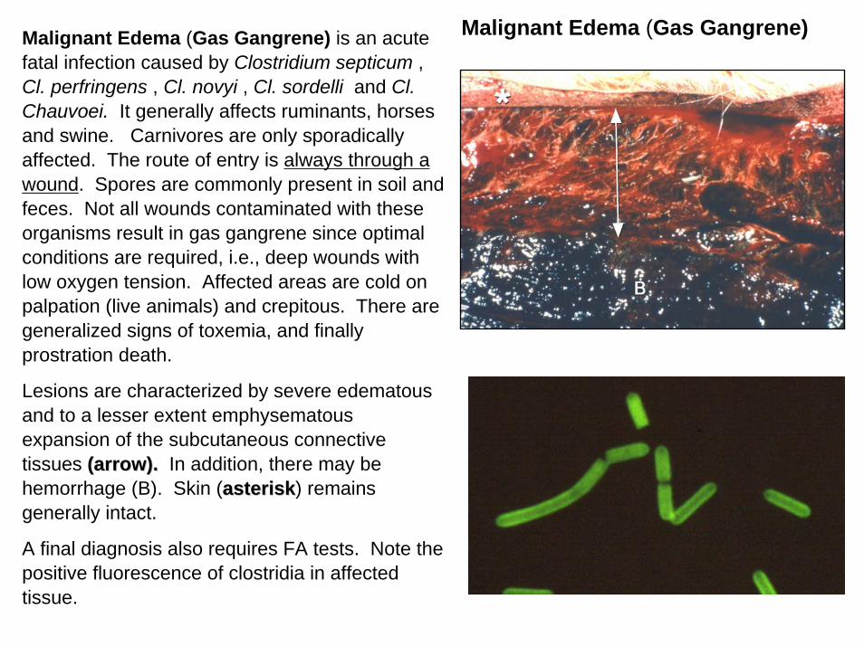
Malignant Edema (Gas Gangrene)Malignant Edema (Gas Gangrene) is an acute fatal infection caused by Clostridium septicum , Cl. perfringens , Cl. novyi , Cl. sordelli and Cl. Chauvoei. It generally affects ruminants, horses and swine. Carnivores are only sporadically affected. The route of entry is always through a wound. Spores are commonly present in soil and feces. Not all wounds contaminated with these organisms result in gas gangrene since optimal conditions are required, i.e., deep wounds with low oxygen tension. Affected areas are cold on palpation (live animals) and crepitous. There are generalized signs of toxemia, and finally prostration death.
Lesions are characterized by severe edematous and to a lesser extent emphysematous expansion of the subcutaneous connective tissues (arrow).(arrow). In addition, there may be hemorrhage (B). Skin (asteriskasterisk) remains generally intact.
A final diagnosis also requires FA tests. Note the positive fluorescence of clostridia in affected tissue.
Parasitic MyositisParasitic Myositis
•• Trichinosis:Trichinosis:
– Trichinella spiralis
•• Cysticercosis:Cysticercosis:
– Cysticercus bovis
– Cyticerucus cellulosae
– Cysticerucs ovis
•• SarcocystisSarcocystis
•• NeosporaNeospora
•• ToxoplasmaToxoplasma
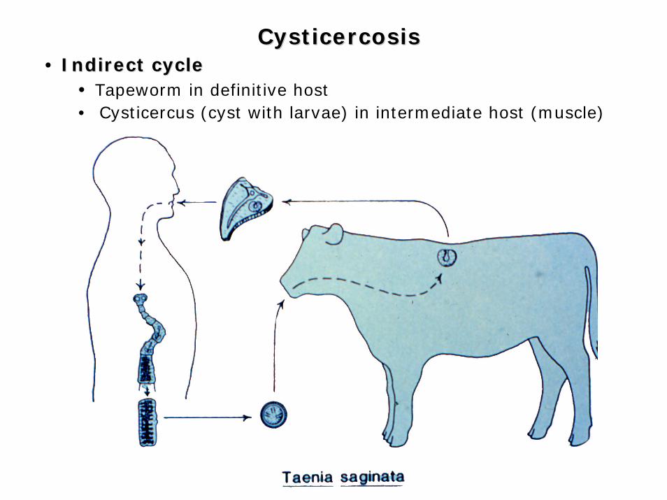
CysticercosisCysticercosis• Indirect cycleIndirect cycle
• Tapeworm in definitive host• Cysticercus (cyst with larvae) in intermediate host (muscle)
CysticercosisCysticercosis
UNAM
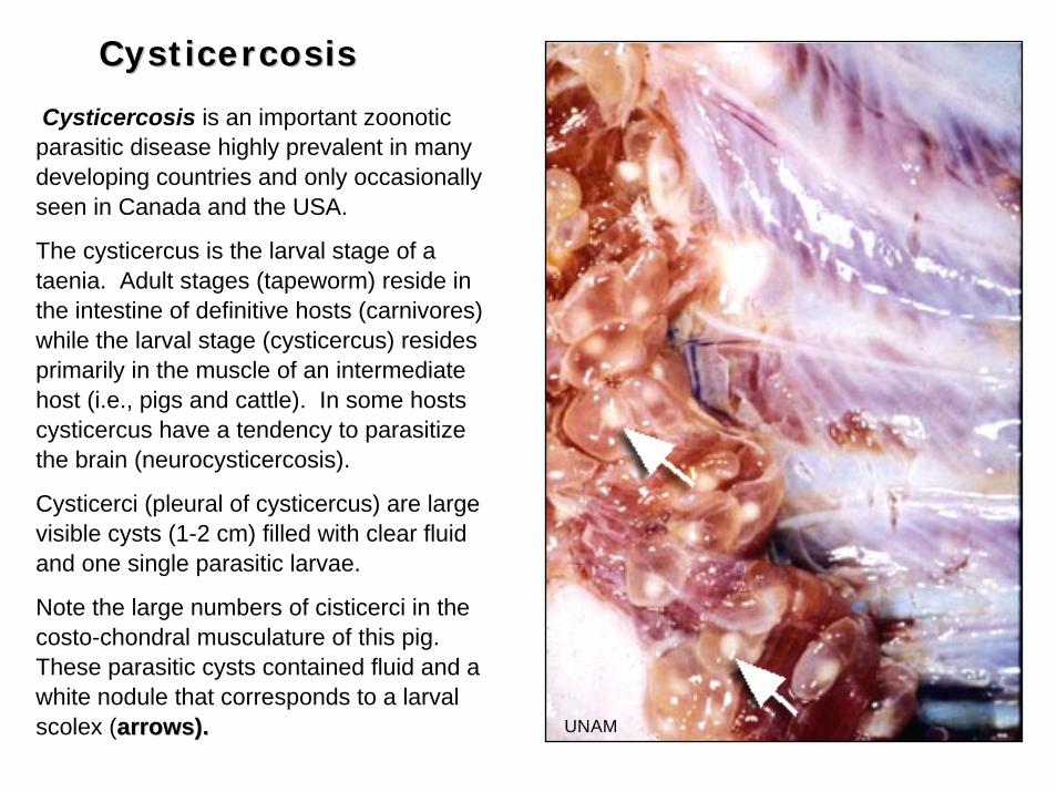
Cysticercosis is an important zoonoticparasitic disease highly prevalent in many developing countries and only occasionally seen in Canada and the USA.
The cysticercus is the larval stage of a taenia. Adult stages (tapeworm) reside in the intestine of definitive hosts (carnivores) while the larval stage (cysticercus) resides primarily in the muscle of an intermediate host (i.e., pigs and cattle). In some hosts cysticercus have a tendency to parasitize the brain (neurocysticercosis).
Cysticerci (pleural of cysticercus) are large visible cysts (1-2 cm) filled with clear fluid and one single parasitic larvae.
Note the large numbers of cisticerci in the costo-chondral musculature of this pig. These parasitic cysts contained fluid and a white nodule that corresponds to a larval scolex (arrows).arrows).
CysticercosisCysticercosis
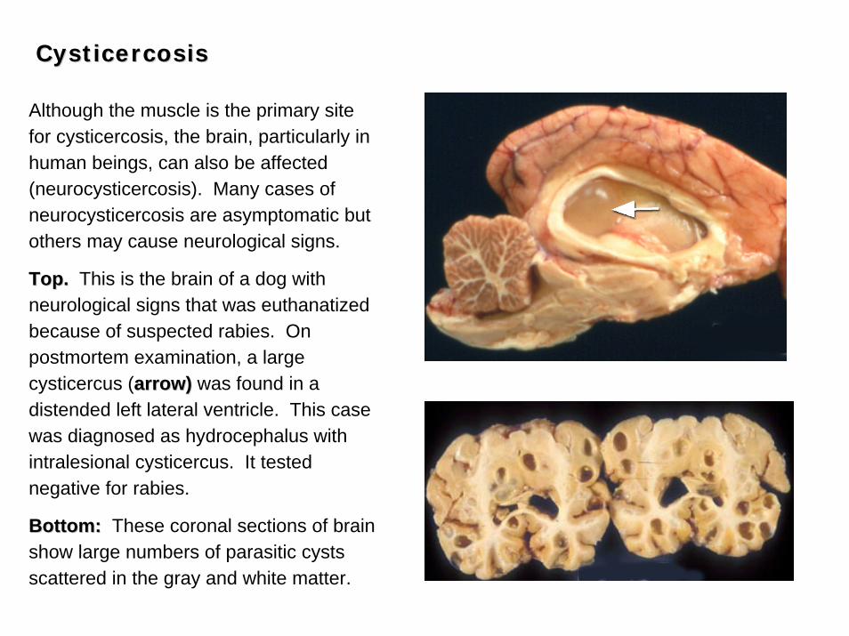
Although the muscle is the primary site for cysticercosis, the brain, particularly in human beings, can also be affected (neurocysticercosis). Many cases of neurocysticercosis are asymptomatic but others may cause neurological signs.
Top.Top. This is the brain of a dog with neurological signs that was euthanatized because of suspected rabies. On postmortem examination, a large cysticercus (arrow)arrow) was found in a distended left lateral ventricle. This case was diagnosed as hydrocephalus with intralesional cysticercus. It tested negative for rabies.
Bottom:Bottom: These coronal sections of brain show large numbers of parasitic cysts scattered in the gray and white matter.
TrichinellaTrichinella spiralisspiralis
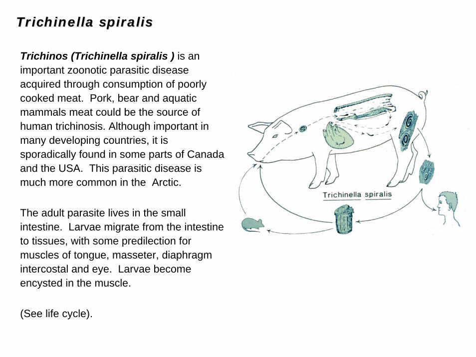
Trichinos (Trichinella spiralis ) is an important zoonotic parasitic disease acquired through consumption of poorly cooked meat. Pork, bear and aquatic mammals meat could be the source of human trichinosis. Although important in many developing countries, it is sporadically found in some parts of Canada and the USA. This parasitic disease is much more common in the Arctic.
The adult parasite lives in the small intestine. Larvae migrate from the intestine to tissues, with some predilection for muscles of tongue, masseter, diaphragm intercostal and eye. Larvae become encysted in the muscle.
(See life cycle).
TrichinellaTrichinella spiralisspiralis
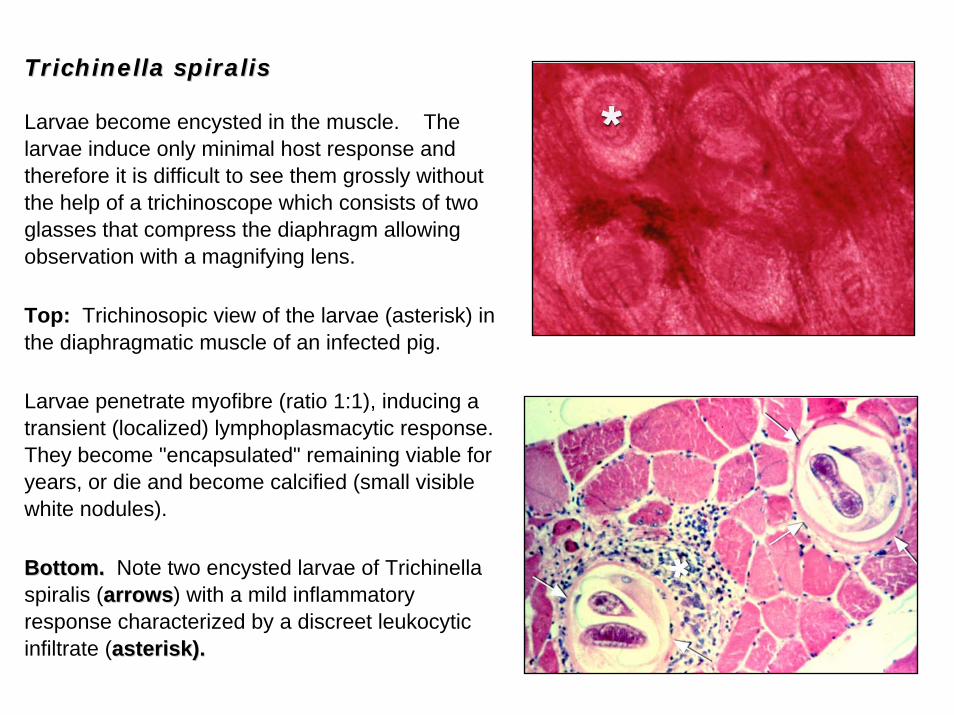
Larvae become encysted in the muscle. The larvae induce only minimal host response and therefore it is difficult to see them grossly without the help of a trichinoscope which consists of two glasses that compress the diaphragm allowing observation with a magnifying lens.
Top: Trichinosopic view of the larvae (asterisk) in the diaphragmatic muscle of an infected pig.
Larvae penetrate myofibre (ratio 1:1), inducing a transient (localized) lymphoplasmacytic response. They become "encapsulated" remaining viable for years, or die and become calcified (small visible white nodules).
Bottom.Bottom. Note two encysted larvae of Trichinellaspiralis (arrowsarrows) with a mild inflammatory response characterized by a discreet leukocytic infiltrate (asterisk).asterisk).
SarcocystisSarcocystis
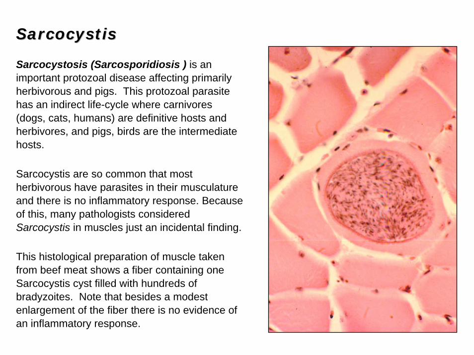
Sarcocystosis (Sarcosporidiosis ) is an important protozoal disease affecting primarily herbivorous and pigs. This protozoal parasite has an indirect life-cycle where carnivores (dogs, cats, humans) are definitive hosts and herbivores, and pigs, birds are the intermediate hosts.
Sarcocystis are so common that most herbivorous have parasites in their musculature and there is no inflammatory response. Because of this, many pathologists considered Sarcocystis in muscles just an incidental finding.
This histological preparation of muscle taken from beef meat shows a fiber containing one Sarcocystis cyst filled with hundreds of bradyzoites. Note that besides a modest enlargement of the fiber there is no evidence of an inflammatory response.
SarcocystisSarcocystis
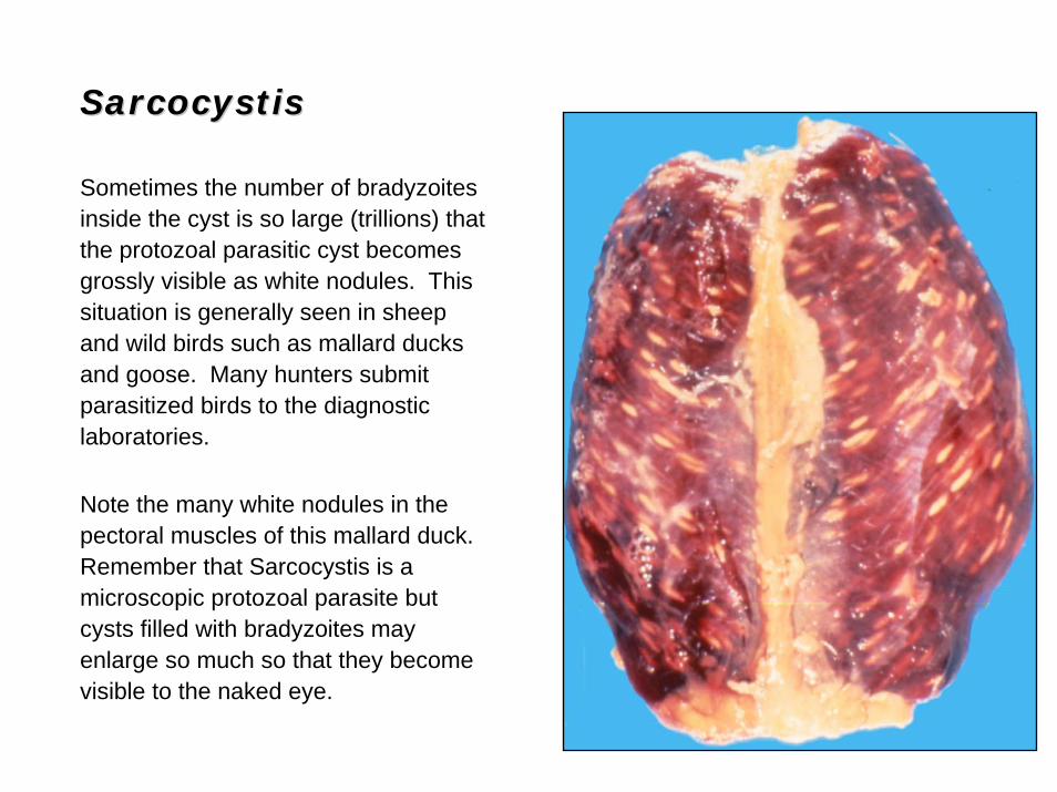
Sometimes the number of bradyzoites inside the cyst is so large (trillions) that the protozoal parasitic cyst becomes grossly visible as white nodules. This situation is generally seen in sheep and wild birds such as mallard ducks and goose. Many hunters submit parasitized birds to the diagnostic laboratories.
Note the many white nodules in the pectoral muscles of this mallard duck. Remember that Sarcocystis is a microscopic protozoal parasite but cysts filled with bradyzoites may enlarge so much so that they become visible to the naked eye.
Eosinophilic MyositisEosinophilic Myositis
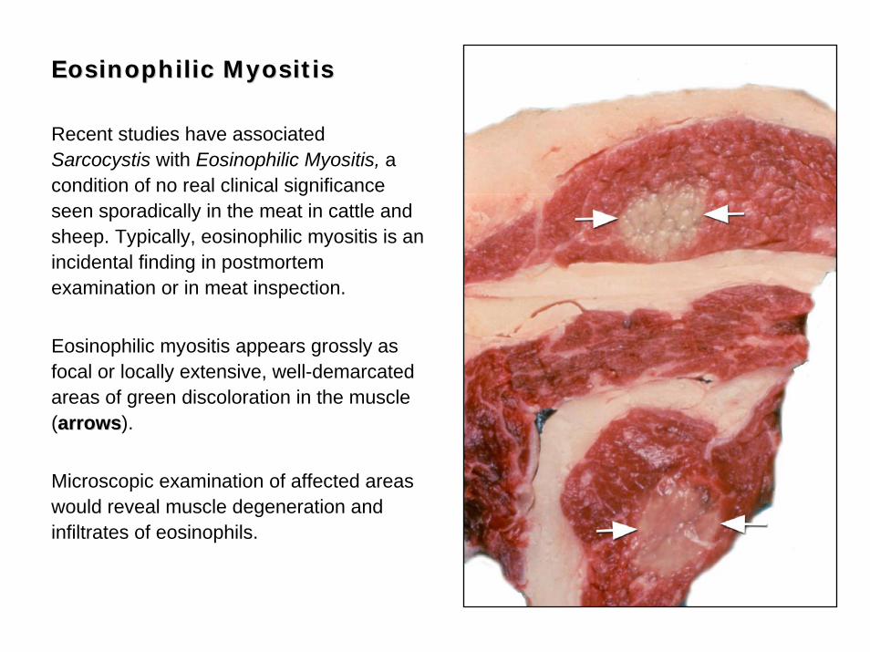
Recent studies have associated Sarcocystis with Eosinophilic Myositis, a condition of no real clinical significance seen sporadically in the meat in cattle and sheep. Typically, eosinophilic myositis is an incidental finding in postmortem examination or in meat inspection.
Eosinophilic myositis appears grossly as focal or locally extensive, well-demarcated areas of green discoloration in the muscle (arrowsarrows).
Microscopic examination of affected areas would reveal muscle degeneration and infiltrates of eosinophils.
Masticatory Muscle Masticatory Muscle MyositisMyositis
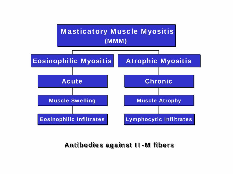
Masticatory Muscle Myositis (MMM) is a rare and acute (relapsing) eosinophilic myositis in dogs characterized by swollen painful jaws, and blood eosinophilia. Another progressive condition called atrophic myositis has also been described in dogs (long-nosed breeds). Some investigators suggest that MMM is the same condition as atrophic myositis but at a different stage of development (Acute vs. Chronic).
The pathogenesis is not completely understood but auto-antibodies against a particular protein present in fibers IIM (unique fiber in masticatory muscles) has been suggested. This specific type of myosin shares antigenic determinants with some bacteria and this may explain why antibodies are formed.
Lesions are typically bilateral. In acute cases there is edema and extensive infiltration of eosinophils in the masseter, temporal, pterygoid muscles. In the chronic form, the cellular infiltrates in these muscles change to lymphocytes and plasma cells and the fibers become atrophic.
Eosinophilic Infiltrates
Muscle Swelling
Acute
Eosinophilic Myositis
Lymphocytic Infiltrates
Muscle Atrophy
Chronic
Atrophic Myositis
Masticatory Muscle Myositis(MMM)
Antibodies against II-M fibersAntibodies against IIAntibodies against II--M fibersM fibers
Masticatory Muscle MyositisMasticatory Muscle Myositis
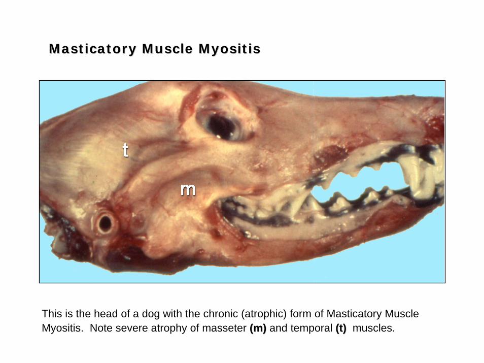
This is the head of a dog with the chronic (atrophic) form of Masticatory Muscle Myositis. Note severe atrophy of masseter (m)(m) and temporal (t)(t) muscles.
Masticatory Muscle MyositisMasticatory Muscle Myositis
HistopathologyHistopathology
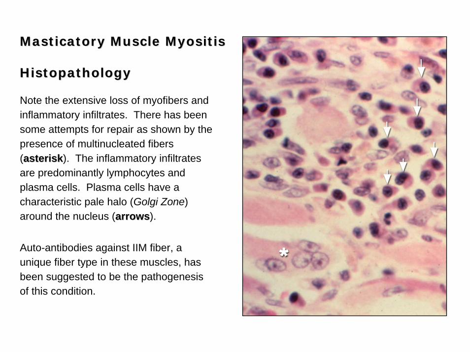
Note the extensive loss of myofibers and inflammatory infiltrates. There has been some attempts for repair as shown by the presence of multinucleated fibers (asteriskasterisk). The inflammatory infiltrates are predominantly lymphocytes and plasma cells. Plasma cells have a characteristic pale halo (Golgi Zone) around the nucleus (arrowsarrows).
Auto-antibodies against IIM fiber, a unique fiber type in these muscles, has been suggested to be the pathogenesis of this condition.
Myasthenia GravisMyasthenia Gravis
•• Rare; Dogs, cats and humansRare; Dogs, cats and humans
•• Neuromuscular junction diseaseNeuromuscular junction disease
• Acethylcholine
•• Clinical signs:Clinical signs:
•• Weakness, fatigue, Weakness, fatigue, dysphagiadysphagia
•• Two types:Two types:
• HereditaryHereditary: Reduced number of Ach receptors
• AcquiredAcquired: Antibodies IgG-Ach receptor
•• LesionsLesions:
• No significant changes in muscle
• Megaesophagus in dogs
• Aspiration pneumonia
Tumors of MuscleTumors of Muscle
•• Spontaneous tumors of striated muscles are rare in veterinary Spontaneous tumors of striated muscles are rare in veterinary
medicine but common in experimental pathology (toxicology). medicine but common in experimental pathology (toxicology).
•• Neoplasia arising in muscle may include tumors from striated musNeoplasia arising in muscle may include tumors from striated muscle cle
((rhabdomyomarhabdomyoma, , rhabdomyosarcomarhabdomyosarcoma), adipose cells (), adipose cells (lipomalipoma/sarcoma), /sarcoma),
fibrous connective tissue (fibrous connective tissue (fibromafibroma/sarcoma), nerves (/sarcoma), nerves (neurofibromaneurofibroma) or ) or
vascular cells (vascular cells (hemangiomahemangioma/sarcoma)./sarcoma).
RhabdomyomaRhabdomyoma
• Benign• Most common in cattle, sheep and pigs.• Generally congenital• Common in the heart• Pedunculated mass

RhabdomyosarcomaRhabdomyosarcoma
• Malignant
• Cattle, sheep, dog and horses
• Large poorly encapsulated nodules
• Prone to metastasis
Note the neoplastic mass infiltrating the muscles in the leg.
Rhabdomyosarcoma are generally pale in color and poorly encapsulated as shown in this photograph (asteriskasterisk).
Histopathology is required for a confirmatory diagnosis (see next frame).
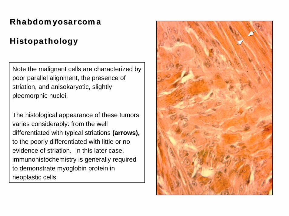
RhabdomyosarcomaRhabdomyosarcoma
HistopathologyHistopathology
Note the malignant cells are characterized by poor parallel alignment, the presence of striation, and anisokaryotic, slightly pleomorphic nuclei.
The histological appearance of these tumors varies considerably: from the well differentiated with typical striations (arrows),(arrows),to the poorly differentiated with little or no evidence of striation. In this later case, immunohistochemistry is generally required to demonstrate myoglobin protein in neoplastic cells.
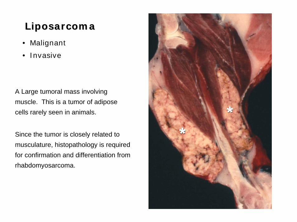
LiposarcomaLiposarcoma
• Malignant
• Invasive
A Large tumoral mass involving muscle. This is a tumor of adipose cells rarely seen in animals.
Since the tumor is closely related to musculature, histopathology is required for confirmation and differentiation from rhabdomyosarcoma.
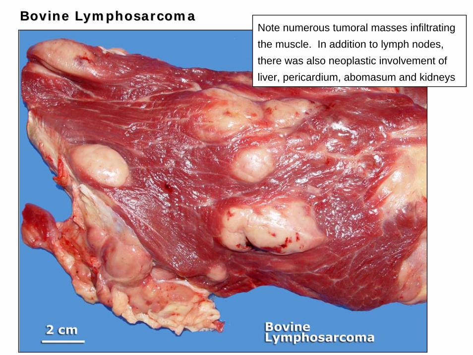
Note numerous tumoral masses infiltrating the muscle. In addition to lymph nodes, there was also neoplastic involvement of liver, pericardium, abomasum and kidneys
Bovine Bovine LymphosarcomaLymphosarcoma

The EndThe End