Main pulmonary artery that is filled with thromboembolic material.
djk23
Text Box
Usually emboli are thrombotic in origin, but they can be from other sources
Pulmonary Embolus with lines of Zahn (H&E)
djk23
Callout
attachment of embolus to vessel wall
djk23
Text Box
the lines of Zahn indicate that the embolus was formed in situ and is not a postmortem clot. They occur over time as RBC's and Platelets mixed with fibrin add onto the thrombus as blood flows over it. Think of it as similar to the way an oyster makes pearls, except that these thrombi can kill you when they embolize get lodged in your lungs.
Pulmonary arteriogram,
with right lower lobe
filling defect
djk23
Callout
looks normal
djk23
Callout
ehh... not so normal here
djk23
Text Box
CT angiograms are probably going to replace this type of arteriogram
Pulmonary infarct, right lung
djk23
Text Box
10% of pulmonary emboli cases lead to a pulmonary infarct. This number is so low because the lung has dual blood supply (remember from normal body)
djk23
Callout
commonly wedge-shaped with apex pointing towards the hilum
djk23
Text Box
usually pulmonary infarcts arise when patients have systemic diseases such as CHF. This type of disease will lead to less blood getting into the lungs through the bronchial arteries (thus less dual supply) and it also predisposes an individual to forming venous clots (usually in deep leg veins) because of an overall increase in stasis. (remember Virchow's triad: Stasis (or other hemodynamic changes like turbulence), endothelial damage, and hypercoagulability)
PULMONARY EMBOLI OF OTHER
TYPES
• Fat emboli (injury to long bones)
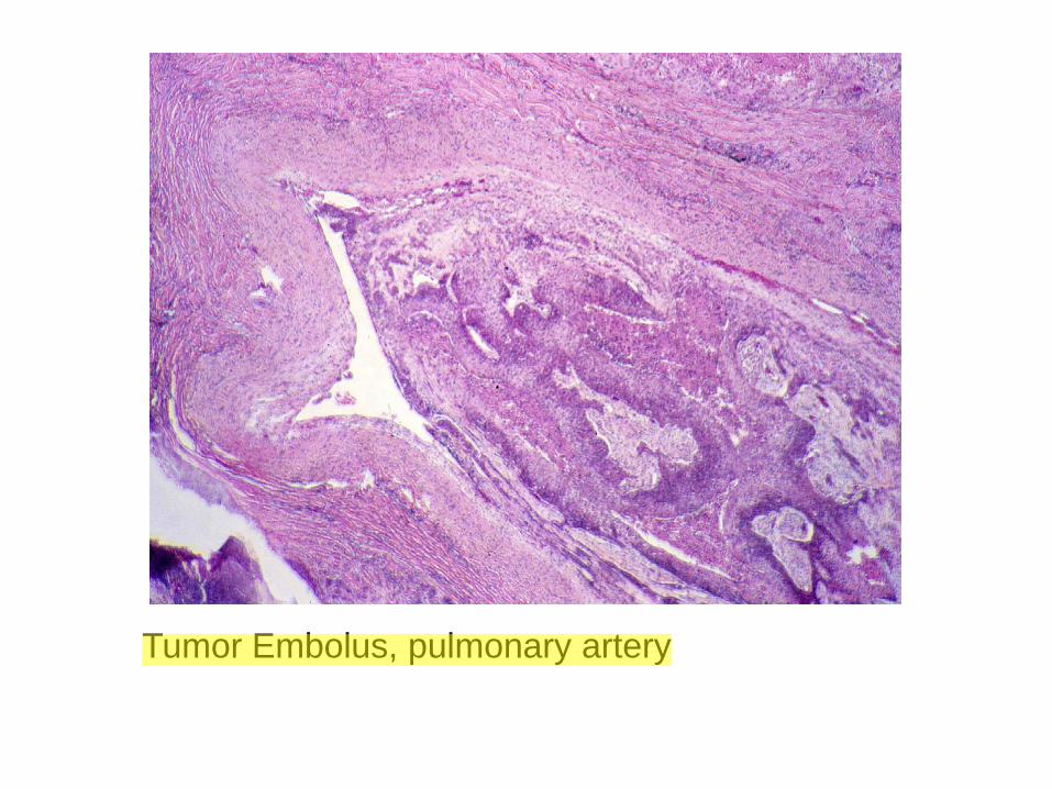
• Tumor emboli (metastatic cancer)
• Air emboli (venous system)
• Talc emboli (IV drug abuse)
• Amniotic fluid emboli (pregnancy
complication)
djk23
Text Box
Also during surgery (like hip replacement)
djk23
Text Box
usually an iatrogenic problem
djk23
Text Box
using drugs meant for oral consumption inappropriately by crushing them up and injecting them. Bad for probably more than one reason, but for this lecture we are worried about the filler material in the drug acting as an embolus
Fat emboli, lung (osmium post fixation)
djk23
Highlight
djk23
Text Box
in addition to the fat blocking the vessels, lipases in the endothelium become activated and convert the fat into fatty acids which can cause further damage downstream in the microvasculature leading to pulmonary edema (add that to your differential diagnosis of pulmonary edema, haha). This is a common problem in people with massive fat emboli. Usually occurs 24 hrs after an ortho procedure.
djk23
Text Box
remember this may happen when long bones are broken or when people have orthopedic surgery (hip replacement)
Can embolize into the small vessels and lead to a granulomatous rxn that can cause pulmonary hypertension.
PULMONARY IN SITU
THROMBOSIS
• Most often occurs in malignancy
(hypercoagulation) or pulmonary
hypertension
• Thrombi are pale and form “casts” of
arterial tree
djk23
Text Box
"very uncommon"
djk23
Highlight
djk23
Highlight
djk23
Highlight
djk23
Underline
djk23
Text Box
made primarily of platelets and fibrin.
djk23
Highlight
djk23
Callout
remember seeing the cast in the roadshow?
Pulmonary artery thrombosis
djk23
Line
djk23
Highlight
djk23
Text Box
Lymph nodes show metastatic disease (note white tissue around the edges of the black lymph nodes). He had gastric cancer which lead to a hypercoagulable state.
PULMONARY
HYPERTENSION
• Primary pulmonary hypertension
– Pulmonary arterial hypertension (PAH)
– Pulmonary veno-occlusive disease
• Secondary pulmonary hypertension
– Chronic pulmonary emboli
– Congenital heart disease (ASD, VSD)
djk23
Text Box
Used to be a reason for lung transplant, but now there are drugs that can help
djk23
Text Box
problem is of unknown origin but normally arises in the pulmonary system itself
djk23
Text Box
showering emboli from leg veins over a long time
djk23
Text Box
leads to higher right sided pressures
jdo8
Text Box
From FA2011: Bosentan is used to treat Pulm HTN; it competitively antagonizes endothelin-1 receptors, decreasing pulmonary vascular resistance
Pulmonary aterial hypertension elastic stain
djk23
Text Box
Hypertrophy of smooth muscle and hyperplasia of the intima in this artery. This leads to a super small lumen that increases resistance and thus pressure
Pulmonary aterial hypertension
plexiform lesion
djk23
Highlight
djk23
Text Box
Hallmark of hypertension
djk23
Text Box
usually indicates emboli that have been re-canalized
Recanalized pulmonary embolus,
chronic thromboembolic disease
djk23
Highlight
Pulmonary veno-occlusive disease,
elastic stain
djk23
Text Box
small veins of the lung become occluded by fibrous tissue. Clinically very difficult to distinguish from the other hypertensive states. This is a problem because it does not respond to the treatment that is used in the other cases
djk23
Text Box
Usually of unknown cause, but can be associated with chemotherapy and inflammation (caused by histoplasmosis for example)
Right ventricular hypertrophy,
primary pulmonary hypertension
djk23
Callout
Thickened and distended RV.
djk23
Text Box
Cor pulmonale: Failure of the right side of the heart due to chronic pulmonary hypertension
DIFFUSE PULMONARY
HEMORRHAGE
• Goodpasture’s syndrome
• Systemic lupus erythematosus
• Wegener’s granulomatosis
• Idiopathic
djk23
Text Box
"not common but important"
djk23
Text Box
usually fatal if not caught in time
djk23
Polygonal Line
djk23
Text Box
both can be treated with corticosteroids and cytotoxic agents
Immunologic Lung Disease
(Gell & Coombs)
Immune
reaction
Mediator Histology Example
Type I Reaginic AB Eosinophils Asthma
Type II Cytotoxic AB Alveolar
hemorrhage
Goodpasture’s
Syn
Type III Immune
complexes
Vasculitis SLE
Type IV Sensitized
lymphs
Granulomas Sarcoidosis
HP
djk23
Text Box
IgE
djk23
Text Box
Ab's directed against glomeruli and alveolar basement membranes
djk23
Text Box
IgG
djk23
Highlight
djk23
Rectangle
djk23
Callout
another example is autoimmune hemolytic anemia
Goodpasture’s
Syndrome
djk23
Text Box
congested blood filled lung
djk23
Text Box
Young males in their 30's have a flu like symptoms and then present with extreme SOB and sometimes hemoptysis
Goodpasture’s Syndrome with
intra-alveolar hemorrhage
djk23
Text Box
Under a light microscope, the alveoli look pretty normal (minus all the blood of course, but the septa look fine). May see "revved" up type 2 cells responding to the blood
Goodpasture’s Syndrome, lung
IgG immunofluorescence
djk23
Highlight
djk23
Text Box
demonstrates linear deposition
Goodpasture’s Syndrome, kidney
IgG immunofluorescence
djk23
Text Box
same type of linear deposition
djk23
Highlight
djk23
Text Box
Side Note: A granular (non-linear) type of deposition is indicative of immune complex deposition (type 3). Such as those formed in SLE
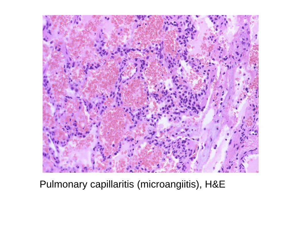
Pulmonary capillaritis (microangiitis), H&E
djk23
Text Box
lupus, wegeners, and iodpathic causes
djk23
Text Box
A case "reverse pneumonia". Neutrophils are in the alveolar capillary walls and RBC's are in the alveolar space
djk23
Text Box
uncommon, but must treat quickly
CLASSIC WEGENER’S
GRANULOMATOSIS
• Necrotizing granulomatous arteritis
of lungs
• Necrotizing inflammation of upper
respiratory tract
• Glomerulonephritis
djk23
Text Box
like the larynx, ear, nose, or eyes
djk23
Text Box
crescentric type glomerulonephritis that is often palci-immune (meaning little or no immune complexes)
Necrobiotic nodule, Wegener’s granulomatosis
djk23
Text Box
fever, chills, SOB, malaise, and loss of apetite
djk23
Highlight
Necrotizing granulomatous arteritis,
Wegener’s (H&E)
djk23
Callout
multinucleate giant cell
Geographic necrosis,
Wegener’s granulomatosis (H&E)
djk23
Text Box
low power
djk23
Text Box
variable in shape and size. Helps distinguish Wegener's from TB and histoplasmosis
Back wall of my den
Obstructive Lung Disease
djk23
Text Box
part 2!
SESSION SPECIFIC OBJECTIVES
• List the major types of obstructive lung
disease
• Recognize and describe the pathology
of obstructive lung disease
– Emphysema, small airways disease, large
airways disease, bronchiectasis, and
asthma
OBSTRUCTIVE LUNG DISEASE
• Emphysema
• Small airways disease
• Large airways disease
• Bronchiectasis
• Asthma
djk23
Polygonal Line
djk23
Text Box
These three can be caused by smoking
CLASSIFICATION OF
EMPHYSEMA
• Centrilobular (85%)
• Panlobular (5%)
• Paracicatricial (5%)
• Localized (5%)
djk23
Text Box
Anatomical definition: dilation of the airways of the lung associated with destruction of the lung parenchyma
djk23
Text Box
Centriacinar
djk23
Text Box
Panacinar
djk23
Text Box
irregular
djk23
Text Box
paraseptal or distal
djk23
Callout
mostly caused by smoking
Centrilobular emphysema (gross)
djk23
Text Box
Commonly begins in the central part of the secondary lobule, but can spread to destroy the whole lobule
djk23
Text Box
example of a bolus (at least 2cm across)
djk23
Callout
What is left of a bronchiole in the center of what used to be a secondary lobule
Whole lobule. Often worse in lower lobes (reason to come)
Liver with globules of alpha-1 antitrypsin
(PAS Stain)
djk23
Highlight
djk23
Highlight
djk23
Text Box
Hepatocytes can't export alpha-1 antitrypsin because of the z mutation in alpha-1-antitrypsin that prevents attachment of carbohydrates important for intracellular trafficking through the ER
Ultrastructure of
liver in alpha-1
antitrypsin deficiency
Paracicatricial
emphysema,
tuberculosis
djk23
Text Box
"beside a scar"
djk23
Text Box
Tb scar that leads to emphysematous destruction around it
Localized emphysema (gross)
djk23
Text Box
only in one area of the lung. Not know why (possibly due to bacteria like pseudomonas that produce elastases). Quick question: who remembers from our single day of antibiotics how to treat pseudomonas? (answer on next slide)
djk23
Text Box
A: Ceftazidime, cefepime, aztreonam, quinolones (but there is rising resistance) aminoglycosides, piperacillin, ticarcillin, carbapenems (but not ertapenem), and probably some others. I think if you remebered any of those you're in good shape
djk23
Text Box
General mechanism of emphysema is an imbalance of proteases and antiproteases. Antiproteases from the blood stream. Proteases released from inflammatory cells
bfg2
Text Box
the lung is the graveyard of neutrophils-- antiproteases are crucial for balancing the release of proteases from dying neutrophils
bfg2
Text Box
smoking also irreversibly blocks the active site of alpha1-antitrypsin, increasing imbalance of protease-antiprotease
Autofluorescent smoker’s macrophages
Ultrastructure of smoker’s macrophages
with numerous secondary lysosomes
bfg2
Text Box
containing particulate material
CONSEQUENCES OF
EMPHYSEMA
• Pulmonary obstruction (TLC,
FEV1)
• Diminished elastic recoil
• Diminished DLCO
djk23
Highlight
djk23
Highlight
bfg2
Text Box
bronchioles held open in expiration by elastic tissue-- without it, they tend to collapse earlier in expiration
Centrilobular emphysema
post-mortem pulmonary
artery injection
bfg2
Text Box
diffusion capacity diminishes with vascular destruction
bfg2
Text Box
larger vessels remain intact, but capillaries and small vessels are lost
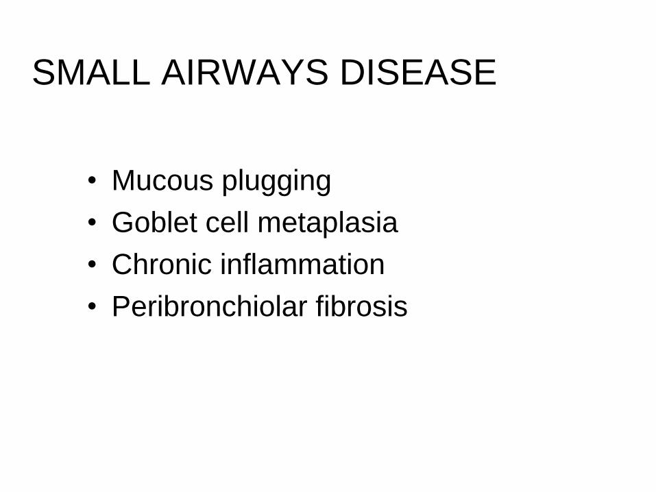
SMALL AIRWAYS DISEASE
• Mucous plugging
• Goblet cell metaplasia
• Chronic inflammation
• Peribronchiolar fibrosis
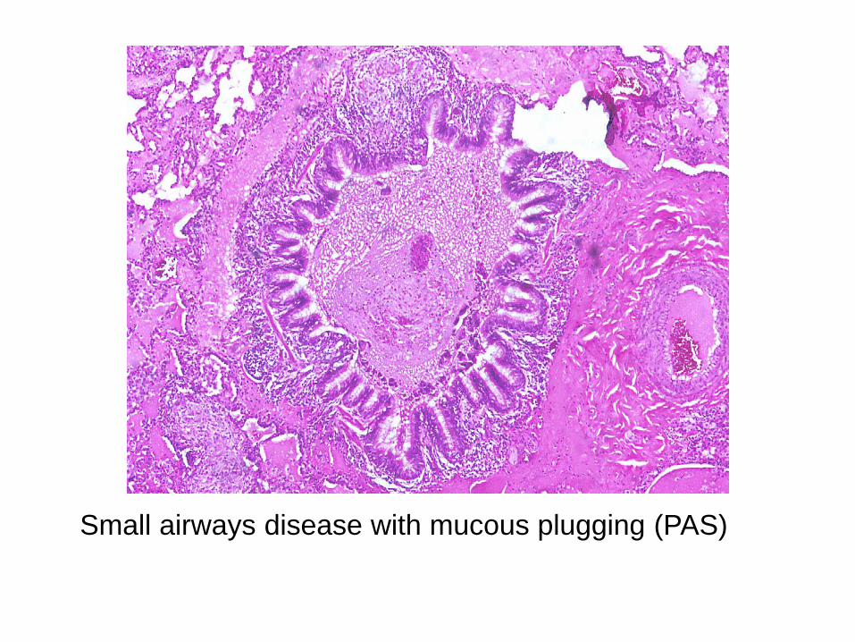
Small airways disease with mucous plugging (PAS)
bfg2
Callout
mononuclear inflammatory infiltrate in the walls
Small airways disease with goblet cell metaplasia
bfg2
Text Box
goblet cells in small airways is a bad thing-- metaplasia
Respiratory bronchiolitis with numerous smokers macrophages
Bronchiolitis obliterans secondary to Adenovirus infection
bfg2
Callout
what's left of the airway
bfg2
Callout
fibrous tissue filling what used to be the bronchiole
bfg2
Callout
Smooth Muscle outline of what was the bronchiole
djk23
Line
bfg2
Text Box
At Duke, it's often due to transplant (lung or BM (GVHD))
bfg2
Text Box
not really associated with cigarette smoking
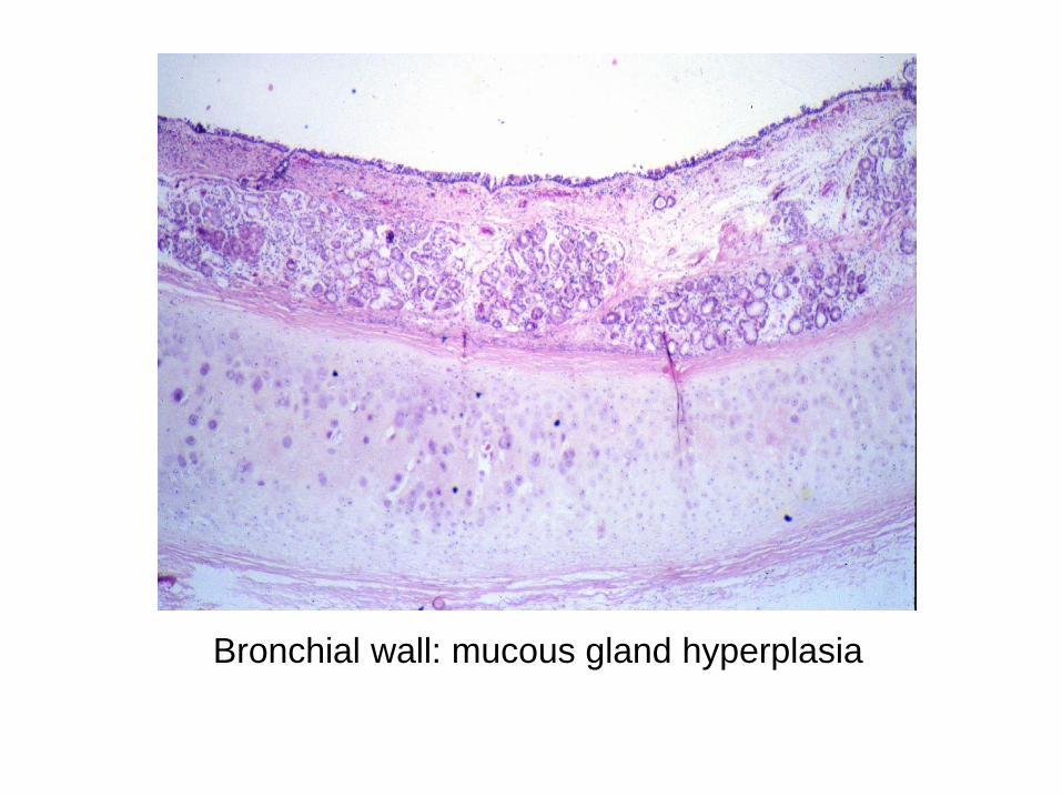
Bronchial wall: mucous gland hyperplasia
bfg2
Text Box
mucosa is more than 50% mucous glands
bfg2
Text Box
chronic bronchitis-- productive cough that occurs for at least 3 months per year for 2 years that doesn't have a better explanation
Chronic bronchitis with dilated mucous duct
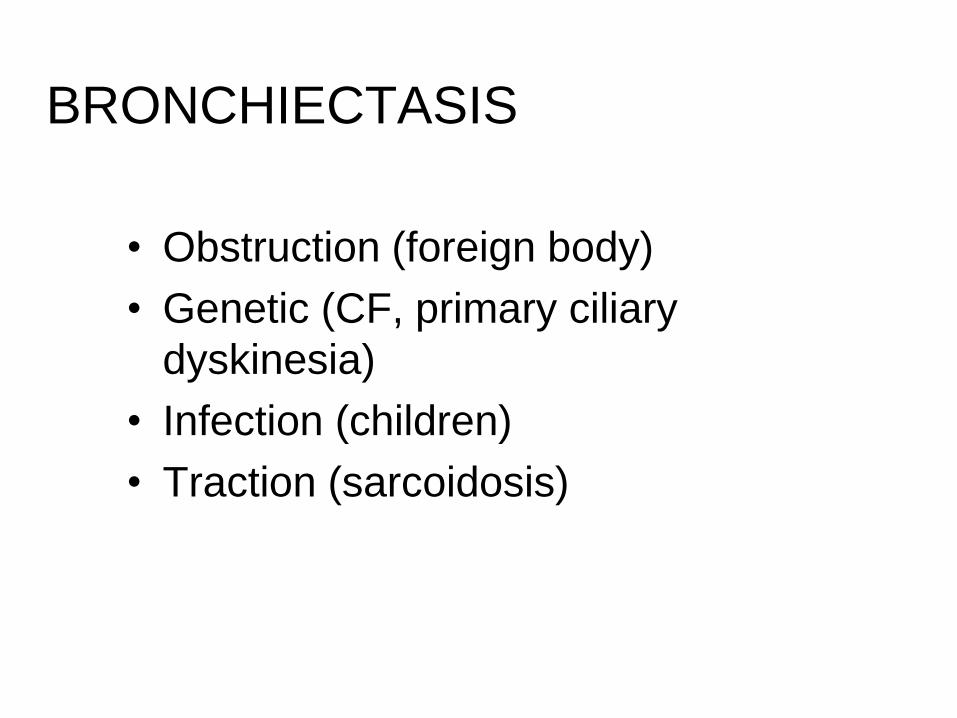
BRONCHIECTASIS
• Obstruction (foreign body)
• Genetic (CF, primary ciliary
dyskinesia)
• Infection (children)
• Traction (sarcoidosis)
bfg2
Text Box
bronchi become abnormally dilated and fail to taper as they go further into the lung
bfg2
Text Box
in fibrotic lung diseases, as fibrotic scars shrink, they pull open the bronchi
Bronchiectasis (gross)
bfg2
Text Box
transverse ribbing-- atrophy of longitudinal smooth muscle and hypertrophy of circular smooth muscle
Bronchiectasis secondary to peanut
aspiration (gross)
bfg2
Callout
note peanut.
Saccular bronchiectasis cystic fibrosis
bfg2
Text Box
many bronchi occluded by mucoid secretions
bfg2
Text Box
red is mucous protein, green is DNA
bfg2
Text Box
DNA is mostly (80%) from host neutrophils, but also some (20%) from bacteria
Bronchiectasis, (H&E)
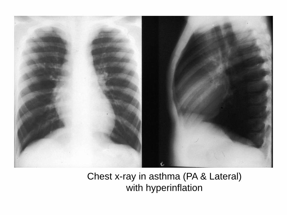
ASTHMA
• Goblet cell metaplasia
• Mucous plugging
• Smooth muscle hyperplasia
• Thickened basement membrane
• ± eosinophils
Chest x-ray in asthma (PA & Lateral)
with hyperinflation
bfg2
Text Box
lungs hyperinflated-- diaphragm shouldn't be flat
Status asthmaticus with mucous plugging
(H&E)
Bronchial mucous plug (asthma) with
Charcot-Leyden crystals
bfg2
Text Box
made by eosinophils, indicative of an allergic rxn


![NWSMT NewsNotes Bowman Gray[1]southernmodified.com/2010/NWSMTBowman_Gray.pdf · Bristol Motor Speedway will host the combination event — bringing to-gether the NASCAR Whelen Modified](https://img.pdfslide.us/doc/110x75/604804256cd9145f7816a03f/nwsmt-newsnotes-bowman-gray1-bristol-motor-speedway-will-host-the-combination.jpg)