Embed Size (px)
Citation preview
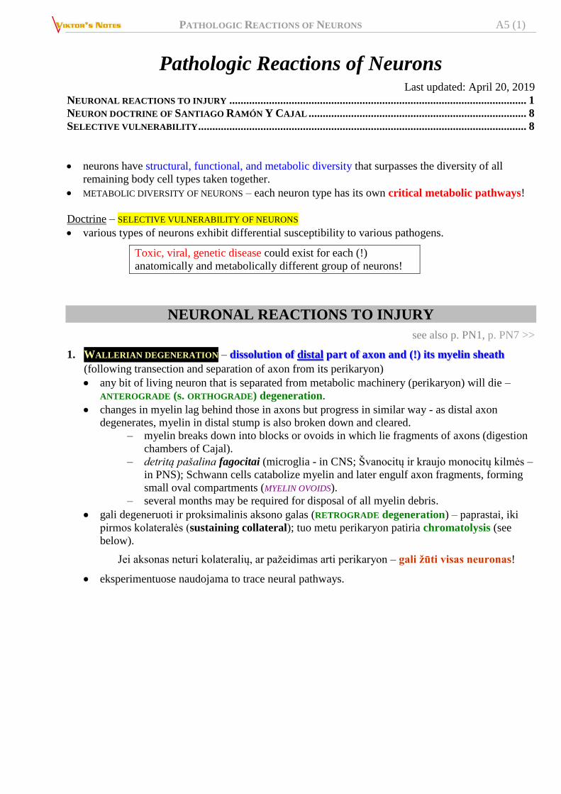
PATHOLOGIC REACTIONS OF NEURONS A5 (1)
Pathologic Reactions of Neurons Last updated: April 20, 2019
NEURONAL REACTIONS TO INJURY ......................................................................................................... 1 NEURON DOCTRINE OF SANTIAGO RAMÓN Y CAJAL ............................................................................. 8 SELECTIVE VULNERABILITY .................................................................................................................... 8
neurons have structural, functional, and metabolic diversity that surpasses the diversity of all
remaining body cell types taken together.
METABOLIC DIVERSITY OF NEURONS – each neuron type has its own critical metabolic pathways!
Doctrine – SELECTIVE VULNERABILITY OF NEURONS
various types of neurons exhibit differential susceptibility to various pathogens.
Toxic, viral, genetic disease could exist for each (!)
anatomically and metabolically different group of neurons!
NEURONAL REACTIONS TO INJURY
see also p. PN1, p. PN7 >>
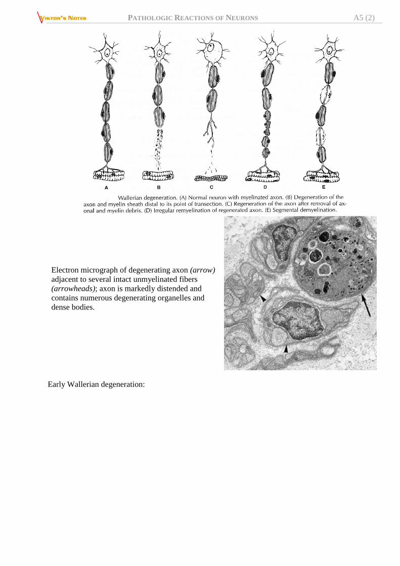
1. WALLERIAN DEGENERATION – dissolution of distal part of axon and (!) its myelin sheath
(following transection and separation of axon from its perikaryon)
any bit of living neuron that is separated from metabolic machinery (perikaryon) will die –
ANTEROGRADE (s. ORTHOGRADE) degeneration.
changes in myelin lag behind those in axons but progress in similar way - as distal axon
degenerates, myelin in distal stump is also broken down and cleared.
– myelin breaks down into blocks or ovoids in which lie fragments of axons (digestion
chambers of Cajal).
– detritą pašalina fagocitai (microglia - in CNS; Švanocitų ir kraujo monocitų kilmės –
in PNS); Schwann cells catabolize myelin and later engulf axon fragments, forming
small oval compartments (MYELIN OVOIDS).
– several months may be required for disposal of all myelin debris.
gali degeneruoti ir proksimalinis aksono galas (RETROGRADE degeneration) – paprastai, iki
pirmos kolateralės (sustaining collateral); tuo metu perikaryon patiria chromatolysis (see
below).
Jei aksonas neturi kolateralių, ar pažeidimas arti perikaryon – gali žūti visas neuronas!
eksperimentuose naudojama to trace neural pathways.
PATHOLOGIC REACTIONS OF NEURONS A5 (2)
Electron micrograph of degenerating axon (arrow)
adjacent to several intact unmyelinated fibers
(arrowheads); axon is markedly distended and
contains numerous degenerating organelles and
dense bodies.
Early Wallerian degeneration:
PATHOLOGIC REACTIONS OF NEURONS A5 (3)
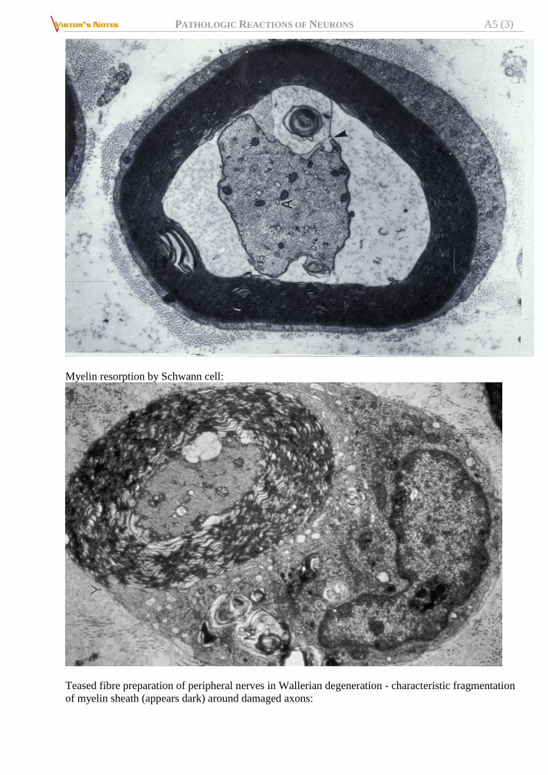
Myelin resorption by Schwann cell:
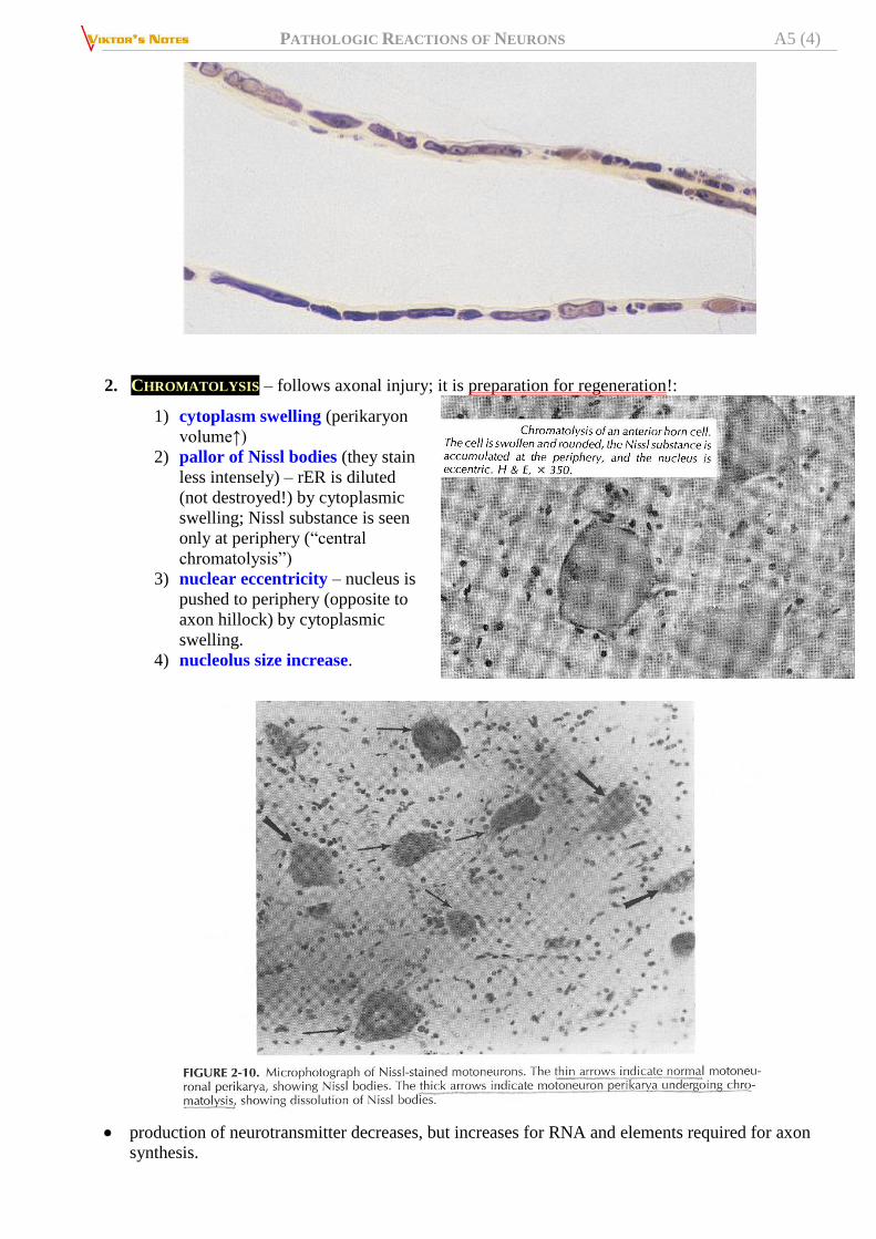
Teased fibre preparation of peripheral nerves in Wallerian degeneration - characteristic fragmentation
of myelin sheath (appears dark) around damaged axons:
PATHOLOGIC REACTIONS OF NEURONS A5 (4)
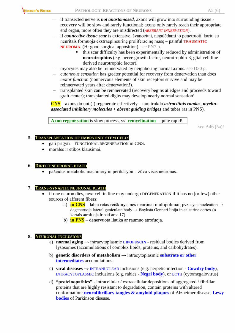
2. CHROMATOLYSIS – follows axonal injury; it is preparation for regeneration!:
1) cytoplasm swelling (perikaryon
volume↑)
2) pallor of Nissl bodies (they stain
less intensely) – rER is diluted
(not destroyed!) by cytoplasmic
swelling; Nissl substance is seen
only at periphery (“central
chromatolysis”)
3) nuclear eccentricity – nucleus is
pushed to periphery (opposite to
axon hillock) by cytoplasmic
swelling.
4) nucleolus size increase.
production of neurotransmitter decreases, but increases for RNA and elements required for axon
synthesis.
PATHOLOGIC REACTIONS OF NEURONS A5 (5)
presynaptic terminals gradually withdraw from such soma and dendrites (synaptic transmission is
reduced).
3. DISTAL DEGENERATION (s. DISTAL AXONOPATHY, “DYING BACK”)
- occurs when neuron metabolic machinery is disrupted; most distal parts begin to degenerate.
4. REGENERATION
NEURONS are postmitotic cells incapable of cell division - mature neurons do not multiply, except:
1) olfactory receptor cells
2) ganglion cells in enteric NS
3) neuroepithelial cells of taste buds
PROXIMAL AXONAL STUMP (vs. PERIKARYON) may regenerate and establish synaptic connection.
PNS – regeneruoti gali ir motoriniai, ir sensoriniai aksonai: also see p. PN7 >>
– įvykus wallerian degeneration, švanocitai nežūva (nors mielinas suyra!) – proliferuoja
ir sudaro guiding Schwann tubes.
– ties jung. audinio randu (jeigu jis nestoras) švanocitai pasidaugina ir formuoja
guiding cellular bridges (BÜNGNER’S bands) link proximal stump.
BÜNGNER’S bands: basal lamina lined endoneurial tubes, enclosing
columns of proliferating Schwann cells.
– AXONAL SPROUTING – 24 val. post injury bėgyje iš proximal stump išauga daug naujų
nemielinizuotų sprouts (neurites); ends of sprouts (growth cones) “sample”
environment, responding positively to facilitatory molecular cues and negatively to
inhibitory ones; several sprouts per cut axon elongate and compete for distal stump
and end-organ; vienas iš jų pataiko į švanocitų kanalą, kiti degeneruoja; 1 axon can
abnormally reinnervate 3-4 end-cells.
Presence of multiple, closely aggregated, thinly myelinated small-caliber
axons is evidence of regeneration (regenerating cluster).
– axon regrowth is slow process (limited by slow component of axonal transport,
movement of tubulin, actin, intermediate filaments):
several days ÷ weeks are required for axon to cross site of anastomosis.
once axons reach distal nerve sheath, regeneration occurs at 1-1.5 mm/d
(axon regeneration near cell body is more rapid than regeneration at
greater distances).
PATHOLOGIC REACTIONS OF NEURONS A5 (6)
– if transected nerve is not anastomosed, axons will grow into surrounding tissue -
recovery will be slow and rarely functional; axons only rarely reach their appropriate
end organ, more often they are misdirected (ABERRANT INNERVATION).
– if connective tissue scar is extensive, švanocitai, negalėdami jo penetruoti, kartu su
neuritais formuoja ekstraepineurinę proliferacinę masę – painful TRAUMATIC
NEUROMA. (H: good surgical apposition). see PN7 p.
this scar difficulty has been experimentally reduced by administration of
neurotrophins (e.g. nerve growth factor, neurotrophin-3, glial cell line-
derived neurotrophic factor).
– myocytes may also be reinnervated by neighboring normal axons. see D30 p.
– cutaneous sensation has greater potential for recovery from denervation than does
motor function (nonnervous elements of skin receptors survive and may be
reinnervated years after denervation!).
– transplanted skin can be reinnervated (recovery begins at edges and proceeds toward
graft center); transplanted digits may develop nearly normal sensation!
CNS – axons do not (!) regenerate effectively – tam trukdo astrocitinis randas, myelin-
associated inhibitory molecules + absent guiding bridges and tubes (as in PNS).
Axon regeneration is slow process, vs. remyelination – quite rapid!
see A46 (5a)!
5. TRANSPLANTATION OF EMBRYONIC STEM CELLS
gali prigyti – FUNCTIONAL REGENERATION in CNS.
moralės ir etikos klausimai.
6. DIRECT NEURONAL DEATH
pažeidus metabolic machinery in perikaryon – žūva visas neuronas.
7. TRANS-SYNAPTIC NEURONAL DEATH
if one neuron dies, next cell in line may undergo DEGENERATION if it has no (or few) other
sources of afferent fibers:
a) in CNS – labai retas reiškinys, nes neuronai multipoliniai; pvz. eye enucleation →
degeneruoja lateral geniculate body → išnyksta Gennari linija in calcarine cortex (o
kartais atrofuoja ir pati area 17) b) in PNS – denervuota liauka ar raumuo atrofuoja.
8. NEURONAL INCLUSIONS a) normal aging → intracytoplasmic LIPOFUSCIN - residual bodies derived from
lysosomes (accumulations of complex lipids, proteins, and carbohydrates).
b) genetic disorders of metabolism → intracytoplasmic substrate or other
intermediates accumulations.
c) viral diseases → INTRANUCLEAR inclusions (e.g. herpetic infection - Cowdry body),
INTRACYTOPLASMIC inclusions (e.g. rabies - Negri body), or BOTH (cytomegalovirus)
d) “proteinopathies” - intracellular / extracellular depositions of aggregated / fibrillar
proteins that are highly resistant to degradation, contain proteins with altered
conformation: neurofibrillary tangles & amyloid plaques of Alzheimer disease, Lewy
bodies of Parkinson disease.

PATHOLOGIC REACTIONS OF NEURONS A5 (7)
9. DENERVATION HYPERSENSITIVITY - hypersensitivity of postsynaptic structure to transmitter previously secreted by damaged axon
ending.
general phenomenon - seen in all types of effector cells:
1) skeletal muscle (muscle also atrophies)
2) smooth muscle (muscle does not atrophy!)
3) exocrine glands (except for sweat glands).
4) lower nervous system centers (after higher centers are destroyed) – hyperactivity is
called "release phenomenon".
mechanism:
1) mainly - synthesis / activation of more receptors.
in denervated skeletal muscle, Acch receptors of fetal γ subunit-containing
type appear over large portions of muscle membrane (normally, only endplate
contains Acch receptors, and they are of adult ε subunit-containing type); these
disappear and sensitivity returns to normal if nerve regrows (motor nerve ending
secretes specific protein agrin); the same is seen during embryonic
development!
N.B. after denervation, endplate sensitivity doesn’t increase!
2) lack of reuptake (at noradrenergic endings) - circulating norepinephrine reaching
receptors has greater effect than it otherwise would.
hypersensitivity is limited to structures immediately innervated by destroyed neurons and fails
to develop in structures farther "downstream";
– suprasegmental spinal cord lesions do not lead to hypersensitivity of paralyzed skeletal
muscles to acetylcholine;
– destruction of preganglionic autonomic nerves does not cause hypersensitivity of denervated
viscera; example in treatment of diseases due to vasospasm: a) if upper extremity is sympathectomized by removing upper part of ganglion chain &
stellate ganglion, hypersensitive vessel walls are stimulated by circulating norepinephrine,
and episodic vasospasm continues to occur.
b) if preganglionic sympathectomy is performed by cutting ganglion chain below 3rd ganglion
(to interrupt ascending preganglionic fibers) & white rami of first three thoracic nerves,
no hypersensitivity results!
Acute neuronal injury (red neuron) 1) cell body shrinkage
2) nucleus pyknosis; nucleus assumes angulated shape of shrunken perikaryon
3) nucleolus disappearance
4) loss of Nissl substance → intense cytoplasm eosinophilia (red neuron)
accompany acute CNS insults that ultimately lead to cell death.
red neurons are evident with H&E preparations at 12-24 hours after irreversible hypoxic /
ischemic insult.
PATHOLOGIC REACTIONS OF NEURONS A5 (8)
"Simple" neuronal atrophy ("degeneration") – selective neuronal death (cell loss) as result of
progressive disease process of long duration → reactive gliosis.
in early stage, cell loss is difficult to detect; associated glial changes are best indicator of
pathologic process at this stage.
GLIOSIS - most important histopathologic indicator of CNS injury (regardless of etiology).
– ASTROCYTES participate by hypertrophy & hyperplasia (reactive, s. gemistocytic
astrocytes).
– in long-standing lesions, astrocytic nuclei lie in dense net of processes (glial "fibrils").
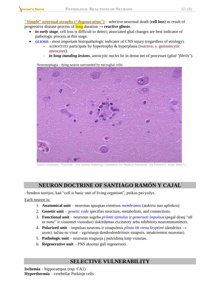
Neuronophagia - dying neuron surrounded by microglial cells:
Source of picture: “WebPath - The Internet Pathology Laboratory for Medical Education” (by Edward C. Klatt, MD) >>
NEURON DOCTRINE OF SANTIAGO RAMÓN Y CAJAL
- bendros teorijos, kad “cell is basic unit of living organism”, puikus pavyzdys.
Each neuron is:
1. Anatomical unit – neuronas apsuptas vientisos membranos (atskiria nuo aplinkos)
2. Genetic unit – genetic code specifies structure, metabolism, and connections.
3. Functional unit – neuronas sugeba priimti stimulus ir generuoti impulsus (pagal dėsnį “all
or none” ir visuomet vienodus) išskirdamas excitatory arba inhibitory neurotransmitters.
4. Polarized unit – impulsas neuronu ir sinapsėmis plinta tik viena kryptimi (dendrites →
axon); tačiau ne visur – egzistuoja dendrodendritinės sinapsės, amakrininiai neuronai).
5. Pathologic unit – neuronas reaguoja į pažeidimą kaip vienetas.
6. Regenerative unit – PNS aksonai gali regeneruoti.
SELECTIVE VULNERABILITY
Ischemia – hippocampus (esp. CA1)
Hyperthermia – cerebellar Purkinje cells

PATHOLOGIC REACTIONS OF NEURONS A5 (9)
Mercury – cerebellar granule cells
BIBLIOGRAPHY for ch. “Neuron, Synapsis, Neurochemistry” → follow this LINK >> NMS Neuroanatomy 1998
Cotran “Robbins Pathologic Basis of Disease”, 6th ed., 1999 (1293-1297 p.)
Ganong “Review of Medical Physiology”, 2002
Viktor’s Notes℠ for the Neurosurgery Resident
Please visit website at www.NeurosurgeryResident.net