Embed Size (px)
Citation preview
Pathogenesis of Human Diffusely Adhering Escherichia coli ExpressingAfa/Dr Adhesins (Afa/Dr DAEC): Current Insights and FutureChallenges
Alain L. Servin
CNRS UMR 8076 BioCIS, Faculty of Pharmacy, Châtenay-Malabry, France, and University Paris-Sud, Faculty of Pharmacy, Châtenay-Malabry, France
SUMMARY . . . . . . . . . . . . . . . . . . . . . . . . . . . . . . . . . . . . . . . . . . . . . . . . . . . . . . . . . . . . . . . . . . . . . . . . . . . . . . . . . . . . . . . . . . . . . . . . . . . . . . . . . . . . . . . . . . . . . . . . . . . . . . . . . . . . . . . . . . . . . . . . . .824INTRODUCTION . . . . . . . . . . . . . . . . . . . . . . . . . . . . . . . . . . . . . . . . . . . . . . . . . . . . . . . . . . . . . . . . . . . . . . . . . . . . . . . . . . . . . . . . . . . . . . . . . . . . . . . . . . . . . . . . . . . . . . . . . . . . . . . . . . . . . . . . . . . .824EPIDEMIOLOGY . . . . . . . . . . . . . . . . . . . . . . . . . . . . . . . . . . . . . . . . . . . . . . . . . . . . . . . . . . . . . . . . . . . . . . . . . . . . . . . . . . . . . . . . . . . . . . . . . . . . . . . . . . . . . . . . . . . . . . . . . . . . . . . . . . . . . . . . . . . .825
Detection. . . . . . . . . . . . . . . . . . . . . . . . . . . . . . . . . . . . . . . . . . . . . . . . . . . . . . . . . . . . . . . . . . . . . . . . . . . . . . . . . . . . . . . . . . . . . . . . . . . . . . . . . . . . . . . . . . . . . . . . . . . . . . . . . . . . . . . . . . . . . . . . .825Urinary Tract Infections . . . . . . . . . . . . . . . . . . . . . . . . . . . . . . . . . . . . . . . . . . . . . . . . . . . . . . . . . . . . . . . . . . . . . . . . . . . . . . . . . . . . . . . . . . . . . . . . . . . . . . . . . . . . . . . . . . . . . . . . . . . . . . . . . . .825Diarrhea . . . . . . . . . . . . . . . . . . . . . . . . . . . . . . . . . . . . . . . . . . . . . . . . . . . . . . . . . . . . . . . . . . . . . . . . . . . . . . . . . . . . . . . . . . . . . . . . . . . . . . . . . . . . . . . . . . . . . . . . . . . . . . . . . . . . . . . . . . . . . . . . . .826Intestinal Asymptomatic Portage. . . . . . . . . . . . . . . . . . . . . . . . . . . . . . . . . . . . . . . . . . . . . . . . . . . . . . . . . . . . . . . . . . . . . . . . . . . . . . . . . . . . . . . . . . . . . . . . . . . . . . . . . . . . . . . . . . . . . . . . .826
VIRULENCE FACTORS . . . . . . . . . . . . . . . . . . . . . . . . . . . . . . . . . . . . . . . . . . . . . . . . . . . . . . . . . . . . . . . . . . . . . . . . . . . . . . . . . . . . . . . . . . . . . . . . . . . . . . . . . . . . . . . . . . . . . . . . . . . . . . . . . . . . . .826Afa/Dr Adhesins . . . . . . . . . . . . . . . . . . . . . . . . . . . . . . . . . . . . . . . . . . . . . . . . . . . . . . . . . . . . . . . . . . . . . . . . . . . . . . . . . . . . . . . . . . . . . . . . . . . . . . . . . . . . . . . . . . . . . . . . . . . . . . . . . . . . . . . . . .826
Afa adhesins . . . . . . . . . . . . . . . . . . . . . . . . . . . . . . . . . . . . . . . . . . . . . . . . . . . . . . . . . . . . . . . . . . . . . . . . . . . . . . . . . . . . . . . . . . . . . . . . . . . . . . . . . . . . . . . . . . . . . . . . . . . . . . . . . . . . . . . . . . .827Dr adhesins . . . . . . . . . . . . . . . . . . . . . . . . . . . . . . . . . . . . . . . . . . . . . . . . . . . . . . . . . . . . . . . . . . . . . . . . . . . . . . . . . . . . . . . . . . . . . . . . . . . . . . . . . . . . . . . . . . . . . . . . . . . . . . . . . . . . . . . . . . . .828F1845 adhesin . . . . . . . . . . . . . . . . . . . . . . . . . . . . . . . . . . . . . . . . . . . . . . . . . . . . . . . . . . . . . . . . . . . . . . . . . . . . . . . . . . . . . . . . . . . . . . . . . . . . . . . . . . . . . . . . . . . . . . . . . . . . . . . . . . . . . . . . .829
Flagella . . . . . . . . . . . . . . . . . . . . . . . . . . . . . . . . . . . . . . . . . . . . . . . . . . . . . . . . . . . . . . . . . . . . . . . . . . . . . . . . . . . . . . . . . . . . . . . . . . . . . . . . . . . . . . . . . . . . . . . . . . . . . . . . . . . . . . . . . . . . . . . . . . .829Secreted Autotransporter Toxin . . . . . . . . . . . . . . . . . . . . . . . . . . . . . . . . . . . . . . . . . . . . . . . . . . . . . . . . . . . . . . . . . . . . . . . . . . . . . . . . . . . . . . . . . . . . . . . . . . . . . . . . . . . . . . . . . . . . . . . . . .829Hemolysin . . . . . . . . . . . . . . . . . . . . . . . . . . . . . . . . . . . . . . . . . . . . . . . . . . . . . . . . . . . . . . . . . . . . . . . . . . . . . . . . . . . . . . . . . . . . . . . . . . . . . . . . . . . . . . . . . . . . . . . . . . . . . . . . . . . . . . . . . . . . . . . .830Other Factors . . . . . . . . . . . . . . . . . . . . . . . . . . . . . . . . . . . . . . . . . . . . . . . . . . . . . . . . . . . . . . . . . . . . . . . . . . . . . . . . . . . . . . . . . . . . . . . . . . . . . . . . . . . . . . . . . . . . . . . . . . . . . . . . . . . . . . . . . . . . .830
MECHANISMS OF PATHOGENICITY . . . . . . . . . . . . . . . . . . . . . . . . . . . . . . . . . . . . . . . . . . . . . . . . . . . . . . . . . . . . . . . . . . . . . . . . . . . . . . . . . . . . . . . . . . . . . . . . . . . . . . . . . . . . . . . . . . . . . . .831Host Cell Receptors for Afa/Dr Adhesins . . . . . . . . . . . . . . . . . . . . . . . . . . . . . . . . . . . . . . . . . . . . . . . . . . . . . . . . . . . . . . . . . . . . . . . . . . . . . . . . . . . . . . . . . . . . . . . . . . . . . . . . . . . . . . . . .831
hDAF. . . . . . . . . . . . . . . . . . . . . . . . . . . . . . . . . . . . . . . . . . . . . . . . . . . . . . . . . . . . . . . . . . . . . . . . . . . . . . . . . . . . . . . . . . . . . . . . . . . . . . . . . . . . . . . . . . . . . . . . . . . . . . . . . . . . . . . . . . . . . . . . . . .831(i) Structure and functions . . . . . . . . . . . . . . . . . . . . . . . . . . . . . . . . . . . . . . . . . . . . . . . . . . . . . . . . . . . . . . . . . . . . . . . . . . . . . . . . . . . . . . . . . . . . . . . . . . . . . . . . . . . . . . . . . . . . . . . . . .831(ii) Receptor for Afa/Dr adhesins . . . . . . . . . . . . . . . . . . . . . . . . . . . . . . . . . . . . . . . . . . . . . . . . . . . . . . . . . . . . . . . . . . . . . . . . . . . . . . . . . . . . . . . . . . . . . . . . . . . . . . . . . . . . . . . . . . . .831(iii) Receptor for microbial pathogens and viruses . . . . . . . . . . . . . . . . . . . . . . . . . . . . . . . . . . . . . . . . . . . . . . . . . . . . . . . . . . . . . . . . . . . . . . . . . . . . . . . . . . . . . . . . . . . . . . . . . .833
hCEACAMs. . . . . . . . . . . . . . . . . . . . . . . . . . . . . . . . . . . . . . . . . . . . . . . . . . . . . . . . . . . . . . . . . . . . . . . . . . . . . . . . . . . . . . . . . . . . . . . . . . . . . . . . . . . . . . . . . . . . . . . . . . . . . . . . . . . . . . . . . . . . .833(i) hCEACAM1 structure and functions . . . . . . . . . . . . . . . . . . . . . . . . . . . . . . . . . . . . . . . . . . . . . . . . . . . . . . . . . . . . . . . . . . . . . . . . . . . . . . . . . . . . . . . . . . . . . . . . . . . . . . . . . . . . . .834(ii) hCEA structure and functions . . . . . . . . . . . . . . . . . . . . . . . . . . . . . . . . . . . . . . . . . . . . . . . . . . . . . . . . . . . . . . . . . . . . . . . . . . . . . . . . . . . . . . . . . . . . . . . . . . . . . . . . . . . . . . . . . . . .834(iii) hCEACAM6 structure and functions . . . . . . . . . . . . . . . . . . . . . . . . . . . . . . . . . . . . . . . . . . . . . . . . . . . . . . . . . . . . . . . . . . . . . . . . . . . . . . . . . . . . . . . . . . . . . . . . . . . . . . . . . . . . .834(iv) Receptors for Afa/Dr adhesins. . . . . . . . . . . . . . . . . . . . . . . . . . . . . . . . . . . . . . . . . . . . . . . . . . . . . . . . . . . . . . . . . . . . . . . . . . . . . . . . . . . . . . . . . . . . . . . . . . . . . . . . . . . . . . . . . . .834(v) Receptors for microbial pathogens . . . . . . . . . . . . . . . . . . . . . . . . . . . . . . . . . . . . . . . . . . . . . . . . . . . . . . . . . . . . . . . . . . . . . . . . . . . . . . . . . . . . . . . . . . . . . . . . . . . . . . . . . . . . . .835
Basement membrane type IV collagen . . . . . . . . . . . . . . . . . . . . . . . . . . . . . . . . . . . . . . . . . . . . . . . . . . . . . . . . . . . . . . . . . . . . . . . . . . . . . . . . . . . . . . . . . . . . . . . . . . . . . . . . . . . . . . . .835�1 Integrin . . . . . . . . . . . . . . . . . . . . . . . . . . . . . . . . . . . . . . . . . . . . . . . . . . . . . . . . . . . . . . . . . . . . . . . . . . . . . . . . . . . . . . . . . . . . . . . . . . . . . . . . . . . . . . . . . . . . . . . . . . . . . . . . . . . . . . . . . . . . .835
Receptor Clustering and Cell Signaling. . . . . . . . . . . . . . . . . . . . . . . . . . . . . . . . . . . . . . . . . . . . . . . . . . . . . . . . . . . . . . . . . . . . . . . . . . . . . . . . . . . . . . . . . . . . . . . . . . . . . . . . . . . . . . . . . . .835Mobilization of adhesin receptors and constituents of cell membrane-associated lipid rafts . . . . . . . . . . . . . . . . . . . . . . . . . . . . . . . . . . . . . . . . . . . . . . . . . . . . . . . . .835hDAF-dependent signaling. . . . . . . . . . . . . . . . . . . . . . . . . . . . . . . . . . . . . . . . . . . . . . . . . . . . . . . . . . . . . . . . . . . . . . . . . . . . . . . . . . . . . . . . . . . . . . . . . . . . . . . . . . . . . . . . . . . . . . . . . . . .838hCEACAM-dependent signaling . . . . . . . . . . . . . . . . . . . . . . . . . . . . . . . . . . . . . . . . . . . . . . . . . . . . . . . . . . . . . . . . . . . . . . . . . . . . . . . . . . . . . . . . . . . . . . . . . . . . . . . . . . . . . . . . . . . . . . .838
Urinary Tract Infections and Pregnancy Complications . . . . . . . . . . . . . . . . . . . . . . . . . . . . . . . . . . . . . . . . . . . . . . . . . . . . . . . . . . . . . . . . . . . . . . . . . . . . . . . . . . . . . . . . . . . . . . . . . .838Internalization . . . . . . . . . . . . . . . . . . . . . . . . . . . . . . . . . . . . . . . . . . . . . . . . . . . . . . . . . . . . . . . . . . . . . . . . . . . . . . . . . . . . . . . . . . . . . . . . . . . . . . . . . . . . . . . . . . . . . . . . . . . . . . . . . . . . . . . . .839Intracellular lifestyle. . . . . . . . . . . . . . . . . . . . . . . . . . . . . . . . . . . . . . . . . . . . . . . . . . . . . . . . . . . . . . . . . . . . . . . . . . . . . . . . . . . . . . . . . . . . . . . . . . . . . . . . . . . . . . . . . . . . . . . . . . . . . . . . . . . .840Cell detachment . . . . . . . . . . . . . . . . . . . . . . . . . . . . . . . . . . . . . . . . . . . . . . . . . . . . . . . . . . . . . . . . . . . . . . . . . . . . . . . . . . . . . . . . . . . . . . . . . . . . . . . . . . . . . . . . . . . . . . . . . . . . . . . . . . . . . . .842Inflammatory responses . . . . . . . . . . . . . . . . . . . . . . . . . . . . . . . . . . . . . . . . . . . . . . . . . . . . . . . . . . . . . . . . . . . . . . . . . . . . . . . . . . . . . . . . . . . . . . . . . . . . . . . . . . . . . . . . . . . . . . . . . . . . . . .842Animal models of UTIs . . . . . . . . . . . . . . . . . . . . . . . . . . . . . . . . . . . . . . . . . . . . . . . . . . . . . . . . . . . . . . . . . . . . . . . . . . . . . . . . . . . . . . . . . . . . . . . . . . . . . . . . . . . . . . . . . . . . . . . . . . . . . . . . .842
Pregnancy Complications . . . . . . . . . . . . . . . . . . . . . . . . . . . . . . . . . . . . . . . . . . . . . . . . . . . . . . . . . . . . . . . . . . . . . . . . . . . . . . . . . . . . . . . . . . . . . . . . . . . . . . . . . . . . . . . . . . . . . . . . . . . . . . . .843Intestinal Tract Infection . . . . . . . . . . . . . . . . . . . . . . . . . . . . . . . . . . . . . . . . . . . . . . . . . . . . . . . . . . . . . . . . . . . . . . . . . . . . . . . . . . . . . . . . . . . . . . . . . . . . . . . . . . . . . . . . . . . . . . . . . . . . . . . . . .843
Structural and functional injuries at the intestinal epithelial barrier . . . . . . . . . . . . . . . . . . . . . . . . . . . . . . . . . . . . . . . . . . . . . . . . . . . . . . . . . . . . . . . . . . . . . . . . . . . . . . . . . . .843(i) Structural lesions at the brush border . . . . . . . . . . . . . . . . . . . . . . . . . . . . . . . . . . . . . . . . . . . . . . . . . . . . . . . . . . . . . . . . . . . . . . . . . . . . . . . . . . . . . . . . . . . . . . . . . . . . . . . . . . . .844(ii) Functional lesions at the brush border. . . . . . . . . . . . . . . . . . . . . . . . . . . . . . . . . . . . . . . . . . . . . . . . . . . . . . . . . . . . . . . . . . . . . . . . . . . . . . . . . . . . . . . . . . . . . . . . . . . . . . . . . . .845(iii) Structural and functional lesions at the junctional domain. . . . . . . . . . . . . . . . . . . . . . . . . . . . . . . . . . . . . . . . . . . . . . . . . . . . . . . . . . . . . . . . . . . . . . . . . . . . . . . . . . . . . .846
(continued)
Address correspondence to [email protected].
This article is dedicated to the memory of Arlette Darfeuille-Michaud (UMR 1071“Microbes, Intestine, Inflammation and Host Susceptibility,” Inserm, Inra, andUniversité d’Auvergne, Clermont-Ferrand, France).
Copyright © 2014, American Society for Microbiology. All Rights Reserved.
doi:10.1128/CMR.00036-14
October 2014 Volume 27 Number 4 Clinical Microbiology Reviews p. 823– 869 cmr.asm.org 823
on August 22, 2019 by guest
http://cmr.asm
.org/D
ownloaded from

Inflammatory responses . . . . . . . . . . . . . . . . . . . . . . . . . . . . . . . . . . . . . . . . . . . . . . . . . . . . . . . . . . . . . . . . . . . . . . . . . . . . . . . . . . . . . . . . . . . . . . . . . . . . . . . . . . . . . . . . . . . . . . . . . . . . . . .846Angiogenesis . . . . . . . . . . . . . . . . . . . . . . . . . . . . . . . . . . . . . . . . . . . . . . . . . . . . . . . . . . . . . . . . . . . . . . . . . . . . . . . . . . . . . . . . . . . . . . . . . . . . . . . . . . . . . . . . . . . . . . . . . . . . . . . . . . . . . . . . . .848EMT events . . . . . . . . . . . . . . . . . . . . . . . . . . . . . . . . . . . . . . . . . . . . . . . . . . . . . . . . . . . . . . . . . . . . . . . . . . . . . . . . . . . . . . . . . . . . . . . . . . . . . . . . . . . . . . . . . . . . . . . . . . . . . . . . . . . . . . . . . . . .848pks-related cell injuries. . . . . . . . . . . . . . . . . . . . . . . . . . . . . . . . . . . . . . . . . . . . . . . . . . . . . . . . . . . . . . . . . . . . . . . . . . . . . . . . . . . . . . . . . . . . . . . . . . . . . . . . . . . . . . . . . . . . . . . . . . . . . . . . .849Host cell defense responses . . . . . . . . . . . . . . . . . . . . . . . . . . . . . . . . . . . . . . . . . . . . . . . . . . . . . . . . . . . . . . . . . . . . . . . . . . . . . . . . . . . . . . . . . . . . . . . . . . . . . . . . . . . . . . . . . . . . . . . . . . .850
CLINICAL CONSIDERATIONS . . . . . . . . . . . . . . . . . . . . . . . . . . . . . . . . . . . . . . . . . . . . . . . . . . . . . . . . . . . . . . . . . . . . . . . . . . . . . . . . . . . . . . . . . . . . . . . . . . . . . . . . . . . . . . . . . . . . . . . . . . . . . .851Reservoir and Transmission. . . . . . . . . . . . . . . . . . . . . . . . . . . . . . . . . . . . . . . . . . . . . . . . . . . . . . . . . . . . . . . . . . . . . . . . . . . . . . . . . . . . . . . . . . . . . . . . . . . . . . . . . . . . . . . . . . . . . . . . . . . . . . .851Treatments . . . . . . . . . . . . . . . . . . . . . . . . . . . . . . . . . . . . . . . . . . . . . . . . . . . . . . . . . . . . . . . . . . . . . . . . . . . . . . . . . . . . . . . . . . . . . . . . . . . . . . . . . . . . . . . . . . . . . . . . . . . . . . . . . . . . . . . . . . . . . . .851Antibiotic Resistance . . . . . . . . . . . . . . . . . . . . . . . . . . . . . . . . . . . . . . . . . . . . . . . . . . . . . . . . . . . . . . . . . . . . . . . . . . . . . . . . . . . . . . . . . . . . . . . . . . . . . . . . . . . . . . . . . . . . . . . . . . . . . . . . . . . . .851Vaccines and Pilicides . . . . . . . . . . . . . . . . . . . . . . . . . . . . . . . . . . . . . . . . . . . . . . . . . . . . . . . . . . . . . . . . . . . . . . . . . . . . . . . . . . . . . . . . . . . . . . . . . . . . . . . . . . . . . . . . . . . . . . . . . . . . . . . . . . . .851
CONCLUDING REMARKS AND FUTURE DIRECTIONS . . . . . . . . . . . . . . . . . . . . . . . . . . . . . . . . . . . . . . . . . . . . . . . . . . . . . . . . . . . . . . . . . . . . . . . . . . . . . . . . . . . . . . . . . . . . . . . . . . . . . .852ACKNOWLEDGMENTS. . . . . . . . . . . . . . . . . . . . . . . . . . . . . . . . . . . . . . . . . . . . . . . . . . . . . . . . . . . . . . . . . . . . . . . . . . . . . . . . . . . . . . . . . . . . . . . . . . . . . . . . . . . . . . . . . . . . . . . . . . . . . . . . . . . . . .853REFERENCES . . . . . . . . . . . . . . . . . . . . . . . . . . . . . . . . . . . . . . . . . . . . . . . . . . . . . . . . . . . . . . . . . . . . . . . . . . . . . . . . . . . . . . . . . . . . . . . . . . . . . . . . . . . . . . . . . . . . . . . . . . . . . . . . . . . . . . . . . . . . . . . .854AUTHOR BIO . . . . . . . . . . . . . . . . . . . . . . . . . . . . . . . . . . . . . . . . . . . . . . . . . . . . . . . . . . . . . . . . . . . . . . . . . . . . . . . . . . . . . . . . . . . . . . . . . . . . . . . . . . . . . . . . . . . . . . . . . . . . . . . . . . . . . . . . . . . . . . . .869
SUMMARY
The pathogenicity and clinical pertinence of diffusely adheringEscherichia coli expressing the Afa/Dr adhesins (Afa/Dr DAEC) inurinary tract infections (UTIs) and pregnancy complications arewell established. In contrast, the implication of intestinal Afa/DrDAEC in diarrhea is still under debate. These strains are age de-pendently involved in diarrhea in children, are apparently notinvolved in diarrhea in adults, and can also be asymptomatic in-testinal microbiota strains in children and adult. This comprehen-sive review analyzes the epidemiology and diagnosis and high-lights recent progress which has improved the understanding ofAfa/Dr DAEC pathogenesis. Here, I summarize the roles of Afa/DrDAEC virulence factors, including Afa/Dr adhesins, flagella, Sattoxin, and pks island products, in the development of specificmechanisms of pathogenicity. In intestinal epithelial polarizedcells, the Afa/Dr adhesins trigger cell membrane receptor cluster-ing and activation of the linked cell signaling pathways, promotestructural and functional cell lesions and injuries in intestinal bar-rier, induce proinflammatory responses, create angiogenesis, in-stigate epithelial-mesenchymal transition-like events, and lead topks-dependent DNA damage. UTI-associated Afa/Dr DAECstrains, following adhesin-membrane receptor cell interactionsand activation of associated lipid raft-dependent cell signalingpathways, internalize in a microtubule-dependent manner withinurinary tract epithelial cells, develop a particular intracellular life-style, and trigger a toxin-dependent cell detachment. In responseto Afa/Dr DAEC infection, the host epithelial cells generate anti-bacterial defense responses. Finally, I discuss a hypothetical role ofintestinal Afa/Dr DAEC strains that can act as “silent pathogens”with the capacity to emerge as “pathobionts” for the developmentof inflammatory bowel disease and intestinal carcinogenesis.
INTRODUCTION
Human Escherichia coli strains are classified as commensal mi-crobiota E. coli, enterovirulent E. coli, and extraintestinal
pathogenic E. coli (ExPEC) on the basis of their genetic featuresand clinical outcomes (1). Their serotypes are based on virulencefactors present in small or large virulence-associated plasmids orchromosomal pathogenicity islands (PAIs) (2) and the molecularand cellular mechanisms by which the intestinal disease is thoughtto be provoked. For the pathogenic enteric E. coli strains, sixpathotypes, i.e., enterotoxigenic E. coli (ETEC), enteropathogenicE. coli (EPEC), enterohemorrhagic E. coli (EHEC), enteroaggre-gative E. coli (EAEC), enteroinvasive E. coli (EIEC), and diffusely
adhering E. coli (DAEC), were first defined by James P. Nataro andJames B. Kaper (3). Recently (4, 5), a seventh group of enteric E.coli strains has been defined, the Crohn’s disease-associated ad-herent-invasive E. coli pathotype (AIEC) (6), which have particu-lar mechanisms of pathogenesis (7). It is noticeable that, distinctfrom enterovirulent E. coli in expressing particular virulence de-terminants and developing pathogenesis in extraintestinal tissues,ExPEC strains include uropathogenic E. coli (UPEC) (8), sepsis-associated E. coli (SEPEC) (9), and neonatal meningitis-associatedE. coli (NEMEC) (10).
The diffusely adherent E. coli (DAEC) class of pathogenic E. coli(1, 3) was previously subdivided into two subclasses: DAEC ex-pressing Afa/Dr adhesins (Afa/Dr DAEC) and DAEC not express-ing Afa/Dr adhesins (11). The subclass of DAEC that does notexpress Afa/Dr adhesins has recently evolved. Indeed, the mainmember of this subclass, i.e., the diarrhea-associated DAEC ex-pressing the aidA gene, encoding an adhesin involved in diffuseadherence (AIDA-I) (12–15), belongs to the newly defined secondclass of EPEC designated “atypical EPEC” (aEPEC) since it is eaepositive. The EPEC class of enterovirulent E. coli has been recentlysubdivided into two subclasses: typical EPEC (tEPEC) and atypi-cal EPEC (aEPEC) (4). The aEPEC subclass (16) comprises eae-positive strains that express a wide range of genes, such as aida-1,fimA, ecpA, csgA, elfA, hcpA, and lda, which code for known adhe-sive factors triggering localized adherence-like (LAL), DA, or ag-gregative (AA) patterns of adhesion, and that do not express-bun-dle forming pili (BFP), a type IV pilus encoded by the EPECadherence factor (EAF) plasmid (pEAF), which allows intercon-nection between bacteria within the dense microcolonies thatform the localized adhesion (LA) pattern of tEPEC.
Afa/Dr DAEC strains are associated with urinary tract infec-tions (UTIs), pregnancy complications, and diarrhea in childrenof ages 18 months to 5 years, but they can also be asymptomaticintestinal microbiota strains in children and adults (11, 17). Fivephylogenetic groups, including the main phylogenetic groups A,B1, B2, and D, have been identified in Gram-negative species us-ing multilocus enzyme electrophoresis and sequence typing meth-ods. Afa/Dr DAEC strains belong to the phylogenetic B2 group(18, 19). In commensal E. coli from humans (in Europe, theUnited States, Australia, and Japan), B2 group E. coli strains arepredominant (20), and it is noteworthy that these E. coli strainsdisplayed a high capacity to colonize epithelia (21–23). The name“Afa/Dr DAEC” was proposed in 2005 to define a family of humanUTI- or diarrhea-associated clinical E. coli isolates harboring ad-
Servin
824 cmr.asm.org Clinical Microbiology Reviews
on August 22, 2019 by guest
http://cmr.asm
.org/D
ownloaded from
hesins encoded by the afa (24–28), dra (29, 30), and daa (31, 32)operons, having a similar genetic organization and displaying asimilar receptor specificity for human decay-accelerating factor(hDAF) and members of the family of human carcinoembryonicantigen cell adhesion molecules (hCEACAMs) (11). It is impor-tant to note that the name “Dr family” has been used by BogdanNowicki and coworkers as dictated by the receptor specificity ofAfa, Dr, and F1845 adhesins for the Dr blood group antigen (33,34). In this review, I summarize recent advances in our under-standing of Afa/Dr DAEC pathogenesis in the urinary and intes-tinal tracts by analyzing how the Afa/Dr DAEC virulence factorscontribute to cause disease in humans.
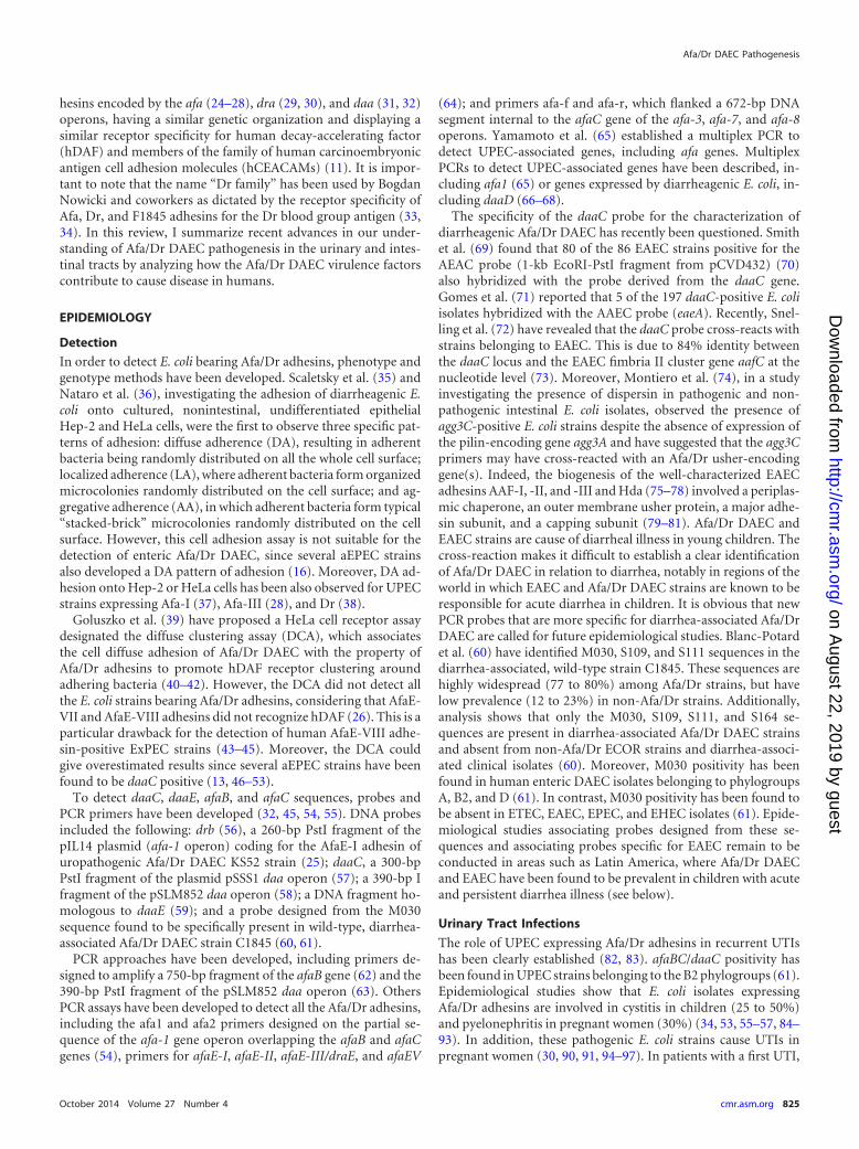
EPIDEMIOLOGY
Detection
In order to detect E. coli bearing Afa/Dr adhesins, phenotype andgenotype methods have been developed. Scaletsky et al. (35) andNataro et al. (36), investigating the adhesion of diarrheagenic E.coli onto cultured, nonintestinal, undifferentiated epithelialHep-2 and HeLa cells, were the first to observe three specific pat-terns of adhesion: diffuse adherence (DA), resulting in adherentbacteria being randomly distributed on all the whole cell surface;localized adherence (LA), where adherent bacteria form organizedmicrocolonies randomly distributed on the cell surface; and ag-gregative adherence (AA), in which adherent bacteria form typical“stacked-brick” microcolonies randomly distributed on the cellsurface. However, this cell adhesion assay is not suitable for thedetection of enteric Afa/Dr DAEC, since several aEPEC strainsalso developed a DA pattern of adhesion (16). Moreover, DA ad-hesion onto Hep-2 or HeLa cells has been also observed for UPECstrains expressing Afa-I (37), Afa-III (28), and Dr (38).
Goluszko et al. (39) have proposed a HeLa cell receptor assaydesignated the diffuse clustering assay (DCA), which associatesthe cell diffuse adhesion of Afa/Dr DAEC with the property ofAfa/Dr adhesins to promote hDAF receptor clustering aroundadhering bacteria (40–42). However, the DCA did not detect allthe E. coli strains bearing Afa/Dr adhesins, considering that AfaE-VII and AfaE-VIII adhesins did not recognize hDAF (26). This is aparticular drawback for the detection of human AfaE-VIII adhe-sin-positive ExPEC strains (43–45). Moreover, the DCA couldgive overestimated results since several aEPEC strains have beenfound to be daaC positive (13, 46–53).
To detect daaC, daaE, afaB, and afaC sequences, probes andPCR primers have been developed (32, 45, 54, 55). DNA probesincluded the following: drb (56), a 260-bp PstI fragment of thepIL14 plasmid (afa-1 operon) coding for the AfaE-I adhesin ofuropathogenic Afa/Dr DAEC KS52 strain (25); daaC, a 300-bpPstI fragment of the plasmid pSSS1 daa operon (57); a 390-bp Ifragment of the pSLM852 daa operon (58); a DNA fragment ho-mologous to daaE (59); and a probe designed from the M030sequence found to be specifically present in wild-type, diarrhea-associated Afa/Dr DAEC strain C1845 (60, 61).
PCR approaches have been developed, including primers de-signed to amplify a 750-bp fragment of the afaB gene (62) and the390-bp PstI fragment of the pSLM852 daa operon (63). OthersPCR assays have been developed to detect all the Afa/Dr adhesins,including the afa1 and afa2 primers designed on the partial se-quence of the afa-1 gene operon overlapping the afaB and afaCgenes (54), primers for afaE-I, afaE-II, afaE-III/draE, and afaEV
(64); and primers afa-f and afa-r, which flanked a 672-bp DNAsegment internal to the afaC gene of the afa-3, afa-7, and afa-8operons. Yamamoto et al. (65) established a multiplex PCR todetect UPEC-associated genes, including afa genes. MultiplexPCRs to detect UPEC-associated genes have been described, in-cluding afa1 (65) or genes expressed by diarrheagenic E. coli, in-cluding daaD (66–68).
The specificity of the daaC probe for the characterization ofdiarrheagenic Afa/Dr DAEC has recently been questioned. Smithet al. (69) found that 80 of the 86 EAEC strains positive for theAEAC probe (1-kb EcoRI-PstI fragment from pCVD432) (70)also hybridized with the probe derived from the daaC gene.Gomes et al. (71) reported that 5 of the 197 daaC-positive E. coliisolates hybridized with the AAEC probe (eaeA). Recently, Snel-ling et al. (72) have revealed that the daaC probe cross-reacts withstrains belonging to EAEC. This is due to 84% identity betweenthe daaC locus and the EAEC fimbria II cluster gene aafC at thenucleotide level (73). Moreover, Montiero et al. (74), in a studyinvestigating the presence of dispersin in pathogenic and non-pathogenic intestinal E. coli isolates, observed the presence ofagg3C-positive E. coli strains despite the absence of expression ofthe pilin-encoding gene agg3A and have suggested that the agg3Cprimers may have cross-reacted with an Afa/Dr usher-encodinggene(s). Indeed, the biogenesis of the well-characterized EAECadhesins AAF-I, -II, and -III and Hda (75–78) involved a periplas-mic chaperone, an outer membrane usher protein, a major adhe-sin subunit, and a capping subunit (79–81). Afa/Dr DAEC andEAEC strains are cause of diarrheal illness in young children. Thecross-reaction makes it difficult to establish a clear identificationof Afa/Dr DAEC in relation to diarrhea, notably in regions of theworld in which EAEC and Afa/Dr DAEC strains are known to beresponsible for acute diarrhea in children. It is obvious that newPCR probes that are more specific for diarrhea-associated Afa/DrDAEC are called for future epidemiological studies. Blanc-Potardet al. (60) have identified M030, S109, and S111 sequences in thediarrhea-associated, wild-type strain C1845. These sequences arehighly widespread (77 to 80%) among Afa/Dr strains, but havelow prevalence (12 to 23%) in non-Afa/Dr strains. Additionally,analysis shows that only the M030, S109, S111, and S164 se-quences are present in diarrhea-associated Afa/Dr DAEC strainsand absent from non-Afa/Dr ECOR strains and diarrhea-associ-ated clinical isolates (60). Moreover, M030 positivity has beenfound in human enteric DAEC isolates belonging to phylogroupsA, B2, and D (61). In contrast, M030 positivity has been found tobe absent in ETEC, EAEC, EPEC, and EHEC isolates (61). Epide-miological studies associating probes designed from these se-quences and associating probes specific for EAEC remain to beconducted in areas such as Latin America, where Afa/Dr DAECand EAEC have been found to be prevalent in children with acuteand persistent diarrhea illness (see below).
Urinary Tract Infections
The role of UPEC expressing Afa/Dr adhesins in recurrent UTIshas been clearly established (82, 83). afaBC/daaC positivity hasbeen found in UPEC strains belonging to the B2 phylogroups (61).Epidemiological studies show that E. coli isolates expressingAfa/Dr adhesins are involved in cystitis in children (25 to 50%)and pyelonephritis in pregnant women (30%) (34, 53, 55–57, 84–93). In addition, these pathogenic E. coli strains cause UTIs inpregnant women (30, 90, 91, 94–97). In patients with a first UTI,
Afa/Dr DAEC Pathogenesis
October 2014 Volume 27 Number 4 cmr.asm.org 825
on August 22, 2019 by guest
http://cmr.asm
.org/D
ownloaded from
the presence of E. coli isolates expressing Afa/Dr adhesins leads toan elevated occurrence of a second UTI (56, 57, 64, 98, 99). Inpatients with pyelonephritis, there was a variable distribution ofafaE subtypes in afa-positive strains (100). Zhang et al. (64) havefound that UTI-associated and fecal E. coli isolates were afaE1positive (18%), afaE2 positive (1.3%), afaE3 positive (1.3%), draEpositive (12%), daaE positive (1.3%), and draE-afaE3 hybrid pos-itive (12%). Some human pyelonephritis E. coli isolates have beenfound to be positive for afa1-afa2 and afa-f-afa-r PCR probes andin some cases to express the afaE1 (5%), afaE8 (39%), and afaEX(20%) operons (45). A UTI-associated E. coli strain generally ex-presses a multiplicity of adhesive factors. Foxman et al. (92), ana-lyzing E. coli strains isolated from women with first-time UTIs,observed that drb-probe positive E. coli isolates displayed positiv-ity with the type 1 pilus probe (80 to 100% positivity) and the Pfimbria probe (50% positivity) and no positivity for the S fimbriaprobe. Szemiako et al. (101) have observed that combinations ofgenes encoding two adherence factors (P and Dr fimbriae or S andDr fimbriae) in UTI-associated E. coli isolates result in an in-creased risk of translocation to the vascular system, leading tobacteremia. Moreover, daaC-positive, UTI-associated clinical iso-lates have been found to express aerobactin (89), hemolysin (56,86, 89, 90, 92, 102, 103), and cytotoxic necrotizing factor (CNF)(19, 56, 89, 92). The same numbers of strains expressing the drbprobe have been found in isolates from the urinary tract or rectumof women with UTIs (104).
Afa/Dr adhesins are frequently found in E. coli associated withpyelonephritis in pregnant women with gestational complications(30, 62, 90, 96, 97). In addition, Afa/Dr DAEC strains are associ-ated with preterm labor/birth (82, 95, 105). Sledzinska et al. (97)have reported the presence of an E. coli strain harboring the com-bination of P and Dr fimbriae in a case of fatal sepsis in a pregnantwoman who developed pyelonephritis.
Diarrhea
It is well established that pathogenic ETEC, tEPEC, aEPEC, andAIEC colonize the small intestine, EAEC colonizes small intestineand/or colon, EHEC colonizes the distal ileum and colon, andEIEC colonizes the colon (4, 5). In contrast, the intestinal site(s)colonized by diarrhea-associated Afa/Dr DAEC currently remainsto be determined. There is an absence of a role of Afa/Dr DAEC indiarrhea in adults. Indeed, when the wild-type strain C1845 wasinoculated in adult volunteers, none of the patients developeddiarrhea, despite the strain being detected in duodenal culturesand stools (106). Moreover, examination of a large number ofhuman diarrhea-associated DAEC strains has shown that only twocarried the daaE gene, suggesting that the F1845 fimbria is rareamong diarrheagenic DAEC strains (59). In addition, epidemio-logical studies in various areas of the world are inconclusive withregard to a role of daaC-positive E. coli strains in diarrhea in chil-dren or adults (13, 48, 50, 99, 107–115). However, the relationshipbetween Afa/Dr DAEC and diarrhea in children as a function ofage has been more convincingly demonstrated in age cross-sec-tional studies showing an increased incidence in children �1 to 5years of age. These studies were conducted in the United States(116), Mexico (117), and different South American countries, in-cluding Chile (118), Brazil (58, 71, 119–121), Colombia (122),Peru (123–127), and Argentina (128), as well as in Thailand (50),Bangladesh (129), Japan (119), New Caledonia (45, 63, 130), var-ious places in Africa (131–133), and European countries, includ-
ing the United Kingdom (134) and France (135, 136). In Afa/DrDAEC strains isolated from stools of children, the sat gene hasbeen found in almost half of the diarrhea-associated Afa/DrDAECstrains and was not present in all the non-diarrhea-associated Afa/DrDAEC strains (137). Why and how Afa/Dr DAEC isolates arepotential pathogens in children with an age-dependent occur-rence remain to be determined. A possible explanation, but onethat does not exclude other possible causes, is that in children inthis age range, the intestinal epithelial barrier is not structurallyand functionally mature, and therefore the strong host defenseresponses against infection by Afa/Dr DAEC, which are describedbelow, are not yet functional.
The observation of daaC positivity of some aEPEC strains isindicative that Afa/Dr adhesins are expressed by enterovirulent E.coli strains other than Afa/Dr DAEC (13, 46–53). Moreover, E. coliisolates displaying daaC and afaBC positivities have been foundamong AIEC, inflammatory bowel disease (IBD)-associated, andintestinal cancer-associated (6, 138–140) strains.
Intestinal Asymptomatic Portage
Most of the epidemiological studies conducted in various areas ofthe world that were intended to identify Afa/Dr DAEC as cause ofdiarrhea in children over 5 years of age and in adults were incon-clusive, as the same numbers of daaC-positive strains were foundin cases and controls (13, 48, 50, 71, 99, 107–115, 141). This ob-servation highlights the existence of asymptomatic carriers of in-testinal Afa/Dr DAEC strains and suggests that these pathogenic E.coli strains can be tolerated or controlled if the mature intestinalepithelial barrier is in a healthy condition.
VIRULENCE FACTORS
Afa/Dr Adhesins
The processes by which epithelia are infected by pathogenic E. colistart by the attachment of bacteria to specific host cells. To do this,pathogenic bacteria express a wide variety of surface-exposed ad-hesins responsible for specific binding to structural or functionalcell membrane-associated molecules (142). The attachments ontothe target host cells allow enteric and urinary tract bacterial patho-gens to resist clearance by peristalsis and micturition, respectively.The bacterial adhesion to target host cells can be more than asimple attachment due to pathogen-specific recognition of hostcell membrane-associated molecules, since several of these mole-cules functioned intrinsically as signaling molecules or after rec-ognition/activation recruited cytosolic signaling molecules (143).Attachment by fimbrial or afimbrial structures allows bacterialpathogens to interact with the host cell membrane to ensure theoptimal delivery of their cytotonic or cytotoxic toxins in the vicin-ity of their membrane-associated receptors, triggering signalingevents that affect transport/secretion functions or the cell struc-tural organization. For other pathogenic bacteria, adhesive factorsallow the intimate association of bacteria with the cell membranethat is necessary for the initiation and completion of signaling-controlled structural lesions, which in turn dramatically impairhost cell functions. For invasive bacterial pathogens, attachmentinitiates an orderly series of signaling-controlled events that leadto host cell membrane rearrangements that are necessary for theachievement of bacterial cell entry followed by the development ofsophisticated bacterial intracellular lifestyles.
Two major classes of adhesins are present on the bacterial sur-
Servin
826 cmr.asm.org Clinical Microbiology Reviews
on August 22, 2019 by guest
http://cmr.asm
.org/D
ownloaded from

face of Gram-negative pathogens: the fimbrial adhesins, consist-ing of linear homopolymers or heteropolymers, and the afimbrialadhesins, formed of single proteins or homotrimers (142). For thecompletion of fimbrial and afimbrial adhesins in Gram-negativepathogens, different secretion systems have been identified, in-cluding Sec-independent and Sec-dependent pathways (144). Themajor families of adhesive proteins (145) include the classicalchaperone/usher pathway-dependent fimbrial adhesins (146,147), the alternate chaperone/usher pathway-dependent E. colisurface pili (148), the extracellular nucleation precipitation-de-pendent curly or thin aggregative fimbrial adhesins (149), the typeI secretion system-dependent afimbrial adhesins (150), the typeIII secretion system-dependent integral outer membrane proteins(151), the polymerization-assembled type IV pili (152), and thetype V secretion system-dependent nonfimbrial trimeric auto-transported adhesins (153).
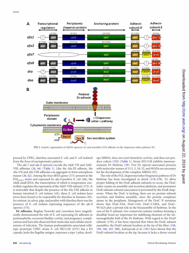
The Afa/Dr family of adhesins contains fimbrial (29, 32, 75, 76,154, 155) or afimbrial (25–27, 30, 37, 44, 64, 84, 156) adhesins(Table 1). These adhesins are encoded by genes present in operonscontaining five major genes, including highly conserved genes Ato D, encoding accessory proteins, and more divergent genes E,encoding the adhesin subunits (Fig. 1A). Assembly via the FGS(with a short F1-G1 loop) and FGL (with a long F1-G1 loop)classes of periplasmic chaperones has been described, and the FGLchaperone/usher protein secretion system assembles Afa/Dr ad-hesins (146, 147) (Fig. 1B). The structural organization of longand short Afa/Dr adhesins develops by the assembly of the bacte-rial membrane usher, successive E adhesin subunits, and one Dsubunit capping the structure. It is interesting to note that thechaperone usher functions in UPEC to form adhesive structuressuch as the P pili, resulting in the orderly assembly of PapA, PapK,PapE, PapF, and adhesive PapG subunits, and type 1 pili, formedby assembly of FimA, FimF, FimG, and adhesive FimH subunits(146, 147). The PapG subunit, localizing at the tip of the fimbria,triggers recognition of host cell membrane-associated globoseriesglycolipids, and the FimH subunit triggers the mannose-depen-dent recognition. Crystallographic and nuclear magnetic reso-nance (NMR) studies coupled or not coupled with mutagenesis
have been used to define the functional domains of the DraE andAfaE-III-Dsc adhesins, which are required for binding to host re-ceptors such as hDAF (157–160), hCEACAMs (160), and collagentype IV (161, 162), and to explain the differential sensitivity tochloramphenicol (161, 162).
Afa adhesins. Agnès Labigne and Chantal Le Bouguénec haveextensively described the pathogenicity mechanisms of E. colistrains bearing the afimbrial adhesins (Afa) encoded by the afaoperons (Table 1). The first Afa adhesin was isolated from thewild-type prototype UPEC strain KS52 by Labigne et al. (25, 37).The 6.7-kb chromosomal DNA fragment essential for a mannose-resistant hemagglutination (MRHA) phenotype in human eryth-rocytes and for adhesion onto uroepithelial cells contains fivegenes: afaA, afaE, afaD, afaB, and afaC (24). Adhesins AfaE-II andAfaE-III were then isolated from two other UPEC strains, A22 andA30, by Labigne et al. (84). Le Bouguénec et al. (27) isolated fromthe UPEC strain A30 a 9-kb plasmid region containing the afa-3gene cluster. The afa-3 gene cluster contains six genes designatedafaA to afaF (163). The AfaE-III and DraE adhesin subunits dis-played 98% identity (of 160 amino acids, 157 are similar) (27, 30),and the afa-3 gene cluster and daa operon are closely similar (31,32, 164, 165). The atomic resolution structure for the AfaE-IIIsubunit has been determined (158, 159, 166, 167). Nuclear mag-netic resolution and biophysical studies have revealed that thestructural organization of Afa-III adhesin develops by assemblyonto the bacterial membrane usher of AfaE-III adhesins subunits(158, 159) capped by the AfaD-III subunit (166–168). The AfaD-III subunit also has the ability to separate from the Dr fimbriae(42). A diffuse and not well-ordered cell surface localization of theAfaD-III subunit has been observed by immunoelectron micros-copy (42, 168, 169). Using chimeras constructed from the afa-3and daa operons, a study has revealed that the afimbrial or fim-brial morphologies of the adhesins were influenced by the order inthe genes coding for the afimbrial or fimbrial adhesin subunits(28). The AfaE-III adhesin subunit is involved in recognizing hostcell receptors (158, 159) such as the DraE and DaaE adhesin sub-units (170). As shown in the analysis of epidemiological studiesbelow, Afa-possessing E. coli strains have been found to be ex-
TABLE 1 Characteristics of Afa/Dr adhesins and Afa/Dr-related adhesins
Adhesin Type Host
Receptors
Type IV collagen hDAF hCEACAMs
AfaE-I Afimbrial Human Negative Positive PositiveAfaE-II Afimbrial Human Unknown Positive UnknownAfaE-III Afimbrial Human Negative Positive PositiveAfaE-V Afimbrial Human Unknown Positive PositiveAfaE-VII Afimbrial Bovine Unknown Negative UnknownAfaE-VIII Afimbrial Human/animal Unknown Negative NegativeDr Fimbrial Human Positive Positive PositiveDr-II Afimbrial Human Negative Positive NegativeF1845 Fimbrial Human Negative Positive Positive
Distant membersa
NFA-I Afimbrial Human Unknown Positive UnknownAAF-I Fimbrial Human Unknown Unknown UnknownAAF-II Fimbrial Human Unknown Unknown UnknownAAF-III Fimbrial Human Unknown Unknown UnknownHdaA Fimbrial Human Unknown Unknown Unknown
a Like Afa/Dr adhesins, AAF-I, -II, and -III and HdaA promote an MRHA phenotype in human erythrocytes (78).
Afa/Dr DAEC Pathogenesis
October 2014 Volume 27 Number 4 cmr.asm.org 827
on August 22, 2019 by guest
http://cmr.asm
.org/D
ownloaded from
pressed by UPEC, diarrhea-associated E. coli, and E. coli isolatedfrom the feces of asymptomatic patients.
The afa-7 and afa-8 operons encode the AfaE-VII and AfaE-VIII adhesins (26, 44) (Table 1). Like the Afa-III adhesin, theAfa-VII and Afa-VIII adhesins can aggregate to form amorphousmasses (26, 42). Among the four sRNA genes (171) present in thePAIAL862 strain and expressed by afa-8-positive E. coli (44), theAfaR small RNA, the transcription of which is temperature con-trolled, regulates the expression of the AfaD-VIII subunit (172). Itis noticeable that despite the presence of the Afa-VIII adhesin inhuman intestinal E. coli isolates (43), these E. coli isolates havenever been found to be responsible for diarrhea in humans (45).In contrast, in calves, pigs, and poultry with diarrhea there was thepresence of E. coli isolates expressing sequences of the afa-8operon (173).
Dr adhesins. Bogdan Nowicki and coworkers have magnifi-cently demonstrated the role of E. coli expressing Dr adhesins inpyelonephritis, recurrent bladder cystitis, and pregnancy compli-cations and have also dissected their molecular and cellular mech-anisms of infection. The human pyelonephritis-associated wild-type prototype UPEC strain E. coli IH11128 (O75) has a K5capsule, lacks the flagellar antigen, expresses a type 1 pilus, devel-
ops MRHA, does not exert hemolytic activity, and does not pro-duce colicin (155) (Table 1). Strain IH11128 exhibits mannose-resistant Dr fimbriae (29). Five Dr operon-associated proteinswith molecular masses of 15.5, 5, 18, 32, and 90 kDa are necessaryfor the development of the complete MRHA (55).
The role of the FGL chaperone/usher biogenesis pathway of Drfimbriae has been investigated in detail (174–178). To allowproper folding of the DraE adhesin subunits to occur, the DraCusher creates an assembly and secretion platform, and prematureDraE subunit-subunit association is prevented by the DraB chap-erone. When the DraC is lacking, there are no protein subunitsecretion and fimbria assembly, since the protein complexesamass in the periplasm. Mutagenesis of the DraC N terminusshows that DraC-F4A, DraC-C64, DraC-C100A, and DraC-W142A play a pivotal role in the bioassembly of fimbriae. In thecase of the E subunit, two conserved cysteine residues forming adisulfide bond are important for stabilizing elements of the im-munoglobulin fold of the Dr fimbriae. With regard to the DraDsubunit (179), it has been reported that when the DraE subunitassembles, the DraD subunit localizes at the tip of the fiber (158,159, 166, 167, 180). Jedrzejczak et al. (181) have shown that theDraD subunit localizes at the tip, because it lacks a donor strand
FIG 1 Genetic organization of Afa/Dr operons (A) and assembly of Dr adhesin via the chaperone-usher pathway (B).
Servin
828 cmr.asm.org Clinical Microbiology Reviews
on August 22, 2019 by guest
http://cmr.asm
.org/D
ownloaded from
and as a consequence functions only as an acceptor. However,expression of the DraD subunit has been found to be independentof the DraC usher, and DraD appears not to be necessary for thepolymerization of DraE subunits (182). Recently, Zalewska-Piatek et al. (183) showed that the DraD subunit can be producedby a chaperone/usher-independent, type II secretion-dependentprocess that allows the translocation of the DraD subunit onto thecell surface of Dr-positive E. coli.
The major structural subunit DraE is involved in host cell re-ceptor recognition (170), as are the AfaE-III (158, 159) and DaaEadhesin subunits (170). Calculation of the electrostatic potentialsof the DraE structure shows an electronegative area around thecluster of amino acids involved in binding onto hDAF (Asp61,Asp63, and Asp75) (157). In the genes encoding Dr fimbriae, sin-gle-nucleotide polymorphisms conferring an adaptive advantagehave been identified (184). Dr fimbriae are unique among Afa/Dradhesins in expressing chloramphenicol sensitivity for bindingonto host cell receptors, whereas binding of Afa-I to -III andF1845 is not affected (27, 185, 186). Korotkova et al. (187) haveinterestingly shown that genes encoding Dr fimbriae form eightstructural groups displaying a high level of amino acid sequencediversity among them. It is noticeable that a functional analysishas revealed the presence of distinctly different binding pheno-types controlling affinity to hDAF, capability to bind collagen typeIV and hCEACAMs, and sensitivity of adhesiveness capacity tochloramphenicol. Since the AfaD/DraD/DaaD subunits localize atthe tip of the Afa/Dr fimbriae, their possible involvement in adhe-sion onto epithelial cells and, in addition, that of the AfaE/DraE/DaaE subunits has been envisaged. Conflicting results have beenobtained. Recombinant DraE�/DraD� or AfaE�/D�-III E. colifailed to adhere to differentiated primary bladder cells (188) andCHO-hDAF-�5�1 cells (189), respectively. In contrast, Zalewska-Piatek et al. (190) have reported that in HeLa cells, DraE�/DraD�
E. coli displays a low level of adhesion, �3-fold lower that ofDraE�/DraD� E. coli. In contrast to the chloramphenicol-sensi-tive adhesion of DraE, the DraD-induced binding is chloram-phenicol insensitive (190).
The Dr-II adhesin has been isolated from the human pyelone-phritis-associated strain EC7372 (Table 1). Compared to themembers of the Afa/Dr adhesin family, the Dr-II adhesin displayspoor sequence identity (17 to 20%) (30). Dr-II has 96% identitywith the nonfimbrial adhesin I (NFA-I) expressed by UTI-associ-ated E. coli (191). Interestingly, NFAs and Afa/Dr adhesins have avery similar genetic organization, and the nfa gene cluster encodesNfaA subunits assembled via the chaperone-usher pathway (191).
F1845 adhesin. Steve L. Moseley and coworkers discovered thediarrhea-associated E. coli expressing F1845 adhesin and beauti-fully described the structural aspects of the interaction betweenAfa/Dr adhesins and their epithelial cell hDAF and hCEACAMreceptors. The human wild-type prototype diarrheagenic strainC1845 expresses a fimbrial adhesin, designated F1845 (Table 1).The order and regulation of the genes necessary for F1845 adhesinassembly have been identified (31, 32, 164, 165, 192–194). TheF1845 and Dr adhesins display 57% identity (91 amino acids of160 are identical) (30). Five polypeptides (10, 95, 27, 15.5, and14.3 kDa) are encoded by daaA, daaB, daaC, daaD, and daaEgenes, respectively. The major structural subunit, DaaE, is in-volved in host cell receptor recognition like the AfaE-III (158, 159)and DraE adhesin subunits (170). Bilge et al. (31) have demon-strated that the fimbrial gene expression in the daa operon was
regulated by both phase variation and environmental regulatorymechanisms. White-Ziegler et al. (193) have reported that in re-sponse to multiple environmental signals, the histone-like H-NSacts as an overall regulator by controlling transcription of the daaoperon.
Flagella
The biogenesis of flagella involves the coordinated structural as-sembly of flagellar proteins (195). A variety of flagellar structuralproteins and capping proteins compose the flagellar propeller(195), and cytoplasmic membrane proteins compose the force-generating unit of the flagellar motor (196). In an aqueous envi-ronment, many bacterial species move by rotating their flagella,allowing individual bacteria to swim in three dimensions (197).Moreover, flagellar swarming coordinates the movement of bac-teria across the host cell surface (198). Flagella expressed by UPECcontribute to colonization of the epithelium, dissemination to thekidney by ascending progression from the bladder, and biofilmformation (199). It has been observed that UPEC strains express-ing type 1 pili or P fimbriae are less flagellated and display re-pressed motility, suggesting that when fimbrial expression isswitched off, UPEC strains are motile (200, 201). Afa/Dr DAECstrains express or do not express flagella. The prototype pyelone-phritis-associated, wild-type Afa/Dr DAEC strain IH11128, ex-pressing a type 1 pilus, does not possess flagellar antigens (155). Incontrast, the UPEC wild-type strain A30, which does expressAfaE-III adhesin, is positive for flagellar antigen (unpublisheddata), and the animal wild-type Afa-VIII-positive strain AL511 isH8 positive (202). It is worth mentioning that that the prototypediarrheagenic wild-type Afa/Dr DAEC strain C1845 (32) does notexpress flagellar antigens (unpublished data). According toArikawa et al. (203), only seven of the 19 afaE1-, afaE2, or afaEX-positive, diarrhea-associated E. coli isolates they examined aremotile. In contrast, Meraz et al. (107), who examined 18 DAECisolates, found that all nine diarrhea-associated, afaE1- or afaEX-positive E. coli isolates are motile. These findings indicate thatUPEC and diarrhea-associated Afa/Dr DAEC display heteroge-neous flagellum expression.
Secreted Autotransporter Toxin
Secreted autotransporter toxin (Sat) belongs to the type V secre-tion pathway-dependent subfamily of serine protease autotrans-porters of Enterobacteriaceae (SPATE) toxins (81, 153, 204). As theresult of differences in the toxins structures and activities, thereare two classes of SPATE toxins. Class I includes plasmid-encodedtoxin (Pet) of EAEC, extracellular serine protease, plasmid en-coded (EspP) of EHEC, EspC of EPEC, SigA of Shigella flexneriand EAEC, Sat of intestinal E. coli and ExPEC, and the hypothet-ical EspC-like SPATE toxins with EcPCN033-C1sp (NCBI acces-sion number EGP21815.1) of ExPEC, EcNA114-C1sp (NCBI ac-cession number AEG39156.1) of UPEC, and EcM605-C1sp(NCBI accession number ZP_08351236.1) of AIEC (204). Class IIincludes protein involved in intestinal colonization (Pic) of Shi-gella, EAEC, and UPEC, SepA of Shigella, EatA of ETEC, vacuo-lating autotransporter toxin (Vat)-like toxins of UPEC, SEPEC,and NEMEC, EcRN587-C2sp (NCBI accession numberEFZ76879.1) of EAEC and EPEC, and EpeA of Shiga toxin-pro-ducing E. coli (204). Class I SPATE toxins are generally cytotoxic,whereas class II display diverse activities, including the cleavage ofmucus, which provides a competitive advantage for host epithe-
Afa/Dr DAEC Pathogenesis
October 2014 Volume 27 Number 4 cmr.asm.org 829
on August 22, 2019 by guest
http://cmr.asm
.org/D
ownloaded from
lium colonization (81, 204). The sat gene has been characterized inthe UPEC prototype strain CFT073 (205), where it resides withinPAI-IICFT073 (206–209). The sat gene is prevalent in UPEC strains,including those bearing Afa/Dr adhesins (8, 56, 137, 206, 208,210–214), resident intestinal microbiota E. coli stains, and patho-genic strains of E. coli, including EAEC (137, 215–218), and Shi-gella isolates (219, 220). The sat gene has been found present indaaC-positive E. coli strains isolated from stools of children withdiarrhea in Brazil and France (137, 218, 221). In Afa/Dr DAECisolates, the sat gene has been found to be expressed equivalentlyby diarrheic and asymptomatic adults (222). Interestingly, the satgene is prevalently expressed in Afa/Dr DAEC isolated from chil-dren in a context of diarrhea (222).
Hemolysin
The pyelonephritogenic strain EC7372, which expresses Dr-II ad-hesin (30), is the only Afa/Dr DAEC strain that produces a func-tional hemolysin. Indeed, unlike other Afa/Dr DAEC strains,strain EC7372 promotes a strong cellular lysis in epithelial cellspreceded by apoptosis (102). On the basis of results reported byBlanc-Potard et al. (60), the hemolysin-positive strain EC7372carries both the hly and pap operons and seems to have acquired alarger part of the PAIsCFT073 (207–209) than Afa/Dr DAEC. Therecombinant E. coli strain EC901, which carries plasmid pBJN406and contains the draA to -E genes involved in expression of Drfimbriae (223), has been observed to display a curious hemolyticactivity. Insertion mutations in draD and draE, but not in draA,draB, and draC, abolish hemolytic activity, indicating that thisactivity is supported by the extracellular domain of Dr fimbriae.This observation is intriguing, since strain IH11128, gestationalpyelonephritis Dr-positive E. coli isolates (94, 155), and clinicalDr-positive E. coli isolates (60) all lack either hemolytic activity orhly gene expression. In contrast, the wild-type O75X strainIH11032 does display hemolytic activity (155). Moreover, fourafaE1-positive and one afaEX-positive diarrheagenic E. coli iso-lates have been found to trigger hemolysis, while 14 other afaE1-positive and one afaEX-positive isolates do not (203). Collectively,these findings show that Afa/Dr DAEC strains are heterogeneousin terms of �-hemolysin expression, suggesting a variable distri-bution of the part of PAICFT073 containing the hly gene among theAfa/Dr DAEC strains.
Other Factors
Blanc-Potard et al. (60) identified several short sequences (73 to495 bp) that are prevalent in Afa/Dr adhesin-positive E. coli clin-ical isolates in comparison with E. coli clinical isolates not express-ing Afa/Dr adhesins (GenBank accession numbers AZ935556 toAZ935604). Several sequences are homologous to virulence genesexpressed in other pathotypes of E. coli, including genes for twosiderophores (irp2 and iuc), a catechol siderophore receptor(iroN), and two transport systems (shu and modD) (60). Interest-ingly, several C1845-specific sequences display no likeness withknown sequences (60). Importantly, the diarrhea-associated wild-type C1845 strain does not express the genes encoding ETEC andEAEC virulence factors and is devoid of genes encoding EPEC andEHEC virulence factors, including the genes of the locus of entero-cyte effacement (LEE) island involved in the type III secretionsystem (T3SS) or T3SS-associated effector proteins and not hy-bridized with eae probes (60). The wild-type C1845 and IH11128strains expressed a part of PAICFT073 (207–209) not including the
hlyA, hlyD, hp1-hp4, papG, or papF sequences (60). A remnant ofthe pap operon which has the F10 papA allele but lacks most of thecentral region of the pap operon has been detected. It is notewor-thy that regions of the PAICFT073 complete genome sequence (207,209, 224) have been found in E. coli strains of the B2 phylogenicgroup (208) and are prevalently expressed in ExPEC strains ofgroup B2 involved in UTIs (8). Moreover, parts of PAICFT073 haverecently been found in intestinal commensal E. coli strains, partic-ularly those of phylogenic group B2 (225–227), and in an AIECstrain (228).
The PAIAL862 expressed by afa-8-positive E. coli strains (44)includes the deoK gene, which confers metabolic adaptability andincreases the competitive advantage with regard to host infectivity(229). The locus designated vpe (virulence-associated phospho-transferase) contains the vpeA, vpeB, and vpeC genes, which en-code, respectively, the EIIA, EIIB, and EIIC constituents of a pu-tative carbohydrate-specific permease of the SgaTBA family (230).This locus is present in the pyelonephritis-associated strainAL511, which expresses the afa-8 operon (43), which confers anability to adapt for kidney and intestinal colonization (231). Thepresence of the vpe locus in other UTI- and diarrhea-associatedAfa/Dr DAEC strains has not been documented.
The capacity to form filamentous forms results from a plasticitycapacity developed by a bacterial pathogen in order to escape hostdefenses when in an intracellular location or to assemble to form abiofilm-like structure that leads to resistance to anti-infectivetreatments, such as antibiotics (199). Some excellent experimentshave demonstrated that type 1 pilus-positive UPEC strains, afterinternalization into superficial epithelial cells known as “umbrellacells” lining the luminal surface of the bladder, form biofilm-likebacterial assemblages designated “intracellular bacterial commu-nities” (IBCs) that function as transient protective structures forUPEC intracellular growth (199, 232). UPEC cells in IBCs consti-tute reservoirs of UPEC, which, after switching to filamentousforms, become detached from the bacterial community and maybe flushed out of the host cells. Zalewska-Piatek et al. (233) werethe first to observe that that Dr-positive E. coli formed biofilms.This phenomenon means that Dr-positive E. coli strains form livefilamentous bacteria, depending on their nutritional environment(190, 233). It has been observed that adhering Dr-positive E. coliforms filamentous forms at the cell surface of CHO-hDAF-�5�1(189) or CHO-hDAF (234) cells. Filamentous bacteria residingwithin the phagosome escaped phagosomal killing as the bacteriamanipulated the phagosome compartment by blocking the acqui-sition of hydrolytic components (235, 236). Even though the in-tracellular vacuole-containing Dr-positive E. coli in HeLa cellslack the characteristics of a degradative compartment (189), nofilamentous forms of Afa- or Dr-positive E. coli residing intracel-lularly have ever been observed. This aspect of Afa/Dr pathogen-esis remains to be explored in the appropriate model of bladderepithelial cells. Bacterial biofilm formed by UPEC after aggrega-tion of three-dimensional structured cells connected by self-pro-duced exopolysaccharide matrix plays a major role in persistentand chronic UTIs (199). Exopolysaccharide production, whichplays a pivotal role in biofilm completion, has been found in UTI-associated E. coli strains expressing Dr (190, 233) or Afa-VIII(231) adhesins. Interestingly, exopolysaccharide production iscontrolled by the vpeBC gene (231), which is present in the vpelocus of afa-8-positive E. coli (43). DraE�/DraD� E. coli strains
Servin
830 cmr.asm.org Clinical Microbiology Reviews
on August 22, 2019 by guest
http://cmr.asm
.org/D
ownloaded from
form dense biofilms, and DraD, whether associated with fimbriaeor not, plays a role in biofilm formation (190, 233).
A large variety of bacteria have been found to produce toxins,named cyclomodulins, that dramatically interfere with the cellcycle (237). Cyclomodulins produced by pathogenic E. coli in-cluded colibactin, cycle-inhibiting factor (Cif), cytotoxic necrotiz-ing factor (CNF), and cytolethal distending toxin (CDT) (238).Currently, the two known genotoxins are colibactin and CDT(238). The cluster of genes known as the “pks island” (239) en-codes a multienzymatic machinery for synthesizing the hybrid,nonribosomal, peptide-polyketide genotoxin colibactin (240). Ithas been suggested that the pks island may affect the host immuneresponse and could be involved in chronic inflammation, in theaccumulation of genomic instability, and in tumor progression(241). Whether the pks island contains other genes encoding ad-ditional bacterial factors and whether the pks-related colibactin isa prototype of a family of molecules or not remain to be investi-gated. The pks genomic island is present in the prototype Afa/DrDAEC wild-type IH11128 and C1845 strains (J. P. Nougayredeand E. Oswald, unpublished result) and in colonic afa-I-positive Ecoli strains isolated from patients with IBD and colorectal cancer(140). The pks island has been also found in ExPEC strains ofphylogenetic group B2 (242), in fecal E. coli strains isolated fromhealthy patients but not in pathogenic EPEC and EHEC isolates(243), in group B2 E. coli strains that are long-term colonizers ofthe intestine (22), in E. coli isolated from the mucosa of patientswith IBD (244), in mucosa-associated or internalized E. coli oftumors and mucosa of colorectal cancer patients (244–246), andin urosepsis E. coli strains (247). It was noted that the intestinalmicrobiota E. coli strain Nissle 1917 expresses the pks genomicisland and displays similarities with the prototype Afa/Dr DAECwild-type C1845 and IH11128 strains, since it harbors parts ofPAICFT073 that lack the expression of �-hemolysin and P fimbriaebut includes iron uptake systems (225, 227). This probiotic E. colistrain with diverse activities (248) is intriguing since its promotionof gut homeostasis activity in response to mucosal injury cannotbe dissociated from the presence of the pks island (249). Whetherthe presence of the pks island in intestinal E. coli and ExPEC strainsis deleterious for the host or without pathological consequencesremains to be investigated.
MECHANISMS OF PATHOGENICITY
Host Cell Receptors for Afa/Dr Adhesins
On the basis of the differential recognition of human epithelial cellmembrane-associated receptors by Afa/Dr adhesins (Table 1),Afa/Dr DAEC strains have been subdivided into two subclasses(11). The first subclass includes E. coli strains harboring the Afa-I(25, 37), Afa-II (27), Afa-III (27), Afa-V (64), Dr (29, 155), Dr-II(30), and F1845 (32) adhesins recognizing hDAF, which also mayor may not recognize members of the hCEACAM family. Thesecond subclass includes strains that express Afa-VII (26, 44) andAfa-VIII (26, 44) adhesins that do not recognize hDAF. In addi-tion, the NFA-I adhesin of UPEC (191) belongs to the Afa/Drfamily of adhesins (Table 1). Moreover, despite a similar geneticorganization with the gene clusters triggering the biogenesis ofAfa/Dr adhesins, the EAEC adhesins AAF-I (77), AAF-II (76),AAF-III (75), and Hda (78) are distant pathogenic factors of theAfa/Dr family of adhesins (Table 1). The four major characteris-tics of EAEC pathogenesis (79–81) are as follows: (i) adherence to
the intestinal mucosa via adhesins (18, 75–78), (ii) the formationof typical “stacked-brick” microcolonies as each bacterium inter-acts with others, (iii) production of enterotoxins and cytotoxins,and (iv) the development of a severe mucosal inflammation. Boi-sen et al. (78), analyzing this superfamily of adhesins, have pro-posed a pertinent phylogram composed of three distinct clusters.The first cluster comprises Afa-I, Afa-II, Afa-III, Afa-V, Dr, Dr-II,and F1845, the second comprises AAF-I, AAF-II, and AAF-III,and the third comprises Afa-VII, Afa-VIII, and Hda. It is worthmentioning that cluster 3 (78) also includes the nonfimbrial M-agglutinin encoded by the bma gene cluster of UPEC (250).
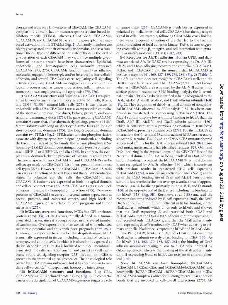
hDAF. Nowicki et al. (33) were the first to report that humandecay-accelerating factor (hDAF) (CD55) expressing the antigensof the Cromer blood group system (251) acts as an epithelial cellreceptor for E. coli expressing Afa/Dr adhesins (Fig. 2) (Table 1).
(i) Structure and functions. DAF is a complement-regulatingprotein with an Mr of 55,000 to 70,000 (251). The physiologicalfunction of DAF is to control the amplification of the complementcascade by a direct interaction with membrane-bound C3b orC4b, which in turn impedes the ulterior uptake of C2 and factor B.Membrane-bound DAF is formed by a membrane glycosylphos-phatidylinositol (GPI) anchor followed by a serine/threonine/proline (STP)-rich region and by four complement control pro-tein repeat (CCP) domains, previously named short consensusrepeats (SCRs) (Fig. 2). Modeling of the extracellular domain ofDAF reveals that CCPs are organized in a helical manner. WhileCCP-1 had no effect on hDAF regulatory activity, deletion ofCCP-2, CCP-3, or CCP-4 entirely abolished the regulatory activ-ity. Interaction of DAF with the convertases is mediated predom-inantly by two patches approximately 13 Å apart, one centeredaround Arg69 and Arg96 on CCP-2 and the other around Phe148and Leu171 on CCP-3 (252). Phe123 and Phe148, localizing at theinterface between CCP-2 and CCP-3, and also Phe154, which ispresent in the CCP-3 cavity, are pivotal for the regulatory activity(253). The GPI anchor increases the lateral mobility of DAFwithin the cell membrane in relation to its localization into mem-brane-associated lipid rafts, and the O-glycosylated STP serves as aspacer for the projection of the hDAF functional domains at thecell membrane (253).
(ii) Receptor for Afa/Dr adhesins. hDAF is one of the receptorsrecognized by Afa/Dr adhesins in epithelial cells (Fig. 2) (Table 1).It is noteworthy that Afa/Dr adhesins bind specifically to hDAFbut not to rodent or pig DAF (254). Dr fimbria binding developsin the digestive, urinary, genital, and respiratory epithelia and inskin (255), consistent with the hDAF expression (251). Only uro-pathogenic and diarrhea-associated E. coli strains bearing theF1845, AfaE-I, AfaE-III, AfaE-V, Dr, and Dr-II adhesins recog-nized hDAF as a receptor (62, 160, 256). In contrast, the Afa-VIIIadhesin expressed by human ExPEC does not recognize hDAF(26, 43, 44). It has been established from functional studies andatomic resolution models that Afa/Dr adhesins recognize theCCP-2 and CCP-3 on hDAF (41, 158, 159, 257–259) (Fig. 2). Incontrast, gestational pyelonephritis-associated E. coli expressingdra-related X adhesins recognized the CCP-3 and CCP-4 domainsof hDAF (62). In the CCP-3, a single point substitution (Ser155-Ala and Ser165-Leu, mimicking the Dra-to-Drb allelic polymor-phisms) results in a complete loss of Dr fimbria binding to hDAF(257, 260). The amino acids (148 to 171), in particular Ser155,present at the surface of CCP-3 controlled the Dr adhesin binding(260). A surface plasmon resonance study of Afa-III adhesin bind-
Afa/Dr DAEC Pathogenesis
October 2014 Volume 27 Number 4 cmr.asm.org 831
on August 22, 2019 by guest
http://cmr.asm
.org/D
ownloaded from

ing onto CCP domains of hDAF has revealed that a constructformed of CCP-1 and -2 did not show any measurable binding tothe AfaE-III adhesin subunit, while constructs formed of CCP-2,-3, and -4, CCP-2 and -3, or CCP-3 and -4 allowed AfaE-III bind-ing with affinities comparable to that for the entire hDAF, con-firming the previously observed importance of the combinedCCP-2 and CCP-3 domains for the recognition of hDAF byAfa/Dr adhesins (158, 159). The Dr adhesin-binding and comple-ment-regulating epitopes of hDAF have been found to be distin-guishable and are approximately 20 Å apart (260). However, An-derson et al. (158) observed that the binding of AfaE-III to hDAFantagonized the hDAF regulatory activity.
The AfaE-I, AfaE-III, AfaE-V, DraE, and DaaE subunits func-tion as receptor ligands for hDAF (27, 160, 170) (Fig. 3A to D). InDraE/AfaE-III subunits, the hDAF-binding site forms a large con-vex surface involving seven � strands (158, 159). The residuesAsp61, Ile73, and Asn77 have been found to be important forbinding to hDAF (158, 159). Mutagenesis and crystallographicstudies of DaaE have been conducted in order to define the de-tailed molecular interactions between Afa/Dr adhesins and hDAF(157). Five daaE mutants (T8N, A60V, D61A, D63V, and T133S)showed a 30 to 50% reduced ability to bind onto CHO cells trans-fected for hDAF expression (157). Mapping the sites of DaaE re-veals that positions Asp61 and Asp63 are necessary for binding tohDAF, and calculation of the electrostatic potentials of the DaaEstructure has revealed an electronegative region around the clusterof amino acids involved in hDAF recognition (Asp61, Asp63, andGlu126) (157). Moreover, the ability of the DraE adhesin to bindhDAF has been found influenced by individual amino acid
changes at positions 10, 63, 65, 75, 77, 79, and 131 of the matureDraE sequence (261).
Binding of the DraE adhesin subunit onto hDAF is sensitive tochloramphenicol, which also inhibits the hDAF-dependentMRHA of human erythrocytes (chloramphenicol-sensitive hem-agglutination [CSHA]) (33, 256). In HeLa cells, the presence ofchloramphenicol diminished the adhesion of DraE�/DraD� E.coli by �3-fold and totally abolished the adhesion of DraE�/DraD� E. coli but did not change the adhesiveness capacity ofDraE�/DraD� E. coli (190). According to Swanson et al. (262), thedomains involved in the CSHA are present within the N-terminaldomain of the DraE subunit. According to Pettigrew et al. (161,162), a hydrophobic pocket including Gly113, Gly42, Pro40,Pro43, Ile111, Tyr115, and Ile114 plays a pivotal role in the chlor-amphenicol-binding site in the DraE subunit. The inhibition ofthe binding of the DraE subunit onto hDAF by chloramphenicolhas received a structural explanation, since by covering the func-tional portion of the adhesin subunit, chloramphenicol disruptsthe recognition of hDAF (161, 162). In contrast to the case for theDr adhesin, chloramphenicol does not affect the hDAF-depen-dent MRHA exerted by the AfaE-I, AfaE-III, and F1845 adhesins(27, 256). This is a result of a difference in expression of aminoacids between the adhesin subunits (161, 162). Moreover, it hasbeen established that binding of chloramphenicol onto the DraEsubunit develops via the interaction of its chlorine “tail” ratherthan its benzene ring (161, 162). Analyzing structural chloram-phenicol modifications, Pettigrew et al. (162) have demonstratedthat acylating the 3-hydroxyl group has no effect on the bindingonto hDAF.
FIG 2 Membrane-associated proteins expressed by human epithelial cells that function as receptors for Afa/Dr adhesins. Center, representations of thestructures of hDAF and hCEACAMs. Left, surface representation of hDAF. Right, homology model of human N-CEA. (Representations of hDAF and N-hCEAreprinted from reference 158 with permission of Elsevier and from reference 160 with permission of the publisher, respectively.)
Servin
832 cmr.asm.org Clinical Microbiology Reviews
on August 22, 2019 by guest
http://cmr.asm
.org/D
ownloaded from

(iii) Receptor for microbial pathogens and viruses. The cellmembrane-bound hDAF is also hijacked by viruses, includingcoxsackievirus serotypes B1, B3, and B5 (263, 264) and coxsacki-evirus A21 (265), enteroviruses (266), and echoviruses (267, 268).Various different hDAF sites are recognized by echoviruses (269).It is worth underlining that like Afa/Dr DAEC (254), echovirusesand coxsackieviruses (270) express high specificity for hDAF. Inaddition, hDAF acts as a receptor for hantavirus (271). Moreover,epithelial hDAF has been identified as a gastric epithelial receptorfor Helicobacter pylori and has been found to be upregulated by thepathogen in relation to inflammatory responses (272, 273).
hCEACAMs. Guignot et al. (41) were the first to show thathCEA (CEACAM5, CD66e) is recruited around the prototype Dradhesin- or F1845 adhesin-positive wild-type Afa/Dr DAECstrains IH11128 and C1845, respectively, adhering to cultured hu-man enterocyte-like Caco-2 cells and that an anti-CD66 antibodyinhibits this bacterial adhesion (Fig. 2) (Table 1). Berger et al.(274), using Chinese hamster ovary (CHO) cells and human cer-vical cancer HeLa cell lines transfected for the expression of eachof the human carcinoembryonic antigen-related cellular adhesionmolecules (hCEACAMs) (CEACAM1 to 8), found that the Dr,F1845, and AfaE-III adhesins bound only to cells expressing epi-
thelial hCEACAM1, hCEA, or hCEACAM6, whereas the AfaE-Iand Dr-II adhesins did not (Fig. 2) (Table 1). Korotkava et al.(160) demonstrated Afa-V adhesin binding to hCEA (Table 1). Inaddition, the Dr, F1845, and AfaE-III adhesins recognize the non-epithelial CEACAM3 as a receptor (188, 189). In contrast, themurine CEACAM1 is not recognized by Afa/Dr adhesins (274).
Twelve members, i.e., CEACAM1 (biliary glycoprotein[BGP], CD66a), CEACAM3 (CEA gene family member 1[CGM1], CD66d), CEACAM4 (CGM7), CEA (carcinoembryonicantigen, CD66e), CEACAM6 (nonspecific cross-reacting antigen[NCA], CD66c), CEACAM7 (CGM2), CEACAM8 (CGM6,CD66b), CEACAM16, and CEACAM18 to -21, compose the fam-ily of CEACAMs (275). CEACAM proteins generally have onevariable (V)-like Ig domain, identified as the N domain (exceptCEACAM16, which has two N domains), but they differ in thenumber of constant C2-like Ig domains as well as in their mem-brane anchorage (Fig. 2). CEACAM5, CEACAM6, CEACAM7,and CEACAM8 are anchored within the cell membrane through aGPI linkage, whereas six other CEACAM family members(CEACAM1, CEACAM3, CEACAM4, CEACAM19, CEACAM20,and CEACAM21) are anchored via bona fide transmembranedomains (275) (Fig. 2). CEACAM16 is devoid of any membrane an-
FIG 3 Receptor clustering by Afa/Dr DAEC. (A to D) Representations of the DraE, DaaE, AfaE-V, and AfaE-I adhesin subunits, respectively. Surface electrostaticpotentials of the DraE, DaaE, AfaE-V, and AfaE-I adhesins (red indicates the negative charges and blue the positive charges) are shown. (Reprinted from reference157 with permission of the publisher.) (E) Representation of DraE adhesin subunit-associated surfaces allowing the specific recognition of hDAF or N-hCEA.Green, surface recognition of hDAF. Red, surface recognition of N-hCEA. Yellow, chloramphenicol bound onto the domain of AfaE-III that recognizes N-hCEA.(Reprinted from reference 160 with permission of the publisher.) (F) Micrographs showing the observation by confocal laser scanning microscopy (CLSM) ofhDAF, hCEACAM1, and hCEA receptor clustering around Dr adhesin-positive E. coli adhering to untransfected HeLa cells constitutively expressing hDAF andto transfected HeLa cells expressing hCEACAM1 and hCEA. Yellow shows colocalization of immunolabeling of Dr adhesin (red) and hDAF, hCEACAM1, orhCEA (green). (Reprinted from reference 274 with permission of the publisher. Copyright 2004 Blackwell Publishing Ltd.) (G) Receptor clustering of hDAF(green) and hCEACAM1 (red) around Dr adhesin-positive E. coli adhering onto transfected HeLa cells expressing hCEACAM1. Yellow, colocalization ofimmunolabelings of hDAF and hCEACAM1. Arrows show immunolabelings of interest around adhering bacteria. (Reprinted from reference 274 with permis-sion of the publisher. Copyright 2004 Blackwell Publishing Ltd.)
Afa/Dr DAEC Pathogenesis
October 2014 Volume 27 Number 4 cmr.asm.org 833
on August 22, 2019 by guest
http://cmr.asm
.org/D
ownloaded from
chorage and is the only known secreted CEACAM. The CEACAM1cytoplasmic domain has immunoreceptor tyrosine-based in-hibitory motifs (ITIMs), whereas CEACAM3, CEACAM4,CEACAM19, and CEACAM20 carry immunoreceptor tyrosine-based activation motifs (ITAMs) (Fig. 2). All family members arehighly glycosylated on their extracellular domains, and as a func-tion of the cell type and differentiation state of the cells, the level ofglycosylation of each CEACAM may vary, since multiple glyco-forms of the same protein have been characterized. Epithelial,endothelial, and hematopoietic cells variously expressedCEACAMs (275, 276). CEACAMs function mainly as adhesionmolecules engaged in homotypic and/or heterotypic intercellularadhesion, and several CEACAMs exert regulating cell signalingactivities (275, 276). CEACAMs are engaged during complex bio-logical processes such as cancer progression, inflammation, im-mune responses, angiogenesis, and apoptosis (275, 276).
(i) hCEACAM1 structure and functions. CEACAM1 was pres-ent in leukocytes, including granulocytes, activated T cells, B cells,and CD16�/CD56� natural killer cells (275). It was present inendothelial cells (275). CEACAM1 was also expressed in epithelialof the stomach, intestine, bile ducts, kidney, prostate, endome-trium, and mammary ducts (275). The gene encoding CEACAM1contains 9 exons that, after alternatively splicing, generate 11 dif-ferent isoforms with long or short cytoplasmic tails and long orshort cytoplasmic domains (275). The long cytoplasmic domaincontains two ITIMs (Fig. 2). ITIMs after tyrosine phosphorylationassociate with diverse cytoplasmic signaling molecules, includingthe tyrosine kinases of the Src family, the tyrosine phosphatase Srchomology 2 (SH2) domain-containing protein tyrosine phospha-tase 1 (SHP-1) or 2 (SHP-2), and Shc (275). In contrast, the cyto-plasmic S domain lacks the presence of tyrosine residues (275).The two major isoforms CEACAM1-L and CEACAM-1S can becell coexpressed, but CEACAM1-L isoforms predominate in mostcell types (275). However, the CEACAM1-L/CEACAM-1S ratiocan vary as a function of the cell types and the cell differentiationstates. In polarized epithelial cells, the CEACAM1-L andCEACAM-1S isoforms are expressed at both the apical domainand cell-cell contact areas (277, 278). CEACAM1 acts as a cell-celladhesion molecule by hemophilic interaction (275). Down-ex-pression of CEACAM1 occurred in several tumor types, such asbreast, prostate, and colorectal cancer, and high levels ofCEACAM1 expression are related to poor prognosis and tumormetastasis (275).
(ii) hCEA structure and functions. hCEA is a GPI-anchoredprotein (275) (Fig. 2). hCEA was initially defined as a tumor-associated marker, since it is overproduced in an elevated numberof carcinomas. Overexpression is often associated with enhancedmetastatic potential and thus with poor prognosis (279, 280).However, it is important to remember that despite its name, hCEAis normally expressed in tissues, including intestinal M cells, en-terocytes, and colonic cells, in which it is abundantly expressed atthe brush border (281). hCEA is localized within cell membrane-associated lipid rafts via its GPI anchor and can act as a cell mem-brane-bound cell signaling receptor (275). In addition, hCEA ispresent in the intestinal apical glycocalyx. The physiological roleplayed by hCEA remains unknown, but it has been shown to me-diate cell-to-cell Ca2�-independent, homotypic interactions.
(iii) hCEACAM6 structure and functions. Like CEA,CEACAM6 is a GPI-anchored protein (275) (Fig. 2). In colorectalcancers, the deregulation of CEACAM6 expression suggests a role
in tumor onset (275). CEACAM6 is brush border expressed inpolarized epithelial intestinal cells. CEACAM6 has the capacity tosignal in cells. For example, following CEACAM6 cross-linking,there was subsequent activation of Src that led to the tyrosinephosphorylation of focal adhesion kinase (FAK), in turn trigger-ing cross talk with �v�3 integrin, and cell interaction with extra-cellular matrix molecules (ECMs) (282, 283).
(iv) Receptors for Afa/Dr adhesins. Human UPEC and diar-rhea-associated Afa/Dr DAEC strains expressing the Dr, Afa-III,Afa-V, and F1845 adhesins recognize the epithelial hCEACAM1,hCEA, and hCEACAM6 and the nonepithelial hCEACAM3 ashost cell receptors (41, 160, 187–189, 274, 284) (Fig. 2) (Table 1).The Afa-I adhesin does not recognize hCEACAMs well, and theDr-II adhesin fails to recognize hCEACAMs (274). It is not knownwhether hCEACAMs are recognized by the Afa-VIII adhesin. Bysurface plasmon resonance (SPR) binding analysis, the N-termi-nal domain of hCEA has been identified as being recognized by theDraE, AfaE-I, AfaE-III, AfaE-V, and DaaE adhesin subunits (160)(Fig. 2). The recognition of the N-terminal domain of nonepithe-lial hCEACAM3 observed by SPR analysis (187) has been con-firmed in transfected cells expressing hCEACAM3 (188). TheAfaE-I subunit displays lower-affinity binding to hCEA than theDraE, AfaE-III, AfaE-V, and DaaE adhesin subunits (160),which is consistent with a previous observation in transfectedhCEACAM-expressing epithelial cells (274). For the hCEA/DraEinteraction, the N-terminal 58 amino acids of hCEA are necessary,since the N-terminal F29I, I91A, and L95A hCEA mutants showeda decreased affinity for the DraE adhesin subunit (160, 284). Cou-pled mutagenesis analysis has identified residues F29, Q44, andD40, localizing in the exposed loops of the GFCC=C� face of theN-terminal domain of hCEA, as being involved in DraE adhesinsubunit binding. In contrast, the hCEACAM8 N-terminal domainis not recognized by Afa/Dr adhesins (160), which is consistentwith previous results in transfected CHO cells expressinghCEACAM8 (274). A nuclear magnetic resonance (NMR) analy-sis of the hCEA binding site of DraE and AfaE-III-dsc adhesinsubunits has revealed a site that overlaps a surface area of approx-imately 1,446 Å, localizing primarily in the A, B, E, and D strands(160) at the opposite end of the � sheet including the binding sitefor hDAF (158) (Fig. 3E). Korotkova et al. (188) analyzed thereceptor clustering induced by E. coli expressing DraE, the DraED61A adhesin subunit mutant deficient in hDAF binding, or theNfaE adhesin subunit, which binds only to hDAF, and showedthat the DraE-expressing E. coli recruited both hDAF andhCEACAMs, that the DraE-D61A adhesin subunit-expressing E.coli recruited only hCEACAMs, and that the NfaE adhesin sub-unit-expressing E. coli recruited only hDAF when adhering to pri-mary epithelial bladder cells expressing hDAF and hCEACAMs.
The P40S, P43V, R86G, G113A, and Y115A mutations in theDraE adhesin subunit severely affect binding to hCEA (160). Asfor hDAF (161, 162, 170, 185, 187, 261), the binding of DraEadhesin subunit-expressing E. coli to hCEA was inhibited bychloramphenicol, whereas the binding of the AfaE adhesin sub-unit III-expressing E. coli to hCEA was resistant to chloramphen-icol (160).
Some hCEACAMs can form homophilic (hCEACAM1/hCEACAM1, hCEA/hCEA, and hCEACAM6/hCEACAM6) andheterophilic (hCEA/hCEACAM1, hCEA/hCEACAM6, and hCEA/hCEACAM8) complexes which form strong intercellular adhesionbonds that are involved in cell-to-cell interactions (275). Al-
Servin
834 cmr.asm.org Clinical Microbiology Reviews
on August 22, 2019 by guest
http://cmr.asm
.org/D
ownloaded from
though no relationship has been established with microbialpathogenesis, the recognition of the N-terminal domain of hCEAby the DraE adhesin subunit leads to an unexpected structuralconsequence (284). The binding of the DraE adhesin subunit tothe N-terminal domains of the hCEA/hCEA dimer is followed bythe dimerization of the complex. It is noteworthy that in the phys-iological situation of the epithelia, the homophilic and hetero-philic dimers of hCEACAM are not accessible to Afa/Dr DAEC,since they are located at the junctional domain of polarized epi-thelial cells.
(v) Receptors for microbial pathogens. hCEACAM1, hCEA,and hCEACAM6 have been shown to be recognized by somestrains of E. coli and some Salmonella species, probably at ex-tramembrane glycosylated domains (285–287). Moreover,hCEACAM6 functions as a cell receptor for the AIEC strain LF82(288). hCEACAMs are important for the pathogenicity of Neisse-ria, since after they are recognized by opacity proteins (Opa), thesemembrane-bound molecules triggered cell signaling, allowingthe bacteria to penetrate into human tissues (289). As describedabove for Afa/Dr adhesins, Opa interactions with hCEACAM1,hCEACAM3, hCEA, and hCEACAM6 have been identified,whereas Opa do not interact with hCEACAM4, hCEACAM7,and hCEACAM8. It is worth underlining that Opa52 bindshCEACAM1, hCEACAM3, hCEA, and hCEACAM6, Opa53 spe-cifically recognizes hCEACAM1, Opa54 binds to hCEACAM1 andhCEA, and Opa55 is hCEA specific. It is important to note thatCEACAM recognition by Neisseria Opa (290) and Afa/Dr ad-hesins (274) is highly human specific. hCEACAM1 is also recog-nized as a receptor by the outer membrane protein P5 of typeableand nontypeable Haemophilus influenzae which can cause diseasesincluding otitis media, conjunctivitis, sinusitis, pneumonia, andchronic bronchitis and the progression of chronic obstructive pul-monary disease (COPD) (291). Moreover, a major outer mem-brane protein of Moraxella catarrhalis strains associated withsinusitis, exacerbations of asthmatic conditions, and otitis and acause of lower respiratory tract infections in adults, especially inpatients with COPD, also interacts with CEACAM1 (292). It isinteresting to note that like Afa/Dr DAEC (160, 284), the N-ter-minal domains of CEACAMs are targeted by the adhesive factorsof Neisseria (293), H. influenzae (291), M. catarrhalis (292), andmouse hepatitis virus strain A59 virus (294, 295). In contrast, theFimH variant of the AIEC prototype wild-type strain LF82 prob-ably recognizes glycosylated epitopes at the IgAs domain ofCEACAM6 (288). In addition, CEACAM1-4L acts as a receptorfor lipopolysaccharide (LPS) and lipooligosaccharide (296), inturn promoting Toll-like receptor 4 (TLR4)-dependent cell sig-naling responses (296, 297).
Basement membrane type IV collagen. Basement membrane-associated proteins include fibronectin, laminin, tenascin, andheparin sulfate proteoglycans and type IV collagen (298). Type IVcollagen interacts with integrins expressed at the membrane basaldomain of polarized intestinal cells to structurally form the epi-thelium (299). Dr adhesin is unique in the Afa/Dr family of ad-hesins as recognizing the 7s domain of the type IV collagen (186,300) (Table 1). By SPR analysis, the resonance signal indicates thatthe DraE adhesin subunit and type IV collagen form a stable com-plex (187). Carnoy and Moseley (185) have shown that mutationsat positions 32, 40, 54, 88, 90, and 113 of the DraE adhesin subunitaffect the type IV collagen binding and chloramphenicol sensitiv-ity of binding, without affecting the hDAF-binding capability. The
amino acids Pro40, Pro43, Ile114, and Tyr115 are also importantfor DraE adhesin subunit/type IV collagen interactions, since mu-tations P40A, P43V, I114A, and Y115A lead to a complete loss ofrecognition (187). Moreover, mutations in the two conserved cys-teine residues forming a disulfide bond, which is necessary forstabilizing elements of the immunoglobulin fold of the Dr adhesin(174), abolish both MRHA and binding to type IV collagen (185).The role of type IV collagen in the pathogenesis of Afa/Dr DAECremains largely elusive. It has been reported that the type IV col-lagen-binding phenotype is necessary for Dr adhesin-positive E.coli to induce pyelonephritis in a mouse model (301). However,the basolateral localization of type IV collagen in the epitheliumprevents it from functioning as a receptor for Dr adhesin duringAfa/Dr DAEC intestinal and urinary tract infections, since epithe-lial colonization strikingly develops at the cell domain facing theluminal compartments, which are devoid of type IV collagen ex-pression. In the context of diseases in which the epithelia are struc-turally deregulated, the basement membrane domain becomesavailable for pathogenic bacteria that recognize basement mem-brane-associated molecules as receptors.
�1 Integrin. AfaED-dsc, but not AfaE-dsc, interacts with twointegrins: �5�1 and �v�3 (167). However, there was a low-affinityinteraction between DraD subunit and �1 integrin as observed bySPR analysis (167, 188). Considering the presence of DGR tripep-tides and an RDG sequence in the AfaD-III subunit, Cota et al.(167) have proposed that the recognition of �1 integrin at a lowlevel of affinity results in binding by these two nonsequential mo-tifs. Intriguingly, when Korotkova et al. (188) used SPR analysis totest the binding to �1 integrin of the whole Dr adhesin, they foundno detectable association, suggesting that the association detectedwith the DraD adhesin subunit cannot reflect the normal bacterialsituation in which the Dr adhesin is well formed and expressed atthe E. coli cell surface.
Receptor Clustering and Cell Signaling
As recently reviewed by Schmick and Bastiaens (302), the signal-ing activity at cellular membranes depends on constant mem-brane reshaping plus interactions with the dynamic cytoskeleton,thereby regulating the potency of molecular reactions betweenmembrane-associated structural components and signaling mol-ecules. The epithelial membrane-bound proteins that function asreceptors for Afa/Dr adhesins are known to trigger cell signalingafter antibody ligation (303, 304) or activation by chemical mol-ecules (305–307). It is noteworthy that the physiological ligandsthat produce cell signaling by hDAF and hCEACAMs are notknown. Adhesive factors of bacterial pathogens and viruses havebeen reported to trigger cell signaling in epithelial cells expressinghDAF and/or hCEACAM1 and CEACAM6 (308, 309). Whethercell signaling is induced following the recognition of hCEA byadhesive factors of bacterial pathogens remains in debate. Afa/Dradhesins have been found to trigger various different cell signalingpathways after recognition of hDAF and hCEACAM1 in epithelialcells, some of which are involved in a wide variety of cellular inju-ries or cell responses.
Mobilization of adhesin receptors and constituents of cellmembrane-associated lipid rafts. Membrane lipid rafts are het-erogeneous sterol-sphingolipid-enriched domains that can dy-namically associate and dissociate (310, 311). How lipids and pro-teins assemble for the structural and functional organization ofthe membrane lipid rafts remains not entirely understood. In the
Afa/Dr DAEC Pathogenesis
October 2014 Volume 27 Number 4 cmr.asm.org 835
on August 22, 2019 by guest
http://cmr.asm
.org/D
ownloaded from

cell membrane, these dynamic entities assemble molecules ex-pressing the GPI anchor, acylation, or certain transmembrane do-mains which, following the recruitment and connection with cy-toplasmic effectors, function as platforms of signal transduction.Different models of lipid raft organization have been proposed,including the model for the apical membrane of epithelial cellsproposed by Kay Simons and coworkers (312) consisting of a con-tinuous lipid raft phase within which isolated non-lipid raft-phasedomains are randomly distributed. It has been proposed that thefamily of integral membrane flotillin/reggie proteins facilitates thephysical organization of lipid raft macrodomains (313–315).Moreover, a particular lattice network of filaments named thecortical actin cytoskeleton underlies the plasma membrane andallows a connection with lipid rafts (316). In addition, other pro-teins, including supervillin, myosin-IIA, myosin IG, and ezrin-radixin-moesin (ERM), associate with lipid rafts to establish aconnection with the cortical actin cytoskeleton (316). By theiradhesins that recognize signaling molecules associated with lipidmembrane rafts, Afa/Dr DAEC activates diverse signaling path-ways that produce deleterious effects on the host cells but also
various cellular defense responses against infection (Fig. 4). Wild-type Afa/Dr DAEC and recombinant strains of E. coli that expressthe AfaE-I, AfaE-II, and AfaE-III Dr adhesin subunits, the Dr-IIadhesin, or the F1845 adhesin promote the hDAF receptor clus-tering around bacteria adhering to the epithelial cell surface (39–41, 45, 102, 188, 189, 258, 317, 318) (Fig. 3F and G). Using draEmutants with impaired type IV collagen and chloramphenicolbinding sensitivity but retaining hDAF-binding capability (185),it has been observed that D54V, D54Y, T90M, and I113T DraEadhesin subunit mutants conserved the property of inducinghDAF receptor clustering around adhering bacteria, while theD54G mutant (Asp54 was replaced with glycine) and the D54Cmutant (Asp54 was replaced with cysteine) lose the receptor-clus-tering activity (41). Das et al. (319) observed that hDAF receptorclustering around adhering bacteria was lower in HeLa and CHO-hDAF cells infected with E. coli mutants in which mutations at theT31A and Q34A amino acids of the DraE adhesin subunit hydro-phobic II domain had been induced. Guignot et al. (41) deter-mined the roles of hDAF epitopes in hDAF receptor mobilizationaround adhering Dr-positive bacteria. Using hDAF mutants ex-
FIG 4 Summary of lipid raft-associated signaling pathways involved in Afa/Dr DAEC pathogenesis. A high-magnification micrograph shows a bacteriuminteracting with a large number of microvilli at an early time postinfection. Afa/Dr adhesins recognize as receptors the GPI-anchored hDAF, hCEA, andhCEACAM6 and the transmembrane hCEACAM1 proteins. hDAF, hCEA, and hCEACAM6 are endogenously associated with lipid rafts, and a part ofhCEACAM1 is translocated within membrane lipid rafts after Afa/Dr DAEC infection. hDAF-dependent signaling involving protein tyrosine kinase(s), phos-pholipase C, phosphatidylinositol 3-kinase (PI3K), protein kinase C, and an increase in [Ca2�]i leads to structural and functional lesions at the brush border ofenterocyte-like cells. hDAF-, hCEA-, and hCEACAM6-dependent signaling involving the Rho GTPase Cdc42 and ERM proteins leads to membrane elongation.hDAF-dependent signaling involving MAPKs and PI3K/Akt lead to HIF-�-dependent VEGF production and epithelial-mesenchymal transition (EMT). hDAF-dependent signaling involving MAPKs leads to proinflammatory cytokines responses, PMNL transmigration, and autophagy followed by cell detachment. Srckinase is necessary for hDAF clustering around adhering bacteria. Phosphorylation of hCEACAM1-4L at ITIMs and recruitment of SHP-2 lead to a negativeregulation of phosphorylation of Src associated with hDAF signaling. The DraE-, DaaE-, and AfaE-triggered dynamic microtubule-dependent internalization ofbacteria is a lipid raft-dependent phenomenon involving hDAF, hCEACAM1, hCEA, and hCEACAM6.
Servin
836 cmr.asm.org Clinical Microbiology Reviews
on August 22, 2019 by guest
http://cmr.asm
.org/D
ownloaded from
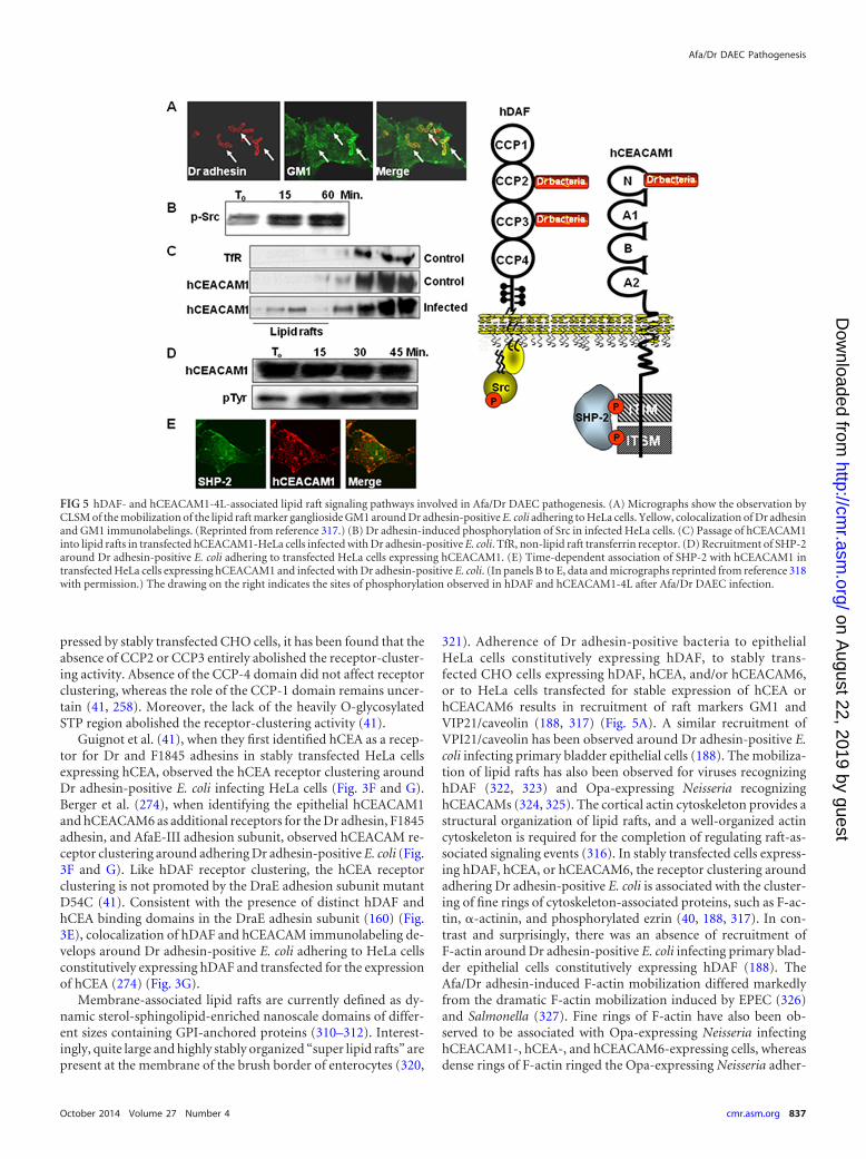
pressed by stably transfected CHO cells, it has been found that theabsence of CCP2 or CCP3 entirely abolished the receptor-cluster-ing activity. Absence of the CCP-4 domain did not affect receptorclustering, whereas the role of the CCP-1 domain remains uncer-tain (41, 258). Moreover, the lack of the heavily O-glycosylatedSTP region abolished the receptor-clustering activity (41).
Guignot et al. (41), when they first identified hCEA as a recep-tor for Dr and F1845 adhesins in stably transfected HeLa cellsexpressing hCEA, observed the hCEA receptor clustering aroundDr adhesin-positive E. coli infecting HeLa cells (Fig. 3F and G).Berger et al. (274), when identifying the epithelial hCEACAM1and hCEACAM6 as additional receptors for the Dr adhesin, F1845adhesin, and AfaE-III adhesion subunit, observed hCEACAM re-ceptor clustering around adhering Dr adhesin-positive E. coli (Fig.3F and G). Like hDAF receptor clustering, the hCEA receptorclustering is not promoted by the DraE adhesion subunit mutantD54C (41). Consistent with the presence of distinct hDAF andhCEA binding domains in the DraE adhesin subunit (160) (Fig.3E), colocalization of hDAF and hCEACAM immunolabeling de-velops around Dr adhesin-positive E. coli adhering to HeLa cellsconstitutively expressing hDAF and transfected for the expressionof hCEA (274) (Fig. 3G).
Membrane-associated lipid rafts are currently defined as dy-namic sterol-sphingolipid-enriched nanoscale domains of differ-ent sizes containing GPI-anchored proteins (310–312). Interest-ingly, quite large and highly stably organized “super lipid rafts” arepresent at the membrane of the brush border of enterocytes (320,
321). Adherence of Dr adhesin-positive bacteria to epithelialHeLa cells constitutively expressing hDAF, to stably trans-fected CHO cells expressing hDAF, hCEA, and/or hCEACAM6,or to HeLa cells transfected for stable expression of hCEA orhCEACAM6 results in recruitment of raft markers GM1 andVIP21/caveolin (188, 317) (Fig. 5A). A similar recruitment ofVPI21/caveolin has been observed around Dr adhesin-positive E.coli infecting primary bladder epithelial cells (188). The mobiliza-tion of lipid rafts has also been observed for viruses recognizinghDAF (322, 323) and Opa-expressing Neisseria recognizinghCEACAMs (324, 325). The cortical actin cytoskeleton provides astructural organization of lipid rafts, and a well-organized actincytoskeleton is required for the completion of regulating raft-as-sociated signaling events (316). In stably transfected cells express-ing hDAF, hCEA, or hCEACAM6, the receptor clustering aroundadhering Dr adhesin-positive E. coli is associated with the cluster-ing of fine rings of cytoskeleton-associated proteins, such as F-ac-tin, �-actinin, and phosphorylated ezrin (40, 188, 317). In con-trast and surprisingly, there was an absence of recruitment ofF-actin around Dr adhesin-positive E. coli infecting primary blad-der epithelial cells constitutively expressing hDAF (188). TheAfa/Dr adhesin-induced F-actin mobilization differed markedlyfrom the dramatic F-actin mobilization induced by EPEC (326)and Salmonella (327). Fine rings of F-actin have also been ob-served to be associated with Opa-expressing Neisseria infectinghCEACAM1-, hCEA-, and hCEACAM6-expressing cells, whereasdense rings of F-actin ringed the Opa-expressing Neisseria adher-
FIG 5 hDAF- and hCEACAM1-4L-associated lipid raft signaling pathways involved in Afa/Dr DAEC pathogenesis. (A) Micrographs show the observation byCLSM of the mobilization of the lipid raft marker ganglioside GM1 around Dr adhesin-positive E. coli adhering to HeLa cells. Yellow, colocalization of Dr adhesinand GM1 immunolabelings. (Reprinted from reference 317.) (B) Dr adhesin-induced phosphorylation of Src in infected HeLa cells. (C) Passage of hCEACAM1into lipid rafts in transfected hCEACAM1-HeLa cells infected with Dr adhesin-positive E. coli. TfR, non-lipid raft transferrin receptor. (D) Recruitment of SHP-2around Dr adhesin-positive E. coli adhering to transfected HeLa cells expressing hCEACAM1. (E) Time-dependent association of SHP-2 with hCEACAM1 intransfected HeLa cells expressing hCEACAM1 and infected with Dr adhesin-positive E. coli. (In panels B to E, data and micrographs reprinted from reference 318with permission.) The drawing on the right indicates the sites of phosphorylation observed in hDAF and hCEACAM1-4L after Afa/Dr DAEC infection.
Afa/Dr DAEC Pathogenesis
October 2014 Volume 27 Number 4 cmr.asm.org 837
on August 22, 2019 by guest
http://cmr.asm
.org/D
ownloaded from

ing to hCEACAM3-expressing cells (328). The fine rings accom-panied the low level of F-actin-independent cell entry intohCEACAM1-, hCEA-, and hCEACAM6-expressing cells, andthe dense rings accompanied the high level of F-actin-depen-dent, small Rac1- and Cdc42 GTPase-triggered cell entry intohCEACAM3-expressing cells (329, 330). It is noticeable that thelow level of Afa/Dr DAEC internalization into hCEA- andhCEACAM6-expressing HeLa and CHO cells is not affected bycytoskeleton blockers (189, 284, 317, 331). Moreover, the recruit-ment of F-actin around adhering Dr adhesin-positive bacteriaplays no role in bacterial internalization, since although the DraEadhesion subunit mutant D54G has impaired F-actin mobiliza-tion, it displays an unchanged level of cell entry (317). Collec-tively, the results obtained with Afa/Dr DAEC indicated that therecruitment of F-actin forming a fine ring around adhering bac-teria probably reflects the physical mobilization of F-actin-con-taining lipid rafts by the adhering bacteria rather than the recruit-ment of F-actin for subsequent bacterium-triggered cellularevents.
During the adhesion step preceding cell entry, the �5�1 integ-rin has been observed to be mobilized so as to form fine ringsaround adhering Dr adhesin- or AfaE-III adhesion subunit-posi-tive bacteria (188, 189, 317, 332). In addition, AfaE/D adhesinsubunit-coated beads adhering onto HeLa cells are decorated byrings of positive immunofluorescence for �5�1 integrin (167).Moreover, it was noted that the clustering of �1 integrin around E.coli adhering to CHO-hDAF-�5�1 cells occurred for bacteria ex-pressing DraE or AfaE-III adhesin subunits alone, regardless of thepresence or absence of DraD or AfaD-III adhesin subunits (189).The mobilization of �1 integrin by Afa/Dr DAEC adhering to thecell surface of undifferentiated epithelial cells could result fromthe mobilization of lipid rafts containing integrins. Indeed, integ-rins have been found in lipid rafts engaged in cell adhesion (333–335), cell migration (336, 337), and contractile forces for cell in-vasion (337–339). As discussed above for F-actin mobilization,the recruitment of �1 integrin by adhering Afa/Dr DAEC canreflect the physical mobilization of �1 integrin-containing lipidrafts by the adhering bacteria.
hDAF-dependent signaling. hDAF has a signal transductioncapacity associating Src tyrosine kinases p56lck and p59fyn (251).Phosphorylation of Src develops in epithelial HeLa cells infectedwith Dr-positive E. coli (318) (Fig. 5B). When examining the in-volvement of Src kinases in Dr adhesin-induced hDAF signaling,Queval et al. (258) observed the recruitment of phosphorylatedSrc kinases together with hDAF around adhering recombinant Dradhesin-positive E. coli in infected hDAF-transfected CHO cellsand in constitutively hDAF-expressing HeLa and human embry-onic kidney HEK293 cells. CCP-4 of hDAF plays a crucial role inthe recruitment of phosphorylated Src kinases and Src kinase ac-tivation, while deletion of CCP-1 had no effect. Moreover, smallinterfering RNA (siRNA) silencing of c-Src in HeLa cells abolisheshDAF clustering around adhering Dr-positive bacteria, whilesiRNA silencing of the Src kinases Yes, Fyn, and Lyn does not(258). Finally, the observation that the D54C DraE adhesion sub-unit mutant fails to induce hDAF clustering and Src recruitmentconfirms the predominant role of this domain of the DraE adhesinsubunit in triggering cell responses.
A variety of cell signaling pathways are activated after the rec-ognition of hDAF by Afa/Dr adhesins. Phosphatidylinositol 3-ki-nase (PI3K) is activated in undifferentiated intestinal INT407 cells
(340), and phosphorylated PI3K is recruited around Dr adhesin-positive E. coli adhering to human differentiated primary bladdercells expressing hDAF and hCEACAMs (188). Triggered by the Drand F1845 adhesins, an hDAF-dependent activation of mitogen-activated protein kinases (MAPKs), including extracellular signal-regulated kinase 1/2 (Erk1/2), p38 mitogen-activated protein ki-nase (p38), and stress-activated protein kinase/c-Jun N-terminalkinase SAPK/JNK (JNK), occurred in enterocyte-like Caco-2 cellsand colonic T84 cells (341–345). Src kinase and NF-B and AKTsignaling pathways are hDAF-dependently activated in C1845-infected T84 cells (345). Moreover, hDAF-dependent activationof tyrosine kinases and protein kinase C (PKC) develops in Afa/DrDAEC-infected polymorphonuclear leukocyte (PMNL)-like cells(346). In addition, MAPK and NF-B activation occurred inPMNL-like cells triggered by type 1 pili and independently ofhDAF recognition (347).
hCEACAM-dependent signaling. Like the Opa52 protein of Neis-seria, which recognizes the N-terminal domain of CEACAM1-4Land promotes its association with the tyrosine phosphatasesSHP-1 and SHP-2 in the cytoplasmic ITIM domains (348), Dradhesin-expressing E. coli binding to HeLa cells transfected for theexpression of hCEACAM1-4L leads to the translocation of a partof CEACAM1-4L into the membrane lipid rafts and phosphory-lation of its ITIM and SHP-2 recruitment (318) (Fig. 5C to E). Inturn, hCEACAM1-4L has been found to play a key role in down-regulating the activity of the protein tyrosine Src kinase associatedwith hDAF signaling (318).
Urinary Tract Infections and Pregnancy Complications
Recurrent cystitis in the bladder and acute pyelonephritis in thekidney, corresponding to 80% of all UTIs, result from infection byUPEC (199, 349). Bacteriuria is the clinical sign of UTI. Recurrentcystitis is a major health problem. Indeed, a recurrence of cystitiswithin 3 to 4 months develops in 20 to 30% of women who haddeveloped a first acute infection. Lower UTIs affect the urethraand bladder, whereas upper UTIs affect the ureters and kidneys,and both can be either uncomplicated or complicated. Cell insultstogether with an intense mucosal inflammatory response, includ-ing the recruitment of neutrophils, lead to cystitis in bladder andpyelonephritis in kidney. It should be noted that asymptomaticbacteriuria (ABU) has been observed in 2 to 20% of the popula-tion, depending on age and gender (199). The infecting E. colistrain involved in ABU colonizes the urothelium and may remainpresent for months or years, resulting in a low-level deleterioustissue attack effect and an innate immune response that is tooweak to cause symptoms. The observation that UPEC virulencegene sequences remained present in ABU strains has led to thesuggestion that the lower virulence of ABU results from a shiftfrom UPEC as a result of genome reduction caused by inactivationof virulence genes as well as by deletions or by the accumulation ofpoint mutations (350, 351), although other causes can also beinvolved (352).
UPEC strains have an elevated organ tropism and ascend theurinary tract from the urethra to the bladder and kidneys (1, 199,232). To do this, UPEC expresses a wide variety of adhesive fac-tors, including type 1 pili, type IV pili, and P, S, F1C, Auf, Yad, Ygi,and F9 fimbriae (199, 353). Moreover, UPEC has developed areciprocal regulation of adhesive factors and motility (199). UPECstrains have developed sophisticated strategies to avoid clearanceby micturition involving colonization of the urothelium and cell
Servin
838 cmr.asm.org Clinical Microbiology Reviews
on August 22, 2019 by guest
http://cmr.asm
.org/D
ownloaded from
internalization, which thus allow them to survive and evade hostinnate immune defenses. For pathogenesis, UPEC expresses PAIsof different sizes containing assemblages of genes encoding viru-lence factors such as adhesins, invasins, capsule, proteases, andmultiple siderophore systems, including aerobactin, IroN, andIreA (199). Moreover, UPEC secretes cytotoxic toxins, includingthe repeat-in-toxin �-hemolysin (HlyA), cytotoxic necrotizingfactor (CNF-1), and diverse SPATE toxins, such as Sat, Pic, PicU,Tsh, and Vat. In addition, UPEC adapts to survive in the urine byexpressing factors involved in scavenging of nutrients.
Internalization. Cell entry of E. coli expressing the Dr, F1845,or Afa-III adhesin has been examined in cervical epithelial HeLacells and endometrial cells constitutively expressing hDAF but nothCEACAMs, in undifferentiated intestinal cells and differentiatedprimary epithelial bladder cells constitutively expressing hDAFand hCEACAMs, and in CHO or HeLa cells transfected for stableexpression of hDAF, hCEACAM1, hCEA, or hCEACAM6 (38, 42,188, 189, 234, 259, 317, 319, 331, 332, 354). Afa/Dr DAEC displaysa rate of cell entry similar to that observed with UPEC expressingtype 1 pili (199, 232). Dr and Afa-III adhesin-positive E. coli useda membrane zipper-like mechanism to enter the HeLa cells (42,317), and Afa-III adhesin-positive E. coli entered the cells via asingle internalization vacuole (42). The cell membrane lipid raftsplay a pivotal role in the internalization of Dr- and Afa-III adhe-sin-positive E. coli into cultured cervical HeLa cells, primary cul-tured human bladder cells, and epithelial cells transfected to ex-press hDAF, hCEACAM1, hCEA, or hCEACAM6 by a mechanisminvolving cell microtubules but not microfilaments (38, 188, 189,259, 317, 331). A particular network of microtubules, i.e., the dy-namically unstable microtubule network, has been found to beinvolved in the cell entry of Dr adhesin-positive E. coli (331). Ranaet al. (234), using a PCR-based proximity ligation assay to detectprotein-protein interactions, observed a strong fluorescent signalof hDAF/tubulin in Dr-positive E. coli-infected HeLa cells result-ing from the proximity between the two molecules, suggestingthat hDAF and microtubules can be physically associated afterinfection. The Afa/Dr DAEC cell entry resembles the lipid raft-and microtubule-dependent zipper-like uptake of Opa-express-ing Neisseria into transfected CHO and HeLa cells expressinghCEA or hCEACAM6 (330). It was noted that a few invasive bac-teria, including, for example, Haemophilus influenzae, Klebsiellapneumoniae, and pilus type 1-expressing UPEC, also require afunctional host microtubule network for the invasion of host ep-ithelial cells (355). Consistent with the F-actin microfilament-in-dependent cell entry of Dr-positive E. coli, the DraE adhesin sub-unit mutant strains D54G and D54C, showing absence andpresence of changes in F-actin mobilization around adhering bac-teria, respectively, displayed unchanged levels of cell internaliza-tion (317). Microtubule-dependent internalization of Dr adhesin-positive E. coli within primary bladder epithelial cells has beenfound to result from the engagement of the actin-binding proteinsezrin/radixin/moesin (ERM) (188). Observations that phosphor-ylation of ERM accompanied the Dr-positive E. coli cell entry(188) agree well with the know role of ERM, which together withRho GTPases act in the remodeling of host cell cytoskeleton (356).It has been noticed that the efficiency of bacterial cell entry result-ing from the action of T3SS-dependent effectors seems to behigher than that resulting from microtubule-dependent processestriggered by bacteria that do not express the T3SS.
Selvaragan et al. (259) have determined the roles of hDAFepitopes in the cell entry of Dr adhesin-positive bacteria usinghDAF deletion mutants expressed by stably transfected CHO cells.The absence of CCP-2 or CCP-3 entirely abolished the cell entry ofDr-positive bacteria, whereas the lack of CCP-1 or CCP-4 had noeffect. Deletion of the heavily O-glycosylated STP region abol-ished the cell entry. In contrast, replacing the GPI anchor with thetransmembrane anchor of HLA-B44 or membrane cofactor pro-tein did not modify the cell entry. Rana et al. (234) have examinedfor adhesion/invasion five hDAF mutants previously used to mapthe extracellular CCP-2/CCP-3 domains of hDAF involved in Dradhesin-positive E. coli adhesion (260). Compared to hDAF, thePhe123-Ala mutant conserves both normal binding activity andinvasion rate, the Ser165-Ala mutant conserves normal bindingactivity but displays a lower invasion rate, and the Gly159-Alamutant shows both reduced binding capacity and a lower invasionrate. In contrast, the Phe148-Ala and Phe154-Ala mutants displaynormal binding capacity combined with an increased rate of in-vasion.
In terms of cell signaling, protein tyrosine kinases (PTKs),phospholipase C (PLC-), and protein kinase C (PKC) blockershave no effect on the cell internalization of Dr adhesin-positive E.coli (317). In contrast, the PI3K-dependent signaling pathway isengaged in the internalization of Dr adhesin-positive E. coli intohCEACAM1-, hCEA-, and hCEACAM6-expressing epithelialcells but not hDAF-expressing cells (188). While Src-dependentsignaling does not play any role in internalization (188), Src andphosphorylated Src have been observed to be recruited aroundadhering Dr-positive E. coli (258).
The �1 integrin is the host cell receptor triggering the zipper-like internalization of type 1 pilus-expressing UPEC (199, 232). Apolyclonal antibody directed against anti-�5�1 integrin abolishesthe basolateral entry of Dr adhesin-positive E. coli within undif-ferentiated intestinal Caco-2 cells (331). Comparison of the cellentry of beads coated with rAfaD-III adhesin subunit into mouseendodermal carcinoma F9-TKO cells stably transfected or not toexpress �1 integrin shows that the presence of the integrin in-creased the bead internalization level 2.3-fold (332). In addition,in HeLa cells subjected to incubation with AfaD-III or AfaD-VIIIadhesin subunits, membrane immunoprecipitation showed thatAfaD-III and -VIII adhesin subunits coprecipitated with �1 integ-rin (332). The role of �5�1 integrin as a receptor for internaliza-tion of E. coli expressing Dr- or Afa-III adhesin subunits into ep-ithelial cells has been recently revisited (189). The overexpressionof �5�1 integrin as a result of transfection in the CHO B2 clone,which does not express �5�1 integrin, does not increase the levelsof adhering and internalized Dr- or Afa-III adhesin-positive E. colicompared to that in untransfected cells. To test whether the coex-pression of �5�1 integrin together with hDAF, hCEACAM1,hCEA, or hCEACAM6 influences the adhesion and/or cell entry ofDr adhesin-positive E. coli, CHO cells transfected for the stableexpression of hDAF, hCEAM1, hCEA, or hCEACAM6 were trans-fected again with the gene coding for the �5 or �1 integrin subunit(189). In all cases, the presence of �5�1 integrin together withhDAF or CEACAMs did not increase the levels of adhering andinternalized Dr adhesin-positive E. coli. In addition, the knock-down of the gene coding for �1 integrin by siRNAs in HeLa cellsconstitutively expressing �5�1 integrin did not affect the level ofadhering and internalized Dr- or Afa-III adhesin-expressing E. colicompared to that in untreated cells (189). It is important to note
Afa/Dr DAEC Pathogenesis
October 2014 Volume 27 Number 4 cmr.asm.org 839
on August 22, 2019 by guest
http://cmr.asm
.org/D
ownloaded from
that the fact that integrins are located exclusively at the basolateraldomain and never at the brush border of the polarized intestinalepithelial cells renders the integrins inaccessible for intestinal ep-ithelium infection by enterovirulent Afa/Dr DAEC residingwithin the luminal compartment.
The identity of the virulence factor(s) of Afa/Dr DAEC in-volved in the invasion of epithelial cells has long been debated. Ithas been proposed by Le Bouguénec and coworkers (357) that theD adhesin subunits encoded by the dra, daa, and afa-1 to -3 oper-ons function as an invasin for the entry of Afa/Dr DAEC intourothelial and bladder cells. DraD and AfaD adhesin subunitshave been identified at the tips of Afa/Dr fibrils (167, 180). AfaDprotein, unlike the AfaE protein, was able to detach from the bac-terium (28, 168, 169). Using recombinant E. coli producing theAfaD or AfaE-III adhesin subunit, it has been observed that theAfaE-III adhesin subunit triggers the binding of the recombinantE. coli to cells and that the AfaD adhesin subunit mediates the cellentry within epithelial HeLa cells (42, 168). When HeLa cells areinfected with an Afa-III adhesin-positive E. coli strain, the AfaE-IIIprotein remains localized at the membranes of infected cells, andonly the AfaD-III protein coats the internalized bacteria residingwithin an internalization vacuole (169). In addition, coating poly-carbonate beads with AfaD protein allows the beads to penetratethe epithelial cells (28). Moreover, using colloidal gold-taggedAfaE-III and AfaD proteins, it has been shown that AfaE-III– goldcomplexes associate with the cell surface, whereas AfaD-goldcomplexes are internalized within the cells (42). In human undif-ferentiated Caco-2, T84, and INT407 cells, urothelial T24 cells,and cervical HeLa cells, rAfaD-III- and rAFaD-VIII adhesin sub-unit-coated beads invaded the cells, whereas rAfaE-III adhesinsubunit-coated beads were not internalized (332). In contrast, ac-cording to Nowicki and coworkers, the DraE adhesin subunitalone is sufficient to allow the entry of Dr adhesin-positive bacte-ria into epithelial cells. Indeed, both the purified Dr fimbriae andlatex beads coated with Dr adhesin are internalized into HeLa cells(259, 319). Moreover, the insertional draE, draC, and draB andadherent draD mutants were unable to enter epithelial cells, andcomplementation of the draE mutation restored the invasionproperty (38). Mapping of the DraE adhesin subunit shows thatamino acids localized on hydrophilic domain II, and in particularthe V28, T31, G33, Q34, L35, T36, and P40 amino acids, reducedor abolished bacterial cell entry into transfected CHO-hDAF cellsand HeLa cells constitutively expressing hDAF but did not affectattachment (319). The role of E and/or D adhesin subunits in theinternalization of Afa/Dr DAEC has been recently reexaminedusing appropriate E. coli mutant strains and cultured unpolarizedepithelial cells and bladder cells (188, 189). Korotkova et al. (188)found that expressing combinations of the DraE�/DraD� andDraE�/DraD� adhesin subunits allowed E. coli to enter differen-tiated primary bladder cells, while the combination of DraE�/DraD� adhesin subunits did not. Guignot et al. (189) have foundthat in CHO cells transfected to express hDAF and �5�1 integrin,recombinant E. coli expressing combinations of DraE�/DraD� orAfaE�/AfaD�-III adhesin subunits adhered and were internalizedand that deletion of the DraD adhesin subunit did not modify thelevel of internalized bacteria. Collectively, these last findings fit inwell with the previous results reported by Nowicki and coworkers(38, 319), and taken together they clearly establish that the DraEand AfaE-III adhesin subunits are necessary and sufficient to pro-mote the cell entry of Dr and Afa-III adhesin-expressing E. coli
into epithelial cells. However, one question remains unresolvedconcerning the functions of the DraD, AfaE, and DaaD adhesinsubunits in the pathogenesis of Afa/Dr DAEC when the proteinsare detached from the fimbriae (42) or when each of the proteinsare present alone at the bacterial cell surface (42, 168, 169). It isnoteworthy that the DraD adhesin subunit, whether fimbria asso-ciated or not, seems to function in the formation of biofilm by Dradhesin-expressing E. coli (190, 233).
Intracellular lifestyle. When internalized within nonphago-cytic epithelial cells, enteric bacterial pathogens reside and survivein the cell cytoplasm in small or large vacuoles, where they may ormay not replicate (358); only a few invasive enteric bacterialpathogens have developed strategies to escape from the vacuole togain access to and proliferate within the host cell cytosol (359).After entering HeLa cells inside a single internalization vacuole,Afa/Dr DAEC survived within large late vacuoles (38, 40, 42, 94,189, 234, 259, 319, 331, 332) (Fig. 6A). As revealed by transmissionelectron microscopy examination, the late large vacuole contain-ing internalized Dr or Afa-III adhesin-expressing bacteria appearsto result from the fusion of early vacuoles formed during the ini-tial step of the internalization process, each containing a singlebacterium (42, 234, 259, 319). Recent data have shown that theinternalization vacuoles containing Dr adhesin-positive bacteriainteracted with the cell endocytic pathway. This pathway includesa complex and multifunctional set of vesicular compartments es-sentially derived from internalization of the plasma membrane,resulting in the formation and maturation of well-formed endo-cytic compartments (360). These compartments are sequentiallymodified by acquiring diverse vesicular elements to form the earlyendosomes and subsequently the late endosomes. In the last step,the intraluminal vesicles are taken for delivery of their contents tothe lysosome, which is a stable organelle that avoids self-degrada-tion (361). The intracellular vacuoles containing Dr adhesin-pos-itive E. coli display some of the characteristics of late endosomes,such as the membrane expression of the Lamp-1, Lamp-2, andCD63 proteins but not of cathepsin D, and are acidic (189) (Fig.6B). This resembles the characteristics of the UPEC UTI89-con-taining vacuoles, which are positive for Lamp-1 and CD63, ca-thepsin D negative, and acidic (362). Surprisingly, despite the factsthat Dr adhesin-positive bacteria bind to membrane-associatedhDAF during the first step of cell association (41, 257–260) andthat hDAF has been observed associated with the membrane in-vagination during the first step of adhesion/internalization (45),the membranes of the large late vacuoles containing the internal-ized Dr-positive bacteria do not contain hDAF (189).
Cellular autophagy (363) is an evolutionarily conserved pro-cess by which cellular cytosolic structures and cell constituents aredegraded and recycled (364). Some intracellular bacteria are de-tected and eliminated via the autophagic pathway, and severalintracellular bacteria have developed sophisticated avoidancestrategies, but this pathway may also serve as a protected nicheproviding a source of nutrients for intracellular bacteria (365).Infecting UPEC was rapidly cleared in the presence of a deficiencyof the key autophagy protein Atg16L1, suggesting that UPEC maysubvert autophagy proteins to establish latency (366). However,the autophagy pathway can favor the pathogen, since internalizedbacteria present within intracellular bacterial communities in su-perficial bladder cells colocalized with Atg16L1 and LC3 puncta(366). No interaction has been observed between the Dr adhesin-positive E. coli-containing vacuoles and the autophagic pathway
Servin
840 cmr.asm.org Clinical Microbiology Reviews
on August 22, 2019 by guest
http://cmr.asm
.org/D
ownloaded from
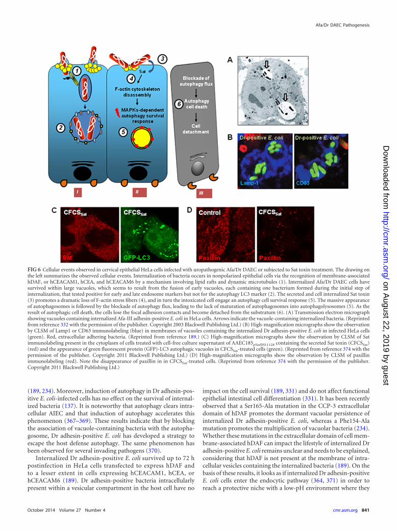
(189, 234). Moreover, induction of autophagy in Dr adhesin-pos-itive E. coli-infected cells has no effect on the survival of internal-ized bacteria (137). It is noteworthy that autophagy clears intra-cellular AIEC and that induction of autophagy accelerates thisphenomenon (367–369). These results indicate that by blockingthe association of vacuole-containing bacteria with the autopha-gosome, Dr adhesin-positive E. coli has developed a strategy toescape the host defense autophagy. The same phenomenon hasbeen observed for several invading pathogens (370).
Internalized Dr adhesin-positive E. coli survived up to 72 hpostinfection in HeLa cells transfected to express hDAF andto a lesser extent in cells expressing hCEACAM1, hCEA, orhCEACAM6 (189). Dr adhesin-positive bacteria intracellularlypresent within a vesicular compartment in the host cell have no
impact on the cell survival (189, 331) and do not affect functionalepithelial intestinal cell differentiation (331). It has been recentlyobserved that a Ser165-Ala mutation in the CCP-3 extracellulardomain of hDAF promotes the dormant vacuolar persistence ofinternalized Dr adhesin-positive E. coli, whereas a Phe154-Alamutation promotes the multiplication of vacuolar bacteria (234).Whether these mutations in the extracellular domain of cell mem-brane-associated hDAF can impact the lifestyle of internalized Dradhesin-positive E. coli remains unclear and needs to be explained,considering that hDAF is not present at the membrane of intra-cellular vesicles containing the internalized bacteria (189). On thebasis of these results, it looks as if internalized Dr adhesin-positiveE. coli cells enter the endocytic pathway (364, 371) in order toreach a protective niche with a low-pH environment where they
FIG 6 Cellular events observed in cervical epithelial HeLa cells infected with uropathogenic Afa/Dr DAEC or subjected to Sat toxin treatment. The drawing onthe left summarizes the observed cellular events. Internalization of bacteria occurs in nonpolarized epithelial cells via the recognition of membrane-associatedhDAF, or hCEACAM1, hCEA, and hCEACAM6 by a mechanism involving lipid rafts and dynamic microtubules (1). Internalized Afa/Dr DAEC cells havesurvived within large vacuoles, which seems to result from the fusion of early vacuoles, each containing one bacterium formed during the initial step ofinternalization, that tested positive for early and late endosome markers but not for the autophagy LC3 marker (2). The secreted and cell internalized Sat toxin(3) promotes a dramatic loss of F-actin stress fibers (4), and in turn the intoxicated cell engage an autophagy cell survival response (5). The massive appearanceof autophagosomes is followed by the blockade of autophagy flux, leading to the lack of maturation of autophagosomes into autophagolysosomes (5). As theresult of autophagic cell death, the cells lose the focal adhesion contacts and become detached from the substratum (6). (A) Transmission electron micrographshowing vacuoles containing internalized Afa-III adhesin-positive E. coli in HeLa cells. Arrows indicate the vacuole-containing internalized bacteria. (Reprintedfrom reference 332 with the permission of the publisher. Copyright 2003 Blackwell Publishing Ltd.) (B) High-magnification micrographs show the observationby CLSM of Lamp1 or CD63 immunolabeling (blue) in membranes of vacuoles containing the internalized Dr adhesin-positive E. coli in infected HeLa cells(green). Red, extracellular adhering bacteria. (Reprinted from reference 189.) (C) High-magnification micrographs show the observation by CLSM of Satimmunolabeling present in the cytoplasm of cells treated with cell-free culture supernatant of AAEC185pSatIH11128 containing the secreted Sat toxin (CFCSSat)(red) and the appearance of green fluorescent protein (GFP)-LC3 autophagic vacuoles in CFCSSat-treated cells (green). (Reprinted from reference 374 with thepermission of the publisher. Copyright 2011 Blackwell Publishing Ltd.) (D) High-magnification micrographs show the observation by CLSM of paxillinimmunolabeling (red). Note the disappearance of paxillin in in CFCSSat-treated cells. (Reprinted from reference 374 with the permission of the publisher.Copyright 2011 Blackwell Publishing Ltd.)
Afa/Dr DAEC Pathogenesis
October 2014 Volume 27 Number 4 cmr.asm.org 841
on August 22, 2019 by guest
http://cmr.asm
.org/D
ownloaded from
can live and remain metabolically active in order to replicate orenter dormancy. The observation that the Dr adhesin-positive E.coli-containing large vacuoles are phenotypically quite similar tolysosomes indicates that internalized bacteria have developed acapacity to actively modify this compartment so as to create adistinct compartment. The mechanism(s) by which internal-ized uropathogenic Afa/Dr DAEC cells modify the vacuolarcompartment and lead it to engage in dormancy or intracellu-lar replication remains to be elucidated. A process designated“nutritional virulence” (372) has recently been defined as usedby several invasive bacteria which, to allow themselves to grow,subvert the host cell machinery so as to create a vesicular de-velopmental niche where they receive components from thesecretory pathway as a source of nutrients. It will be interestingto investigate whether internalized Afa/Dr DAEC cells do or donot develop a “nutritional virulence” mechanism in order topermit their intracellular residence and survival. The observa-tion of an intracellular lifestyle of Dr adhesin-positive E. coliwith intracellular survival is interesting in term of persistence andrecurrence of infection. Indeed, it is worth mentioning that type 1pilus-positive UPEC after cell entry resided in endocytic vesicles inwhich its replication is restricted and which form an intracellularniche that is protected from host immunity responses and killingof intracellular bacteria by antibiotics (232). This intracellular life-style permits the long-term maintenance of quiescent UPEC in thebladder cells (232).
Cell detachment. To protect the epithelia against bacterial col-onization and invasion and to maintain tissue homeostasis, thehost has developed defense systems including exfoliation and celldeath of bacterially infected cells (370). The eviction of cells fromthe epithelium can occur by a process known as extrusion follow-ing detachment from the extracellular matrix and loss of cell-to-cell contact or toward the basal surface by delamination after dis-ruption of cell-to-cell contacts (373). For example, exfoliation offully differentiated umbrella epithelial cells infected by FimH-pos-itive E. coli is an innate host defense mechanism that has the effectof removing adhering and invaded bacteria from the bladder (199,232). However, some invasive bacterial pathogens have developedcountermeasures intended to antagonize the host epithelial turn-over to exploit infected epithelial cells as a survival and replicativeniche (370). As an adverse effect for the host, the detachment ofcells containing intracellular UPEC promotes the reinitiation ofthe UPEC infectious cycle (199, 232).
A cell detachment effect triggered by the Sat toxin expressed byAfa/Dr DAEC has been recently described (137, 206, 210–212).Sat promotes a profound disorganization of the F-actin cytoskel-eton in HeLa cells that is followed by a host cell survival responseinvolving the noncanonical autophagy pathway (374). Theautophagy pathway induced is not intended to destroy the intra-cellular toxin, since the toxin is never present in autophagic vesi-cles. The Sat-intoxicated cells displayed a massive intracellularappearance of autophagic vacuoles which, at the autophagosomestep, fail to mature into autophagolysomes. In turn, the Sat-intox-icated cells overexpressing autophagosomes die as a result of au-tophagy cell death. A similar effect has been reported for �-hemo-lysin of Streptococcus aureus (375). The Sat-induced autophagycell death is accompanied by a dramatic disassembly of the focaladhesion-associated vinculin and paxillin, which finally promotescell detachment (374). It is noteworthy that Capello et al. (376)have observed that the class I SPATE toxin Pet of EAEC induces
the cleavage of FAK and a deterioration of focal adhesion com-plexes resulting from the redistribution of paxillin and vinculinand the depletion of phosphotyrosines. Together with a loss ofF-actin stress fibers and �-actinin and spectrin network disassem-bly, these dramatic rearrangements of structural and functionalproteins result in cell rounding and detachment of Pet-intoxicatedcells from the substratum (376–378). Interestingly, the UPECpore-forming HylA triggering cell exfoliation of bladder epithelialcells induces cell rounding and a complete loss of the microtubulenetwork and F-actin stress fibers, accompanied by the degradationof paxillin (379).
Inflammatory responses. New information on immune re-sponses accompanying UPEC infection have recently been ob-tained in mice models (380, 381). UPEC infection is accompaniedby a strong inflammatory response, including the production ofcytokines such as tumor necrosis factor alpha (TNF-�), interleu-kin-6 (IL-6), IL-8, IL-17a, and granulocyte colony-stimulatingfactor (199, 382). Moreover, neutrophils and macrophages play apivotal role in the defense against UTI (383). Although not ob-served in urinary cell models, it is important to note that the UTI-associated wild-type Afa/Dr DAEC strain IH11128 promotes anhDAF- and MAPK-dependent production of IL-8 (341) followedby transepithelial migration of PMNLs (341), triggering the pro-duction of TNF-� (342).
Animal models of UTIs. Dr adhesin-positive E. coli leads tochronic pyelonephritis in experimental mice (301, 384). In con-trast, wild-type strain IH11128, an isogenic mutant that is devoidof Dr adhesin, does not cause kidney infection or cellular lesionsand is gradually eliminated compared to the wild-type strain. Inthe kidney tissue of Dr adhesin-positive E. coli-infected mice, Drantigen was present in the injured parenchymal regions character-ized by histological changes indicating tubulointerstitial nephritis(384). It is notable that mouse DAF (mDAF) probably plays norole in this Dr adhesin-induced chronic ascending pyelonephritisin mice, since Hudault et al. (254) have demonstrated that Dradhesin binds specifically to hDAF but not to rodent DAF. Impor-tantly, Selvarangan et al. (301) have demonstrated the crucial roleof the type IV collagen-binding capability of Dr adhesin for patho-genesis in mouse kidney. Indeed, an isogenic DraE adhesin sub-unit mutant lacking binding to type IV collagen fails to persistwithin the mouse renal tissues, and the transcomplementation ofthe mutant strain, restoring the type IV collagen-binding activity,allows the reestablishment of a long-term renal infection. It wasnoticed that immunization of LPS-nonresponder C3H/HeJ micewith purified Dr protein resulted in delayed mortality when theimmunized mice were infected by instilling wild-type Dr adhesin-positive strain IH11128 into the bladder, and incubation ofIH11128 with sera of immunized mice resulted in a marked de-crease in bacterial adhesion to ex vivo specimens of mouse blad-ders and kidneys (385). However, additional bacterial factors mayalso be involved, since Meitinen et al. (386) observed that afterinjection of O75X fimbriae in mice, there was an absence of his-tological damage despite persistent O75X deposits in glomeruli.Interestingly, the Sat toxin of the prototype UPEC wild-type strainCFT073 has been found to promote renal histological lesions inmice (387). It remains to be determined whether Sat expressed bystrain IH11128 plays a role in the IH11128-induced mice kidneylesions.
Servin
842 cmr.asm.org Clinical Microbiology Reviews
on August 22, 2019 by guest
http://cmr.asm
.org/D
ownloaded from
Pregnancy Complications
Bogdan Nowicki and coworkers have illuminated the risk gener-ated by UTIs caused by UPEC harboring the Dr adhesin in womenwho have undergone pregnancy termination or preterm labor(105), and this is related to the levels of tissue expression of hDAF(82, 388–390) and TLR4 (390, 391). Dr adhesin-positive E. colistrains have been found to be often present in pyelonephritis-associated E. coli strains isolated from pregnant females (30, 62,90, 91, 96, 97). Nowicki and coworkers (82, 105) have convinc-ingly demonstrated the role of Dr-positive UPEC in pregnancycomplications. In the uterus, hDAF is present in the endometrialglands, spiral arterioles, and myometrial arteries. In these tissues,the density of hDAF expression correlates with a high level ofregulation of the complement-induced cell damage (392, 393).Moreover, it has been demonstrated that interindividual differ-ences in hDAF density in the endometrium influence the tissuecolonization by Dr adhesin-expressing E. coli (393, 394).
A set of elegant studies have provided a clear demonstration ofthe role of UPEC expressing the Dr and Afa adhesins in the lethaloutcome of gestational infection. Nowicki et al. (395) have shownthat the level of uterine Dr adhesin-positive E. coli infection inpregnant rats was higher than that in nonpregnant rats. In anexperimental model of nonpregnant lipopolysaccharide-hypore-sponder C3H/HeJ mice, Kaul et al. (396) observed that Dr adhe-sin-positive E. coli-induced chronic pyelonephritis resulted in ahigh level of preterm deliveries compared to that in mice infectedwith an E. coli isogenic Dr mutant. A high rate of maternal mor-tality has been observed in rats infected with an AfaE�/AfaD�
strain that causes high rates of maternal mortality (397). Miceinfected with the AfaE�/AfaD� strain show a 2-fold-decreasedlevel of maternal mortality and infection with AfaE�/AfaD� strainresults in a 5-fold decrease in death, while in contrast, the AfaE�/AfaD� double mutant fails to promote death, even though all themutants showed an equal infection level in uteri. The observationthat the decrease in the mortality rate paralleled the decrease in theinvasiveness capacity of a Dr adhesin-positive E. coli strain (397)correlates well with the in vitro observation by Goluszko et al. of ahigh rate of invasiveness capability in Dr adhesin-positive E. coliisolated from pregnant women with UTIs (94).
It has been proposed that infectious complications of preg-nancy such as host gestation-dependent sensitivity to UPEC arerelated to the host nitric oxide (NO) status. When Dr-positive E.coli invades human endometrial adenocarcinoma Ishikawa cells,there is a decrease of cell entry after an induction of NO produc-tion and an increase of cell entry after NO inhibition (354). Inter-estingly, elevation of NO production is accompanied by a signifi-cantly reduced expression of hDAF protein and mRNA (354).Mechanistically, NO triggers a displacement of hDAF from themembrane lipid rafts coupled to its internalization within humanendometrial cells (398). In rodents, an increase in rat uterine NOsynthase (NOS) activity has been observed during pregnancy, andone function of NO generated in the uterus, which declines atterm (399), is to inhibit uterine contractility (400). It has beenobserved, interestingly, that urogenital tract colonization by Dradhesin-positive E. coli is followed by a defense mechanism in-volving the production of NO (401). Moreover, there is a localizedincrease in type II NOS expression and NO production after in-trauterine Dr-positive E. coli infection in pregnant versus non-pregnant rats (402). Comparing LPS responder (C3H/HeN) and
nonresponder (C3H/HeJ) mice and Dr adhesin-positive E. coliand P fimbria-positive E. coli infections, Nowicki et al. (403) ob-served that the infection level in the Dr-positive E. coli-infectedC3H/HeN group treated with an inhibitor of NO, nitro-L-argininemethyl ester (L-NAME), was about 100-fold higher than that inthe P adhesin-positive E. coli-infected, L-NAME-treated group. Dradhesin-positive E. coli infection in mice (403) and AfaE/AfaDadhesin subunit-positive E. coli infection in rats (404) are followedby complications in pregnancy and death. Moreover, the deathrate was increased by treatment with the NO blocker L-NAME inboth mice and rat (403, 404). As indicated above for Dr adhesin-induced pyelonephritis in the mouse model, the role of the mouseDAF in Dr adhesin-induced complications during pregnancy inthe rat model is intriguing, considering that the rodent DAF isclearly not recognized by Afa/Dr adhesins (254). Banadakoppa etal. (405) recently demonstrated an NO-independent regulation ofhDAF expression in endometrial Ishikawa and cervical HeLa cellsinvolving the PI3K/Akt pathway engaging the PI3K/Akt regula-tory protein PTEN. Interestingly, the PI3K/Akt pathway nega-tively regulated the membrane expression of hDAF and conse-quently downregulated the adhesion of Dr adhesin-positive E.coli. As underlined by Nowicki and coworkers, two independenthost cell systems, NO and PI3K/Akt, by the downregulation ofhDAF expression at cell membrane level represent a functionalhost pathogen strategy to achieve a well-controlled limited level ofinfection by Dr adhesin-positive E. coli.
Intestinal Tract Infection
Facing the luminal compartment, the intestinal epithelium islined by a monolayer of highly polarized epithelial cells, includingfour extremely specialized cell phenotypes, each with specializedfunctions: enterocytes, neuroendocrine cells, goblet cells, andPaneth cells. Enterocytes, neuroendocrine cells, and goblet cellsare constantly renewed by detachment from the villus tip (373) viaa specific type of apoptosis known as “anoikis” (406) and are re-placed by a cell cycle renewal characterized by a differentiation/migration process occurring along the crypt-villus axis and start-ing from stem cells localizing at the crypt of the villus (407).Polarized intestinal epithelial cells express an apical domain facingthe luminal compartment. In the cell lateral domain, the cell-to-cell junctional domain, including the tight junction (TJ), the ad-herens junctions (AJs), and the desmosome, establish tight con-tacts with neighboring cells and seal the intestinal cell barrier (299,408). In addition, the basal domain establishes a connection withthe basement membrane. Structural or functional breaches of theintestinal epithelial barrier by enterovirulent bacteria lead to dis-eases (409).
Structural and functional injuries at the intestinal epithelialbarrier. Afa/Dr DAEC strains infecting cultured enterocyte-likeCaco-2 (410–412) and colonic T84 (342) cells attach at the brushborder. Adhesion of F1845 adhesin-positive E. coli strain C1845parallels the cell differentiation-dependent appearance of thebrush border (413). As the result of the recognition of brush bor-der-associated hDAF, recombinant Dr adhesin- and Afa-I adhe-sin-positive E. coli strains adhere to cultured human enterocyte-like HT-29 and Caco-2 cells (414). Similarly, Adlerberth et al.(415) have found that Dr adhesin-positive E. coli cells adhere at thebrush border of freshly isolated ileal enterocytes or colonic cells.The wild-type strain C1845 displays a low level of cell entry wheninfecting the permissive epithelial Hep-2 cell line, 20-fold lower
Afa/Dr DAEC Pathogenesis
October 2014 Volume 27 Number 4 cmr.asm.org 843
on August 22, 2019 by guest
http://cmr.asm
.org/D
ownloaded from
than that of the prototype invasive AIEC wild-type strain LF82 butequaling the rate of internalization of noninvasive EHEC andEAEC strains (416). Moreover, Afa/Dr DAEC strains are nonin-vasive when they infect cultured enterocyte-like cells via the nat-ural apical infection route. Indeed, Guignot et al. (331) haveshown that after apical infection of cultured human enterocyte-like Caco-2/TC7 cells, the level of internalized Afa/Dr DAEC wasvery low, indicating that these bacteria are noninvasive when incontact with the brush border of human enterocytes lining theintestinal epithelial barrier. In contrast, when Afa/Dr DAEC in-fected the Caco-2/TC7 cells basolaterally, it was internalized by amechanism involving the basolaterally expressed �1 integrin. Itshould be noted, however, that Afa/Dr DAEC never uses thebasolateral domain of intestinal epithelial barrier as its natural wayof infection. However, treating Caco-2/TC7 cells with Ca2�-freemedium containing EGTA, disrupting intercellular junctions andexposing the junctional domain of the cells, results in an increasedlevel of internalized Afa/Dr DAEC infecting the brush border.This indicates that when Afa/Dr DAEC infects a diseased intestinalepithelial barrier in which the closure at the junctional domain isimpaired, it can enter the cells.
(i) Structural lesions at the brush border. The highly differen-tiated apical pole structure of enterocytes is composed of a densearray of microvilli, considerably increasing the intestinal surfacearea (299). The formation of the brush border in enterocytes dur-ing the establishment of the apical-basal polarity results from acomplex and highly regulated epithelial polarity program (408).Microvilli are formed by assembly of parallel arrays of actin fila-ments that create the actin bundle, and myosin-1a links the actinnetwork to the plasma membrane (417, 418). In addition, zonulaoccludens (ZO) proteins required for TJ assembly also regulatethe organization of the apical cytoskeleton, particularly the peri-junctional antimyosin ring, and in turn function in the polarizedorganization of the cells (419).
The molecular and cellular mechanisms of the intestinalpathogenesis of Afa/Dr DAEC have been investigated using cul-tured fully differentiated human intestinal Caco-2 cells and T84cells, which structurally and functionally mimic enterocytes andcolonic cells of the intestinal barrier (420, 421). Unlike that ofother enteric pathogenic E. coli strains (4, 5, 422), the intestinalpathogenesis of Afa/Dr DAEC is triggered predominantly via in-teractions between Afa/Dr adhesin and brush border membrane-associated proteins functioning as signaling receptors (410–412).Adhering diarrhea-associated wild-type strain C1845 and recom-binant F1845 adhesin-positive E. coli induced hDAF-dependentinjuries in microvilli in fully differentiated Caco-2 and T84 cells(341, 410) (Fig. 7A to F). Bacteria at the sites of attachment weretrapped by an elevated number of microvilli as a result of contactwith the tips of microvilli (Fig. 4). Elongation of microvilli occurs,and finally, the lesion results in a loss of microvilli (Fig. 7C to F).This brush border injury results in rearrangements of structuralbrush border proteins, including the disappearance of the F-actincytoskeleton and clumping of villin, fimbrin, and �-actinin withincytoplasmic aggregates (410, 412). Loss of the brush border hasbeen also observed after apical infection of human enterocyte andcultured enterocyte-like cells by EPEC and EHEC (326, 423). TheEPEC- and EHEC-triggered attaching and effacing (A/E) lesionsat the microvilli of the intestinal brush border follow a commonmechanism of virulence resulting from the expression of the PAIlocus of enterocyte effacement (LEE) controlling via a type III
secretion system (T3SS) the completion of a bacterial syringe al-lowing the translocation of virulence factors into target intestinalhost cells (4, 5, 422). Even though the disappearance of brushborder microvilli induced by Afa/Dr DAEC, EPEC, and EHECinfection is morphologically similar, the mechanism controllingthe deleterious effect of Afa/Dr DAEC at the brush border is verydifferent from those for EPEC and EHEC, since Afa/Dr DAECdoes not express any T3SS and is devoid of the EPEC and EHECvirulence factors involved in A/E lesions (60).
Mechanistically, the lesions induced by diarrhea-associatedwild-type strain C1845 in the F-actin cytoskeleton result fromF1845 adhesin-triggered, hDAF-dependent signaling events. Inpermanently undifferentiated human embryonic intestinalINT407 and undifferentiated intestinal Caco-2 cells, both express-ing hDAF, infection by the wild-type strain C1845 and by anF1845 adhesin-expressing recombinant E. coli strain triggers F-ac-tin cytoskeleton disassembly accompanied by the appearance ofcytoplasmic phosphotyrosylated proteins and the activation ofsignaling molecules, including PTKs, phospholipase C, PI3K,PKC, and Ca2� (340). In enterocyte-like Caco-2 cells, the F1845adhesin-triggered disassembly of brush border cytoskeleton-asso-ciated proteins is Ca2� signaling dependent (412).
Elongation of microvilli precedes the disappearance of brushborder in diarrhea-associated wild-type strain C1845-infected en-terocyte-like Caco-2 cells (410). Cell membrane extensions at-tached or entrapping adhering Afa/Dr DAEC have been observedin infected epithelial cells. Yamamoto et al. (51) observed that cellmembrane extensions are connected to the daaC-positive strainD2 adhering to Hep-2 and HeLa cells. Cookson and Nataro (424)observed cell membrane extensions in close contact with adheringbacteria in C1845-infected Hep-2 cells. Berger et al. (274) havedissected the mechanism by which Afa/Dr DAEC promotes cellmembrane extensions. In epithelial cells infected with a Dr adhe-sin-expressing E. coli strain, the phenomenon results from therecognition by Dr adhesin of membrane-associated GPI-an-chored receptor hDAF, hCEA, or hCEACAM6. In contrast, thephenomenon does not occur after recognition of the transmem-brane receptor hCEACAM1. Mechanistically, the Dr adhesin-in-duced cell membrane extension is microfilament dependent andfollows the activation of the small GTPase Cdc42 and the phos-phorylation of ERM (274). The observation that phosphorylationof ERM accompanied the Dr adhesin-induced cell membrane ex-tensions (274) is consistent with the known roles of Rho GTPasesand ERM in the completion of the actin cytoskeleton (356). In-deed, the low-molecular-weight GTPases RhoA, Rac, and Cdc42are enzymes that in the host cells control a wide range of physio-logical processes, including membrane trafficking, cytoskeletaldynamics, and nuclear importation and signal transduction path-ways (425). The endogenous activators of the Rho family GTPasesare guanine nucleotide exchange factors and GTPase-activatingproteins (426). Bacterial pathogens hijacking the low-molecular-weight GTPases are critical targets of bacterial effector proteins(427).
After infection of cultured human enterocyte-like Caco-2 cellswith the diarrhea-associated wild-type strain C1845, there was anoriginal cell lesion characterized by the release of the tips of mi-crovilli that were in close contact with the infecting bacteria (410).Indeed, disrupted tips of microvilli in contact with adhering bac-teria vesiculated and remained attached to the bacteria. The re-leased C1845 bacteria are newly positive for the brush border-
Servin
844 cmr.asm.org Clinical Microbiology Reviews
on August 22, 2019 by guest
http://cmr.asm
.org/D
ownloaded from
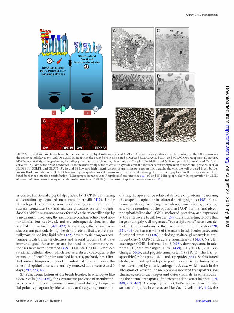
associated functional dipeptidylpeptidase IV (DPP IV), indicatinga decoration by detached membrane microvilli (410). Underphysiological conditions, vesicles expressing membrane-boundsucrase-isomaltase (SI) and maltase-glucoamylase aminopepti-dase N (APN) are spontaneously formed at the microvillar tips bya mechanism involving the membrane-binding actin-based mo-tor Myo1a, but not Myo2, and are subsequently shed into theluminal compartment (428, 429). Interestingly, the released vesi-cles contain particularly high levels of proteins that are preferen-tially partitioned into lipid rafts (429). Several vesicle cargoes con-taining brush border hydrolases and several proteins that haveimmunological function or are involved in inflammatory re-sponses have been identified (429). This Afa/Dr DAEC-inducedsacrificial cellular effect, which has as a direct consequence theextrusion of brush border-attached bacteria, probably has a lim-ited and/or temporary impact on intestinal function, since theintestinal epithelial cells are entirely renewed at between 3 and 5days (299, 373, 406).
(ii) Functional lesions at the brush border. In enterocyte-likeCaco-2 cells (430–434), the asymmetric presence of membrane-associated functional proteins is monitored during the epithe-lial polarity program by biosynthetic and recycling routes me-
diating the apical or basolateral delivery of proteins possessingthese specific apical or basolateral sorting signals (408). Func-tional proteins, including hydrolases, transporters, exchang-ers, some members of the aquaporin (AQP) family, and glyco-phosphatidylinositol (GPI)-anchored proteins, are expressedat the enterocyte brush border (299). It is interesting to note thatlarge and highly well-organized “super lipid rafts” have been de-tected at the membrane of the brush border of enterocytes (320,321, 435) containing some of the major brush border-associatedfunctional proteins (436), including maltase-glucoamylase ami-nopeptidase N (APN) and sucrase-isomaltase (SI) (437), Na�/H�
exchanger (NHE) isoforms 1 to 3 (438), downregulated in ade-noma Cl�/base exchanger (DRA) (439), Cl�/HCO3
�/OH� ex-changer (440), and peptide transporter 1 (PEPT1), which is re-sponsible for the uptake of di- and tripeptides (441). Sophisticatedstrategies including the hijacking of the cellular machinery havebeen developed by enteric pathogenic E. coli, which result in thealteration of activities of membrane-associated transporters, ionchannels, and/or exchangers and water channels, in turn modify-ing the normal transports of nutrients and the water balance (4, 5,409, 422, 442). Accompanying the C1845-induced brush borderstructural injuries in enterocyte-like Caco-2 cells (410, 412), the
FIG 7 Structural and functional brush border lesions caused by diarrhea-associated Afa/Dr DAEC in enterocyte-like cells. The drawing on the left summarizesthe observed cellular events. Afa/Dr DAEC interact with the brush border-associated hDAF and hCEACAM1, hCEA, and hCEACAM6 receptors (1). In turn,hDAF-associated signaling pathways, including protein tyrosine kinase(s), phospholipase C, phosphatidylinositol 3-kinase, protein kinase C, and Ca2�, areactivated (2). Loss of the brush border results in the disassembly of the microvillus cytoskeleton and induces defective expression of functional proteins, such asSI, DPP IV, SGLT1, and GLUT5 (3). (A and B) Low and high magnifications of transmission electron micrographs showing the well-ordered brush bordermicrovilli of uninfected cells. (C to F) Low and high magnifications of transmission electron and scanning electron micrographs show the disappearance of thebrush border at a late time postinfection. (Micrographs in panels A to F reprinted from reference 410.) (G and H) Micrographs show the observation by CLSMof immunofluorescence labeling of brush border-associated DPP IV (x-y section). (Reprinted from reference 412.)
Afa/Dr DAEC Pathogenesis
October 2014 Volume 27 Number 4 cmr.asm.org 845
on August 22, 2019 by guest
http://cmr.asm
.org/D
ownloaded from
distribution at the brush border of functional intestinal proteinssuch as SI and DPP IV hydrolases, sodium/glucose cotransporter 1(SGLT1), and the fructose GLUT5 transporter was profoundlyimpaired (412) (Fig. 7H and I). The default of SI and DPP IVexpression at the brush border induced by strain C1845 relatesdirectly to the promoted disassembly of the brush border cytoskel-eton, since stabilizing the F-actin cytoskeleton by jaspakinolinetreatment abolishes the disappearance of SI and DPP IV (411). InC1845-infected cells, there is a strong decrease of enzyme activityof SI and DPP IV (411, 412). It was logical to assume that thisdecrease in enzyme activity was related to the disorganization ofthe microvillus cytoskeleton and the disappearance of microvilli.However, it was not. Indeed, unlike C1845-induced, signaling-dependent cytoskeleton disassembly (340, 412), the loss of SI andDPP IV enzyme activity is independent of PTK, phospholipaseC, PI3K, PKC, and Ca2� signaling pathways and also of C1845-induced cytoskeleton disassembly (411). This indicates that whenSI and DPP IV are delocalized from the brush border in infectedenterocyte-like cells, the hydrolases relocated into the cytoplasmlose their enzyme activity. In addition, the biosynthesis of the twohydrolases is severely affected without any change in mRNA levelsand protein stability (411).
(iii) Structural and functional lesions at the junctional do-main. The junctional domain in epithelia plays a critical role inhealth, and barrier dysfunction at the junctional domain can leadto disease (409). The adherence junctions (AJs) and desmosomesact as adhesive domains between intestinal epithelial cells. Indeed,cadherin-based cell-cell junctions are mechanical connectionscreating contractile force which, after interaction with the con-tractile antimyosin cortex, actively couples neighboring cells inthe intestinal epithelial barrier (443, 444). The most apical junc-tional complex is TJs, which are highly regulated and formed bythe assembly of specialized proteins such as the cytoplasmic ZO-1,ZO-2, and ZO-3 proteins connected with the F-actin cytoskele-ton, the transmembrane occludin connected both with ZO pro-teins, cytoskeleton, and junctional adhesion molecules, and clau-dins connected with ZO proteins (445, 446). Functionally, TJs actas a “fence” separating the apical and basolateral membrane do-mains of polarized intestinal cells. This function results in thesegregation in each membrane domain of cell proteins and lipids.Moreover, TJs, by the sealing of intercellular space, act as a “gate”regulating the paracellular passage of small particles and solutes.On the basis of functional studies (447), it has been recently evi-denced that the gate activity includes two functional paracellularpathways: the first one, named the leaky pathway, engaging occlu-din that controls the paracellular passage of larger molecules, andthe second, named the high-capacity pore pathway, engaging theclaudin family of proteins functioning as cation-selective and an-ion-selective protein-forming channels and as protein-formingchannels without a clear established selectivity. Enterovirulentbacteria have developed sophisticated strategies to breach the in-testinal barrier by means of effectors that by signaling-dependentmechanisms target the structural molecules that compose the TJs(448). Pathogen-induced intestinal barrier deficiencies have beenlinked to the onset of inflammation and diarrhea (409).
The infection of fully differentiated Caco-2/TC7 cell monolay-ers by strain C1845, which expresses F1845 adhesin, is followed byan elevated level of paracellular permeability without affecting thetransepithelial resistance (449). At an early stage of infection, thedistributions of TJ-associated occludin and ZO-1 proteins are
profoundly modified, showing the disappearance of the proteinsat the TJs, whereas there was no change in expression of the AJ-associated E-cadherin. The TJ lesions develop from an F1845 ad-hesin-induced, hDAF-dependent mechanism. Guignot et al.(137) identified the toxin Sat of wild-type strain IH11128 as thevirulence factor that triggers the TJ lesions (Fig. 8A). Sat inducesrearrangements of the TJ-associated proteins ZO-1, ZO-3, andoccludin without modifying significantly the claudin-1 distribu-tion. In Sat-treated cells, there was a dramatic decrease of themembrane expression of ZO-1 and ZO-3 and a slight decrease inthe membrane expression of phosphorylated and nonphosphory-lated forms of occludin. In turn, Sat increases the paracellularpermeability, since there is elevation the paracellular passage ofmannitol without affecting the passage of nonionic moleculeswith higher molecular masses (137, 449). In addition, Sat inducesthe formation of fluid domes by increasing the transcellular pas-sage (137) (Fig. 8B). These fluid-filled, blister-like structuresknown as fluid domes are randomly distributed areas that evolvepermanently into the cell monolayers as the result of transcellularpassage of fluids accumulating basolaterally (450, 451). This resultis interesting since Taddei et al. (221) have reported that Sat caninduce secretory activity, resembling the enterotoxic activity ofETEC, in rabbit ileum tissues mounted in an Ussing chamber,with a dramatic fluid accumulation in rabbit ileum loops.
Inflammatory responses. Diverse host cell proinflammatoryresponses followed the infection of enterocyte-like and coloniccell lines by Afa/Dr DAEC. Flagella expressed by EPEC, EAEC, andEHEC contribute to favor epithelium colonization and proin-flammatory responses, such as the induction of proinflammatoryIL-8 production (4, 5). Motile, AfaE1-, AfaE2-, or afaEX-positivediarrhea-associated strains induced the production of high levelsof IL-8 in T84 and Caco-2 cells (203, 452). Meraz et al. (107) havereported that nine motile diarrhea-associated AfaE1- or AfaEX-positive E. coli strains induced the production of IL-8 in T84 cells.Paul Hofman and coworkers (341, 342, 344, 453) have nicely dis-sected the Afa/Dr adhesin-triggered, hDAF-dependent signalingpathways controlling the production of proinflammatory cyto-kines and related cellular events in colonic T84 cells. The nonflag-ellated, wild-type Afa/Dr DAEC C1845 and IH11128 strains wereable to promote the basolateral secretion of IL-8 in monolayers ofpolarized intestinal T84 cells (341). The production is daa or draoperon dependent, involves binding onto brush border-associ-ated hDAF, and develops by activation of the extracellular signal-regulated protein kinase (Erk1/2), p38, and c-Jun NH2-terminalkinase signaling pathways. Arikawa et al. (203) observed that 12nonmotile, AfaE1-, AfaE2-. or AfaEX-positive, diarrhea-associ-ated strains also induced the production of IL-8, but at a level ninetimes lower than that found in the motile strain. Flagella isolatedfrom motile Afa/Dr DAEC to produce IL-8 recognize the basolat-eral Toll-like receptor 5 (TLR5), and in addition, TJ function isobserved to be reduced (454). To explain this phenomenon, it hasbeen proposed that an unknown additional virulence factor caus-ing the structural opening of TJs allows flagellin to reach basolat-erally expressed TLR5 (454).
PMNLs play a pivotal role in maintaining intestinal homeosta-sis and are critical actors in the innate immune response thatprotects the host against microbial pathogens by generating adiversified arsenal of antimicrobial molecules, including reac-tive oxygen species, antimicrobial peptides, myeloperoxidase, hy-drolytic enzymes, proteases, cationic phospholipase, and metal
Servin
846 cmr.asm.org Clinical Microbiology Reviews
on August 22, 2019 by guest
http://cmr.asm
.org/D
ownloaded from

chelators, and by forming cell extensions known as neutrophilextracellular traps (NETs) (455). PMNL infiltration resultingfrom recruitment by chemokines produced by macrophages orepithelial cells at the site of insult is a hallmark of the host inflam-matory response to infection with various different enteric bacte-rial pathogens. The effects of Afa/Dr DAEC strains on the trans-epithelial migration of PMNLs have been demonstrated inpolarized monolayers of human colonic T84 cells cocultured withfreshly isolated PMNLs (341). The transepithelial migration ofPMNLs induced by Afa/Dr DAEC follows F1845 and Dr adhesin-induced hDAF-dependent basolateral production of IL-8 after ac-tivation of Erk1/2, p38, and JNK MAPKs. Afa/Dr DAEC-inducedPMNL transmigration triggers cell synthesis of TNF-�, and IL-1�,in turn inducing the up-expression of hDAF at the brush border ofthe cells and increasing the adhesion of Afa/Dr DAEC bacteria(342). In addition, there was an abnormal appearance of hDAF atthe basolateral domain of cells (342). The Afa/Dr DAEC-inducedtransmigration of PMNLs leading to a cytokine-triggered up-ex-pression of hDAF was consistent with previously observed IL-1�-induced up-expression of hDAF (456–459). The up-expression ofhDAF at the brush border is probably a cell defense response, sinceit has been demonstrated that hDAF functions as an antiadhesivemolecule accelerating the release from the luminal surface ofPMNLs that have undergone transepithelial migration (460, 461).It remains to be determined whether the abnormal basolateralexpression of hDAF in infected colonic T84 cells is also a cell
defense response that in turn can block the basolateral recruit-ment and transmigration of PMNLs.
The major histocompatibility complex (MHC) class I-relatedmolecules A and B (MICA and MICB) are distant homologues ofMHC class I molecules (462). Below the epithelium, resident lym-phocytes can be activated by these nonclassical MHC class I mol-ecules to display a diverse array of immune responses (463). To-gether with UL-16-binding proteins, MICA and MICB are ligandsof human NKG2D (464), an activating natural killer (NK) recep-tor expressed on both tumor-infiltrating lymphocytes and tumorscells. NKG2D exerts cytolytic destruction of cells through recog-nition of its cognate ligands. NK cells circulate through the blood,lymphatics, and tissues, patrolling for the presence of pathogen-infected cells (465). MICA and MICB are more highly expressed atthe surface of epithelial cells in colonic biopsy specimens fromCrohn’s disease (CD)-affected patients than in those from con-trols (466). Tieng et al. (467) have observed that the upregulationof MICA in enterocyte-like Caco-2 cells infected by AfaE-III ad-hesin subunit-positive E. coli is mediated by the specific interac-tion between the adhesin subunit and hDAF.
Pathogenic bacteria use quorum sensing (QS) to regulate sev-eral traits that allow them to establish and maintain infection intheir host; these include motility, biofilm formation, and viru-lence-specific genes (468). In E. coli, QS involves autoinducersAI-2 and AI-3, depending on the function encoded by the luxSgene. It has been observed that the Dr adhesin-positive IH11128
FIG 8 Structural and functional injuries at the tight junction caused by Afa/Dr DAEC in enterocyte-like cells. The drawing on the left summarizes the observedcellular events. The secreted and internalized Sat toxin induces the reorganization of the TJ-associated proteins ZO-1 and occludin without affecting E-cadherinexpression (1). Sat induces an increase in transcellular and paracellular passages (2). (A) Low- and high-magnification micrographs show the immunofluores-cence labeling of structural TJ-associated ZO-1 protein observed by CLMS in control and AAEC185psat-infected cells. Note the Sat-induced disappearance of theprotein from the cell-to-cell contacts. (Reprinted from reference 137 with permission.) (B) Fluid domes observed by phase-contrast CLSM in control andAAEC185psat-infected cells. Note the Sat-induced increase of the fluid dome height and surface.
Afa/Dr DAEC Pathogenesis
October 2014 Volume 27 Number 4 cmr.asm.org 847
on August 22, 2019 by guest
http://cmr.asm
.org/D
ownloaded from
strain produces high levels of AI-2 at the end of the exponentialphase of growth (unpublished result). Sperandio et al. (469), ob-serving that an EHEC luxS mutant responds to a eukaryotic cellsignal activating the expression of its virulence genes, have iden-tified epinephrine as promoting a “language” by which bacteriaand host cells communicate. Diard et al. (343, 470) have demon-strated that norepinephrine is involved in Afa/Dr DAEC patho-genesis. In the wild-type strain IH11128, the thermoregulatedproduction of Dr adhesin, which is optimal during the logarith-mic phase of anaerobic growth, has been found to be accompaniedby a release of Dr fimbriae without cell lysis (470). Norepineph-rine increases the Dr adhesin release by affecting the production ofthe fimbrial subunits. Indeed, norepinephrine promotes the dif-ferential induction of genes draC and draE by a regulatory mech-anism; i.e., the level of the draE transcript is highly increased, whileexpression of draC transcript is increased to a lesser extent, relativeto basal expression (343). Like with the wild-type IH11128 (341),the released Dr adhesin induces the phosphorylation of Erk1/2, inturn promoting the production of IL-8 in enterocyte-like Caco-2/TC7 cells (343).
Proinflammatory effects of Afa/Dr DAEC after direct contactwith PMNL-like PLB-985 cells have been described by SylvieChollet-Martin and coworkers (346, 347, 471). A rapid and mas-sive release of reactive oxygen species and preformed intragranu-lar mediators (myeloperoxidase and IL-8) develops in PMNL-likePLB-985 cells infected with the wild-type IH11128 and C1845strains (347). The phenomenon is triggered not by Afa/Dr ad-hesins but by the type 1 pili (347) expressed by wild-type Afa/DrDAEC strains (155).
Adherence to hDAF expressed at the membrane of freshly iso-lated PMNLs mediated by Dr adhesin did not lead to a significantincrease of bacterial killing, indicating that the Afa/Dr DAECcould overcome the microbicidal activity of PMNLs (472). In con-trast with the fact that PMNLs that have transmigrated displaygreater phagocytic activity against E. coli (473), PMNLs that havenot transmigrated or transmigrated across T84 cell monolayersafter Afa/Dr DAEC infection display low phagocytic activity, sug-gesting that Afa/Dr DAEC can block this activity (453).
Interaction of the wild-type Afa/Dr DAEC C1845 and IH11128strains or recombinant Dr adhesin-positive E. coli with freshlyisolated human PMNLs results in an increased rate of cell apop-tosis evidenced by morphological nuclear changes, DNA frag-mentation, cleavage of pro-caspase 3 and stimulation of thecaspase activity, and up-expression of annexin V (453, 472). Sincethis phenomenon is not blocked by anti-hDAF and -hCEA anti-bodies, it seems to be linked to a leukocyte agglutination processrather than to the recognition of these signaling receptors. Intrigu-ingly, phosphatidylserine appears at the cell membrane of PMNL-like PLB-985 cells, resulting from an Afa/Dr adhesin-triggered,hDAF-dependent activation of tyrosine kinase, and protein kinaseC signaling occurs, but without apoptosis (346). Low levels ofphagocytosis of Afa/Dr DAEC bacteria were observed in bothnontransmigrated and transmigrated PMNLs, suggesting a bacte-rium-triggered diminished leukocyte phagocytic capacity to es-cape host defenses (453).
Angiogenesis. Angiogenesis is a new component of IBD patho-genesis. Cane et al. (345) have reported that infection of culturedhuman colonic T84 cells by the diarrhea-associated E. coli strainC1845 is followed by an increase in vascular endothelial growthfactor (VEGF) mRNA expression and production of VEGF pro-
tein. Members of the VEGF family are secreted, dimeric glycopro-teins of �40 kDa consisting of five members, VEGF-A, -B, -C, and-D and a placenta growth factor, which function as regulators ofvasculogenesis, i.e., the vascular development that occurs duringembryogenesis, and the angiogenesis process that forms bloodvessels (474). Importantly, the VEGF secreted during C1845-in-fection of T84 cells is bioactive, since the cell-free spent culturemedium is able to induce tubulogenesis. This phenomenon resultsfrom the recognition of hDAF by F1845 adhesin followed by acti-vation of a Src protein kinase upstream of the activation of theRas/Raf/MAPK and PI3K/Akt signaling pathways (345). Upregu-lation of VEGF has also been observed in epithelial cells infectedwith an Afa-I adhesin-positive E. coli strain (140). Moreover, theF1845 adhesin-triggered, DAF-dependent up-production ofVEGF and also IL-8 in T84 cells has been found to be controlled byan MAPK- and PI3K-dependent induction of the hypoxia-in-duced factor 1-� (HIF-1�), revealing a connection betweenhDAF-associated signaling and a translational mechanism(s)(344). It is noteworthy that HIF-1 consists of two subunits,HIF-1� and HIF-1�, and that HIF-1�, which is degraded via anoxygen-dependent process involving prolyl hydroxylases, func-tions as an oxygen sensor (475). It is interesting to note that theCD-associated AIEC strain LF82 also induces, by an unknownfactor, the production of HIF-1 � protein and the activation ofVEGF/VEGR receptor (VEGFR) signaling in T84 cells (476). Allthe above-reported cell responses to Afa/Dr DAEC infection arerelated mainly to recognition of intestinal epithelial cell-expressedhDAF by Afa/Dr adhesins. Considering that hCEACAM1, hCEA,and hCEACAM6 are known to be involved in immune responses(477), it remains to be shown whether these hCEACAMs alsotrigger host cell immune responses after recognition by Afa/Dradhesins.
EMT events. Under physiological conditions, the intestinal ep-ithelium undergoes controlled constant cell shedding. In epithe-lia, a programmed cell death process known as “anoikis,” a Greekword meaning “homelessness,” occurs when polarized epithelialcells forming an epithelial barrier are detached from the appropri-ate lateral domain and extracellular matrix (406). The control ofanoikis in differentiated epithelial intestinal cells is a differentia-tion-dependent process involving the engagement of �1 and �4integrins and activation of FAK, Src kinase, and the Erk1/2 andPi3K/Akt signaling pathways (406). The diarrhea-associatedC1845 affects the AJs of intestinal epithelial polarized colonic T84cells. Indeed, an F1845 adhesin-triggered, hDAF-dependent lossof AJ-associated E-cadherin protein and cytokeratin 18 (CK18)has been observed, resulting from activation of HIF-1� (344).Moreover, CK18 plays a cytoskeletal function as a component ofthe intermediate filaments and acts as a target for caspase-medi-ated cleavage during cell apoptosis (478). In parallel, C1845 infec-tion promotes a rise in fibronectin expression, accompanied bythe up-expression of Twist1 mRNA (344), a helix-loop-helix tran-scriptional factor belonging to a small group of core transcriptionfactors that also includes Snail, Slug, and Sip1 and which are in-volved in controlling the epithelial-mesenchymal transition(EMT) (479). The EMT is a transcriptional and morphologicalprogram observed during the progression of diseases, includingcancers (480, 481). Three distinct subtypes of epithelial cells thattransition into mesenchymal cells have been defined, dependingon the physiological tissue context (481). Type 1 occurs in em-bryogenesis and organ development, type 2 develops in tissue re-
Servin
848 cmr.asm.org Clinical Microbiology Reviews
on August 22, 2019 by guest
http://cmr.asm
.org/D
ownloaded from

generation and organ fibrosis, and type 3 is associated with cancerprogression. However, partial EMT can develop as a function ofthe tissue and signaling contexts, resulting in a loss of only some ofthe polarized characteristics such as cell contact dissolution andactin cytoskeleton reorganization. The two major hallmarks ofEMT are the dissolution of epithelial cell-cell adhesion contactsand the massive actin cytoskeleton reorganization followed by theloss of cell polarization. Transcription factors drive EMT bydownregulating genes, including those encoding proteins main-taining epithelial cell-cell adhesion domains. After a long time ofinfection, C1845 infection promotes depolarization of the entero-cyte-like Caco-2 cells, which is characterized by the complete dis-organization of the apical domain and the loss of cell-to-cell con-tacts accompanied by the appearance of undifferentiated cells thatbecome detached from the cell monolayers (Fig. 9A) (unpub-lished results). The C1845-induced cell shedding resembles thecascapase-1-dependent cell shedding observed in T84 cells (482)and the TNF-�-induced cell shedding observed in mouse intesti-nal and Madin-Darby canine kidney cells (482, 483). It has beenmentioned above that in C1845-infected T84 cells, the induced
transepithelial migration of PMNLs is followed by the productionof TNF-� (342). Collectively, these results show that diarrhea-associated Afa/Dr DAEC promotes cell depolarization and inju-ries at the two major sites of the junctional domain of polarizedepithelial cells forming the intestinal epithelial cell barrier, i.e., TJsand AJs, and in turn induces EMT-like behavior.
pks-related cell injuries. The presence of the pks genomic islandcoding for the genotoxin colibactin in the prototype wild-typeAfa/Dr DAEC strains leads to the appearance of known pks-re-lated cell changes (240). Indeed, in C1845-infected undifferenti-ated Caco-2/TC7 cells there was an enlargement of the cell bodyand nucleus and an increase of the nuclear phosphorylation ofhistone H2A variant H2AX (Fig. 9B) (Nougayrede and Oswald,unpublished result). These cell injuries were abrogated when thecells were infected with a mutant colibactin strain (clbA::frt) de-leted for the clbA gene, encoding the phosphopantetheinyl trans-ferase ClbA within the pks island (484), and were restored by thecomplementation of the mutant strain with p-clbA (Nougayredeand Oswald, unpublished result). It is noteworthy that the pks-triggered enlarged cells cell have been recently characterized as
FIG 9 Diarrheagenic Afa/Dr DAEC strain C1845 promotes epithelial-mesenchymal transition (EMT)-like behavior, cell extrusion, and pks-dependent damagein human intestinal cells. The drawing on the left summarizes the observed EMT-related cellular events. An F1845-triggered hDAF-, MAPKs-, PI3K-, andHIF-1�-dependent production of VEGF develops in colonic-like T84 cells (1 and 2). An F1845-triggered hDAF-, MAPKs-, PI3K-, and HIF-1�-dependentEMT-like behavior develops, characterized by the typical changes in expression of mesenchymal markers, such as the upregulation of fibronectin, the down-regulation of CK18, and the disappearance of AJ-associated E-cadherin (1 and 3). Finally, depolarized intestinal cells lose the lateral cell-to-cell and basal contactsand become detached from the cell monolayer. (A) Low-magnification transmission electron micrographs show the dedifferentiation of Caco-2/TC7 cellsinfected with the diarrhea-associated strain C1845, characterized by a disorganization of the apical domain and by the appearance of nucleus fragmentation andloss of nucleus dense electron material, indicating cell death (left micrograph). The boxed area indicates a cell with a funnel form engaging in shedding from thecell monolayer as the result of the wide opening of the lateral cell-to-cell junctional domain and detachment at the basal domain. The micrograph on the rightshows a high magnification of the cell engaging in detachment and containing a high number of vesicles in the cytoplasm. (B) Low- and high-magnificationmicrographs show the morphological changes in Giemsa-stained C1845-infected undifferentiated Caco-2/TC7, cells characterized by an enlargement of the cellbody and nucleus. Low-magnification micrographs show the increase of the phosphorylation of nuclear H2AX, the most sensitive marker of DNA damage.(Courtesy of J. P. Nougayrede and E. Oswald, reproduced with permission.)
Afa/Dr DAEC Pathogenesis
October 2014 Volume 27 Number 4 cmr.asm.org 849
on August 22, 2019 by guest
http://cmr.asm
.org/D
ownloaded from
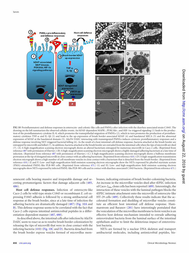
senescent cells bearing massive and irreparable damage and se-creting protumorigenic factors that damage adjacent cells (485,486).
Host cell defense responses. Infection of enterocyte-likeCaco-2 cells by wild-type strain C1845 or recombinant E. coli ex-pressing F1845 adhesin is followed by a strong antibacterial cellresponse at the brush border, since at a late time of infection theadhering bacteria are dramatically damaged (487) (Fig. 10A andB). This defense response seems to be correlated with the fact thatCaco-2 cells express intestinal antimicrobial peptides in a differ-entiation-dependent manner (487, 488).
As described above, the intestinal cells after infection by Afa/DrDAEC seem to react so as to resolve brush border colonization byreleasing the tips of microvilli that were in close contact with theinfecting bacteria (410) (Fig. 10C and D). Bacteria detached fromthe brush border express vesicles formed of microvillus mem-
branes, indicating extrusion of brush border-colonizing bacteria.An increase in the microvillar vesicles shed after tEPEC infectionof Caco-2BBe clone cells has been reported (489). Interestingly, theinteraction of these vesicles with the luminal pathogen blocks thetEPEC intimate attachment onto the microvilli of enterocyte-likeHT-29 cells (489). Collectively these results indicate that the ac-celerated formation and shedding of microvillar vesicles consti-tute an efficient host intestinal cell defense response. Ham-marstrom and Baranov (281) have interestingly postulated thatthe vesiculation of the microvillus membrane to form vesicles is aneffective host defense mechanism intended to extrude adheringenterovirulent bacteria from the luminal surface of the intestinalepithelium and/or to limit the deleterious impact of enteroviru-lent bacteria.
NETs are formed by a nuclear DNA skeleton and transportantibacterial molecules, including antimicrobial peptides, his-
FIG 10 Proinflammatory and defense responses in enterocyte- and colonic-like cells and PMNLs after infection with the diarrhea-associated strain C1845. Thedrawing on the left summarizes the observed cellular events. An hDAF-dependent MAPK-, PI3K/Akt-, and HIF-1�-triggered signaling (1) leads to the produc-tion of the proinflammatory cytokine IL-8, which promotes the transepithelial migration of PMNLs (2), which in turn promotes the production of proinflam-matory cytokines TNF-� and IL-1� (3) and leads to the up-expression of brush border-associated hDAF (4) and basolateral MICA (5) and the abnormalexpression of hDAF at the basolateral domain (5). Afa/Dr DAEC interacting with transmigrated PMNLs induces cytotoxic proinflammatory responses and adefense response involving NET-triggered bacterial killing (6). As the result of a host intestinal cell defense response, bacteria attached at the brush border areentrapped by microvilli and killed (7). In addition, bacteria attached at the brush border are extruded from the intestinal cells when the tips of microvilli are shed(7). (A) A high-magnification scanning electron micrograph shows an altered bacterium entrapped by numerous microvilli in Caco-2 cells. (Reprinted fromreference 487 with permission of Elsevier.) (B) A high-magnification scanning electron micrograph shows a highly damaged adhering bacterium at a late time ofinfection. (Reprinted from reference 487 with permission of Elsevier.) (C) A high-magnification scanning electron micrograph shows a bulbous membraneprotrusion at the tip of elongated microvilli in close contact with an adhering bacterium. (Reprinted from reference 410.) (D) A high-magnification transmissionelectron micrograph shows a high number of cell membrane vesicles in close contact with a bacterium that is detached from the brush border. (Reprinted fromreference 410.) (E and F) Low- and high-magnification field emission scanning electron micrographs show the NETs expressed by phorbol myristate acetate(PMA)-stimulated PMNL-like PLB-985 cells. (Reprinted from reference 471.) (G and H) Low- and high-magnification field emission scanning electronmicrographs show NETs expressed by infected PMNL-like PLB-985 cells and in contact with diarrhea-associated C1845 bacteria. (Reprinted from reference 471.)
Servin
850 cmr.asm.org Clinical Microbiology Reviews
on August 22, 2019 by guest
http://cmr.asm
.org/D
ownloaded from

tones, and proteases (490, 491). In the human myeloid cell linePLB-985, which has the capability to differentiate into fully ma-ture neutrophil-like cells, wild-type strain C1845 infection is fol-lowed by the projection of NETs, which by entrapment of bacte-rial cells trigger the killing of bacteria (471) (Fig. 10E to H).However, this defense response has an unexpected cell cytotoxicconsequence, since the induced NETs promote a profound disor-ganization of the F-actin cytoskeleton by contact with the Caco-2/TC7 cells (471). It is worth mentioning that NETs induce celldeath in endothelial cells (492, 493).
CLINICAL CONSIDERATIONS
Reservoir and Transmission
There has been no report of the isolation of E. coli expressing themajor human AfaE-I to -III, Dr, or F1845 adhesins in a largevariety of animal species. In contrast, the afimbrial Afa-VII andAfa-VIII adhesins are present in diarrhea- or septicemia-associ-ated E. coli strains in calves (26, 44). Moreover, the AfaE-VIIIadhesin is expressed in ExPEC infecting calves (43, 54, 494). Thegene afa-8 and afa/dr-related genes have been also been found inporcine, poultry, and cattle E. coli isolates (45, 173, 494–496).Examination of E. coli isolates from dogs with UTIs has revealedthe presence of P- and S-positive isolates and the absence of Afa-positive isolates (497). There is an absence of evidence of directanimal-to-human transmission.
Treatments
Historically, antibiotics have provided a very effective way ofresolving UTIs. The current routine treatment for UTIs is anti-biotic therapy, most commonly trimethoprim-sulfamethoxazole(TMP-SMX) or ciprofloxacin. However, patients with chronic orrecurrent cystitis require long-term treatment with antibiotics,and as UPEC strains have an unusual intracellular lifestyle, con-ventional antibiotics are not very effective when the bacteria residewithin intracellular vacuoles or are present in biofilm-like struc-tures (498–505).
Afa/Dr DAEC-associated diarrhea in children has been de-scribed as being generally limited in duration. Diarrhea, as definedby the World Health Organization (WHO), is characterized by thepresence of three or more stools or watery stools within a 1-dayperiod. Diarrhea is defined as being acute when it lasts less than 14days and as persistent when the episode has lasted 14 days or more.In general, to prevent dehydration and nutritional default in chil-dren with infectious acute diarrhea, it has been recommended toprovide an oral rehydration solution (ORS) and to continue feed-ing (506). Since ORS administration has no effect on the duration,severity, or frequency of infectious diarrheal episodes, ORS ad-ministration has been associated with adjuvant therapies such asluminally acting antisecretory drugs and antibiotics (507). How-ever, these treatments can cause adverse effects outside or insidethe gastrointestinal tract; one such effect that occurs if broad-spectrum antibiotics are used is the emergence of Clostridium dif-ficile-associated diarrhea resulting from an antibiotic-induced im-balance of the intestinal microbiota (508, 509). It should be notedthat cost and availability are limiting factors for the use of antise-cretory drugs and antibiotics, particularly in developing countries.Another therapeutic strategy used to accompany ORS (which islow cost but for which availability may be a problem, particularlyin developing countries) is the use of human intestinal microbiota
probiotic Lactobacillus strains secreting antibiotic-like moleculesthat have demonstrated experimental in vitro and in vivo effectsagainst the major diarrhea-associated bacterial pathogens (510).
Antibiotic Resistance
The increasing prevalence of antibiotic resistance of bacterialpathogens is a major health problem (511, 512). There has been asteady and rapid increase of UPEC resistance to antibiotics ac-companied by the problematic occurrence of multidrug-resistantUPEC strains over the last decade (199, 349, 498). For examples, arecent international study revealed that more than 10% of E. colicystitis isolates are resistant to at least three classes of antibiotics(513). Moreover, a recent survey of more than 12 million clinicalisolates across the United States from 2000 to 2011 revealed anincrease of resistance to 9 antibiotics, including TMP-SMX, cip-rofloxacin, nitrofurantoin, and ceftriaxone (514). Not surpris-ingly, resistance to antibiotics has been reported for Afa/DrDAEC, including resistance to ampicillin (90, 124, 515, 516),TMP-SMX (515–518), fosfomycin (517), piperacillin (515), tetra-cycline (124, 515), ciprofloxacin (518), co-trimoxazole (124),nitrofurantoin (518), fosfomycin (517), tetracycline (519), peni-cillin (516), oxacillin (516), bactericin (516), cloxacillin (516),chloramphenicol (519), and nalidixic acid (124, 516). In addition,the fact that Dr adhesin-positive E. coli has the capability to formbiofilm (190, 233) suggested that like FimH-positive UPEC (199,232), uropathogenic Afa/Dr DAEC strains have developed strate-gies to escape antimicrobial host defense systems and to reducetheir sensitivity to antibiotics.
Vaccines and Pilicides
A therapeutic strategy has been developed to disarm pathogens inthe host by the use of substances that mimic bacterial virulencefactors such as adhesive factors, bacterial effectors, and toxins orthat display antagonistic activities against the bacterial productionof these factors (520, 521). Touchon et al. (522) underlined thatthis could make it difficult to develop a vaccine against ExPECinfections. It is noteworthy that an emerging therapeutic strategyconsists of developing antagonists of QS systems in order to in-hibit the bacterium-bacterium and bacterium-host cell commu-nications involved in virulence (523). Brumbaugh and Mobley(520) have recently summarized the advances in vaccine strategyagainst UPEC and enteric virulent E. coli. These include com-pounds targeting surface polysaccharides and formulations com-posed of several virulence factors such as hemolysin, type 1, P, Dr,and S fimbrial adhesins, CNF-1, siderophores, and markers ofPAIsCFT073 and which display serotypes O1, -4, -6, -17, -75, and-77, K1, -3, -5, -13, and -95, and flagellar H:1, H:5, H:7, and H:33.Other strategies are the targeting of membrane proteins fromUPEC strains or the use of mixtures of inactivated UPEC strains orgenetically engineered vaccines. Although promising, the use ofantagonists of adhesive factors to combat bacterial colonization ofepithelia is complicated by the fact that a single pathogenic E. colistrain expresses a multiplicity of adhesive factors, each of whichdisplays specific host cell receptor recognition. Consequently, itappears evident that it may be necessary to target more than onevirulence factor for successful vaccine or pilicide therapies. An-other subunit vaccine strategy against ExPEC has been designed totarget immunodominant epitopes of the virulence-associatedExPEC proteins FyuA, IroN, ChuA, IreA, Iha, and Usp (524). ForAfa/Dr DAEC infection, a vaccine strategy has been experimen-
Afa/Dr DAEC Pathogenesis
October 2014 Volume 27 Number 4 cmr.asm.org 851
on August 22, 2019 by guest
http://cmr.asm
.org/D
ownloaded from
tally tested using the Dr adhesin in the mouse pyelonephritismodel (385). High titers of serum anti-Dr antibodies develop aftervaccinating mice with purified Dr adhesin, and this is accompa-nied by a significant reduction in mortality but, surprisingly, with-out affecting the levels of bladder or kidney colonization by Dradhesin-positive E. coli. Ex vivo, preincubating Dr adhesin-posi-tive E. coli with Dr adhesin-immunized mouse serum reduces thebacterial adherence to mouse bladders and kidneys, whereas pre-incubating urine from Dr adhesin-immunized mice fails to pro-duce this inhibitory activity.
Therapeutic-strategy-based pilicides have been developed inorder to inhibit the completion of fimbrial and nonfimbrial ad-hesins at the cell surface of bacterial pathogens, including UPECand enteric pathogenic E. coli (525, 526). Pilicides are a class oflow-molecular-weight compounds which, by blocking chaperoneand usher functions, inhibit pilus/fimbria completion withoutkilling or affecting the growth of bacteria. Consequently, pilicidescan prevent the first critical adhesion step required for the success-ful urinary tract colonization by UPEC strains involved in cystitisand pyelonephritis. On the bases that AfaC/DraC/DaaC ushers arepivotal for Afa/Dr adhesins biogenesis to occur and that the draCand afaC-III genes encoding the Afa/Dr ushers display 100% iden-tity, it has been postulated that 2-pyridone pilicide compoundscould inhibit the biogenesis of Afa/Dr adhesins (83, 176, 177).This pilicide strategy has been found to be effective against Dradhesin biogenesis, where it produces a marked reduction of theexpression of Dr adhesin and the disappearance of its properties ofadherence to hDAF (175, 527).
CONCLUDING REMARKS AND FUTURE DIRECTIONS
The importance of human Afa/Dr DAEC in UTIs and pregnancycomplications has been convincingly demonstrated both experi-mentally and clinically. Moreover, increasing epidemiological ev-idences has been reported during the last decade on the role ofhuman intestinal Afa/Dr DAEC in triggering established acutediarrhea in infants �5 years. In contrast, epidemiological studiesindicate that human intestinal Afa/Dr DAEC strains are probablynot responsible for established acute diarrhea in adults. A robustPCR method for detecting and identifying diarrhea-associatedAfa/Dr DAEC strains remains to be developed, as the PCR meth-ods currently used in a clinical setting are problematic with regardto the recently observed cross-reaction between the most com-monly used Afa/Dr DAEC daaC primer and EAEC primers.
In spite of the remarkable results obtained over the past de-cade, the cellular and molecular mechanisms of Afa/Dr DAECpathogenesis remain incompletely elucidated. Several aspects de-serve particular attention in the future. Whole-genome sequenc-ing is called for to identify more potential virulence factors in thewild-type Afa/Dr DAEC prototype strains in order to completeour understanding of the pathogenesis of these pathogenic E. colistrains. Much of our current knowledge about Afa/Dr DAEC in-testinal pathogenesis has been obtained from in vitro observationsusing cultured human epithelial colon cancer cells, which, despitemimicking the situation in vivo, do not entirely reflect the healthyhuman situation and have several drawbacks (421). The highspecificity of Afa/Dr adhesins for human epithelium-associatedmolecules acting as receptors and triggering cell signalization andcellular effects complicates the experimental examination of thecellular and molecular mechanisms of Afa/Dr DAEC pathogene-sis. In comparison with the in vitro situation, the use of transgenic
mouse models constructed for the epithelial expression of hDAFand hCEACAMs has not given convincing results in terms of cel-lular lesions even in the presence of Afa/Dr DAEC intestinal col-onization (unpublished results). This is probably due to the en-dogenous presence of mouse DAF and CEACAMs and to theobservation of a less-than-optimal expression of the membrane-bound human receptors (unpublished results) in the mouse epi-thelial tissue and/or defective connection to the mouse endoge-nous signaling pathways that have been observed in vitro to beinvolved in Afa/Dr DAEC pathogenesis. To overcome these prob-lems, human intestinal in vitro organ culture (IVOC) models(528) can offer valuable tools for the experimental investigation ofthe mechanisms of virulence of intestinal Afa/Dr DAEC. VariousIVOC systems corresponding to an ex vivo human situation havebeen described, including those constituting polarized IVOCs es-tablished from pediatric duodenum biopsy specimens or speci-mens of human colon taken at the vicinity of tumors. These IVOCsystems have been used mainly to study the mechanisms of viru-lence of enterovirulent E. coli, including ETEC, EPEC, EHEC,EAEC, and AIEC (528).
Repeated observations in epidemiological studies of the intes-tinal presence of daaC-positive DAEC strains in adults in the ab-sence of acute diarrhea are intriguing. This asymptomatic carriagedeserves further exploration. Indeed, the Afa/Dr DAEC strains areclearly pathogenic with regard to the deleterious effects observedin vitro in human intestinal cell models, some of which resemblethe pathogenic effects of EPEC and EHEC even though they resultfrom different cellular and molecular pathogenic mechanisms.This reinforces our previous hypothesis that intestinal Afa/DrDAEC strains act as “silent pathogens” that are well controlledunder healthy conditions by the intestinal antimicrobial mole-cules generated by dedicated cells of the host intestinal barrier andby the barrier effect exerted by the intestinal microbiota (11). Theabove-described deleterious cellular effects of intestinal Afa/DrDAEC make us think that these microbiota-resident E. coli inhealthy humans could be good candidates for belonging to therecently defined class of “pathobionts” (529). Indeed, recent evi-dence suggests that the observed deleterious cellular effects may becaused by specific bacterial species with pathogenic potential thatwere present as symbionts in the healthy host intestinal microbi-ota. Although the pathophysiological mechanisms of pathobiontsremain largely unknown, it appears that genetic factors, environ-mental factors, and/or changes in host defense systems may ex-pose the host to developing diseases triggered by these potentiallypathogenic microbiotal bacteria. Observations that some strongcell defense responses, including the extrusion of infecting bacte-ria from the brush border, cell detachment of intoxicated cells,and killing by NETs of activated PMNLs developed after Afa/DrDAEC infection, all support the existence of efficient control bythe host of these pathogenic E. coli strains in healthy situations.This leads to the question of how a resident intestinal microbiotaAfa/Dr DAEC strain can emerge as an enterovirulent pathogen.Several circumstances allow the occurrence of diseases triggeredby pathobionts. For example, dysbiosis, a shift in the makeup ofthe intestinal microbiota community and/or its abundance, leadsto a change in the equilibrium between putative protective speciesand harmful pathogens (530). Another E. coli pathobiont candi-date is the E. coli strain C25, isolated from the feces of a healthyhuman (531, 532). This strain expresses the SPATE toxin Pic(533), translocates across colonic T84 cell monolayers, promotes
Servin
852 cmr.asm.org Clinical Microbiology Reviews
on August 22, 2019 by guest
http://cmr.asm
.org/D
ownloaded from
alterations in localization of claudin-1, activates NF-B signaling,and induces the production of IL-6, IL-8, and TNF-� (533–535),all effects that are exacerbated in a proinflammatory situation(536).
The causes of IBD have not been entirely identified but areknown to be related to a susceptible human genome (537), a dys-function of the immune system (537), a massive and/or selectivemodification in the composition of the intestinal microbiota(538), and the intervention of opportunistic enterovirulent bacte-ria (529, 539). With regard to the observed asymptomatic carriageof intestinal daaC-positive E. coli strains, some of the above-re-ported deleterious cellular effects of intestinal Afa/DrDAEC, in-cluding marked proinflammatory responses, indicated that thesestrains could play a role in intestinal inflammation. Afa/Dr DAECbelongs to phylogenic group B2, which has been observed to beprevalently associated with IBD (138, 217). It is difficult to addressthis question experimentally because of the lack of experimentalanimal models mimicking the human situation. Furthermore, ep-idemiological clinical studies are difficult to conduct because thiswould require isolating the mucosa-associated intestinal Afa/DrDAEC in patients with chronic inflammatory diseases. Indeed, thein vitro results show that these strains are very effectively excludedfrom the brush border by the host defenses. Moreover, the sam-pling techniques used to isolate the mucosa-associated bacteria,which are localized exclusively on the luminal surface of the intes-tinal barrier, are also problematic, since these bacteria can be easilylost during the technical process. It has been found to be easier toisolate AIEC from patients with chronic inflammatory diseasessimply because it is intracellularly localized in cells lining the epi-thelial barrier. Future progress can be expected to provide answersto these questions.
There are emerging links between microbial and parasite infec-tions and oncogenesis (540). Moreover, the relationships andmechanisms through which disturbance of intestinal microbiota,infection, and inflammation increase cancer risks and promotetumor development were recently reviewed (541, 542). A smallnumber of microbial pathogens have been identified as criticalcauses of specific chronic inflammatory conditions or malignan-cies. The best-characterized example of a bacterial contribution tocancer is H. pylori infection, which is well established as a majorrisk factor for gastritis leading to gastric cancer as the result of thedeleterious activity of its cytotoxin-associated gene A protein(543). However, the simple observation that some bacteria arepresent at the site of cancerous intestinal tumors does not consti-tute a proof of causality. A link of causality is difficult to demon-strate here because there is a long time lag between initiation of acellular process of carcinogenesis and the onset of overt disease.Indeed, the bacterium-triggered event can occur long before anysign of a cancer tumor manifests itself, and indeed the pathogenmay no longer be present. It is becoming clear that some proin-flammatory bacterial pathogens can contribute to initiating, pro-moting, and/or progressing cancer by their virulence factors, in-cluding toxins, which directly disrupt cellular signaling pathwaysthat control inflammatory processes and damage DNA. Somedemonstrated aspects of the mechanisms of pathogenicity of in-testinal and uropathogenic Afa/Dr DAEC strains suggest a possi-ble connection with the initiation or development of intestinaland urinary tract cancers. Some of the deleterious cellular re-sponses triggered by Afa/Dr DAEC described above suggest thatthey can be connected to the initiation of cellular events in relation
to intestinal cancer. The pks island (238, 540) is present in Afa/DrDAEC, and it has been found that pks-positive E. coli strains be-longing to the phylogenic B2 group are prevalently present inbiopsy specimens of patients with intestinal cancers and induceDNA double-strand breaks in intestinal cells as well as triggerchromosomal instability, gene mutations, and cell transformation(22, 240, 246, 544). Afa/Dr DAEC infection is accompanied by thetransepithelial migration of PMNLs, and in the intestinal tractmigration of PMNLs across the epithelial lining is a hallmark ofIBD and can create a deleterious inflammatory situation (455) andpreneoplastic conditions, since transmigrated PMNLs exert a di-rect cytotoxic effect by releasing products such as oxidative re-agents and elastase (545). Moreover, after Afa/Dr DAEC-inducedtransmigration of PMNLs across a monolayer of intestinal cells,the epithelial cells displayed deregulated expression of mem-brane-associated hDAF with altered functions, some of which areimplicated in carcinogenesis (546). On the other hand, it shouldbe noted that hCEACAMs, the receptors of Afa/Dr adhesins, areknown to be implicated in carcinogenesis (275). In addition, theEMT and angiogenesis observed to be triggered by intestinalAfa/Dr DAEC are indicative that these bacteria play a role in car-cinogenesis (480). An important function of autophagy in canceris the limitation of inflammation, tissue damage, and genome in-stability, and when the autophagy process is blocked at the degra-dative autophagosome step, cells accumulate cytotoxic material,thus promoting DNA mutation and carcinogenesis (547, 548).Consequently, the observed blockade of autophagosome matura-tion triggered by the Afa/Dr DAEC Sat toxin may play a role incarcinogenesis. It is noteworthy that this deleterious effect of au-tophagy has been observed for H. pylori in relation to its role in thedevelopment of gastric cancer (549), since the vacuolating cyto-toxin VacA induces autophagy and disrupts autophagosome mat-uration after prolonged exposure (550). UPEC-induced cell exfo-liation in turn induces the upregulation of urothelial turnover,and it has been postulated that it may play a role in patients’predisposition to bladder cancer (504, 551, 552). It is tempting tohypothesize that Afa/Dr DAEC is involved in the development ofbladder cancer, given the observed cell exfoliation effect triggeredby the Afa/Dr DAEC Sat toxin. Collectively, these results provide agroup of facts but do not amount to evidence of a possible linkbetween Afa/Dr DAEC infection and intestinal or bladder carci-nogenesis.
ACKNOWLEDGMENTS
I express my earnest thanks to Marie-Françoise Bernet-Camard, JulieGuignot, Isabelle Peiffer, Cedric N. Berger, Imad Kansau, Sylvie Chollet-Martin, Anne-Béatrice Blanc-Potard, Sylvie Hudault, Clémence Rougeaux,Sophie Kerneis, and Vanessa Liévin-Le Moal for their outstanding contri-butions that have illuminated the understanding of the cellular and mo-lecular mechanisms of pathogenesis of Afa/Dr DAEC (Research TeamsInserm and University Paris-Sud, CJF 94.07 and Units 510 and 756 at theUniversity Paris-Sud, Faculty of Pharmacy, Châtenay-Malabry, France).Particular thanks go to Steve L. Moseley and Natalia Korotkova (Univer-sity of Washington, Seattle, WA, USA), Bogdan Nowicki (Meharry Med-ical College, Nashville, TN, USA), Steve Matthews (Imperial CollegeLondon, London, United Kingdom), and Paul Hofman and ValérieVouret-Craviari (Centre Hospitalo-Universitaire de Nice and Institute ofResearch on Cancer and Ageing, University of Nice, Nice, France), Chan-tal Le Bouguénec (Pasteur Institute, Paris, France), Oliver Billker (Well-come Trust Sanger Institute, Cambridge, United Kingdom), Doug Lublin(Washington University School of Medicine, St. Louis, MO, USA), and
Afa/Dr DAEC Pathogenesis
October 2014 Volume 27 Number 4 cmr.asm.org 853
on August 22, 2019 by guest
http://cmr.asm
.org/D
ownloaded from
Jean-Philippe Nougayrède and Eric Oswald (Inserm, UMR1043, Centrede Physiopathologie de Purpan, Université de Toulouse, Toulouse,France) for their invaluable collaborative contributions.
REFERENCES1. Kaper JB, Nataro JP, Mobley HL. 2004. Pathogenic Escherichia coli. Nat.
Rev. Microbiol. 2:123–140. http://dx.doi.org/10.1038/nrmicro818.2. Johnson TJ, Nolan LK. 2009. Pathogenomics of the virulence plasmids
of Escherichia coli. Microbiol. Mol. Biol. Rev. 73:750 –774. http://dx.doi.org/10.1128/MMBR.00015-09.
3. Nataro JP, Kaper JB. 1998. Diarrheagenic Escherichia coli. Clin. Micro-biol. Rev. 11:142–201.
4. Croxen MA, Law RJ, Scholz R, Keeney KM, Wlodarska M, Finlay BB.2013. Recent advances in understanding enteric pathogenic Escherichiacoli. Clin. Microbiol. Rev. 26:822– 880. http://dx.doi.org/10.1128/CMR.00022-13.
5. Clements A, Young JC, Constantinou N, Frankel G. 2012. Infectionstrategies of enteric pathogenic Escherichia coli. Gut Microbes 3:71– 87.http://dx.doi.org/10.4161/gmic.19182.
6. Darfeuille-Michaud A, Neut C, Barnich N, Lederman E, Di Martino P,Desreumaux P, Gambiez L, Joly B, Cortot A, Colombel JF. 1998.Presence of adherent Escherichia coli strains in ileal mucosa of patientswith Crohn’s disease. Gastroenterology 115:1405–1413. http://dx.doi.org/10.1016/S0016-5085(98)70019-8.
7. Barnich N, Darfeuille-Michaud A. 2007. Adherent-invasive Escherichiacoli and Crohn’s disease. Curr. Opin. Gastroenterol. 23:16 –20. http://dx.doi.org/10.1097/MOG.0b013e3280105a38.
8. Lloyd AL, Henderson TA, Vigil PD, Mobley HL. 2009. Genomicislands of uropathogenic Escherichia coli contribute to virulence. J. Bac-teriol. 191:3469 –3481. http://dx.doi.org/10.1128/JB.01717-08.
9. Mokady D, Gophna U, Ron EZ. 2005. Virulence factors of septicemicEscherichia coli strains. Int. J. Med. Microbiol. 295:455– 462. http://dx.doi.org/10.1016/j.ijmm.2005.07.007.
10. Kim KS. 2008. Mechanisms of microbial traversal of the blood-brainbarrier. Nat. Rev. Microbiol. 6:625– 634. http://dx.doi.org/10.1038/nrmicro1952.
11. Servin AL. 2005. Pathogenesis of Afa/Dr diffusely adhering Escherichiacoli. Clin. Microbiol. Rev. 18:264 –292. http://dx.doi.org/10.1128/CMR.18.2.264-292.2005.
12. Benz I, Schmidt MA. 1989. Cloning and expression of an adhesin(AIDA-I) involved in diffuse adherence of enteropathogenic Escherichiacoli. Infect. Immun. 57:1506 –1511.
13. Beinke C, Laarmann S, Wachter C, Karch H, Greune L, Schmidt MA.1998. Diffusely adhering Escherichia coli strains induce attaching andeffacing phenotypes and secrete homologs of Esp proteins. Infect. Im-mun. 66:528 –539.
14. Laarmann S, Schmidt MA. 2003. The Escherichia coli AIDA autotrans-porter adhesin recognizes an integral membrane glycoprotein as re-ceptor. Microbiology 149:1871–1882. http://dx.doi.org/10.1099/mic.0.26264-0.
15. Schmidt MA. 2010. LEEways: tales of EPEC, ATEC and EHEC. Cell.Microbiol. 12:1544 –1552. http://dx.doi.org/10.1111/j.1462-5822.2010.01518.x.
16. Hernandes RT, Elias WP, Vieira MA, Gomes TA. 2009. An overview ofatypical enteropathogenic Escherichia coli. FEMS Microbiol. Lett. 297:137–149. http://dx.doi.org/10.1111/j.1574-6968.2009.01664.x.
17. Le Bouguenec C, Servin AL. 2006. Diffusely adherent Escherichia colistrains expressing Afa/Dr adhesins (Afa/Dr DAEC): hitherto unrecog-nized pathogens. FEMS Microbiol. Lett. 256:185–194. http://dx.doi.org/10.1111/j.1574-6968.2006.00144.x.
18. Czeczulin JR, Whittam TS, Henderson IR, Navarro-Garcia F, NataroJP. 1999. Phylogenetic analysis of enteroaggregative and diffusely adher-ent Escherichia coli. Infect. Immun. 67:2692–2699.
19. Johnson JR, Stell AL. 2000. Extended virulence genotypes of Escherichiacoli strains from patients with urosepsis in relation to phylogeny andhost compromise. J. Infect. Dis. 181:261–272. http://dx.doi.org/10.1086/315217.
20. Tenaillon O, Skurnik D, Picard B, Denamur E. 2010. The populationgenetics of commensal Escherichia coli. Nat. Rev. Microbiol. 8:207–217.http://dx.doi.org/10.1038/nrmicro2298.
21. Nowrouzian FL, Adlerberth I, Wold AE. 2006. Enhanced persistence inthe colonic microbiota of Escherichia coli strains belonging to phyloge-
netic group B2: role of virulence factors and adherence to colonic cells.Microbes Infect. 8:834 – 840. http://dx.doi.org/10.1016/j.micinf.2005.10.011.
22. Nowrouzian FL, Oswald E. 2012. Escherichia coli strains with the capac-ity for long-term persistence in the bowel microbiota carry the poten-tially genotoxic pks island. Microb. Pathog. 53:180 –182. http://dx.doi.org/10.1016/j.micpath.2012.05.011.
23. Nowrouzian FL, Wold AE, Adlerberth I. 2005. Escherichia coli strainsbelonging to phylogenetic group B2 have superior capacity to persist inthe intestinal microflora of infants. J. Infect. Dis. 191:1078 –1083. http://dx.doi.org/10.1086/427996.
24. Labigne-Roussel A, Schmidt MA, Walz W, Falkow S. 1985. Geneticorganization of the afimbrial adhesin operon and nucleotide sequencefrom a uropathogenic Escherichia coli gene encoding an afimbrial adhe-sin. J. Bacteriol. 162:1285–1292.
25. Labigne-Roussel AF, Lark D, Schoolnik G, Falkow S. 1984. Cloningand expression of an afimbrial adhesin (AFA-I) responsible for P bloodgroup-independent, mannose-resistant hemagglutination from a pyelo-nephritic Escherichia coli strain. Infect. Immun. 46:251–259.
26. Lalioui L, Jouve M, Gounon P, Le Bouguenec C. 1999. Molecularcloning and characterization of the afa-7 and afa-8 gene clusters encod-ing afimbrial adhesins in Escherichia coli strains associated with diarrheaor septicemia in calves. Infect. Immun. 67:5048 –5059.
27. Le Bouguenec C, Garcia MI, Ouin V, Desperrier JM, Gounon P,Labigne A. 1993. Characterization of plasmid-borne afa-3 gene clustersencoding afimbrial adhesins expressed by Escherichia coli strains associ-ated with intestinal or urinary tract infections. Infect. Immun. 61:5106 –5114.
28. Garcia MI, Gounon P, Courcoux P, Labigne A, Le Bouguenec C. 1996.The afimbrial adhesive sheath encoded by the afa-3 gene cluster of patho-genic Escherichia coli is composed of two adhesins. Mol. Microbiol. 19:683– 693. http://dx.doi.org/10.1046/j.1365-2958.1996.394935.x.
29. Nowicki B, Barrish JP, Korhonen T, Hull RA, Hull SI. 1987. Molecularcloning of the Escherichia coli O75X adhesin. Infect. Immun. 55:3168 –3173.
30. Pham TQ, Goluszko P, Popov V, Nowicki S, Nowicki BJ. 1997.Molecular cloning and characterization of Dr-II, a nonfimbrial adhesin-I-like adhesin isolated from gestational pyelonephritis-associated Esche-richia coli that binds to decay-accelerating factor. Infect. Immun. 65:4309 – 4318.
31. Bilge SS, Apostol JM, Jr, Fullner KJ, Moseley SL. 1993. Transcriptionalorganization of the F1845 fimbrial adhesin determinant of Escherichiacoli. Mol. Microbiol. 7:993–1006. http://dx.doi.org/10.1111/j.1365-2958.1993.tb01191.x.
32. Bilge SS, Clausen CR, Lau W, Moseley SL. 1989. Molecular character-ization of a fimbrial adhesin, F1845, mediating diffuse adherence of di-arrhea-associated Escherichia coli to HEp-2 cells. J. Bacteriol. 171:4281–4289.
33. Nowicki B, Moulds J, Hull R, Hull S. 1988. A hemagglutinin of uro-pathogenic Escherichia coli recognizes the Dr blood group antigen. Infect.Immun. 56:1057–1060.
34. Nowicki B, Selvarangan R, Nowicki S. 2001. Family of Escherichia coliDr adhesins: decay-accelerating factor receptor recognition and inva-siveness. J. Infect. Dis. 183(Suppl 1):S24 –S27. http://dx.doi.org/10.1086/318846.
35. Scaletsky IC, Silva ML, Trabulsi LR. 1984. Distinctive patterns of ad-herence of enteropathogenic Escherichia coli to HeLa cells. Infect. Im-mun. 45:534 –536.
36. Nataro JP, Scaletsky IC, Kaper JB, Levine MM, Trabulsi LR. 1985.Plasmid-mediated factors conferring diffuse and localized adherence ofenteropathogenic Escherichia coli. Infect. Immun. 48:378 –383.
37. Walz W, Schmidt MA, Labigne-Roussel AF, Falkow S, Schoolnik G.1985. AFA-I, a cloned afimbrial X-type adhesin from a human pyelone-phritic Escherichia coli strain. Purification and chemical, functional andserologic characterization. Eur. J. Biochem. 152:315–321.
38. Goluszko P, Popov V, Selvarangan R, Nowicki S, Pham T, Nowicki BJ.1997. Dr fimbriae operon of uropathogenic Escherichia coli mediate mi-crotubule-dependent invasion to the HeLa epithelial cell line. J. Infect.Dis. 176:158 –167. http://dx.doi.org/10.1086/514018.
39. Goluszko P, Selvarangan R, Nowicki BJ, Nowicki S, Hart A, PawelczykE, Nguyen K. 2001. Rapid receptor-clustering assay to detect uropatho-genic and diarrheal Escherichia coli isolates bearing adhesins of the Dr
Servin
854 cmr.asm.org Clinical Microbiology Reviews
on August 22, 2019 by guest
http://cmr.asm
.org/D
ownloaded from
family. J. Clin. Microbiol. 39:2317–2320. http://dx.doi.org/10.1128/JCM.39.6.2317-2320.2001.
40. Goluszko P, Selvarangan R, Popov V, Pham T, Wen JW, Singhal J.1999. Decay-accelerating factor and cytoskeleton redistribution patternin HeLa cells infected with recombinant Escherichia coli strains express-ing Dr family of adhesins. Infect. Immun. 67:3989 –3997.
41. Guignot J, Peiffer I, Bernet-Camard MF, Lublin DM, Carnoy C,Moseley SL, Servin AL. 2000. Recruitment of CD55 and CD66e brushborder-associated glycosylphosphatidylinositol-anchored proteins bymembers of the Afa/Dr diffusely adhering family of Escherichia coli thatinfect the human polarized intestinal Caco-2/TC7 cells. Infect. Immun.68:3554 –3563. http://dx.doi.org/10.1128/IAI.68.6.3554-3563.2000.
42. Jouve M, Garcia MI, Courcoux P, Labigne A, Gounon P, Le Bougue-nec C. 1997. Adhesion to and invasion of HeLa cells by pathogenicEscherichia coli carrying the afa-3 gene cluster are mediated by the AfaEand AfaD proteins, respectively. Infect. Immun. 65:4082– 4089.
43. Girardeau JP, Lalioui L, Said AM, De Champs C, Le Bouguenec C.2003. Extended virulence genotype of pathogenic Escherichia coli isolatescarrying the afa-8 operon: evidence of similarities between isolates fromhumans and animals with extraintestinal infections. J. Clin. Microbiol.41:218 –226. http://dx.doi.org/10.1128/JCM.41.1.218-226.2003.
44. Lalioui L, Le Bouguenec C. 2001. afa-8 gene cluster is carried by apathogenicity island inserted into the tRNA(Phe) of human and bovinepathogenic Escherichia coli isolates. Infect. Immun. 69:937–948. http://dx.doi.org/10.1128/IAI.69.2.937-948.2001.
45. Le Bouguenec C, Lalioui L, du Merle L, Jouve M, Courcoux P, BouzariS, Selvarangan R, Nowicki BJ, Germani Y, Andremont A, Gounon P,Garcia MI. 2001. Characterization of AfaE adhesins produced by ex-traintestinal and intestinal human Escherichia coli isolates: PCR assays fordetection of Afa adhesins that do or do not recognize Dr blood groupantigens. J. Clin. Microbiol. 39:1738 –1745. http://dx.doi.org/10.1128/JCM.39.5.1738-1745.2001.
46. Piva IC, Pereira AL, Ferraz LR, Silva RS, Vieira AC, Blanco JE, BlancoM, Blanco J, Giugliano LG. 2003. Virulence markers of enteroaggrega-tive Escherichia coli isolated from children and adults with diarrhea inBrasilia, Brazil. J. Clin. Microbiol. 41:1827–1832. http://dx.doi.org/10.1128/JCM.41.5.1827-1832.2003.
47. Keller R, Ordonez JG, de Oliveira RR, Trabulsi LR, Baldwin TJ,Knutton S. 2002. Afa, a diffuse adherence fibrillar adhesin associatedwith enteropathogenic Escherichia coli. Infect. Immun. 70:2681–2689.http://dx.doi.org/10.1128/IAI.70.5.2681-2689.2002.
48. Scaletsky IC, Pedroso MZ, Morais MB, Carvalho RL, Silva RM,Fabbricotti SH, Fagundes-Neto U. 1999. Association of patterns ofEscherichia coli adherence to HEp-2 cells with acute and persistent diar-rhea. Arq. Gastroenterol. 36:54 – 60.
49. Jallat C, Darfeuille-Michaud A, Rich C, Joly B. 1994. Survey of clinicalisolates of diarrhoeogenic Escherichia coli: diffusely adhering E. colistrains with multiple adhesive factors. Res. Microbiol. 145:621– 632.http://dx.doi.org/10.1016/0923-2508(94)90079-5.
50. Echeverria P, Serichantalerg O, Changchawalit S, Baudry B, LevineMM, Orskov F, Orskov I. 1992. Tissue culture-adherent Escherichia coliin infantile diarrhea. J. Infect. Dis. 165:141–143. http://dx.doi.org/10.1093/infdis/165.1.141.
51. Yamamoto T, Kaneko M, Changchawalit S, Serichantalergs O, Ijuin S,Echeverria P. 1994. Actin accumulation associated with clustered andlocalized adherence in Escherichia coli isolated from patients with diar-rhea. Infect. Immun. 62:2917–2929.
52. Cantey JR, Moseley SL. 1991. HeLa cell adherence, actin aggregation,and invasion by nonenteropathogenic Escherichia coli possessing the eaegene. Infect. Immun. 59:3924 –3929.
53. Archambaud M, Courcoux P, Labigne-Roussel A. 1988. Detection bymolecular hybridization of pap, afa, and sfa adherence systems in Esche-richia coli strains associated with urinary and enteral infections. Ann.Inst. Pasteur Microbiol. 139:575–588. http://dx.doi.org/10.1016/0769-2609(88)90156-1.
54. Le Bouguenec C, Archambaud M, Labigne A. 1992. Rapid and specificdetection of the pap, afa, and sfa adhesin-encoding operons in uropatho-genic Escherichia coli strains by polymerase chain reaction. J. Clin. Mi-crobiol. 30:1189 –1193.
55. Nowicki B, Svanborg-Eden C, Hull R, Hull S. 1989. Molecular analysisand epidemiology of the Dr hemagglutinin of uropathogenic Escherichiacoli. Infect. Immun. 57:446 – 451.
56. Foxman B, Zhang L, Palin K, Tallman P, Marrs CF. 1995. Bacterial
virulence characteristics of Escherichia coli isolates from first-time uri-nary tract infection. J. Infect. Dis. 171:1514 –1521. http://dx.doi.org/10.1093/infdis/171.6.1514.
57. Stapleton A, Moseley S, Stamm WE. 1991. Urovirulence determinantsin Escherichia coli isolates causing first-episode and recurrent cystitis inwomen. J. Infect. Dis. 163:773–779. http://dx.doi.org/10.1093/infdis/163.4.773.
58. Spano LC, Sadovsky AD, Segui PN, Saick KW, Kitagawa SM, PereiraFE, Fagundes-Neto U, Scaletsky IC. 2008. Age-specific prevalence ofdiffusely adherent Escherichia coli in Brazilian children with acute diar-rhoea. J. Med. Microbiol. 57:359 –363. http://dx.doi.org/10.1099/jmm.0.47660-0.
59. Campos LC, Vieira MA, Trabulsi LR, da Silva LA, Monteiro-Neto V,Gomes TA. 1999. Diffusely adhering Escherichia coli (DAEC) strains offecal origin rarely express F1845 adhesin. Microbiol. Immunol. 43:167–170. http://dx.doi.org/10.1111/j.1348-0421.1999.tb02388.x.
60. Blanc-Potard AB, Tinsley C, Scaletsky I, Le Bouguenec C, Guignot J,Servin AL, Nassif X, Bernet-Camard MF. 2002. Representational dif-ference analysis between Afa/Dr diffusely adhering Escherichia coli andnonpathogenic E. coli K-12. Infect. Immun. 70:5503–5511. http://dx.doi.org/10.1128/IAI.70.10.5503-5511.2002.
61. Escobar-Paramo P, Clermont O, Blanc-Potard AB, Bui H, Le Bougue-nec C, Denamur E. 2004. A specific genetic background is required foracquisition and expression of virulence factors in Escherichia coli. Mol.Biol. Evol. 21:1085–1094. http://dx.doi.org/10.1093/molbev/msh118.
62. Pham T, Kaul A, Hart A, Goluszko P, Moulds J, Nowicki S, LublinDM, Nowicki BJ. 1995. dra-related X adhesins of gestational pyelone-phritis-associated Escherichia coli recognize SCR-3 and SCR-4 domainsof recombinant decay-accelerating factor. Infect. Immun. 63:1663–1668.
63. Germani Y, Begaud E, Duval P, Le Bouguenec C. 1996. Prevalenceof enteropathogenic, enteroaggregative, and diffusely adherent Esch-erichia coli among isolates from children with diarrhea in new Caledonia.J. Infect. Dis. 174:1124 –1126. http://dx.doi.org/10.1093/infdis/174.5.1124.
64. Zhang L, Foxman B, Tallman P, Cladera E, Le Bouguenec C, MarrsCF. 1997. Distribution of drb genes coding for Dr binding adhesinsamong uropathogenic and fecal Escherichia coli isolates and identifica-tion of new subtypes. Infect. Immun. 65:2011–2018.
65. Yamamoto S, Terai A, Yuri K, Kurazono H, Takeda Y, Yoshida O.1995. Detection of urovirulence factors in Escherichia coli by multiplexpolymerase chain reaction. FEMS Immunol. Med. Microbiol. 12:85–90.http://dx.doi.org/10.1111/j.1574-695X.1995.tb00179.x.
66. Barletta F, Ochoa TJ, Cleary TG. 2013. Multiplex real-time PCR (MRT-PCR) for diarrheagenic. Methods Mol. Biol. 943:307–314. http://dx.doi.org/10.1007/978-1-60327-353-4_21.
67. Barletta F, Ochoa TJ, Ecker L, Gil AI, Lanata CF, Cleary TG. 2009.Validation of five-colony pool analysis using multiplex real-time PCR fordetection of diarrheagenic Escherichia coli. J. Clin. Microbiol. 47:1915–1917. http://dx.doi.org/10.1128/JCM.00608-09.
68. Guion CE, Ochoa TJ, Walker CM, Barletta F, Cleary TG. 2008.Detection of diarrheagenic Escherichia coli by use of melting-curve anal-ysis and real-time multiplex PCR. J. Clin. Microbiol. 46:1752–1757. http://dx.doi.org/10.1128/JCM.02341-07.
69. Smith HR, Scotland SM, Willshaw GA, Rowe B, Cravioto A, Eslava C.1994. Isolates of Escherichia coli O44:H18 of diverse origin are enteroag-gregative. J. Infect. Dis. 170:1610 –1613. http://dx.doi.org/10.1093/infdis/170.6.1610.
70. Baudry B, Savarino SJ, Vial P, Kaper JB, Levine MM. 1990. A sensitiveand specific DNA probe to identify enteroaggregative Escherichia coli, arecently discovered diarrheal pathogen. J. Infect. Dis. 161:1249 –1251.http://dx.doi.org/10.1093/infdis/161.6.1249.
71. Gomes TA, Vieira MA, Abe CM, Rodrigues D, Griffin PM, Ramos SR.1998. Adherence patterns and adherence-related DNA sequences inEscherichia coli isolates from children with and without diarrhea in SaoPaulo city, Brazil. J. Clin. Microbiol. 36:3609 –3613.
72. Snelling AM, Macfarlane-Smith LR, Fletcher JN, Okeke IN. 2009. Thecommonly-used DNA probe for diffusely-adherent Escherichia colicross-reacts with a subset of enteroaggregative E. coli. BMC Microbiol.9:269. http://dx.doi.org/10.1186/1471-2180-9-269.
73. Jenkins C, Chart H, Willshaw GA, Cheasty T, Tompkins DS. 2007.Association of putative pathogenicity genes with adherence characteris-tics and fimbrial genotypes in typical enteroaggregative Escherichia colifrom patients with and without diarrhoea in the United Kingdom. Eur. J.
Afa/Dr DAEC Pathogenesis
October 2014 Volume 27 Number 4 cmr.asm.org 855
on August 22, 2019 by guest
http://cmr.asm
.org/D
ownloaded from
Clin. Microbiol. Infect. Dis. 26:901–906. http://dx.doi.org/10.1007/s10096-007-0388-z.
74. Monteiro BT, Campos LC, Sircili MP, Franzolin MR, Bevilacqua LF,Nataro JP, Elias WP. 2009. The dispersin-encoding gene (aap) is notrestricted to enteroaggregative Escherichia coli. Diagn. Microbiol. Infect.Dis. 65:81– 84. http://dx.doi.org/10.1016/j.diagmicrobio.2009.05.011.
75. Bernier C, Gounon P, Le Bouguenec C. 2002. Identification of anaggregative adhesion fimbria (AAF) type III-encoding operon in entero-aggregative Escherichia coli as a sensitive probe for detecting the AAF-encoding operon family. Infect. Immun. 70:4302– 4311. http://dx.doi.org/10.1128/IAI.70.8.4302-4311.2002.
76. Czeczulin JR, Balepur S, Hicks S, Phillips A, Hall R, Kothary MH,Navarro-Garcia F, Nataro JP. 1997. Aggregative adherence fimbria II, asecond fimbrial antigen mediating aggregative adherence in enteroaggre-gative Escherichia coli. Infect. Immun. 65:4135– 4145.
77. Nataro JP, Deng Y, Maneval DR, German AL, Martin WC, LevineMM. 1992. Aggregative adherence fimbriae I of enteroaggregative Esch-erichia coli mediate adherence to HEp-2 cells and hemagglutination ofhuman erythrocytes. Infect. Immun. 60:2297–2304.
78. Boisen N, Struve C, Scheutz F, Krogfelt KA, Nataro JP. 2008. Newadhesin of enteroaggregative Escherichia coli related to the Afa/Dr/AAFfamily. Infect. Immun. 76:3281–3292. http://dx.doi.org/10.1128/IAI.01646-07.
79. Estrada-Garcia T, Navarro-Garcia F. 2012. Enteroaggregative Esche-richia coli pathotype: a genetically heterogeneous emerging foodborneenteropathogen. FEMS Immunol. Med. Microbiol. 66:281–298. http://dx.doi.org/10.1111/j.1574-695X.2012.01008.x.
80. Harrington SM, Dudley EG, Nataro JP. 2006. Pathogenesis of entero-aggregative Escherichia coli infection. FEMS Microbiol. Lett. 254:12–18.http://dx.doi.org/10.1111/j.1574-6968.2005.00005.x.
81. Navarro-Garcia F, Elias WP. 2011. Autotransporters and virulence ofenteroaggregative E. coli. Gut Microbes 2:13–24.
82. Nowicki B, Nowicki S. 2013. DAF as a therapeutic target for steroidhormones: implications for host-pathogen interactions. Adv. Exp. Med.Biol. 735:83–96. http://dx.doi.org/10.1007/978-1-4614-4118-2_5.
83. Zalewska-Piatek BM. 2011. Urinary tract infections of Escherichia colistrains of chaperone-usher system. Pol. J. Microbiol. 60:279 –285.
84. Labigne-Roussel A, Falkow S. 1988. Distribution and degree of hetero-geneity of the afimbrial-adhesin-encoding operon (afa) among uro-pathogenic Escherichia coli isolates. Infect. Immun. 56:640 – 648.
85. Arthur M, Johnson CE, Rubin RH, Arbeit RD, Campanelli C, Kim C,Steinbach S, Agarwal M, Wilkinson R, Goldstein R. 1989. Molecularepidemiology of adhesin and hemolysin virulence factors among uro-pathogenic Escherichia coli. Infect. Immun. 57:303–313.
86. Daigle F, Harel J, Fairbrother JM, Lebel P. 1994. Expression anddetection of pap-, sfa-, and afa-encoded fimbrial adhesin systems amonguropathogenic Escherichia coli. Can. J. Microbiol. 40:286 –291. http://dx.doi.org/10.1139/m94-046.
87. D’Orazio SE, Collins CM. 1998. Molecular pathogenesis of urinary tractinfections. Curr. Top. Microbiol. Immunol. 225:137–164.
88. Usein CR, Damian M, Tatu-Chitoiu D, Capusa C, Fagaras R, MircescuG. 2003. Comparison of genomic profiles of Escherichia coli isolates fromurinary tract infections. Roum. Arch. Microbiol. Immunol. 62:137–154.
89. Usein CR, Damian M, Tatu-Chitoiu D, Capusa C, Fagaras R, Tudo-rache D, Nica M, Le Bouguenec C. 2001. Prevalence of virulence genesin Escherichia coli strains isolated from Romanian adult urinary tractinfection cases. J. Cell. Mol. Med. 5:303–310. http://dx.doi.org/10.1111/j.1582-4934.2001.tb00164.x.
90. Hart A, Nowicki BJ, Reisner B, Pawelczyk E, Goluszko P, Urvil P,Anderson G, Nowicki S. 2001. Ampicillin-resistant Escherichia coli ingestational pyelonephritis: increased occurrence and association with thecolonization factor Dr adhesin. J. Infect. Dis. 183:1526 –1529. http://dx.doi.org/10.1086/320196.
91. Hart A, Pham T, Nowicki S, Whorton EB, Jr, Martens MG, AndersonGD, Nowicki BJ. 1996. Gestational pyelonephritis-associated Esche-richia coli isolates represent a nonrandom, closely related population.Am. J. Obstet. Gynecol. 174:983–989. http://dx.doi.org/10.1016/S0002-9378(96)70337-X.
92. Foxman B, Zhang L, Tallman P, Palin K, Rode C, Bloch C, GillespieB, Marrs CF. 1995. Virulence characteristics of Escherichia coli causingfirst urinary tract infection predict risk of second infection. J. Infect. Dis.172:1536 –1541. http://dx.doi.org/10.1093/infdis/172.6.1536.
93. Rice JC, Peng T, Kuo YF, Pendyala S, Simmons L, Boughton J,
Ishihara K, Nowicki S, Nowicki BJ. 2006. Renal allograft injury isassociated with urinary tract infection caused by Escherichia coli bearingadherence factors. Am. J. Transplant. 6:2375–2383. http://dx.doi.org/10.1111/j.1600-6143.2006.01471.x.
94. Goluszko P, Niesel D, Nowicki B, Selvarangan R, Nowicki S, Hart A,Pawelczyk E, Das M, Urvil P, Hasan R. 2001. Dr operon-associatedinvasiveness of Escherichia coli from pregnant patients with pyelonephri-tis. Infect. Immun. 69:4678 – 4680. http://dx.doi.org/10.1128/IAI.69.7.4678-4680.2001.
95. Nowicki B. 2002. Urinary tract infection in pregnant women: old dog-mas and current concepts regarding pathogenesis. Curr. Infect. Dis. Rep.4:529 –535. http://dx.doi.org/10.1007/s11908-002-0041-z.
96. Nowicki B, Martens M, Hart A, Nowicki S. 1994. Gestational age-dependent distribution of Escherichia coli fimbriae in pregnant patientswith pyelonephritis. Ann. N. Y. Acad. Sci. 730:290 –291. http://dx.doi.org/10.1111/j.1749-6632.1994.tb44268.x.
97. Sledzinska A, Mielech A, Krawczyk B, Samet A, Nowicki B, Nowicki S,Jankowski Z, Kur J. 2011. Fatal sepsis in a pregnant woman with pye-lonephritis caused by Escherichia coli bearing Dr and P adhesins: diagno-sis based on postmortem strain genotyping. BJOG 118:266 –269. http://dx.doi.org/10.1111/j.1471-0528.2010.02732.x.
98. Zhang L, Foxman B. 2003. Molecular epidemiology of Escherichia colimediated urinary tract infections. Front. Biosci. 8:e235– e244. http://dx.doi.org/10.2741/1007.
99. Forestier C, Meyer M, Favre-Bonte S, Rich C, Malpuech G, Le Bou-guenec C, Sirot J, Joly B, De Champs C. 1996. Enteroadherent Esche-richia coli and diarrhea in children: a prospective case-control study. J.Clin. Microbiol. 34:2897–2903.
100. Ishitoya S, Yamamoto S, Kanamaru S, Kurazono H, Habuchi T,Ogawa O, Terai A. 2003. Distribution of afaE adhesins in Escherichia coliisolated from Japanese patients with urinary tract infection. J. Urol. 169:1758 –1761. http://dx.doi.org/10.1097/01.ju.0000057968.53213.b1.
101. Szemiako K, Krawczyk B, Samet A, Sledzinska A, Nowicki B, NowickiS, Kur J. 2013. A subset of two adherence systems, acute pro-inflammatory pap genes and invasion coding dra, fim, or sfa, increasesthe risk of Escherichia coli translocation to the bloodstream. Eur. J. Clin.Microbiol. Infect. Dis. 32:1579 –1582. http://dx.doi.org/10.1007/s10096-013-1913-x.
102. Guignot J, Breard J, Bernet-Camard MF, Peiffer I, Nowicki BJ, ServinAL, Blanc-Potard AB. 2000. Pyelonephritogenic diffusely adhering Esch-erichia coli EC7372 harboring Dr-II adhesin carries classical uropatho-genic virulence genes and promotes cell lysis and apoptosis in polarizedepithelial Caco-2/TC7 cells. Infect. Immun. 68:7018 –7027. http://dx.doi.org/10.1128/IAI.68.12.7018-7027.2000.
103. Nimmich W, Voigt W, Seltmann G. 1997. Characterization of urinaryEscherichia coli O75 strains. J. Clin. Microbiol. 35:1112–1117.
104. Xie J, Foxman B, Zhang L, Marrs CF. 2006. Molecular epidemiologicidentification of Escherichia coli genes that are potentially involved inmovement of the organism from the intestinal tract to the vagina andbladder. J. Clin. Microbiol. 44:2434 –2441. http://dx.doi.org/10.1128/JCM.00397-06.
105. Nowicki B, Sledzinska A, Samet A, Nowicki S. 2011. Pathogenesis ofgestational urinary tract infection: urinary obstruction versus immuneadaptation and microbial virulence. BJOG 118:109 –112. http://dx.doi.org/10.1111/j.1471-0528.2010.02706.x.
106. Tacket CO, Moseley SL, Kay B, Losonsky G, Levine MM. 1990.Challenge studies in volunteers using Escherichia coli strains with diffuseadherence to HEp-2 cells. J. Infect. Dis. 162:550 –552. http://dx.doi.org/10.1093/infdis/162.2.550.
107. Meraz IM, Jiang ZD, Ericsson CD, Bourgeois AL, Steffen R, TaylorDN, Hernandez N, DuPont HL. 2008. Enterotoxigenic Escherichia coliand diffusely adherent E. coli as likely causes of a proportion of pathogen-negative travelers’ diarrhea—a PCR-based study. J. Travel Med. 15:412–418. http://dx.doi.org/10.1111/j.1708-8305.2008.00249.x.
108. Fujihara S, Arikawa K, Aota T, Tanaka H, Nakamura H, Wada T, HaseA, Nishikawa Y. 2009. Prevalence and properties of diarrheagenic Esch-erichia coli among healthy individuals in Osaka City, Japan. Jpn. J. Infect.Dis. 62:318 –323.
109. Lopes LM, Fabbricotti SH, Ferreira AJ, Kato MA, Michalski J, Sca-letsky IC. 2005. Heterogeneity among strains of diffusely adherent Esch-erichia coli isolated in Brazil. J. Clin. Microbiol. 43:1968 –1972. http://dx.doi.org/10.1128/JCM.43.4.1968-1972.2005.
110. Albert MJ, Faruque AS, Faruque SM, Sack RB, Mahalanabis D. 1999.
Servin
856 cmr.asm.org Clinical Microbiology Reviews
on August 22, 2019 by guest
http://cmr.asm
.org/D
ownloaded from

Case-control study of enteropathogens associated with childhood diar-rhea in Dhaka, Bangladesh. J. Clin. Microbiol. 37:3458 –3464.
111. Vargas M, Gascon J, Gallardo F, Jimenez De Anta MT, Vila J. 1998.Prevalence of diarrheagenic Escherichia coli strains detected by PCR inpatients with travelers’ diarrhea. Clin. Microbiol. Infect. 4:682– 688. http://dx.doi.org/10.1111/j.1469-0691.1998.tb00652.x.
112. Schultsz C, Moussa M, van Ketel R, Tytgat GN, Dankert J. 1997.Frequency of pathogenic and enteroadherent Escherichia coli in patientswith inflammatory bowel disease and controls. J. Clin. Pathol. 50:573–579. http://dx.doi.org/10.1136/jcp.50.7.573.
113. Kang G, Mathan MM, Mathan VI. 1995. Evaluation of a simplifiedHEp-2 cell adherence assay for Escherichia coli isolated from south Indianchildren with acute diarrhea and controls. J. Clin. Microbiol. 33:2204 –2205.
114. Morelli R, Baldassarri L, Falbo V, Donelli G, Caprioli A. 1994. Detec-tion of enteroadherent Escherichia coli associated with diarrhoea in Italy.J. Med. Microbiol. 41:399 – 404. http://dx.doi.org/10.1099/00222615-41-6-399.
115. Brook MG, Smith HR, Bannister BA, McConnell M, Chart H, Scot-land SM, Sawyer A, Smith M, Rowe B. 1994. Prospective study ofverocytotoxin-producing, enteroaggregative and diffusely adherentEscherichia coli in different diarrhoeal states. Epidemiol. Infect. 112:63–67. http://dx.doi.org/10.1017/S0950268800057423.
116. Cohen MB, Nataro JP, Bernstein DI, Hawkins J, Roberts N, Staat MA.2005. Prevalence of diarrheagenic Escherichia coli in acute childhood en-teritis: a prospective controlled study. J. Pediatr. 146:54 – 61. http://dx.doi.org/10.1016/j.jpeds.2004.08.059.
117. Giron JA, Jones T, Millan-Velasco F, Castro-Munoz E, Zarate L, Fry J,Frankel G, Moseley SL, Baudry B, Kaper JB, et al. 1991. Diffuse-adhering Escherichia coli (DAEC) as a putative cause of diarrhea in Ma-yan children in Mexico. J. Infect. Dis. 163:507–513. http://dx.doi.org/10.1093/infdis/163.3.507.
118. Levine MM, Ferreccio C, Prado V, Cayazzo M, Abrego P, Martinez J,Maggi L, Baldini MM, Martin W, Maneval D, et al. 1993. Epidemio-logic studies of Escherichia coli diarrheal infections in a low socioeco-nomic level peri-urban community in Santiago, Chile. Am. J. Epidemiol.138:849 – 869.
119. Meraz IM, Arikawa K, Nakamura H, Ogasawara J, Hase A, Ni-shikawa Y. 2007. Association of IL-8-inducing strains of diffuselyadherent Escherichia coli with sporadic diarrheal patients with less than 5years of age. Braz. J. Infect. Dis. 11:44 – 49. http://dx.doi.org/10.1590/S1413-86702007000100012.
120. Scaletsky IC, Fabbricotti SH, Carvalho RL, Nunes CR, Maranhao HS,Morais MB, Fagundes-Neto U. 2002. Diffusely adherent Escherichia colias a cause of acute diarrhea in young children in Northeast Brazil: acase-control study. J. Clin. Microbiol. 40:645– 648. http://dx.doi.org/10.1128/JCM.40.2.645-648.2002.
121. Rosa AC, Mariano AT, Pereira AM, Tibana A, Gomes TA, Andrade JR.1998. Enteropathogenicity markers in Escherichia coli isolated from in-fants with acute diarrhoea and healthy controls in Rio de Janeiro, Brazil.J. Med. Microbiol. 47:781–790. http://dx.doi.org/10.1099/00222615-47-9-781.
122. Gomez-Duarte OG, Arzuza O, Urbina D, Bai J, Guerra J, Montes O,Puello M, Mendoza K, Castro GY. 2010. Detection of Escherichia colienteropathogens by multiplex polymerase chain reaction from children’sdiarrheal stools in two Caribbean-Colombian cities. Foodborne Pathog.Dis. 7:199 –206. http://dx.doi.org/10.1089/fpd.2009.0355.
123. Ochoa TJ, Mercado EH, Durand D, Rivera FP, Mosquito S, ContrerasC, Riveros M, Lluque A, Barletta F, Prada A, Ruiz J. 2011. Frequencyand pathotypes of diarrheagenic Escherichia coli in Peruvian childrenwith and without diarrhea. Rev. Peru Med. Exp. Salud Publica 28:13–20.http://dx.doi.org/10.1590/S1726-46342011000100003.
124. Ochoa TJ, Ruiz J, Molina M, Del Valle LJ, Vargas M, Gil AI, Ecker L,Barletta F, Hall E, Cleary TG, Lanata CF. 2009. High frequency ofantimicrobial drug resistance of diarrheagenic Escherichia coli in infantsin Peru. Am. J. Trop. Med. Hyg. 81:296 –301.
125. Ochoa TJ, Rivera FP, Bernal M, Meza R, Ecker L, Gil AI, Cepeda D,Mosquito S, Mercado E, Maves RC, Hall ER, Svennerholm AM,McVeigh A, Savarino S, Lanata CF. 2010. Detection of the CS20 colo-nization factor antigen in diffuse-adhering Escherichia coli strains. FEMSImmunol. Med. Microbiol. 60:186 –189. http://dx.doi.org/10.1111/j.1574-695X.2010.00730.x.
126. Riveros L, Barletta F, Cabello M, Durand D, Mercado EH, Con-
treras C, Rivera FP, Mosquito S, Lluque Á, Ochoa TJ. 2011.Patrones de adherencia de cepas de Escherichia coli difusamente adher-ente (DAEC) provenientes de ninos con y sin diarrhea. Rev. PeruMed. Exp. Salud Publica 28:21–28. http://dx.doi.org/10.1590/S1726-46342011000100004.
127. Riveros M, Barletta F, Cabello M, Durand D, Mercado EH, Contreras C,Rivera FP, Mosquito S, Lluque A, Ochoa TJ. 2011. Adhesion patterns indiffusely adherent Escherichia coli (DAEC) strains isolated from childrenwith and without diarrhea. Rev. Peru Med. Exp. Salud Publica 28:21–28.http://dx.doi.org/10.1590/S1726-46342011000100004.
128. Quiroga M, Oviedo P, Chinen I, Pegels E, Husulak E, Binztein N,Rivas M, Schiavoni L, Vergara M. 2000. Asymptomatic infections bydiarrheagenic Escherichia coli in children from Misiones, Argentina, dur-ing the first twenty months of their lives. Rev. Inst. Med. Trop. Sao Paulo42:9 –15.
129. Baqui AH, Sack RB, Black RE, Haider K, Hossain A, Alim AR, YunusM, Chowdhury HR, Siddique AK. 1992. Enteropathogens associatedwith acute and persistent diarrhea in Bangladeshi children less than 5years of age. J. Infect. Dis. 166:792–796. http://dx.doi.org/10.1093/infdis/166.4.792.
130. Germani Y, Begaud E, Duval P, Le Bouguenec C. 1997. An Escherichiacoli clone carrying the adhesin-encoding afa operon is involved in bothdiarrhoea and cystitis in twins. Trans. R. Soc. Trop. Med. Hyg. 91:573.http://dx.doi.org/10.1016/S0035-9203(97)90031-6.
131. Valentiner-Branth P, Steinsland H, Fischer TK, Perch M, Scheutz F,Dias F, Aaby P, Molbak K, Sommerfelt H. 2003. Cohort study ofGuinean children: incidence, pathogenicity, conferred protection, andattributable risk for enteropathogens during the first 2 years of life. J.Clin. Microbiol. 41:4238 – 4245. http://dx.doi.org/10.1128/JCM.41.9.4238-4245.2003.
132. Okeke IN. 2009. Diarrheagenic Escherichia coli in sub-Saharan Africa:status, uncertainties and necessities. J. Infect. Dev. Ctries. 3:817– 842.
133. Okeke IN, Lamikanra A, Steinruck H, Kaper JB. 2000. Characterizationof Escherichia coli strains from cases of childhood diarrhea in provincialsouthwestern Nigeria. J. Clin. Microbiol. 38:7–12.
134. Knutton S, Shaw R, Phillips AD, Smith HR, Willshaw GA, Watson P,Price E. 2001. Phenotypic and genetic analysis of diarrhea-associatedEscherichia coli isolated from children in the United Kingdom. J. Pediatr.Gastroenterol. Nutr. 33:32– 40. http://dx.doi.org/10.1097/00005176-200107000-00006.
135. Poitrineau P, Forestier C, Meyer M, Jallat C, Rich C, Malpuech G, DeChamps C. 1995. Retrospective case-control study of diffusely adheringEscherichia coli and clinical features in children with diarrhea. J. Clin.Microbiol. 33:1961–1962.
136. Jallat C, Livrelli V, Darfeuille-Michaud A, Rich C, Joly B. 1993.Escherichia coli strains involved in diarrhea in France: high prevalenceand heterogeneity of diffusely adhering strains. J. Clin. Microbiol. 31:2031–2037.
137. Guignot J, Chaplais C, Coconnier-Polter MH, Servin AL. 2007. Thesecreted autotransporter toxin, Sat, functions as a virulence factor inAfa/Dr diffusely adhering Escherichia coli by promoting lesions in tightjunction of polarized epithelial cells. Cell. Microbiol. 9:204 –221. http://dx.doi.org/10.1111/j.1462-5822.2006.00782.x.
138. Martinez-Medina M, Aldeguer X, Lopez-Siles M, Gonzalez-Huix F,Lopez-Oliu C, Dahbi G, Blanco JE, Blanco J, Garcia-Gil LJ, Darfeuille-Michaud A. 2009. Molecular diversity of Escherichia coli in the humangut: new ecological evidence supporting the role of adherent-invasive E.coli (AIEC) in Crohn’s disease. Inflamm. Bowel Dis. 15:872– 882. http://dx.doi.org/10.1002/ibd.20860.
139. Martinez-Medina M, Mora A, Blanco M, Lopez C, Alonso MP, Bona-corsi S, Nicolas-Chanoine MH, Darfeuille-Michaud A, Garcia-Gil J,Blanco J. 2009. Similarity and divergence among adherent-invasiveEscherichia coli and extraintestinal pathogenic E. coli strains. J. Clin. Mi-crobiol. 47:3968 –3979. http://dx.doi.org/10.1128/JCM.01484-09.
140. Prorok-Hamon M, Friswell MK, Alswied A, Roberts CL, Song F,Flanagan PK, Knight P, Codling C, Marchesi JR, Winstanley C, HallN, Rhodes JM, Campbell BJ. 2014. Colonic mucosa-associated diffuselyadherent afaC� Escherichia coli expressing lpfA and pks are increased ininflammatory bowel disease and colon cancer. Gut 63:581–594. http://dx.doi.org/10.1136/gutjnl-2013-304739.
141. Muller D, Greune L, Heusipp G, Karch H, Fruth A, Tschape H,Schmidt MA. 2007. Identification of unconventional intestinal patho-genic Escherichia coli isolates expressing intermediate virulence factor
Afa/Dr DAEC Pathogenesis
October 2014 Volume 27 Number 4 cmr.asm.org 857
on August 22, 2019 by guest
http://cmr.asm
.org/D
ownloaded from
profiles by using a novel single-step multiplex PCR. Appl. Environ. Mi-crobiol. 73:3380 –3390. http://dx.doi.org/10.1128/AEM.02855-06.
142. Kline KA, Falker S, Dahlberg S, Normark S, Henriques-Normark B.2009. Bacterial adhesins in host-microbe interactions. Cell Host Microbe5:580 –592. http://dx.doi.org/10.1016/j.chom.2009.05.011.
143. Hayward RD, Leong JM, Koronakis V, Campellone KG. 2006.Exploiting pathogenic Escherichia coli to model transmembrane recep-tor signalling. Nat. Rev. Microbiol. 4:358 –370. http://dx.doi.org/10.1038/nrmicro1391.
144. Gerlach RG, Hensel M. 2007. Protein secretion systems and adhesins:the molecular armory of Gram-negative pathogens. Int. J. Med. Micro-biol. 297:401– 415. http://dx.doi.org/10.1016/j.ijmm.2007.03.017.
145. Thanassi DG, Bliska JB, Christie PJ. 2012. Surface organelles assembledby secretion systems of Gram-negative bacteria: diversity in structureand function. FEMS Microbiol. Rev. 36:1046 –1082. http://dx.doi.org/10.1111/j.1574-6976.2012.00342.x.
146. Waksman G, Hultgren SJ. 2009. Structural biology of the chaperone-usher pathway of pilus biogenesis. Nat. Rev. Microbiol. 7:765–774. http://dx.doi.org/10.1038/nrmicro2220.
147. Zav’yalov V, Zavialov A, Zav’yalova G, Korpela T. 2010. Adhesiveorganelles of Gram-negative pathogens assembled with the classicalchaperone/usher machinery: structure and function from a clinicalstandpoint. FEMS Microbiol. Rev. 34:317–378. http://dx.doi.org/10.1111/j.1574-6976.2009.00201.x.
148. Soto GE, Hultgren SJ. 1999. Bacterial adhesins: common themes andvariations in architecture and assembly. J. Bacteriol. 181:1059 –1071.
149. Fronzes R, Remaut H, Waksman G. 2008. Architectures and biogenesisof non-flagellar protein appendages in Gram-negative bacteria. EMBO J.27:2271–2280. http://dx.doi.org/10.1038/emboj.2008.155.
150. Holland IB, Schmitt L, Young J. 2005. Type 1 protein secretion inbacteria, the ABC-transporter dependent pathway. Mol. Membr. Biol.22:29 –39. http://dx.doi.org/10.1080/09687860500042013.
151. Lai Y, Rosenshine I, Leong JM, Frankel G. 2013. Intimate host attach-ment: enteropathogenic and enterohaemorrhagic Escherichia coli. Cell.Microbiol. 15:1796 –1808. http://dx.doi.org/10.1111/cmi.12179.
152. Voth DE, Broederdorf LJ, Graham JG. 2012. Bacterial type IV secretionsystems: versatile virulence machines. Future Microbiol. 7:241–257. http://dx.doi.org/10.2217/fmb.11.150.
153. Henderson IR, Navarro-Garcia F, Desvaux M, Fernandez RC,Ala’Aldeen D. 2004. Type V protein secretion pathway: the autotrans-porter story. Microbiol. Mol. Biol. Rev. 68:692–744. http://dx.doi.org/10.1128/MMBR.68.4.692-744.2004.
154. Elias WP, Suzart S, Trabulsi LR, Nataro JP, Gomes TA. 1999. Distri-bution of aggA and aafA gene sequences among Escherichia coli isolateswith genotypic or phenotypic characteristics, or both, of enteroaggrega-tive E. coli. J. Med. Microbiol. 48:597–599. http://dx.doi.org/10.1099/00222615-48-6-597.
155. Vaisanen-Rhen V. 1984. Fimbria-like hemagglutinin of Escherichia coliO75 strains. Infect. Immun. 46:401– 407.
156. Abe K, Miyazaki M, Koji T, Furusu A, Ozono Y, Harada T, Sakai H,Nakane PK, Kohno S. 1998. Expression of decay accelerating factormRNA and complement C3 mRNA in human diseased kidney. KidneyInt. 54:120 –130. http://dx.doi.org/10.1046/j.1523-1755.1998.00961.x.
157. Korotkova N, Le Trong I, Samudrala R, Korotkov K, Van Loy CP, BuiAL, Moseley SL, Stenkamp RE. 2006. Crystal structure and mutationalanalysis of the DaaE adhesin of Escherichia coli. J. Biol. Chem. 281:22367–22377. http://dx.doi.org/10.1074/jbc.M604646200.
158. Anderson KL, Billington J, Pettigrew D, Cota E, Simpson P, Roversi P,Chen HA, Urvil P, du Merle L, Barlow PN, Medof ME, Smith RA,Nowicki B, Le Bouguenec C, Lea SM, Matthews S. 2004. An atomicresolution model for assembly, architecture, and function of the Dr ad-hesins. Mol. Cell 15:647– 657. http://dx.doi.org/10.1016/j.molcel.2004.08.003.
159. Anderson KL, Cota E, Simpson P, Chen HA, Du Merle L, BouguenecCL, Matthews S. 2004. Complete resonance assignments of a ‘donor-strand complemented’ AfaE: the afimbrial adhesin from diffusely adher-ent E. coli. J. Biomol. NMR 29:409 – 410. http://dx.doi.org/10.1023/B:JNMR.0000032498.94441.08.
160. Korotkova N, Cota E, Lebedin Y, Monpouet S, Guignot J, Servin AL,Matthews S, Moseley SL. 2006. A subfamily of Dr adhesins of Escherichiacoli bind independently to decay-accelerating factor and the N-domainof carcinoembryonic antigen. J. Biol. Chem. 281:29120 –29130. http://dx.doi.org/10.1074/jbc.M605681200.
161. Pettigrew D, Anderson KL, Billington J, Cota E, Simpson P, Urvil P,Rabuzin F, Roversi P, Nowicki B, du Merle L, Le Bouguenec C,Matthews S, Lea SM. 2004. High resolution studies of the Afa/Dr adhe-sin DraE and its interaction with chloramphenicol. J. Biol. Chem. 279:46851– 46857. http://dx.doi.org/10.1074/jbc.M409284200.
162. Pettigrew DM, Roversi P, Davies SG, Russell AJ, Lea SM. 2009. Astructural study of the interaction between the Dr haemagglutinin DraEand derivatives of chloramphenicol. Acta Crystallogr. Section D Biol.Cryst. 65:513–522. http://dx.doi.org/10.1107/S0907444909005113.
163. Garcia MI, Labigne A, Le Bouguenec C. 1994. Nucleotide sequence ofthe afimbrial-adhesin-encoding afa-3 gene cluster and its translocationvia flanking IS1 insertion sequences. J. Bacteriol. 176:7601–7613.
164. Bilge SS, Apostol JM, Jr, Aldape MA, Moseley SL. 1993. mRNAprocessing independent of RNase III and RNase E in the expression of theF1845 fimbrial adhesin of Escherichia coli. Proc. Natl. Acad. Sci. U. S. A.90:1455–1459. http://dx.doi.org/10.1073/pnas.90.4.1455.
165. Loomis WP, Moseley SL. 1998. Translational control of mRNA process-ing in the F1845 fimbrial operon of Escherichia coli. Mol. Microbiol.30:843– 853. http://dx.doi.org/10.1046/j.1365-2958.1998.01117.x.
166. Cota E, Chen HA, Anderson KL, Simpson P, Du Merle L, Bernier-Febreau C, Piaatek R, Zalewska B, Nowicki B, Kur J, Le Bouguenec C,Matthews S. 2004. Complete resonance assignments of the ‘donor-strand complemented’ AfaD: the afimbrial invasin from diffusely adher-ent E. coli. J. Biomol. NMR 29:411– 412. http://dx.doi.org/10.1023/B:JNMR.0000032499.61022.45.
167. Cota E, Jones C, Simpson P, Altroff H, Anderson KL, du Merle L,Guignot J, Servin A, Le Bouguenec C, Mardon H, Matthews S. 2006.The solution structure of the invasive tip complex from Afa/Dr fibrils.Mol. Microbiol. 62:356 –366. http://dx.doi.org/10.1111/j.1365-2958.2006.05375.x.
168. Garcia MI, Jouve M, Nataro JP, Gounon P, Le Bouguenec C. 2000.Characterization of the AfaD-like family of invasins encoded bypathogenic Escherichia coli associated with intestinal and extra-intestinalinfections. FEBS Lett. 479:111–117. http://dx.doi.org/10.1016/S0014-5793(00)01898-6.
169. Gounon P, Jouve M, Le Bouguenec C. 2000. Immunocytochemistry ofthe AfaE adhesin and AfaD invasin produced by pathogenic Escherichiacoli strains during interaction of the bacteria with HeLa cells by high-resolution scanning electron microscopy. Microbes Infect. 2:359 –365.http://dx.doi.org/10.1016/S1286-4579(00)00331-2.
170. Van Loy CP, Sokurenko EV, Moseley SL. 2002. The major structuralsubunits of Dr and F1845 fimbriae are adhesins. Infect. Immun. 70:1694 –1702. http://dx.doi.org/10.1128/IAI.70.4.1694-1702.2002.
171. Pichon C, du Merle L, Caliot ME, Trieu-Cuot P, Le Bouguenec C.2012. An in silico model for identification of small RNAs in whole bac-terial genomes: characterization of antisense RNAs in pathogenic Esche-richia coli and Streptococcus agalactiae strains. Nucleic Acids Res. 40:2846 –2861. http://dx.doi.org/10.1093/nar/gkr1141.
172. Pichon C, du Merle L, Lequeutre I, Le Bouguenec C. 2013. The AfaRsmall RNA controls expression of the AfaD-VIII invasin in pathogenicEscherichia coli strains. Nucleic Acids Res. 41:5469 –5482. http://dx.doi.org/10.1093/nar/gkt208.
173. Gerardin J, Lalioui L, Jacquemin E, Le Bouguenec C, Mainil JG. 2000.The afa-related gene cluster in necrotoxigenic and other Escherichia colifrom animals belongs to the afa-8 variant. Vet. Microbiol. 76:175–184.http://dx.doi.org/10.1016/S0378-1135(00)00234-0.
174. Piatek R, Bruzdziak P, Wojciechowski M, Zalewska-Piatek B, Kur J.2010. The noncanonical disulfide bond as the important stabilizing ele-ment of the immunoglobulin fold of the Dr fimbrial DraE subunit. Bio-chemistry 49:1460 –1468. http://dx.doi.org/10.1021/bi901896b.
175. Piatek R, Bruzdziak P, Zalewska-Piatek B, Kur J, Stangret J. 2009.Preclusion of irreversible destruction of Dr adhesin structures by a highactivation barrier for the unfolding stage of the fimbrial DraE subunit.Biochemistry 48:11807–11816. http://dx.doi.org/10.1021/bi900920k.
176. Piatek R, Zalewska B, Bury K, Kur J. 2005. The chaperone-usherpathway of bacterial adhesin biogenesis—from molecular mechanism tostrategies of anti-bacterial prevention and modern vaccine design. ActaBiochim. Pol. 52:639 – 646.
177. Piatek R, Zalewska B, Kolaj O, Ferens M, Nowicki B, Kur J. 2005.Molecular aspects of biogenesis of Escherichia coli Dr fimbriae: charac-terization of DraB-DraE complexes. Infect. Immun. 73:135–145. http://dx.doi.org/10.1128/IAI.73.1.135-145.2005.
178. Zalewska-Piatek B, Kur M, Wilkanowicz S, Piatek R, Kur J. 2010. The
Servin
858 cmr.asm.org Clinical Microbiology Reviews
on August 22, 2019 by guest
http://cmr.asm
.org/D
ownloaded from
DraC usher in Dr fimbriae biogenesis of uropathogenic E. coli Dr(�)strains. Arch. Microbiol. 192:351–363. http://dx.doi.org/10.1007/s00203-010-0564-x.
179. Zalewska B, Piatek R, Cieslinski H, Nowicki B, Kur J. 2001. Cloning,expression, and purification of the uropathogenic Escherichia coli invasinDraD. Protein Expr. Purif. 23:476 – 482. http://dx.doi.org/10.1006/prep.2001.1536.
180. Piatek RJ, Bruzdziak P, Zalewska-Piatek BM, Wojciechowski MA,Namiesnik JM, Kur JW. 2011. Analysis of the unique structural andphysicochemical properties of the DraD/AfaD invasin in the context ofits belonging to the family of chaperone/usher type fimbrial subunits.BMC Struct. Biol. 11:25. http://dx.doi.org/10.1186/1472-6807-11-25.
181. Jedrzejczak R, Dauter Z, Dauter M, Piatek R, Zalewska B, Mroz M,Bury K, Nowicki B, Kur J. 2006. Structure of DraD invasin fromuropathogenic Escherichia coli: a dimer with swapped beta-tails. ActaCrystallogr. D Biol. Cryst. 62:157–164. http://dx.doi.org/10.1107/S0907444905036747.
182. Zalewska B, Piatek R, Bury K, Samet A, Nowicki B, Nowicki S, Kur J.2005. A surface-exposed DraD protein of uropathogenic Escherichia colibearing Dr fimbriae may be expressed and secreted independently fromDraC usher and DraE adhesin. Microbiology 151:2477–2486. http://dx.doi.org/10.1099/mic.0.28083-0.
183. Zalewska-Piatek B, Bury K, Piatek R, Bruzdziak P, Kur J. 2008. TypeII secretory pathway for surface secretion of DraD invasin from the uro-pathogenic Escherichia coli Dr� strain. J. Bacteriol. 190:5044 –5056. http://dx.doi.org/10.1128/JB.00224-08.
184. Weissman SJ, Moseley SL, Dykhuizen DE, Sokurenko EV. 2003. Entero-bacterial adhesins and the case for studying SNPs in bacteria. Trends Micro-biol. 11:115–117. http://dx.doi.org/10.1016/S0966-842X(03)00010-6.
185. Carnoy C, Moseley SL. 1997. Mutational analysis of receptor bindingmediated by the Dr family of Escherichia coli adhesins. Mol. Microbiol.23:365–379. http://dx.doi.org/10.1046/j.1365-2958.1997.2231590.x.
186. Westerlund B, Kuusela P, Risteli J, Risteli L, Vartio T, Rauvala H,Virkola R, Korhonen TK. 1989. The O75X adhesin of uropathogenicEscherichia coli is a type IV collagen-binding protein. Mol. Microbiol.3:329 –337. http://dx.doi.org/10.1111/j.1365-2958.1989.tb00178.x.
187. Korotkova N, Chattopadhyay S, Tabata TA, Beskhlebnaya V, Vigdo-rovich V, Kaiser BK, Strong RK, Dykhuizen DE, Sokurenko EV,Moseley SL. 2007. Selection for functional diversity drives accumulationof point mutations in Dr adhesins of Escherichia coli. Mol. Microbiol.64:180 –194. http://dx.doi.org/10.1111/j.1365-2958.2007.05648.x.
188. Korotkova N, Yarova-Yarovaya Y, Tchesnokova V, Yazvenko N, CarlMA, Stapleton AE, Moseley SL. 2008. Escherichia coli DraE adhesin-associated bacterial internalization by epithelial cells is promoted inde-pendently by decay-accelerating factor and carcinoembryonic antigen-related cell adhesion molecule binding and does not require the DraDinvasin. Infect. Immun. 76:3869 –3880. http://dx.doi.org/10.1128/IAI.00427-08.
189. Guignot J, Hudault S, Kansau I, Chau I, Servin AL. 2009. Humandecay-accelerating factor and CEACAM receptor-mediated internaliza-tion and intracellular lifestyle of Afa/Dr diffusely adhering Escherichiacoli in epithelial cells. Infect. Immun. 77:517–531. http://dx.doi.org/10.1128/IAI.00695-08.
190. Zalewska-Piatek B, Wilkanowicz S, Bruzdziak P, Piatek R, Kur J. 2013.Biochemical characteristic of biofilm of uropathogenic Escherichia coliDr(�) strains. Microbiol. Res. 168:367–378. http://dx.doi.org/10.1016/j.micres.2013.01.001.
191. Ahrens R, Ott M, Ritter A, Hoschutzky H, Buhler T, Lottspeich F,Boulnois GJ, Jann K, Hacker J. 1993. Genetic analysis of the gene clusterencoding nonfimbrial adhesin I from an Escherichia coli uropathogen.Infect. Immun. 61:2505–2512.
192. Loomis WP, Koo JT, Cheung TP, Moseley SL. 2001. A tripeptidesequence within the nascent DaaP protein is required for mRNA process-ing of a fimbrial operon in Escherichia coli. Mol. Microbiol. 39:693–707.http://dx.doi.org/10.1046/j.1365-2958.2001.02241.x.
193. White-Ziegler CA, Villapakkam A, Ronaszeki K, Young S. 2000. H-NScontrols pap and daa fimbrial transcription in Escherichia coli in responseto multiple environmental cues. J. Bacteriol. 182:6391– 6400. http://dx.doi.org/10.1128/JB.182.22.6391-6400.2000.
194. van der Woude MW, Low DA. 1994. Leucine-responsive regulatoryprotein and deoxyadenosine methylase control the phase variationand expression of the sfa and daa pili operons in Escherichia coli.
Mol. Microbiol. 11:605– 618. http://dx.doi.org/10.1111/j.1365-2958.1994.tb00340.x.
195. Chevance FF, Hughes KT. 2008. Coordinating assembly of a bacterialmacromolecular machine. Nat. Rev. Microbiol. 6:455– 465. http://dx.doi.org/10.1038/nrmicro1887.
196. Minamino T, Imada K, Namba K. 2008. Molecular motors of thebacterial flagella. Curr. Opin. Struct. Biol. 18:693–701. http://dx.doi.org/10.1016/j.sbi.2008.09.006.
197. Josenhans C, Suerbaum S. 2002. The role of motility as a virulence factorin bacteria. Int. J. Med. Microbiol. 291:605– 614. http://dx.doi.org/10.1078/1438-4221-00173.
198. Patrick JE, Kearns DB. 2012. Swarming motility and the control ofmaster regulators of flagellar biosynthesis. Mol. Microbiol. 83:14 –23.http://dx.doi.org/10.1111/j.1365-2958.2011.07917.x.
199. Hannan TJ, Totsika M, Mansfield KJ, Moore KH, Schembri MA,Hultgren SJ. 2012. Host-pathogen checkpoints and population bottle-necks in persistent and intracellular uropathogenic Escherichia coli blad-der infection. FEMS Microbiol. Rev. 36:616 – 648. http://dx.doi.org/10.1111/j.1574-6976.2012.00339.x.
200. Lindberg S, Xia Y, Sonden B, Goransson M, Hacker J, Uhlin BE. 2008.Regulatory interactions among adhesin gene systems of uropathogenicEscherichia coli. Infect. Immun. 76:771–780. http://dx.doi.org/10.1128/IAI.01010-07.
201. Simms AN, Mobley HL. 2008. Multiple genes repress motility in uro-pathogenic Escherichia coli constitutively expressing type 1 fimbriae. J.Bacteriol. 190:3747–3756. http://dx.doi.org/10.1128/JB.01870-07.
202. Pichon C, Hechard C, du Merle L, Chaudray C, Bonne I, GuadagniniS, Vandewalle A, Le Bouguenec C. 2009. Uropathogenic Escherichia coliAL511 requires flagellum to enter renal collecting duct cells. Cell. Micro-biol. 11:616 – 628. http://dx.doi.org/10.1111/j.1462-5822.2008.01278.x.
203. Arikawa K, Meraz IM, Nishikawa Y, Ogasawara J, Hase A. 2005.Interleukin-8 secretion by epithelial cells infected with diffusely adherentEscherichia coli possessing Afa adhesin-coding genes. Microbiol. Immu-nol. 49:493–503. http://dx.doi.org/10.1111/j.1348-0421.2005.tb03754.x.
204. Ruiz-Perez F, Nataro JP. 2014. Bacterial serine proteases secreted by theautotransporter pathway: classification, specificity, and role in virulence.Cell. Mol. Life Sci. 71:745–770. http://dx.doi.org/10.1007/s00018-013-1355-8.
205. Mobley HL, Green DM, Trifillis AL, Johnson DE, Chippendale GR,Lockatell CV, Jones BD, Warren JW. 1990. Pyelonephritogenic Esche-richia coli and killing of cultured human renal proximal tubular epithelialcells: role of hemolysin in some strains. Infect. Immun. 58:1281–1289.
206. Guyer DM, Henderson IR, Nataro JP, Mobley HL. 2000. Identificationof sat, an autotransporter toxin produced by uropathogenic Escherichiacoli. Mol. Microbiol. 38:53– 66. http://dx.doi.org/10.1046/j.1365-2958.2000.02110.x.
207. Guyer DM, Kao JS, Mobley HL. 1998. Genomic analysis of a pathoge-nicity island in uropathogenic Escherichia coli CFT073: distribution ofhomologous sequences among isolates from patients with pyelonephri-tis, cystitis, and catheter-associated bacteriuria and from fecal samples.Infect. Immun. 66:4411– 4417.
208. Parham NJ, Pollard SJ, Chaudhuri RR, Beatson SA, Desvaux M,Russell MA, Ruiz J, Fivian A, Vila J, Henderson IR. 2005. Prevalenceof pathogenicity island II CFT073 genes among extraintestinal clinicalisolates of Escherichia coli. J. Clin. Microbiol. 43:2425–2434. http://dx.doi.org/10.1128/JCM.43.5.2425-2434.2005.
209. Welch RA, Burland V, Plunkett G, III, Redford P, Roesch P, Rasko D,Buckles EL, Liou SR, Boutin A, Hackett J, Stroud D, Mayhew GF, RoseDJ, Zhou S, Schwartz DC, Perna NT, Mobley HL, Donnenberg MS,Blattner FR. 2002. Extensive mosaic structure revealed by the completegenome sequence of uropathogenic Escherichia coli. Proc. Natl. Acad. Sci.U. S. A. 99:17020 –17024. http://dx.doi.org/10.1073/pnas.252529799.
210. Olesen B, Hansen DS, Nilsson F, Frimodt-Moller J, Leihof RF, StruveC, Scheutz F, Johnston B, Krogfelt KA, Johnson JR. 2013. Prevalenceand characteristics of the epidemic multiresistant Escherichia coli ST131clonal group among extended-spectrum beta-lactamase-producing E.coli isolates in Copenhagen, Denmark. J. Clin. Microbiol. 51:1779 –1785.http://dx.doi.org/10.1128/JCM.00346-13.
211. Ruiz J, Simon K, Horcajada JP, Velasco M, Barranco M, Roig G,Moreno-Martinez A, Martinez JA, Jimenez de Anta T, Mensa J, Vila J.2002. Differences in virulence factors among clinical isolates of Esche-richia coli causing cystitis and pyelonephritis in women and prostatitis in
Afa/Dr DAEC Pathogenesis
October 2014 Volume 27 Number 4 cmr.asm.org 859
on August 22, 2019 by guest
http://cmr.asm
.org/D
ownloaded from

men. J. Clin. Microbiol. 40:4445– 4449. http://dx.doi.org/10.1128/JCM.40.12.4445-4449.2002.
212. Vila J, Simon K, Ruiz J, Horcajada JP, Velasco M, Barranco M,Moreno A, Mensa J. 2002. Are quinolone-resistant uropathogenic Esch-erichia coli less virulent? J. Infect. Dis. 186:1039 –1042. http://dx.doi.org/10.1086/342955.
213. Guyer DM, Gunther NWt Mobley HL. 2001. Secreted proteins andother features specific to uropathogenic Escherichia coli. J. Infect. Dis.183(Suppl 1):S32–S35. http://dx.doi.org/10.1086/318854.
214. Parham NJ, Pollard SJ, Desvaux M, Scott-Tucker A, Liu C, Fivian A,Henderson IR. 2005. Distribution of the serine protease autotransport-ers of the Enterobacteriaceae among extraintestinal clinical isolates ofEscherichia coli. J. Clin. Microbiol. 43:4076 – 4082. http://dx.doi.org/10.1128/JCM.43.8.4076-4082.2005.
215. Abreu AG, Bueris V, Porangaba TM, Sircili MP, Navarro-Garcia F,Elias WP. 2013. Autotransporter protein-encoding genes of diarrhea-genic Escherichia coli are found in both typical and atypical enteropatho-genic E. coli strains. Appl. Environ. Microbiol. 79:411– 414. http://dx.doi.org/10.1128/AEM.02635-12.
216. Boisen N, Ruiz-Perez F, Scheutz F, Krogfelt KA, Nataro JP. 2009. Highprevalence of serine protease autotransporter cytotoxins among strainsof enteroaggregative Escherichia coli. Am. J. Trop. Med. Hyg. 80:294 –301.
217. Kotlowski R, Bernstein CN, Sepehri S, Krause DO. 2007. High preva-lence of Escherichia coli belonging to the B2�D phylogenetic group ininflammatory bowel disease. Gut 56:669 – 675. http://dx.doi.org/10.1136/gut.2006.099796.
218. Taddei CR, Moreno AC, Fernandes Filho A, Montemor LP, Mar-tinez MB. 2003. Prevalence of secreted autotransporter toxin geneamong diffusely adhering Escherichia coli isolated from stools of chil-dren. FEMS Microbiol. Lett. 227:249 –253. http://dx.doi.org/10.1016/S0378-1097(03)00688-8.
219. Roy S, Thanasekaran K, Dutta Roy AR, Sehgal SC. 2006. Distributionof Shigella enterotoxin genes and secreted autotransporter toxin geneamong diverse species and serotypes of shigella isolated from AndamanIslands, India. Trop. Med. Int. Health 11:1694 –1698. http://dx.doi.org/10.1111/j.1365-3156.2006.01723.x.
220. Ruiz J, Navia MM, Vila J, Gascon J. 2002. Prevalence of the Sat geneamong clinical isolates of Shigella spp. causing travelers’ diarrhea: geo-graphical and specific differences. J. Clin. Microbiol. 40:1565–1566. http://dx.doi.org/10.1128/JCM.40.4.1565-1566.2002.
221. Taddei CR, Fasano A, Ferreira AJ, Trabulsi LR, Martinez MB. 2005.Secreted autotransporter toxin produced by a diffusely adhering Esche-richia coli strain causes intestinal damage in animal model assays. FEMSMicrobiol. Lett. 250:263–269. http://dx.doi.org/10.1016/j.femsle.2005.07.013.
222. Mansan-Almeida R, Pereira AL, Giugliano LG. 2013. Diffusely adher-ent Escherichia coli strains isolated from children and adults constitutetwo different populations. BMC Microbiol. 13:22. http://dx.doi.org/10.1186/1471-2180-13-22.
223. Goluszko P, Nowicki S, Kaul AK, Pham T, Nowicki BJ. 1995. Drfimbriae coding region associated hemolytic activity of Escherichia coli.FEMS Microbiol. Lett. 130:13–17. http://dx.doi.org/10.1016/0378-1097(95)00177-7.
224. Rasko DA, Phillips JA, Li X, Mobley HL. 2001. Identification of DNAsequences from a second pathogenicity island of uropathogenic Esche-richia coli CFT073: probes specific for uropathogenic populations. J. In-fect. Dis. 184:1041–1049. http://dx.doi.org/10.1086/323602.
225. Grozdanov L, Raasch C, Schulze J, Sonnenborn U, Gottschalk G,Hacker J, Dobrindt U. 2004. Analysis of the genome structure of thenonpathogenic probiotic Escherichia coli strain Nissle 1917. J. Bacteriol.186:5432–5441. http://dx.doi.org/10.1128/JB.186.16.5432-5441.2004.
226. Sabate M, Moreno E, Perez T, Andreu A, Prats G. 2006. Pathogenicityisland markers in commensal and uropathogenic Escherichia coli isolates.Clin. Microbiol. Infect. 12:880 – 886. http://dx.doi.org/10.1111/j.1469-0691.2006.01461.x.
227. Hancock V, Vejborg RM, Klemm P. 2010. Functional genomics ofprobiotic Escherichia coli Nissle 1917 and 83972, and UPEC strainCFT073: comparison of transcriptomes, growth and biofilm formation.Mol. Genet. Genomics 284:437– 454. http://dx.doi.org/10.1007/s00438-010-0578-8.
228. Krause DO, Little AC, Dowd SE, Bernstein CN. 2011. Complete ge-nome sequence of adherent invasive Escherichia coli UM146 isolated
from ileal Crohn’s disease biopsy tissue. J. Bacteriol. 193:583. http://dx.doi.org/10.1128/JB.01290-10.
229. Bernier-Febreau C, du Merle L, Turlin E, Labas V, Ordonez J, GillesAM, Le Bouguenec C. 2004. Use of deoxyribose by intestinal and ex-traintestinal pathogenic Escherichia coli strains: a metabolic adaptationinvolved in competitiveness. Infect. Immun. 72:6151– 6156. http://dx.doi.org/10.1128/IAI.72.10.6151-6156.2004.
230. Zhang Z, Aboulwafa M, Smith MH, Saier MH, Jr. 2003. The ascorbatetransporter of Escherichia coli. J. Bacteriol. 185:2243–2250. http://dx.doi.org/10.1128/JB.185.7.2243-2250.2003.
231. Martinez-Jehanne V, Pichon C, du Merle L, Poupel O, Cayet N,Bouchier C, Le Bouguenec C. 2012. Role of the vpe carbohydrate per-mease in Escherichia coli urovirulence and fitness in vivo. Infect. Immun.80:2655–2666. http://dx.doi.org/10.1128/IAI.00457-12.
232. Bower JM, Eto DS, Mulvey MA. 2005. Covert operations of uropatho-genic Escherichia coli within the urinary tract. Traffic 6:18 –31. http://dx.doi.org/10.1111/j.1600-0854.2004.00251.x.
233. Zalewska-Piatek BM, Wilkanowicz SI, Piatek RJ, Kur JW. 2009. Bio-film formation as a virulence determinant of uropathogenic Escherichiacoli Dr� strains. Pol. J. Microbiol. 58:223–229.
234. Rana T, Hasan RJ, Nowicki S, Venkatarajan MS, Singh R, Urvil PT,Popov V, Braun WA, Popik W, Goodwin JS, Nowicki BJ. 2014.Complement protective epitopes and CD55-microtubule complexes fa-cilitate the invasion and intracellular persistence of uropathogenic Esch-erichia coli. J. Infect. Dis. 209:1066 –1076. http://dx.doi.org/10.1093/infdis/jit619.
235. Prashar A, Bhatia S, Gigliozzi D, Martin T, Duncan C, Guyard C,Terebiznik MR. 2013. Filamentous morphology of bacteria delays thetiming of phagosome morphogenesis in macrophages. J. Cell Biol. 203:1081–1097. http://dx.doi.org/10.1083/jcb.201304095.
236. Horvath DJ, Jr, Li B, Casper T, Partida-Sanchez S, Hunstad DA,Hultgren SJ, Justice SS. 2011. Morphological plasticity promotes resis-tance to phagocyte killing of uropathogenic Escherichia coli. MicrobesInfect. 13:426 – 437. http://dx.doi.org/10.1016/j.micinf.2010.12.004.
237. Oswald E, Nougayrede JP, Taieb F, Sugai M. 2005. Bacterial toxins thatmodulate host cell-cycle progression. Curr. Opin. Microbiol. 8:83–91.http://dx.doi.org/10.1016/j.mib.2004.12.011.
238. Nougayrede JP, Taieb F, De Rycke J, Oswald E. 2005. Cyclomodulins:bacterial effectors that modulate the eukaryotic cell cycle. Trends Micro-biol. 13:103–110. http://dx.doi.org/10.1016/j.tim.2005.01.002.
239. Homburg S, Oswald E, Hacker J, Dobrindt U. 2007. Expression anal-ysis of the colibactin gene cluster coding for a novel polyketide in Esche-richia coli. FEMS Microbiol. Lett. 275:255–262. http://dx.doi.org/10.1111/j.1574-6968.2007.00889.x.
240. Nougayrede JP, Homburg S, Taieb F, Boury M, Brzuszkiewicz E,Gottschalk G, Buchrieser C, Hacker J, Dobrindt U, Oswald E. 2006.Escherichia coli induces DNA double-strand breaks in eukaryotic cells.Science 313:848 – 851. http://dx.doi.org/10.1126/science.1127059.
241. Nougayrede JP, Oswald E. 2011. Microbiota and colorectal cancer:genotoxic bacteria in the intestinal tract. Bull. Acad. Natl. Med. 195:1295–1304.
242. Johnson JR, Johnston B, Kuskowski MA, Nougayrede JP, Oswald E.2008. Molecular epidemiology and phylogenetic distribution of the Esch-erichia coli pks genomic island. J. Clin. Microbiol. 46:3906 –3911. http://dx.doi.org/10.1128/JCM.00949-08.
243. Putze J, Hennequin C, Nougayrede JP, Zhang W, Homburg S, KarchH, Bringer MA, Fayolle C, Carniel E, Rabsch W, Oelschlaeger TA,Oswald E, Forestier C, Hacker J, Dobrindt U. 2009. Genetic structureand distribution of the colibactin genomic island among members of thefamily Enterobacteriaceae. Infect. Immun. 77:4696 – 4703. http://dx.doi.org/10.1128/IAI.00522-09.
244. Arthur JC, Perez-Chanona E, Muhlbauer M, Tomkovich S, UronisJM, Fan TJ, Campbell BJ, Abujamel T, Dogan B, Rogers AB,Rhodes JM, Stintzi A, Simpson KW, Hansen JJ, Keku TO, FodorAA, Jobin C. 2012. Intestinal inflammation targets cancer-inducingactivity of the microbiota. Science 338:120 –123. http://dx.doi.org/10.1126/science.1224820.
245. Bonnet M, Buc E, Sauvanet P, Darcha C, Dubois D, Pereira B,Dechelotte P, Bonnet R, Pezet D, Darfeuille-Michaud A. 2014. Colo-nization of the human gut by E. coli and colorectal cancer risk. Clin.Cancer Res. 20:859 – 867. http://dx.doi.org/10.1158/1078-0432.CCR-13-1343.
246. Buc E, Dubois D, Sauvanet P, Raisch J, Delmas J, Darfeuille-Michaud
Servin
860 cmr.asm.org Clinical Microbiology Reviews
on August 22, 2019 by guest
http://cmr.asm
.org/D
ownloaded from

A, Pezet D, Bonnet R. 2013. High prevalence of mucosa-associated E.coli producing cyclomodulin and genotoxin in colon cancer. PLoS One8:e56964. http://dx.doi.org/10.1371/journal.pone.0056964.
247. Dubois D, Delmas J, Cady A, Robin F, Sivignon A, Oswald E, BonnetR. 2010. Cyclomodulins in urosepsis strains of Escherichia coli. J. Clin.Microbiol. 48:2122–2129. http://dx.doi.org/10.1128/JCM.02365-09.
248. Jacobi CA, Malfertheiner P. 2011. Escherichia coli Nissle 1917 (Muta-flor): new insights into an old probiotic bacterium. Dig. Dis. 29:600 –607. http://dx.doi.org/10.1159/000333307.
249. Olier M, Marcq I, Salvador-Cartier C, Secher T, Dobrindt U, Boury M,Bacquie V, Penary M, Gaultier E, Nougayrede JP, Fioramonti J,Oswald E. 2012. Genotoxicity of Escherichia coli Nissle 1917 strain can-not be dissociated from its probiotic activity. Gut Microbes 3:501–509.http://dx.doi.org/10.4161/gmic.21737.
250. Rhen M, Vaisanen-Rhen V, Saraste M, Korhonen TK. 1986. Organi-zation of genes expressing the blood-group-M-specific hemagglutinin ofEscherichia coli: identification and nucleotide sequence of the M-agglu-tinin subunit gene. Gene 49:351–360. http://dx.doi.org/10.1016/0378-1119(86)90371-9.
251. Lublin DM. 2005. Cromer and DAF: role in health and disease. Immu-nohematology 21:39 – 47.
252. Kuttner-Kondo L, Hourcade DE, Anderson VE, Muqim N, MitchellL, Soares DC, Barlow PN, Medof ME. 2007. Structure-based map-ping of DAF active site residues that accelerate the decay of C3 con-vertases. J. Biol. Chem. 282:18552–18562. http://dx.doi.org/10.1074/jbc.M611650200.
253. Coyne KE, Hall SE, Thompson S, Arce MA, Kinoshita T, Fujita T,Anstee DJ, Rosse W, Lublin DM. 1992. Mapping of epitopes, glycosy-lation sites, and complement regulatory domains in human decay accel-erating factor. J. Immunol. 149:2906 –2913.
254. Hudault S, Spiller OB, Morgan BP, Servin AL. 2004. Human diffuselyadhering Escherichia coli expressing Afa/Dr adhesins that use humanCD55 (decay-accelerating factor) as a receptor does not bind the rodentand pig analogues of CD55. Infect. Immun. 72:4859 – 4863. http://dx.doi.org/10.1128/IAI.72.8.4859-4863.2004.
255. Nowicki B, Truong L, Moulds J, Hull R. 1988. Presence of the Drreceptor in normal human tissues and its possible role in the pathogen-esis of ascending urinary tract infection. Am. J. Pathol. 133:1– 4.
256. Nowicki B, Labigne A, Moseley S, Hull R, Hull S, Moulds J. 1990. TheDr hemagglutinin, afimbrial adhesins AFA-I and AFA-III, and F1845fimbriae of uropathogenic and diarrhea-associated Escherichia coli be-long to a family of hemagglutinins with Dr receptor recognition. Infect.Immun. 58:279 –281.
257. Nowicki B, Hart A, Coyne KE, Lublin DM, Nowicki S. 1993. Shortconsensus repeat-3 domain of recombinant decay-accelerating factor isrecognized by Escherichia coli recombinant Dr adhesin in a model of acell-cell interaction. J. Exp. Med. 178:2115–2121. http://dx.doi.org/10.1084/jem.178.6.2115.
258. Queval CJ, Nicolas V, Beau I. 2011. Role of Src kinases in mobilizationof glycosylphosphatidylinositol-anchored decay-accelerating factor byDr fimbria-positive adhering bacteria. Infect. Immun. 79:2519 –2534.http://dx.doi.org/10.1128/IAI.01052-10.
259. Selvarangan R, Goluszko P, Popov V, Singhal J, Pham T, Lublin DM,Nowicki S, Nowicki B. 2000. Role of decay-accelerating factor domainsand anchorage in internalization of Dr-fimbriated Escherichia coli. Infect.Immun. 68:1391–1399. http://dx.doi.org/10.1128/IAI.68.3.1391-1399.2000.
260. Hasan RJ, Pawelczyk E, Urvil PT, Venkatarajan MS, Goluszko P, KurJ, Selvarangan R, Nowicki S, Braun WA, Nowicki BJ. 2002. Structure-function analysis of decay-accelerating factor: identification of residuesimportant for binding of the Escherichia coli Dr adhesin and complementregulation. Infect. Immun. 70:4485– 4493. http://dx.doi.org/10.1128/IAI.70.8.4485-4493.2002.
261. Van Loy CP, Sokurenko EV, Samudrala R, Moseley SL. 2002. Identi-fication of amino acids in the Dr adhesin required for binding to decay-accelerating factor. Mol. Microbiol. 45:439 – 452. http://dx.doi.org/10.1046/j.1365-2958.2002.03022.x.
262. Swanson TN, Bilge SS, Nowicki B, Moseley SL. 1991. Molecular struc-ture of the Dr adhesin: nucleotide sequence and mapping of receptor-binding domain by use of fusion constructs. Infect. Immun. 59:261–268.
263. Shafren DR, Bates RC, Agrez MV, Herd RL, Burns GF, Barry RD.1995. Coxsackieviruses B1, B3, and B5 use decay accelerating factor as areceptor for cell attachment. J. Virol. 69:3873–3877.
264. Bergelson JM, Mohanty JG, Crowell RL, St John NF, Lublin DM,Finberg RW. 1995. Coxsackievirus B3 adapted to growth in RD cellsbinds to decay-accelerating factor (CD55). J. Virol. 69:1903–1906.
265. Shafren DR, Dorahy DJ, Ingham RA, Burns GF, Barry RD. 1997.Coxsackievirus A21 binds to decay-accelerating factor but requires inter-cellular adhesion molecule 1 for cell entry. J. Virol. 71:4736 – 4743.
266. Karnauchow TM, Tolson DL, Harrison BA, Altman E, Lublin DM,Dimock K. 1996. The HeLa cell receptor for enterovirus 70 is decay-accelerating factor (CD55). J. Virol. 70:5143–5152.
267. Clarkson NA, Kaufman R, Lublin DM, Ward T, Pipkin PA, Minor PD,Evans DJ, Almond JW. 1995. Characterization of the echovirus 7 recep-tor: domains of CD55 critical for virus binding. J. Virol. 69:5497–5501.
268. Stuart AD, McKee TA, Williams PA, Harley C, Shen S, Stuart DI,Brown TD, Lea SM. 2002. Determination of the structure of a decayaccelerating factor-binding clinical isolate of echovirus 11 allows map-ping of mutants with altered receptor requirements for infection. J. Virol.76:7694 –7704. http://dx.doi.org/10.1128/JVI.76.15.7694-7704.2002.
269. Williams P, Chaudhry Y, Goodfellow IG, Billington J, Powell R, SpillerOB, Evans DJ, Lea S. 2003. Mapping CD55 function. The structure oftwo pathogen-binding domains at 1.7 A. J. Biol. Chem. 278:10691–10696. http://dx.doi.org/10.1074/jbc.M212561200.
270. Spiller OB, Goodfellow IG, Evans DJ, Almond JW, Morgan BP. 2000.Echoviruses and coxsackie B viruses that use human decay-acceleratingfactor (DAF) as a receptor do not bind the rodent analogues of DAF. J.Infect. Dis. 181:340 –343. http://dx.doi.org/10.1086/315210.
271. Krautkramer E, Zeier M. 2008. Hantavirus causing hemorrhagic feverwith renal syndrome enters from the apical surface and requires decay-accelerating factor (DAF/CD55). J. Virol. 82:4257– 4264. http://dx.doi.org/10.1128/JVI.02210-07.
272. O’Brien DP, Israel DA, Krishna U, Romero-Gallo J, Nedrud J, MedofME, Lin F, Redline R, Lublin DM, Nowicki BJ, Franco AT, Ogden S,Williams AD, Polk DB, Peek RM, Jr. 2006. The role of decay-accelerating factor as a receptor for Helicobacter pylori and a mediator ofgastric inflammation. J. Biol. Chem. 281:13317–13323. http://dx.doi.org/10.1074/jbc.M601805200.
273. O’Brien DP, Romero-Gallo J, Schneider BG, Chaturvedi R, Delgado A,Harris EJ, Krishna U, Ogden SR, Israel DA, Wilson KT, Peek RM, Jr.2008. Regulation of the Helicobacter pylori cellular receptor decay-accelerating factor. J. Biol. Chem. 283:23922–23930. http://dx.doi.org/10.1074/jbc.M801144200.
274. Berger CN, Billker O, Meyer TF, Servin AL, Kansau I. 2004. Differ-ential recognition of members of the carcinoembryonic antigen family byAfa/Dr adhesins of diffusely adhering Escherichia coli (Afa/Dr DAEC).Mol. Microbiol. 52:963–983. http://dx.doi.org/10.1111/j.1365-2958.2004.04033.x.
275. Beauchemin N, Arabzadeh A. 2013. Carcinoembryonic antigen-relatedcell adhesion molecules (CEACAMs) in cancer progression and metas-tasis. Cancer Metastasis Rev. 32:643– 671. http://dx.doi.org/10.1007/s10555-013-9444-6.
276. Hammarstrom S. 1999. The carcinoembryonic antigen (CEA) family:structures, suggested functions and expression in normal and malignanttissues. Semin. Cancer Biol. 9:67– 81. http://dx.doi.org/10.1006/scbi.1998.0119.
277. Sundberg U, Obrink B. 2002. CEACAM1 isoforms with different cyto-plasmic domains show different localization, organization and adhesiveproperties in polarized epithelial cells. J. Cell Sci. 115:1273–1284.
278. Sundberg U, Beauchemin N, Obrink B. 2004. The cytoplasmic domainof CEACAM1-L controls its lateral localization and the organization ofdesmosomes in polarized epithelial cells. J. Cell Sci. 117:1091–1104. http://dx.doi.org/10.1242/jcs.00944.
279. Chan CH, Camacho-Leal P, Stanners CP. 2007. Colorectal hyperplasiaand dysplasia due to human carcinoembryonic antigen (CEA) familymember expression in transgenic mice. PLoS One 2:e1353. http://dx.doi.org/10.1371/journal.pone.0001353.
280. Blumenthal RD, Leon E, Hansen HJ, Goldenberg DM. 2007. Expres-sion patterns of CEACAM5 and CEACAM6 in primary and metastaticcancers. BMC Cancer 7:2. http://dx.doi.org/10.1186/1471-2407-7-2.
281. Hammarstrom S, Baranov V. 2001. Is there a role for CEA in innateimmunity in the colon? Trends Microbiol. 9:119 –125. http://dx.doi.org/10.1016/S0966-842X(01)01952-7.
282. Duxbury MS, Ito H, Ashley SW, Whang EE. 2004. CEACAM6 cross-linking induces caveolin-1-dependent, Src-mediated focal adhesion ki-
Afa/Dr DAEC Pathogenesis
October 2014 Volume 27 Number 4 cmr.asm.org 861
on August 22, 2019 by guest
http://cmr.asm
.org/D
ownloaded from

nase phosphorylation in BxPC3 pancreatic adenocarcinoma cells. J. Biol.Chem. 279:23176 –23182. http://dx.doi.org/10.1074/jbc.M402051200.
283. Duxbury MS, Ito H, Ashley SW, Whang EE. 2004. c-Src-dependentcross-talk between CEACAM6 and alphavbeta3 integrin enhances pan-creatic adenocarcinoma cell adhesion to extracellular matrix compo-nents. Biochem. Biophys. Res. Commun. 317:133–141. http://dx.doi.org/10.1016/j.bbrc.2004.03.018.
284. Korotkova N, Yang Y, Le Trong I, Cota E, Demeler B, Marchant J,Thomas WE, Stenkamp RE, Moseley SL, Matthews S. 2008. Binding ofDr adhesins of Escherichia coli to carcinoembryonic antigen triggers re-ceptor dissociation. Mol. Microbiol. 67:420 – 434. http://dx.doi.org/10.1111/j.1365-2958.2007.06054.x.
285. Leusch HG, Drzeniek Z, Markos-Pusztai Z, Wagener C. 1991. Bindingof Escherichia coli and Salmonella strains to members of the carcinoem-bryonic antigen family: differential binding inhibition by aromatic al-pha-glycosides of mannose. Infect. Immun. 59:2051–2057.
286. Leusch HG, Hefta SA, Drzeniek Z, Hummel K, Markos-Pusztai Z,Wagener C. 1990. Escherichia coli of human origin binds to carcinoem-bryonic antigen (CEA) and non-specific crossreacting antigen (NCA).FEBS Lett. 261:405– 409. http://dx.doi.org/10.1016/0014-5793(90)80603-G.
287. Sauter SL, Rutherfurd SM, Wagener C, Shively JE, Hefta SA. 1991.Binding of nonspecific cross-reacting antigen, a granulocyte membraneglycoprotein, to Escherichia coli expressing type 1 fimbriae. Infect. Im-mun. 59:2485–2493.
288. Barnich N, Carvalho FA, Glasser AL, Darcha C, Jantscheff P, Allez M,Peeters H, Bommelaer G, Desreumaux P, Colombel JF, Darfeuille-Michaud A. 2007. CEACAM6 acts as a receptor for adherent-invasive E.coli, supporting ileal mucosa colonization in Crohn disease. J. Clin. In-vest. 117:1566 –1574. http://dx.doi.org/10.1172/JCI30504.
289. Virji M. 2009. Pathogenic Neisseriae: surface modulation, pathogenesisand infection control. Nat. Rev. Microbiol. 7:274 –286. http://dx.doi.org/10.1038/nrmicro2097.
290. Voges M, Bachmann V, Kammerer R, Gophna U, Hauck CR. 2010.CEACAM1 recognition by bacterial pathogens is species-specific. BMCMicrobiol. 10:117. http://dx.doi.org/10.1186/1471-2180-10-117.
291. Virji M, Evans D, Griffith J, Hill D, Serino L, Hadfield A, Watt SM.2000. Carcinoembryonic antigens are targeted by diverse strains of typ-able and non-typable Haemophilus influenzae. Mol. Microbiol. 36:784 –795. http://dx.doi.org/10.1046/j.1365-2958.2000.01885.x.
292. Hill DJ, Virji M. 2003. A novel cell-binding mechanism of Moraxellacatarrhalis ubiquitous surface protein UspA: specific targeting of the N-domain of carcinoembryonic antigen-related cell adhesion molecules byUspA1. Mol. Microbiol. 48:117–129. http://dx.doi.org/10.1046/j.1365-2958.2003.03433.x.
293. Virji M, Watt SM, Barker S, Makepeace K, Doyonnas R. 1996. TheN-domain of the human CD66a adhesion molecule is a target for Opaproteins of Neisseria meningitidis and Neisseria gonorrhoeae. Mol. Micro-biol. 22:929 –939. http://dx.doi.org/10.1046/j.1365-2958.1996.01548.x.
294. Dveksler GS, Dieffenbach CW, Cardellichio CB, McCuaig K, PensieroMN, Jiang GS, Beauchemin N, Holmes KV. 1993. Several members ofthe mouse carcinoembryonic antigen-related glycoprotein family arefunctional receptors for the coronavirus mouse hepatitis virus-A59. J.Virol. 67:1– 8.
295. Nedellec P, Dveksler GS, Daniels E, Turbide C, Chow B, Basile AA,Holmes KV, Beauchemin N. 1994. Bgp2, a new member of the carcino-embryonic antigen-related gene family, encodes an alternative receptorfor mouse hepatitis viruses. J. Virol. 68:4525– 4537.
296. Liévin-Le Moal V, Beau I, Rougeaux C, Kansau I, Fabrega S, Brice C,Korotkova N, Moseley SL, Servin AL. 2011. Apical expression of humanfull-length hCEACAM1-4L protein renders the Madin Darby canine kid-ney cells responsive to lipopolysaccharide leading to TLR4-dependentErk1/2 and p38 MAPK signalling. Cell. Microbiol. 13:764 –785. http://dx.doi.org/10.1111/j.1462-5822.2011.01575.x.
297. Lu R, Pan H, Shively JE. 2012. CEACAM1 negatively regulates IL-1betaproduction in LPS activated neutrophils by recruiting SHP-1 to a SYK-TLR4-CEACAM1 complex. PLoS Pathog. 8:e1002597. http://dx.doi.org/10.1371/journal.ppat.1002597.
298. Yurchenco PD. 2011. Basement membranes: cell scaffoldings and sig-naling platforms. Cold Spring Harb. Perspect. Biol. 3:a004911. http://dx.doi.org/10.1101/cshperspect.a004911.
299. Turner JR. 2009. Intestinal mucosal barrier function in health and dis-ease. Nat. Rev. Immunol. 9:799 – 809. http://dx.doi.org/10.1038/nri2653.
300. Westerlund B, Korhonen TK. 1993. Bacterial proteins binding to the
mammalian extracellular matrix. Mol. Microbiol. 9:687– 694. http://dx.doi.org/10.1111/j.1365-2958.1993.tb01729.x.
301. Selvarangan R, Goluszko P, Singhal J, Carnoy C, Moseley S, Hud-son B, Nowicki S, Nowicki B. 2004. Interaction of Dr adhesin withcollagen type IV is a critical step in Escherichia coli renal persistence.Infect. Immun. 72:4827– 4835. http://dx.doi.org/10.1128/IAI.72.8.4827-4835.2004.
302. Schmick M, Bastiaens PIH. 2014. The interdependence of membraneshape and cellular signal processing. Cell 156:1132–1138. http://dx.doi.org/10.1016/j.cell.2014.02.007.
303. Shenoy-Scaria AM, Gauen LK, Kwong J, Shaw AS, Lublin DM. 1993.Palmitylation of an amino-terminal cysteine motif of protein tyrosinekinases p56lck and p59fyn mediates interaction with glycosyl-phosphatidylinositol-anchored proteins. Mol. Cell. Biol. 13:6385– 6392.
304. Shenoy-Scaria AM, Kwong J, Fujita T, Olszowy MW, Shaw AS, LublinDM. 1992. Signal transduction through decay-accelerating factor. Inter-action of glycosyl-phosphatidylinositol anchor and protein tyrosine ki-nases p56lck and p59fyn 1. J. Immunol. 149:3535–3541.
305. Beauchemin N, Kunath T, Robitaille J, Chow B, Turbide C, Daniels E,Veillette A. 1997. Association of biliary glycoprotein with protein ty-rosine phosphatase SHP-1 in malignant colon epithelial cells. Oncogene14:783–790. http://dx.doi.org/10.1038/sj.onc.1200888.
306. Huber M, Izzi L, Grondin P, Houde C, Kunath T, Veillette A, Beau-chemin N. 1999. The carboxyl-terminal region of biliary glycoproteincontrols its tyrosine phosphorylation and association with protein-tyrosine phosphatases SHP-1 and SHP-2 in epithelial cells. J. Biol. Chem.274:335–344. http://dx.doi.org/10.1074/jbc.274.1.335.
307. Obrink B, Sawa H, Scheffrahn I, Singer BB, Sigmundsson K, SundbergU, Heymann R, Beauchemin N, Weng G, Ram P, Iyengar R. 2002.Computational analysis of isoform-specific signal regulation byCEACAM1—a cell adhesion molecule expressed in PC12 cells. Ann.N. Y. Acad. Sci. 971:597– 607. http://dx.doi.org/10.1111/j.1749-6632.2002.tb04536.x.
308. Kuespert K, Pils S, Hauck CR. 2006. CEACAMs: their role in physiologyand pathophysiology. Curr. Opin. Cell Biol. 18:565–571. http://dx.doi.org/10.1016/j.ceb.2006.08.008.
309. Lea S. 2002. Interactions of CD55 with non-complement ligands.Biochem. Soc. Trans. 30:1014 –1019.
310. Lingwood D, Kaiser HJ, Levental I, Simons K. 2009. Lipid rafts asfunctional heterogeneity in cell membranes. Biochem. Soc. Trans. 37:955–960. http://dx.doi.org/10.1042/BST0370955.
311. Lingwood D, Simons K. 2010. Lipid rafts as a membrane-organizing prin-ciple. Science 327:46–50. http://dx.doi.org/10.1126/science.1174621.
312. Meder D, Moreno MJ, Verkade P, Vaz WL, Simons K. 2006. Phasecoexistence and connectivity in the apical membrane of polarized epithe-lial cells. Proc. Natl. Acad. Sci. U. S. A. 103:329 –334. http://dx.doi.org/10.1073/pnas.0509885103.
313. Langhorst MF, Reuter A, Stuermer CA. 2005. Scaffolding microdo-mains and beyond: the function of reggie/flotillin proteins. Cell. Mol.Life Sci. 62:2228 –2240. http://dx.doi.org/10.1007/s00018-005-5166-4.
314. Langhorst MF, Solis GP, Hannbeck S, Plattner H, Stuermer CA. 2007.Linking membrane microdomains to the cytoskeleton: regulation of thelateral mobility of reggie-1/flotillin-2 by interaction with actin. FEBSLett. 581:4697– 4703. http://dx.doi.org/10.1016/j.febslet.2007.08.074.
315. Morrow IC, Parton RG. 2005. Flotillins and the PHB domain proteinfamily: rafts, worms and anaesthetics. Traffic 6:725–740. http://dx.doi.org/10.1111/j.1600-0854.2005.00318.x.
316. Chichili GR, Rodgers W. 2009. Cytoskeleton-membrane interactions inmembrane raft structure. Cell. Mol. Life Sci. 66:2319 –2328. http://dx.doi.org/10.1007/s00018-009-0022-6.
317. Kansau I, Berger C, Hospital M, Amsellem R, Nicolas V, Servin AL,Bernet-Camard MF. 2004. Zipper-like internalization of Dr-positiveEscherichia coli by epithelial cells is preceded by an adhesin-induced mo-bilization of raft-associated molecules in the initial step of adhesion.Infect. Immun. 72:3733–3742. http://dx.doi.org/10.1128/IAI.72.7.3733-3742.2004.
318. Rougeaux C, Berger CN, Servin AL. 2008. hCEACAM1-4L downregu-lates hDAF-associated signalling after being recognized by the Dr adhesinof diffusely adhering Escherichia coli. Cell. Microbiol. 10:632– 654. http://dx.doi.org/10.1111/j.1462-5822.2007.01072.x.
319. Das M, Hart-Van Tassell A, Urvil PT, Lea S, Pettigrew D, AndersonKL, Samet A, Kur J, Matthews S, Nowicki S, Popov V, Goluszko P,Nowicki BJ. 2005. Hydrophilic domain II of Escherichia coli Dr fimbriae
Servin
862 cmr.asm.org Clinical Microbiology Reviews
on August 22, 2019 by guest
http://cmr.asm
.org/D
ownloaded from
facilitates cell invasion. Infect. Immun. 73:6119 – 6126. http://dx.doi.org/10.1128/IAI.73.9.6119-6126.2005.
320. Braccia A, Villani M, Immerdal L, Niels-Christiansen LL, Nystrom BT,Hansen GH, Danielsen EM. 2003. Microvillar membrane microdo-mains exist at physiological temperature. Role of galectin-4 as lipid raftstabilizer revealed by “superrafts.” J. Biol. Chem. 278:15679 –15684. http://dx.doi.org/10.1074/jbc.M211228200.
321. Danielsen EM, Hansen GH. 2006. Lipid raft organization and functionin brush borders of epithelial cells. Mol. Membr. Biol. 23:71–79. http://dx.doi.org/10.1080/09687860500445604.
322. Patel KP, Coyne CB, Bergelson JM. 2009. Dynamin- and lipid raft-dependent entry of decay-accelerating factor (DAF)-binding and non-DAF-binding coxsackieviruses into nonpolarized cells. J. Virol. 83:11064 –11077. http://dx.doi.org/10.1128/JVI.01016-09.
323. Stuart AD, Eustace HE, McKee TA, Brown TD. 2002. A novel cell entrypathway for a DAF-using human enterovirus is dependent on lipid rafts.J. Virol. 76:9307–9322. http://dx.doi.org/10.1128/JVI.76.18.9307-9322.2002.
324. Muenzner P, Bachmann V, Kuespert K, Hauck CR. 2008. TheCEACAM1 transmembrane domain, but not the cytoplasmic domain,directs internalization of human pathogens via membrane microdo-mains. Cell. Microbiol. 10:1074 –1092. http://dx.doi.org/10.1111/j.1462-5822.2007.01106.x.
325. Schmitter T, Pils S, Weibel S, Agerer F, Peterson L, Buntru A, KoppK, Hauck CR. 2007. Opa proteins of pathogenic neisseriae initiate Srckinase-dependent or lipid raft-mediated uptake via distinct human car-cinoembryonic antigen-related cell adhesion molecule isoforms. Infect.Immun. 75:4116 – 4126. http://dx.doi.org/10.1128/IAI.01835-06.
326. Knutton S, Baldwin T, Williams PH, McNeish AS. 1989. Actin accu-mulation at sites of bacterial adhesion to tissue culture cells: basis of anew diagnostic test for enteropathogenic and enterohemorrhagic Esche-richia coli. Infect. Immun. 57:1290 –1298.
327. Finlay BB, Ruschkowski S, Dedhar S. 1991. Cytoskeletal rearrange-ments accompanying Salmonella entry into epithelial cells. J. Cell Sci.99:283–296.
328. Billker O, Popp A, GrayOwen SD, Meyer TF. 2000. The structural basisof CEACAM-receptor targeting by neisserial Opa proteins. Trends Mi-crobiol. 8:258 –260. (Discussion, 8:260 –251.)
329. Billker O, Popp A, Brinkmann V, Wenig G, Schneider J, Caron E,Meyer TF. 2002. Distinct mechanisms of internalization of Neisseriagonorrhoeae by members of the CEACAM receptor family involvingRac1- and Cdc42-dependent and -independent pathways. EMBO J. 21:560 –571. http://dx.doi.org/10.1093/emboj/21.4.560.
330. McCaw SE, Liao EH, Gray-Owen SD. 2004. Engulfment of Neisseriagonorrhoeae: revealing distinct processes of bacterial entry by individualcarcinoembryonic antigen-related cellular adhesion molecule family re-ceptors. Infect. Immun. 72:2742–2752. http://dx.doi.org/10.1128/IAI.72.5.2742-2752.2004.
331. Guignot J, Bernet-Camard MF, Pous C, Plancon L, Le Bouguenec C,Servin AL. 2001. Polarized entry of uropathogenic Afa/Dr diffusely ad-hering Escherichia coli strain IH11128 into human epithelial cells: evi-dence for alpha5beta1 integrin recognition and subsequent internaliza-tion through a pathway involving caveolae and dynamic unstablemicrotubules. Infect. Immun. 69:1856 –1868. http://dx.doi.org/10.1128/IAI.69.3.1856-1868.2001.
332. Plancon L, Du Merle L, Le Friec S, Gounon P, Jouve M, Guignot J,Servin A, Le Bouguenec C. 2003. Recognition of the cellular beta1-chainintegrin by the bacterial AfaD invasin is implicated in the internalizationof afa-expressing pathogenic Escherichia coli strains. Cell. Microbiol.5:681– 693. http://dx.doi.org/10.1046/j.1462-5822.2003.00308.x.
333. Norambuena A, Schwartz MA. 2011. Effects of integrin-mediated celladhesion on plasma membrane lipid raft components and signaling.Mol. Biol. Cell 22:3456 –3464. http://dx.doi.org/10.1091/mbc.E11-04-0361.
334. van Zanten TS, Cambi A, Koopman M, Joosten B, Figdor CG,Garcia-Parajo MF. 2009. Hotspots of GPI-anchored proteins andintegrin nanoclusters function as nucleation sites for cell adhesion.Proc. Natl. Acad. Sci. U. S. A. 106:18557–18562. http://dx.doi.org/10.1073/pnas.0905217106.
335. Yoon J, Terada A, Kita H. 2007. CD66b regulates adhesion and activa-tion of human eosinophils. J. Immunol. 179:8454 – 8462. http://dx.doi.org/10.4049/jimmunol.179.12.8454.
336. Fang Z, Takizawa N, Wilson KA, Smith TC, Delprato A, Davidson
MW, Lambright DG, Luna EJ. 2010. The membrane-associated protein,supervillin, accelerates F-actin-dependent rapid integrin recycling andcell motility. Traffic 11:782–799. http://dx.doi.org/10.1111/j.1600-0854.2010.01062.x.
337. Runz S, Mierke CT, Joumaa S, Behrens J, Fabry B, Altevogt P. 2008.CD24 induces localization of beta1 integrin to lipid raft domains.Biochem. Biophys. Res. Commun. 365:35– 41. http://dx.doi.org/10.1016/j.bbrc.2007.10.139.
338. Mierke CT, Bretz N, Altevogt P. 2011. Contractile forces contribute toincreased glycosylphosphatidylinositol-anchored receptor CD24-facilitated cancer cell invasion. J. Biol. Chem. 286:34858 –34871. http://dx.doi.org/10.1074/jbc.M111.245183.
339. Lipscomb EA, Mercurio AM. 2005. Mobilization and activation of asignaling competent alpha6beta4integrin underlies its contribution tocarcinoma progression. Cancer Metastasis Rev. 24:413– 423. http://dx.doi.org/10.1007/s10555-005-5133-4.
340. Peiffer I, Servin AL, Bernet-Camard MF. 1998. Piracy of decay-accelerating factor (CD55) signal transduction by the diffusely adheringstrain Escherichia coli C1845 promotes cytoskeletal F-actin rearrange-ments in cultured human intestinal INT407 cells. Infect. Immun. 66:4036 – 4042.
341. Betis F, Brest P, Hofman V, Guignot J, Bernet-Camard MF, Rossi B,Servin A, Hofman P. 2003. The Afa/Dr adhesins of diffusely adheringEscherichia coli stimulate interleukin-8 secretion, activate mitogen-activated protein kinases, and promote polymorphonuclear transepithe-lial migration in T84 polarized epithelial cells. Infect. Immun. 71:1068 –1074. http://dx.doi.org/10.1128/IAI.71.3.1068-1074.2003.
342. Betis F, Brest P, Hofman V, Guignot J, Kansau I, Rossi B, Servin A,Hofman P. 2003. Afa/Dr diffusely adhering Escherichia coli infection inT84 cell monolayers induces increased neutrophil transepithelial migra-tion, which in turn promotes cytokine-dependent upregulation of decay-accelerating factor (CD55), the receptor for Afa/Dr adhesins. Infect.Immun. 71:1774 –1783. http://dx.doi.org/10.1128/IAI.71.4.1774-1783.2003.
343. Diard S, Liévin-Le Moal V, Toribio AL, Boum Y, Vigier F, Servin AL,Bouvet O. 2009. Norepinephrine-dependently released Dr fimbriae ofdiffusely adhering Escherichia coli strain IH11128 promotes a mitogen-activated protein kinase ERK1/2-dependent production of pro-inflammatory cytokine, IL-8 in human intestinal Caco-2/TC7 cells. Mi-crobes Infect. 11:886 – 894. http://dx.doi.org/10.1016/j.micinf.2009.05.010.
344. Cane G, Ginouves A, Marchetti S, Busca R, Pouyssegur J, Berra E,Hofman P, Vouret-Craviari V. 2010. HIF-1alpha mediates the induc-tion of IL-8 and VEGF expression on infection with Afa/Dr diffuselyadhering E. coli and promotes EMT-like behaviour. Cell. Microbiol. 12:640 – 653. http://dx.doi.org/10.1111/j.1462-5822.2009.01422.x.
345. Cane G, Liévin-Le Moal V, Pages G, Servin AL, Hofman P, Vouret-Craviari V. 2007. Up-regulation of intestinal vascular endothelialgrowth factor by Afa/Dr diffusely adhering Escherichia coli. PLoS One2:e1359. http://dx.doi.org/10.1371/journal.pone.0001359.
346. Semiramoth N, Gleizes A, Turbica I, Sandre C, Marin-Esteban V,Gorges R, Servin A, Chollet-Martin S. 2010. Afa/Dr-expressing, dif-fusely adhering Escherichia coli strain C1845 triggers F1845 fimbria-dependent phosphatidylserine externalization on neutrophil-like differ-entiated PLB-985 cells through an apoptosis-independent mechanism.Infect. Immun. 78:2974 –2983. http://dx.doi.org/10.1128/IAI.01354-09.
347. Semiramoth N, Gleizes A, Turbica I, Sandre C, Gorges R, Kansau I,Servin A, Chollet-Martin S. 2009. Escherichia coli type 1 pili trigger lateIL-8 production by neutrophil-like differentiated PLB-985 cells througha Src family kinase- and MAPK-dependent mechanism. J. Leukoc. Biol.85:310 –321. http://dx.doi.org/10.1189/jlb.0608350.
348. Boulton IC, Gray-Owen SD. 2002. Neisserial binding to CEACAM1arrests the activation and proliferation of CD4� T lymphocytes. Nat.Immunol. 3:229 –236. http://dx.doi.org/10.1038/ni769.
349. Foxman B. 2010. The epidemiology of urinary tract infection. Nat. Rev.Urol. 7:653– 660. http://dx.doi.org/10.1038/nrurol.2010.190.
350. Salvador E, Wagenlehner F, Kohler CD, Mellmann A, Hacker J,Svanborg C, Dobrindt U. 2012. Comparison of asymptomatic bacteri-uria Escherichia coli isolates from healthy individuals versus those fromhospital patients shows that long-term bladder colonization selects forattenuated virulence phenotypes. Infect. Immun. 80:668 – 678. http://dx.doi.org/10.1128/IAI.06191-11.
351. Zdziarski J, Svanborg C, Wullt B, Hacker J, Dobrindt U. 2008. Mo-
Afa/Dr DAEC Pathogenesis
October 2014 Volume 27 Number 4 cmr.asm.org 863
on August 22, 2019 by guest
http://cmr.asm
.org/D
ownloaded from

lecular basis of commensalism in the urinary tract: low virulence or vir-ulence attenuation? Infect. Immun. 76:695–703. http://dx.doi.org/10.1128/IAI.01215-07.
352. Koves B, Salvador E, Gronberg-Hernandez J, Zdziarski J, Wullt B,Svanborg C, Dobrindt U. 2014. Rare emergence of symptoms duringlong-term asymptomatic E. coli 83972 carriage, without altered virulencefactor repertoire. J. Urol. 191:519 –528. http://dx.doi.org/10.1016/j.juro.2013.07.060.
353. Wiles TJ, Kulesus RR, Mulvey MA. 2008. Origins and virulence mech-anisms of uropathogenic Escherichia coli. Exp. Mol. Pathol. 85:11–19.http://dx.doi.org/10.1016/j.yexmp.2008.03.007.
354. Fang L, Nowicki BJ, Urvil P, Goluszko P, Nowicki S, Young SL,Yallampalli C. 2004. Epithelial invasion by Escherichia coli bearing Drfimbriae is controlled by nitric oxide-regulated expression of CD55.Infect. Immun. 72:2907–2914. http://dx.doi.org/10.1128/IAI.72.5.2907-2914.2004.
355. Yoshida S, Sasakawa C. 2003. Exploiting host microtubule dynamics: anew aspect of bacterial invasion. Trends Microbiol. 11:139 –143. http://dx.doi.org/10.1016/S0966-842X(03)00023-4.
356. Ivetic A, Ridley AJ. 2004. Ezrin/radixin/moesin proteins and RhoGTPase signalling in leucocytes. Immunology 112:165–176. http://dx.doi.org/10.1111/j.1365-2567.2004.01882.x.
357. Le Bouguenec C. 2005. Adhesins and invasins of pathogenic Escherichiacoli. Int. J. Med. Microbiol. 295:471– 478. http://dx.doi.org/10.1016/j.ijmm.2005.07.001.
358. Watson KG, Holden DW. 2010. Dynamics of growth and disseminationof Salmonella in vivo. Cell. Microbiol. 12:1389 –1397. http://dx.doi.org/10.1111/j.1462-5822.2010.01511.x.
359. Ray K, Marteyn B, Sansonetti PJ, Tang CM. 2009. Life on the inside: theintracellular lifestyle of cytosolic bacteria. Nat. Rev. Microbiol. 7:333–340. http://dx.doi.org/10.1038/nrmicro2112.
360. Huotari J, Helenius A. 2011. Endosome maturation. EMBO J. 30:3481–3500. http://dx.doi.org/10.1038/emboj.2011.286.
361. Luzio JP, Pryor PR, Bright NA. 2007. Lysosomes: fusion and func-tion. Nat. Rev. Mol. Cell. Biol. 8:622– 632. http://dx.doi.org/10.1038/nrm2217.
362. Eto DS, Sundsbak JL, Mulvey MA. 2006. Actin-gated intracellulargrowth and resurgence of uropathogenic Escherichia coli. Cell. Microbiol.8:704 –717. http://dx.doi.org/10.1111/j.1462-5822.2006.00691.x.
363. He C, Klionsky DJ. 2009. Regulation mechanisms and signaling path-ways of autophagy. Annu. Rev. Genet. 43:67–93. http://dx.doi.org/10.1146/annurev-genet-102808-114910.
364. Lamb CA, Dooley HC, Tooze SA. 2013. Endocytosis and autophagy:shared machinery for degradation. Bioessays 35:34 – 45. http://dx.doi.org/10.1002/bies.201200130.
365. Deretic V, Saitoh T, Akira S. 2013. Autophagy in infection, inflamma-tion and immunity. Nat. Rev. Immunol. 13:722–737. http://dx.doi.org/10.1038/nri3532.
366. Wang C, Mendonsa GR, Symington JW, Zhang Q, Cadwell K, VirginHW, Mysorekar IU. 2012. Atg16L1 deficiency confers protection fromuropathogenic Escherichia coli infection in vivo. Proc. Natl. Acad. Sci.U. S. A. 109:11008 –11013. http://dx.doi.org/10.1073/pnas.1203952109.
367. Chargui A, Cesaro A, Mimouna S, Fareh M, Brest P, Naquet P,Darfeuille-Michaud A, Hebuterne X, Mograbi B, Vouret-Craviari V,Hofman P. 2012. Subversion of autophagy in adherent invasive Esche-richia coli-infected neutrophils induces inflammation and cell death.PLoS One 7:e51727. http://dx.doi.org/10.1371/journal.pone.0051727.
368. Lapaquette P, Bringer MA, Darfeuille-Michaud A. 2012. Defects in au-tophagy favour adherent-invasive Escherichia coli persistence within mac-rophages leading to increased pro-inflammatory response. Cell. Micro-biol. 14:791– 807. http://dx.doi.org/10.1111/j.1462-5822.2012.01768.x.
369. Lapaquette P, Glasser AL, Huett A, Xavier RJ, Darfeuille-Michaud A.2010. Crohn’s disease-associated adherent-invasive E. coli are selectivelyfavoured by impaired autophagy to replicate intracellularly. Cell. Micro-biol. 12:99 –113. http://dx.doi.org/10.1111/j.1462-5822.2009.01381.x.
370. Kim M, Ashida H, Ogawa M, Yoshikawa Y, Mimuro H, Sasakawa C.2010. Bacterial interactions with the host epithelium. Cell Host Microbe8:20 –35. http://dx.doi.org/10.1016/j.chom.2010.06.006.
371. Gutierrez MG. 2013. Functional role(s) of phagosomal Rab GTPases.Small GTPases 4:148 –158. http://dx.doi.org/10.4161/sgtp.25604.
372. Abu Kwaik Y, Bumann D. 2013. Microbial quest for food in vivo:‘nutritional virulence’ as an emerging paradigm. Cell. Microbiol. 15:882–890. http://dx.doi.org/10.1111/cmi.12138.
373. Vincent JP, Fletcher AG, Baena-Lopez LA. 2013. Mechanisms andmechanics of cell competition in epithelia. Nat. Rev. Mol. Cell. Biol.14:581–591. http://dx.doi.org/10.1038/nrm3639.
374. Liévin-Le Moal V, Comenge Y, Ruby V, Amsellem R, Nicolas V, ServinAL. 2011. Secreted autotransporter toxin (Sat) triggers autophagy inepithelial cells that relies on cell detachment. Cell. Microbiol. 13:992–1013. http://dx.doi.org/10.1111/j.1462-5822.2011.01595.x.
375. Mestre MB, Fader CM, Sola C, Colombo MI. 2010. Alpha-hemolysin isrequired for the activation of the autophagic pathway in Staphylococcusaureus-infected cells. Autophagy 6:110 –125. http://dx.doi.org/10.4161/auto.6.1.10698.
376. Cappello RE, Estrada-Gutierrez G, Irles C, Giono-Cerezo S, Bloch RJ,Nataro JP. 2011. Effects of the plasmid-encoded toxin of enteroaggrega-tive Escherichia coli on focal adhesion complexes. FEMS Immunol. Med.Microbiol. 61:301–314. http://dx.doi.org/10.1111/j.1574-695X.2010.00776.x.
377. Navarro-Garcia F, Sears C, Eslava C, Cravioto A, Nataro JP. 1999.Cytoskeletal effects induced by pet, the serine protease enterotoxin ofenteroaggregative Escherichia coli. Infect. Immun. 67:2184 –2192.
378. Villaseca JM, Navarro-Garcia F, Mendoza-Hernandez G, Nataro JP,Cravioto A, Eslava C. 2000. Pet toxin from enteroaggregative Escherichiacoli produces cellular damage associated with fodrin disruption. Infect.Immun. 68:5920 –5927. http://dx.doi.org/10.1128/IAI.68.10.5920-5927.2000.
379. Dhakal BK, Mulvey MA. 2012. The UPEC pore-forming toxin alpha-hemolysin triggers proteolysis of host proteins to disrupt cell adhesion,inflammatory, and survival pathways. Cell Host Microbe 11:58 – 69. http://dx.doi.org/10.1016/j.chom.2011.12.003.
380. Chassin C, Goujon JM, Darche S, du Merle L, Bens M, Cluzeaud F,Werts C, Ogier-Denis E, Le Bouguenec C, Buzoni-Gatel D, Vande-walle A. 2006. Renal collecting duct epithelial cells react to pyelonephri-tis-associated Escherichia coli by activating distinct TLR4-dependent and-independent inflammatory pathways. J. Immunol. 177:4773– 4784.http://dx.doi.org/10.4049/jimmunol.177.7.4773.
381. Chassin C, Tourneur E, Bens M, Vandewalle A. 2011. A role for collectingduct epithelial cells in renal antibacterial defences. Cell. Microbiol. 13:1107–1113. http://dx.doi.org/10.1111/j.1462-5822.2011.01614.x.
382. Ingersoll MA, Albert ML. 2013. From infection to immunotherapy: hostimmune responses to bacteria at the bladder mucosa. Mucosal Immunol.6:1041–1053. http://dx.doi.org/10.1038/mi.2013.72.
383. Schiwon M, Weisheit C, Franken L, Gutweiler S, Dixit A, Meyer-Schwesinger C, Pohl JM, Maurice NJ, Thiebes S, Lorenz K, Quast T,Fuhrmann M, Baumgarten G, Lohse MJ, Opdenakker G, Bernhagen J,Bucala R, Panzer U, Kolanus W, Grone HJ, Garbi N, Kastenmuller W,Knolle PA, Kurts C, Engel DR. 2014. Crosstalk between sentinel andhelper macrophages permits neutrophil migration into infected uroepi-thelium. Cell 156:456 – 468. http://dx.doi.org/10.1016/j.cell.2014.01.006.
384. Goluszko P, Moseley SL, Truong LD, Kaul A, Williford JR, Sel-varangan R, Nowicki S, Nowicki B. 1997. Development of experi-mental model of chronic pyelonephritis with Escherichia coli O75:K5:H-bearing Dr fimbriae: mutation in the dra region preventedtubulointerstitial nephritis. J. Clin. Invest. 99:1662–1672. http://dx.doi.org/10.1172/JCI119329.
385. Goluszko P, Goluszko E, Nowicki B, Nowicki S, Popov V, Wang HQ.2005. Vaccination with purified Dr fimbriae reduces mortality associatedwith chronic urinary tract infection due to Escherichia coli bearing Dradhesin. Infect. Immun. 73:627– 631. http://dx.doi.org/10.1128/IAI.73.1.627-631.2005.
386. Miettinen A, Westerlund B, Tarkkanen AM, Tornroth T, Ljungberg P,Renkonen OV, Korhonen TK. 1993. Binding of bacterial adhesins to ratglomerular mesangium in vivo. Kidney Int. 43:592– 600. http://dx.doi.org/10.1038/ki.1993.87.
387. Guyer DM, Radulovic S, Jones FE, Mobley HL. 2002. Sat, the secretedautotransporter toxin of uropathogenic Escherichia coli, is a vacuolatingcytotoxin for bladder and kidney epithelial cells. Infect. Immun. 70:4539 – 4546. http://dx.doi.org/10.1128/IAI.70.8.4539-4546.2002.
388. Nowicki S, Izban MG, Pawelczyk E, Agboto VK, Pratap S, Olson G,Nowicki B. 2009. Preterm labor: CD55 in maternal blood leukocytes.Am. J. Reprod. Immunol. 61:360 –367. http://dx.doi.org/10.1111/j.1600-0897.2009.00702.x.
389. Pacheco LD, Hankins GD, Costantine MM, Anderson GD, PawelczykE, Nowicki S, Nowicki BJ. 2011. The role of human decay-accelerating
Servin
864 cmr.asm.org Clinical Microbiology Reviews
on August 22, 2019 by guest
http://cmr.asm
.org/D
ownloaded from

factor in the pathogenesis of preterm labor. Am. J. Perinatol. 28:565–570.http://dx.doi.org/10.1055/s-0031-1274510.
390. Pratap S, Brown LE, Izban MG, Nowicki S, Nowicki BJ. 2013. Accuratepreterm labor diagnosis using a CD55-TLR4 combination biomarkermodel. J. Biomed. Sci. Eng. 6:253–257. http://dx.doi.org/10.4236/jbise.2013.63031.
391. Pawelczyk E, Nowicki BJ, Izban MG, Pratap S, Sashti NA, SandersonM, Nowicki S. 2010. Spontaneous preterm labor is associated with anincrease in the proinflammatory signal transducer TLR4 receptor onmaternal blood monocytes. BMC Pregn. Childbirth 10:66. http://dx.doi.org/10.1186/1471-2393-10-66.
392. Kaul A, Nagamani M, Nowicki B. 1995. Decreased expression of endo-metrial decay accelerating factor (DAF), a complement regulatory pro-tein, in patients with luteal phase defect. Am. J. Reprod. Immunol. 34:236 –240. http://dx.doi.org/10.1111/j.1600-0897.1995.tb00947.x.
393. Kaul A, Nowicki BJ, Martens MG, Goluszko P, Hart A, Nagamani M,Kumar D, Pham TQ, Nowicki S. 1994. Decay-accelerating factor isexpressed in the human endometrium and may serve as the attachmentligand for Dr pili of Escherichia coli. Am. J. Reprod. Immunol. 32:194 –199. http://dx.doi.org/10.1111/j.1600-0897.1994.tb01114.x.
394. Kaul AK, Kumar D, Nagamani M, Goluszko P, Nowicki S, Nowicki BJ.1996. Rapid cyclic changes in density and accessibility of endometrialligands for Escherichia coli Dr fimbriae. Infect. Immun. 64:611– 615.
395. Nowicki B, Fang L, Singhal J, Nowicki S, Yallampalli C. 1997. Lethaloutcome of uterine infection in pregnant but not in nonpregnant ratsand increased death rate with inhibition of nitric oxide. Am. J. Re-prod. Immunol. 38:309 –312. http://dx.doi.org/10.1111/j.1600-0897.1997.tb00521.x.
396. Kaul AK, Khan S, Martens MG, Crosson JT, Lupo VR, Kaul R. 1999.Experimental gestational pyelonephritis induces preterm births and lowbirth weights in C3H/HeJ mice. Infect. Immun. 67:5958 –5966.
397. Wroblewska-Seniuk K, Selvarangan R, Hart A, Pladzyk R, Goluszko P,Jafari A, du Merle L, Nowicki S, Yallampalli C, Le Bouguenec C,Nowicki B. 2005. Dra/AfaE adhesin of uropathogenic Dr/Afa� Esche-richia coli mediates mortality in pregnant rats. Infect. Immun. 73:7597–7601. http://dx.doi.org/10.1128/IAI.73.11.7597-7601.2005.
398. Banadakoppa M, Goluszko P, Liebenthal D, Yallampalli C. 2012.Nitric oxide induces segregation of decay accelerating factor (DAF orCD55) from the membrane lipid-rafts and its internalization in humanendometrial cells. Cell Biol. Int. 36:901–907. http://dx.doi.org/10.1042/CBI20110586.
399. Riemer RK, Buscher C, Bansal RK, Black SM, He Y, Natuzzi ES. 1997.Increased expression of nitric oxide synthase in the myometrium of thepregnant rat uterus. Am. J. Physiol. 272:E1008 –E1015.
400. Yallampalli C, Dong YL, Gangula PR, Fang L. 1998. Role andregulation of nitric oxide in the uterus during pregnancy and partu-rition. J. Soc. Gynecol. Invest. 5:58 – 67. http://dx.doi.org/10.1016/S1071-5576(97)00106-8.
401. Fang L, Nowicki BJ, Dong YL, Yallampalli C. 1999. Localizedincrease in nitric oxide production and the expression of nitric oxidesynthase isoforms in rat uterus with experimental intrauterine infec-tion. Am. J. Obstet. Gynecol. 181:601– 609. http://dx.doi.org/10.1016/S0002-9378(99)70499-0.
402. Fang L, Nowicki B, Yallampalli C. 2001. Differential expression ofuterine NO in pregnant and nonpregnant rats with intrauterine bacterialinfection. Am. J. Physiol. Regul. Integr. Comp. Physiol. 280:R1356 –R1363.
403. Nowicki B, Singhal J, Fang L, Nowicki S, Yallampalli C. 1999. Inverserelationship between severity of experimental pyelonephritis and nitricoxide production in C3H/HeJ mice. Infect. Immun. 67:2421–2427.
404. Wroblewska-Seniuk K, Nowicki S, Le Bouguenec C, Nowicki B, Yal-lampalli C. 2011. Maternal/fetal mortality and fetal growth restriction:role of nitric oxide and virulence factors in intrauterine infection in rats.Am. J. Obstet. Gynecol. 205:83e81– e87. http://dx.doi.org/10.1016/j.ajog.2011.02.049.
405. Banadakoppa M, Goluszko P, Liebenthal D, Nowicki BJ, Nowicki S,Yallampalli C. 2014. PI3K/Akt pathway restricts epithelial adhesion ofDr� Escherichia coli by down-regulating the expression of decay accel-erating factor. Exp. Biol. Med. 239:581–594. http://dx.doi.org/10.1177/1535370214522183.
406. Taddei ML, Giannoni E, Fiaschi T, Chiarugi P. 2012. Anoikis: anemerging hallmark in health and diseases. J. Pathol. 226:380 –393. http://dx.doi.org/10.1002/path.3000.
407. Barker N. 2014. Adult intestinal stem cells: critical drivers of epithelialhomeostasis and regeneration. Nat. Rev. Mol. Cell. Biol. 15:19 –33.
408. Rodriguez-Boulan E, Macara IG. 2014. Organization and execution ofthe epithelial polarity programme. Nat. Rev. Mol. Cell. Biol. 15:225–242.http://dx.doi.org/10.1038/nrm3775.
409. Marchiando AM, Graham WV, Turner JR. 2010. Epithelial barriers inhomeostasis and disease. Annu. Rev. Pathol. 5:119 –144. http://dx.doi.org/10.1146/annurev.pathol.4.110807.092135.
410. Bernet-Camard MF, Coconnier MH, Hudault S, Servin AL. 1996.Pathogenicity of the diffusely adhering strain Escherichia coli C1845:F1845 adhesin-decay accelerating factor interaction, brush border mi-crovillus injury, and actin disassembly in cultured human intestinal ep-ithelial cells. Infect. Immun. 64:1918 –1928.
411. Peiffer I, Bernet-Camard MF, Rousset M, Servin AL. 2001. Impair-ments in enzyme activity and biosynthesis of brush border-associatedhydrolases in human intestinal Caco-2/TC7 cells infected by members ofthe Afa/Dr family of diffusely adhering Escherichia coli. Cell. Microbiol.3:341–357. http://dx.doi.org/10.1046/j.1462-5822.2001.00121.x.
412. Peiffer I, Guignot J, Barbat A, Carnoy C, Moseley SL, Nowicki BJ,Servin AL, Bernet-Camard MF. 2000. Structural and functional lesionsin brush border of human polarized intestinal Caco-2/TC7 cells infectedby members of the Afa/Dr diffusely adhering family of Escherichia coli.Infect. Immun. 68:5979 –5990. http://dx.doi.org/10.1128/IAI.68.10.5979-5990.2000.
413. Kerneis S, Bilge SS, Fourel V, Chauviere G, Coconnier MH, Servin AL.1991. Use of purified F1845 fimbrial adhesin to study localization andexpression of receptors for diffusely adhering Escherichia coli during en-terocytic differentiation of human colon carcinoma cell lines HT-29 andCaco-2 in culture. Infect. Immun. 59:4013– 4018.
414. Kerneis S, Gabastou JM, Bernet-Camard MF, Coconnier MH, NowickiBJ, Servin AL. 1994. Human cultured intestinal cells express attachmentsites for uropathogenic Escherichia coli bearing adhesins of the Dr adhe-sin family. FEMS Microbiol. Lett. 119:27–32. http://dx.doi.org/10.1111/j.1574-6968.1994.tb06862.x.
415. Adlerberth I, Hanson LA, Svanborg C, Svennerholm AM, Nordgren S,Wold AE. 1995. Adhesins of Escherichia coli associated with extra-intestinal pathogenicity confer binding to colonic epithelial cells. Mi-crob. Pathog. 18:373–385. http://dx.doi.org/10.1006/mpat.1995.0034.
416. Boudeau J, Glasser AL, Masseret E, Joly B, Darfeuille-Michaud A.1999. Invasive ability of an Escherichia coli strain isolated from the ilealmucosa of a patient with Crohn’s disease. Infect. Immun. 67:4499 – 4509.
417. Peterson MD, Bement WM, Mooseker MS. 1993. An in vitro model forthe analysis of intestinal brush border assembly. II. Changes in expres-sion and localization of brush border proteins during cell contact-induced brush border assembly in Caco-2BBe cells. J. Cell Sci. 105:461–472.
418. Peterson MD, Mooseker MS. 1993. An in vitro model for the analysis ofintestinal brush border assembly. I. Ultrastructural analysis of cell con-tact-induced brush border assembly in Caco-2BBe cells. J. Cell Sci. 105:445– 460.
419. Fanning AS, Van Itallie CM, Anderson JM. 2012. Zonula occludens-1and -2 regulate apical cell structure and the zonula adherens cytoskeletonin polarized epithelia. Mol. Biol. Cell 23:577–590. http://dx.doi.org/10.1091/mbc.E11-09-0791.
420. Law RJ, Gur-Arie L, Rosenshine I, Finlay BB. 2013. In vitro and in vivomodel systems for studying enteropathogenic Escherichia coli infections.Cold Spring Harb. Perspect. Med. 3:a009977. http://dx.doi.org/10.1101/cshperspect.a009977.
421. Liévin-Le Moal V, Servin AL. 2013. Pathogenesis of human enteroviru-lent bacteria: lessons from cultured, fully differentiated human coloncancer cell lines. Microbiol. Mol. Biol. Rev. 77:380 – 439. http://dx.doi.org/10.1128/MMBR.00064-12.
422. Navarro-Garcia F, Serapio-Palacios A, Ugalde-Silva P, Tapia-PastranaG, Chavez-Duenas L. 2013. Actin cytoskeleton manipulation by effectorproteins secreted by diarrheagenic Escherichia coli pathotypes. Biomed.Res. Int. 2013:374395. http://dx.doi.org/10.1155/2013/374395.
423. Knutton S, Lloyd DR, McNeish AS. 1987. Adhesion of enteropatho-genic Escherichia coli to human intestinal enterocytes and cultured hu-man intestinal mucosa. Infect. Immun. 55:69 –77.
424. Cookson ST, Nataro JP. 1996. Characterization of HEp-2 cell projectionformation induced by diffusely adherent Escherichia coli. Microb.Pathog. 21:421– 434. http://dx.doi.org/10.1006/mpat.1996.0073.
Afa/Dr DAEC Pathogenesis
October 2014 Volume 27 Number 4 cmr.asm.org 865
on August 22, 2019 by guest
http://cmr.asm
.org/D
ownloaded from
425. Hall A. 2012. Rho family GTPases. Biochem. Soc. Trans. 40:1378 –1382.http://dx.doi.org/10.1042/BST20120103.
426. Cherfils J, Zeghouf M. 2013. Regulation of small GTPases by GEFs,GAPs, and GDIs. Physiol. Rev. 93:269 –309. http://dx.doi.org/10.1152/physrev.00003.2012.
427. Lemichez E, Aktories K. 2013. Hijacking of Rho GTPases during bacte-rial infection. Exp. Cell Res. 319:2329 –2336. http://dx.doi.org/10.1016/j.yexcr.2013.04.021.
428. McConnell RE, Tyska MJ. 2007. Myosin-1a powers the sliding of apicalmembrane along microvillar actin bundles. J. Cell Biol. 177:671– 681.http://dx.doi.org/10.1083/jcb.200701144.
429. McConnell RE, Higginbotham JN, Shifrin DA, Jr, Tabb DL, Coffey RJ,Tyska MJ. 2009. The enterocyte microvillus is a vesicle-generatingorganelle. J. Cell Biol. 185:1285–1298. http://dx.doi.org/10.1083/jcb.200902147.
430. Gilbert T, Rodriguez-Boulan E. 1991. Induction of vacuolar apical com-partments in the Caco-2 intestinal epithelial cell line. J. Cell Sci. 100:451–458.
431. Gilbert T, Le Bivic A, Quaroni A, Rodriguez-Boulan E. 1991. Micro-tubular organization and its involvement in the biogenetic pathways ofplasma membrane proteins in Caco-2 intestinal epithelial cells. J. CellBiol. 113:275–288. http://dx.doi.org/10.1083/jcb.113.2.275.
432. Le Bivic A, Quaroni A, Nichols B, Rodriguez-Boulan E. 1990. Bioge-netic pathways of plasma membrane proteins in Caco-2, a human intes-tinal epithelial cell line. J. Cell Biol. 111:1351–1361. http://dx.doi.org/10.1083/jcb.111.4.1351.
433. Lisanti MP, Le Bivic A, Saltiel AR, Rodriguez-Boulan E. 1990. Pre-ferred apical distribution of glycosyl-phosphatidylinositol (GPI) an-chored proteins: a highly conserved feature of the polarized epithelialcell phenotype. J. Membr. Biol. 113:155–167. http://dx.doi.org/10.1007/BF01872889.
434. Lisanti MP, Caras IW, Davitz MA, Rodriguez-Boulan E. 1989. Aglycophospholipid membrane anchor acts as an apical targeting signal inpolarized epithelial cells. J. Cell Biol. 109:2145–2156. http://dx.doi.org/10.1083/jcb.109.5.2145.
435. Wrackmeyer U, Hansen GH, Seya T, Danielsen EM. 2006. Intelectin: anovel lipid raft-associated protein in the enterocyte brush border. Bio-chemistry 45:9188 –9197. http://dx.doi.org/10.1021/bi060570x.
436. Danielsen EM, Hansen GH. 2003. Lipid rafts in epithelial brush borders:atypical membrane microdomains with specialized functions. Biochim.Biophys. Acta 1617:1–9. http://dx.doi.org/10.1016/j.bbamem.2003.09.005.
437. Hansen GH, Niels-Christiansen LL, Thorsen E, Immerdal L, DanielsenEM. 2000. Cholesterol depletion of enterocytes. Effect on the Golgi com-plex and apical membrane trafficking. J. Biol. Chem. 275:5136 –5142.http://dx.doi.org/10.1074/jbc.275.7.5136.
438. Cavet ME, Akhter S, Murtazina R, Sanchez de Medina F, Tse CM,Donowitz M. 2001. Half-lives of plasma membrane Na(�)/H(�) ex-changers NHE1-3: plasma membrane NHE2 has a rapid rate of degrada-tion. Am. J. Physiol. Cell Physiol. 281:C2039 –C2048.
439. Musch MW, Arvans DL, Wu GD, Chang EB. 2009. Functional couplingof the downregulated in adenoma Cl�/base exchanger DRA and theapical Na�/H� exchangers NHE2 and NHE3. Am. J. Physiol. Gastroin-test. Liver Physiol. 296:G202–G210. http://dx.doi.org/10.1152/ajpgi.90350.2008.
440. Saksena S, Tyagi S, Goyal S, Gill RK, Alrefai WA, Ramaswamy K,Dudeja PK. 2010. Stimulation of apical Cl(�)/HCO(3)(�)(OH(�))exchanger, SLC26A3 by neuropeptide Y is lipid raft dependent. Am. J.Physiol. Gastrointest. Liver Physiol. 299:G1334 –1343. http://dx.doi.org/10.1152/ajpgi.00039.2010.
441. Nguyen HT, Charrier-Hisamuddin L, Dalmasso G, Hiol A, SitaramanS, Merlin D. 2007. Association of PepT1 with lipid rafts differentlymodulates its transport activity in polarized and nonpolarized cells. Am.J. Physiol. Gastrointest. Liver Physiol. 293:G1155–G1165. http://dx.doi.org/10.1152/ajpgi.00334.2007.
442. Viswanathan VK, Hodges K, Hecht G. 2009. Enteric infection meetsintestinal function: how bacterial pathogens cause diarrhoea. Nat. Rev.Microbiol. 7:110 –119. http://dx.doi.org/10.1038/nrmicro2053.
443. Harris TJ, Tepass U. 2010. Adherens junctions: from molecules to mor-phogenesis. Nat. Rev. Mol. Cell. Biol. 11:502–514. http://dx.doi.org/10.1038/nrm2927.
444. Ivanov AI, Naydenov NG. 2013. Dynamics and regulation of epithelialadherens junctions: recent discoveries and controversies. Int. Rev. Cell
Mol. Biol. 303:27–99. http://dx.doi.org/10.1016/B978-0-12-407697-6.00002-7.
445. Anderson JM, Van Itallie CM. 2009. Physiology and function of thetight junction. Cold Spring Harb. Perspect. Biol. 1:a002584.
446. Shen L. 2012. Tight junctions on the move: molecular mechanisms forepithelial barrier regulation. Ann. N. Y. Acad. Sci. 1258:9 –18. http://dx.doi.org/10.1111/j.1749-6632.2012.06613.x.
447. Weber CR. 2012. Dynamic properties of the tight junction barrier. Ann.N. Y. Acad. Sci. 1257:77– 84. http://dx.doi.org/10.1111/j.1749-6632.2012.06528.x.
448. Guttman JA, Finlay BB. 2009. Tight junctions as targets of infectiousagents. Biochim. Biophys. Acta 1788:832– 841. http://dx.doi.org/10.1016/j.bbamem.2008.10.028.
449. Peiffer I, Blanc-Potard AB, Bernet-Camard MF, Guignot J, Barbat A,Servin AL. 2000. Afa/Dr diffusely adhering Escherichia coli C1845 infec-tion promotes selective injuries in the junctional domain of polarizedhuman intestinal Caco-2/TC7 cells. Infect. Immun. 68:3431–3442. http://dx.doi.org/10.1128/IAI.68.6.3431-3442.2000.
450. Grasset E, Bernabeu J, Pinto M. 1985. Epithelial properties of humancolonic carcinoma cell line Caco-2: effect of secretagogues. Am. J.Physiol. 248:C410 –C418.
451. Grasset E, Pinto M, Dussaulx E, Zweibaum A, Desjeux JF. 1984.Epithelial properties of human colonic carcinoma cell line Caco-2: elec-trical parameters. Am. J. Physiol. 247:C260 –C267.
452. Tanimoto Y, Arikawa K, Nishikawa Y. 2013. Effect of diffusely adherentEscherichia coli strains isolated from diarrhoeal patients and healthy car-riers on IL-8 secretion and tight junction barrier integrity of Caco-2 cells.Vet. Immunol. Immunopathol. 152:183–188. http://dx.doi.org/10.1016/j.vetimm.2012.09.031.
453. Brest P, Betis F, Cuburu N, Selva E, Herrant M, Servin A, Auberger P,Hofman P. 2004. Increased rate of apoptosis and diminished phagocyticability of human neutrophils infected with Afa/Dr diffusely adheringEscherichia coli strains. Infect. Immun. 72:5741–5749. http://dx.doi.org/10.1128/IAI.72.10.5741-5749.2004.
454. Arikawa K, Nishikawa Y. 2010. Interleukin-8 induction due to diffuselyadherent Escherichia coli possessing Afa/Dr genes depends on flagella andepithelial Toll-like receptor 5. Microbiol. Immunol. 54:491–501. http://dx.doi.org/10.1111/j.1348-0421.2010.00244.x.
455. Fournier BM, Parkos CA. 2012. The role of neutrophils during intesti-nal inflammation. Mucosal Immunol. 5:354 –366. http://dx.doi.org/10.1038/mi.2012.24.
456. Andoh A, Fujiyama Y, Sumiyoshi K, Sakumoto H, Okabe H, Bamba T.1997. Tumour necrosis factor-alpha up-regulates decay-accelerating fac-tor gene expression in human intestinal epithelial cells. Immunology90:358 –363. http://dx.doi.org/10.1111/j.1365-2567.1997.00358.x.
457. Bjorge L, Jensen TS, Matre R. 1996. Characterisation of the comple-ment-regulatory proteins decay-accelerating factor (DAF, CD55) andmembrane cofactor protein (MCP, CD46) on a human colonic adeno-carcinoma cell line. Cancer Immunol. Immunother. 42:185–192. http://dx.doi.org/10.1007/s002620050269.
458. Nasu J, Mizuno M, Uesu T, Takeuchi K, Inaba T, Ohya S, Kawada M,Shimo K, Okada H, Fujita T, Tsuji T. 1998. Cytokine-stimulated releaseof decay-accelerating factor (DAF;CD55) from HT-29 human intestinalepithelial cells. Clin. Exp. Immunol. 113:379 –385. http://dx.doi.org/10.1046/j.1365-2249.1998.00660.x.
459. Spiller OB, Criado-Garcia O, Rodriguez De Cordoba S, Morgan BP.2000. Cytokine-mediated up-regulation of CD55 and CD59 protects hu-man hepatoma cells from complement attack. Clin. Exp. Immunol. 121:234 –241. http://dx.doi.org/10.1046/j.1365-2249.2000.01305.x.
460. Lawrence DW, Bruyninckx WJ, Louis NA, Lublin DM, Stahl GL,Parkos CA, Colgan SP. 2003. Antiadhesive role of apical decay-accelerating factor (CD55) in human neutrophil transmigration acrossmucosal epithelia. J. Exp. Med. 198:999 –1010. http://dx.doi.org/10.1084/jem.20030380.
461. Louis NA, Hamilton KE, Kong T, Colgan SP. 2005. HIF-dependentinduction of apical CD55 coordinates epithelial clearance of neutrophils.FASEB J. 19:950 –959. http://dx.doi.org/10.1096/fj.04-3251com.
462. Choy MK, Phipps ME. 2010. MICA polymorphism: biology and impor-tance in immunity and disease. Trends Mol. Med. 16:97–106. http://dx.doi.org/10.1016/j.molmed.2010.01.002.
463. Shao L, Kamalu O, Mayer L. 2005. Non-classical MHC class I moleculeson intestinal epithelial cells: mediators of mucosal crosstalk. Immunol.Rev. 206:160 –176. http://dx.doi.org/10.1111/j.0105-2896.2005.00295.x.
Servin
866 cmr.asm.org Clinical Microbiology Reviews
on August 22, 2019 by guest
http://cmr.asm
.org/D
ownloaded from
464. Gonzalez S, Groh V, Spies T. 2006. Immunobiology of human NKG2Dand its ligands. Curr. Top. Microbiol. Immunol. 298:121–138.
465. Lanier LL. 2008. Up on the tightrope: natural killer cell activation andinhibition. Nat. Immunol. 9:495–502. http://dx.doi.org/10.1038/ni1581.
466. Glas J, Martin K, Brunnler G, Kopp R, Folwaczny C, Weiss EH, AlbertED. 2001. MICA, MICB and C1_4_1 polymorphism in Crohn’s diseaseand ulcerative colitis. Tissue Antigens. 58:243–249. http://dx.doi.org/10.1034/j.1399-0039.2001.580404.x.
467. Tieng V, Le Bouguenec C, du Merle L, Bertheau P, Desreumaux P,Janin A, Charron D, Toubert A. 2002. Binding of Escherichia coli adhe-sin AfaE to CD55 triggers cell-surface expression of the MHC class I-re-lated molecule MICA. Proc. Natl. Acad. Sci. U. S. A. 99:2977–2982. http://dx.doi.org/10.1073/pnas.032668099.
468. Kendall MM, Sperandio V. 2007. Quorum sensing by enteric pathogens.Curr. Opin. Gastroenterol. 23:10 –15. http://dx.doi.org/10.1097/MOG.0b013e3280118289.
469. Sperandio V, Torres AG, Jarvis B, Nataro JP, Kaper JB. 2003. Bacteria-host communication: the language of hormones. Proc. Natl. Acad. Sci.U. S. A. 100:8951– 8956. http://dx.doi.org/10.1073/pnas.1537100100.
470. Diard S, Toribio AL, Boum Y, Vigier F, Kansau I, Bouvet O, Servin A.2006. Environmental signals implicated in Dr fimbriae release by patho-genic Escherichia coli. Microbes Infect. 8:1851–1858. http://dx.doi.org/10.1016/j.micinf.2006.02.023.
471. Marin-Esteban V, Turbica I, Dufour G, Semiramoth N, Gleizes A,Gorges R, Beau I, Servin AL, Liévin-Le Moal V, Sandre C, Chollet-Martin S. 2012. Afa/Dr diffusely adhering Escherichia coli strain C1845induces neutrophil extracellular traps that kill bacteria and damage hu-man enterocyte-like cells. Infect. Immun. 80:1891–1899. http://dx.doi.org/10.1128/IAI.00050-12.
472. Johnson JR, Skubitz KM, Nowicki BJ, Jacques-Palaz K, Rakita RM.1995. Nonlethal adherence to human neutrophils mediated by Dr anti-gen-specific adhesins of Escherichia coli. Infect. Immun. 63:309 –316.
473. Hofman P, Piche M, Far DF, Le Negrate G, Selva E, Landraud L,Alliana-Schmid A, Boquet P, Rossi B. 2000. Increased Escherichia coliphagocytosis in neutrophils that have transmigrated across a culturedintestinal epithelium. Infect. Immun. 68:449 – 455. http://dx.doi.org/10.1128/IAI.68.2.449-455.2000.
474. Olsson AK, Dimberg A, Kreuger J, Claesson-Welsh L. 2006. VEGFreceptor signalling—in control of vascular function. Nat. Rev. Mol. Cell.Biol. 7:359 –371. http://dx.doi.org/10.1038/nrm1911.
475. Weidemann A, Johnson RS. 2008. Biology of HIF-1alpha. Cell. DeathDiffer. 15:621– 627. http://dx.doi.org/10.1038/cdd.2008.12.
476. Mimouna S, Goncalves D, Barnich N, Darfeuille-Michaud A, HofmanP, Vouret-Craviari V. 2011. Crohn disease-associated Escherichia colipromote gastrointestinal inflammatory disorders by activation of HIF-dependent responses. Gut Microbes 2:335–346. http://dx.doi.org/10.4161/gmic.18771.
477. Shao L, Allez M, Park MS, Mayer L. 2006. Immunomodulatory roles ofthe carcinoembryonic antigen family of glycoproteins. Ann. N. Y. Acad.Sci. 1072:194 –209. http://dx.doi.org/10.1196/annals.1326.037.
478. Pan X, Hobbs RP, Coulombe PA. 2013. The expanding significance ofkeratin intermediate filaments in normal and diseased epithelia. Curr.Opin. Cell Biol. 25:47–56. http://dx.doi.org/10.1016/j.ceb.2012.10.018.
479. Lander R, Nordin K, LaBonne C. 2011. The F-box protein Ppa is acommon regulator of core EMT factors Twist, Snail, Slug, and Sip1. J.Cell Biol. 194:17–25. http://dx.doi.org/10.1083/jcb.201012085.
480. Hofman P, Vouret-Craviari V. 2012. Microbes-induced EMT at thecrossroad of inflammation and cancer. Gut Microbes 3:176 –185. http://dx.doi.org/10.4161/gmic.20288.
481. Lamouille S, Xu J, Derynck R. 2014. Molecular mechanisms of epithe-lial-mesenchymal transition. Nat. Rev. Mol. Cell. Biol. 15:178 –196. http://dx.doi.org/10.1038/nrm3758.
482. Liu JJ, Davis EM, Wine E, Lou Y, Rudzinski JK, Alipour M, BoulangerP, Thiesen AL, Sergi C, Fedorak RN, Muruve D, Madsen KL, Irvin RT.2013. Epithelial cell extrusion leads to breaches in the intestinal epithe-lium. Inflamm. Bowel Dis. 19:912–921. http://dx.doi.org/10.1097/MIB.0b013e3182807600.
483. Marchiando AM, Shen L, Graham WV, Edelblum KL, Duckworth CA,Guan Y, Montrose MH, Turner JR, Watson AJ. 2011. The epithelialbarrier is maintained by in vivo tight junction expansion during patho-logic intestinal epithelial shedding. Gastroenterology 140:1208 –1218.http://dx.doi.org/10.1053/j.gastro.2011.01.004.
484. Martin P, Marcq I, Magistro G, Penary M, Garcie C, Payros D, Boury
M, Olier M, Nougayrede JP, Audebert M, Chalut C, Schubert S,Oswald E. 2013. Interplay between siderophores and colibactin geno-toxin biosynthetic pathways in Escherichia coli. PLoS Pathog. 9:e1003437.http://dx.doi.org/10.1371/journal.ppat.1003437.
485. Cougnoux A, Dalmasso G, Martinez R, Buc E, Delmas J, Gibold L,Sauvanet P, Darcha C, Dechelotte P, Bonnet M, Pezet D, Wodrich H,Darfeuille-Michaud A, Bonnet R. 21 March 2014. Bacterial genotoxincolibactin promotes colon tumour growth by inducing a senescence-associated secretory phenotype. Gut http://dx.doi.org/10.1136/gutjnl-2013-305257.
486. Secher T, Samba-Louaka A, Oswald E, Nougayrede JP. 2013. Esche-richia coli producing colibactin triggers premature and transmissible se-nescence in mammalian cells. PLoS One 8:e77157. http://dx.doi.org/10.1371/journal.pone.0077157.
487. Bernet-Camard MF, Coconnier MH, Hudault S, Servin AL. 1996.Differentiation-associated antimicrobial functions in human colon ade-nocarcinoma cell lines. Exp. Cell Res. 226:80 – 89. http://dx.doi.org/10.1006/excr.1996.0205.
488. Hase K, Eckmann L, Leopard JD, Varki N, Kagnoff MF. 2002. Celldifferentiation is a key determinant of cathelicidin LL-37/human cat-ionic antimicrobial protein 18 expression by human colon epithelium.Infect. Immun. 70:953–963. http://dx.doi.org/10.1128/IAI.70.2.953-963.2002.
489. Shifrin DA, Jr, McConnell RE, Nambiar R, Higginbotham JN, CoffeyRJ, Tyska MJ. 2012. Enterocyte microvillus-derived vesicles detoxifybacterial products and regulate epithelial-microbial interactions. Curr.Biol. 22:627– 631. http://dx.doi.org/10.1016/j.sbi.2012.07.006.
490. Brinkmann V, Zychlinsky A. 2012. Neutrophil extracellular traps: isimmunity the second function of chromatin? J. Cell Biol. 198:773–783.http://dx.doi.org/10.1083/jcb.201203170.
491. Kaplan MJ, Radic M. 2012. Neutrophil extracellular traps: double-edged swords of innate immunity. J. Immunol. 189:2689 –2695. http://dx.doi.org/10.4049/jimmunol.1201719.
492. Gupta AK, Joshi MB, Philippova M, Erne P, Hasler P, Hahn S, ResinkTJ. 2010. Activated endothelial cells induce neutrophil extracellular trapsand are susceptible to NETosis-mediated cell death. FEBS Lett. 584:3193–3197. http://dx.doi.org/10.1016/j.febslet.2010.06.006.
493. Saffarzadeh M, Juenemann C, Queisser MA, Lochnit G, Barreto G,Galuska SP, Lohmeyer J, Preissner KT. 2012. Neutrophil extracellulartraps directly induce epithelial and endothelial cell death: a predominantrole of histones. PLoS One 7:e32366. http://dx.doi.org/10.1371/journal.pone.0032366.
494. Le Bouguenec C, Bertin Y. 1999. AFA and F17 adhesins produced bypathogenic Escherichia coli strains in domestic animals. Vet. Res. 30:317–342.
495. Hur J, Jeon BW, Kim YJ, Oh IG, Lee JH. 2013. Escherichia coli isolatesfrom calf diarrhea in Korea and their virulent genetic characteristics. J.Vet. Med. Sci. 75:519 –522. http://dx.doi.org/10.1292/jvms.12-0378.
496. Mainil JG, Jacquemin E, Herault F, Oswald E. 1997. Presence of pap-,sfa-, and afa-related sequences in necrotoxigenic Escherichia coli isolatesfrom cattle: evidence for new variants of the AFA family. Can. J. Vet. Res.61:193–199.
497. Maluta RP, Stella AE, Riccardi K, Rigobelo EC, Marin JM, CarvalhoMB, de Avila FA. 2012. Phenotypical characterization and adhesin iden-tification in Escherichia coli strains isolated from dogs with urinary tractinfections. Braz. J. Microbiol. 43:375–381. http://dx.doi.org/10.1590/S1517-83822012000100045.
498. Blango MG, Mulvey MA. 2010. Persistence of uropathogenic Esche-richia coli in the face of multiple antibiotics. Antimicrob. Agents Che-mother. 54:1855–1863. http://dx.doi.org/10.1128/AAC.00014-10.
499. Anderson GG, Dodson KW, Hooton TM, Hultgren SJ. 2004. Intracel-lular bacterial communities of uropathogenic Escherichia coli in urinarytract pathogenesis. Trends Microbiol. 12:424 – 430. http://dx.doi.org/10.1016/j.tim.2004.07.005.
500. Anderson GG, Martin SM, Hultgren SJ. 2004. Host subversion byformation of intracellular bacterial communities in the urinary tract.Microbes Infect. 6:1094 –1101. http://dx.doi.org/10.1016/j.micinf.2004.05.023.
501. Anderson GG, Palermo JJ, Schilling JD, Roth R, Heuser J, Hultgren SJ.2003. Intracellular bacterial biofilm-like pods in urinary tract infections.Science 301:105–107. http://dx.doi.org/10.1126/science.1084550.
502. Justice SS, Hung C, Theriot JA, Fletcher DA, Anderson GG, FooterMJ, Hultgren SJ. 2004. Differentiation and developmental pathways of
Afa/Dr DAEC Pathogenesis
October 2014 Volume 27 Number 4 cmr.asm.org 867
on August 22, 2019 by guest
http://cmr.asm
.org/D
ownloaded from
uropathogenic Escherichia coli in urinary tract pathogenesis. Proc. Natl.Acad. Sci. U. S. A. 101:1333–1338. http://dx.doi.org/10.1073/pnas.0308125100.
503. Mulvey MA, Schilling JD, Hultgren SJ. 2001. Establishment of a per-sistent Escherichia coli reservoir during the acute phase of a bladder in-fection. Infect. Immun. 69:4572– 4579. http://dx.doi.org/10.1128/IAI.69.7.4572-4579.2001.
504. Mulvey MA, Schilling JD, Martinez JJ, Hultgren SJ. 2000. Bad bugs andbeleaguered bladders: interplay between uropathogenic Escherichia coliand innate host defenses. Proc. Natl. Acad. Sci. U. S. A. 97:8829 – 8835.http://dx.doi.org/10.1073/pnas.97.16.8829.
505. Schilling JD, Mulvey MA, Hultgren SJ. 2001. Dynamic interactionsbetween host and pathogen during acute urinary tract infections. Urol-ogy 57:56 – 61. http://dx.doi.org/10.1016/S0090-4295(01)01130-X.
506. Guarino A, Albano F, Ashkenazi S, Gendrel D, Hoekstra JH, ShamirR, Szajewska H, European Society for Paediatric GastroenterologyHepatology, and Nutrition, European Society for Paediatric InfectiousDiseases. 2008. European Society for Paediatric Gastroenterology,Hepatology, and Nutrition/European Society for Paediatric InfectiousDiseases evidence-based guidelines for the management of acute gastro-enteritis in children in Europe. J. Pediatr. Gastroenterol. Nutr. 46:S81–S122. http://dx.doi.org/10.1097/MPG.0b013e31816f7b16.
507. Menees S, Saad R, Chey WD. 2012. Agents that act luminally to treatdiarrhoea and constipation. Nat. Rev. Gastroenterol. Hepatol. 9:661–674. http://dx.doi.org/10.1038/nrgastro.2012.162.
508. Rupnik M, Wilcox MH, Gerding DN. 2009. Clostridium difficile infec-tion: new developments in epidemiology and pathogenesis. Nat. Rev.Microbiol. 7:526 –536. http://dx.doi.org/10.1038/nrmicro2164.
509. Willing BP, Russell SL, Finlay BB. 2011. Shifting the balance: antibioticeffects on host-microbiota mutualism. Nat. Rev. Microbiol. 9:233–243.http://dx.doi.org/10.1038/nrmicro2536.
510. Liévin-Le Moal V, Servin AL. 2014. Anti-infective activities of humanintestinal microbiota Lactobacillus strains: from probiotics to gastroin-testinal anti-infectious biotherapeutic agents. Clin. Microbiol. Rev. 27:167–199. http://dx.doi.org/10.1128/CMR.00080-13.
511. Beceiro A, Tomas M, Bou G. 2013. Antimicrobial resistance and viru-lence: a successful or deleterious association in the bacterial world? Clin.Microbiol. Rev. 26:185–230. http://dx.doi.org/10.1128/CMR.00059-12.
512. Depardieu F, Podglajen I, Leclercq R, Collatz E, Courvalin P. 2007.Modes and modulations of antibiotic resistance gene expression. Clin.Microbiol. Rev. 20:79 –114. http://dx.doi.org/10.1128/CMR.00015-06.
513. Schito GC, Naber KG, Botto H, Palou J, Mazzei T, Gualco L, MarcheseA. 2009. The ARESC study: an international survey on the antimicrobialresistance of pathogens involved in uncomplicated urinary tract infec-tions. Int. J. Antimicrob. Agents 34:407– 413. http://dx.doi.org/10.1016/j.ijantimicag.2009.04.012.
514. Sanchez GV, Master RN, Karlowsky JA, Bordon JM. 2012. In vitroantimicrobial resistance of urinary Escherichia coli isolates among U.S.outpatients from 2000 to 2010. Antimicrob. Agents Chemother. 56:2181–2183. http://dx.doi.org/10.1128/AAC.06060-11.
515. Koren J, Curova K, Kmetova M, Siegfried L, Janko V, Kovacs L,Hupkova H, Luha J. 2013. Involvement of virulence properties andantibiotic resistance in Escherichia coli strains causing pyelonephritis inchildren. Folia Microbiol. (Praha) 58:53–59. http://dx.doi.org/10.1007/s12223-012-0176-8.
516. Kachroo BB. 2001. Association between antibiotic resistance and theexpression of Dr adhesin among uropathogenic Escherichia coli. Chemo-therapy 47:97–103. http://dx.doi.org/10.1159/000048507.
517. Oteo J, Orden B, Bautista V, Cuevas O, Arroyo M, Martinez-Ruiz R,Perez-Vazquez M, Alcaraz M, Garcia-Cobos S, Campos J. 2009. CTX-M-15-producing urinary Escherichia coli O25b-ST131-phylogroup B2has acquired resistance to fosfomycin. J. Antimicrob. Chemother. 64:712–717. http://dx.doi.org/10.1093/jac/dkp288.
518. Gibreel TM, Dodgson AR, Cheesbrough J, Fox AJ, Bolton FJ,Upton M. 2012. Population structure, virulence potential and anti-biotic susceptibility of uropathogenic Escherichia coli from NorthwestEngland. J. Antimicrob. Chemother. 67:346 –356. http://dx.doi.org/10.1093/jac/dkr451.
519. Starcic Erjavec M, Rijavec M, Krizan-Hergouth V, Fruth A, Zgur-Bertok D. 2007. Chloramphenicol- and tetracycline-resistant uro-pathogenic Escherichia coli (UPEC) exhibit reduced virulence potential.Int. J. Antimicrob. Agents 30:436 – 442. http://dx.doi.org/10.1016/j.ijantimicag.2007.06.025.
520. Brumbaugh AR, Mobley HL. 2012. Preventing urinary tract infection:progress toward an effective Escherichia coli vaccine. Expert Rev. Vac-cines 11:663– 676. http://dx.doi.org/10.1586/erv.12.36.
521. Cegelski L, Marshall GR, Eldridge GR, Hultgren SJ. 2008. The biologyand future prospects of antivirulence therapies. Nat. Rev. Microbiol.6:17–27. http://dx.doi.org/10.1038/nrmicro1818.
522. Touchon M, Hoede C, Tenaillon O, Barbe V, Baeriswyl S, Bidet P,Bingen E, Bonacorsi S, Bouchier C, Bouvet O, Calteau A, Chiapello H,Clermont O, Cruveiller S, Danchin A, Diard M, Dossat C, Karoui ME,Frapy E, Garry L, Ghigo JM, Gilles AM, Johnson J, Le Bouguenec C,Lescat M, Mangenot S, Martinez-Jehanne V, Matic I, Nassif X, OztasS, Petit MA, Pichon C, Rouy Z, Ruf CS, Schneider D, Tourret J,Vacherie B, Vallenet D, Medigue C, Rocha EP, Denamur E. 2009.Organised genome dynamics in the Escherichia coli species results inhighly diverse adaptive paths. PLoS Genet. 5:e1000344. http://dx.doi.org/10.1371/journal.pgen.1000344.
523. Njoroge J, Sperandio V. 2009. Jamming bacterial communication: newapproaches for the treatment of infectious diseases. EMBO Mol. Med.1:201–210. http://dx.doi.org/10.1002/emmm.200900032.
524. Wieser A, Romann E, Magistro G, Hoffmann C, Norenberg D, Wein-ert K, Schubert S. 2010. A multiepitope subunit vaccine conveys pro-tection against extraintestinal pathogenic Escherichia coli in mice. Infect.Immun. 78:3432–3442. http://dx.doi.org/10.1128/IAI.00174-10.
525. Aberg V, Almqvist F. 2007. Pilicides—small molecules targeting bacte-rial virulence. Org. Biomol. Chem. 5:1827–1834. http://dx.doi.org/10.1039/b702397a.
526. Chorell E, Pinkner JS, Bengtsson C, Banchelin TS, Edvinsson S,Linusson A, Hultgren SJ, Almqvist F. 2012. Mapping pilicide anti-virulence effect in Escherichia coli, a comprehensive structure-activitystudy. Bioorg. Med. Chem. 20:3128 –3142. http://dx.doi.org/10.1016/j.bmc.2012.01.048.
527. Piatek R, Zalewska-Piatek B, Dzierzbicka K, Makowiec S, Pilipczuk J,Szemiako K, Cyranka-Czaja A, Wojciechowski M. 2013. Pilicides in-hibit the FGL chaperone/usher assisted biogenesis of the Dr fimbrialpolyadhesin from uropathogenic Escherichia coli. BMC Microbiol. 13:131. http://dx.doi.org/10.1186/1471-2180-13-131.
528. Fang S-H, Schuller S, Phillips AD. 2013. Human intestinal in vitroorgan culture as a model for investigation of bacteria– host interac-tions. J. Exp. Clin. Med. 5:43–50. http://dx.doi.org/10.1016/j.jecm.2013.02.006.
529. Chow J, Tang H, Mazmanian SK. 2011. Pathobionts of the gastrointes-tinal microbiota and inflammatory disease. Curr. Opin. Immunol. 23:473– 480. http://dx.doi.org/10.1016/j.coi.2011.07.010.
530. Stecher B, Maier L, Hardt WD. 2013. ‘Blooming’ in the gut: howdysbiosis might contribute to pathogen evolution. Nat. Rev. Microbiol.11:277–284. http://dx.doi.org/10.1038/nrmicro2989.
531. Hentges DJ, Freter R. 1962. In vivo and in vitro antagonism of intestinalbacteria against Shigella flexneri. I. Correlation between various tests. J.Infect. Dis. 110:30 –37.
532. Freter R. 1962. In vivo and in vitro antagonism of intestinal bacteriaagainst Shigella flexneri. II. The inhibitory mechanism. J. Infect. Dis. 110:38 – 46.
533. Macutkiewicz C, Carlson G, Clark E, Dobrindt U, Roberts I, WarhurstG. 2008. Characterisation of Escherichia coli strains involved in transcy-tosis across gut epithelial cells exposed to metabolic and inflammatorystress. Microbes Infect. 10:424 – 431. http://dx.doi.org/10.1016/j.micinf.2008.01.001.
534. Zareie M, Riff J, Donato K, McKay DM, Perdue MH, Soderholm JD,Karmali M, Cohen MB, Hawkins J, Sherman PM. 2005. Novel effects ofthe prototype translocating Escherichia coli, strain C25 on intestinal epi-thelial structure and barrier function. Cell. Microbiol. 7:1782–1797. http://dx.doi.org/10.1111/j.1462-5822.2005.00595.x.
535. Michalsky MP, Deitch EA, Ding J, Lu Q, Huang Q. 1997. Interleukin-6and tumor necrosis factor production in an enterocyte cell model(Caco-2) during exposure to Escherichia coli. Shock 7:139 –146. http://dx.doi.org/10.1097/00024382-199702000-00010.
536. Clark E, Hoare C, Tanianis-Hughes J, Carlson GL, Warhurst G. 2005.Interferon gamma induces translocation of commensal Escherichia coliacross gut epithelial cells via a lipid raft-mediated process. Gastroenter-ology 128:1258 –1267. http://dx.doi.org/10.1053/j.gastro.2005.01.046.
537. Cho JH. 2008. The genetics and immunopathogenesis of inflammatorybowel disease. Nat. Rev. Immunol. 8:458 – 466. http://dx.doi.org/10.1038/nri2340.
Servin
868 cmr.asm.org Clinical Microbiology Reviews
on August 22, 2019 by guest
http://cmr.asm
.org/D
ownloaded from
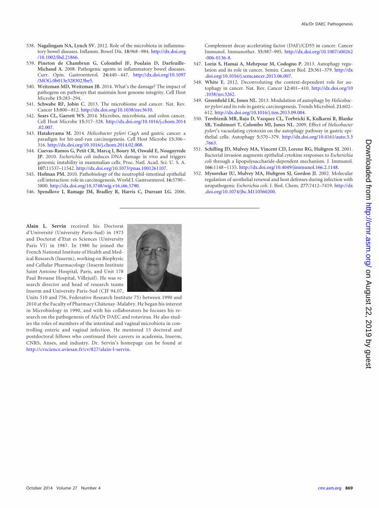
538. Nagalingam NA, Lynch SV. 2012. Role of the microbiota in inflamma-tory bowel diseases. Inflamm. Bowel Dis. 18:968 –984. http://dx.doi.org/10.1002/ibd.21866.
539. Pineton de Chambrun G, Colombel JF, Poulain D, Darfeuille-Michaud A. 2008. Pathogenic agents in inflammatory bowel diseases.Curr. Opin. Gastroenterol. 24:440 – 447. http://dx.doi.org/10.1097/MOG.0b013e3283023be5.
540. Weitzman MD, Weitzman JB. 2014. What’s the damage? The impact ofpathogens on pathways that maintain host genome integrity. Cell HostMicrobe 15:283–294.
541. Schwabe RF, Jobin C. 2013. The microbiome and cancer. Nat. Rev.Cancer 13:800 – 812. http://dx.doi.org/10.1038/nrc3610.
542. Sears CL, Garrett WS. 2014. Microbes, microbiota, and colon cancer.Cell Host Microbe 15:317–328. http://dx.doi.org/10.1016/j.chom.2014.02.007.
543. Hatakeyama M. 2014. Helicobacter pylori CagA and gastric cancer: aparadigm for hit-and-run carcinogenesis. Cell Host Microbe 15:306 –316. http://dx.doi.org/10.1016/j.chom.2014.02.008.
544. Cuevas-Ramos G, Petit CR, Marcq I, Boury M, Oswald E, NougayredeJP. 2010. Escherichia coli induces DNA damage in vivo and triggersgenomic instability in mammalian cells. Proc. Natl. Acad. Sci. U. S. A.107:11537–11542. http://dx.doi.org/10.1073/pnas.1001261107.
545. Hofman PM. 2010. Pathobiology of the neutrophil-intestinal epithelialcell interaction: role in carcinogenesis. World J. Gastroenterol. 16:5790 –5800. http://dx.doi.org/10.3748/wjg.v16.i46.5790.
546. Spendlove I, Ramage JM, Bradley R, Harris C, Durrant LG. 2006.
Complement decay accelerating factor (DAF)/CD55 in cancer. CancerImmunol. Immunother. 55:987–995. http://dx.doi.org/10.1007/s00262-006-0136-8.
547. Lorin S, Hamai A, Mehrpour M, Codogno P. 2013. Autophagy regu-lation and its role in cancer. Semin. Cancer Biol. 23:361–379. http://dx.doi.org/10.1016/j.semcancer.2013.06.007.
548. White E. 2012. Deconvoluting the context-dependent role for au-tophagy in cancer. Nat. Rev. Cancer 12:401– 410. http://dx.doi.org/10.1038/nrc3262.
549. Greenfield LK, Jones NL. 2013. Modulation of autophagy by Helicobac-ter pylori and its role in gastric carcinogenesis. Trends Microbiol. 21:602–612. http://dx.doi.org/10.1016/j.tim.2013.09.004.
550. Terebiznik MR, Raju D, Vazquez CL, Torbricki K, Kulkarni R, BlankeSR, Yoshimori T, Colombo MI, Jones NL. 2009. Effect of Helicobacterpylori’s vacuolating cytotoxin on the autophagy pathway in gastric epi-thelial cells. Autophagy 5:370 –379. http://dx.doi.org/10.4161/auto.5.3.7663.
551. Schilling JD, Mulvey MA, Vincent CD, Lorenz RG, Hultgren SJ. 2001.Bacterial invasion augments epithelial cytokine responses to Escherichiacoli through a lipopolysaccharide-dependent mechanism. J. Immunol.166:1148 –1155. http://dx.doi.org/10.4049/jimmunol.166.2.1148.
552. Mysorekar IU, Mulvey MA, Hultgren SJ, Gordon JI. 2002. Molecularregulation of urothelial renewal and host defenses during infection withuropathogenic Escherichia coli. J. Biol. Chem. 277:7412–7419. http://dx.doi.org/10.1074/jbc.M110560200.
Alain L. Servin received his Doctoratd’Université (University Paris-Sud) in 1973and Doctorat d’Etat es Sciences (UniversityParis VI) in 1987. In 1980 he joined theFrench National Institute of Health and Med-ical Research (Inserm), working on Biophysicand Cellular Pharmacology (Inserm InstituteSaint Antoine Hospital, Paris, and Unit 178Paul Brousse Hospital, Villejuif). He was re-search director and head of research teamsInserm and University Paris-Sud (CJF 94.07,Units 510 and 756, Federative Research Institute 75) between 1990 and2010 at the Faculty of Pharmacy Châtenay-Malabry. He began his interestin Microbiology in 1990, and with his collaborators he focuses his re-search on the pathogenesis of Afa/Dr DAEC and rotavirus. He also stud-ies the roles of members of the intestinal and vaginal microbiota in con-trolling enteric and vaginal infection. He mentored 15 doctoral andpostdoctoral fellows who continued their careers in academia, Inserm,CNRS, Anses, and industry. Dr. Servin’s homepage can be found athttp://cvscience.aviesan.fr/cv/827/alain-l-servin.
Afa/Dr DAEC Pathogenesis
October 2014 Volume 27 Number 4 cmr.asm.org 869
on August 22, 2019 by guest
http://cmr.asm
.org/D
ownloaded from