Embed Size (px)
Citation preview
S114 • JID 2010:201 (Suppl 2) • Darville and Hiltke
S U P P L E M E N T A R T I C L E
Pathogenesis of Genital Tract DiseaseDue to Chlamydia trachomatis
Toni Darville1 and Thomas J. Hiltke2
1Departments of Pediatrics and Immunology, University of Pittsburgh Medical Center, and 2Sexually Transmitted Diseases Branch,National Institute of Allergy and Infectious Diseases, National Institutes of Health, Bethesda, Maryland
Although the pathologic consequences of C. trachomatis genital infection are well-established, the mechanism(s)that result in chlamydia-induced tissue damage are not fully understood. We reviewed in vitro, animal, andhuman data related to the pathogenesis of chlamydial disease to better understand how reproductive sequelaeresult from C. trachomatis infection. Abundant in vitro data suggest that the inflammatory response tochlamydiae is initiated and sustained by actively infected nonimmune host epithelial cells. The mouse modelindicates a critical role for chlamydia activation of the innate immune receptor, Toll-like receptor 2, andsubsequent inflammatory cell influx and activation, which contributes to the development of chronic genitaltract tissue damage. Data from recent vaccine studies in the murine model and from human immunoepide-miologic studies support a role for chlamydia-specific CD4 Th1-interferon-g-producing cells in protectionfrom infection and disease. However, limited evidence obtained using animal models of repeated infectionindicates that, although the adaptive T cell response is a key mechanism involved in controlling or eliminatinginfection, it may have a double-edged nature and contribute to tissue damage. Important immunologic ques-tions include whether anamnestic CD4 T cell responses drive disease rather than protect against disease andthe role of specific immune cells and inflammatory mediators in the induction of tissue damage with primaryand repeated infections. Continued study of the complex molecular and cellular interactions between chla-mydiae and their host and large-scale prospective immunoepidemiologic and immunopathologic studies areneeded to address gaps in our understanding of pathogenesis that thwart development of optimally effectivecontrol programs, including vaccine development.
Sexually transmitted Chlamydia trachomatis infection is
a widespread public health concern because of its prev-
alence and potentially devastating reproductive con-
sequences, including pelvic inflammatory disease
(PID), infertility, and ectopic pregnancy. Although the
pathologic consequences of infection are well estab-
Potential conflicts of interest: None reported.Financial support: National Institute of Allergy and Infectious Diseases
(AI054624 and AI084024 to T.D.) and the Department of Pediatrics, Children’sHospital of Pittsburgh of University of Pittsburgh Medical Center (to T.D.).
Supplement sponsorship: This article is part of a supplement entitled “Chlamydiatrachomatis Genital Infection: Natural History, Immunobiology, and Implications forControl Programs,” which was sponsored by the Centers for Disease Control andPrevention.
Presented in part: C. trachomatis Immunobiology Meeting, sponsored by theCenters for Disease Control and Prevention, Atlanta, GA, May 2009.
Reprints or correspondence: Dr Toni Darville, Div of Infectious Diseases, Deptof Pediatrics, Children’s Hospital of Pittsburgh of University of Pittsburgh MedicalCenter, 45th and Penn Ave, Pittsburgh, PA 15201 ([email protected]).
The Journal of Infectious Diseases 2010; 201(S2):S114–S125� 2010 by the Infectious Diseases Society of America. All rights reserved.0022-1899/2010/20112S2-0006$15.00DOI: 10.1086/652397
lished, the mechanism(s) of chlamydia-induced tissue
damage are not fully understood. Histological exami-
nation of tissue samples from women with PID caused
by C. trachomatis revealed neutrophils in endometrial
surface epithelium and in gland lumens, dense sub-
epithelial stromal lymphocytic infiltration, stromal
plasma cells, and germinal centers containing trans-
formed lymphocytes [1]. The prominence of both neu-
trophils and chronic inflammatory cells in infected hu-
man female genital tract tissue samples does not assist
in the determination of specific responses responsible
for disease sequelae.
Because of the inherent difficulties in acquiring hu-
man tissue samples for study, researchers have taken
advantage of multiple animal models of chlamydial in-
fection to examine the nature and timing of the in-
flammatory response that occurs in the female genital
tract after in vivo infection. Mouse and guinea pig mod-
els show that the response to primary chlamydial in-
Chlamydial Pathogenesis • JID 2010:201 (Suppl 2) • S115
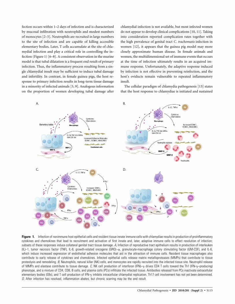
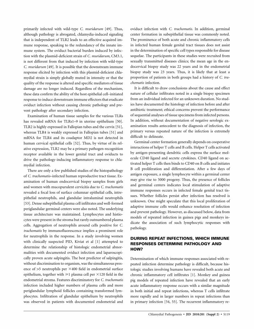
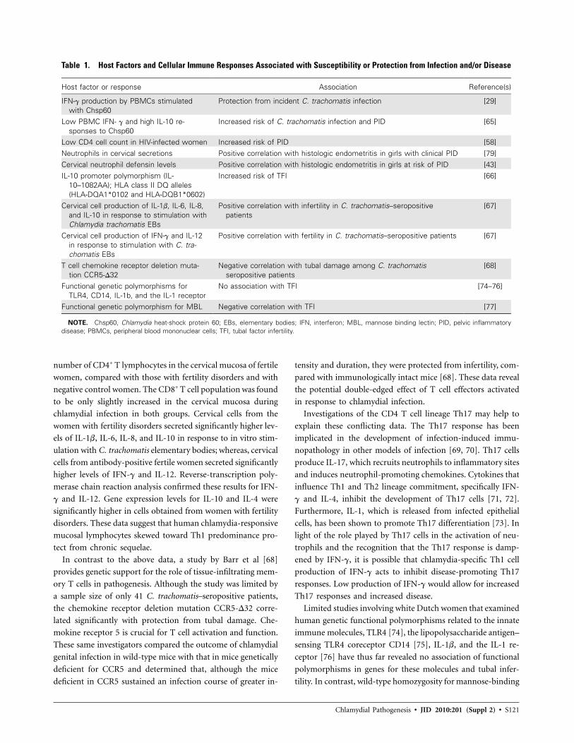
Figure 1. Infection of nonimmune host epithelial cells and resident tissue innate immune cells with chlamydiae results in production of proinflammatorycytokines and chemokines that lead to recruitment and activation of first innate and, later, adaptive immune cells to effect resolution of infection;subsets of these responses induce collateral genital tract tissue damage. A, Infection of reproductive tract epithelium results in production of interleuken(IL)–1, tumor necrosis factor (TNF), IL-8, growth-related oncogene (GRO)–a, granulocyte-macrophage colony stimulating factor (GM-CSF), and IL-6,which induce increased expression of endothelial adhesion molecules that aid in the attraction of immune cells. Resident tissue macrophages alsocontribute to early release of cytokines and chemokines. Infected epithelial cells release matrix metalloproteases (MMPs) that contribute to tissueproteolysis and remodeling. B, Neutrophils, natural killer (NK) cells, and monocytes are rapidly recruited into the infected tissue site. Neutrophil releaseof MMPs and elastase contribute to tissue damage. C, NK cell production of interferon (IFN)–g drives CD4 T cells toward the Th1 (IFN-g–producing)phenotype, and a mixture of CD4, CD8, B cells, and plasma cells (PCs) infiltrate the infected tissue. Antibodies released from PCs inactivate extracellularelementary bodies (EBs), and T cell production of IFN-g inhibits intracellular chlamydial replication. Th17 cell involvement has not yet been determined.D, After infection has resolved, inflammation abates, but chronic scarring may be the end result.
fection occurs within 1–2 days of infection and is characterized
by mucosal infiltration with neutrophils and modest numbers
of monocytes [2–5]. Neutrophils are recruited in large numbers
to the site of infection and are capable of killing accessible
elementary bodies. Later, T cells accumulate at the site of chla-
mydial infection and play a critical role in controlling the in-
fection (Figure 1) [6–8]. A consistent observation in the murine
model is that tubal dilatation is a frequent end result of primary
infection. Thus, the inflammatory process resulting from a sin-
gle chlamydial insult may be sufficient to induce tubal damage
and infertility. In contrast, in female guinea pigs, the host re-
sponse to primary infection results in long-term tissue damage
in a minority of infected animals [5, 9]. Analogous information
on the proportion of women developing tubal damage after
chlamydial infection is not available, but most infected women
do not appear to develop clinical complications [10, 11]. Taking
into consideration reported complication rates together with
the high prevalence of genital tract C. trachomatis infection in
women [12], it appears that the guinea pig model may more
closely approximate human disease. In female animals and
women, the multidimensional set of immune events that occurs
at the time of infection ultimately results in an acquired im-
mune response. Unfortunately, the adaptive response induced
by infection is not effective in preventing reinfection, and the
host’s oviducts remain vulnerable to repeated inflammatory
insult.
The cellular paradigm of chlamydia pathogenesis [13] states
that the host response to chlamydiae is initiated and sustained
S116 • JID 2010:201 (Suppl 2) • Darville and Hiltke
by epithelial cells that are the primary targets of chlamydial
infection. Infected host epithelial cells act as first responders,
initiating and propagating immune responses. They secrete che-
mokines that recruit inflammatory leukocytes to the site of
infection and cytokines that induce and augment the cellular
inflammatory response [14], and these mediators induce direct
damage to the tissues. At the time of reinfection, host cell release
of chemokines leads to recruitment of chlamydia-specific im-
mune cells that rapidly amplify the response. The release of
proteases, clotting factors, and tissue growth factors from in-
fected host cells and infiltrating inflammatory cells leads to
tissue damage and eventual scarring—the hallmark of chla-
mydia-induced oviduct disease. The cellular paradigm makes
no distinction between damage induced by professional innate
immune cells (neutrophils and monocytes) and adaptive lym-
phocyte populations but assumes that both cell populations
contribute to pathogenesis. Chronic chlamydial infections are
common [15] and would lead to ongoing release of mediators
that promote continued influx of inflammatory cells, damage
to host epithelium, scarring, and ultimately, fibrosis and scar-
ring. Because reinfection with chlamydiae is a frequent occur-
rence [16], repeated inflammatory responses may lead to re-
peated insult to the tissues and may promote tissue scarring.
Studies using the murine model of C. trachomatis genital
tract infection have established that resolution of genital chla-
mydial infection is dependent on an influx of interferon (IFN)-
g–producing CD4+ Th1 cells [8, 17–20]. The immunological
paradigm for pathogenesis is based on the premise that T cell
responses that are essential to host defense may also cause
collateral tissue damage [21]. It was speculated that host T cell
responses induced on primary infection to a species-specific
antigen were increased with subsequent infections, promoting
tissue damage and scarring [22]. Chlamydia heat shock protein
60 (Chsp60) has been investigated as a potential antigen re-
sponsible for induction of delayed type hypersensitivity–in-
duced disease. This molecule attracted attention as a candidate
pathogenic antigen after studies in immune guinea pigs and
monkeys suggested direct eye inoculation with this sensitizing
antigen promoted heightened inflammation of the conjunctiva
[23, 24]. However, residual Triton-X detergent contaminating
the extracts proved to be the inducer of disease. Later studies
conducted in the guinea pig model of trachoma revealed a
protective role for vaccination with Chsp60 [25], and although
human studies have revealed detection of elevated titers of an-
tibody to Chsp60 in those with more severe disease [26, 27],
this may simply reflect increased exposure to chlamydia
through chronic or repeated infection. A recent large prospec-
tive study of women with PID did not reveal a correlation of
increased antibody titers to Chsp60 with worse outcome [28].
Furthermore, IFN-g production by peripheral blood T cells
stimulated with Chsp60 predicts protection from incident in-
fection in women at high risk of repeated infection [29].
Despite the lack of evidence for a specific chlamydial path-
ogenic antigen that primes anamnestic T cell responses, in the
presence of chronic or repeated infection, an ongoing or aug-
mented memory T cell response might heighten disease de-
velopment. Monkey [30] and guinea pig [31] models of re-
peated infection indicate that CD4 and CD8 T cells infiltrate
more rapidly and in larger numbers than do neutrophils during
repeat oviduct infections, and this recurrent inflammatory re-
action ultimately culminates in fibrosis and scarring. Of im-
portance, because guinea pigs develop sufficient immunity after
primary infection to significantly limit bacterial burden during
a secondary vaginal infection, this indicates that very small
amounts of chlamydiae may be sufficient to induce an enhanced
T cell response in the oviduct that culminates in disease. Al-
though CD4 and CD8 T cells have been observed in both of
these models of repeated infection, with CD8 T cells being
predominant in the monkey model, no data exist on the specific
role of CD8 T cells in pathogenesis in humans.
Because the ultimate goal of chlamydia control programs is
to prevent reproductive tract complications, a more complete
understanding of how C. trachomatis infection leads to sequelae
is needed. The cellular paradigm of pathogenesis does not in-
voke professional innate or adaptive immune cells as being
more or less responsible for disease development. Instead, the
central player of pathogenesis is assumed to be the host epi-
thelial cell that drives the inflammatory response through its
recognition of chlamydia infection. Epithelial cells possess sur-
face and intracellular innate immune receptors that enable them
to recognize conserved chlamydial ligands and initiate inflam-
mation. Thus, the infected epithelial cell serves as a key innate
responder cell. Therefore, a determination of genetic poly-
morphisms that result in heightened innate inflammatory re-
sponses to chlamydia may serve to identify persons at high risk
of disease development and in need of increased levels of
screening and treatment. Furthermore, because tissue-damag-
ing responses begin as soon as the bacterium infects the oviduct
epithelium and the infected epithelial cells will continue to drive
inflammation as long as the pathogen remains, control pro-
grams should seek to provide treatment before infection of the
oviduct occurs or to shorten the duration of oviduct infection
as much as possible.
The long-term morbidity associated with chlamydial infec-
tion primarily results from tissue damage at the level of the
oviduct. The cellular paradigm imposes a prerequisite for in-
fection of the oviduct to occur for disease to develop. Thus,
an adaptive immune response (induced by infection at the
cervix or by vaccination) that prevents ascension of bacteria to
the oviduct after initial or repeated infection would effectively
prevent disease. This has important implications for the design
Chlamydial Pathogenesis • JID 2010:201 (Suppl 2) • S117
of screening and vaccine control programs for women, because
it indicates that infection of the cervix does not correlate with
risk of disease as long as a strong adaptive immune response
is documented in the same patient. Therefore, a vaccine that
induces responses potent enough to prevent oviduct infection
while containing infection to the cervix might also be effective
in preventing disease sequelae. A determination of responses
that predict protection from reinfection in women at high risk
because of exposure is a realistic goal. If these responses could
be demonstrated to be the same as those that protect an in-
dividual from infection of the upper genital tract, these re-
sponses could be used as markers of women at low risk of
infection and disease. Women without such markers would be
considered to be at high risk and in need of an increased fre-
quency of screening. Because ample data indicate that chla-
mydia-specific CD4 Th1-IFN-g–producing cells are key me-
diators of host defense, a goal for vaccine development should
be to determine chlamydia antigens and adjuvants that induce
a strong CD4 Th1 memory response. Of importance, the in-
duction of sterilizing immunity would not be required to pro-
tect women from chlamydia disease.
On the other hand, if as suggested by the immunological
paradigm, a small antigenic insult at the site of the oviduct
after rechallenge induces tissue-damaging memory T cell re-
sponses, it would be essential for a vaccine to prevent even
minimal infection of the oviduct. Thus, sterilizing immunity
will be required for a vaccine to be protective. In this instance,
chlamydia control programs would have to pay vigorous at-
tention to preventing repeat infection in women with evidence
of a strong CD4 Th1 memory response.
A review of published studies revealed possible avenues to
the identification of specific cytokine and cellular responses that
predict sequelae. Determination of biomarkers of disease will
allow control efforts to be intensified for individuals identified
at highest risk. We reviewed in vitro, animal, and human data
related to the pathogenesis of chlamydial disease to better un-
derstand how reproductive sequelae result from C. trachomatis
infection. We approached our review of the literature with sev-
eral specific questions in mind.
WHICH INFLAMMATORY AND/OR IMMUNERESPONSES OCCUR DURING AN INITIALC. TRACHOMATIS INFECTION AND HOW DOTHEY LEAD TO PATHOLOGY?
The concept of the epithelial cell as an important and early
component of the host response to chlamydial infection was
first revealed by Rasmussen et al [14], who showed that in vitro
infection of cervical and colonic epithelial cells with C. tra-
chomatis induced the secretion of an array of proinflammatory
cytokines that have chemoattractant and proinflammatory
functions. Moreover, in contrast to invasion by other bacteria
that induce a rapid but transient proinflammatory cytokine
response at entry [32], chlamydial invasion alone was not suf-
ficient to elicit a response. Instead, intracellular chlamydial
growth was required, and the epithelial cytokine response was
sustained throughout the chlamydial developmental cycle. En-
docervical epithelial cells released interleuken (IL)–1a after in-
fection, and the induced proinflammatory cytokine cascade
could be inhibited by specific anti–IL-1a antibodies [14]. Thus,
IL-1a, released on epithelial cell lysis, may act to amplify the
inflammatory response by stimulating additional cytokine pro-
duction. These findings formed the basis for Richard Stephens’
[13] “cellular paradigm of chlamydial pathogenesis.” Stephens
theorized that “the inflammatory processes of chlamydial path-
ogenesis are elicited by infected host cells and are necessary
and sufficient to account for chronic inflammation and the
promotion of cellular proliferation, tissue remodeling and scar-
ring—the ultimate cause of disease sequelae” [13, p 44].
Since the article by Rasmussen et al [14], others have con-
firmed the elicitation of inflammatory mediators from non-
immune host cells infected in vitro with C. trachomatis. Infec-
tion of Fallopian tube organ cultures results in epithelial cell
release of IL-1 and cellular damage, independent of inflam-
matory cell influx. The addition of IL-1 receptor antagonist to
the cultures completely eliminates tissue destruction induced
by infection, indicating a direct role for this cytokine in path-
ogenesis [33]. In vitro infection of Fallopian tube epithelium
also results in the production of tumor necrosis factor (TNF)
[34] and increased expression of adhesion molecules on oviduct
endothelial cells [35]. This milieu could easily lead to activation
and recruitment of first innate and, later, adaptive immune
cells to effect resolution of infection. However, subsets of these
responses may also induce collateral damage to genital tract
tissue.
Proinflammatory chemokines and cytokines have been doc-
umented in murine and guinea pig models of genital tract
chlamydial infection [4, 36, 37]. The up-regulation of integrins
in the murine genital tract is coincident with the onset of
infection [2]. Detection of the neutrophil chemokine macro-
phage inflammatory protein-2 (MIP-2), a chemokine analo-
gous to IL-8 in humans, in genital tract secretions of infected
mice coincides with a rapid influx of neutrophils into the lower
genital tract. Thus, data to date suggest that the inflammatory
response to chlamydiae begins and is sustained by actively in-
fected nonimmune host epithelial cells. Defining the specific
responses that promote tissue damage and differentiating them
from those that lead to benign resolution of infection is an
important ongoing research goal.
Murine genital tract studies indicate that the intensity of
neutrophil influx into the oviduct (pyosalpinx) correlates di-
rectly with eventual development of hydrosalpinx [38]. Pro-
longed infiltration of neutrophils into the oviduct correlates
S118 • JID 2010:201 (Suppl 2) • Darville and Hiltke
with an increased incidence of severe hydrosalpinx in an im-
munologically normal mouse strain that exhibits increased dis-
ease susceptibility [39, 40]. Data by Morrison et al [8] revealed
that genital chlamydial infection of mice with disrupted b2-
microglobulin, I-A, or CD4 genes resulted in tubal ectasia and
hydrosalpinx similar to infection of immunologically intact
mice. A common feature in all mouse strains tested was the
accompaniment of ascending infection of the genital tract with
a marked neutrophilic inflammatory response. In class II�/-
mice, a persistent prominent acute-subacute inflammatory re-
sponse failed to resolve infection but led to development of
hydrosalpinx. Although these data do not exclude a role for
adaptive immune responses in the development of pathology,
they indicate that chronic genital tract tissue damage can occur
independently of T cell responses and implicates a role for acute
inflammatory cells in its development. Activation of phagocyte
oxidase in myeloid cells causes release of superoxide molecules
that are damaging to cells and tissues. Mice with a deletion of
a key component of phagocyte oxidase (p47phox�/-) sustain
lower rates of hydrosalpinx [41] after chlamydial infection.
Other potentially important factors are matrix metalloprotein-
ases (MMPs), which are expressed by neutrophils and mono-
cytes and are involved in the proteolysis and resynthesis of the
extracellular matrix. Ramsey et al [39] implicated neutrophil
production of MMP-9 in the development of scarring and fi-
brosis of the murine oviduct after chlamydial infection.
Studies in humans also indicate a role for MMPs and neu-
trophils in production of tissue damage. Fallopian tube epi-
thelial cells infected in vitro with C. trachomatis produce MMP-
2, and infected oviduct stromal cells produce MMP-9 [42]. The
importance of neutrophil activation in the inflammatory pro-
cess associated with C. trachomatis infection of the female re-
productive tract was also revealed in a study of women at risk
of PID in which vaginal levels of neutrophil a-defensins
(HNP1–3), markers of neutrophil activation, were strongly as-
sociated with the presence of endometritis [43]. Absolute num-
bers of vaginal neutrophils were associated with endometritis
only in the presence of elevated defensin levels. Although ocu-
logenital serovars are killed by neutrophils, in human endo-
metrial epithelial cell cultures monitored for 11 month after
exposure to azithromycin (a chlamydiacidal antibiotic)–loaded
neutrophils, residual chlamydial envelopes and major outer
membrane protein and lipopolysaccharide antigens were de-
tected and continued to stimulate neutrophil chemotaxis [44].
Thus, antigens may persist for some time after the organisms
are killed, inducing continued neutrophil inflammation and
ongoing release of tissue-damaging molecules in the host.
Taken together, these data imply that ascension of chlamydiae
from the lower to the upper genital tract and, ultimately, in-
fection of oviduct epithelium are prerequisites for the devel-
opment of long-term sequelae. Support of this hypothesis
comes from the lack of detection of oviduct pathology in mice
infected vaginally with human serovars [4], where infection of
the cervix and endometrium occur but oviduct infection does
not [4, 45]. Human serovars elicit oviduct pathology in the
mouse when they are inoculated directly under the ovarian
bursa [46]. On the other hand, infection of the oviduct does
not guarantee disease development. Rank and Sanders [9] doc-
umented that oviduct infection occurred in 78% of vaginally
inoculated guinea pigs, but chronic oviduct pathology was seen
in only 12%. Thus, additional host factors must influence the
development of disease.
Researchers have begun to determine the cellular receptors
involved in C. trachomatis–induced stimulation of cytokine re-
lease. Toll-like receptors (TLRs) act as pathogen-recognition
receptors that enable cells to recognize conserved bacterial, vi-
ral, and fungal structural elements. In vitro, C. trachomatis
infection of HEK cells transfected with the adaptor molecule
MyD88 and the pathogen molecular pattern receptors TLR2
and TLR4/MD-2 revealed that TLR2 was required for IL-8
secretion and that the role of TLR4/MD-2 was minimal. This
was reproduced with chlamydial infection of immortalized hu-
man ectocervical epithelial cells [47]. Activation was dependent
on live, replicating bacteria, because infection with ultra violet–
irradiated bacteria and treatment of infected cells with chlo-
ramphenicol (no inclusions), but not ampicillin (jammed in
reticulate body form), abrogated the induction of IL-8 secre-
tion. The response was largely dependent on the MyD88 adap-
tor molecule. Confocal microscopy experiments revealed that
both TLR2 and MyD88 colocalize with the intracellular chla-
mydial inclusion, suggesting that TLR2 is actively engaged in
signaling from this intracellular location.
Examination of the outcomes of genital tract infection in
wild-type mice and mice genetically deficient in TLR2 and
TLR4 confirmed a dominant role for TLR2, compared with
TLR4, in the recognition and response to Chlamydia murida-
rum in the genital tract [48]. TLR4-/- mice responded to in-
fection similarly to wild-type controls and developed similar
pathology. Eradication of lower genital tract infection was
equivalent in TLR2-/- mice and wild-type mice, even in the
early days after infection. However, significantly lower levels of
TNF-a and MIP-2 were detected in genital tract secretions of
TLR2-/- mice during the first week of infection, and there was
a significant reduction in oviduct and mesosalpinx pathology
at later times. Thus, the cytokine response that remained in
the absence of TLR2 signaling was sufficient to recruit effector
cells to the genital tract, but these data indicate a protective
role for TLR2 deficiency in genital tract infection sequelae due
to C. trachomatis. Wild-type mice that are infected with plas-
mid-deficient C. muridarum that fail to signal via TLR2 develop
a predominant Th1 response, and they are as resistant to sec-
ondary challenge infection as immunologically normal mice
Chlamydial Pathogenesis • JID 2010:201 (Suppl 2) • S119
primarily infected with wild-type C. muridarum [49]. Thus,
although pathology is abrogated, chlamydia-induced signaling
that is independent of TLR2 leads to an effective acquired im-
mune response, speaking to the redundancy of the innate im-
mune system. The oviduct bacterial burden induced by infec-
tion with the plasmid-deficient strain of C. muridarum, CM3.1,
is not different from that induced by infection with wild-type
C. muridarum [49]. It is possible that the downstream immune
response elicited by infection with this plasmid-deficient chla-
mydial strain is simply globally muted in intensity or that the
quality of the response is altered and specific mediators of tissue
damage are no longer induced. Regardless of the mechanism,
these data confirm the ability of the host epithelial cell–initiated
response to induce downstream immune effectors that eradicate
oviduct infection without causing chronic pathology and pre-
vent pathology after secondary infection.
Examination of human tissue samples for the various TLRs
has revealed mRNA for TLRs1–9 in uterine epithelium [50].
TLR2 is highly expressed in Fallopian tubes and the cervix [51],
whereas TLR4 is weakly expressed in Fallopian tubes [51] and
mRNA for TLR4 and its coadaptor MD2 is not detected in
human cervical epithelial cells [52]. Thus, by virtue of its rel-
ative expression, TLR2 may be a primary pathogen-recognition
receptor available in the lower genital tract and oviducts to
drive the pathology-inducing inflammatory response to chla-
mydial infection.
There are only a few published studies of the histopathology
of C. trachomatis–infected human reproductive tract tissue. Ex-
amination of human endocervical biopsy samples from girls
and women with mucopurulent cervicitis due to C. trachomatis
revealed a focal loss of surface columnar epithelial cells, intre-
pithelial neutrophils, and glandular intraluminal neutrophils
[53]. Dense subepithelial plasma cell infiltrates and well-formed
periglandular germinal centers were also noted. The underlying
tissue architecture was maintained. Lymphocytes and histio-
cytes were present in the stroma but rarely outnumbered plasma
cells. Aggregation of neutrophils around cells positive for C.
trachomatis by immunofluorescence implies a prominent role
for neutrophils in the response. In a study involving women
with clinically suspected PID, Kiviat et al [1] attempted to
determine the relationship of histologic endometrial abnor-
malities with documented oviduct infection and laparoscopi-
cally proven acute salpingitis. The best predictor of salpingitis,
without discrimination to organism, was the simultaneous pres-
ence of 15 neutrophils per �400 field in endometrial surface
epithelium, together with �1 plasma cell per �120 field in the
endometrial stroma. Features discriminatory for C. trachomatis
infection included higher numbers of plasma cells and more
periglandular lymphoid follicles containing transformed lym-
phocytes. Infiltration of glandular epithelium by neutrophils
was observed in patients with documented endometrial and
oviduct infection with C. trachomatis. In addition, germinal
center formation in subepithelial tissue was commonly noted.
The prominence of both acute and chronic inflammatory cells
in infected human female genital tract tissues does not assist
in the determination of specific cell types responsible for disease
sequelae. The participants in these studies were recruited from
sexually transmitted diseases clinics; the mean age in the en-
docervical biopsy study was 22 years and in the endometrial
biopsy study was 23 years. Thus, it is likely that at least a
proportion of patients in both groups had a history of C. tra-
chomatis infection.
It is difficult to draw conclusions about the cause and effect
nature of cellular infiltrates noted in a single biopsy specimen
from an individual infected for an unknown duration. No stud-
ies have documented the histology of infection before and after
antibiotic treatment; ethical concerns prevent the performance
of sequential analyses of tissue specimens from infected persons.
In addition, without documentation of negative serologic ex-
amination results antecedent to the diagnosis of infection, the
primary versus repeated nature of the infection is extremely
difficult to delineate.
Germinal center formation generally depends on cooperative
interactions of helper T cells and B cells. Helper T cells activated
by antigen-presenting dendritic cells express the surface mol-
ecule CD40 ligand and secrete cytokines. CD40 ligand on ac-
tivated helper T cells then binds to CD40 on B cells and initiates
B cell proliferation and differentiation. After a few days of
antigen exposure, a single lymphocyte within a germinal center
may give rise to 5000 progeny. Thus, the presence of follicles
and germinal centers indicates local stimulation of adaptive
immune responses occurs in infected female genital tract tis-
sues. Whether follicles persist after infection has resolved is
unknown. One might speculate that this local proliferation of
adaptive immune cells would enhance resolution of infection
and prevent pathology. However, as discussed below, data from
models of repeated infection in guinea pigs and monkeys in-
dicate the association of such lymphocytic responses with
pathology.
DURING REPEAT INFECTIONS, WHICH IMMUNERESPONSES DETERMINE PATHOLOGY ANDHOW?
Determination of which immune responses associated with re-
peated infection determine pathology is difficult, because his-
tologic studies involving humans have revealed both acute and
chronic inflammatory cell infiltrates [1]. Monkey and guinea
pig models of repeated infection have revealed that an early
acute inflammatory response occurs with a similar magnitude
in both initial and repeat infections, whereas T cells infiltrate
more rapidly and in larger numbers in repeat infections than
in primary infection [54, 55]. The recurrent inflammatory re-
S120 • JID 2010:201 (Suppl 2) • Darville and Hiltke
action ultimately culminates in fibrosis and scarring [56]. Pri-
mary infection in the mouse model induces such severe oviduct
pathology that this model is somewhat impractical for address-
ing the role of specific cell types in induction of pathology with
repeat infection.
Reinfection in the macaque salpingeal pocket model resulted
in a rapid lymphocytic infiltration, resulting in notable epithe-
lial cell destruction and the formation of lymphoid follicles
[55]. Tertiary infection of pockets reignites lymphocytic activity
with focal areas of epithelium destruction and the formation
of extensive fibrosis and lymphoid follicles in the deep stroma
[30]. Additional data from this model indicate that the robust
inflammatory response to repeat infection may be mediated by
cytotoxic CD8 T cells primed against Chsp60 [57]. Thus, this
direct inoculation nonhuman primate model displays immune-
mediated destruction of genital tract tissue that is enhanced
with each repeat infection. Human epidemiologic studies have
indicated increased risk of disease with repeated infection [58–
60]. However, this may simply be a cumulative process rather
than the result of enhanced destruction with each repeat in-
fection, as seen in this primate model. In addition, it should
be noted that these models differ from human infections in
that the oviducts are infected directly rather than by ascension
from the lower genital tract. During natural infection, the time
required for ascension of the bacterium to the oviducts may
allow for homing of protective memory T cells, leading to a
decrease in the infectious inoculum and consequent damaging
inflammation at this vulnerable tissue site. In addition, animals
are not treated with antibiotics between infections; thus, these
data cannot be extrapolated to humans who sustain repeat
infection after treatment of an initial infection.
Primary vaginal infection of guinea pigs with Chlamydia cav-
iae (formally known as the guinea pig inclusion conjunctivitis
serovar of Chlamydia psittaci) leads to an ascending infection
that is cleared by ∼20 days after infection [9]. Although infec-
tion of at least 1 oviduct can be documented in most animals
after a primary infection, less than half develop notable tubal
pathology. Both cellular and humoral immune mechanisms
generated in a primary infection protected animals from re-
infection for up to 30 days; however, this immunity waned
such that animals were susceptible beginning on day 77 [61–
63]. Although secondary infections were noted to be markedly
abbreviated with a significant reduction in bacterial burden, an
increased number of animals developed oviduct dilatation with
repeat infection [31]. One potential caveat is that oviduct pa-
thology was evaluated 105–112 days after primary infection in
challenged animals and 75–85 days after primary infection in
nonchallenged animals. It is possible that increased scarring
occurred over time, leading to the detection of an increased
frequency of oviduct dilation in the challenged animals. How-
ever, definite enhancement of cell-mediated inflammation after
rechallenge was observed in a subsequent study using the guinea
pig model in which flow cytometric analysis revealed that sig-
nificantly higher levels of T and B cells, but not Mac-1–positive
cells, were recruited to the oviduct in rechallenged animals,
compared with animals infected only once [64]. Of interest,
little to no C. caviae reached the oviduct during repeat infection,
suggesting that this organ is exceptionally susceptible to a small
antigenic insult after priming through an initial infection and
providing strong evidence for a cell-mediated mechanism of
pathology in repeat infection. Further development of this
model and the use of guinea pig–specific reagents may provide
a useful surrogate to investigate the possibility that repeat in-
fection leads to an accelerated inflammatory response resulting
in enhanced disease.
WHAT IS THE ROLE OF HOST FACTORS INPATHOGENESIS? ARE THERE RELIABLEMARKERS OF PATHOGENESIS ORPATHOLOGY?
Studies have suggested an association between specific host
immune responses and susceptibility to chlamydial infection
and/or disease (Table 1). In a prospective cohort study involving
women at high risk of C. trachomatis infection, Cohen et al
[29] found that, at baseline and after adjustment for age and
other potential confounding variables, production of IFN-g by
peripheral-blood mononuclear cells (PBMCs) stimulated with
Chsp60 strongly correlated with protection against incident C.
trachomatis infection, although there was no information re-
garding development of sequelae. In a small study involving
Australian women attending a sexually transmitted diseases
clinic, Debattista et al [65] found that low PBMC IFN-g and
high IL-10 responses to Chsp60 were markers for increased risk
of chlamydial infection and PID. In human immunodeficiency
virus–seropositive women, a CD4 lymphocyte count of !400
cells/mm3 was determined to be an independent risk factor for
C. trachomatis PID (odds ratio, 21.7; 95% confidence interval,
1.2–383; ) [58]. These studies support a role for chla-P p .036
mydia-responsive CD4 Th1–IFN-g–producing cells in protec-
tion from disease. In women with chlamydia-related tubal in-
fertility, T cell responses to Chsp60 were associated with a
specific IL-10 promoter polymorphism (IL-10–1082AA) and
with specific HLA class II DQ alleles (HLA-DQA1*0102 and
HLA-DQB1*0602) [66]. Thus, genetic factors that regulate the
induction and activation of Th1 cells may be important in
directing a protective host response.
A recent study by Agrawal et al [67] examined cervical lym-
phocyte cytokine responses of 255 C. trachomatis antibody–
positive women with or without fertility disorders (infertility
and multiple spontaneous abortions) and of healthy control
women negative for C. trachomatis serum IgM or IgG. Flow
cytometric analysis revealed a significant increase in the mean
Chlamydial Pathogenesis • JID 2010:201 (Suppl 2) • S121
Table 1. Host Factors and Cellular Immune Responses Associated with Susceptibility or Protection from Infection and/or Disease
Host factor or response Association Reference(s)
IFN-g production by PBMCs stimulatedwith Chsp60
Protection from incident C. trachomatis infection [29]
Low PBMC IFN- g and high IL-10 re-sponses to Chsp60
Increased risk of C. trachomatis infection and PID [65]
Low CD4 cell count in HIV-infected women Increased risk of PID [58]Neutrophils in cervical secretions Positive correlation with histologic endometritis in girls with clinical PID [79]Cervical neutrophil defensin levels Positive correlation with histologic endometritis in girls at risk of PID [43]IL-10 promoter polymorphism (IL-
10–1082AA); HLA class II DQ alleles(HLA-DQA1*0102 and HLA-DQB1*0602)
Increased risk of TFI [66]
Cervical cell production of IL-1b, IL-6, IL-8,and IL-10 in response to stimulation withChlamydia trachomatis EBs
Positive correlation with infertility in C. trachomatis–seropositivepatients
[67]
Cervical cell production of IFN-g and IL-12in response to stimulation with C. tra-chomatis EBs
Positive correlation with fertility in C. trachomatis–seropositive patients [67]
T cell chemokine receptor deletion muta-tion CCR5-D32
Negative correlation with tubal damage among C. trachomatisseropositive patients
[68]
Functional genetic polymorphisms forTLR4, CD14, IL-1b, and the IL-1 receptor
No association with TFI [74–76]
Functional genetic polymorphism for MBL Negative correlation with TFI [77]
NOTE. Chsp60, Chlamydia heat-shock protein 60; EBs, elementary bodies; IFN, interferon; MBL, mannose binding lectin; PID, pelvic inflammatorydisease; PBMCs, peripheral blood mononuclear cells; TFI, tubal factor infertility.
number of CD4+ T lymphocytes in the cervical mucosa of fertile
women, compared with those with fertility disorders and with
negative control women. The CD8+ T cell population was found
to be only slightly increased in the cervical mucosa during
chlamydial infection in both groups. Cervical cells from the
women with fertility disorders secreted significantly higher lev-
els of IL-1b, IL-6, IL-8, and IL-10 in response to in vitro stim-
ulation with C. trachomatis elementary bodies; whereas, cervical
cells from antibody-positive fertile women secreted significantly
higher levels of IFN-g and IL-12. Reverse-transcription poly-
merase chain reaction analysis confirmed these results for IFN-
g and IL-12. Gene expression levels for IL-10 and IL-4 were
significantly higher in cells obtained from women with fertility
disorders. These data suggest that human chlamydia-responsive
mucosal lymphocytes skewed toward Th1 predominance pro-
tect from chronic sequelae.
In contrast to the above data, a study by Barr et al [68]
provides genetic support for the role of tissue-infiltrating mem-
ory T cells in pathogenesis. Although the study was limited by
a sample size of only 41 C. trachomatis–seropositive patients,
the chemokine receptor deletion mutation CCR5-D32 corre-
lated significantly with protection from tubal damage. Che-
mokine receptor 5 is crucial for T cell activation and function.
These same investigators compared the outcome of chlamydial
genital infection in wild-type mice with that in mice genetically
deficient for CCR5 and determined that, although the mice
deficient in CCR5 sustained an infection course of greater in-
tensity and duration, they were protected from infertility, com-
pared with immunologically intact mice [68]. These data reveal
the potential double-edged effect of T cell effectors activated
in response to chlamydial infection.
Investigations of the CD4 T cell lineage Th17 may help to
explain these conflicting data. The Th17 response has been
implicated in the development of infection-induced immu-
nopathology in other models of infection [69, 70]. Th17 cells
produce IL-17, which recruits neutrophils to inflammatory sites
and induces neutrophil-promoting chemokines. Cytokines that
influence Th1 and Th2 lineage commitment, specifically IFN-
g and IL-4, inhibit the development of Th17 cells [71, 72].
Furthermore, IL-1, which is released from infected epithelial
cells, has been shown to promote Th17 differentiation [73]. In
light of the role played by Th17 cells in the activation of neu-
trophils and the recognition that the Th17 response is damp-
ened by IFN-g, it is possible that chlamydia-specific Th1 cell
production of IFN-g acts to inhibit disease-promoting Th17
responses. Low production of IFN-g would allow for increased
Th17 responses and increased disease.
Limited studies involving white Dutch women that examined
human genetic functional polymorphisms related to the innate
immune molecules, TLR4 [74], the lipopolysaccharide antigen–
sensing TLR4 coreceptor CD14 [75], IL-1b, and the IL-1 re-
ceptor [76] have thus far revealed no association of functional
polymorphisms in genes for these molecules and tubal infer-
tility. In contrast, wild-type homozygosity for mannose-binding
S122 • JID 2010:201 (Suppl 2) • Darville and Hiltke
lectin allele A has been associated with protection against tubal
occlusion in white Hungarian women [77]. Binding of man-
nose-binding lectin to sugar groups of the C. trachomatis major
outer membrane protein blocks attachment of organisms to
host cells [78]. Thus, low levels of mannose-binding lectin may
lead to an increased infectious burden and/or increased ascen-
sion to the oviducts, resulting in increased risk for tubal factor
infertility. Examination for functional polymorphisms in genes
for TLR2 (shown to be essential for induction of oviduct pa-
thology in the mouse model) and the TLR2 coreceptors TLR1
and TLR6 with use of banked specimens from women with
and without infertility previously enrolled in the PID evaluation
and clinical health study (PEACH) [28] are ongoing (T. Darville
and C. L. Haggerty, personal communication).
Researchers have attempted to determine factors identifiable
using cervical samples that predict upper genital tract pathol-
ogy. As reported above, vaginal neutrophil defensin levels have
been shown to correlate with presence of endometritis in
women with subclinical PID [43]. An earlier study revealed a
strong correlation between detection of neutrophils in cervical
secretions and presence of endometritis as determined by ex-
amination of biopsy samples from girls with clinically symp-
tomatic PID [79]. However, it remains to be determined
whether leucorrhea and/or elevated cervical mucus defensin
levels predict salpingitis and/or increased risk of chronic tubal
damage.
One important question is that of the relationship of anti-
chlamydial antibodies and disease risk. Risk factors for C. tra-
chomatis PID that were established in prospective studies in-
volving female sex workers include positive serum antibody to
Chsp60 [58, 80] and repeated C. trachomatis infection [58].
Among women with mild to moderate PID, antibody titers to
C. trachomatis elementary bodies measured in the highest tertile
at follow-up, but not to Chsp60, were independently associated
with elevated rates of recurrent PID and reduced rates of preg-
nancy [28]. Although these studies linked positive antibody
responses to chlamydial proteins to sequelae, these responses
may simply be markers for increased exposure to chlamydiae.
Increased exposure could be in the form of repeated or chronic
infection. Monkeys inoculated in their oviducts with C. tra-
chomatis and subsequently treated with antibiotics were ex-
amined for antibodies to Chsp60. A persistent Chsp60 antibody
response was correlated with having culture- or ligase chain
reaction–positive oviduct samples after treatment, which sug-
gests that antibody positivity is a useful marker of chronic
infection [81]. These data indicate that prolonged or repeated
exposure to chlamydiae leads to increased risk for disease and
increased detection of anti-chlamydial antibodies, rather than
implicating antibody formation directly in pathogenesis.
Another possibility is that female individuals who are prone
to develop high titers of antibody (Th2) are less likely to develop
a protective cell-mediated (Th1) immune response. Although
high antibody responses to Chsp60 have been correlated with
increased susceptibility to chlamydial PID [58, 80], IFN-g re-
sponses to this highly conserved protein have been correlated
with protection among the same group of women [58]. Al-
though logistically very difficult, more correlative longitudinal
data on antibody and T cell responses are needed in adolescent
and adult women who sustain an initial or repeated infection.
SUMMARY AND CONCLUSIONS
The most conclusive pathogenesis data to date reveal that ac-
tively infected nonimmune host cells are the driving force in
the inflammatory response to chlamydia. In vitro and mouse
data indicate that infected host epithelial cells not only drive
influx of inflammatory cells, but also release tissue-damaging
molecules directly. Mouse and in vitro data implicate neutro-
phils and proteinases released from activated neutrophils in
tissue pathogenesis and indicate an important role for chla-
mydia-induced activation of TLR2 in initiation of these re-
sponses. However, continued release of chemokines from in-
fected host cells drives recruitment of not only innate
neutrophils and monocytes, but also adaptive T cells and B
cells. The intracellular niche of chlamydia makes it logical that
T cell responses would be important for successful eradication
of infection, and multiple studies involving mice and humans
suggest that the CD4 Th1 response is the principle mechanism
of host defense. Thus, a key question that needs to be answered
is whether anamnestic CD4 Th1 responses induced after re-
infection, with ongoing chronic infection, or after vaccination
cause disease.
Limited evidence from animal models indicates that the
adaptive T cell response may promote pathology, especially with
ongoing chronic or repeat infection. However, recent vaccine
studies in mice indicate that induction of a Th1 immune re-
sponse to vaccine antigens is essential for the vaccine to protect
challenged animals from disease [82, 83]. In addition, human
immunoepidemiologic studies have shown that women at high
risk of exposure that develop chlamydia-specific CD4+ Th1–
IFN-g–producing cells are protected from incident infection
[29]. Because it is unlikely that a vaccine that induces primarily
antibody responses will be effective against a pathogen that
replicates in a protective vacuole intracellularly, conclusive evi-
dence that induction of Th1 immunity is solely protective is
crucial for vaccine development.
Although CD8 T cells have been detected in guinea pig and
monkey models of repeat infection in association with oviduct
disease, the role of these cells in human disease needs to be
defined. In addition, chlamydia-specific Th17 cells should be
examined as potential mediators of pathogenesis. These cells
activate potentially tissue-damaging neutrophils, and their ac-
Chlamydial Pathogenesis • JID 2010:201 (Suppl 2) • S123
tivation has been associated with increased inflammation and
disease in other infectious disease models.
The application of multicolor flow cytometric methods to
determine specific cell types, cell function, and the kinetics of
these responses in genital tract tissues with use of established
animal models should assist in answering these questions. These
same techniques can be applied to human specimens, such as
peripheral blood, endocervical brush, and endometrial biopsy
samples, in conjunction with collection of clinical and epide-
miological data. Furthermore, knowledge gained from the use
of genetically engineered knock-out mouse models related to
pathogen-host cell interactions that stimulate disease-inducing
immune responses should be investigated in humans. Eluci-
dation of specific cytokine and cellular responses as predictors
of sequelae would allow control efforts to be intensified for
individuals identified to be at highest risk of disease. Ap-
proaches include examinations conducted with human cells and
tissue samples ex vivo and large-scale immunoepidemiologic,
immunopathologic, and genetic studies correlated with clinical
data on infection and disease status.
Acknowledgments
We thank Sami Gottlieb, Robert Brunham, and Gerald Byrne, for theirthoughtful suggestions regarding content, and artists at the Centers forDisease Control and Prevention, for assistance with the illustration.
References
1. Kiviat NB, Wolner-Hanssen P, Eschenbach DA, et al. Endometrial his-topathology in patients with culture-proved upper genital tract infec-tion and laparoscopically diagnosed acute salpingitis. Am J Surg Pathol1990; 14(2):167–175.
2. Kelly KA, Rank RG. Identification of homing receptors that mediatethe recruitment of CD4 T cells to the genital tract following intravaginalinfection with Chlamydia trachomatis. Infect Immun 1997; 65(12):5198–5208.
3. Morrison SG, Morrison RP. In situ analysis of the evolution of theprimary immune response in murine Chlamydia trachomatis genitaltract infection. Infect Immun 2000; 68(5):2870–2879.
4. Darville T, Andrews CW Jr, Laffoon KK, Shymasani W, Kishen LR,Rank RG. Mouse strain-dependent variation in the course and outcomeof chlamydial genital tract infection is associated with differences inhost response. Infect Immun 1997; 65(8):3065–3073.
5. Soloff BL, Rank RG, Barron AL. Ultrastructural studies of chlamydialinfection in guinea pig urogenital tract. J Comp Pathol 1982; 92:547–558.
6. Rank RG, Barron AL. Effect of antithymocyte serum on the course ofchlamydial genital infection in female guinea pigs. Infect Immun1983; 41(2):876–879.
7. Ramsey KH, Rank RG. Resolution of chlamydial genital infection withantigen-specific T- lymphocyte lines. Infect Immun 1991; 59(3):925–931.
8. Morrison RP, Feilzer K, Tumas DB. Gene knockout mice establish aprimary protective role for major histocompatibility complex class II-restricted responses in Chlamydia trachomatis genital tract infection.Infect Immun 1995; 63(12):4661–4668.
9. Rank RG, Sanders MM. Pathogenesis of endometritis and salpingitis
in a guinea pig model of chlamydial genital infection. Am J Pathol1992; 140(4):927–936.
10. Paavonen J, Eggert-Kruse W. Chlamydia trachomatis: impact on humanreproduction. Hum Reprod Update 1999; 5(5):433–447.
11. van Valkengoed IG, Morre SA, van den Brule AJ, Meijer CJ, BouterLM, Boeke AJ. Overestimation of complication rates in evaluations ofChlamydia trachomatis screening programmes–implications for cost-effectiveness analyses. Int J Epidemiol 2004; 33(2):416–425.
12. World Health Organization. Global prevalence and incidence of se-lected curable sexually transmitted diseases: overview and estimates.Geneva: world Health Organization, 2001:1–34.
13. Stephens RS. The cellular paradigm of chlamydial pathogenesis. TrendsMicrobiol 2003; 11:44–51.
14. Rasmussen SJ, Eckmann L, Quayle AJ, et al. Secretion of proinflam-matory cytokines by epithelial cells in response to Chlamydia infectionsuggests a central role for epithelial cells in chlamydial pathogenesis.J Clin Invest 1997; 99(1):77–87.
15. Molano M, Meijer CJ, Weiderpass E, et al. The natural course of Chla-mydia trachomatis infection in asymptomatic Colombian women: a 5-year follow-up study. J Infect Dis 2005; 191(6):907–916.
16. Burstein GR, Gaydos CA, Diener-West M, Howell MR, Zenilman JM,Quinn TC. Incident Chlamydia trachomatis infections among inner-city adolescent females [see comments]. JAMA 1998; 280(6):521–526.
17. Cain TK, Rank RG. Local Th1-like responses are induced by intra-vaginal infection of mice with the mouse pneumonitis biovar of Chla-mydia trachomatis. Infect Immun 1995; 63(5):1784–1789.
18. Perry LL, Feilzer K, Caldwell HD. Immunity to Chlamydia trachomatisis mediated by T helper 1 cells through IFN-gamma-dependent and -independent pathways. J Immunol 1997; 158(7):3344–3352.
19. Cotter TW, Ramsey KH, Miranpuri GS, Poulsen CE, Byrne GI. Dis-semination of Chlamydia trachomatis chronic genital tract infection ingamma interferon gene knockout mice. Infect Immun 1997; 65(6):2145–2152.
20. Morrison SG, Su H, Caldwell HD, Morrison RP. Immunity to murineChlamydia trachomatis genital tract reinfection involves B cells andCD4(+) T cells but not CD8(+) T cells. Infect Immun 2000; 68(12):6979–6987.
21. Brunham RC, Rey-Ladino J. Immunology of Chlamydia infection: im-plications for a Chlamydia trachomatis vaccine. Nat Rev Immunol2005; 5(2):149–161.
22. Grayston JT, Wang SP, Yeh LJ, Kuo CC. Importance of reinfection inthe pathogenesis of trachoma. Rev Infect Dis 1985; 7(6):717–725.
23. Watkins NG, Hadlow WJ, Moos AB, Caldwell HD. Ocular delayedhypersensitivity: a pathogenetic mechanism of chlamydial conjuncti-vitis in guinea pigs. Proc Natl Acad Sci U S A 1986; 83:7480–7484.
24. Morrison RP, Lyng K, Caldwell HD. Chlamydial disease pathogenesis:ocular hypersensitivity elicited by a genus-specific 57-kD protein. J ExpMed 1989; 169(3):663–675.
25. Rank RG, Dascher C, Bowlin AK, Bavoil PM. Systemic immunizationwith Hsp60 alters the development of chlamydial ocular disease. InvestOphthalmol Vis Sci 1995; 36(7):1344–1351.
26. Peeling RW, Kimani J, Plummer F et al. Antibody to chlamydial hsp60predicts an increased risk for chlamydial pelvic inflammatory disease.J Infect Dis 1997; 175(5):1153–1158.
27. Toye B, Laferriere C, Claman P, Jessamine P, Peeling R. Associationbetween antibody to the chlamydial heat-shock protein and tubal in-fertility. J Infect Dis 1993; 168:1236–1240.
28. Ness RB, Soper DE, Richter HE, et al. Chlamydia antibodies, chlamydiaheat shock protein, and adverse sequelae after pelvic inflammatorydisease: the PID Evaluation and Clinical Health (PEACH) Study. SexTransm Dis 2008; 35(2):129–135.
29. Cohen CR, Koochesfahani KM, Meier AS, et al. Immunoepidemiologicprofile of Chlamydia trachomatis infection: importance of heat-shockprotein 60 and interferon-gamma. J Infect Dis 2005; 192(4):591–599.
30. Van Voorhis WC, Barrett LK, Sweeney YT, Kuo CC, Patton DL. Re-peated Chlamydia trachomatis infection of Macaca nemestrina fallopian
S124 • JID 2010:201 (Suppl 2) • Darville and Hiltke
tubes produces a Th1-like cytokine response associated with fibrosisand scarring. Infect Immun 1997; 65(6):2175–2182.
31. Rank RG, Sanders MM, Patton DL. Increased incidence of oviductpathology in the guinea pig after repeat vaginal inoculation with thechlamydial agent of guinea pig inclusion conjunctivitis. Sex TransmDis 1995; 22(1):48–54.
32. Eckmann L, Kagnoff MF, Fierer J. Epithelial cells secrete the chemokineinterleukin-8 in response to bacterial entry. Infect Immun 1993; 61:4569–4574.
33. Hvid M, Baczynska A, Deleuran B, et al. Interleukin-1 is the initiatorof fallopian tube destruction during Chlamydia trachomatis infection.Cell Microbiol 2007; 9:2795–2803.
34. Ault KA, Tawfik OW, Smith-King MM, Gunter J, Terranova PF. Tumornecrosis factor-alpha response to infection with Chlamydia trachomatisin human fallopian tube organ culture. Am J Obstet Gynecol 1996;175(5):1242–1245.
35. Kelly KA, Natarajan S, Ruther P, Wisse A, Chang MH, Ault KA. Chla-mydia trachomatis infection induces mucosal addressin cell adhesionmolecule-1 and vascular cell adhesion molecule-1, providing an im-munologic link between the fallopian tube and other mucosal tissues.J Infect Dis 2001; 184(7):885–891.
36. Belay T, Eko FO, Ananaba GA et al. Chemokine and chemokine re-ceptor dynamics during genital chlamydial infection. Infect Immun2002; 70(2):844–850.
37. Darville T, Lafoon KK, Kishen LR, Rank RG. Tumor necrosis factor-alpha activity in genital tract secretions of guinea pigs infected withchlamydiae. Infect Immun 1995; 63:4675–4681.
38. Shah AA, Schripsema JH, Imtiaz MT, et al. Histopathologic changesrelated to fibrotic oviduct occlusion after genital tract infection of micewith Chlamydia muridarum. Sex Transm Dis 2005; 32(1):49–56.
39. Ramsey KH, Sigar IM, Schripsema JH, Shaba N, Cohoon KP. Expres-sion of matrix metalloproteinases subsequent to urogenital Chlamydiamuridarum infection of mice. Infect Immun 2005; 73(10):6962–6973.
40. Darville T, Andrews CW, Jr., Sikes JD, Fraley PL, Rank RG. Early localcytokine profiles in strains of mice with different outcomes from chla-mydial genital tract infection. Infect Immun 2001; 69(6):3556–3561.
41. Ramsey KH, Sigar IM, Rana SV, Gupta J, Holland SM, Byrne GI. Rolefor inducible nitric oxide synthase in protection from chronic Chla-mydia trachomatis urogenital disease in mice and its regulation byoxygen free radicals. Infect Immun 2001; 69(12):7374–7379.
42. Ault KA, Kelly KA, Ruther PE, et al. Chlamydia trachomatis enhancesthe expression of matrix metalloproteinases in an in vitro model ofthe human fallopian tube infection. Am J Obstet Gynecol 2002; 187(5):1377–1383.
43. Wiesenfeld HC, Heine RP, Krohn MA, et al. Association between el-evated neutrophil defensin levels and endometritis. J Infect Dis2002; 186(6):792–797.
44. Wyrick PB, Knight ST, Paul TR, Rank RG, Barbier CS. Persistent chla-mydial envelope antigens in antibiotic-exposed infected cells triggerneutrophil chemotaxis. J Infect Dis 1999; 179(4):954–966.
45. Ito JI Jr, Lyons JM, Airo-Brown LP. Variation in virulence amongoculogenital serovars of Chlamydia trachomatis in experimental genitaltract infection. Infect Immun 1990; 58(6):2021–2023.
46. Tuffrey M, Falder P, Taylor-Robinson D. Genital-tract infection anddisease in nude and immunologically competent mice after inoculationof a human strain of Chlamydia trachomatis. Br J Exp Pathol 1982;63(5):539–546.
47. O’Connell CM, Ionova IA, Quayle AJ, Visintin A, Ingalls RR. Local-ization of TLR2 and MyD88 to Chlamydia trachomatis inclusions: evi-dence for signaling by intracellular TLR2 during infection with anobligate intracellular pathogen. J Biol Chem 2006; 281(3):1652–1659.
48. Darville T, O’Neill JM, Andrews CW Jr, Nagarajan UM, Stahl L, OjciusDM. Toll-like receptor-2, but not toll-like receptor-4, is essential fordevelopment of oviduct pathology in chlamydial genital tract infection.J Immunol 2003; 171(11):6187–6197.
49. O’Connell CM, Ingalls RR, Andrews CW Jr, Skurlock AM, Darville T.Plasmid-deficient Chlamydia muridarum fail to induce immune pa-
thology and protect against oviduct disease. J Immunol 2007; 179(6):4027–4034.
50. Schaefer TM, Fahey JV, Wright JA, Wira CR. Innate immunity in thehuman female reproductive tract: antiviral response of uterine epithe-lial cells to the TLR3 agonist poly(I:C). J Immunol 2005; 174(2):992–1002.
51. Pioli PA, Amiel E, Schaefer TM, Connolly JE, Wira CR, Guyre PM.Differential expression of Toll-like receptors 2 and 4 in tissues of thehuman female reproductive tract. Infect Immun 2004; 72(10):5799–5806.
52. Fichorova RN, Cronin AO, Lien E, Anderson DJ, Ingalls RR. Responseto Neisseria gonorrhoeae by cervicovaginal epithelial cells occurs in theabsence of toll-like receptor 4-mediated signaling. J Immunol 2002;168(5):2424–2432.
53. Kiviat NB, Paavonen JA, Wolner-Hanssen P, et al. Histopathology ofendocervical infection caused by Chlamydia trachomatis, herpes sim-plex virus, Trichomonas vaginalis, and Neisseria gonorrhoeae. HumPathol 1990; 21(8):831–837.
54. Rank RG, Bowlin AK, Kelly KA. Characterization of lymphocyte re-sponse in the female genital tract during ascending chlamydial genitalinfection in the guinea pig model. Infect Immun 2000; 68(9):5293–5298.
55. Patton DL, Kuo CC. Histopathology of Chlamydia trachomatis salpin-gitis after primary and repeated reinfections in the monkey subcuta-neous pocket model. J Reprod Fertil 1989; 85(2):647–656.
56. Rank RG, Sanders MM, Patton DL. Increased incidence of oviductpathology in the guinea pig after repeat vaginal inoculation with thechlamydial agent of guinea pig inclusion conjunctivitis. Sex TransmDis 1995; 22(1):48–54.
57. Lichtenwalner AB, Patton DL, Van Voorhis WC, Sweeney YT, Kuo CC.Heat shock protein 60 is the major antigen which stimulates delayed-type hypersensitivity reaction in the macaque model of Chlamydiatrachomatis salpingitis. Infect Immun 2004; 72(2):1159–1161.
58. Kimani J, Maclean IW, Bwayo JJ, et al. Risk factors for Chlamydiatrachomatis pelvic inflammatory disease among sex workers in Nairobi,Kenya. J Infect Dis 1996; 173(6):1437–1444.
59. Hillis SD, Owens LM, Marchbanks PA, Amsterdam LE, Mac KenzieWR. Recurrent chlamydial infections increase the risks of hospitali-zation for ectopic pregnancy and pelvic inflammatory disease. Am JObstet Gynecol 1997; 176:103–107.
60. Bakken IJ, Skjeldestad FE, Lydersen S, Nordbo SA. Births and ectopicpregnancies in a large cohort of women tested for Chlamydia tra-chomatis. Sex Transm Dis 2007; 34(10):739–743.
61. Rank RG, Batteiger BE, Soderberg LS. Susceptibility to reinfection aftera primary chlamydial genital infection. Infect Immun 1988; 56(9):2243–2249.
62. Rank RG, Soderberg LS, Sanders MM, Batteiger BE. Role of cell-me-diated immunity in the resolution of secondary chlamydial genitalinfection in guinea pigs infected with the agent of guinea pig inclusionconjunctivitis. Infect Immun 1989; 57(3):706–710.
63. Batteiger BE, Rank RG. Analysis of the humoral immune response tochlamydial genital infection in guinea pigs. Infect Immun 1987; 55(8):1767–1773.
64. Rank RG, Bowlin AK, Kelly KA. Characterization of lymphocyte re-sponse in the female genital tract during ascending Chlamydial genitalinfection in the guinea pig model. Infect Immun 2000; 68(9):5293–5298.
65. Debattista J, Timms P, Allan J, Allan J. Reduced levels of gamma-interferon secretion in response to chlamydial 60 kDa heat shock pro-tein amongst women with pelvic inflammatory disease and a historyof repeated Chlamydia trachomatis infections. Immunol Lett 2002;81(3):205–210.
66. Kinnunen A, Surcel H, Lehtinen M, et al. HLA DQ alleles and inter-leukin-10 polymorphism associated with Chlamydia trachoma-tis–related tubal factor infertility: a case-control study. Hum Reprod2002; 17(8):2073–2078.
67. Agrawal T, Gupta R, Dutta R et al. Protective or pathogenic immune
Chlamydial Pathogenesis • JID 2010:201 (Suppl 2) • S125
response to genital chlamydial infection in women—a possible role ofcytokine secretion profile of cervical mucosal cells. Clin Immunol2009; 130:347–354.
68. Barr EL, Ouburg S, Igietseme JU, et al. Host inflammatory responseand development of complications of Chlamydia trachomatis genitalinfection in CCR5-deficient mice and subfertile women with theCCR5delta32 gene deletion. J Microbiol Immunol Infect 2005; 38(4):244–254.
69. Dubin PJ, Kolls JK. IL-23 mediates inflammatory responses to mucoidPseudomonas aeruginosa lung infection in mice. Am J Physiol LungCell Mol Physiol 2007; 292(2):L519-L528.
70. Koenders MI, Kolls JK, Oppers-Walgreen B, et al. Interleukin-17 re-ceptor deficiency results in impaired synovial expression of interleukin-1 and matrix metalloproteinases 3, 9, and 13 and prevents cartilagedestruction during chronic reactivated streptococcal cell wall-inducedarthritis. Arthritis Rheum 2005; 52(10):3239–3247.
71. Kolls JK, Linden A. Interleukin-17 family members and inflammation.Immunity 2004; 21(4):467–476.
72. Cruz A, Khader SA, Torrado E, et al. Cutting edge: IFN-gamma reg-ulates the induction and expansion of IL-17-producing CD4 T cellsduring mycobacterial infection. J Immunol 2006; 177(3):1416–1420.
73. Benwell RK, Lee DR. Essential and synergistic roles of IL1 and IL6 inhuman Th17 differentiation directed by TLR ligand-activated dendriticcells. Clin Immunol 2010; 134:178–187.
74. Morre SA, Murillo LS, Bruggeman CA, Pena AS. The role that thefunctional Asp299Gly polymorphism in the toll-like receptor-4 geneplays in susceptibility to Chlamydia trachomatis–associated tubal in-fertility. J Infect Dis 2003; 187(2):341–342.
75. Ouburg S, Spaargaren J, den Hartog JE, et al. The CD14 functionalgene polymorphism �260 C1T is not involved in either the suscep-tibility to Chlamydia trachomatis infection or the development of tubalpathology. BMC Infect Dis 2005; 5:114.
76. Murillo LS, Land JA, Pleijster J, Bruggeman CA, Pena AS, Morre SA.Interleukin-1B (IL-1B) and interleukin-1 receptor antagonist (IL-1RN)gene polymorphisms are not associated with tubal pathology and Chla-mydia trachomatis-related tubal factor subfertility. Hum Reprod2003; 18(11):2309–2314.
77. Sziller I, Babula O, Ujhazy A, et al. Chlamydia trachomatis infection,Fallopian tube damage and a mannose-binding lectin codon 54 genepolymorphism. Hum Reprod 2007; 22(7):1861–1865.
78. Swanson AF, Ezekowitz RA, Lee A, Kuo CC. Human mannose-bindingprotein inhibits infection of HeLa cells by Chlamydia trachomatis. InfectImmun 1998; 66(4):1607–1612.
79. Peipert JF, Boardman L, Hogan JW, Sung J, Mayer KH. Laboratoryevaluation of acute upper genital tract infection. Obstet Gynecol1996; 87:730–736.
80. Peeling RW, Kimani J, Plummer F, et al. Antibody to chlamydial hsp60predicts an increased risk for chlamydial pelvic inflammatory disease.J Infect Dis 1997; 175(5):1153–1158.
81. Peeling RW, Patton DL, Cosgrove Sweeney YT et al. Antibody responseto the chlamydial heat-shock protein 60 in an experimental model ofchronic pelvic inflammatory disease in monkeys (Macaca nemestrina).J Infect Dis 1999; 180(3):774–779.
82. Pal S, Peterson EM, De La Maza LM. Vaccination with the Chlamydiatrachomatis major outer membrane protein can elicit an immune re-sponse as protective as that resulting from inoculation with live bac-teria. Infect Immun 2005; 73(12):8153–8160.
83. Murthy AK, Chambers JP, Meier PA, Zhong G, Arulanandam BP. In-tranasal vaccination with a secreted chlamydial protein enhances res-olution of genital Chlamydia muridarum infection, protects againstoviduct pathology, and is highly dependent upon endogenous gammainterferon production. Infect Immun 2007; 75(2):666–676.