Embed Size (px)
Citation preview
CLINICAL MICROBIOLOGY REVIEWS, July 2011, p. 557–591 Vol. 24, No. 30893-8512/11/$12.00 doi:10.1128/CMR.00008-11Copyright © 2011, American Society for Microbiology. All Rights Reserved.
Pathogenesis and Pathophysiology of Pneumococcal MeningitisBarry B. Mook-Kanamori,† Madelijn Geldhoff,† Tom van der Poll, and Diederik van de Beek*
Department of Neurology and Center for Experimental and Molecular Medicine (CEMM), Center ofInfection and Immunity Amsterdam (CINIMA), Academic Medical Center, P.O. Box 22660,
1100DD Amsterdam, The Netherlands
INTRODUCTION .......................................................................................................................................................558COLONIZATION .......................................................................................................................................................558
Mucosal Colonization.............................................................................................................................................558Natural Barrier Evasion ........................................................................................................................................558Host Mucosal Immune System .............................................................................................................................558Binding to Epithelium............................................................................................................................................560Cocolonization .........................................................................................................................................................561
INVASIVE DISEASE..................................................................................................................................................561Patients at Risk.......................................................................................................................................................561Invading Host Endothelial and Epithelial Cells ................................................................................................562Extracellular Matrix...............................................................................................................................................562
BLOODSTREAM SURVIVAL...................................................................................................................................563Complement System ...............................................................................................................................................563Recognition by the Host Immune System ...........................................................................................................563Initiation of Coagulation .......................................................................................................................................563
CENTRAL NERVOUS SYSTEM INVASION .........................................................................................................564Intracellular Translocation across the Blood-Brain Barrier............................................................................564Intercellular Translocation across the Blood-Brain Barrier ............................................................................565
CENTRAL NERVOUS SYSTEM IMMUNE RESPONSE ....................................................................................565Immune Activation .................................................................................................................................................565Anatomical Localization of Blood-Brain Barrier Invasion by Leukocytes .....................................................565Pattern Recognition Receptors .............................................................................................................................566Downstream Signaling Molecules.........................................................................................................................567Proinflammatory Cytokines...................................................................................................................................568Anti-Inflammatory Cytokines ................................................................................................................................569Chemokines..............................................................................................................................................................569Leukocyte Migration Adhesion Molecules...........................................................................................................570Other Chemoattractants ........................................................................................................................................570The Complement System .......................................................................................................................................571MMPs .......................................................................................................................................................................571Oxidative Stress ......................................................................................................................................................571Coagulation..............................................................................................................................................................572
NEURONAL DAMAGE AND HISTOPATHOLOGY.............................................................................................572Neuronal Damage/Histopathology ........................................................................................................................572Microgial Activation ...............................................................................................................................................572Neuronal Apoptosis ................................................................................................................................................574Sepsis and Hippocampal Damage ........................................................................................................................575Cochlear Damage and Hearing Loss ...................................................................................................................575Cerebrovascular Complications ............................................................................................................................575
TARGETS FOR ADJUNCTIVE THERAPY............................................................................................................576Inhibition of Complement Activation...................................................................................................................576Inhibition of Proinflammatory Cytokines............................................................................................................576Inhibition of Pattern Recognition Receptors ......................................................................................................577Inhibition of Leukocyte Influx into the CNS ......................................................................................................577Inhibition of Caspases ...........................................................................................................................................578Adjunctive Dexamethasone Therapy ....................................................................................................................578Adjunctive Glycerol Therapy .................................................................................................................................579
* Corresponding author. Mailing address: Department of Neurol-ogy, Center of Infection and Immunity Amsterdam (CINIMA), Aca-demic Medical Center, University of Amsterdam, P.O. Box 22660,1100DD Amsterdam, The Netherlands. Phone: 31205663842. Fax:31205669374. E-mail: [email protected].
† B.B.M.-K. and M.G. contributed equally in the writing of thispaper.
557
on June 16, 2019 by guesthttp://cm
r.asm.org/
Dow
nloaded from
Nonbacteriolytic Antibiotics ..................................................................................................................................579Radical Scavenging.................................................................................................................................................580
CONCLUSIONS .........................................................................................................................................................580ACKNOWLEDGMENT..............................................................................................................................................580REFERENCES ............................................................................................................................................................580
INTRODUCTION
Community-acquired bacterial meningitis continues to exacta heavy toll, even in developed countries, despite the imple-mentation of childhood vaccination programs and effectiveantimicrobial agents (71, 497). The most common etiologicagents are Streptococcus pneumoniae and Neisseria meningiti-dis, with the first being responsible for two-thirds of cases inEurope and the United States (18, 70, 496). Today, despiteadvances in medical care, mortality from pneumococcal men-ingitis ranges from 16 to 37%, and neurological sequelae, in-cluding hearing loss, focal neurological deficits, and cognitiveimpairment, are estimated to occur in 30 to 52% of survivingpatients (231, 496, 500, 526, 528).
During past decades, experimental animal models haveshown that the outcome of bacterial meningitis is related to theseverity of inflammation in the subarachnoid space and thatthe outcome can be improved by modulation of the inflamma-tory response, e.g., with dexamethasone (471). Many random-ized clinical trials of dexamethasone in bacterial meningitishave been performed, but the results remain ambiguous (70,115, 148, 324, 442, 494). An individual patient data meta-analysis of 5 large recent trials showed no effect of dexameth-asone (499). However, a prospective cohort study showed adecrease in mortality from 30 to 20% in adults with pneumo-coccal meningitis after successful nationwide implementationof dexamethasone in The Netherlands (69). Nevertheless, newadjunctive therapies are needed to improve the prognosis ofbacterial meningitis.
Previously, we reviewed the epidemiology, diagnosis, andantimicrobial treatment of acute bacterial meningitis (70). Inthe current review, we focus on current understandings of thepathophysiology and pathogenic mechanisms associated withpneumococcal meningitis. Finally, we discuss targets for futuretherapeutic strategies.
COLONIZATION
Mucosal Colonization
The human nasopharynx is the main reservoir for S. pneu-moniae, where it usually leads to asymptomatic colonization.Carriage rates of S. pneumoniae are highest among youngchildren (37%) and may rise to up to 58% in crowded situa-tions such as day care centers (50). In adults, crowding mayalso lead to increased carriage rates, specifically in hospitals,long-term care facilities, shelters, and prisons, where carriagerates of up to 40% have been reported (199, 208), compared to4% in the general adult population (410). The bacterium istransferred between people mainly by coughing and sneezing.During colonization, adherence, nutrition, and replication arethe pneumococcus’ main priorities. To reach these objectives,the pneumococcus is confronted with the host’s natural barri-
ers at the respiratory mucosa, the host’s immune system, andother pathogens colonizing the same niche.
Natural Barrier Evasion
Two important natural barriers preventing pneumococcifrom binding to the respiratory mucosal surface are the respi-ratory mucus and lysozyme (98, 350, 449). The pneumococcushas evolved several strategies to overcome these barriers andreach the respiratory epithelial cell layer.
Mucus entrapment and subsequent clearing may be pre-vented by the pneumococcus by three ways. First, the capsuleof the pneumococcus repulses the sialic acid residues of mucusby its negative charge, thereby decreasing the likelihood ofentrapment (350). Second, the pneumococcus expresses sev-eral exoglycosidases, including neuraminidase A (NanA), beta-galactosidase A (BgaA), beta-N-acetylglucosaminidase (StrH),and neuraminidase B (NanB), which are capable of deglyco-sylating mucus glycoconjugates, thereby decreasing mucus vis-cosity and preventing mucus entrapment (79, 240, 480). Third,pneumolysin (Ply), a pore-forming toxin, decreases epithelialcell ciliary beating, thereby enabling the pneumococcus to bindto epithelial cells without being removed with the mucus (Fig.1A) (144, 145).
Lysozyme is a muramidase which cleaves peptidoglycan, apolymer of sugars and amino acids present in the cell wall ofmany pathogens, including S. pneumoniae (112). Acetylatedpeptidoglycan molecules of the pneumococcal cell wall (PCW)are specifically prone to lysozyme destruction. The pneumo-coccus expresses two enzymes, peptidoglycan N-acetylgluco-samine-deacetylase A (PdgA) and an O-acetyltransferase(Adr), which are able to deacetylate peptidoglycan moleculeson the pneumococcal surface, rendering the bacterium resis-tant to lysozyme (Fig. 1B) (101, 112, 515). Both enzymes havebeen shown to be important during colonization, as PdgA orAdr knockout pneumococci are more prone to exogenous ly-sozyme and are outcompeted by wild-type (WT) pneumococciin an intranasal model of pneumococcal colonization (112).
Host Mucosal Immune System
At the nasopharyngeal mucosal site, the pneumococcus istargeted by components of the host innate immune system,such as secretory IgA (sIgA), (212), lactoferrin (447), andcomponents of the complement system (51, 390).
sIgA interferes with binding of the pneumococcus to thenasopharyngeal mucosa (223, 274) and facilitates opsonizationof bacteria, which enables phagocytosis by antigen-presentingcells (APCs) and neutrophils (212). Pneumococci have severalmethods to limit opsonization by sIgA. First, the capsule itselfprevents binding of sIgA (141). Second, capsule-bound IgA iscleaved by a pneumococcal IgA1 protease. This proteasecleaves sIgA at the hinge region, inhibiting IgA-mediated op-sonization and promoting binding to the respiratory mucosa
558 MOOK-KANAMORI ET AL. CLIN. MICROBIOL. REV.
on June 16, 2019 by guesthttp://cm
r.asm.org/
Dow
nloaded from

(429, 523). The remaining Fab fragment of sIgA binds to thePCW, thereby exposing choline-binding proteins (Cbps) anddecreasing the negative charge of the capsule, which also fa-cilitates bacterial adhesion to the epithelial cell (Fig. 1B) (523).
Lactoferrin is an iron scavenger present in multiple humanbody fluids, including saliva and nasal secretions (405). Lacto-ferrin acts bacteriostatically by depleting iron necessary forbacterial metabolism. Unbound lactoferrin (apolactoferrin)also has direct bactericidal properties, independent of ironscavenging, toward various pathogens, including S. pneu-moniae (20, 21, 447). The mechanism by which apolactoferrindestroys bacteria is not completely clear, but it appears todisrupt the bacterial cell, leading to cell lysis (446). Lactoferrinis also present in neutrophils and may enhance bacterialphagocytosis and killing (140). The peumococcus preventsapolactoferrin-mediated killing by the expression of pneumo-coccal surface protein A (PspA), a choline-binding proteinexpressed on the outer surface of the pneumococcal cell. PspAbinds human apolactoferrin at its active site, thereby inhibitingapolactoferrin-mediated bacterial killing (447).
A third, important component of the mucosal innate im-mune system is the complement cascade. Activation of thecomplement pathway results in cleavage of several comple-ment factors, leading to bacterial opsonization and phagocy-tosis, leukocyte recruitment, and the assembly of a membraneattack complex (MAC) which forms pores in the pathogen’smembrane, inducing cell lysis (211). Complement plays animportant role in the immune response against S. pneumoniae,since mice as well as humans with complement deficiencies aremore susceptible to the transition of pneumococcal coloniza-tion to invasive disease (51, 390, 488).
C-reactive protein (CRP) serves as an important innate im-mune defense mechanism of the respiratory tract (174). CRP isa protein produced by the liver in the acute phase of an infec-tion (211). CRP binds to phosphorylcholine on apoptotic cells(238) and several bacteria, including the pneumococcus (534).Through binding on the bacterial cell surface, CRP can acti-vate the classical complement pathway through complementfactor 1q (C1q) (465). Subsequent opsonophagocytosis by thecomplement system leads to more effective phagocytosis bymacrophages. In addition, CRP can bind the Fc� receptor(Fc�R) on macrophages and dendritic cells, thereby enhancingphagocytosis (339, 475) and macrophage cytokine production(334).
The complement cascade is activated in three ways: theclassical complement pathway, the alternative complementpathway, and the lectin-induced complement pathway. Theclassical complement pathway is characteristically activated byantibody-antigen complexes. Natural IgM, a part of which isdirected against pneumococcal C polysaccharides (teichoicacid), contributes to the activation of the classical pathway(335). However, the classical pathway may also be activatedthrough other mechanisms, such as by the binding of acute-phase proteins such as CRP to the pneumococcal surface andsubsequent binding of complement component C1q, directbinding of C1q to the bacterium (211), and binding of C1q tothe C-type lectin SIGN-R1 (224). When C1q was depletedfrom human serum, in vitro opsonophagocytosis of S. pneu-moniae was severely affected (544). In addition, C1q-deficientmice showed a severely impaired immune response and worseoutcomes in an experimental model of pneumococcal menin-gitis (428). Furthermore, mice deficient in the pattern recog-
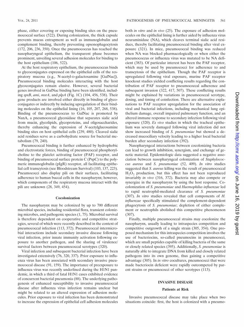
FIG. 1. (A) Mucus breakdown. S. pneumoniae colonization of the nasopharynx is facilitated by mucus degradation by the enzymes NanA, BgaA,StrH, and NanB. Ply decreases epithelial cell ciliary beating, enhancing bacterial adherence. (B) Evasion of proteolytic enzymes. Pneumococcalcell wall peptidoglycans may be destroyed by lysozyme. PdgA and Adr deacetylate pneumococcal cell surface petidoglycan molecules, renderingthem resistant to lysozyme. (C) Epithelial cell binding. S. pneumoniae binds host GalNac by using SpxB, Smi, MsrA, and PlpA. (D) Intracellulartranslocation. By binding the pIgR with PspC (or PAF receptor [PAFr] with ChoP), pneumococci can use the pIgR or PAF receptor recyclingpathway to be transported through the epithelial cell layer. (E) Inter- and pericellular translocation. Plasminogen bound by Gly3Ph, CbpE, andenolase enhances epithelial cell binding and degrades interepithelial adherens junctions, allowing pericellular migration.
VOL. 24, 2011 PATHOGENESIS OF PNEUMOCOCCAL MENINGITIS 559
on June 16, 2019 by guesthttp://cm
r.asm.org/
Dow
nloaded from
nition receptor SIGN-R1 had reduced activation of the classi-cal complement pathway (224). In this study, C1q was directlyactivated upon activation of SIGN-R1 by pneumococcal poly-saccharides in the spleen, leading to activation of the classicalcomplement cascade and complement component C3 activa-tion, with subsequent pneumococcal opsonization (224).SIGN-R1 is highly abundant on cells of the splenic red pulpand is an important factor in the spleen’s function to controlinvasive pneumococcal disease. Another study showed thatsplenic macrophages of SIGN-R1 knockout mice were unableto activate splenic B cells to produce pneumococcus-specificIgM (259). Therefore, splenic SIGN-R1-mediated activation ofB cells may explain, at least partially, the susceptibility ofsplenectomized patients to invasive pneumococcal disease.
Activation of C1q by the classical or mannose-binding lectin(MBL) pathway leads to cleavage of complement componentC2. In a Swedish cohort, 40 patients with a homozygous C2deficiency due to a deletion in the C2 gene were described(218). Invasive infections, mainly pneumococcal infections,were found in 23 (58%) of these patients (218).
The alternative pathway is also activated during infectionwith S. pneumoniae and occurs by the direct binding of com-plement component C3 to the pneumococcal surface (533).The importance of the alternative pathway in pneumococcalopsonization was shown in mice made deficient in factor D, apeptidase involved in activation of the alternative pathway(539). Opsonophagocytosis of S. pneumoniae was delayed infactor D-deficient mice compared to wild-type mice, indicatingan important role for this complement pathway in the earlyphase of infection (539). In line with this, a recent studyshowed that mice deficient in complement factor B, anotherpeptidase involved in activation of the alternative complementpathway, were more susceptible to pneumococcal otitis media(481).
The lectin-induced complement pathway appears to be lessimportant in pneumococcal disease than the classical and al-ternative pathways. Polymorphisms in MBL, one of the mostimportant activators of the lectin complement pathway, werenot associated with increased risk of pneumococcal invasivedisease in a genetic association study (331). A larger cohortshowed a significant increase in risk for pneumococcal invasivedisease, with three codon variants in the MBL locus (426). Ina third study, 140 patients with invasive pneumococcal disease,defined by positive blood culture for S. pneumoniae, were as-sessed for three structural variant MBL alleles and one pro-moter allele (269). In this study, no association was foundbetween susceptibility or outcome of invasive pneumococcaldisease and any of the structural MBL variants or promoteralleles. In a subgroup analysis of the 22 patients in the cohortwith pneumococcal invasive disease and meningitis, there wasno association between susceptibility or outcome and the MBLgenotype (269). However, a meta-analysis combining the re-sults of the above three studies demonstrated an associationbetween susceptibility to invasive pneumococcal disease andhomozygosity for one of the three structural variants in theMBL gene, with an odds ratio (OR) of 2.57 (95% confidenceinterval [CI], 1.38 to 4.80) (68). In a cohort of 57 HIV-positivepatients, an increased risk for invasive pneumococcal diseasewas found to be associated neither with MBL polymorphismsnor with polymorphisms in the downstream molecule MBL-
associated serine protease 2 (MASP-2) (203). One geneticassociation study has been performed regarding outcome andMBL genotypes. This study included only 60 patients withcommunity-acquired pneumococcal pneumonia and did notdetect an association between MBL genotype and outcome(138). Experimental studies showed weak to no binding ofMBL to S. pneumoniae compared to other bacteria (267, 352).Another experimental study showed that although MBL boundto S. pneumoniae, it did not increase opsonophagocytosis, andthat complement activation by the classical pathway was muchmore important (73).
Another group of proteins that can activate the lectin-in-duced complement pathway are ficolins. Two ficolin variants,H-ficolin and L-ficolin, have been studied for the capability ofbinding to S. pneumoniae; only L-ficolin was found to bindsome of the pneumococcal strains tested (267). However, nofrequency differences were found for polymorphisms in L-ficolin among 290 patients with invasive pneumococcal diseasecompared to 720 controls from a similar population (89).
The pneumococcus has evolved several strategies to limitcomplement-mediated opsonophagocytosis. The pneumococ-cal capsule plays a central role by limiting the amount ofcomplement deposited on the pneumococcal surface and im-peding the access to cell-bound complement (205). Further-more, pneumolysin has been shown to decrease complementopsonization of the pneumococcal cell (400). This is thought toresult from the consumption of complement factors by re-leased pneumolysin. In addition, several other pneumococcalouter surface proteins have been shown to affect complementdeposition on the pneumococcus, including pneumococcal sur-face protein C (PspC), PspA, PsaA, and PhpA (111, 213, 232,356, 399, 400, 411, 547).
PspC, also referred to as CbpA or SpsA, a choline-bindingprotein attached to the cell wall, is able to bind complementcomponent C3b, thereby preventing opsonization (111, 213,232, 399). Furthermore, PspC binds human factor H, a factorwhich inhibits activation of two complement components ofthe alternative and lectin pathways. By binding and activatingfactor H, the pneumococcus locally blocks the unfolding ofthese two complement pathways (110, 348, 399, 543). In addi-tion, PspC binds the complement inhibitor C4b-binding pro-tein, which blocks activation of the classical complement path-way (122). PspA has been shown to interfere with the bindingof complement component C3 on the bacterial surface, therebyinhibiting complement-mediated opsonization (356, 400, 411).PhpA is a pneumococcal surface protein with C3-degradingproperties (547). Since activation of the complement cascade iscrucial in the defense against pneumococcal invasive disease,pneumococcal complement binding proteins are important tar-gets for vaccine development (65, 109, 147, 336).
Binding to Epithelium
The pneumococcal capsule is advantageous in circumventingthe host barriers and reaching the respiratory mucosa but cov-ers PCW binding sites for epithelial cell binding. The pneumo-coccus adjusts its binding properties to its environmentthrough a process called phase variation (106, 296, 522). In thisprocess, the amount of polysaccharide in the capsule variesfrom an opaque (thick capsule) to a transparent (thin capsule)
560 MOOK-KANAMORI ET AL. CLIN. MICROBIOL. REV.
on June 16, 2019 by guesthttp://cm
r.asm.org/
Dow
nloaded from
phase, either covering or exposing binding sites on the pneu-mococcal surface (522). During colonization, the thick capsuleprevents mucus entrapment as well as immunoglobulin andcomplement binding, thereby preventing opsonophagocytosis(172, 206, 236, 350). Once the pneumococcus has reached thenasopharyngeal epithelium, the transparent phase becomesprominent, unveiling several adhesion molecules for binding tothe host epithelium (106, 522).
At the host respiratory epithelium, the pneumococcus bindsto glycoconjugates expressed on the epithelial cells of the res-piratory mucosa (e.g., N-acetyl-D-galactosamine [GalNac]).Pneumococcal binding molecules interacting with the hostglycoconjugates remain elusive. However, several bacterialgenes involved in GalNac binding have been identified, includ-ing spxB, ami, msrA, and plpA (Fig. 1C) (104, 456, 538). Theirgene products are involved either directly in binding of glyco-conjugates or indirectly by inducing upregulation of their bind-ing molecules on the epithelial lining (16, 105, 207, 268, 538).Binding of the pneumococcus to GalNac is promoted byNanA, a pneumococcal glycosidase that separates sialic acidfrom mucin, glycolipids, glycoproteins, and oligosaccharides,thereby enhancing the expression of N-acetylglucosaminebinding sites on host epithelial cells (239, 480). Cleaved sialicacid residues serve as a carbohydrate source for bacterial me-tabolism (79, 240).
Pneumococcal binding is further enhanced by hydrophobicand electrostatic forces, binding of pneumococcal phosphoryl-choline to the platelet activating factor (PAF) receptor, andbinding of pneumococcal surface protein C (PspC) to the poly-meric immunoglobulin (pIgR) receptor, all facilitating epithe-lial cell transcytosis (see Bloodstream Survival) (103, 137, 223).Pneumococci also display pili on their surfaces, facilitatingadherence to human buccal cells in the nasopharynx; however,which components of the respiratory mucosa interact with thepili are unknown (28, 349, 454).
Cocolonization
The nasopharynx may be colonized by up to 700 differentmicrobial species, including residential flora, transient coloniz-ing microbes, and pathogenic species (1, 75). Microbial survivalis therefore dependent on cooperative and competitive strat-egies, several of which were recently described in the context ofpneumococcal infection (113, 372). Pneumococcal intermicro-bial interactions include secondary invasive disease followingviral infection, prior innate immunity activation following ex-posure to another pathogen, and the sharing of virulence/survival factors between pneumococcal serotypes (320).
Viral infection and subsequent bacterial infection have beeninvestigated extensively (76, 320, 337). Prior exposure to influ-enza virus has been associated with secondary invasive pneu-mococcal disease (91, 159). The importance of preexposure toinfluenza virus was recently underlined during the H1N1 pan-demic, in which a third of fatal H1N1 cases exhibited evidenceof concurrent bacterial pneumonia (88). The underlying patho-genesis of enhanced susceptibility to invasive pneumococcaldisease after influenza virus infection remains unclear butmight be related to an altered expression of adhesion mole-cules. Prior exposure to viral infection has been demonstratedto increase the expression of epithelial cell adhesion molecules
both in vitro and in vivo (25). The exposure of adhesion mol-ecules on the epithelial lining is further aided by influenza virusneuraminidase (NA), which cleaves terminal sialic acid resi-dues, thereby facilitating pneumococcal binding after viral ex-posure (321). In mice, pneumococcal binding was reducedwhen NA was blocked pharmacologically or when either thepneumococcus or influenza virus was mutated to be NA defi-cient (383). Of particular interest has been the PAF receptor,which may be used by pneumococci for adherence to andtranscytosis of the epithelium. Though the PAF receptor isupregulated following viral exposure, murine PAF receptorknockout studies yielded conflicting results regarding the con-tribution of PAF receptor to pneumococcal adherence andsubsequent invasion (322, 417, 507). These conflicting resultsmight be explained by variations in pneumococcal serotype,dosing, and timing of coinfection. There are alternative expla-nations to PAF receptor upregulation for the association ofviral and bacterial infections, including mechanical lung epi-thelium damage, overall impaired pulmonary function, and analtered immune response to secondary infection following viralexposure (320). Ex vivo studies in which the tracheal epithe-lium was severely damaged following viral infection did notshow increased binding of S. pneumoniae but showed a de-creased mucociliary velocity leading to a higher local bacterialburden after secondary infection (392).
Nasopharyngeal interactions between cocolonizing bacteriacan lead to growth inhibition, synergism, and exchange of ge-netic material. Epidemiologic data suggested a negative asso-ciation between nasopharyngeal colonization of Staphylococ-cus aureus and S. pneumoniae (52, 409). In vitro studiessuggested that S. aureus killing was the result of pneumococcalH2O2 production, but this effect has not been reproducedinvariably in vivo (316, 372). Bacteria may also compete orsynergize in the nasopharynx by using the host response. Co-colonization of S. pneumoniae and Haemophilus influenzae ledto rapid neutrophil-mediated clearance of S. pneumoniae(307). In vitro studies revealed that cell components of H.influenzae specifically stimulated the complement-dependentphagocytosis of S. pneumoniae; depletion of either comple-ment or neutrophils abolished this competitive phenomenon(307).
Finally, multiple pneumococcal strains may cocolonize thenasopharynx, usually leading to intraspecies competition andcompetitive outgrowth of a single strain (305, 354). One pro-posed mechanism for this intraspecies competition involves theuse of bacteriocins, so-called pneumocins in pneumococci,which are small peptides capable of killing bacteria of the sameor closely related species (395). Additionally, S. pneumoniae isnaturally able to integrate DNA from killed and closely relatedpathogens into its own genome, thus gaining a competitiveadvantage (305). In in vitro cocultures, pneumococci that weremade bacteriocin deficient were rapidly outcompeted by par-ent strains or pneumococci of other serotypes (113).
INVASIVE DISEASE
Patients at Risk
Invasive pneumococcal disease may take place when twosituations coincide: first, the host is colonized with a pneumo-
VOL. 24, 2011 PATHOGENESIS OF PNEUMOCOCCAL MENINGITIS 561
on June 16, 2019 by guesthttp://cm
r.asm.org/
Dow
nloaded from

coccal strain that it has not yet established immunity to, andsecond, an alteration of the natural barriers or host immunesystem has occurred (49, 312). Invasive pneumococcal diseaseis seen during the extremes of age (less than 2 or more than 50years of age); in patients with underlying conditions, such assplenectomy or asplenic states, sickle cell disease, multiplemyeloma, hypogammaglobulinemia, alcoholism, chronic liveror kidney disease, malignancy, malnutrition, Wiskott-Aldrichsyndrome, thalassemia major, diabetes mellitus, and basilarskull fracture with leakage of cerebrospinal fluid (CSF); and inchildren with cochlear implants (3, 19, 42, 71, 161, 265, 329,341, 364, 422, 497, 498, 524, 527). The use of immunosuppres-sive drugs, a history of splenectomy, or the presence of diabe-tes mellitus, alcoholism, or infection with HIV is found in 20%of adults with pneumococcal meningitis (364, 524). Further-more, damage to the naso- and oropharyngeal mucosae may beelicited by local pneumococcal infection, such as sinusitis orotitis, by viral respiratory infections (specifically by influenzavirus [see “Cocolonization”), by smoking, or by allergy (219,355, 519, 528).
Invading Host Endothelial and Epithelial Cells
Pneumococci are relatively ineffective at invading host en-dothelial and epithelial cells. However, pressures of the hostnatural barriers, cocolonization of other micoorganisms, andan activated innate immune response drive pathogens to de-velop new strategies. Epithelial endo- and transcytosis is animportant strategy of invasion and also allows intraepithelialbacterial reservoirs and subsequent recolonization of the na-sopharynx. Two mechanisms of epithelial transmigration by S.pneumoniae have been described (Fig. 1D). First, pneumococ-cal phosphorylcholine (ChoP) may bind to the PAF receptoron activated epithelial and endothelial cells (103). ChoP is acomponent of cell wall-associated acids and lipoteichoic acids(LTAs) on the surfaces of transparent pneumococci (221). Bybinding the PAF receptor, the pneumococcus may enter thePAF receptor recycling pathway, which transports the bacte-rium to the basal membrane of the host epithelial cell, whichmay lead to invasive disease (103, 402). Intranasal challenge ofmice deficient in the PAF receptor resulted in reduced rates ofpneumococcal colonization, pneumonia, and invasive disease(417).
A second mechanism involves the binding of the pneumo-coccal choline-binding protein PspC (also known as CbpA orSpsA) to the extracellular portion of epithelial pIgR, refered toas “secretory component” (137, 223). Following attachment,the pneumococcus uses the pIgR recycling pathway, analogousto the PAF receptor pathway, to be transported between theapical and basal membranes of the epithelial cell (223, 546).Pneumococcal expression of PspC has been shown to be animportant factor for colonization and invasive disease, al-though its effect on virulence may vary between pneumococcalstrains (67, 190, 232, 424, 546). The PspC binding of pIg re-ceptor is observed only in humans, not in mice, rats, or rabbits(223). In addition, PspC also binds sialic acid and lacto-N-neotetraose on respiratory epithelial cells, further facilitatingcolonization (424). The level of pIg receptor directly correlateswith the degree of pneumococcal attachment and epithelialinvasion (546). pIg receptors are expressed in a decreasing
gradient from the upper to the lower respiratory tract, whilethe opposite pattern is observed for the PAF receptor (325,546). Therefore, it has been suggested that where pIg receptorserves mainly as a pneumococcal receptor in the nasopharynx,the PAF receptor acts as a ligand for attachment and invasionof the pulmonary epithelium (546).
Inter- or pericellular migration is another mechanism bywhich bacteria may cross epithelial or endothelial cell layers(Fig. 1E) (371). Plasminogen, bound by the pneumococcalreceptors enolase, Gly3Ph, and CbpE, plays a central role inthis process and has been shown to serve two purposes (24, 35,36). First, plasminogen increases adhesion of pneumococci tothe epithelial surface (23). Second, bound plasmin is able tocleave proteins involved in the intercellular adherens junctions,which bind epithelial cells together to form a mechanical bar-rier to underlying tissues (23). This disruption is mediated bythe degradation of cadherin, an essential component of inte-repithelial adherens junctions (23). Murine pneumococcal na-sopharyngeal colonization studies demonstrated that epithelialbarrier function was diminished through the downregulation ofcadherins in a Toll-like receptor (TLR)-dependent manner(32). Third, epithelial permeability is also modulated by theinnate immune system in a transforming growth factor beta(TGF-�)-dependent manner, possibly to allow for adequatemigration of immune cells and inflammatory mediators intoinfected areas (31). Thus, the breakdown of the tight junctions,though necessary for an adequate immune response, may allowfor pneumococcal access to the basal membrane and subse-quent invasive disease.
Extracellular Matrix
At the basal side of the epithelium or endothelium lies thebasement membrane, which is comprised mainly of a networkof collagen type I, laminin, and proteoglygans (9). Like manybacteria, pneumococci use hyaluronan lyase to degrade majorcomponents of the extracellular matrix (ECM), hyaluronan,and certain chondroitins, thereby facilitating invasive disease(215). The importance of hyaluronan lyase for the develop-ment of invasive pneumococcal disease was demonstrated inmice, as intranasally administered hyaluronidase adjuvant en-hanced the development of invasive disease after an otherwisenoninvasive intranasal inoculation of pneumococci (552).Moreover, pneumococci isolated from patients with pneumo-coccal meningitis expressed higher levels of hyaluronidase thanpneumococci isolated from asymptomatic carriers (263).
Fibronectin, a large multidomain ECM glycoprotein, isfound in nearly every human tissue environment that the pneu-mococcus is likely to encounter and is bound by several pneu-mococcal adhesins, among which the most important are thepneumococcal adhesion and virulence A (PavA) and B (PavB)proteins (200, 216). In murine infection models, PavA-defi-cient pneumococci had impaired adherence to murine epithe-lium and endothelial cells and were unable to sustain long-term nasopharyngeal colonization (220, 394). Furthermore,although pneumococci lacking PavA showed similar growth toWT pneumococci in a sepsis model, PavA mutants were rap-idly cleared from the central nervous system (CNS) after in-tracranial infections (220). Possibly, PavA not only serves todirectly bind fibronectin but also plays a role in the effective
562 MOOK-KANAMORI ET AL. CLIN. MICROBIOL. REV.
on June 16, 2019 by guesthttp://cm
r.asm.org/
Dow
nloaded from
adherence and virulence mediated by other, so far unknowndeterminants (394).
BLOODSTREAM SURVIVAL
Complement System
Once in the bloodstream, pneumococci are confronted withadditional host defense mechanisms. Complement represents thefirst step of innate immunity against bacteremia. The classicalcomplement pathway plays a dominant role in pneumococcalclearance, although the classical and alternative complementpathways are also activated by streptococcal species (214, 374).Pneumococci have developed two ways to minimize complement-mediated opsonization and phagocytosis. First, pneumococci un-dergo a second phase variation and become encapsulated. Thepolysaccharide capsule serves as a nonspecific barrier, signifi-cantly reducing complement deposition on the bacterial surfaceand limiting subsequent interaction with phagocytes (2, 221). Inmurine studies, systemically administered unencapsulated pneu-mococci were shown to be avirulent (395).
Second, pneumococcal surface proteins PspA, PspC, andpneumolysin target specific complement components, therebyreducing complement-mediated bacterial clearance. PspA,which is expressed ubiquitously among pneumococci, inhibitsC1q and subsequent C3b deposition (214). PspC binds humanfactor H, thereby blocking the formation of C3 convertase(C3bBb), leading to lower C3b production and limiting op-sonophagocytosis (292, 548). Pneumococci can also attach toerythrocytes through a process called immune adherence,which is dependent on the binding of complement componentsC3b, C4b, C1q, and MBL to both the pneumococcus anderythrocyte receptor CR1 (189, 292, 351). Immune complexescontaining pneumococci, bound by complement to erythro-cytes, are then transferred to macrophages, after which theerythrocytes are returned to the circulation (99). Recent invitro studies showed that PspA and PspC work synergisticallyto limit complement-mediated adherence and transfer tophagocytes (292).
Pneumolysin, released during pneumococcal autolysis, read-ily binds the Fc portion of IgG, thereby potently activating theclassical complement pathway, increasing bacterial virulenceby independently depleting complement factors away from thebacterium, and limiting opsonophagocytosis (13). Murine bac-teremia studies showed that pneumolysin-deficient pneumo-cocci are either cleared from the bloodstream or allowed todevelop into chronic bacteremia (359). Furthermore, serumcomplement depletion may be particularly important in cir-cumstances of overall limited complement availability, such asliver cirrhosis (12), and may further increase pneumococcalvirulence at sites of limited complement presence, such as thenasopharynx (318).
Lastly, the acute-phase CRP binds phosphorylcholine (Chop)on the PCW (4, 514) and subsequently interacts with C1q, leadingto the activation of the classical complement pathway (93, 451). Inmice, CRP is not an acute-phase protein, and treatment withhuman CRP reduced mortality following pneumococcal infection(467, 468). In vitro studies showed that CRP reduced pneumo-coccal binding to the epithelial cell PAF receptor (175).
Recognition by the Host Immune System
Pneumococci are recognized by APCs through the bindingof pattern recognition receptors, which are specifically directedtoward general motifs of molecules expressed by pathogensthat are essential for pathogen survival. Pattern recognitionreceptors involved in sensing pneumococci include TLR2 (192,286, 333, 441, 511, 541), TLR4 (59, 278, 311), TLR9 (10, 278,333), and nucleotide oligomerization domain 1 (Nod1) (357,549). Upon activation of these receptors, APCs release variouscytokines, which induce a cascade of inflammatory reactions,including the recruitment of neutrophils (211). The most im-portant cytokines released by phagocytic cells are tumor ne-crosis factor alpha (TNF-�), interleukin-1 (IL-1), and IL-6(419). IL-1� and TNF-� act on local vascular endothelial cells,increasing vascular permeability and vasodilatation and up-regulating adhesion molecules such as E-selectin, P-selectin,and vascular cell adhesion molecule 1 (VCAM-1) to enable theinflux of neutrophils and other lymphocytes from the blood tothe site of infection (Fig. 2) (142, 470).
Initiation of Coagulation
Most patients with invasive pneumococcal disease show ev-idence of coagulation activation (288, 313). Inflammation-in-duced thrombin generation is not dependent on direct inter-action of bacteria and the coagulation cascade but rather onthe exposure of blood to tissue factor (TF) (290). TF is ex-pressed primarily on cells outside the vasculature (128, 149)and is exposed to coagulation factors during vascular damage.Low levels of circulating TF have been detected in healthyindividuals (167), in whom the role of TF in thrombin gener-ation remains uncertain (81, 195, 407). The expression of TF inblood cells is limited to monocytes and can be elevated con-siderably during inflammation or sepsis (370). The upregula-tion of TF is largely IL-6 dependent, as studies have shownabrogation of TF-dependent thrombin generation when IL-6 isblocked (506).
Upon exposure to blood, TF forms a complex with factorVII and catalyzes the conversion of factor X into factor Xa.Factor Xa allows prothrombin conversion to thrombin, al-though this reaction occurs to a significant extent only afterthrombin-induced feedback activation of factor VIII and factorV, nonenzymatic cofactors in the tenase and prothrombinasecomplexes, respectively (81, 290). The prothrombinase andtenase complexes convert prothrombin (factor II) into throm-bin (factor IIa), which then leads to the conversion of fibrin-ogen to the clot-forming fibrin protein (289). The activity ofprothrombinase and tenase complexes is markedly enhancedby the presence of activated platelets, which become activatedduring inflammation but may also be activated directly bythrombin itself (427).
Inflammation-mediated thrombin formation is regulated bythree anticoagulant mechanisms: antithrombin (AT), the pro-tein C system, and tissue factor pathway inhibitor (TFPI), all ofwhich may be impaired during systemic infection (290). Anti-thrombin inhibits thrombin and factor Xa, though during se-vere infection antithrombin levels are markedly lower due toimpaired synthesis, degradation, and consumption duringthrombin generation (291). Circulating protein C, which upon
VOL. 24, 2011 PATHOGENESIS OF PNEUMOCOCCAL MENINGITIS 563
on June 16, 2019 by guesthttp://cm
r.asm.org/
Dow
nloaded from
conversion to activated protein C by the thrombin-thrombo-modulin complex degrades the essential coagulation factors Vaand VIIIa, is hampered during severe inflammation by enzy-matic degradation by neutrophil-derived elastase and by im-paired synthesis as well as decreased activation by depressedlevels of thrombomodulin (135, 143). Lastly, the importance ofTFPI has been demonstrated in studies in healthy human vol-unteers injected with endotoxin, in whom administration ofTFPI induced a marked inhibition of coagulation (117). Ani-mal studies showed that rabbits deficient in TFPI were moresusceptible to severe disseminated intravascular coagulation(DIC), and primates infused with TFPI were able to surviveexposure to otherwise lethal amounts of Escherichia coli (404).
The degradation of fibrin clots is mediated by plasmin, theactive form of plasminogen, which is activated by tissue-typeplasminogen activator (tPA) and urokinase-type plaminogenactivator (uPA), both of which are stimulated by the inflam-matory cytokines TNF-� and IL-1� (505). During severe in-fection, these cytokines subsequently induce plasminogen ac-tivator inhibitor type 1 (PAI-1), thereby limiting fibrinolysisand resulting in a net procoagulant state (505). Higher levels ofPAI-1 in patients with meningococcal septicemia or dissemi-
nated intravascular coagulation have been shown to be associ-ated with poor outcomes and mortality (326, 530).
At relatively high concentrations, thrombin forms a complexwith thrombomodulin and activates thrombin-activatable fibri-nolysis inhibitor (TAFI; also known as plasma carboxypepti-dase B, carboxypeptidase U, and carboxypeptidase R) (48,518). Activated TAFI inhibits fibrinolysis by limiting plasminformation through the inhibition of plasminogen and tPA in-corporation into fibrin clots (338). Furthermore, TAFI is ableto inhibit several proinflammatory substrates, such as brady-kinin and complement components C3 and C5a (84). Theimportance of TAFI and C5a was first demonstrated in amouse model in which TAFI knockout mice showed a highermortality when challenged with sublethal doses of lipopolysac-charide (LPS) and cobra venom factor (22).
CENTRAL NERVOUS SYSTEM INVASION
Intracellular Translocation across the Blood-Brain Barrier
Cerebral vascular endothelial cells show marked differencesfrom their systemic counterparts. They exhibit very tight junc-
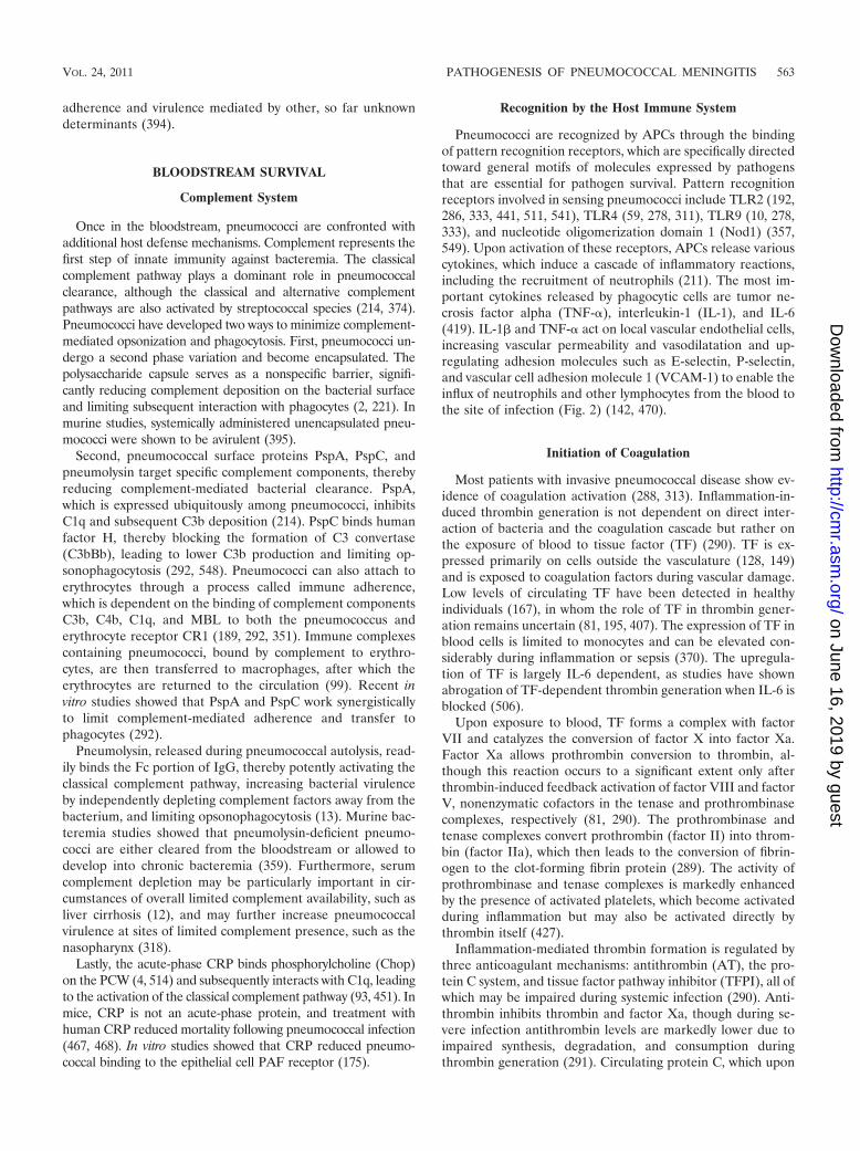
FIG. 2. S. pneumoniae adheres to endothelial cells by using PspC, which binds laminin and pIgR, enabling transcytosis across the endothelium. Oncein the CSF, pneumococci multiply freely and release bacterial products such as LTA and Ply, which are recognized by TLR2 and TLR4 on circulatingAPCs. The subsequent release of proinflammatory cytokines and chemokines from macrophages and microglial cells results in upregulation ofendothelial cell P- and E-selectin and ICAM (which binds MAC-1 on leukocytes), leading to increased neutrophil recruitment into the CSF.
564 MOOK-KANAMORI ET AL. CLIN. MICROBIOL. REV.
on June 16, 2019 by guesthttp://cm
r.asm.org/
Dow
nloaded from
tions, low rates of pinocytosis, and relatively large numbers ofmitochondria (398). In human brain microvascular endothelialcell cultures, the pneumococcus was able to adhere to thevascular endothelial PAF receptor, allowing transmigrationthrough the endothelial cell to the basolateral site (418). Thismechanism of transcytosis is similar to that seen at the pulmo-nary epithelium (see Invasive Disease) and is mediated bybinding of pneumococcal phosphorylcholine to the PAF recep-tor (103, 417). Pneumococci in the transparent phase are moreefficient at invading the brain endothelial cell layer thanopaque variants, which are dependent on the expression ofphosphorylcholine (418). Concordantly, PAF receptor-defi-cient mice showed less translocation of pneumococci across theblood-brain barrier and, therefore, a decreased incidence ofpneumococcal meningitis after intravenous challenge (402).Many of these studies have been performed with brain vascularendothelial cells. However, another important site of entrymight be the choroid plexus epithelium, as shown for Strepto-coccus suis, which induces epithelial cell death and blood-brainbarrier disruption in porcine choroid plexus epithelium (473)but may also translocate intracellularly across the plexus epi-thelium (474).
Nasopharyngeal colonization models demonstrated bindingof pneumococcal PspC to pIgR on local epithelial cells, facil-itating pneumococcal invasion (546). However, in a cell line ofhuman brain microvascular endothelial cells, the pIgR was notexpressed (546). In vitro and animal experiments showed thatpneumococcal PspC may bind the laminin receptor on brainmicrovascular endothelial cells (360). This receptor, by whichendothelial cells are bound to the major component of base-ment membranes, laminin, was also shown to be a ligand forneurotropic viruses and prions (6, 158, 360). Laminin appearsto be involved in binding of bacteria that may cause meningitis,such as S. pneumoniae, N. meningitidis, and H. influenzae, tobrain microvascular endothelial cells (360). PneumococcalPspC binds to laminin, and in a mouse model of pneumococcalsepsis, a pneumococcal PspC mutant caused a decreased fre-quency of pneumococcal meningitis (360). These results indi-cate that the interaction between laminin and pneumococcalPspC plays a role in intracellular translocation of pneumococciacross the blood-brain barrier.
Intercellular Translocation across the Blood-Brain Barrier
Pneumococci may translocate into the CSF intercellularly,by disruption of the interepithelial tight junctions. In an animalmodel of pneumococcal meningitis, tight junctions betweenbrain microvascular endothelial cells became disrupted in thecourse of the disease (398). This may be due to damage causedby the pneumococcus or by factors of the host immune re-sponse (153, 448, 558). Analogous to the nasopharyngeal set-ting, pneumolysin was capable of disrupting an endothelial celllayer in an in vitro endothelial cell culture, which may enhanceblood-brain barrier disruption in vivo (558).
After crossing the dense vascular endothelial cell lining,pneumococci have several methods of disrupting and invadingthe basement membrane. The first involves binding of plasmin-ogen to the bacterial surface, which may subsequently be ac-tivated by tPA (129). In patients with bacterial meningitis,levels of uPA correlated with breakdown of the blood-brain
barrier and pleocytosis (536). In vitro models showed thatpneumococcus-mediated activation of plasminogen resulted indamage of extracellular matrix components and the basementmembrane (129), although conversely, an in vivo mouse modelfailed to demonstrate an effect of tPA or uPA receptor onpneumococcal transmigration across the blood-brain barrier(379). Finally, pneumococci may bind fibronectin (502), vitro-nectin, and collagen in the extracellular matrix, which mayenhance blood-brain barrier disruption (34, 262).
CENTRAL NERVOUS SYSTEM IMMUNE RESPONSE
Immune Activation
During multiplication, pneumococci concurrently undergoautolysis, which eventually leads to a stationary phase wheremultiplication and autolysis rates are similar (479). The re-leased bacterial products are highly immunogenic and maylead to an increased inflammatory response in the host (489).Bactericidal antibiotics causing bacterial lysis may also inducea similar effect and lead to a temporarily increased host in-flammatory response and increased disease severity (344, 345,483).
A variety of pneumococcal compounds are proinflamma-tory. The pathophysiological aspects of the different com-pounds may be reproduced by intracisternal inoculation ofheat-killed unencapsulated pneumococci, purified PCW, cellwall lipoteichoic acid, or cell wall peptidoglycan (490). Heat-killed encapsulated pneumococci or purified pneumococcalcapsular polysaccharides inoculated intracisternally into rab-bits did not cause meningitis, indicating that the pneumococcalcapsule is not immunogenic in the CSF (490). Inoculation withknockout pneumococcal strains is another way to study theimmunogenicity of pneumococcal compounds. In a murinemodel of pneumococcal meningitis, intracisternal inoculationwith pneumolysin-deficient pneumococci resulted in lower bac-terial loads, better clinical scores, and longer survival of thehost (529). However, histological inflammatory changes in thisstudy were similar to those induced by wild-type pneumococci(529).
Anatomical Localization of Blood-Brain BarrierInvasion by Leukocytes
Neutrophils are thought to cross the blood-brain barriermainly at the venous side of the penetrating cerebral bloodvessels (182). Here they migrate to the perivascular space,which is continuous with the subarachnoid space. However,some neutrophils penetrate the brain parenchyma. Neutro-philic infiltrates in the brain have been seen primarily in spacesadjacent to CSF, such as the corpus callosum, periventricularspace, and the meninges (482). Neutrophils mediate bacterialkilling by phagocytosis of opsonized bacteria (211). Phagocy-tosis is initiated by recognition and binding of bacteria by aneutrophil and is facilitated by opsonization of the bacteria bycomplement and antibody. Following binding, the neutrophilengulfs the bacteria, after which the cell membrane closesaround the pathogens and is cut off, forming a free membrane-covered entity within the cell called an endosome (211). In theactivated neutrophil, the endosome containing the pathogens
VOL. 24, 2011 PATHOGENESIS OF PNEUMOCOCCAL MENINGITIS 565
on June 16, 2019 by guesthttp://cm
r.asm.org/
Dow
nloaded from
is fused with a lysosome present in the cell, which containsseveral bactericidal mediators, including nitric and oxygen spe-cies, but also activated lysozymes, and the bacteria are killed.In addition to intracellular killing, neutrophils also secretenitric and oxygen species, establishing a bactericidal milieuaround the cell (211). Adversely, these nitric and oxygen spe-cies may damage the surrounding tissue when they are presentin large amounts and may be responsible, at least in part, forthe neuronal damage seen in pneumococcal meningitis. Thistopic is discussed further in Neuronal Damage and Histopa-thology.
Pattern Recognition Receptors
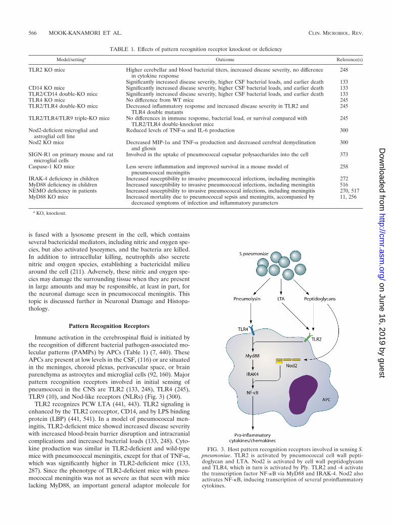
Immune activation in the cerebrospinal fluid is initiated bythe recognition of different bacterial pathogen-associated mo-lecular patterns (PAMPs) by APCs (Table 1) (7, 440). TheseAPCs are present at low levels in the CSF, (116) or are situatedin the meninges, choroid plexus, perivascular space, or brainparenchyma as astrocytes and microglial cells (92, 160). Majorpattern recognition receptors involved in initial sensing ofpneumococci in the CNS are TLR2 (133, 248), TLR4 (245),TLR9 (10), and Nod-like receptors (NLRs) (Fig. 3) (300).
TLR2 recognizes PCW LTA (441, 443). TLR2 signaling isenhanced by the TLR2 coreceptor, CD14, and by LPS bindingprotein (LBP) (441, 541). In a model of pneumococcal men-ingitis, TLR2-deficient mice showed increased disease severitywith increased blood-brain barrier disruption and intracranialcomplications and increased bacterial loads (133, 248). Cyto-kine production was similar in TLR2-deficient and wild-typemice with pneumococcal meningitis, except for that of TNF-�,which was significantly higher in TLR2-deficient mice (133,287). Since the phenotype of TLR2-deficient mice with pneu-mococcal meningitis was not as severe as that seen with micelacking MyD88, an important general adaptor molecule for
TABLE 1. Effects of pattern recognition receptor knockout or deficiency
Model/settinga Outcome Reference(s)
TLR2 KO mice Higher cerebellar and blood bacterial titers, increased disease severity, no differencein cytokine response
248
Significantly increased disease severity, higher CSF bacterial loads, and earlier death 133CD14 KO mice Significantly increased disease severity, higher CSF bacterial loads, and earlier death 133TLR2/CD14 double-KO mice Significantly increased disease severity, higher CSF bacterial loads, and earlier death 133TLR4 KO mice No difference from WT mice 245TLR2/TLR4 double-KO mice Decreased inflammatory response and increased disease severity in TLR2 and
TLR4 double mutants245
TLR2/TLR4/TLR9 triple-KO mice No differences in immune response, bacterial load, or survival compared withTLR2/TLR4 double-knockout mice
245
Nod2-deficient microglial andastroglial cell line
Reduced levels of TNF-� and IL-6 production 300
Nod2 KO mice Decreased MIP-1� and TNF-� production and decreased cerebral demyelinationand gliosis
300
SIGN-R1 on primary mouse and ratmicroglial cells
Involved in the uptake of pneumococcal capsular polysaccharides into the cell 373
Caspase-1 KO mice Less severe inflammation and improved survival in a mouse model ofpneumococcal meningitis
258
IRAK-4 deficiency in children Increased susceptibility to invasive pneumococcal infections, including meningitis 272MyD88 deficiency in children Increased susceptibility to invasive pneumococcal infections, including meningitis 516NEMO deficiency in patients Increased susceptibility to invasive pneumococcal infections, including meningitis 270, 517MyD88 KO mice Increased mortality due to pneumococcal sepsis and meningitis, accompanied by
decreased symptoms of infection and inflammatory parameters11, 256
a KO, knockout.
FIG. 3. Host pattern recognition receptors involved in sensing S.pneumoniae. TLR2 is activated by pneumococcal cell wall pepti-doglycan and LTA. Nod2 is activated by cell wall peptidoglycansand TLR4, which in turn is activated by Ply. TLR2 and -4 activatethe transcription factor NF-�B via MyD88 and IRAK-4. Nod2 alsoactivates NF-�B, inducing transcription of several proinflammatorycytokines.
566 MOOK-KANAMORI ET AL. CLIN. MICROBIOL. REV.
on June 16, 2019 by guesthttp://cm
r.asm.org/
Dow
nloaded from
TLR signaling, it was proposed that other TLRs besides TLR2may play a role in sensing pneumococci in the CNS (248, 256).
TLR4 recognizes pneumococcal pneumolysin (311). TLR4-deficient mice did not differ significantly from wild-type mice intheir host immune response, cerebrovascular changes, or out-come during pneumococcal meningitis (245). However, in micedeficient in both TLR2 and TLR4, a marked reduction ininflammatory mediators, increased bacterial replication in theCNS, and reduced survival were seen compared to those forwild-type mice or mice with a single TLR deficiency (245).Thus, in meningitis, both TLR2 and TLR4 are important re-ceptors in detecting the pneumococcus and initiating a robustinflammatory response to the pathogen, and one receptor maycompensate for the absence of the other (245).
TLR9 is an intracellular pattern recognition receptor and isactivated by CpG repeats in bacterial DNA (196). In vitro, S.pneumoniae was able to activate alveolar and peripheral mac-rophages through TLR9 and induced IL-8 production inTLR9-transfected human embryonic kidney cells (10, 333). Invivo, TLR9-deficient mice showed reduced resistance to S.pneumoniae after intranasal challenge (10). However, in amodel of pneumococcal meningitis, triple mutant TLR2/TLR4/TLR9-deficient mice did not show significant differencesin immune response, bacterial load, or survival compared withTLR2/TLR4-deficient mice (245). Therefore, TLR9 appears toplay a minor role in pneumococcal meningitis, although thiswas assessed only in TLR triple-knockout mice.
NLRs are a second group of intracellular pattern recogni-tion receptors involved in detecting pneumococci (357). NLRsbelong to a family of receptors which, upon activation, induceactivation of NF-�B or mitogen-activated protein kinase(MAPK) pathways and inflammatory caspases (357). In humanembryonic kidney 293 cells, Nod2 was activated by internalizedpneumococci through sensing of meso-diaminopimelic acid(meso-DAP) motifs of the bacterial peptidoglycan (151, 357).In vitro experiments showed that microglial and astroglial cellsare activated by S. pneumoniae through Nod2 (300). Murinemicroglial and astroglial cells deficient in Nod2 showed re-duced levels of TNF-� and IL-6 production (300). With in vivoexperiments using a pneumococcal meningitis model, Nod2activation of primary murine glial cells induced macrophageinflammatory protein 1� (MIP-1�) and TNF-� production andenhanced cerebral demyelination and gliosis (300). Thus, ac-tivation of Nod2 appears to be one of the contributing factorsleading to cerebral damage in bacterial meningitis.
Another group of NLRs are the inflammasomes, which in-clude a complex of various pattern recognition receptors shar-ing the caspase adaptor apoptosis-associated speck-like pro-tein (ASC) and leading to caspase-1 activation when triggered(150). Cleavage and activation of caspase-1 lead to cleavage ofdifferent procytokines into their active forms, including IL-1�and IL-18 (8, 123, 408). In addition, inflammasome activationmay lead to a specific form of controlled cell death, differentfrom apoptosis, called pyroptosis (146). Inflammasomes areintracellular pattern recognition receptors and can be activatedby several endogenous and exogenous ligands, including bac-teria (486), bacterial DNA (237), bacterial toxins (187), endog-enous reactive oxygen species (ROS) produced by macro-phages in response to infection (431), and uric acid releasedthrough cell injury during inflammation (157). Little is known
about the role of inflammasomes in bacterial meningitis. Inpatients suffering from bacterial meningitis, cerebrospinal fluidlevels of caspase-1 were increased (258). In children with bac-terial meningitis, as well as a rat model of pneumococcal men-ingitis, increased IL-1� levels were measured in the CSF (30,86). Koedel et al. showed that mice lacking caspase-1 displayedless severe inflammation and improved survival in a pneumo-coccal meningitis mouse model (258). Similar results werefound in a pneumococcal meningitis model with IL-18 knock-out mice (554), indicating a role for inflammasome activationin the pathophysiology of pneumococcal meningitis.
A fourth group of pathogen recognition receptors involvedin sensing S. pneumoniae are the C-type lectins, which arehighly expressed on splenic dendritic cells and also on perito-neal macrophages (276). A member of this group, SIGN-R1,was shown to facilitate phagocytosis by recognition of thepneumococcal capsular polysaccharide (225, 276). Mice lack-ing functional SIGN-R1 fail to effectively phagocytose S. pneu-moniae, leading to an inability to clear the infection and re-sulting in increased inflammatory parameters and reducedsurvival in both a model of pneumococcal peritoneal sepsis(276) and one of intranasally induced pneumonia (260). Fur-thermore, SIGN-R1 plays a role in the activation of the clas-sical complement pathway by binding C1q (224). Park et al.showed the presence of SIGN-R1 on microglial cells in mouseand rat brains, which was functionally active in taking up pneu-mococcal capsular polysaccharides into the cell (373). There-fore, SIGN-R1 may be an important pathogen recognitionreceptor in the brain during pneumococcal meningitis.
Downstream Signaling Molecules
Upon stimulation of one of the above pattern recognitionreceptors, an intracellular cascade is activated and leads to theproduction of inflammatory molecules, usually cytokines orchemokines, which modulate the immune response by activat-ing or attracting specialized immune cells. Deficiencies andpolymorphisms in the pathogen recognition receptor down-stream signaling cascade in humans have been associated withinvasive pneumococcal disease, including meningitis.
The most extensively characterized TLR downstream signal-ing protein in pneumococcal invasive disease is IRAK-4 (Fig.3) (391). This adaptor protein is one of the links in TLR- andIL-1 receptor (IL-1R)-induced activation of MyD88 and NF-�B, which ultimately results in cytokine production (420, 545).Specifically, children with IRAK-4 deficiency are susceptible to(recurrent) invasive pneumococcal infections, which are asso-ciated with high mortality (272). In a group of pediatric pa-tients with normally expressed IRAK-4 but with recurrent in-vasive pneumococcal disease, deficiencies in the commonadaptor molecule of TLR and IL-1R pathways, MyD88, werefound (516). Deficiencies in IRAK-4 and MyD88 give indistin-guishable phenotypes. Both patient groups are unresponsive toall TLR1, -2, -5, -6, -7, and -8 agonists (516), TLR9 agonists(323), and IL-1R agonists (271). In IRAK-4- or MyD88-defi-cient patients, the TLR3 signaling pathway is not affected, andthe TLR4 pathway is affected only partially. Both TLR3 and -4can still signal through the MyD88-independent TRIF path-way, leading to cytokine production (516). Stimulation ofwhole blood of IRAK-4- or MyD88-deficient patients with
VOL. 24, 2011 PATHOGENESIS OF PNEUMOCOCCAL MENINGITIS 567
on June 16, 2019 by guesthttp://cm
r.asm.org/
Dow
nloaded from
several different TLR agonists showed impaired production ofIL-1�, IL-6, IL-8, IL-10, IL-12, monocyte chemoattractant pro-tein 1 (MCP-1), MIP-1�, and MIP-1� (516). Stimulation witha TLR3 or TLR4 agonist showed impaired production of IL-6,IL-10, and IL-12, as well as that of IL-8 in the case of TLR3stimulation and IL-1� in the case of TLR4 stimulation (516).Among patients with an IRAK-4 or MyD88 deficiency, 68%suffer from invasive pneumococcal disease, and S. pneumoniaeis responsible for 53% of all episodes of infectious episodes inthese patients (391). Invasive bacterial disease in these patientsconsists of meningitis in 41% of IRAK-4-deficient patients and52% of MyD88-deficient patients (391). IRAK-4 and MyD88appear to be specifically important at a young age, as no fataldisease has been reported after the age of 8 years, with noinvasive infections after the age of 14 years (391). Two patientshave been described as having a homozygous mutation in thegene encoding NEMO, an adaptor molecule of the MyD88-dependent TLR, IL-1R, and TNF receptor (TNF-R) signalingpathways, and this mutation is associated with invasive pneu-mococcal disease (270, 517).
In mice, MyD88 deficiency resulted in increased suscep-tibility to systemic infection after colonization and increasedmortality due to pneumococcal sepsis and meningitis (11,256). Pneumococcal infection in MyD88�/� mice was ac-companied by decreased symptoms of infection and inflam-matory parameters (256), similar to the phenotype seen inpatients lacking functional MyD88 or IRAK-M (391, 517).Deficiencies in the TLR and IL-1R signaling pathways havebeen associated with recurrent pneumococcal disease (68),illustrating the importance of these pathways in controllingpneumococcal infection.
Proinflammatory Cytokines
The early response cytokines IL-1, TNF-�, and IL-6 areproduced after pneumococcal recognition (472, 508). Sev-eral cells have been found to be capable of sensing pneu-mococci and produce proinflammatory cytokines: perivascu-lar and meningeal macrophages (393, 557), vascularendothelial cells (153), astrocytes (154), and microglial cells(193, 413). These early-phase cytokines induce upregulationof several adhesion factors on the vascular endothelium,mediating leukocyte influx (see above) (142, 470). The ma-jority of leukocytes recruited to the CSF are polymorpho-nuclear neutrophils, and influx occurs largely in the first 6 hof infection (557).
TNF-� is an important early proinflammatory response cy-tokine. Patients with bacterial meningitis have increased CSFTNF-� levels early in the course of disease (66, 169, 285, 448,493). Intrathecal levels of TNF-� correlated with severity ofblood-brain barrier disruption, disease severity, and neurologicsequelae in a study including 48 patients with bacterial men-ingitis (448). In this study, TNF-� levels decreased within 24 hafter the onset of antibiotic treatment (448). In animal modelsof pneumococcal meningitis, TNF-� was produced mainly inthe first 6 to 24 h of the immune response (29, 363). One hourafter intrathecal injection of recombinant TNF-�, CSF leuko-cyte recruitment was observed in a rabbit model (433). Intra-thecal administration of anti-TNF-� antibody together with S.pneumoniae reduced CSF leukocytosis, protein content, and
brain edema in these experiments (433). TNF-� administeredintravenously also mediated blood-brain barrier opening, fa-cilitating bacterial traversal into the CSF (484). However,TNF-� production is also essential for defense, as TNF-�-deficient mice showed decreased survival in a pneumococcalmeningitis model (163). Thus, TNF-� has been shown to be amarker of the acute inflammatory response and is associatedwith inflammation-related complications of bacterial meningi-tis but is also essential for an adequate host response to theinfection.
IL-1� is a proinflammatory cytokine produced by, e.g.,perivascular and meningeal macrophages (557). CSF IL-1�levels are increased in the first 18 h of infection (438). Pro-IL-1� is cleaved into its active form by caspase-1, which isregulated by a group of different receptors called the inflam-masome (408). Reported data on the role of IL-1� in bacterialmeningitis are somewhat contradictory. Levels of IL-1� werenot associated with the degree of blood-brain barrier disrup-tion in patients with bacterial meningitis (448). However, apneumococcal model using caspase-1 knockout mice showeddecreased levels of IL-1� and decreased intracranial pressure(ICP), leukocyte recruitment, and brain edema compared tothose in WT mice (258). IL-1� administered intrathecally didnot lead to CSF pleocytosis or brain edema in a rabbit modelof pneumococcal meningitis (433). However, antibodiesagainst IL-1� decreased leukocyte influx induced by TNF-�(433). Mice deficient in the receptor for IL-1� and IL-1�(IL-1R) showed impaired survival and decreased cytokine re-sponses without alterations in CSF pleocytosis (551). Thus,although IL-1� did not influence CSF pleocytosis in pneumo-coccal meningitis, other caspase-1-cleaved cytokines may beresponsible for the reduced pleocytosis observed in caspase-1knockout mice.
IL-6 is a proinflammatory as well as anti-inflammatory cy-tokine and has been shown to be upregulated in the acutephase of many infection models (155). In a mouse pneumo-coccal meningitis model, IL-6 knockout mice displayed in-creased CSF pleocytosis but decreased cerebral edema, blood-brain barrier disruption, and intracranial pressure (376). Thiswas also described for a model of pneumococcal pneumoniawhere IL-6 was shown to downregulate multiple proinflamma-tory as well as anti-inflammatory cytokines (504). Thus, inpneumococcal meningitis, IL-6 attenuates CSF leukocyte re-cruitment but does not inhibit complications related to fluidshift.
Gamma interferon (IFN-�) is one of the major cytokines ofthe T-helper 1 (Th1) pathway. IFN-� was increased in the CSFof patients with pneumococcal meningitis (170, 261). IFN-�was also expressed in brain tissue of rats with pneumococcalmeningitis (121). The exact role of IFN-� in pneumococcalmeningitis remains unclear. IL-12p70, an important stimulusfor IFN-� production, could be detected in patients with pneu-mococcal meningitis (261) and in animal models of pneumo-coccal meningitis (121). Macrophage inflammatory factor(MIF) was found to be increased in the CSF of patients withpneumococcal meningitis and has also been associated withdisease severity (361), suggesting a role for MIF in the patho-physiology of pneumococcal meningitis (162).
568 MOOK-KANAMORI ET AL. CLIN. MICROBIOL. REV.
on June 16, 2019 by guesthttp://cm
r.asm.org/
Dow
nloaded from
Anti-Inflammatory Cytokines
Anti-inflammatory cytokines include IL-10 and TGF-� (120,277, 466, 476). IL-6 may act partially as an anti-inflammatorycytokine and has been discussed earlier (504). IL-10 is ananti-inflammatory cytokine with multiple effects, includingdownregulation of proinflammatory cytokines and costimula-tory molecules on macrophages (120, 476) and impairment ofneutrophil phagocytosis and killing (275). IL-10 has beenshown to downregulate TNF-�, IL-6, and keratinocyte-derivedchemokine (KC), thereby reducing CSF pleocytosis in pneu-mococcal meningitis (555). Nonetheless, in experimental pneu-mococcal meningitis, IL-10 knockout mice did not have alteredbacterial loads or survival (555). This anti-inflammatory cyto-kine has been described as an important repressor of sepsis-associated neuronal damage. Its pathophysiology is unclear,but it appears that inflammatory mediators as well as bacterialcomponents cross the blood-brain barrier and induce a localinflammatory response (358, 492, 509). In mice overexpressingIL-10, the development of sepsis-associated neuronal damageas a result of pneumococcal sepsis has been shown to be de-creased (358). In line with this, Koedel et al. showed thatintravenously administered recombinant IL-10, as opposed tointracisternally administered IL-10, reduced the levels of CSFproinflammatory cytokines, CSF pleocytosis, cerebral edema,and intracranial pressure in a rat model of pneumococcal men-ingitis (249). Interestingly, intracisternally administered IL-10had the opposite effect, as it increased CSF pleocytosis in ratswith pneumococcal meningitis and induced an inflammatoryresponse in uninfected rats (249). Thus, systemic IL-10 reducescerebral inflammation and secondary complications in pneu-mococcal meningitis.
TGF-� is an anti-inflammatory cytokine with multiple func-tions, including differentiation and maintenance of regulatoryT cells (Tregs), differentiation of Th17 T cells, and inhibitionof Th1 and Th2 T-cell maturation and differentiation (295), butTGF-� also suppresses macrophage activation and productionof several proinflammatory cytokines, such as IL-1�, IL-6, andTNF, by microglial cells (277, 466). Activated Tregs produceTGF-� in an autocrine fashion and are thought to modulatethe immune response in such a way that the host’s tissues areminimally damaged while the invading pathogen is effectivelyeliminated, by downregulating the acute inflammatory re-sponse. In a mouse model of pneumococcal meningitis, TGF-�was associated with cerebral vasculitis, a frequent complicationin patients with meningitis (231, 310). Mice with leukocytesdeficient in TGF-� receptor II (TGF-�RII) showed increasedneutrophil influx into the subarachnoid space, which was ac-companied by increased bacterial clearance and survival of thehost (310). In addition, TGF-�RII knockout mice showed de-creased blood-brain barrier disruption, intracranial pressure,and cerebral vasculitis (310). However, when TGF-�2 orTGF-�1 was administered intraperitoneally in a rat model ofsterile meningitis induced by a PCW lysate, cerebral edema,intracranial pressure, and cerebral blood flow (CBF) de-creased (387). Thus, leukocyte TGF-�RII signaling has anunfavorable effect on the course of pneumococcal meningitis,although systemic TGF-� production appears to decrease thecomplications of meningitis.
Chemokines
Chemokines are a subgroup of cytokines with chemotacticactivity recruiting effector immune cells to the site of infection(211). Multiple chemokines have been reported to be upregu-lated in the CSF of patients with pneumococcal meningitis,including MIP-1delta (CCL15), NAP-2 (CXCL7), MIF,MCP-2 (CCL8), PARC (CCL18), MIP-3� (CCL20) (226),ENA-78 (CXCL5), GRO-� (CXCL-1) (455, 553), IL-8(CXCL-8) (201, 363, 455, 493, 553), MCP-1 (CCL2), MIP-1�(CCL3), and MIP-1� (CCL4) (455). In animal models of pneu-mococcal meningitis, additional chemokines have been identi-fied by protein arrays for brain tissue, including MIP-1�(CCL9), MIP-2 (CXCL-2), lymphotactin (XCL-1), TCA-3(CCL1), eotaxin (CCL11), MCP-5 (CCL12), eotaxin-2(CCL24), TECK (CCL25), PF-4 (CXCL4), CRG-2 (CXCL10),SDF-1� (CXCL12), BLC (CXCL13), and CXCL16 (246). Therole in pneumococcal meningitis of many of these chemokineshas not been elucidated yet.
IL-8 is one of the well-characterized chemokines involved inpneumococcal meningitis. IL-8 was found to be chemotacticfor neutrophils in the CSF of patients with bacterial meningitis(455). Furthermore, CSF IL-8 levels increased as a result ofblocking leukocyte recruitment in rabbits with pneumococcalmeningitis, indicating local production of chemotactic cyto-kines (368). In patients with bacterial meningitis, no correla-tion was found between the CSF white blood cell (WBC) countand IL-8 (455). Ostergaard et al. showed that not intracisternalbut rather systemic IL-8 levels induced CSF pleocytosis in arabbit model of pneumococcal meningitis (369). Thus, IL-8appears to regulate CSF pleocytosis from the systemic com-partment, comparable with the proinflammatory cytokineTNF-� and the anti-inflammatory cytokines IL-10 and TGF-�.
The CCL chemokines MCP-1, MIP-1�, and MIP-2 wereproduced in vitro by astrocytes (193) and microglial cells inresponse to PCW structures (397). In vitro, antibodies againstMCP-1, MIP-1�, and MIP-1� inhibited monocyte chemotacticproperties of CSF from patients with pneumococcal meningitis(455). Furthermore, intracisternal inoculation of recombinantMIP-1 or MIP-2 induced blood-brain barrier disruption, CSFleukocytosis, and cerebral edema in a rabbit model of pneu-mococcal meningitis; blocking MIP-1 or MIP-2 delayed theseinflammatory alterations by 2 h (433). Another experimentshowed that blocking the receptor for MIP-1, i.e., CCR2, spe-cifically reduced the influx of monocytes into the subarachnoidspace in a mouse model of pneumococcal meningitis, while notchanging bacterial clearing (328). Thus, both MIP-1 and MIP-2are produced by immune cells resident in the brain and attractmonocytes and neutrophils from the bloodstream into the CSFin the acute stage of infection. The role of MCP-1 in pneumo-coccal meningitis has not been studied extensively.
Of the CXCL chemokines, ENA-78 was found to be upregu-lated in patients with bacterial meningitis and exhibited spe-cifically neutrophil chemotactic properties together with IL-8(553). GRO-� was also found at high levels in the CSF ofpatients with bacterial meningitis (553), as well as in a ratmodel of pneumococcal meningitis, but it did not exert anychemotactic activity (30, 553).
In summary, multiple chemokines have been shown to beupregulated in pneumococcal meningitis. Most of them have a
VOL. 24, 2011 PATHOGENESIS OF PNEUMOCOCCAL MENINGITIS 569
on June 16, 2019 by guesthttp://cm
r.asm.org/
Dow
nloaded from
role in attracting leukocytes to the CSF. However, the roles ofmany other chemokines have not been investigated extensively.
Leukocyte Migration Adhesion Molecules
In response to proinflammatory cytokines, selectins and in-tegrins are upregulated on the blood endothelium and leuko-cytes are attracted from the bloodstream (85, 182). Cerebralperivascular and meningeal macrophages play a key role inattracting leukocytes across the blood-brain barrier into theCSF (393). About 90% of the attracted leukocyte populationconsists of neutrophilic granulocytes, with the other 10% beingpredominantly monocytes (287, 328, 393). Rats depleted ofperivascular and meningeal macrophages by use of clodronateshowed decreased leukocyte recruitment into the CSF despiteincreased expression of MIP-2, IL-6, and VCAM-1 (393). Fur-thermore, these depleted rats showed increased bacterial out-growth in the CSF and poorer clinical scores than those forcontrol rats with pneumococcal meningitis (393). Thus, leuko-cyte attraction to the subarachnoid space seems to be crucialfor efficacious clearing of S. pneumoniae from the subarach-noid space and dependent on perivascular and meningeal mac-rophage activation but appears to be mediated by cytokinesother than IL-6 and MIP-2.
Other cytokines and chemokines attracting leukocytes to thesubarachnoid space are TNF-�, IL-8 (systemic), MIP-2, andENA-78 (see above) (433, 553). Monocytes are also attractedfrom the bloodstream into the CSF but appear to play a minorrole in the pathogenesis of pneumococcal meningitis (328).
Leukocytes cross the blood-brain barrier by binding to se-lectins on the endothelium (182). Binding to P- and E-selectinpromotes leukocyte rolling across the endothelium (182).Blocking L-selectin by fucoidin treatment reduced leukocytosisand disruption of the blood-brain barrier in rabbits challengedintrathecally with pneumococcal antigen (182). Integrins arealso upregulated on the vascular endothelium, facilitatingbinding of leukocytes and subsequent blood-brain barrier mi-gration (85). An important integrin involved in leukocyte re-cruitment in pneumococcal meningitis is ICAM-1, which isknown to bind MAC-1 (CD11b/CD18) on the leukocyte sur-face (82, 491). Rabbits treated intravenously with antibodiesagainst CD18 showed decreased CSF leukocytosis (82), blood-brain barrier permeability, and brain edema and improvedsurvival after intracisternal challenge with PCW or S. pneu-moniae (491). Interestingly, antibodies directed against CD11bdid not alter CSF leukocytosis in the same rabbit model ofpneumococcal meningitis (491), which may implicate a role forCD11a/CD18 or CD11c/CD18. In line with this, CD11a/CD18-deficient mice showed increased rates of meningitis and otitismedia following intraperitoneal infection with S. pneumoniae(396).
The integrin ICAM-1 was shown to be expressed on brainvascular endothelial cells in response to PCW, through anautocrine loop involving TNF-� (153). In a rat model of men-ingitis induced by PCW, antibodies against ICAM-1 reducedthe increase in CBF, increase in ICP, brain edema, and CSFleukocyte counts observed in the first hours after induction ofmeningitis (520). In an infant mouse model of pneumococcalperitonitis, ICAM-1 deficiency did not reduce the incidence ofmeningitis, and histopathologically there was no difference in
the severity of inflammation (469). Thus, ICAM-1 is not solelyresponsible for leukocyte recruitment to the brain.
Other Chemoattractants
PAF is a protein produced by neutrophils and endothelialcells in response to inflammatory stimuli, and it facilitatesadhesion of leukocytes to the vascular endothelium (43, 308,460). In rabbits, PAF administered intrathecally inducedblood-brain barrier permeability and cerebral edema at dosesmuch lower than those at which it induced leukocytosis (82).Antibodies against CD18 blocked these effects (82).
In response to pneumococci, endothelial cells and neutro-phils are stimulated to produce reactive nitrogen species(RNS), such as NO, by endothelial nitric oxide synthetase(eNOS) and inducible nitric oxide synthetase (iNOS), respec-tively (153, 537). The cerebral vasculature appears to be themain location where ROS are active (434), and increased levelsof ROS are associated with blood-brain barrier disruption(254). In patients with meningitis, positive correlations werefound between CSF derivatives of NO production and CSFleukocyte counts and protein concentrations in a group of 27children with bacterial meningitis; however, only 2 of thesechildren had confirmed pneumococcal meningitis (340). Micedeficient for iNOS showed decreased blood-brain barrier dis-ruption and decreased IL-1�, IL-6, TNF-�, MIP-1�, andMIP-2 mRNA levels in the brain (537). The opposite was truefor eNOS-deficient mice, which showed more profound leuko-cyte infiltrates, increased cytokine levels, and decreased sur-vival due to pneumococcal meningitis (253). A third form ofNOS, neuronal NOS (nNOS), appears to play a minor role influid balance-related complications of pneumococcal meningi-tis (375). In addition to RNS, ROS such as O2
� are producedby the enzyme NADPH oxidase in neutrophils, macrophages,and endothelial cells in response to infection (435). In micedeficient for the subunit of NADPH oxidase in nonphagocyticcells, such as endothelial cells (p47), detrimental effects onblood-brain barrier permeability, subarachnoid space inflam-mation, and bacterial outgrowth were found (435). Mice defi-cient for the subunit of NADPH oxidase in phagocytic cells(gp91) did not show any inflammatory differences from WTmice in the course of pneumococcal meningitis (435). Thus,RNS/ROS produced specifically by cerebral endothelial cells,as opposed to granulocytes and macrophages, contribute to theblood-brain barrier damage and associated complications ob-served during pneumococcal meningitis.
The fibrinolysis factor uPA is also implicated in leukocyterecruitment to the brain in pneumococcal meningitis. In agroup of 12 patients with bacterial meningitis (67% of caseswere caused by S. pneumoniae), CSF uPA levels were associ-ated with leukocyte recruitment and blood-brain barrier dis-ruption (536). In this study, serum uPA levels correlated withunfavorable clinical outcomes for these patients with bacterialmeningitis (536). Mice deficient in uPA showed reduced CSFleukocytosis, although blood-brain barrier permeability, ICP,expression of chemokines, bacterial killing, and clinical out-comes were not different from those for WT mice (379). In-terestingly, deficiency in tPA did not have any implications ina mouse pneumococcal meningitis model (379).
570 MOOK-KANAMORI ET AL. CLIN. MICROBIOL. REV.
on June 16, 2019 by guesthttp://cm
r.asm.org/
Dow
nloaded from
The Complement System
A fourth chemoattractant factor for CSF leukocytes is thecomplement system. Low or undetectable CSF levels of C3,C4, and B were found in uninfected control subjects (478). Inresponse to infection, the liver produces an array of acute-phase proteins which includes several complement compo-nents (188, 461). Circulating monocytes, macrophages (95,461), and epithelial cells of the pulmonary and gastrointestinaltracts (96, 462) also produce substantial amounts of comple-ment components. In addition, brain resident macrophagesand monocytes recruited to the CSF during meningitis mayalso locally produce complement components. C3 was alsofound to be produced by astrocytes and neurons in response toHIV or proinflammatory cytokines (74, 314, 457). Culturedhuman brain pericytes from a patient with Alzheimer’s diseasehave been shown to produce C1q (512), and activated astro-glial cells can produce C1q, which has been associated withincreased blood-brain barrier damage in a rat model of neu-rotoxicity (306). Microglial cells have been shown to upregu-late C1q, C3, C4, and C5a production in response to injury(183). Thus, levels of complement components are increased inthe peripheral blood but may also be produced locally in thebrain during infection or inflammation.
During infection or inflammation, the immune response inthe brain compartment may do more harm than good. Undernormal circumstances, the brain expresses multiple inhibitoryfactors for complement activation. One of these, factor H, wasfound to be expressed constitutively in neurons, brain endo-thelial cells, microglial cells, and astrocytes in mice (184). In amouse model of antibody-mediated inflammation, expressionof factor H in these cells was suppressed; when recombinantfactor H was administered, complement opsonization, axonalinjury, and leukocyte infiltration decreased (184). Thus, in ad-dition to monocytes and macrophages, brain resident cells maycontribute to the production of complement factors leading toleukocyte influx during inflammation.
In a rabbit model of pneumococcal meningitis where cobravenom (known to consume complement factor 3) was admin-istered systemically, treated mice showed decreased survivalaccompanied by increased bacterial outgrowth in the CSF(488). CSF pleocytosis was similar between the treated anduntreated groups, but neutrophils were severely impaired inphagocytosis and killing of bacteria (488). In line with theseresults, mice deficient in C1q or C3 also showed increasedbacterial outgrowth in the CSF in a pneumococcal meningitismodel, which was accompanied by decreased survival (428).C1q and C3 knockout mice displayed a tempered inflammatoryresponse which was reflected by a decreased leukocyte count inthe CSF, decreased brain cytokine and chemokine levels, andfewer meningitis-associated intracranial complications. How-ever, survival was decreased in this model as a result of morefulminant sepsis accompanied by systemic complications (428).Similar results were found in mice deficient for the C3b recep-tor (CR3), which is also an integrin involved in binding ofleukocytes to the endothelium (378). CR3�/� mice showedincreased bacterial outgrowth compared to WT mice, withdecreased survival, as a result of decreased neutrophilic super-oxidase production in the CR3�/� mice leading to ineffectivebacterial killing, while CSF pleocytosis was not different be-
tween groups (378). In rabbits, intracisternally administeredC5a resulted in rapid CSF pleocytosis and increased CSF pro-tein levels which peaked 1 h after injection (222). Furthermore,the CSF of rabbits with pneumococcal meningitis lost its che-motactic activity to neutrophils after incubation with an anti-body against C5a (139).
MMPs
Matrix metalloproteinases (MMPs) are Zn2�- and Ca2�-dependent endopeptidases capable of breaking down and re-modeling extracellular matrix components such as fibronectin,laminin, proteoglycans, and type IV collagen (423, 540). MMPsare produced mainly by activated neutrophils and, to a lesserextent, also by macrophages, monocytes, and possibly TNF-�-stimulated endothelial cells (194, 233). Furthermore, constitu-tive MMP expression is found on microglial cells and astro-cytes and may be modulated during neuroinflammation andmeningitis (102, 173, 464).
The action of MMPs was initially believed to be limited tothe breakdown of ECM during leukocyte migration across thesubendothelial layer (319). However, experimental bacterialmeningitis models revealed that inhibition of MMPs did notresult in a reduction of CSF pleocytosis, although blood-brainbarrier permeability disruption was attenuated (377). Morerecent studies have revealed a wide range of MMP substrates,such as chemokines, growth factors, and adhesion molecules,as well as cytokines and cytokine receptors, allowing MMPs toinfluence the course of various inflammatory conditions (319,510).
In patients with bacterial meningitis, CSF levels of MMP-8and MMP-9 were elevated (285). Moreover, higher levels ofMMP-9 were detected in children with meningitis who devel-oped hearing impairment or secondary epilepsy than in thosewho recovered without neurological deficit (285).
To avoid unwanted proteolytic activity, the activity of allMMPs is tightly regulated by binding to inhibitory proteinscalled tissue inhibitors of metalloproteinases (TIMPs).MMP-9, for instance, forms complexes with and is inacti-vated by TIMP-1, both of which are upregulated duringpneumococcal meningitis (377). In a murine model of pneu-mococcal meningitis, the induction of TIMP-1 was delayedin relation to that of MMP-9, favoring increased collagen IVdegradation and subsequent increased blood-brain barrierpermeability (445). Thus, treatment options including drugsspecifically targeting MMPs are being investigated (281).Interestingly, in a rat model of pneumococcal meningitis,adjuvant treatment of pneumococcal meningitis with dexa-methasone resulted in lower MMP-9 mRNA expression,suggesting a possible mechanism of corticosteroids as anadjuvant treatment for bacterial meningitis (301).
Oxidative Stress
One of the characterizing features of pneumococcal menin-gitis is the marked recruitment of leukocytes into the CSF (364,524). The subsequent release of large amounts of RNS andROS has been documented for patient populations as well asin animal models and plays a central role in the developmentof intracranial complications and brain damage (119, 229, 283).
VOL. 24, 2011 PATHOGENESIS OF PNEUMOCOCCAL MENINGITIS 571
on June 16, 2019 by guesthttp://cm
r.asm.org/
Dow
nloaded from
In the past decade, ROS and RNS have been investigatedextensively as potential targets for adjuvant treatment (re-viewed by Klein et al. [242]).
During pneumococcal meningitis, RNS are produced byiNOS and eNOS (537). NOS inhibition studies with experi-mental pneumococcal meningitis models have yielded contra-dictory results, at least in part due to a lack of drug specificityfor single isoforms of NOS (436). Moreover, iNOS and eNOSknockout studies show that the source of RNS is pivotal indetermining its function during disease progression. Thus, NOderived from iNOS appears to contribute to blood-brain bar-rier disruption and to production of proinflammatory media-tors (such as IL-1-�, TNF-�, and MIP-2), whereas eNOS-derived NO plays a largely protective role (253, 537).
Reactive oxygen species, such as superoxide anion (O2�),
hydrogen peroxide (H2O2), and hydroxyl radicals, are pro-duced by brain resident immune cells as part of the host re-sponse to invasive bacterial infections (27). Pneumococcithemselves are also an important source of H2O2, which notonly is able to cause direct cytotoxic damage but also interactswith host NO to form the highly reactive species peroxynitrite(ONOO�) (37, 63, 198). When present in large quantities,ROS overwhelm the resident antioxidant mechanisms (such assuperoxide dismutase and glutathione), leading to tissue expo-sure to oxidative stress (27). Interventions aimed at scavengingROS or enhancing antioxidant activity have generally resultedin reductions of intracranial complications such as elevatedICP, increased CBF, brain edema, and neuronal injury (242,436).
Peroxynitrite, which is formed by the combination of super-oxide radicals and NO, is a very reactive, short-lived moleculewhose direct detection has proven difficult; the compound ni-trotyrosine (which is formed by the reaction of NOO� withtyrosine) is widely used as a marker (401). In patients withbacterial meningitis, elevated CSF levels of nitrotyrosine wereassociated with an unfavorable outcome and with lower CSFconcentrations of the antioxidant ascorbic acid, suggesting an-tioxidant depletion by the RNS (229). Furthermore, in autopsystudies, nitrotyrosine was detected in the leptomeninges, sub-arachnoid granulocytes, and penetrating cortical and lepto-meningeal vasculature.
Peroxynitrite can damage neurons and glial cells in two ways.First, it causes damage by means of lipid peroxidation and cellmembrane destabilization, which occurs by peroxynitrite attackon lipid peroxidation and is consistently seen in brain homog-enates of rats with pneumococcal meningitis (242, 257). Block-ing of lipid peroxidation with aminosteroids limits neuronaldamage (257). Alternatively, peroxynitrite can cause DNAfragmentation and subsequent poly(ADP-ribose) polymerase(PARP) activation, which leads to cell energy depletion andcell death (436). PARP knockout mice, as well as mice treatedwith a PARP inhibitor, demonstrated lower levels of inflam-mation and a better clinical course during pneumococcal men-ingitis (257, 436).
Klein et al. (242) have reviewed the mechanisms of oxidativedamage (activation of cytokines and chemokines, neutrophilactivation, lipid peroxidation, DNA and mitochondrial dam-age, tyrosine nitration, MMP activation/TIMP inactivation, K�
channel activity alterations, and prostaglandin synthesis) as
well as the resulting pathophysiologic alterations in pneumo-coccal meningitis (242).
Coagulation
The importance of coagulation and fibrinolytic dysregulationduring pneumococcal meningitis is illustrated by the largenumber of cerebrovascular complications, which occur in up toone-third of patients (524). Analysis of CSF in patients withbacterial meningitis revealed increased levels of both coagula-tion and fibrinolytic factors (Table 2) (535, 536). More re-cently, PAI-1 was shown to be associated with the occurrenceof brain infarctions (525), though no causal relationship wasdetermined. Furthermore, in a recent autopsy series of pa-tients who died of pneumococcal meningitis, fibrin thrombiand cerebral infarctions were found in the absence of inflam-matory vessel wall infiltrates, suggesting that disseminatedcerebral intravascular coagulation might be an additional ex-planation for ischemic stroke in pneumococcal meningitis(513). The precise mechanism of cerebral infarction remainsunclear but may include mechanisms such as vascular endo-thelial swelling, local vascular inflammation, and cerebral in-travascular coagulopathy (386, 513).
Microhemorrhages are also frequently observed in the lep-tomeninges, cortex, and white matter and are located mostlyaround congested small veins and capillaries (513). It may behypothesized that the massive clotting process results in localdepletion of clotting factors, thereby inducing the local forma-tion of microhemorrhages. In severe cases of disseminatedcerebral intravascular coagulation, these microhemorrhagesmight potentially lead to clinically manifested intracerebralmacrohemorrhages, which are rarely observed in patients withbacterial meningitis.
NEURONAL DAMAGE AND HISTOPATHOLOGY
Neuronal Damage/Histopathology
Human observational studies have repeatedly found long-term sequelae after pneumococcal meningitis, including sen-somotor deficit, hearing loss, and cognitive impairment, whichmay occur in up to 30% of surviving patients (202, 241, 496,497, 500, 526). Human histopathological data showed that theparenchymal damage was caused by increased ICP, cytotoxicand vasogenic edema, herniation, and local leukocyte infiltra-tion or abscess formation, as well as by cortical necrosis andhippocampal neuronal loss (346, 513). Experimental animalmodels of pneumococcal meningitis have demonstrated largevariations in histopathological features (Fig. 4 and Table 3),most likely due to different combinations of bacterialstrains, infected animal species, methods of inoculation, andstages of infection (64, 178).
Microgial Activation
Microglial cells, a specific subset of cells related to mono-cytes and dendritic cells, form the initial line of defense ofbrain parenchyma against damage, injury, or infection and playa pivotal role in tissue repair, removal of dead debris, andrecruitment of other immune cells to the site of infection (90,353, 403). Together with meningeal and perivascular macro-
572 MOOK-KANAMORI ET AL. CLIN. MICROBIOL. REV.
on June 16, 2019 by guesthttp://cm
r.asm.org/
Dow
nloaded from
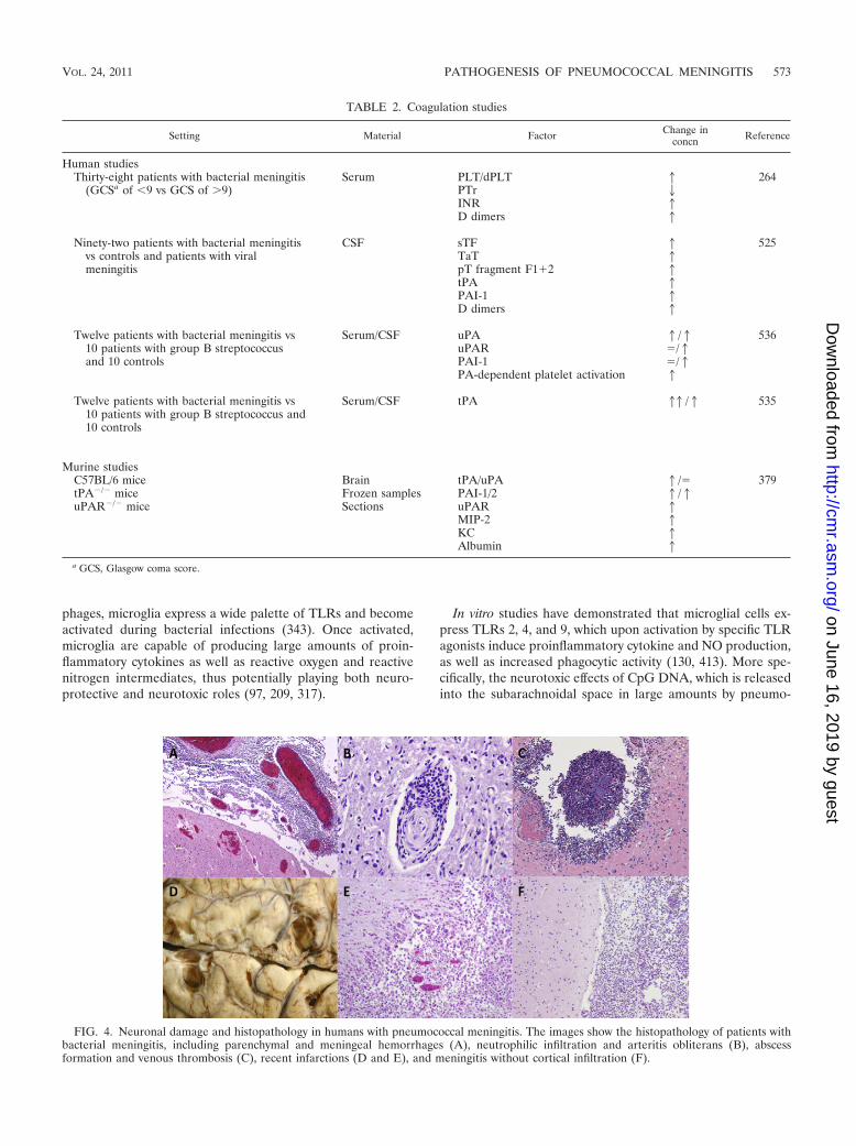
phages, microglia express a wide palette of TLRs and becomeactivated during bacterial infections (343). Once activated,microglia are capable of producing large amounts of proin-flammatory cytokines as well as reactive oxygen and reactivenitrogen intermediates, thus potentially playing both neuro-protective and neurotoxic roles (97, 209, 317).
In vitro studies have demonstrated that microglial cells ex-press TLRs 2, 4, and 9, which upon activation by specific TLRagonists induce proinflammatory cytokine and NO production,as well as increased phagocytic activity (130, 413). More spe-cifically, the neurotoxic effects of CpG DNA, which is releasedinto the subarachnoidal space in large amounts by pneumo-
TABLE 2. Coagulation studies
Setting Material Factor Change inconcn Reference
Human studiesThirty-eight patients with bacterial meningitis Serum PLT/dPLT 1 264
(GCSa of �9 vs GCS of �9) PTr 2INR 1D dimers 1
Ninety-two patients with bacterial meningitis CSF sTF 1 525vs controls and patients with viral TaT 1meningitis pT fragment F1�2 1
tPA 1PAI-1 1D dimers 1
Twelve patients with bacterial meningitis vs Serum/CSF uPA 1/1 53610 patients with group B streptococcus uPAR /1and 10 controls PAI-1 /1
PA-dependent platelet activation 1
Twelve patients with bacterial meningitis vs10 patients with group B streptococcus and10 controls
Serum/CSF tPA 11/1 535
Murine studiesC57BL/6 mice Brain tPA/uPA 1/ 379tPA�/� mice Frozen samples PAI-1/2 1/1uPAR�/� mice Sections uPAR 1
MIP-2 1KC 1Albumin 1
a GCS, Glasgow coma score.
FIG. 4. Neuronal damage and histopathology in humans with pneumococcal meningitis. The images show the histopathology of patients withbacterial meningitis, including parenchymal and meningeal hemorrhages (A), neutrophilic infiltration and arteritis obliterans (B), abscessformation and venous thrombosis (C), recent infarctions (D and E), and meningitis without cortical infiltration (F).
VOL. 24, 2011 PATHOGENESIS OF PNEUMOCOCCAL MENINGITIS 573
on June 16, 2019 by guesthttp://cm
r.asm.org/
Dow
nloaded from
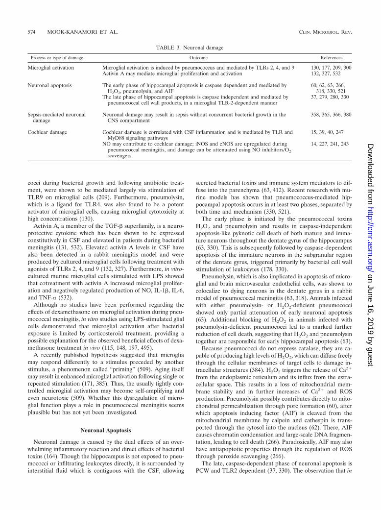
cocci during bacterial growth and following antibiotic treat-ment, were shown to be mediated largely via stimulation ofTLR9 on microglial cells (209). Furthermore, pneumolysin,which is a ligand for TLR4, was also found to be a potentactivator of microglial cells, causing microglial cytotoxicity athigh concentrations (130).
Activin A, a member of the TGF-� superfamily, is a neuro-protective cytokine which has been shown to be expressedconstitutively in CSF and elevated in patients during bacterialmeningitis (131, 532). Elevated activin A levels in CSF havealso been detected in a rabbit meningitis model and wereproduced by cultured microglial cells following treatment withagonists of TLRs 2, 4, and 9 (132, 327). Furthermore, in vitro-cultured murine microglial cells stimulated with LPS showedthat cotreatment with activin A increased microglial prolifer-ation and negatively regulated production of NO, IL-1�, IL-6,and TNF-� (532).
Although no studies have been performed regarding theeffects of dexamethasone on microglial activation during pneu-mococcal meningitis, in vitro studies using LPS-stimulated glialcells demonstrated that microglial activation after bacterialexposure is limited by corticosteroid treatment, providing apossible explanation for the observed beneficial effects of dexa-methasone treatment in vivo (115, 148, 197, 495).
A recently published hypothesis suggested that microgliamay respond differently to a stimulus preceded by anotherstimulus, a phenomenon called “priming” (509). Aging itselfmay result in enhanced microglial activation following single orrepeated stimulation (171, 385). Thus, the usually tightly con-trolled microglial activation may become self-amplifying andeven neurotoxic (509). Whether this dysregulation of micro-glial function plays a role in pneumococcal meningitis seemsplausible but has not yet been investigated.
Neuronal Apoptosis
Neuronal damage is caused by the dual effects of an over-whelming inflammatory reaction and direct effects of bacterialtoxins (164). Though the hippocampus is not exposed to pneu-mococci or infiltrating leukocytes directly, it is surrounded byinterstitial fluid which is contiguous with the CSF, allowing
secreted bacterial toxins and immune system mediators to dif-fuse into the parenchyma (63, 412). Recent research with mu-rine models has shown that pneumococcus-mediated hip-pocampal apoptosis occurs in at least two phases, separated byboth time and mechanism (330, 521).
The early phase is initiated by the pneumococcal toxinsH2O2 and pneumolysin and results in caspase-independentapoptosis-like pyknotic cell death of both mature and imma-ture neurons throughout the dentate gyrus of the hippocampus(63, 330). This is subsequently followed by caspase-dependentapoptosis of the immature neurons in the subgranular regionof the dentate gyrus, triggered primarily by bacterial cell wallstimulation of leukocytes (178, 330).
Pneumolysin, which is also implicated in apoptosis of micro-glial and brain microvascular endothelial cells, was shown tocolocalize to dying neurons in the dentate gyrus in a rabbitmodel of pneumococcal meningitis (63, 318). Animals infectedwith either pneumolysin- or H2O2-deficient pneumococcishowed only partial attenuation of early neuronal apoptosis(63). Additional blocking of H2O2 in animals infected withpneumolysin-deficient pneumococci led to a marked furtherreduction of cell death, suggesting that H2O2 and pneumolysintogether are responsible for early hippocampal apoptosis (63).
Because pneumococci do not express catalase, they are ca-pable of producing high levels of H2O2, which can diffuse freelythrough the cellular membranes of target cells to damage in-tracellular structures (384). H2O2 triggers the release of Ca2�
from the endoplasmic reticulum and its influx from the extra-cellular space. This results in a loss of mitochondrial mem-brane stability and in further increases of Ca2� and ROSproduction. Pneumolysin possibly contributes directly to mito-chondrial permeabilization through pore formation (60), afterwhich apoptosis inducing factor (AIF) is cleaved from themitochondrial membrane by calpein and cathespin is trans-ported through the cytosol into the nucleus (62). There, AIFcauses chromatin condensation and large-scale DNA fragmen-tation, leading to cell death (266). Paradoxically, AIF may alsohave antiapoptotic properties through the regulation of ROSthrough peroxide scavenging (266).
The late, caspase-dependent phase of neuronal apoptosis isPCW and TLR2 dependent (37, 330). The observation that in
TABLE 3. Neuronal damage
Process or type of damage Outcome References
Microglial activation Microglial activation is induced by pneumococcus and mediated by TLRs 2, 4, and 9 130, 177, 209, 300Activin A may mediate microglial proliferation and activation 132, 327, 532
Neuronal apoptosis The early phase of hippocampal apoptosis is caspase dependent and mediated byH2O2, pneumolysin, and AIF
60, 62, 63, 266,318, 330, 521
The late phase of hippocampal apoptosis is caspase independent and mediated bypneumococcal cell wall products, in a microglial TLR-2-dependent manner
37, 279, 280, 330
Sepsis-mediated neuronaldamage
Neuronal damage may result in sepsis without concurrent bacterial growth in theCNS compartment
358, 365, 366, 380
Cochlear damage Cochlear damage is correlated with CSF inflammation and is mediated by TLR andMyD88 signaling pathways
15, 39, 40, 247
NO may contribute to cochlear damage; iNOS and eNOS are upregulated duringpneumococcal meningitis, and damage can be attenuated using NO inhibitors/O2scavengers
14, 227, 241, 243
574 MOOK-KANAMORI ET AL. CLIN. MICROBIOL. REV.
on June 16, 2019 by guesthttp://cm
r.asm.org/
Dow
nloaded from

vitro exposure of isolated cultured neurons to PCW productsdoes not lead to cell death led to the hypothesis that late-phaseapoptosis may be dependent on the host inflammatory re-sponse (330). Neurons themselves do not express TLR2 or -4and are not sensitive to exposure to the corresponding bacte-rial ligands. However, when cocultured with microglial cells,neurons revealed caspase-dependent, TLR-mediated lateapoptosis when they were exposed to PCW or LPS (279, 280).Therefore, the inflammatory response of microglial cells andinvading neutrophils may underlie the caspase-dependent hip-pocampal apoptosis during pneumococcal meningitis.
Several experimental treatments aimed at reducing hip-pocampal apoptosis have been studied in animal models ofpneumococcal meningitis. Much attention has gone to nonbac-teriolytic antibiotic therapies, such as rifampin, daptomycin,and clindamycin (see Targets for Adjunctive Therapy). In ex-perimental rabbit models, rifampin given either alone or as apretreatment before ceftriaxone resulted in a reduction of hip-pocampal apoptosis, though no reduction in mortality was ob-served (54, 165, 458). Likewise, both daptomycin and clinda-mycin treatments yielded less hippocampal neuronal damagethan that with ceftriaxone treatment in rabbit and rat models,respectively (55, 177). Finally, inhibition of MMP and TNF-�converting enzyme (TACE) was also shown to reduce corticaldamage and neuronal apoptosis, as well as preserving learningperformance in rats with pneumococcal meningitis (281).
Sepsis and Hippocampal Damage
Recent findings with experimental animal models have sug-gested that neuronal damage may also be caused by pneumo-coccal growth outside the CNS compartment (365). Mice ex-posed intravascularly to purified PCW developed hippocampalapoptosis at 6 h postinoculation, an effect that was not repro-duced in TLR2- or NOD2-deficient mice and was limited inmice overexpressing IL-10 (358). These findings suggest a pos-sible third, earlier, IL-10-repressible mechanism of neuronaldamage, which may precede pneumococcal invasion of theCNS (358). Additionally, in the setting of experimental men-ingitis, bacteremia has been shown to contribute not only toincreased hippocampal apoptosis but also to dysregulation ofCBF autoregulation, reduced meningeal inflammation, and at-tenuated CSF pleocytosis (366, 380). The mechanisms involvedremain largely unclear.
Cochlear Damage and Hearing Loss
Hearing loss is a common long-term complication in survi-vors of bacterial meningitis. Up to 30% of survivors of pneu-mococcal meningitis experience uni- or bilateral hearing loss,which is often permanent and may be quite severe (125, 273,415, 524).
The underlying pathophysiology has been studied in severalanimal models (39, 40, 45, 244, 421, 477) Cochlear involvementmay result from direct spread of pneumococcal infection fromthe meninges, CSF, and cochlear perilymphatic system (40,227, 244). Alternatively, a hematogenous route may occur fol-lowing bacteremia or sepsis (227, 244).
The resulting cochlear infiltration of pneumoccoci and neu-trophils results in a severe granulocytic inflammation of the
perilymphatic spaces and in the release of proinflammatorycytokines and cytotoxic mediators (56). Hearing loss was cor-related with the level of CSF inflammation in a rabbit model ofpneumococcal meningitis (40), and intrathecal administrationof TNF-� alone was sufficient to induce cochlear injury similarto that observed with bacterial meningitis (15). Furthermore,TLR and MyD88 knockout mice demonstrated less hearingloss following pneumococcal meningitis (247). In a rat model,cochlear expression of iNOS and eNOS was upregulated fol-lowing pneumococcal meningitis, and RNS-mediated cochleardamage could be attenuated both electrophysiologically andhistopathologically by RNS scavengers (227, 241, 243). Earlierstudies with guinea pigs showed that local perfusion of thescala tympani with NO donor compounds resulted in cochleardamage and could be attenuated by NO inhibitors or O2 scav-engers (14).
Cerebrovascular Complications
Cerebrovascular complications are very common duringpneumococcal meningitis (497, 528). Arterial stroke occurs inup to 30% of patients, cerebral venous thrombosis in 9%, andintracerebral hemorrhage in up to 9% (231, 524, 531). Autopsystudies in the 1930s through 1960s showed inflammatory infil-trations of cerebral arteries and veins (77, 83, 126). Takingthese together with angiographic descriptions of segmentalarterial narrowing in patients with ischemic stroke complicat-ing pneumococcal meningitis, the general assumption has beenthat infarctions during bacterial meningitis are caused by vas-culitis.
In a recent human histopathological analysis of patients withpneumococcal meningitis, among whom half of patients hadevidence of cerebral infarctions and 67% showed microhem-orrhages, there was no evidence of large-vessel vasculitis (513).Moreover, the observed small-vessel vasculitis did not colocal-ize with areas of infarction, and in this series, no evidence ofdisseminated intravascular coagulation in the systemic com-partment was observed (513). These results suggest the possi-bility of cerebral intravascular coagulation, independent of sys-temic coagulopathy or cerebral vasculitis, as the cause for bothcerebral infarctions and hemorrhages.
The pathogenesis of cerebral infarction remains unclear andis the subject of ongoing research, which has focused largely ontwo areas: first, the dysregulation of the coagulation and fi-brinolytic pathways, not only systemically but also locally, asexemplified by the upregulation of PAI-1 and elevated levels ofprothrombin fragments F1 and -2 and soluble tissue factor inthe CSF of patients with pneumococcal meningitis (264, 525,536); and second, endothelial cell dysfunction, which may leadto localized swelling and release of procoagulant factors andproinflammatory cytokines. Also, endothelin, which is one ofseveral potent vasoactive peptides, has been shown to be ele-vated in CSF during acute stages of infection (33, 251, 389). Ina rat model, treatment with bosentan (an endothelin antago-nist) normalized otherwise reduced CBF. Although endothelininhibition lowered cortical necrosis, no effect on hippocampaldamage was observed (389).
VOL. 24, 2011 PATHOGENESIS OF PNEUMOCOCCAL MENINGITIS 575
on June 16, 2019 by guesthttp://cm
r.asm.org/
Dow
nloaded from
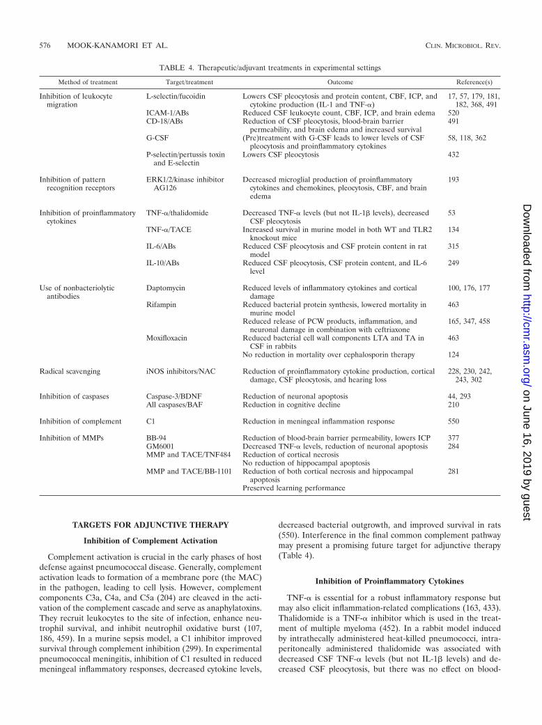
TARGETS FOR ADJUNCTIVE THERAPY
Inhibition of Complement Activation
Complement activation is crucial in the early phases of hostdefense against pneumococcal disease. Generally, complementactivation leads to formation of a membrane pore (the MAC)in the pathogen, leading to cell lysis. However, complementcomponents C3a, C4a, and C5a (204) are cleaved in the acti-vation of the complement cascade and serve as anaphylatoxins.They recruit leukocytes to the site of infection, enhance neu-trophil survival, and inhibit neutrophil oxidative burst (107,186, 459). In a murine sepsis model, a C1 inhibitor improvedsurvival through complement inhibition (299). In experimentalpneumococcal meningitis, inhibition of C1 resulted in reducedmeningeal inflammatory responses, decreased cytokine levels,
decreased bacterial outgrowth, and improved survival in rats(550). Interference in the final common complement pathwaymay present a promising future target for adjunctive therapy(Table 4).
Inhibition of Proinflammatory Cytokines
TNF-� is essential for a robust inflammatory response butmay also elicit inflammation-related complications (163, 433).Thalidomide is a TNF-� inhibitor which is used in the treat-ment of multiple myeloma (452). In a rabbit model inducedby intrathecally administered heat-killed pneumococci, intra-peritoneally administered thalidomide was associated withdecreased CSF TNF-� levels (but not IL-1� levels) and de-creased CSF pleocytosis, but there was no effect on blood-
TABLE 4. Therapeutic/adjuvant treatments in experimental settings
Method of treatment Target/treatment Outcome Reference(s)
Inhibition of leukocytemigration
L-selectin/fucoidin Lowers CSF pleocytosis and protein content, CBF, ICP, andcytokine production (IL-1 and TNF-�)
17, 57, 179, 181,182, 368, 491
ICAM-1/ABs Reduced CSF leukocyte count, CBF, ICP, and brain edema 520CD-18/ABs Reduction of CSF pleocytosis, blood-brain barrier
permeability, and brain edema and increased survival491
G-CSF (Pre)treatment with G-CSF leads to lower levels of CSFpleocytosis and proinflammatory cytokines
58, 118, 362
P-selectin/pertussis toxinand E-selectin
Lowers CSF pleocytosis 432
Inhibition of patternrecognition receptors
ERK1/2/kinase inhibitorAG126
Decreased microglial production of proinflammatorycytokines and chemokines, pleocytosis, CBF, and brainedema
193
Inhibition of proinflammatorycytokines
TNF-�/thalidomide Decreased TNF-� levels (but not IL-1� levels), decreasedCSF pleocytosis
53
TNF-�/TACE Increased survival in murine model in both WT and TLR2knockout mice
134
IL-6/ABs Reduced CSF pleocytosis and CSF protein content in ratmodel
315
IL-10/ABs Reduced CSF pleocytosis, CSF protein content, and IL-6level
249
Use of nonbacteriolyticantibodies
Daptomycin Reduced levels of inflammatory cytokines and corticaldamage
100, 176, 177
Rifampin Reduced bacterial protein synthesis, lowered mortality inmurine model
463
Reduced release of PCW products, inflammation, andneuronal damage in combination with ceftriaxone
165, 347, 458
Moxifloxacin Reduced bacterial cell wall components LTA and TA inCSF in rabbits
463
No reduction in mortality over cephalosporin therapy 124
Radical scavenging iNOS inhibitors/NAC Reduction of proinflammatory cytokine production, corticaldamage, CSF pleocytosis, and hearing loss
228, 230, 242,243, 302
Inhibition of caspases Caspase-3/BDNF Reduction of neuronal apoptosis 44, 293All caspases/BAF Reduction in cognitive decline 210
Inhibition of complement C1 Reduction in meningeal inflammation response 550
Inhibition of MMPs BB-94 Reduction of blood-brain barrier permeability, lowers ICP 377GM6001 Decreased TNF-� levels, reduction of neuronal apoptosis 284MMP and TACE/TNF484 Reduction of cortical necrosis
No reduction of hippocampal apoptosisMMP and TACE/BB-1101 Reduction of both cortical necrosis and hippocampal
apoptosis281
Preserved learning performance
576 MOOK-KANAMORI ET AL. CLIN. MICROBIOL. REV.
on June 16, 2019 by guesthttp://cm
r.asm.org/
Dow
nloaded from
brain barrier permeability (80). When TNF-� was blocked by aTACE inhibitor in an experimental mouse model of pneumo-coccal meningitis, survival was increased in WT and TLR2�/�
mice (134). Thus, TACE inhibition may improve survival, evenin a host with deficient TLR2 signaling (134).
Blocking IL-6 intravenously in a rat model of pneumococcalmeningitis reduced CSF pleocytosis and protein content (315).Similar results were found with antibodies against IL-10 ad-ministered intravenously, which also decreased CSF IL-6 levels(249). Administration of recombinant TGF-�2 intraperitone-ally in the acute phase of pneumococcal meningitis in ratsreduced the subarachnoid inflammatory response by inhibitingthe increase in CBF and brain water content (387).
Inhibition of Pattern Recognition Receptors
Inhibiting TLR signaling with the aim of decreasing subse-quent cytokine responses presents a promising strategy. Thekinase inhibitor tyrphostin AG126 was shown to inhibit phos-phorylation of the signaling molecule extracellular signal-reg-ulated kinase 1/2 (ERK1/2) in microglial cells (193). ERK1/2 isactivated in blood monocytes in response to LPS through ac-tivation of CD14 and TLR4 (185). In microglial cells, treat-ment with this kinase inhibitor resulted in decreased produc-tion of proinflammatory cytokines and chemokines (193).When the kinase inhibitor was administered intraperitoneallyin a mouse pneumococcal meningitis model, leukocyte recruit-ment to the CSF, CBF, brain edema, and TNF-� productionwere reduced (193). This provided evidence that blocking theTLR signaling pathway may reduce the severity of disease inpneumococcal meningitis.
Inhibition of Leukocyte Influx into the CNS
One of the strategies to prevent brain damage is to limitleukocyte recruitment to the CSF or to increase leukocyteapoptosis. Recruitment of leukocytes (mainly polymorphonu-clear leukocytes [PMNs]) to the subarachnoid space results inthe clearance of bacteria, which is accompanied by the produc-tion of several toxic mediators that may induce damage notonly to the bacteria but also to the brain.
The first step in extravasation of PMNs involves binding ofthe leukocytes to selectins on the vessel endothelium. Fucoidinis a polysaccharide that blocks the leukocyte receptor L-selec-tin. Intravenous treatment with fucoidin in several animalmodels of pneumococcal meningitis reduced CSF pleocytosis(17, 179, 182), in association with reduced CSF protein content(179, 182), modestly decreased CSF lactate (179, 182), anddecreased CBF and ICP, without influencing blood-brain bar-rier permeability, cerebral edema, and outcome (17). Further-more, fucoidin prevented the increase in CSF TNF-� and IL-1levels in response to intrathecally administered PCW in rabbits(181); however, when live bacteria were administered intrathe-cally and the rabbits were treated with ampicillin, fucoidin hadno effect on cytokine production (181). A similar study withoutantibiotic treatment reported a reduction in IL-1 and an in-crease in CSF IL-8 in fucoidin-treated rabbits with pneumo-coccal meningitis (368). In contrast to these studies, in a ratmodel of pneumococcal meningitis, fucoidin treatment led todecreased survival (57). The leukocyte concentration in CSF
was lower in fucoidin-treated rats, but systemic leukocytosiswas increased, as was systemic bacterial outgrowth. However,rats in this study were pretreated with fucoidin, which mayhave led to the differences in the systemic immune response(57).
After binding of leukocytes to selectins, firm adhesion to thevascular endothelium is mediated by ICAM-1. In a rat modelof meningitis with PCW component-induced inflammation, an-tibodies against ICAM-1 reduced CSF leukocyte counts, CBF,ICP, and brain edema in the first 6 h (520). In experimentalpneumococcal meningitis, an intravenously administered anti-body against CD18, a subunit on leukocytes for bindingICAM-1, induced either by live bacteria or by PCW, reducedCSF leukocyte counts, blood-brain barrier permeability, andbrain edema and increased survival (491). However, anti-CD18antibodies tended only to inhibit CSF leukocyte counts in arabbit model of PCW-induced inflammation (180).
Vascular endothelial growth factor (VEGF) is a peptideinvolved in angiogenesis, but it has also been described tofunction as a macrophage and granulocyte chemoattractant(485). Levels of VEGF were associated with CSF leukocytecounts in experimental meningitis induced by heat-killed pneu-mococci (503). However, blocking of VEGF in rabbits withpneumococcal meningitis did not reduce the extent of brainedema, leukocyte influx, or blood-brain barrier permeability(503).
A different approach involves the induction of an increasedsystemic proliferation of leukocytes, which would lead to bettercontrol of systemic infection and, subsequently, to enhancedcontrol of CNS infection. In patients with meningitis, granulo-cyte colony-stimulating factor (G-CSF) and macrophage colo-ny-stimulating factor (M-CSF) have been found to be elevatedin the CSF (156, 450). In these studies, G-CSF and M-CSFlevels correlated with CSF leukocytosis (450). In mice withpneumococcal meningitis, expression of granulocyte-macro-phage colony-stimulating factor (GM-CSF) in the brain wasincreased (376). Rabbits pretreated with G-CSF intravenously1 h before intrathecal inoculation with S. pneumoniae showedincreased peripheral but not subarachnoid leukocytosis andincreased CSF levels of TNF and IL-1 but no reduction ofsubarachnoid bacterial outgrowth or neuron-specific enolase,an indicator of neuronal cell damage (438). A similar experi-ment showed no influence on subarachnoid bacterial killing,but systemic pleocytosis and bacterial killing were increased(362). CSF leukocytosis and protein content and levels of IL-8,TNF-�, and IL-1� were decreased in G-CSF-pretreated ani-mals, indicating a decreased subarachnoid inflammatory re-sponse (362). Similar results were found in a study with rats(58). However, late administration of G-CSF (28 h after infec-tion) did not have any influence on disease parameters (58). In22 patients with pneumococcal meningitis, adjunctive treat-ment with recombinant G-CSF was performed together withstandard treatment with ceftriaxone and dexamethasone. G-CSF was continued for 4 days unless leukocyte counts ex-ceeded 40 109 cells/liter. Lactate and glucose levels returnedto normal more quickly in G-CSF-treated patients than inhistorical controls, and no adverse events were recorded. How-ever, this was not a randomized controlled study (118).
Another approach to limit neutrophil-mediated damage inpneumococcal meningitis is to induce apoptosis in neutrophils
VOL. 24, 2011 PATHOGENESIS OF PNEUMOCOCCAL MENINGITIS 577
on June 16, 2019 by guesthttp://cm
r.asm.org/
Dow
nloaded from
by use of roscovitine. Mice treated with a combination ofantibiotics and roscovitine showed increased resolution of in-flammation, decreased cerebral hemorrhages, and faster re-coveries (250).
In meningitis caused by Cryptococcus neoformans, the fun-gal capsular polysaccharide glucuronoxylomannan (GMX)inhibited leukocyte extravasation despite high IL-8 levels inthe CSF (297). The mechanisms by which GMX inhibitsleukocytosis are unknown; however, when GMX was admin-istered intravenously in an experimental rabbit model ofmeningeal inflammation with heat-killed pneumococci, CSFTNF-� levels and leukocytosis decreased, in association withreduced brain edema and inflammation on brain histopa-thology (298).
A similar mechanism has been described for pertussis toxin,which interferes with the binding of PMNs to P-selectin andE-selectin, the first steps of diapedesis. Intravenous treatmentwith pertussis toxin in a rabbit model of meningeal inflamma-tion induced by heat-killed pneumococci altered CSF pleocy-tosis compared to that in untreated animals (432).
Inhibition of Caspases
Caspase activation has been implicated in the activation ofproinflammatory cytokines (258) as well as in the mediation ofprogrammed cell death of cerebral endothelial cells and neu-rons in the hippocampal dendate gyrus (37, 330). Experimentalmodels of pneumococcal meningitis using mice deficient incaspase-1 or after pharmacological blocking of caspase-1 dem-onstrated lower levels of IL-1� and subsequent diminishedproinflammatory cytokine production, as well as fewer menin-gitis-induced complications (258). The protective effect ofcaspase-3 inhibition on the development of neuronal damagewas demonstrated in a rat model of pneumococcal meningitisand was independent of cytokine modulation (166). Also, thebroad-spectrum caspase inhibitor z-VAD-fmk was shown toreduce hippocampal neuronal apoptosis in a rabbit model ofpneumococcal meningitis compared to that in untreated con-trols (61). Moreover, rats inoculated with group B streptococcidemonstrated less cognitive decline following adjunctive treat-ment with the pan-caspase inhibitor bocaspartyl (OMe)-fluo-romethylketone (210). An interesting observation is that exog-enous administration of brain-derived neutrotrophic factor(BDNF), which has been shown to block caspase-3 (191), re-duced neuronal apoptosis in both rat and murine models (44,293). BDNF was found to be upregulated naturally duringbacterial meningitis and after treatment with antibiotics withadjunctive dexamethasone yet lowered during standard antibi-otic treatment, suggesting a possible mechanism of corticoste-roid therapy (294). So far, no clinical trials have been per-formed with caspase inbitors as adjunctive therapy.
Adjunctive Dexamethasone Therapy
Dexamethasone is a widely used anti-inflammatory drug.The mechanisms by which dexamethasone inhibits inflamma-tion are not clear, but it decreases proinflammatory cytokineproduction in monocytes, dendritic cells, astroglial cells, andneutrophils (108, 154, 217, 425), increases the production ofanti-inflammatory cytokines such as IL-10 (108), inhibits ROS
production by leukocytes (108), and decreases leukocyte ad-herence (153, 303). Dexamethasone acts on multiple moleculesof the TLR downstream signaling cascade, including TAK-1,ERK1/2, MAPK, NF-�B, and STAT3 (41, 542). Astroglial cellsstimulated with PCW components produced decreasedamounts of NO and TNF-� when they were treated with dexa-methasone (38, 154, 444). Brain microvascular endothelialcells also showed reduced levels of TNF-� and IL-1 and de-creased expression of ICAM-1 (153). On the molecular level,in peripheral blood mononuclear cells dexamethasone wasshown to inhibit S. pneumoniae-induced I�B� phosphorylationand degradation and binding of NF-�B to DNA, both of whichare downstream effector mechanisms of TLR signaling (332).Dexamethasone induced increased levels of I�B� mRNA,which may bind and inhibit the p65 subunit of NF-�B. Theseeffects resulted in decreased IL-8 production by peripheralblood mononuclear cells (332).
In experimental pneumococcal meningitis, adjunctive dexa-methasone reduced TNF-�, lactate (304), and NO (119a) lev-els when it was administered together with antibiotics. Further-more, dexamethasone decreased ICP, brain edema, and CSFpleocytosis in rats with PCW-induced meningeal inflammation(255, 388). In a rabbit model, neuron-specific enolase, amarker of overall neuronal damage, was reduced in animalstreated with ceftriaxone and dexamethasone compared tothose treated with ceftriaxone alone, though an increase inhippocampal apoptosis was also observed (556). Moreover,in rats treated with adjunctive dexamethasone, the observedincrease in hippocampal apoptosis was accompanied by im-paired learning performance (282), and uninfected rats treatedwith dexamethasone also demonstrated increased hippocam-pal damage (47). In an analysis of several prospective multi-center trials in which patients with bacterial meningitis weretreated with either adjunctive dexamethasone or placebo, theuse of dexamethasone was not associated with cognitive im-pairment (202, 526).
Adjuvant treatment with dexamethasone resulted in a re-duction of hearing loss in rabbit and gerbil models of pneu-mococcal meningitis (235, 406) but did not have a significanteffect in infant rats (94). A recent meta-analysis of human trialsevaluating adjuvant dexamethasone treatment suggested thatdexamethasone may reduce hearing loss among survivors(499). Clearly, further trials are necessary to assess the effectsof dexamethasone on cochlear injury and hearing loss.
In children with bacterial meningitis, CSF TNF-� and IL-1levels were decreased if patients had been treated with adjunc-tive dexamethasone therapy (342). In a large randomized con-trolled trial in Vietnam investigating the efficacy of dexameth-asone addition to conventional antibiotic regimens in adultswith bacterial meningitis, CSF samples from a large group ofpatients were examined (309). For 195 of a total of 341 patientsincluded in this study, CSF was available at baseline (whentherapy was started) and at a follow-up puncture 1 to 4 dayslater. Of these 195 patients, 88 had received dexamethasonealong with antibiotics starting at baseline, and 107 receivedplacebo and antibiotics. For 24% of patients, S. pneumoniaewas confirmed as the causative agent by CSF culture. Othercausative agents included S. suis (43%) and N. meningitidis(8%). CSF samples from these patients were analyzed for IL-6,IL-8, IL-10, IL-12, IL-1�, and TNF-�. Cytokine levels in CSF
578 MOOK-KANAMORI ET AL. CLIN. MICROBIOL. REV.
on June 16, 2019 by guesthttp://cm
r.asm.org/
Dow
nloaded from
from the first lumbar puncture at baseline were similar be-tween both groups. Dexamethasone treatment reduced thelevels of IL-6 and IL-8 and increased the levels of IL-10 (me-dian, 37 pg/ml versus 33 pg/ml; P value 0.01) in the CSF atfollow-up compared to those with placebo (309). Levels ofIL-12, IL-1�, and TNF-� were similar between both groups atfollow-up. In addition to the differences in anti-inflammatorycytokine profiles, opening pressure of the follow-up lumbarpuncture was reduced and the CSF glucose level and CSF/plasma glucose ratio were restored sooner in patients receivingdexamethasone. CSF lactate and protein levels as well as leu-kocyte counts were similar at follow-up in the dexamethasoneand placebo groups (309).
Many randomized clinical trials of dexamethasone for treat-ment of bacterial meningitis have been performed, but theresults have remained somewhat ambiguous (70, 115, 494, 495,499). An individual patient data meta-analysis of 5 large recenttrials showed no effect of dexamethasone (499). A prospectivecohort study showed a decrease in mortality from 30 to 20%for adults with pneumococcal meningitis after nationwide im-plementation of dexamethasone therapy in the Netherlands(69). Dexamethasone treatment has been implemented as rou-tine therapy for patients with suspected or proven pneumococ-cal meningitis in many countries (487, 497).
Adjunctive Glycerol Therapy
Glycerol (glycerine 1-2-3-propanetriol), a hyperosmolarcompound, is an essential component of cell membranes. It hasbeen used in neurosurgery, neurology, and ophthalmology todecrease raised tissue pressures (26, 78, 87, 168, 416, 501).Toxicological data show that it is safe and is associated withfew, rare, mild, mostly gastrointestinal side effects (152). Fur-thermore, glycerol is inexpensive and readily available, facili-tating widespread implementation if it is effective. The effectsof glycerol in the neurological/neurosurgical setting have beenhypothesized to lie in the resulting increase of plasma osmo-lality, which was shown to reduce the excretion of CSF by some20 to 30%, leading to increased cerebral blood flow and im-provement of brain oxygenation (453). For acute stroke, glyc-erol has been shown to confer only a short-term advantage,without demonstrating benefits in the long-term outcome(416).
The first clinical trial evaluating glycerol as a potential ad-juvant treatment for bacterial meningitis was conducted with122 Finnish children with bacterial meningitis and suggested areduction in hearing impairment as well as in long-term neu-rological sequelae (234). More recently, a large South Amer-ican trial using adjuvant glycerol for the treatment of childrenwith bacterial meningitis provided additional support for itsefficacy in the reduction of neurological sequelae, althoughhearing loss and mortality were not diminished (381, 382).However, reliable interpretation of the findings was compro-mised by several methodological problems (430). Nevertheless,the results fueled additional trials, most recently a study inMalawi where 256 adults with bacterial meningitis were ran-domized to receive either placebo or glycerol as adjuvant treat-ment (5). The trial was halted prematurely when an interimanalysis after 100 deaths showed increased death in the glyc-erol group (5). The discrepancies in the various studies most
likely lie in the variation in study populations, such as age,comorbidity, and causative pathogen, as well as in the variationin treatment regimens and methods of assessing outcome pa-rameters.
Relatively few animal studies have been performed withglycerol, and they have not been able to demonstrate clinical orhistological benefits of adjuvant glycerol therapy (46). More-over, in a rabbit model, pneumococcal meningitis treated withglycerol alone was associated with an increased level of hip-pocampal neuronal apoptosis (439). A study in which healthymice were treated with high doses of glycerol (much higherthan trial dosages) showed that treatment was associated withthe occurrence of seizures (127), which were also seen more inthe glycerol-treated patients in the Malawi trial (5). This find-ing was not in concurrence with the South American trial andmay be explained by either the shorter glycerol regimen (2 daysinstead of 4 days) or increased disease severity in the Malawigroup.
Though glycerol seemed attractive as a potential adjuvanttreatment for bacterial meningitis, the lack of effectiveness inexperimental models, combined with its harmful effects in theMalawi trial, question the value of additional studies on ad-junctive glycerol for the treatment of adult meningitis (72).
Nonbacteriolytic Antibiotics
The massive inflammatory response following pneumococcalmeningitis has been shown to play a key role in the develop-ment of brain damage and subsequent poor outcomes (252). Inpart, the inflammatory response is determined by bacterial lysisproducts, as shown in experimental pneumococcal meningitismodels where inoculation with PCW led to massive inflamma-tion and neuronal damage (489, 490). Although bacteriolyticantibiotic regimens may limit the overall amount of release ofbacterial products, temporary increases in the release of bac-terial components have been documented following treatment(344). These observations have fueled the study of nonbacte-riolytic antimicrobials as future therapy options.
Specifically, recent research of nonbacteriolytic antimicrobi-als has focused on the potential use of daptomycin, rifampin,and moxifloxacin. Daptomycin, a lipopeptide antibiotic, hasbeen shown to effectively clear S. pneumoniae in experimentalrat, mouse, and rabbit meningitis models (100, 177). Treatmentwith daptomycin led to lower levels of inflammatory cytokinesand possibly to less cortical brain damage and neuronal apop-tosis than those for treatment with ceftriaxone alone (176,177). Although adjunctive treatment with dexamethasone didnot result in a reduction of overall pneumococcal clearancefrom the CSF, daptomycin penetration into the inflamed me-ninges was reduced in the presence of adjunctive corticosteroidtherapy (136).
Rifampin inhibits bacterial protein synthesis and was shownto lead to diminished levels of PCW product release in vitro(463). In a murine model of pneumococcal meningitis, a de-crease in mortality was observed in mice treated with rifampinversus ceftriaxone, an effect which was most notable during thefirst hours following antibiotic therapy (347). Moreover, recentstudies with a rabbit model demonstrated that short-term pre-treatment with rifampin before ceftriaxone reduced the releaseof PCW products, inflammation, and neuronal damage com-
VOL. 24, 2011 PATHOGENESIS OF PNEUMOCOCCAL MENINGITIS 579
on June 16, 2019 by guesthttp://cm
r.asm.org/
Dow
nloaded from
pared to those with treatment with ceftriaxone alone (54, 165,458).
In a rabbit model of pneumococcal meningitis, treatmentwith moxifloxacin, a relatively novel quinolone, was shown toresult in lower levels of the proinflammatory cell wall compo-nents LTA and TA in CSF than those obtained by treatmentwith ceftriaxone (463). Pneumococcal clearance from the CSFwas comparable after treatment with either moxifloxacin orceftriaxone, and drug levels were not reduced following ad-junctive treatment with dexamethasone (367, 437). However,in a murine model, moxifloxacin failed to reduce mortalitycompared to standard cephalosporin therapy (124).
Radical Scavenging
Investigations of potential adjunctive antioxidant therapieshave been limited to animal studies and so far lack sufficientevidence for clinical testing. A recent comprehensive review byKlein et al. summarizes the efforts and findings in this area ofpotential treatment options (242). Among the most promisingcandidates for future clinical applications are the iNOS inhib-itors, the peroxynitrite scavenger Mn(III)tetrakis(4-benzoic ac-id)-porphyrin (MnTBAP), uric acid, alpha-pheny-tert-butyl-ni-trone (PBN), and N-acetyl-L-cysteine (NAC) (242). In spite ofsome beneficial effects, such as reductions of proinflammatorycytokine pleocytosis (230), reductions in cortical damage (302),attenuated CSF pleocytosis (228, 230), and a diminished inci-dence of hearing loss (243), adverse outcomes have been re-ported, such as impaired bacterial killing, increased neuronalapoptosis, and impaired learning function (302). Of all poten-tial antioxidant agents, only NAC is currently used routinely inclinical practice (for the treatment of acetaminophen intoxica-tions), and therefore it is a likely candidate for evaluation in aclinical setting.
CONCLUSIONS
Despite significant advances in treatment and vaccinations,pneumococcal meningitis remains one of the most importantinfectious diseases worldwide and continues to be associatedwith high mortality and morbidity. The growing emergence ofdrug resistance as well as shifts in serotype incidence is fuelingfurther development of novel antibiotic and adjuvant treat-ment strategies. In addition to the widespread introduction ofdexamethasone, other options for adjuvant drugs may lie inmodulating ROS/RNS-mediated damage, in caspase inhibi-tion, or in drugs targeting specific mediators in the inflamma-tory, complement, or coagulation cascades. Extensive researchin this area is being performed using experimental animalmeningitis models, though so far no clinical treatment trialswith humans have been performed. Although the limitations ofanimal models of meningitis lie in the great variability betweenspecies, inoculation methods, and ages of infected animals,experimental medicine continues to provide the backbone forboth intervention and pathophysiology studies and will hope-fully pave the way to new knowledge and treatment of thisdeadly disease.
ACKNOWLEDGMENT
We have no conflicts of interest.
REFERENCES
1. Aas, J. A., B. J. Paster, L. N. Stokes, I. Olsen, and F. E. Dewhirst. 2005.Defining the normal bacterial flora of the oral cavity. J. Clin. Microbiol.43:5721–5732.
2. Abeyta, M., G. G. Hardy, and J. Yother. 2003. Genetic alteration of capsuletype but not PspA type affects accessibility of surface-bound complementand surface antigens of Streptococcus pneumoniae. Infect. Immun. 71:218–225.
3. Adriani, K. S., D. van de Beek, M. C. Brouwer, L. Spanjaard, and J. deGans. 2007. Community-acquired recurrent bacterial meningitis in adults.Clin. Infect. Dis. 45:e46–e51.
4. Agrawal, A., M. J. Simpson, S. Black, M. P. Carey, and D. Samols. 2002. AC-reactive protein mutant that does not bind to phosphocholine and pneu-mococcal C-polysaccharide. J. Immunol. 169:3217–3222.
5. Ajdukiewicz, K. M., et al. 2011. Glycerol adjuvant therapy in adults withbacterial meningitis in a high HIV seroprevalence setting in Malawi: adouble-blind, randomised controlled trial. Lancet Infect. Dis. 11:293–300.
6. Akache, B., et al. 2006. The 37/67-kilodalton laminin receptor is a receptorfor adeno-associated virus serotypes 8, 2, 3, and 9. J. Virol. 80:9831–9836.
7. Akira, S., K. Hoshino, and T. Kaisho. 2000. The role of Toll-like receptorsand MyD88 in innate immune responses. J. Endotoxin Res. 6:383–387.
8. Akita, K., et al. 1997. Involvement of caspase-1 and caspase-3 in the pro-duction and processing of mature human interleukin 18 in monocyticTHP.1 cells. J. Biol. Chem. 272:26595–26603.
9. Alberts, B. 2009. Essential cell biology, 3rd ed. Garland Science, NewYork, NY.
10. Albiger, B., et al. 2007. Toll-like receptor 9 acts at an early stage in hostdefence against pneumococcal infection. Cell. Microbiol. 9:633–644.
11. Albiger, B., et al. 2005. Myeloid differentiation factor 88-dependent signal-ling controls bacterial growth during colonization and systemic pneumo-coccal disease in mice. Cell. Microbiol. 7:1603–1615.
12. Alcantara, R. B., L. C. Preheim, and M. J. Gentry. 1999. Role of pneumo-lysin’s complement-activating activity during pneumococcal bacteremia incirrhotic rats. Infect. Immun. 67:2862–2866.
13. Alcantara, R. B., L. C. Preheim, and M. J. Gentry-Nielsen. 2001. Pneumo-lysin-induced complement depletion during experimental pneumococcalbacteremia. Infect. Immun. 69:3569–3575.
14. Amaee, F. R., S. D. Comis, M. P. Osborne, S. Drew, and M. J. Tarlow. 1997.Possible involvement of nitric oxide in the sensorineural hearing loss ofbacterial meningitis. Acta Otolaryngol. 117:329–336.
15. Aminpour, S., S. P. Tinling, and H. A. Brodie. 2005. Role of tumor necrosisfactor-alpha in sensorineural hearing loss after bacterial meningitis. Otol.Neurotol. 26:602–609.
16. Andersson, B., et al. 1983. Identification of an active disaccharide unit of aglycoconjugate receptor for pneumococci attaching to human pharyngealepithelial cells. J. Exp. Med. 158:559–570.
17. Angstwurm, K., et al. 1995. Fucoidin, a polysaccharide inhibiting leukocyterolling, attenuates inflammatory responses in experimental pneumococcalmeningitis in rats. Neurosci. Lett. 191:1–4.
18. Arda, B., O. R. Sipahi, S. Atalay, and S. Ulusoy. 2008. Pooled analysis of2,408 cases of acute adult purulent meningitis from Turkey. Med. Princ.Pract. 17:76–79.
19. Arditi, M., et al. 1998. Three-year multicenter surveillance of pneumococcalmeningitis in children: clinical characteristics, and outcome related to pen-icillin susceptibility and dexamethasone use. Pediatrics 102:1087–1097.
20. Arnold, R. R., M. Brewer, and J. J. Gauthier. 1980. Bactericidal activity ofhuman lactoferrin: sensitivity of a variety of microorganisms. Infect. Im-mun. 28:893–898.
21. Arnold, R. R., J. E. Russell, W. J. Champion, M. Brewer, and J. J. Gauthier.1982. Bactericidal activity of human lactoferrin: differentiation from thestasis of iron deprivation. Infect. Immun. 35:792–799.
22. Asai, S., et al. 2004. Absence of procarboxypeptidase R induces comple-ment-mediated lethal inflammation in lipopolysaccharide-primed mice.J. Immunol. 173:4669–4674.
23. Attali, C., C. Durmort, T. Vernet, and A. M. Di Guilmi. 2008. The interac-tion of Streptococcus pneumoniae with plasmin mediates transmigrationacross endothelial and epithelial monolayers by intercellular junction cleav-age. Infect. Immun. 76:5350–5356.
24. Attali, C., et al. 2008. Streptococcus pneumoniae choline-binding protein Einteraction with plasminogen/plasmin stimulates migration across the ex-tracellular matrix. Infect. Immun. 76:466–476.
25. Avadhanula, V., et al. 2006. Respiratory viruses augment the adhesion ofbacterial pathogens to respiratory epithelium in a viral species- and celltype-dependent manner. J. Virol. 80:1629–1636.
26. Awasthi, P., and S. N. Srivastava. 1965. Role of oral glycerol in glaucoma.Br. J. Ophthalmol. 49:660–666.
27. Aycicek, A., A. Iscan, O. Erel, M. Akcali, and A. R. Ocak. 2007. Oxidant andantioxidant parameters in the treatment of meningitis. Pediatr. Neurol.37:117–120.
28. Bagnoli, F., et al. 2008. A second pilus type in Streptococcus pneumoniae
580 MOOK-KANAMORI ET AL. CLIN. MICROBIOL. REV.
on June 16, 2019 by guesthttp://cm
r.asm.org/
Dow
nloaded from
is prevalent in emerging serotypes and mediates adhesion to host cells. J.Bacteriol. 190:5480–5492.
29. Barichello, T., et al. 2009. Tumor necrosis factor alpha (TNF-alpha) levelsin the brain and cerebrospinal fluid after meningitis induced by Strepto-coccus pneumoniae. Neurosci. Lett. 467:217–219.
30. Barichello, T., et al. 2010. TNF-alpha, IL-1beta, IL-6, and cinc-1 levels inrat brain after meningitis induced by Streptococcus pneumoniae. J. Neu-roimmunol. 221:42–45.
31. Beisswenger, C., C. B. Coyne, M. Shchepetov, and J. N. Weiser. 2007. Roleof p38 MAP kinase and transforming growth factor-beta signaling in trans-epithelial migration of invasive bacterial pathogens. J. Biol. Chem. 282:28700–28708.
32. Beisswenger, C., E. S. Lysenko, and J. N. Weiser. 2009. Early bacterialcolonization induces Toll-like receptor-dependent transforming growth fac-tor signaling in the epithelium. Infect. Immun. 77:2212–2220.
33. Berg, R. M. G., et al. 2009. Circulating levels of vasoactive peptides inpatients with acute bacterial meningitis. Intensive Care Med. 35:1604–1608.
34. Bergmann, S., et al. 2009. Integrin-linked kinase is required for vitronectin-mediated internalization of Streptococcus pneumoniae by host cells. J. CellSci. 122:256–267.
35. Bergmann, S., M. Rohde, G. S. Chhatwal, and S. Hammerschmidt. 2001.Alpha-enolase of Streptococcus pneumoniae is a plasmin(ogen)-bindingprotein displayed on the bacterial cell surface. Mol. Microbiol. 40:1273–1287.
36. Bergmann, S., M. Rohde, and S. Hammerschmidt. 2004. Glyceraldehyde-3-phosphate dehydrogenase of Streptococcus pneumoniae is a surface-displayed plasminogen-binding protein. Infect. Immun. 72:2416–2419.
37. Bermpohl, D., et al. 2005. Bacterial programmed cell death of cerebralendothelial cells involves dual death pathways. J. Clin. Invest. 115:1607–1615.
38. Bernatowicz, A., U. Kodel, K. Frei, A. Fontana, and H. W. Pfister. 1995.Production of nitrite by primary rat astrocytes in response to pneumococci.J. Neuroimmunol. 60:53–61.
39. Bhatt, S., et al. 1991. Hearing loss and pneumococcal meningitis: an animalmodel. Laryngoscope 101:1285–1292.
40. Bhatt, S. M., et al. 1993. Progression of hearing loss in experimental pneu-mococcal meningitis: correlation with cerebrospinal fluid cytochemistry. J.Infect. Dis. 167:675–683.
41. Bhattacharyya, S., et al. 2010. TAK1 targeting by glucocorticoids deter-mines JNK and IkappaB regulation in Toll-like receptor-stimulated mac-rophages. Blood 115:1921–1931.
42. Biernath, K. R., et al. 2006. Bacterial meningitis among children withcochlear implants beyond 24 months after implantation. Pediatrics 117:284–289.
43. Biffl, W. L., et al. 1996. Interleukin-6 stimulates neutrophil production ofplatelet-activating factor. J. Leukoc. Biol. 59:569–574.
44. Bifrare, Y.-D., J. Kummer, P. Joss, M. G. Tauber, and S. L. Leib. 2005.Brain-derived neurotrophic factor protects against multiple forms of braininjury in bacterial meningitis. J. Infect. Dis. 191:40–45.
45. Blank, A. L., G. L. Davis, T. R. VanDeWater, and R. J. Ruben. 1994. AcuteStreptococcus pneumoniae meningogenic labyrinthitis. An experimentalguinea pig model and literature review. Arch. Otolaryngol. Head NeckSurg. 120:1342–1346.
46. Blaser, C., et al. 2010. Adjuvant glycerol is not beneficial in experimentalpneumococcal meningitis. BMC Infect. Dis. 10:84.
47. Blaser, C., M. Wittwer, D. Grandgirard, and S. L. Leib. 2011. Adjunctivedexamethasone affects the expression of genes related to inflammation,neurogenesis and apoptosis in infant rat pneumococcal meningitis. PLoSOne 6:e17840.
48. Boffa, M. B., W. Wang, L. Bajzar, and M. E. Nesheim. 1998. Plasma andrecombinant thrombin-activable fibrinolysis inhibitor (TAFI) and activatedTAFI compared with respect to glycosylation, thrombin/thrombomodulin-dependent activation, thermal stability, and enzymatic properties. J. Biol.Chem. 273:2127–2135.
49. Bogaert, D., R. De Groot, and P. W. M. Hermans. 2004. Streptococcuspneumoniae colonisation: the key to pneumococcal disease. Lancet Infect.Dis. 4:144–154.
50. Bogaert, D., et al. 2001. Pneumococcal carriage in children in The Nether-lands: a molecular epidemiological study. J. Clin. Microbiol. 39:3316–3320.
51. Bogaert, D., C. M. Thompson, K. Trzcinski, R. Malley, and M. Lipsitch.2010. The role of complement in innate and adaptive immunity to pneu-mococcal colonization and sepsis in a murine model. Vaccine 28:681–685.
52. Bogaert, D., et al. 2004. Colonisation by Streptococcus pneumoniae andStaphylococcus aureus in healthy children. Lancet 363:1871–1872.
53. Bogdan, I., S. L. Leib, M. Bergeron, L. Chow, and M. G. Tauber. 1997.Tumor necrosis factor-alpha contributes to apoptosis in hippocampal neu-rons during experimental group B streptococcal meningitis. J. Infect. Dis.176:693–697.
54. Bottcher, T., et al. 2000. Rifampin reduces production of reactive oxygenspecies of cerebrospinal fluid phagocytes and hippocampal neuronal apop-tosis in experimental Streptococcus pneumoniae meningitis. J. Infect. Dis.181:2095–2098.
55. Bottcher, T., et al. 2004. Clindamycin is neuroprotective in experimentalStreptococcus pneumoniae meningitis compared with ceftriaxone. J. Neu-rochem. 91:1450–1460.
56. Brandt, C. T., et al. 2006. Hearing loss and cochlear damage in experimen-tal pneumococcal meningitis, with special reference to the role of neutro-phil granulocytes. Neurobiol. Dis. 23:300–311.
57. Brandt, C. T., et al. 2005. Blocking of leukocyte accumulation in the cere-brospinal fluid augments bacteremia and increases lethality in experimentalpneumococcal meningitis. J. Neuroimmunol. 166:126–131.
58. Brandt, C. T., et al. 2004. Attenuation of the bacterial load in blood bypretreatment with granulocyte-colony-stimulating factor protects rats fromfatal outcome and brain damage during Streptococcus pneumoniae men-ingitis. Infect. Immun. 72:4647–4653.
59. Branger, J., et al. 2004. Role of Toll-like receptor 4 in gram-positive andgram-negative pneumonia in mice. Infect. Immun. 72:788–794.
60. Braun, J. S., et al. 2007. Pneumolysin causes neuronal cell death throughmitochondrial damage. Infect. Immun. 75:4245–4254.
61. Braun, J. S., et al. 1999. Neuroprotection by a caspase inhibitor in acutebacterial meningitis. Nat. Med. 5:298–302.
62. Braun, J. S., et al. 2001. Apoptosis-inducing factor mediates microglial andneuronal apoptosis caused by pneumococcus. J. Infect. Dis. 184:1300–1309.
63. Braun, J. S., et al. 2002. Pneumococcal pneumolysin and H(2)O(2) mediatebrain cell apoptosis during meningitis. J. Clin. Invest. 109:19–27.
64. Braun, J. S., and E. I. Tuomanen. 1999. Molecular mechanisms of braindamage in bacterial meningitis. Adv. Pediatr. Infect. Dis. 14:49–71.
65. Briles, D. E., et al. 2003. Immunizations with pneumococcal surface proteinA and pneumolysin are protective against pneumonia in a murine model ofpulmonary infection with Streptococcus pneumoniae. J. Infect. Dis. 188:339–348.
66. Brivet, F. G., F. M. Jacobs, and B. Megarbane. 2005. Cerebral output ofcytokines in patients with pneumococcal meningitis. Crit. Care Med. 33:2721–2722.
67. Brock, S. C., P. A. McGraw, P. F. Wright, and J. E. Crowe, Jr. 2002. Thehuman polymeric immunoglobulin receptor facilitates invasion of epithelialcells by Streptococcus pneumoniae in a strain-specific and cell type-specificmanner. Infect. Immun. 70:5091–5095.
68. Brouwer, M. C., et al. 2009. Host genetic susceptibility to pneumococcaland meningococcal disease: a systematic review and meta-analysis. LancetInfect. Dis. 9:31–44.
69. Brouwer, M. C., et al. 2010. Nationwide implementation of adjunctivedexamethasone therapy for pneumococcal meningitis. Neurology 75:1533–1539.
70. Brouwer, M. C., P. McIntyre, J. de Gans, K. Prasad, and D. van de Beek.2010. Corticosteroids for acute bacterial meningitis. Cochrane DatabaseSyst. Rev. 9:CD004405.
71. Brouwer, M. C., A. R. Tunkel, and D. van de Beek. 2010. Epidemiology,diagnosis, and antimicrobial treatment of acute bacterial meningitis. Clin.Microbiol. Rev. 23:467–492.
72. Brouwer, M. C., and D. van de Beek. 2011. Glycerol in bacterial meningitis:one strike and out? Lancet Infect. Dis. 11:257–258.
73. Brouwer, N., et al. 2008. Mannose-binding lectin (MBL) facilitates op-sonophagocytosis of yeasts but not of bacteria despite MBL binding. J. Im-munol. 180:4124–4132.
74. Bruder, C., et al. 2004. HIV-1 induces complement factor C3 synthesis inastrocytes and neurons by modulation of promoter activity. Mol. Immunol.40:949–961.
75. Brugger, S. D., L. J. Hathaway, and K. Muhlemann. 2009. Detection ofStreptococcus pneumoniae strain cocolonization in the nasopharynx.J. Clin. Microbiol. 47:1750–1756.
76. Brundage, J. F. 2006. Interactions between influenza and bacterial respi-ratory pathogens: implications for pandemic preparedness. Lancet Infect.Dis. 6:303–312.
77. Buchan, G. C., and E. C. Alvord, Jr. 1969. Diffuse necrosis of subcorticalwhite matter associated with bacterial meningitis. Neurology 19:1–9.
78. Buckell, M., and L. Walsh. 1964. Effect of glycerol by mouth on raisedintracranial pressure in man. Lancet ii:1151–1152.
79. Burnaugh, A. M., L. J. Frantz, and S. J. King. 2008. Growth of Strepto-coccus pneumoniae on human glycoconjugates is dependent upon the se-quential activity of bacterial exoglycosidases. J. Bacteriol. 190:221–230.
80. Burroughs, M. H., et al. 1995. Effect of thalidomide on the inflammatoryresponse in cerebrospinal fluid in experimental bacterial meningitis. Mi-crob. Pathog. 19:245–255.
81. Butenas, S., T. Orfeo, and K. G. Mann. 2009. Tissue factor in coagulation:which? where? when? Arterioscler. Thromb. Vasc. Biol. 29:1989–1996.
82. Cabellos, C., et al. 1992. Differing roles for platelet-activating factor duringinflammation of the lung and subarachnoid space. The special case ofStreptococcus pneumoniae. J. Clin. Invest. 90:612–618.
83. Cairns, H., and D. S. Russell. 1946. Cerebral arteritis and phlebitis inpneumococcal meningitis. J. Pathol. Bacteriol. 58:649–665.
84. Campbell, W. D., E. Lazoura, N. Okada, and H. Okada. 2002. Inactivationof C3a and C5a octapeptides by carboxypeptidase R and carboxypeptidaseN. Microbiol. Immunol. 46:131–134.
VOL. 24, 2011 PATHOGENESIS OF PNEUMOCOCCAL MENINGITIS 581
on June 16, 2019 by guesthttp://cm
r.asm.org/
Dow
nloaded from
85. Carlos, T. M., and J. M. Harlan. 1994. Leukocyte-endothelial adhesionmolecules. Blood 84:2068–2101.
86. Carrol, E. D., et al. 2007. High pneumococcal DNA loads are associatedwith mortality in Malawian children with invasive pneumococcal disease.Pediatr. Infect. Dis. J. 26:416–422.
87. Casey, T. A., and P. D. Trevor-Roper. 1963. Oral glycerol in glaucoma. Br.Med. J. 2:851–852.
88. CDC. 2009. Bacterial coinfections in lung tissue specimens from fatal casesof 2009 pandemic influenza A (H1N1)—United States, May-August 2009.MMWR Morb. Mortal. Wkly. Rep. 58:1071–1074.
89. Chapman, S. J., et al. 2007. Functional polymorphisms in the FCN2 geneare not associated with invasive pneumococcal disease. Mol. Immunol.44:3267–3270.
90. Chauhan, V. S., D. G. Sterka, Jr., S. R. Furr, A. B. Young, and I. Marriott.2009. NOD2 plays an important role in the inflammatory responses ofmicroglia and astrocytes to bacterial CNS pathogens. Glia 57:414–423.
91. Chien, Y.-W., K. P. Klugman, and D. M. Morens. 2009. Bacterial pathogensand death during the 1918 influenza pandemic. N. Engl. J. Med. 361:2582–2583.
92. Chinnery, H. R., M. J. Ruitenberg, and P. G. McMenamin. 2010. Novelcharacterization of monocyte-derived cell populations in the meninges andchoroid plexus and their rates of replenishment in bone marrow chimericmice. J. Neuropathol. Exp. Neurol. 69:896–909.
93. Claus, D. R., J. Siegel, K. Petras, A. P. Osmand, and H. Gewurz. 1977.Interactions of C-reactive protein with the first component of human com-plement. J. Immunol. 119:187–192.
94. Coimbra, R. S., G. Loquet, and S. L. Leib. 2007. Limited efficacy of adjuvanttherapy with dexamethasone in preventing hearing loss due to experimentalpneumococcal meningitis in the infant rat. Pediatr. Res. 62:291–294.
95. Colten, H. R. 1984. Expression of the MHC class III genes. Philos. Trans.R. Soc. Lond. B Biol. Sci. 306:355–366.
96. Colten, H. R., J. M. Gordon, H. J. Rapp, and T. Borsos. 1968. Synthesis ofthe first component of guinea pig complement by columnar epithelial cellsof the small intestine. J. Immunol. 100:788–792.
97. Colton, C. A., J. Snell, O. Chernyshev, and D. L. Gilbert. 1994. Induction ofsuperoxide anion and nitric oxide production in cultured microglia. Ann.N. Y. Acad. Sci. 738:54–63.
98. Coonrod, J. D., R. Varble, and K. Yoneda. 1991. Mechanism of killing ofpneumococci by lysozyme. J. Infect. Dis. 164:527–532.
99. Cornacoff, J. B., et al. 1983. Primate erythrocyte-immune complex-clearingmechanism. J. Clin. Invest. 71:236–247.
100. Cottagnoud, P., et al. 2004. Daptomycin is highly efficacious against peni-cillin-resistant and penicillin- and quinolone-resistant pneumococci in ex-perimental meningitis. Antimicrob. Agents Chemother. 48:3928–3933.
101. Crisostomo, M. I., et al. 2006. Attenuation of penicillin resistance in apeptidoglycan O-acetyl transferase mutant of Streptococcus pneumoniae.Mol. Microbiol. 61:1497–1509.
102. Crocker, S. J., R. Milner, N. Pham-Mitchell, and I. L. Campbell. 2006. Celland agonist-specific regulation of genes for matrix metalloproteinases andtheir tissue inhibitors by primary glial cells. J. Neurochem. 98:812–823.
103. Cundell, D. R., N. P. Gerard, C. Gerard, I. Idanpaan-Heikkila, and E. I.Tuomanen. 1995. Streptococcus pneumoniae anchor to activated humancells by the receptor for platelet-activating factor. Nature 377:435–438.
104. Cundell, D. R., B. J. Pearce, J. Sandros, A. M. Naughton, and H. R.Masure. 1995. Peptide permeases from Streptococcus pneumoniae affectadherence to eucaryotic cells. Infect. Immun. 63:2493–2498.
105. Cundell, D. R., and E. I. Tuomanen. 1994. Receptor specificity of adherenceof Streptococcus pneumoniae to human type-II pneumocytes and vascularendothelial cells in vitro. Microb. Pathog. 17:361–374.
106. Cundell, D. R., J. N. Weiser, J. Shen, A. Young, and E. I. Tuomanen. 1995.Relationship between colonial morphology and adherence of Streptococcuspneumoniae. Infect. Immun. 63:757–761.
107. Czermak, B. J., et al. 1998. Role of complement in in vitro and in vivo lunginflammatory reactions. J. Leukoc. Biol. 64:40–48.
108. Dandona, P., et al. 1999. Effect of dexamethasone on reactive oxygenspecies generation by leukocytes and plasma interleukin-10 concentrations:a pharmacodynamic study. Clin. Pharmacol. Ther. 66:58–65.
109. Daniels, C. C., et al. 2010. The proline-rich region of pneumococcal surfaceproteins A and C contains surface-accessible epitopes common to all pneu-mococci and elicits antibody-mediated protection against sepsis. Infect.Immun. 78:2163–2172.
110. Dave, S., A. Brooks-Walter, M. K. Pangburn, and L. S. McDaniel. 2001.PspC, a pneumococcal surface protein, binds human factor H. Infect. Im-mun. 69:3435–3437.
111. Dave, S., S. Carmicle, S. Hammerschmidt, M. K. Pangburn, and L. S.McDaniel. 2004. Dual roles of PspC, a surface protein of Streptococcuspneumoniae, in binding human secretory IgA and factor H. J. Immunol.173:471–477.
112. Davis, K. M., H. T. Akinbi, A. J. Standish, and J. N. Weiser. 2008. Resis-tance to mucosal lysozyme compensates for the fitness deficit of peptidogly-can modifications by Streptococcus pneumoniae. PLoS Pathog. 4:e1000241.
113. Dawid, S., A. M. Roche, and J. N. Weiser. 2007. The blp bacteriocins of
Streptococcus pneumoniae mediate intraspecies competition both in vitroand in vivo. Infect. Immun. 75:443–451.
114. Reference deleted.115. de Gans, J., and D. van de Beek. 2002. Dexamethasone in adults with
bacterial meningitis. N. Engl. J. Med. 347:1549–1556.116. de Graaf, M. T., et al. 2011. Central memory CD4(�) T cells dominate the
normal cerebrospinal fluid. Cytometry B Clin. Cytom. 80:43–50.117. de Jonge, E., et al. 2000. Tissue factor pathway inhibitor dose-dependently
inhibits coagulation activation without influencing the fibrinolytic and cy-tokine response during human endotoxemia. Blood 95:1124–1129.
118. de Lalla, F., R. Nicolin, and L. Lazzarini. 2000. Safety and efficacy ofrecombinant granulocyte colony-stimulating factor as an adjunctive therapyfor Streptococcus pneumoniae meningitis in non-neutropenic adult pa-tients: a pilot study. J. Antimicrob. Chemother. 46:843–846.
119. de Menezes, C. C., et al. 2009. Oxidative stress in cerebrospinal fluid ofpatients with aseptic and bacterial meningitis. Neurochem. Res. 34:1255–1260.
119a.Destache, C. J., C. B. Pakiz, A. K. Dash, and C. Larsen. 1998. Nitric oxideconcentrations and cerebrospinal fluid parameters in an experimental an-imal model of Streptococcus pneumoniae meningitis. Pharmacotherapy18:612–619.
120. de Waal Malefyt, R., J. Abrams, B. Bennett, C. G. Figdor, and J. E. de Vries.1991. Interleukin 10 (IL-10) inhibits cytokine synthesis by human mono-cytes: an autoregulatory role of IL-10 produced by monocytes. J. Exp. Med.174:1209–1220.
121. Diab, A., et al. 1997. Haemophilus influenzae and Streptococcus pneu-moniae induce different intracerebral mRNA cytokine patterns during thecourse of experimental bacterial meningitis. Clin. Exp. Immunol. 109:233–241.
122. Dieudonne-Vatran, A., et al. 2009. Clinical isolates of Streptococcus pneu-moniae bind the complement inhibitor C4b-binding protein in a PspCallele-dependent fashion. J. Immunol. 182:7865–7877.
123. Dinarello, C. A. 2005. An IL-1 family member requires caspase-1 processingand signals through the ST2 receptor. Immunity 23:461–462.
124. Djukic, M., et al. 2005. Moxifloxacin in experimental Streptococcus pneu-moniae cerebritis and meningitis. Neurocrit. Care 2:325–329.
125. Dodge, P. R., et al. 1984. Prospective evaluation of hearing impairment asa sequela of acute bacterial meningitis. N. Engl. J. Med. 311:869–874.
126. Dodge, P. R., and M. N. Swartz. 1965. Bacterial meningitis—a review ofselected aspects. II. Special neurologic problems, postmeningitic complica-tions and clinicopathological correlations. N. Engl. J. Med. 272:1003–1010.
127. Donnelly, S., C. Loscher, K. H. Mills, and M. A. Lynch. 1999. Glycerol-induced seizure: involvement of IL-1beta and glutamate. Neuroreport 10:1821–1825.
128. Drake, T. A., J. H. Morrissey, and T. S. Edgington. 1989. Selective cellularexpression of tissue factor in human tissues. Implications for disorders ofhemostasis and thrombosis. Am. J. Pathol. 134:1087–1097.
129. Eberhard, T., G. Kronvall, and M. Ullberg. 1999. Surface bound plasminpromotes migration of Streptococcus pneumoniae through reconstitutedbasement membranes. Microb. Pathog. 26:175–181.
130. Ebert, S., et al. 2005. Dose-dependent activation of microglial cells byToll-like receptor agonists alone and in combination. J. Neuroimmunol.159:87–96.
131. Ebert, S., et al. 2006. Activin A concentrations in human cerebrospinal fluidare age-dependent and elevated in meningitis. J. Neurol. Sci. 250:50–57.
132. Ebert, S., M. Zeretzke, R. Nau, and U. Michel. 2007. Microglial cells andperitoneal macrophages release activin A upon stimulation with Toll-likereceptor agonists. Neurosci. Lett. 413:241–244.
133. Echchannaoui, H., et al. 2002. Toll-like receptor 2-deficient mice are highlysusceptible to Streptococcus pneumoniae meningitis because of reducedbacterial clearing and enhanced inflammation. J. Infect. Dis. 186:798–806.
134. Echchannaoui, H., S. L. Leib, U. Neumann, and R. M. Landmann. 2007.Adjuvant TACE inhibitor treatment improves the outcome of TLR2�/�mice with experimental pneumococcal meningitis. BMC Infect. Dis. 7:25.
135. Eckle, I., R. Seitz, R. Egbring, G. Kolb, and K. Havemann. 1991. Protein Cdegradation in vitro by neutrophil elastase. Biol. Chem. Hoppe Seyler372:1007–1013.
136. Egermann, U., et al. 2009. Combination of daptomycin plus ceftriaxone ismore active than vancomycin plus ceftriaxone in experimental meningitisafter addition of dexamethasone. Antimicrob. Agents Chemother. 53:3030–3033.
137. Elm, C., et al. 2004. Ectodomains 3 and 4 of human polymeric immuno-globulin receptor (hpIgR) mediate invasion of Streptococcus pneumoniaeinto the epithelium. J. Biol. Chem. 279:6296–6304.
138. Endeman, H., et al. 2008. Mannose-binding lectin genotypes in susceptibil-ity to community-acquired pneumonia. Chest 134:1135–1140.
139. Ernst, J. D., K. T. Hartiala, I. M. Goldstein, and M. A. Sande. 1984.Complement (C5)-derived chemotactic activity accounts for accumulationof polymorphonuclear leukocytes in cerebrospinal fluid of rabbits withpneumococcal meningitis. Infect. Immun. 46:81–86.
140. Esposito, A. L., C. A. Clark, and W. J. Poirier. 1990. An assessment of the
582 MOOK-KANAMORI ET AL. CLIN. MICROBIOL. REV.
on June 16, 2019 by guesthttp://cm
r.asm.org/
Dow
nloaded from
factors contributing to the killing of type 3 Streptococcus pneumoniae byhuman polymorphonuclear leukocytes in vitro. APMIS 98:111–121.
141. Fasching, C. E., et al. 2007. Impact of the molecular form of immunoglob-ulin A on functional activity in defense against Streptococcus pneumoniae.Infect. Immun. 75:1801–1810.
142. Fassbender, K., et al. 1997. Endothelial-derived adhesion molecules inbacterial meningitis: association to cytokine release and intrathecal leuko-cyte-recruitment. J. Neuroimmunol. 74:130–134.
143. Faust, S. N., et al. 2001. Dysfunction of endothelial protein C activation insevere meningococcal sepsis. N. Engl. J. Med. 345:408–416.
144. Feldman, C., et al. 2002. The effects of pneumolysin and hydrogen peroxide,alone and in combination, on human ciliated epithelium in vitro. Respir.Med. 96:580–585.
145. Feldman, C., et al. 1990. The effect of Streptococcus pneumoniae pneumo-lysin on human respiratory epithelium in vitro. Microb. Pathog. 9:275–284.
146. Fernandes-Alnemri, T., et al. 2007. The pyroptosome: a supramolecularassembly of ASC dimers mediating inflammatory cell death via caspase-1activation. Cell Death Differ. 14:1590–1604.
147. Ferreira, D. M., et al. 2009. Characterization of protective mucosal andsystemic immune responses elicited by pneumococcal surface protein PspAand PspC nasal vaccines against a respiratory pneumococcal challenge inmice. Clin. Vaccine Immunol. 16:636–645.
148. Fitch, M. T., and D. van de Beek. 2008. Drug insight: steroids in CNSinfectious diseases—new indications for an old therapy. Nat. Clin. Pract.Neurol. 4:97–104.
149. Fleck, R. A., L. V. Rao, S. I. Rapaport, and N. Varki. 1990. Localization ofhuman tissue factor antigen by immunostaining with monospecific, poly-clonal anti-human tissue factor antibody. Thromb. Res. 59:421–437.
150. Franchi, L., T. Eigenbrod, R. Munoz-Planillo, and G. Nunez. 2009. Theinflammasome: a caspase-1-activation platform that regulates immune re-sponses and disease pathogenesis. Nat. Immunol. 10:241–247.
151. Franchi, L., N. Warner, K. Viani, and G. Nunez. 2009. Function of Nod-likereceptors in microbial recognition and host defense. Immunol. Rev. 227:106–128.
152. Frank, M. S., M. C. Nahata, and M. D. Hilty. 1981. Glycerol: a review of itspharmacology, pharmacokinetics, adverse reactions, and clinical use. Phar-macotherapy 1:147–160.
153. Freyer, D., et al. 1999. Cerebral endothelial cells release TNF-alpha afterstimulation with cell walls of Streptococcus pneumoniae and regulate in-ducible nitric oxide synthase and ICAM-1 expression via autocrine loops.J. Immunol. 163:4308–4314.
154. Freyer, D., et al. 1996. Pneumococcal cell wall components induce nitricoxide synthase and TNF-alpha in astroglial-enriched cultures. Glia 16:1–6.
155. Gadient, R. A., and P. H. Patterson. 1999. Leukemia inhibitory factor,interleukin 6, and other cytokines using the GP130 transducing receptor:roles in inflammation and injury. Stem Cells 17:127–137.
156. Gallo, P., et al. 1990. Macrophage-colony stimulating factor (M-CSF) in thecerebrospinal fluid. J. Neuroimmunol. 29:105–112.
157. Gasse, P., et al. 2009. Uric acid is a danger signal activating NALP3inflammasome in lung injury inflammation and fibrosis. Am. J. Respir. Crit.Care Med. 179:903–913.
158. Gauczynski, S., et al. 2001. The 37-kDa/67-kDa laminin receptor acts as thecell-surface receptor for the cellular prion protein. EMBO J. 20:5863–5875.
159. Geddes, A. M. 2009. Influenza and bacterial pneumonia. Int. J. Antimicrob.Agents 34:293–294.
160. Gehrmann, J., Y. Matsumoto, and G. W. Kreutzberg. 1995. Microglia:intrinsic immuneffector cell of the brain. Brain Res. Brain Res. Rev. 20:269–287.
161. Geiseler, P. J., K. E. Nelson, S. Levin, K. T. Reddi, and V. K. Moses. 1980.Community-acquired purulent meningitis: a review of 1,316 cases duringthe antibiotic era, 1954–1976. Rev. Infect. Dis. 2:725–745.
162. Geldhoff, M., B. B. Mook-Kanamori, and D. van de Beek. 2009. Macro-phage migration inhibitory factor, infection, the brain, and corticosteroids.Crit. Care 13:170.
163. Gerber, J., et al. 2004. Increased mortality and spatial memory deficits inTNF-alpha-deficient mice in ceftriaxone-treated experimental pneumococ-cal meningitis. Neurobiol. Dis. 16:133–138.
164. Gerber, J., and R. Nau. 2010. Mechanisms of injury in bacterial meningitis.Curr. Opin. Neurol. 23:312–318.
165. Gerber, J., K. Pohl, V. Sander, S. Bunkowski, and R. Nau. 2003. Rifampinfollowed by ceftriaxone for experimental meningitis decreases lipoteichoicacid concentrations in cerebrospinal fluid and reduces neuronal damage incomparison to ceftriaxone alone. Antimicrob. Agents Chemother. 47:1313–1317.
166. Gianinazzi, C., et al. 2003. Caspase-3 mediates hippocampal apoptosis inpneumococcal meningitis. Acta Neuropathol. 105:499–507.
167. Giesen, P. L., et al. 1999. Blood-borne tissue factor: another view of throm-bosis. Proc. Natl. Acad. Sci. U. S. A. 96:2311–2315.
168. Gilsanz, V., J. L. Rebollar, J. Buencuerpo, and M. T. Chantres. 1975.Controlled trial of glycerol versus dexamethasone in the treatment of ce-rebral oedema in acute cerebral infarction. Lancet i:1049–1051.
169. Glimaker, M., P. Kragsbjerg, M. Forsgren, and P. Olcen. 1993. Tumor
necrosis factor-alpha (TNF alpha) in cerebrospinal fluid from patients withmeningitis of different etiologies: high levels of TNF alpha indicate bacte-rial meningitis. J. Infect. Dis. 167:882–889.
170. Glimaker, M., P. Olcen, and B. Andersson. 1994. Interferon-gamma incerebrospinal fluid from patients with viral and bacterial meningitis. Scand.J. Infect. Dis. 26:141–147.
171. Godbout, J. P., et al. 2005. Exaggerated neuroinflammation and sicknessbehavior in aged mice following activation of the peripheral innate immunesystem. FASEB J. 19:1329–1331.
172. Goldenberg, H. B., T. L. McCool, and J. N. Weiser. 2004. Cross-reactivity ofhuman immunoglobulin G2 recognizing phosphorylcholine and evidencefor protection against major bacterial pathogens of the human respiratorytract. J. Infect. Dis. 190:1254–1263.
173. Gottschall, P. E., and S. Deb. 1996. Regulation of matrix metalloproteinaseexpressions in astrocytes, microglia and neurons. Neuroimmunomodulation3:69–75.
174. Gould, J. M., and J. N. Weiser. 2001. Expression of C-reactive protein in thehuman respiratory tract. Infect. Immun. 69:1747–1754.
175. Gould, J. M., and J. N. Weiser. 2002. The inhibitory effect of C-reactiveprotein on bacterial phosphorylcholine platelet-activating factor receptor-mediated adherence is blocked by surfactant. J. Infect. Dis. 186:361–371.
176. Grandgirard, D., K. Oberson, A. Buhlmann, R. Gaumann, and S. L. Leib.2010. Attenuation of cerebrospinal fluid inflammation by the nonbacterio-lytic antibiotic daptomycin versus that by ceftriaxone in experimental pneu-mococcal meningitis. Antimicrob. Agents Chemother. 54:1323–1326.
177. Grandgirard, D., C. Schurch, P. Cottagnoud, and S. L. Leib. 2007. Preven-tion of brain injury by the nonbacteriolytic antibiotic daptomycin in exper-imental pneumococcal meningitis. Antimicrob. Agents Chemother. 51:2173–2178.
178. Grandgirard, D., O. Steiner, M. G. Tauber, and S. L. Leib. 2007. An infantmouse model of brain damage in pneumococcal meningitis. Acta Neuro-pathol. 114:609–617.
179. Granert, C., J. Raud, and L. Lindquist. 1998. The polysaccharide fucoidininhibits the antibiotic-induced inflammatory cascade in experimental pneu-mococcal meningitis. Clin. Diagn. Lab. Immunol. 5:322–324.
180. Granert, C., J. Raud, L. Lindquist, and L. Lindbom. 2002. Treatment withan anti-CD18 monoclonal antibody in rabbits inhibits pneumococcal-in-duced leukocyte recruitment in the skin, but not in the meninges. Med.Microbiol. Immunol. 191:97–100.
181. Granert, C., J. Raud, A. Waage, and L. Lindquist. 1999. Effects of poly-saccharide fucoidin on cerebrospinal fluid interleukin-1 and tumor necrosisfactor alpha in pneumococcal meningitis in the rabbit. Infect. Immun.67:2071–2074.
182. Granert, C., J. Raud, X. Xie, L. Lindquist, and L. Lindbom. 1994. Inhibitionof leukocyte rolling with polysaccharide fucoidin prevents pleocytosis inexperimental meningitis in the rabbit. J. Clin. Invest. 93:929–936.
183. Griffin, R. S., et al. 2007. Complement induction in spinal cord microgliaresults in anaphylatoxin C5a-mediated pain hypersensitivity. J. Neurosci.27:8699–8708.
184. Griffiths, M. R., J. W. Neal, M. Fontaine, T. Das, and P. Gasque. 2009.Complement factor H, a marker of self protects against experimentalautoimmune encephalomyelitis. J. Immunol. 182:4368–4377.
185. Guha, M., and N. Mackman. 2001. LPS induction of gene expression inhuman monocytes. Cell. Signal. 13:85–94.
186. Guo, R. F., et al. 2006. In vivo regulation of neutrophil apoptosis by C5aduring sepsis. J. Leukoc. Biol. 80:1575–1583.
187. Gurcel, L., L. Abrami, S. Girardin, J. Tschopp, and F. G. van der Goot.2006. Caspase-1 activation of lipid metabolic pathways in response to bac-terial pore-forming toxins promotes cell survival. Cell 126:1135–1145.
188. Hall, R. E., and H. R. Colten. 1978. Genetic defect in biosynthesis of theprecursor form of the fourth component of complement. Science 199:69–70.
189. Hament, J. M., et al. 2003. Pneumococcal immune adherence to humanerythrocytes. Eur. J. Clin. Invest. 33:169–175.
190. Hammerschmidt, S., M. P. Tillig, S. Wolff, J. P. Vaerman, and G. S.Chhatwal. 2000. Species-specific binding of human secretory component toSpsA protein of Streptococcus pneumoniae via a hexapeptide motif. Mol.Microbiol. 36:726–736.
191. Han, B. H., et al. 2000. BDNF blocks caspase-3 activation in neonatalhypoxia-ischemia. Neurobiol. Dis. 7:38–53.
192. Han, F., et al. 2009. Role for Toll-like receptor 2 in the immune responseto Streptococcus pneumoniae infection in mouse otitis media. Infect. Im-mun. 77:3100–3108.
193. Hanisch, U. K., et al. 2001. The protein tyrosine kinase inhibitor AG126prevents the massive microglial cytokine induction by pneumococcal cellwalls. Eur. J. Immunol. 31:2104–2115.
194. Harkness, K. A., et al. 2000. Dexamethasone regulation of matrix metallo-proteinase expression in CNS vascular endothelium. Brain 123:698–709.
195. Hathcock, J. J., and Y. Nemerson. 2004. Platelet deposition inhibits tissuefactor activity: in vitro clots are impermeable to factor Xa. Blood 104:123–127.
VOL. 24, 2011 PATHOGENESIS OF PNEUMOCOCCAL MENINGITIS 583
on June 16, 2019 by guesthttp://cm
r.asm.org/
Dow
nloaded from
196. Hemmi, H., et al. 2000. A Toll-like receptor recognizes bacterial DNA.Nature 408:740–745.
197. Hinkerohe, D., et al. 2010. Dexamethasone prevents LPS-induced micro-glial activation and astroglial impairment in an experimental bacterial men-ingitis co-culture model. Brain Res. 1329:45–54.
198. Hoffmann, O., et al. 2006. Interplay of pneumococcal hydrogen peroxideand host-derived nitric oxide. Infect. Immun. 74:5058–5066.
199. Hoge, C. W., et al. 1994. An epidemic of pneumococcal disease in anovercrowded, inadequately ventilated jail. N. Engl. J. Med. 331:643–648.
200. Holmes, A. R., et al. 2001. The pavA gene of Streptococcus pneumoniaeencodes a fibronectin-binding protein that is essential for virulence. Mol.Microbiol. 41:1395–1408.
201. Holub, M., et al. 2007. Cortisol levels in cerebrospinal fluid correlate withseverity and bacterial origin of meningitis. Crit. Care 11:R41.
202. Hoogman, M., D. van de Beek, M. Weisfelt, J. de Gans, and B. Schmand.2007. Cognitive outcome in adults after bacterial meningitis. J. Neurol.Neurosurg. Psychiatry 78:1092–1096.
203. Horcajada, J. P., et al. 2009. Polymorphic receptors of the innate immunesystem (MBL/MASP-2 and TLR2/4) and susceptibility to pneumococcalbacteremia in HIV-infected patients: a case-control study. Curr. HIV Res.7:218–223.
204. Hugli, T. E. 1986. Biochemistry and biology of anaphylatoxins. Complement3:111–127.
205. Hyams, C., E. Camberlein, J. M. Cohen, K. Bax, and J. S. Brown. 2010. TheStreptococcus pneumoniae capsule inhibits complement activity and neu-trophil phagocytosis by multiple mechanisms. Infect. Immun. 78:704–715.
206. Hyams, C., et al. 2010. Streptococcus pneumoniae resistance to comple-ment-mediated immunity is dependent on the capsular serotype. Infect.Immun. 78:716–725.
207. Idanpaan-Heikkila, I., et al. 1997. Oligosaccharides interfere with the es-tablishment and progression of experimental pneumococcal pneumonia. J.Infect. Dis. 176:704–712.
208. Ihekweazu, C., et al. 2010. Outbreaks of serious pneumococcal disease inclosed settings in the post-antibiotic era: a systematic review. J. Infect.61:21–27.
209. Iliev, A. I., A. K. Stringaris, R. Nau, and H. Neumann. 2004. Neuronalinjury mediated via stimulation of microglial Toll-like receptor-9 (TLR9).FASEB J. 18:412–414.
210. Irazuzta, J., R. K. Pretzlaff, and B. Zingarelli. 2008. Caspases inhibitiondecreases neurological sequelae in meningitis. Crit. Care Med. 36:1603–1606.
211. Janeway, C. 2005. Immunobiology: the immune system in health and dis-ease, 6th ed. Garland Science, New York, NY.
212. Janoff, E. N., et al. 1999. Killing of Streptococcus pneumoniae by capsularpolysaccharide-specific polymeric IgA, complement, and phagocytes.J. Clin. Invest. 104:1139–1147.
213. Jarva, H., et al. 2002. Streptococcus pneumoniae evades complement attackand opsonophagocytosis by expressing the pspC locus-encoded Hic proteinthat binds to short consensus repeats 8–11 of factor H. J. Immunol. 168:1886–1894.
214. Jarva, H., T. S. Jokiranta, R. Wurzner, and S. Meri. 2003. Complementresistance mechanisms of streptococci. Mol. Immunol. 40:95–107.
215. Jedrzejas, M. J. 2007. Unveiling molecular mechanisms of bacterial surfaceproteins: Streptococcus pneumoniae as a model organism for structuralstudies. Cell. Mol. Life Sci. 64:2799–2822.
216. Jensch, I., et al. 2010. PavB is a surface-exposed adhesin of Streptococcuspneumoniae contributing to nasopharyngeal colonization and airways in-fections. Mol. Microbiol. 77:22–43.
217. Jensen, S. S., and M. Gad. 2010. Differential induction of inflammatorycytokines by dendritic cells treated with novel TLR-agonist and cytokinebased cocktails: targeting dendritic cells in autoimmunity. J. Inflamm. (Lon-don) 7:37.
218. Jonsson, G., et al. 2005. Hereditary C2 deficiency in Sweden: frequentoccurrence of invasive infection, atherosclerosis, and rheumatic disease.Medicine (Baltimore) 84:23–34.
219. Juhn, Y. J., et al. 2008. Increased risk of serious pneumococcal disease inpatients with asthma. J. Allergy Clin. Immunol. 122:719–723.
220. Kadioglu, A., et al. 2010. Pneumococcal protein PavA is important fornasopharyngeal carriage and development of sepsis. Mol. Oral Microbiol.25:50–60.
221. Kadioglu, A., J. N. Weiser, J. C. Paton, and P. W. Andrew. 2008. The roleof Streptococcus pneumoniae virulence factors in host respiratory coloni-zation and disease. Nat. Rev. Microbiol. 6:288–301.
222. Kadurugamuwa, J. L., B. Hengstler, M. A. Bray, and O. Zak. 1989. Inhi-bition of complement-factor-5a-induced inflammatory reactions by prosta-glandin E2 in experimental meningitis. J. Infect. Dis. 160:715–719.
223. Kaetzel, C. S. 2001. Polymeric Ig receptor: defender of the fort or Trojanhorse? Curr. Biol. 11:R35–R38.
224. Kang, Y. S., et al. 2006. A dominant complement fixation pathway forpneumococcal polysaccharides initiated by SIGN-R1 interacting with C1q.Cell 125:47–58.
225. Kang, Y. S., et al. 2004. The C-type lectin SIGN-R1 mediates uptake of the
capsular polysaccharide of Streptococcus pneumoniae in the marginal zoneof mouse spleen. Proc. Natl. Acad. Sci. U. S. A. 101:215–220.
226. Kastenbauer, S., B. Angele, B. Sporer, H.-W. Pfister, and U. Koedel. 2005.Patterns of protein expression in infectious meningitis: a cerebrospinal fluidprotein array analysis. J. Neuroimmunol. 164:134–139.
227. Kastenbauer, S., M. Klein, U. Koedel, and H. W. Pfister. 2001. Reactivenitrogen species contribute to blood-labyrinth barrier disruption in suppu-rative labyrinthitis complicating experimental pneumococcal meningitis inthe rat. Brain Res. 904:208–217.
228. Kastenbauer, S., U. Koedel, B. F. Becker, and H. W. Pfister. 2001. Exper-imental meningitis in the rat: protection by uric acid at human physiologicalblood concentrations. Eur. J. Pharmacol. 425:149–152.
229. Kastenbauer, S., U. Koedel, B. F. Becker, and H. W. Pfister. 2002. Oxidativestress in bacterial meningitis in humans. Neurology 58:186–191.
230. Kastenbauer, S., U. Koedel, B. F. Becker, and H. W. Pfister. 2002. Pneu-mococcal meningitis in the rat: evaluation of peroxynitrite scavengers foradjunctive therapy. Eur. J. Pharmacol. 449:177–181.
231. Kastenbauer, S., and H.-W. Pfister. 2003. Pneumococcal meningitis inadults: spectrum of complications and prognostic factors in a series of 87cases. Brain 126:1015–1025.
232. Kerr, A. R., et al. 2006. The contribution of PspC to pneumococcal viru-lence varies between strains and is accomplished by both complementevasion and complement-independent mechanisms. Infect. Immun. 74:5319–5324.
233. Kieseier, B. C., et al. 1999. Differential expression of matrix metalloprotei-nases in bacterial meningitis. Brain 122:1579–1587.
234. Kilpi, T., H. Peltola, T. Jauhiainen, and M. J. Kallio. 1995. Oral glyceroland intravenous dexamethasone in preventing neurologic and audiologicsequelae of childhood bacterial meningitis. The Finnish Study Group. Pe-diatr. Infect. Dis. J. 14:270–278.
235. Kim, H. H., J. Addison, E. Suh, D. R. Trune, and C. P. Richter. 2007.Otoprotective effects of dexamethasone in the management of pneumococ-cal meningitis: an animal study. Laryngoscope 117:1209–1215.
236. Kim, J. O., et al. 1999. Relationship between cell surface carbohydrates andintrastrain variation on opsonophagocytosis of Streptococcus pneumoniae.Infect. Immun. 67:2327–2333.
237. Kim, S., et al. 2010. Listeria monocytogenes is sensed by the NLRP3 andAIM2 inflammasome. Eur. J. Immunol. 40:1545–1551.
238. Kim, S. J., D. Gershov, X. Ma, N. Brot, and K. B. Elkon. 2003. Opsonizationof apoptotic cells and its effect on macrophage and T cell immune re-sponses. Ann. N. Y. Acad. Sci. 987:68–78.
239. King, S. J., et al. 2004. Phase variable desialylation of host proteins thatbind to Streptococcus pneumoniae in vivo and protect the airway. Mol.Microbiol. 54:159–171.
240. King, S. J., K. R. Hippe, and J. N. Weiser. 2006. Deglycosylation of humanglycoconjugates by the sequential activities of exoglycosidases expressed byStreptococcus pneumoniae. Mol. Microbiol. 59:961–974.
241. Klein, M., U. Koedel, S. Kastenbauer, and H.-W. Pfister. 2008. Nitrogenand oxygen molecules in meningitis-associated labyrinthitis and hearingimpairment. Infection 36:2–14.
242. Klein, M., U. Koedel, and H.-W. Pfister. 2006. Oxidative stress in pneumo-coccal meningitis: a future target for adjunctive therapy? Prog. Neurobiol.80:269–280.
243. Klein, M., U. Koedel, H. W. Pfister, and S. Kastenbauer. 2003. Meningitis-associated hearing loss: protection by adjunctive antioxidant therapy. Ann.Neurol. 54:451–458.
244. Klein, M., U. Koedel, H. W. Pfister, and S. Kastenbauer. 2003. Morpho-logical correlates of acute and permanent hearing loss during experimentalpneumococcal meningitis. Brain Pathol. 13:123–132.
245. Klein, M., et al. 2008. Innate immunity to pneumococcal infection of thecentral nervous system depends on Toll-like receptor (TLR) 2 and TLR4.J. Infect. Dis. 198:1028–1036.
246. Klein, M., et al. 2006. Protein expression pattern in experimental pneumo-coccal meningitis. Microbes Infect. 8:974–983.
247. Klein, M., et al. 2007. MyD88-dependent immune response contributes tohearing loss in experimental pneumococcal meningitis. J. Infect. Dis. 195:1189–1193.
248. Koedel, U., et al. 2003. Toll-like receptor 2 participates in mediation ofimmune response in experimental pneumococcal meningitis. J. Immunol.170:438–444.
249. Koedel, U., A. Bernatowicz, K. Frei, A. Fontana, and H. W. Pfister. 1996.Systemically (but not intrathecally) administered IL-10 attenuates patho-physiologic alterations in experimental pneumococcal meningitis. J. Immu-nol. 157:5185–5191.
250. Koedel, U., et al. 2009. Apoptosis is essential for neutrophil functionalshutdown and determines tissue damage in experimental pneumococcalmeningitis. PLoS Pathog. 5:e1000461.
251. Koedel, U., C. Gorriz, S. Lorenzl, and H. W. Pfister. 1997. Increasedendothelin levels in cerebrospinal fluid samples from adults with bacterialmeningitis. Clin. Infect. Dis. 25:329–330.
252. Koedel, U., M. Klein, and H.-W. Pfister. 2010. New understandings on the
584 MOOK-KANAMORI ET AL. CLIN. MICROBIOL. REV.
on June 16, 2019 by guesthttp://cm
r.asm.org/
Dow
nloaded from

pathophysiology of bacterial meningitis. Curr. Opin. Infect. Dis. 23:217–223.
253. Koedel, U., et al. 2001. Lack of endothelial nitric oxide synthase aggravatesmurine pneumococcal meningitis. J. Neuropathol. Exp. Neurol. 60:1041–1050.
254. Koedel, U., and H. W. Pfister. 1999. Superoxide production by primary ratcerebral endothelial cells in response to pneumococci. J. Neuroimmunol.96:190–200.
255. Koedel, U., H. W. Pfister, and A. Tomasz. 1994. Methylprednisolone atten-uates inflammation, increase of brain water content and intracranial pres-sure, but does not influence cerebral blood flow changes in experimentalpneumococcal meningitis. Brain Res. 644:25–31.
256. Koedel, U., et al. 2004. MyD88 is required for mounting a robust hostimmune response to Streptococcus pneumoniae in the CNS. Brain 127:1437–1445.
257. Koedel, U., W. M. Scheld, and H.-W. Pfister. 2002. Pathogenesis and patho-physiology of pneumococcal meningitis. Lancet Infect. Dis. 2:721–736.
258. Koedel, U., et al. 2002. Role of caspase-1 in experimental pneumococcalmeningitis: evidence from pharmacologic caspase inhibition and caspase-1-deficient mice. Ann. Neurol. 51:319–329.
259. Koppel, E. A., M. Litjens, V. C. van den Berg, Y. van Kooyk, and T. B.Geijtenbeek. 2008. Interaction of SIGNR1 expressed by marginal zonemacrophages with marginal zone B cells is essential to early IgM responsesagainst Streptococcus pneumoniae. Mol. Immunol. 45:2881–2887.
260. Koppel, E. A., et al. 2005. Specific ICAM-3 grabbing nonintegrin-related 1(SIGNR1) expressed by marginal zone macrophages is essential for defenseagainst pulmonary Streptococcus pneumoniae infection. Eur. J. Immunol.35:2962–2969.
261. Kornelisse, R. F., et al. 1997. Intrathecal production of interleukin-12 andgamma interferon in patients with bacterial meningitis. Infect. Immun.65:877–881.
262. Kostrzynska, M., and T. Wadstrom. 1992. Binding of laminin, type IVcollagen, and vitronectin by Streptococcus pneumoniae. Zentralbl. Bakte-riol. 277:80–83.
263. Kostyukova, N. N., M. O. Volkova, V. V. Ivanova, and A. S. Kvetnaya. 1995.A study of pathogenic factors of Streptococcus pneumoniae strains causingmeningitis. FEMS Immunol. Med. Microbiol. 10:133–137.
264. Kowalik, M. M., T. Smiatacz, M. Hlebowicz, R. Pajuro, and H. Trocha.2007. Coagulation, coma, and outcome in bacterial meningitis—an obser-vational study of 38 adult cases. J. Infect. 55:141–148.
265. Kragsbjerg, P., J. Kallman, and P. Olcen. 1994. Pneumococcal meningitisin adults. Scand. J. Infect. Dis. 26:659–666.
266. Krantic, S., N. Mechawar, S. Reix, and R. Quirion. 2007. Apoptosis-induc-ing factor: a matter of neuron life and death. Prog. Neurobiol. 81:179–196.
267. Krarup, A., U. B. Sorensen, M. Matsushita, J. C. Jensenius, and S. Thiel.2005. Effect of capsulation of opportunistic pathogenic bacteria on bindingof the pattern recognition molecules mannan-binding lectin, L-ficolin, andH-ficolin. Infect. Immun. 73:1052–1060.
268. Krivan, H. C., D. D. Roberts, and V. Ginsburg. 1988. Many pulmonarypathogenic bacteria bind specifically to the carbohydrate sequence GalNAcbeta 1-4Gal found in some glycolipids. Proc. Natl. Acad. Sci. U. S. A.85:6157–6161.
269. Kronborg, G., et al. 2002. Variant mannose-binding lectin alleles are notassociated with susceptibility to or outcome of invasive pneumococcal in-fection in randomly included patients. J. Infect. Dis. 185:1517–1520.
270. Ku, C. L., et al. 2005. NEMO mutations in 2 unrelated boys with severeinfections and conical teeth. Pediatrics 115:e615–e619.
271. Ku, C. L., et al. 2007. IRAK4 and NEMO mutations in otherwise healthychildren with recurrent invasive pneumococcal disease. J. Med. Genet.44:16–23.
272. Ku, C. L., et al. 2007. Selective predisposition to bacterial infections inIRAK-4-deficient children: IRAK-4-dependent TLRs are otherwise redun-dant in protective immunity. J. Exp. Med. 204:2407–2422.
273. Kulahli, I., et al. 1997. Evaluation of hearing loss with auditory brainstemresponses in the early and late period of bacterial meningitis in children. J.Laryngol. Otol. 111:223–227.
274. Kurono, Y., K. Shimamura, H. Shigemi, and G. Mogi. 1991. Inhibition ofbacterial adherence by nasopharyngeal secretions. Ann. Otol. Rhinol.Laryngol. 100:455–458.
275. Laichalk, L. L., J. M. Danforth, and T. J. Standiford. 1996. Interleukin-10inhibits neutrophil phagocytic and bactericidal activity. FEMS Immunol.Med. Microbiol. 15:181–187.
276. Lanoue, A., et al. 2004. SIGN-R1 contributes to protection against lethalpneumococcal infection in mice. J. Exp. Med. 200:1383–1393.
277. Ledeboer, A., J. J. Breve, S. Poole, F. J. Tilders, and A. M. Van Dam. 2000.Interleukin-10, interleukin-4, and transforming growth factor-beta differ-entially regulate lipopolysaccharide-induced production of pro-inflamma-tory cytokines and nitric oxide in co-cultures of rat astroglial and microglialcells. Glia 30:134–142.
278. Lee, K. S., C. A. Scanga, E. M. Bachelder, Q. Chen, and C. M. Snapper.2007. TLR2 synergizes with both TLR4 and TLR9 for induction of the
MyD88-dependent splenic cytokine and chemokine response to Strepto-coccus pneumoniae. Cell. Immunol. 245:103–110.
279. Lehnardt, S., et al. 2006. A mechanism for neurodegeneration induced bygroup B streptococci through activation of the TLR2/MyD88 pathway inmicroglia. J. Immunol. 177:583–592.
280. Lehnardt, S., et al. 2003. Activation of innate immunity in the CNS triggersneurodegeneration through a Toll-like receptor 4-dependent pathway.Proc. Natl. Acad. Sci. U. S. A. 100:8514–8519.
281. Leib, S. L., et al. 2001. Inhibition of matrix metalloproteinases and tumournecrosis factor alpha converting enzyme as adjuvant therapy in pneumo-coccal meningitis. Brain 124:1734–1742.
282. Leib, S. L., C. Heimgartner, Y.-D. Bifrare, J. M. Loeffler, and M. G.Taauber. 2003. Dexamethasone aggravates hippocampal apoptosis andlearning deficiency in pneumococcal meningitis in infant rats. Pediatr. Res.54:353–357.
283. Leib, S. L., Y. S. Kim, L. L. Chow, R. A. Sheldon, and M. G. Tauber. 1996.Reactive oxygen intermediates contribute to necrotic and apoptotic neuro-nal injury in an infant rat model of bacterial meningitis due to group Bstreptococci. J. Clin. Invest. 98:2632–2639.
284. Leib, S. L., D. Leppert, J. Clements, and M. G. Tauber. 2000. Matrixmetalloproteinases contribute to brain damage in experimental pneumo-coccal meningitis. Infect. Immun. 68:615–620.
285. Leppert, D., et al. 2000. Matrix metalloproteinase (MMP)-8 and MMP-9 incerebrospinal fluid during bacterial meningitis: association with blood-brainbarrier damage and neurological sequelae. Clin. Infect. Dis. 31:80–84.
286. Letiembre, M., et al. 2005. Toll-like receptor 2 deficiency delays pneumo-coccal phagocytosis and impairs oxidative killing by granulocytes. Infect.Immun. 73:8397–8401.
287. Letiembre, M., H. Echchannaoui, F. Ferracin, S. Rivest, and R. Landmann.2005. Toll-like receptor-2 deficiency is associated with enhanced brain TNFgene expression during pneumococcal meningitis. J. Neuroimmunol. 168:21–33.
288. Levi, M., and S. M. Opal. 2006. Coagulation abnormalities in critically illpatients. Crit. Care 10:222.
289. Levi, M., and H. Ten Cate. 1999. Disseminated intravascular coagulation.N. Engl. J. Med. 341:586–592.
290. Levi, M., and T. van der Poll. 2010. Inflammation and coagulation. Crit.Care Med. 38:S26–S34.
291. Levi, M., T. van der Poll, and H. R. Buller. 2004. Bidirectional relationbetween inflammation and coagulation. Circulation 109:2698–2704.
292. Li, J., D. T. Glover, A. J. Szalai, S. K. Hollingshead, and D. E. Briles. 2007.PspA and PspC minimize immune adherence and transfer of pneumococcifrom erythrocytes to macrophages through their effects on complementactivation. Infect. Immun. 75:5877–5885.
293. Li, L., Q. X. Shui, K. Liang, and H. Ren. 2007. Brain-derived neurotrophicfactor rescues neurons from bacterial meningitis. Pediatr. Neurol. 36:324–329.
294. Li, L., Q. X. Shui, and Z. Y. Zhao. 2003. Regulation of brain-derivedneurotrophic factor (BDNF) expression following antibiotic treatment ofexperimental bacterial meningitis. J. Child Neurol. 18:828–834.
295. Li, M. O., and R. A. Flavell. 2008. Contextual regulation of inflammation:a duet by transforming growth factor-beta and interleukin-10. Immunity28:468–476.
296. Li-Korotky, H.-S., et al. 2008. Interaction of pneumococcal phase variationand middle ear pressure/gas composition: an in vitro model of simulatedotitis media. Microb. Pathog. 45:201–206.
297. Lipovsky, M. M., et al. 1998. Cryptococcal glucuronoxylomannan inducesinterleukin (IL)-8 production by human microglia but inhibits neutrophilmigration toward IL-8. J. Infect. Dis. 177:260–263.
298. Lipovsky, M. M., et al. 2000. Cryptococcal glucuronoxylomannan delaystranslocation of leukocytes across the blood-brain barrier in an animalmodel of acute bacterial meningitis. J. Neuroimmunol. 111:10–14.
299. Liu, D., et al. 2007. C1 inhibitor-mediated protection from sepsis. J. Im-munol. 179:3966–3972.
300. Liu, X., V. S. Chauhan, A. B. Young, and I. Marriott. 2010. NOD2 mediatesinflammatory responses of primary murine glia to Streptococcus pneu-moniae. Glia 58:839–847.
301. Liu, X., Q. Han, R. Sun, and Z. Li. 2008. Dexamethasone regulation ofmatrix metalloproteinase expression in experimental pneumococcal men-ingitis. Brain Res. 1207:237–243.
302. Loeffler, J. M., R. Ringer, M. Hablutzel, M. G. Tauber, and S. L. Leib. 2001.The free radical scavenger alpha-phenyl-tert-butyl nitrone aggravates hip-pocampal apoptosis and learning deficits in experimental pneumococcalmeningitis. J. Infect. Dis. 183:247–252.
303. Lorenzl, S., U. Koedel, U. Dirnagl, G. Ruckdeschel, and H. W. Pfister. 1993.Imaging of leukocyte-endothelium interaction using in vivo confocal laserscanning microscopy during the early phase of experimental pneumococcalmeningitis. J. Infect. Dis. 168:927–933.
304. Lutsar, I., et al. 2003. Factors influencing the anti-inflammatory effect ofdexamethasone therapy in experimental pneumococcal meningitis. J. An-timicrob. Chemother. 52:651–655.
305. Lux, T., M. Nuhn, R. Hakenbeck, and P. Reichmann. 2007. Diversity of
VOL. 24, 2011 PATHOGENESIS OF PNEUMOCOCCAL MENINGITIS 585
on June 16, 2019 by guesthttp://cm
r.asm.org/
Dow
nloaded from
bacteriocins and activity spectrum in Streptococcus pneumoniae. J. Bacte-riol. 189:7741–7751.
306. Lynch, N. J., et al. 2004. Microglial activation and increased synthesis ofcomplement component C1q precedes blood-brain barrier dysfunction inrats. Mol. Immunol. 40:709–716.
307. Lysenko, E. S., A. J. Ratner, A. L. Nelson, and J. N. Weiser. 2005. The roleof innate immune responses in the outcome of interspecies competition forcolonization of mucosal surfaces. PLoS Pathog. 1:e1.
308. Macconi, D., et al. 1995. PAF mediates neutrophil adhesion to thrombin orTNF-stimulated endothelial cells under shear stress. Am. J. Physiol. 269:C42–C47.
309. Mai, N. T., et al. 2009. Immunological and biochemical correlates of ad-junctive dexamethasone in Vietnamese adults with bacterial meningitis.Clin. Infect. Dis. 49:1387–1392.
310. Malipiero, U., U. Koedel, W. Pfister, and A. Fontana. 2007. Bacterialmeningitis: the role of transforming growth factor-beta in innate immunityand secondary brain damage. Neurodegener. Dis. 4:43–50.
311. Malley, R., et al. 2003. Recognition of pneumolysin by Toll-like receptor 4confers resistance to pneumococcal infection. Proc. Natl. Acad. Sci. U. S. A.100:1966–1971.
312. Malley, R., et al. 2001. Intranasal immunization with killed unencapsulatedwhole cells prevents colonization and invasive disease by capsulated pneu-mococci. Infect. Immun. 69:4870–4873.
313. Mammen, E. F. 1998. The haematological manifestations of sepsis. J. An-timicrob. Chemother. 41(Suppl. A):17–24.
314. Maranto, J., J. Rappaport, and P. K. Datta. 2008. Regulation of comple-ment component C3 in astrocytes by IL-1beta and morphine. J. Neuroim-mune Pharmacol. 3:43–51.
315. Marby, D., G. R. Lockhart, R. Raymond, and J. G. Linakis. 2001. Anti-interleukin-6 antibodies attenuate inflammation in a rat meningitis model.Acad. Emerg. Med. 8:946–949.
316. Margolis, E. 2009. Hydrogen peroxide-mediated interference competitionby Streptococcus pneumoniae has no significant effect on Staphylococcusaureus nasal colonization of neonatal rats. J. Bacteriol. 191:571–575.
317. Marques, C. P., M. C. Cheeran, J. M. Palmquist, S. Hu, and J. R. Lokens-gard. 2008. Microglia are the major cellular source of inducible nitric oxidesynthase during experimental herpes encephalitis. J. Neurovirol. 14:229–238.
318. Marriott, H. M., T. J. Mitchell, and D. H. Dockrell. 2008. Pneumolysin: adouble-edged sword during the host-pathogen interaction. Curr. Mol. Med.8:497–509.
319. McCawley, L. J., and L. M. Matrisian. 2001. Matrix metalloproteinases:they’re not just for matrix anymore! Curr. Opin. Cell Biol. 13:534–540.
320. Mccullers, J. A. 2006. Insights into the interaction between influenza virusand pneumococcus. Clin. Microbiol. Rev. 19:571–582.
321. Mccullers, J. A., and K. C. Bartmess. 2003. Role of neuraminidase in lethalsynergism between influenza virus and Streptococcus pneumoniae. J. In-fect. Dis. 187:1000–1009.
322. Mccullers, J. A., A. R. Iverson, R. Mckeon, and P. J. Murray. 2008. Theplatelet activating factor receptor is not required for exacerbation of bac-terial pneumonia following influenza. Scand. J. Infect. Dis. 40:11–17.
323. McDonald, D. R., D. Brown, F. A. Bonilla, and R. S. Geha. 2006. Interleukinreceptor-associated kinase-4 deficiency impairs Toll-like receptor-depen-dent innate antiviral immune responses. J. Allergy Clin. Immunol. 118:1357–1362.
324. McIntyre, P. 2010. Adjunctive dexamethasone in meningitis: does valuedepend on clinical setting? Lancet Neurol. 9:229–231.
325. Mestecky, J. 2005. Mucosal immunology, 3rd ed. Elsevier Academic Press,Amsterdam, Netherlands.
326. Mesters, R. M., N. Florke, H. Ostermann, and J. Kienast. 1996. Increase ofplasminogen activator inhibitor levels predicts outcome of leukocytopenicpatients with sepsis. Thromb. Haemost. 75:902–907.
327. Michel, U., et al. 2003. Increased activin levels in cerebrospinal fluid ofrabbits with bacterial meningitis are associated with activation of microglia.J. Neurochem. 86:238–245.
328. Mildner, A., et al. 2008. Ly-6G�CCR2� myeloid cells rather than Ly-6ChighCCR2� monocytes are required for the control of bacterial infec-tion in the central nervous system. J. Immunol. 181:2713–2722.
329. Miller, M. L., G. Gao, T. Pestina, D. Persons, and E. Tuomanen. 2007.Hypersusceptibility to invasive pneumococcal infection in experimentalsickle cell disease involves platelet-activating factor receptor. J. Infect. Dis.195:581–584.
330. Mitchell, L., et al. 2004. Dual phases of apoptosis in pneumococcal men-ingitis. J. Infect. Dis. 190:2039–2046.
331. Moens, L., et al. 2006. Mannose-binding lectin genotype and invasive pneu-mococcal infection. Hum. Immunol. 67:605–611.
332. Mogensen, T. H., R. S. Berg, S. R. Paludan, and L. Ostergaard. 2008.Mechanisms of dexamethasone-mediated inhibition of Toll-like receptorsignaling induced by Neisseria meningitidis and Streptococcus pneumoniae.Infect. Immun. 76:189–197.
333. Mogensen, T. H., S. R. Paludan, M. Kilian, and L. Ostergaard. 2006. LiveStreptococcus pneumoniae, Haemophilus influenzae, and Neisseria men-
ingitidis activate the inflammatory response through Toll-like receptors 2, 4,and 9 in species-specific patterns. J. Leukoc. Biol. 80:267–277.
334. Mold, C., and T. W. Du Clos. 2006. C-reactive protein increases cytokineresponses to Streptococcus pneumoniae through interactions with Fcgamma receptors. J. Immunol. 176:7598–7604.
335. Mold, C., B. Rodic-Polic, and T. W. Du Clos. 2002. Protection from Strep-tococcus pneumoniae infection by C-reactive protein and natural antibodyrequires complement but not Fc gamma receptors. J. Immunol. 168:6375–6381.
336. Moreno, A. T., et al. 2010. Immunization of mice with single PspA frag-ments induces antibodies capable of mediating complement deposition ondifferent pneumococcal strains and cross-protection. Clin. Vaccine Immu-nol. 17:439–446.
337. Morens, D. M., J. K. Taubenberger, and A. S. Fauci. 2008. Predominantrole of bacterial pneumonia as a cause of death in pandemic influenza:implications for pandemic influenza preparedness. J. Infect. Dis. 198:962–970.
338. Morser, J., E. C. Gabazza, T. Myles, and L. L. Leung. 2010. What has beenlearnt from the thrombin-activatable fibrinolysis inhibitor-deficient mouse?J. Thromb. Haemost. 8:868–876.
339. Mortensen, R. F., and J. A. Duszkiewicz. 1977. Mediation of CRP-depen-dent phagocytosis through mouse macrophage Fc-receptors. J. Immunol.119:1611–1616.
340. Murawska-Cialowicz, E., Z. Szychowska, and B. Trbusiewicz. 2000. Nitricoxide production during bacterial and viral meningitis in children. Int.J. Clin. Lab. Res. 30:127–131.
341. Musher, D. M. 1992. Infections caused by Streptococcus pneumoniae: clin-ical spectrum, pathogenesis, immunity, and treatment. Clin. Infect. Dis.14:801–807.
342. Mustafa, M. M., et al. 1989. Prostaglandins E2 and I2, interleukin 1-beta,and tumor necrosis factor in cerebrospinal fluid in infants and children withbacterial meningitis. Pediatr. Infect. Dis. J. 8:921–922.
343. Nau, R., and W. Bruck. 2002. Neuronal injury in bacterial meningitis:mechanisms and implications for therapy. Trends Neurosci. 25:38–45.
344. Nau, R., and H. Eiffert. 2005. Minimizing the release of proinflammatoryand toxic bacterial products within the host: a promising approach toimprove outcome in life-threatening infections. FEMS Immunol. Med.Microbiol. 44:1–16.
345. Nau, R., and H. Eiffert. 2002. Modulation of release of proinflammatorybacterial compounds by antibacterials: potential impact on course of in-flammation and outcome in sepsis and meningitis. Clin. Microbiol. Rev.15:95–110.
346. Nau, R., A. Soto, and W. Bruck. 1999. Apoptosis of neurons in the dentategyrus in humans suffering from bacterial meningitis. J. Neuropathol. Exp.Neurol. 58:265–274.
347. Nau, R., et al. 1999. Rifampin reduces early mortality in experimentalStreptococcus pneumoniae meningitis. J. Infect. Dis. 179:1557–1560.
348. Neeleman, C., et al. 1999. Resistance to both complement activation andphagocytosis in type 3 pneumococci is mediated by the binding of comple-ment regulatory protein factor H. Infect. Immun. 67:4517–4524.
349. Nelson, A. L., et al. 2007. RrgA is a pilus-associated adhesin in Streptococ-cus pneumoniae. Mol. Microbiol. 66:329–340.
350. Nelson, A. L., et al. 2007. Capsule enhances pneumococcal colonization bylimiting mucus-mediated clearance. Infect. Immun. 75:83–90.
351. Nelson, R. A., Jr. 1953. The immune-adherence phenomenon; an immuno-logically specific reaction between microorganisms and erythrocytes leadingto enhanced phagocytosis. Science 118:733–737.
352. Neth, O., et al. 2000. Mannose-binding lectin binds to a range of clinicallyrelevant microorganisms and promotes complement deposition. Infect. Im-mun. 68:688–693.
353. Nimmerjahn, A., F. Kirchhoff, and F. Helmchen. 2005. Resting microglialcells are highly dynamic surveillants of brain parenchyma in vivo. Science308:1314–1318.
354. Nobbs, A. H., R. J. Lamont, and H. F. Jenkinson. 2009. Streptococcusadherence and colonization. Microbiol. Mol. Biol. Rev. 73:407–450.
355. Nuorti, J. P., et al. 2000. Cigarette smoking and invasive pneumococcaldisease. Active Bacterial Core Surveillance Team. N. Engl. J. Med. 342:681–689.
356. Ochs, M. M., et al. 2008. Vaccine-induced human antibodies to PspAaugment complement C3 deposition on Streptococcus pneumoniae. Mi-crob. Pathog. 44:204–214.
357. Opitz, B., et al. 2004. Nucleotide-binding oligomerization domain proteinsare innate immune receptors for internalized Streptococcus pneumoniae.J. Biol. Chem. 279:36426–36432.
358. Orihuela, C. J., et al. 2006. Cell wall-mediated neuronal damage in earlysepsis. Infect. Immun. 74:3783–3789.
359. Orihuela, C. J., G. Gao, K. P. Francis, J. Yu, and E. I. Tuomanen. 2004.Tissue-specific contributions of pneumococcal virulence factors to patho-genesis. J. Infect. Dis. 190:1661–1669.
360. Orihuela, C. J., et al. 2009. Laminin receptor initiates bacterial contact withthe blood brain barrier in experimental meningitis models. J. Clin. Invest.119:1638–1646.
586 MOOK-KANAMORI ET AL. CLIN. MICROBIOL. REV.
on June 16, 2019 by guesthttp://cm
r.asm.org/
Dow
nloaded from
361. Ostergaard, C., and T. Benfield. 2009. Macrophage migration inhibitoryfactor in cerebrospinal fluid from patients with central nervous systeminfection. Crit. Care 13:R101.
362. Ostergaard, C., et al. 1999. Pretreatment with granulocyte colony-stimulat-ing factor attenuates the inflammatory response but not the bacterial loadin cerebrospinal fluid during experimental pneumococcal meningitis in rab-bits. Infect. Immun. 67:3430–3436.
363. Ostergaard, C., C. Brandt, H. B. Konradsen, and S. Samuelsson. 2004.Differences in survival, brain damage, and cerebrospinal fluid cytokinekinetics due to meningitis caused by 3 different Streptococcus pneumoniaeserotypes: evaluation in humans and in 2 experimental models. J. Infect.Dis. 190:1212–1220.
364. Østergaard, C., H. B. Konradsen, and S. Samuelsson. 2005. Clinical pre-sentation and prognostic factors of Streptococcus pneumoniae meningitisaccording to the focus of infection. BMC Infect. Dis. 5:93.
365. Østergaard, C., S. L. Leib, I. Rowland, and C. T. Brandt. 2010. Bacteremiacauses hippocampal apoptosis in experimental pneumococcal meningitis.BMC Infect. Dis. 10:1.
366. Østergaard, C., T. O’Reilly, C. Brandt, N. Frimodt-Møller, and J. D. Lun-dgren. 2006. Influence of the blood bacterial load on the meningeal inflam-matory response in Streptococcus pneumoniae meningitis. BMC Infect.Dis. 6:78.
367. Ostergaard, C., T. K. Sorensen, J. D. Knudsen, and N. Frimodt-Moller.1998. Evaluation of moxifloxacin, a new 8-methoxyquinolone, for treatmentof meningitis caused by a penicillin-resistant pneumococcus in rabbits.Antimicrob. Agents Chemother. 42:1706–1712.
368. Ostergaard, C., et al. 2000. Inhibition of leukocyte entry into the brain bythe selectin blocker fucoidin decreases interleukin-1 (IL-1) levels but in-creases IL-8 levels in cerebrospinal fluid during experimental pneumococ-cal meningitis in rabbits. Infect. Immun. 68:3153–3157.
369. Ostergaard, C., et al. 2000. Treatment with a monoclonal antibody to IL-8attenuates the pleocytosis in experimental pneumococcal meningitis in rab-bits when given intravenously, but not intracisternally. Clin. Exp. Immunol.122:207–211.
370. Osterud, B. 2010. Tissue factor expression in blood cells. Thromb. Res.125(Suppl. 1):S31–S34.
371. Pancholi, V., P. Fontan, and H. Jin. 2003. Plasminogen-mediated group Astreptococcal adherence to and pericellular invasion of human pharyngealcells. Microb. Pathog. 35:293–303.
372. Park, B., V. Nizet, and G. Y. Liu. 2008. Role of Staphylococcus aureuscatalase in niche competition against Streptococcus pneumoniae. J. Bacte-riol. 190:2275–2278.
373. Park, J. Y., et al. 2009. The C-type lectin CD209b is expressed on microgliaand it mediates the uptake of capsular polysaccharides of Streptococcuspneumoniae. Neurosci. Lett. 450:246–251.
374. Paterson, G. K., and T. J. Mitchell. 2006. Innate immunity and the pneu-mococcus. Microbiology (Reading, England) 152:285–293.
375. Paul, R., U. Koedel, and H. W. Pfister. 1997. 7-Nitroindazole inhibits pialarteriolar vasodilation in a rat model of pneumococcal meningitis. J. Cereb.Blood Flow Metab. 17:985–991.
376. Paul, R., et al. 2003. Lack of IL-6 augments inflammatory response butdecreases vascular permeability in bacterial meningitis. Brain 126:1873–1882.
377. Paul, R., et al. 1998. Matrix metalloproteinases contribute to the blood-brain barrier disruption during bacterial meningitis. Ann. Neurol. 44:592–600.
378. Paul, R., et al. 2008. Myeloid Src kinases regulate phagocytosis and oxida-tive burst in pneumococcal meningitis by activating NADPH oxidase.J. Leukoc. Biol. 84:1141–1150.
379. Paul, R., et al. 2005. Urokinase-type plasminogen activator receptor regu-lates leukocyte recruitment during experimental pneumococcal meningitis.J. Infect. Dis. 191:776–782.
380. Pedersen, M., et al. 2008. The effect of S. pneumoniae bacteremia oncerebral blood flow autoregulation in rats. J. Cereb. Blood Flow Metab.28:126–134.
381. Peltola, H., et al. 2010. Hearing impairment in childhood bacterial menin-gitis is little relieved by dexamethasone or glycerol. Pediatrics 125:e1–e8.
382. Peltola, H., et al. 2007. Adjuvant glycerol and/or dexamethasone to improvethe outcomes of childhood bacterial meningitis: a prospective, randomized,double-blind, placebo-controlled trial. Clin. Infect. Dis. 45:1277–1286.
383. Peltola, V. T., and J. A. Mccullers. 2004. Respiratory viruses predisposing tobacterial infections: role of neuraminidase. Pediatr. Infect. Dis. J. 23:S87–S97.
384. Pericone, C. D., K. Overweg, P. W. Hermans, and J. N. Weiser. 2000.Inhibitory and bactericidal effects of hydrogen peroxide production byStreptococcus pneumoniae on other inhabitants of the upper respiratorytract. Infect. Immun. 68:3990–3997.
385. Perry, V. H. 2007. Stress primes microglia to the presence of systemicinflammation: implications for environmental influences on the brain. BrainBehav. Immun. 21:45–46.
386. Pfister, H. W., G. D. Borasio, U. Dirnagl, M. Bauer, and K. M. Einhaupl.
1992. Cerebrovascular complications of bacterial meningitis in adults. Neu-rology 42:1497–1504.
387. Pfister, H. W., et al. 1992. Transforming growth factor beta 2 inhibitscerebrovascular changes and brain edema formation in the tumor necrosisfactor alpha-independent early phase of experimental pneumococcal men-ingitis. J. Exp. Med. 176:265–268.
388. Pfister, H. W., et al. 1990. Superoxide dismutase inhibits brain oedemaformation in experimental pneumococcal meningitis. Acta Neurochir.Suppl. (Vienna) 51:378–380.
389. Pfister, L. A., et al. 2000. Endothelin inhibition improves cerebral bloodflow and is neuroprotective in pneumococcal meningitis. Ann. Neurol.47:329–335.
390. Picard, C., A. Puel, J. Bustamante, C. L. Ku, and J. L. Casanova. 2003.Primary immunodeficiencies associated with pneumococcal disease. Curr.Opin. Allergy Clin. Immunol. 3:451–459.
391. Picard, C., et al. 2010. Clinical features and outcome of patients withIRAK-4 and MyD88 deficiency. Medicine (Baltimore) 89:403–425.
392. Pittet, L. A., L. Hall-Stoodley, M. R. Rutkowski, and A. G. Harmsen. 2010.Influenza virus infection decreases tracheal mucociliary velocity and clear-ance of Streptococcus pneumoniae. Am. J. Respir. Cell Mol. Biol. 42:450–460.
393. Polfliet, M. M., et al. 2001. Meningeal and perivascular macrophages of thecentral nervous system play a protective role during bacterial meningitis.J. Immunol. 167:4644–4650.
394. Pracht, D., et al. 2005. PavA of Streptococcus pneumoniae modulatesadherence, invasion, and meningeal inflammation. Infect. Immun. 73:2680–2689.
395. Preston, J. A., and D. H. Dockrell. 2008. Virulence factors in pneumococcalrespiratory pathogenesis. Future Microbiol. 3:205–221.
396. Prince, J. E., et al. 2001. The differential roles of LFA-1 and Mac-1 in hostdefense against systemic infection with Streptococcus pneumoniae. J. Im-munol. 166:7362–7369.
397. Prinz, M., et al. 1999. Microglial activation by components of gram-positiveand -negative bacteria: distinct and common routes to the induction of ionchannels and cytokines. J. Neuropathol. Exp. Neurol. 58:1078–1089.
398. Quagliarello, V. J., W. J. Long, and W. M. Scheld. 1986. Morphologicalterations of the blood-brain barrier with experimental meningitis in therat. Temporal sequence and role of encapsulation. J. Clin. Invest. 77:1084–1095.
399. Quin, L. R., et al. 2005. In vivo binding of complement regulator factor Hby Streptococcus pneumoniae. J. Infect. Dis. 192:1996–2003.
400. Quin, L. R., Q. C. Moore III, and L. S. McDaniel. 2007. Pneumolysin, PspA,and PspC contribute to pneumococcal evasion of early innate immuneresponses during bacteremia in mice. Infect. Immun. 75:2067–2070.
401. Radi, R. 2004. Nitric oxide, oxidants, and protein tyrosine nitration. Proc.Natl. Acad. Sci. U. S. A. 101:4003–4008.
402. Radin, J. N., et al. 2005. Beta-arrestin 1 participates in platelet-activatingfactor receptor-mediated endocytosis of Streptococcus pneumoniae. Infect.Immun. 73:7827–7835.
403. Raivich, G. 2005. Like cops on the beat: the active role of resting microglia.Trends Neurosci. 28:571–573.
404. Randolph, M. M., et al. 1998. Attenuation of tissue thrombosis and hem-orrhage by ala-TFPI does not account for its protection against E. coli—acomparative study of treated and untreated non-surviving baboons chal-lenged with LD100 E. coli. Thromb. Haemost. 79:1048–1053.
405. Raphael, G. D., et al. 1989. Pathophysiology of rhinitis. Lactoferrin andlysozyme in nasal secretions. J. Clin. Invest. 84:1528–1535.
406. Rappaport, J. M., S. M. Bhatt, R. F. Burkard, S. N. Merchant, and J. B.Nadol, Jr. 1999. Prevention of hearing loss in experimental pneumococcalmeningitis by administration of dexamethasone and ketorolac. J. Infect.Dis. 179:264–268.
407. Rauch, U., and Y. Nemerson. 2000. Tissue factor, the blood, and the arterialwall. Trends Cardiovasc. Med. 10:139–143.
408. Ray, C. A., et al. 1992. Viral inhibition of inflammation: cowpox virusencodes an inhibitor of the interleukin-1 beta converting enzyme. Cell69:597–604.
409. Regev-Yochay, G., et al. 2004. Association between carriage of Streptococ-cus pneumoniae and Staphylococcus aureus in children. JAMA 292:716–720.
410. Regev-Yochay, G., et al. 2004. Nasopharyngeal carriage of Streptococcuspneumoniae by adults and children in community and family settings. Clin.Infect. Dis. 38:632–639.
411. Ren, B., et al. 2004. The virulence function of Streptococcus pneumoniaesurface protein A involves inhibition of complement activation and impair-ment of complement receptor-mediated protection. J. Immunol. 173:7506–7512.
412. Rennels, M. L., T. F. Gregory, O. R. Blaumanis, K. Fujimoto, and P. A.Grady. 1985. Evidence for a ‘paravascular’ fluid circulation in the mamma-lian central nervous system, provided by the rapid distribution of tracerprotein throughout the brain from the subarachnoid space. Brain Res.326:47–63.
413. Ribes, S., et al. 2010. Toll-like receptor stimulation enhances phagocytosis
VOL. 24, 2011 PATHOGENESIS OF PNEUMOCOCCAL MENINGITIS 587
on June 16, 2019 by guesthttp://cm
r.asm.org/
Dow
nloaded from
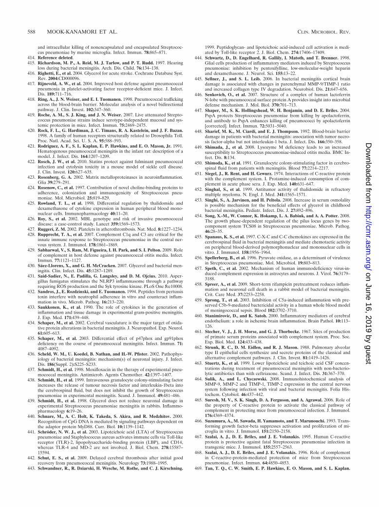
and intracellular killing of nonencapsulated and encapsulated Streptococ-cus pneumoniae by murine microglia. Infect. Immun. 78:865–871.
414. Reference deleted.415. Richardson, M. P., A. Reid, M. J. Tarlow, and P. T. Rudd. 1997. Hearing
loss during bacterial meningitis. Arch. Dis. Child. 76:134–138.416. Righetti, E., et al. 2004. Glycerol for acute stroke. Cochrane Database Syst.
Rev. 2004:CD000096.417. Rijneveld, A. W., et al. 2004. Improved host defense against pneumococcal
pneumonia in platelet-activating factor receptor-deficient mice. J. Infect.Dis. 189:711–716.
418. Ring, A., J. N. Weiser, and E. I. Tuomanen. 1998. Pneumococcal traffickingacross the blood-brain barrier. Molecular analysis of a novel bidirectionalpathway. J. Clin. Invest. 102:347–360.
419. Roche, A. M., S. J. King, and J. N. Weiser. 2007. Live attenuated Strepto-coccus pneumoniae strains induce serotype-independent mucosal and sys-temic protection in mice. Infect. Immun. 75:2469–2475.
420. Rock, F. L., G. Hardiman, J. C. Timans, R. A. Kastelein, and J. F. Bazan.1998. A family of human receptors structurally related to Drosophila Toll.Proc. Natl. Acad. Sci. U. S. A. 95:588–593.
421. Rodriguez, A. F., S. L. Kaplan, E. P. Hawkins, and E. O. Mason, Jr. 1991.Hematogenous pneumococcal meningitis in the infant rat: description of amodel. J. Infect. Dis. 164:1207–1209.
422. Rosch, J. W., et al. 2010. Statins protect against fulminant pneumococcalinfection and cytolysin toxicity in a mouse model of sickle cell disease.J. Clin. Invest. 120:627–635.
423. Rosenberg, G. A. 2002. Matrix metalloproteinases in neuroinflammation.Glia 39:279–291.
424. Rosenow, C., et al. 1997. Contribution of novel choline-binding proteins toadherence, colonization and immunogenicity of Streptococcus pneu-moniae. Mol. Microbiol. 25:819–829.
425. Rowland, T. L., et al. 1998. Differential regulation by thalidomide anddexamethasone of cytokine expression in human peripheral blood mono-nuclear cells. Immunopharmacology 40:11–20.
426. Roy, S., et al. 2002. MBL genotype and risk of invasive pneumococcaldisease: a case-control study. Lancet 359:1569–1573.
427. Ruggeri, Z. M. 2002. Platelets in atherothrombosis. Nat. Med. 8:1227–1234.428. Rupprecht, T. A., et al. 2007. Complement C1q and C3 are critical for the
innate immune response to Streptococcus pneumoniae in the central ner-vous system. J. Immunol. 178:1861–1869.
429. Sabharwal, V., S. Ram, M. Figueira, I. H. Park, and S. I. Pelton. 2009. Roleof complement in host defense against pneumococcal otitis media. Infect.Immun. 77:1121–1127.
430. Saez-Llorens, X., and G. H. McCracken. 2007. Glycerol and bacterial men-ingitis. Clin. Infect. Dis. 45:1287–1289.
431. Said-Sadier, N., E. Padilla, G. Langsley, and D. M. Ojcius. 2010. Asper-gillus fumigatus stimulates the NLRP3 inflammasome through a pathwayrequiring ROS production and the Syk tyrosine kinase. PLoS One 5:e10008.
432. Sandros, J., E. Rozdzinski, and E. Tuomanen. 1994. Peptides from pertussistoxin interfere with neutrophil adherence in vitro and counteract inflam-mation in vivo. Microb. Pathog. 16:213–220.
433. Saukkonen, K., et al. 1990. The role of cytokines in the generation ofinflammation and tissue damage in experimental gram-positive meningitis.J. Exp. Med. 171:439–448.
434. Schaper, M., et al. 2002. Cerebral vasculature is the major target of oxida-tive protein alterations in bacterial meningitis. J. Neuropathol. Exp. Neurol.61:605–613.
435. Schaper, M., et al. 2003. Differential effect of p47phox and gp91phoxdeficiency on the course of pneumococcal meningitis. Infect. Immun. 71:4087–4092.
436. Scheld, W. M., U. Koedel, B. Nathan, and H.-W. Pfister. 2002. Pathophys-iology of bacterial meningitis: mechanism(s) of neuronal injury. J. Infect.Dis. 186(Suppl. 2):S225–S233.
437. Schmidt, H., et al. 1998. Moxifloxacin in the therapy of experimental pneu-mococcal meningitis. Antimicrob. Agents Chemother. 42:1397–1407.
438. Schmidt, H., et al. 1999. Intravenous granulocyte colony-stimulating factorincreases the release of tumour necrosis factor and interleukin-1beta intothe cerebrospinal fluid, but does not inhibit the growth of Streptococcuspneumoniae in experimental meningitis. Scand. J. Immunol. 49:481–486.
439. Schmidt, H., et al. 1998. Glycerol does not reduce neuronal damage inexperimental Streptococcus pneumoniae meningitis in rabbits. Inflammo-pharmacology 6:19–26.
440. Schnare, M., A. C. Holt, K. Takeda, S. Akira, and R. Medzhitov. 2000.Recognition of CpG DNA is mediated by signaling pathways dependent onthe adaptor protein MyD88. Curr. Biol. 10:1139–1142.
441. Schroder, N. W. J., et al. 2003. Lipoteichoic acid (LTA) of Streptococcuspneumoniae and Staphylococcus aureus activates immune cells via Toll-likereceptor (TLR)-2, lipopolysaccharide-binding protein (LBP), and CD14,whereas TLR-4 and MD-2 are not involved. J. Biol. Chem. 278:15587–15594.
442. Schut, E. S., et al. 2009. Delayed cerebral thrombosis after initial goodrecovery from pneumococcal meningitis. Neurology 73:1988–1995.
443. Schwandner, R., R. Dziarski, H. Wesche, M. Rothe, and C. J. Kirschning.
1999. Peptidoglycan- and lipoteichoic acid-induced cell activation is medi-ated by Toll-like receptor 2. J. Biol. Chem. 274:17406–17409.
444. Schwartz, D., D. Engelhard, R. Gallily, I. Matoth, and T. Brenner. 1998.Glial cells production of inflammatory mediators induced by Streptococcuspneumoniae: inhibition by pentoxifylline, low-molecular-weight heparinand dexamethasone. J. Neurol. Sci. 155:13–22.
445. Sellner, J., and S. L. Leib. 2006. In bacterial meningitis cortical braindamage is associated with changes in parenchymal MMP-9/TIMP-1 ratioand increased collagen type IV degradation. Neurobiol. Dis. 21:647–656.
446. Senkovich, O., et al. 2007. Structure of a complex of human lactoferrinN-lobe with pneumococcal surface protein A provides insight into microbialdefense mechanism. J. Mol. Biol. 370:701–713.
447. Shaper, M., S. K. Hollingshead, W. H. Benjamin, and D. E. Briles. 2004.PspA protects Streptococcus pneumoniae from killing by apolactoferrin,and antibody to PspA enhances killing of pneumococci by apolactoferrin[corrected]. Infect. Immun. 72:5031–5040.
448. Sharief, M. K., M. Ciardi, and E. J. Thompson. 1992. Blood-brain barrierdamage in patients with bacterial meningitis: association with tumor necro-sis factor-alpha but not interleukin-1 beta. J. Infect. Dis. 166:350–358.
449. Shimada, J., et al. 2008. Lysozyme M deficiency leads to an increasedsusceptibility to Streptococcus pneumoniae-induced otitis media. BMC In-fect. Dis. 8:134.
450. Shimoda, K., et al. 1991. Granulocyte colony-stimulating factor in cerebro-spinal fluid from patients with meningitis. Blood 77:2214–2217.
451. Siegel, J., R. Rent, and H. Gewurz. 1974. Interactions of C-reactive proteinwith the complement system. I. Protamine-induced consumption of com-plement in acute phase sera. J. Exp. Med. 140:631–647.
452. Singhal, S., et al. 1999. Antitumor activity of thalidomide in refractorymultiple myeloma. N. Engl. J. Med. 341:1565–1571.
453. Singhi, S., A. Jarvinen, and H. Peltola. 2008. Increase in serum osmolalityis possible mechanism for the beneficial effects of glycerol in childhoodbacterial meningitis. Pediatr. Infect. Dis. J. 27:892–896.
454. Song, X.-M., W. Connor, K. Hokamp, L. A. Babiuk, and A. A. Potter. 2008.The growth phase-dependent regulation of the pilus locus genes by two-component system TCS08 in Streptococcus pneumoniae. Microb. Pathog.46:28–35.
455. Spanaus, K. S., et al. 1997. C-X-C and C-C chemokines are expressed in thecerebrospinal fluid in bacterial meningitis and mediate chemotactic activityon peripheral blood-derived polymorphonuclear and mononuclear cells invitro. J. Immunol. 158:1956–1964.
456. Spellerberg, B., et al. 1996. Pyruvate oxidase, as a determinant of virulencein Streptococcus pneumoniae. Mol. Microbiol. 19:803–813.
457. Speth, C., et al. 2002. Mechanism of human immunodeficiency virus-in-duced complement expression in astrocytes and neurons. J. Virol. 76:3179–3188.
458. Spreer, A., et al. 2009. Short-term rifampicin pretreatment reduces inflam-mation and neuronal cell death in a rabbit model of bacterial meningitis.Crit. Care Med. 37:2253–2258.
459. Sprong, T., et al. 2003. Inhibition of C5a-induced inflammation with pre-served C5b-9-mediated bactericidal activity in a human whole blood modelof meningococcal sepsis. Blood 102:3702–3710.
460. Stanimirovic, D., and K. Satoh. 2000. Inflammatory mediators of cerebralendothelium: a role in ischemic brain inflammation. Brain Pathol. 10:113–126.
461. Stecher, V. J., J. H. Morse, and G. J. Thorbecke. 1967. Sites of productionof primate serum proteins associated with complement system. Proc. Soc.Exp. Biol. Med. 124:433–438.
462. Strunk, R. C., D. M. Eidlen, and R. J. Mason. 1988. Pulmonary alveolartype II epithelial cells synthesize and secrete proteins of the classical andalternative complement pathways. J. Clin. Invest. 81:1419–1426.
463. Stuertz, K., et al. 1999. Lower lipoteichoic and teichoic acid CSF concen-trations during treatment of pneumococcal meningitis with non-bacterio-lytic antibiotics than with ceftriaxone. Scand. J. Infect. Dis. 31:367–370.
464. Sulik, A., and L. Chyczewski. 2008. Immunohistochemical analysis ofMMP-9, MMP-2 and TIMP-1, TIMP-2 expression in the central nervoussystem following infection with viral and bacterial meningitis. Folia His-tochem. Cytobiol. 46:437–442.
465. Suresh, M. V., S. K. Singh, D. A. Ferguson, and A. Agrawal. 2006. Role ofthe property of C-reactive protein to activate the classical pathway ofcomplement in protecting mice from pneumococcal infection. J. Immunol.176:4369–4374.
466. Suzumura, A., M. Sawada, H. Yamamoto, and T. Marunouchi. 1993. Trans-forming growth factor-beta suppresses activation and proliferation of mi-croglia in vitro. J. Immunol. 151:2150–2158.
467. Szalai, A. J., D. E. Briles, and J. E. Volanakis. 1995. Human C-reactiveprotein is protective against fatal Streptococcus pneumoniae infection intransgenic mice. J. Immunol. 155:2557–2563.
468. Szalai, A. J., D. E. Briles, and J. E. Volanakis. 1996. Role of complementin C-reactive-protein-mediated protection of mice from Streptococcuspneumoniae. Infect. Immun. 64:4850–4853.
469. Tan, T. Q., C. W. Smith, E. P. Hawkins, E. O. Mason, and S. L. Kaplan.
588 MOOK-KANAMORI ET AL. CLIN. MICROBIOL. REV.
on June 16, 2019 by guesthttp://cm
r.asm.org/
Dow
nloaded from
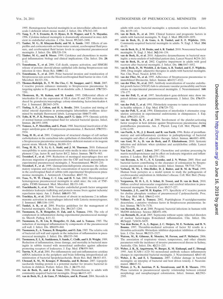
1995. Hematogenous bacterial meningitis in an intercellular adhesion mol-ecule-1-deficient infant mouse model. J. Infect. Dis. 171:342–349.
470. Tang, T., P. S. Frenette, R. O. Hynes, D. D. Wagner, and T. N. Mayadas.1996. Cytokine-induced meningitis is dramatically attenuated in mice defi-cient in endothelial selectins. J. Clin. Invest. 97:2485–2490.
471. Tauber, M. G., H. Khayam-Bashi, and M. A. Sande. 1985. Effects of am-picillin and corticosteroids on brain water content, cerebrospinal fluid pres-sure, and cerebrospinal fluid lactate levels in experimental pneumococcalmeningitis. J. Infect. Dis. 151:528–534.
472. Tauber, M. G., and B. Moser. 1999. Cytokines and chemokines in menin-geal inflammation: biology and clinical implications. Clin. Infect. Dis. 28:1–11.
473. Tenenbaum, T., et al. 2006. Cell death, caspase activation, and HMGB1release of porcine choroid plexus epithelial cells during Streptococcus suisinfection in vitro. Brain Res. 1100:1–12.
474. Tenenbaum, T., et al. 2009. Polar bacterial invasion and translocation ofStreptococcus suis across the blood-cerebrospinal fluid barrier in vitro. Cell.Microbiol. 11:323–336.
475. Thomas-Rudolph, D., T. W. Du Clos, C. M. Snapper, and C. Mold. 2007.C-reactive protein enhances immunity to Streptococcus pneumoniae bytargeting uptake to Fc gamma R on dendritic cells. J. Immunol. 178:7283–7291.
476. Thomssen, H., M. Kahan, and M. Londei. 1995. Differential effects ofinterleukin-10 on the expression of HLA class II and CD1 molecules in-duced by granulocyte/macrophage colony-stimulating factor/interleukin-4.Eur. J. Immunol. 25:2465–2470.
477. Tinling, S. P., J. Colton, and H. A. Brodie. 2004. Location and timing ofinitial osteoid deposition in postmeningitic labyrinthitis ossificans deter-mined by multiple fluorescent labels. Laryngoscope 114:675–680.
478. Tofte, R. W., P. K. Peterson, Y. Kim, and P. G. Quie. 1979. Opsonic activityof normal human cerebrospinal fluid for selected bacterial species. Infect.Immun. 26:1093–1098.
479. Tomasz, A., P. Moreillon, and G. Pozzi. 1988. Insertional inactivation of themajor autolysin gene of Streptococcus pneumoniae. J. Bacteriol. 170:5931–5934.
480. Tong, H. H., et al. 2001. Comparison of structural changes of cell surfacecarbohydrates in the eustachian tube epithelium of chinchillas infected witha Streptococcus pneumoniae neuraminidase-deficient mutant or its isogenicparent strain. Microb. Pathog. 31:309–317.
481. Tong, H. H., Y. X. Li, G. L. Stahl, and J. M. Thurman. 2010. Enhancedsusceptibility to acute pneumococcal otitis media in mice deficient in com-plement C1qa, factor B, and factor B/C2. Infect. Immun. 78:976–983.
482. Trostdorf, F., et al. 1999. Reduction of meningeal macrophages does notdecrease migration of granulocytes into the CSF and brain parenchyma inexperimental pneumococcal meningitis. J. Neuroimmunol. 99:205–210.
483. Trostdorf, F., et al. 1999. Quinupristin/dalfopristin attenuates the inflam-matory response and reduces the concentration of neuron-specific enolasein the cerebrospinal fluid of rabbits with experimental Streptococcus pneu-moniae meningitis. J. Antimicrob. Chemother. 43:87–94.
484. Tsao, N., W. W. Chang, C. C. Liu, and H. Y. Lei. 2002. Development ofhematogenous pneumococcal meningitis in adult mice: the role of TNF-alpha. FEMS Immunol. Med. Microbiol. 32:133–140.
485. Tsuchihashi, S., et al. 2006. Vascular endothelial growth factor antagonistmodulates leukocyte trafficking and protects mouse livers against ischemia/reperfusion injury. Am. J. Pathol. 168:695–705.
486. Tsuchiya, K., et al. 2010. Involvement of absent in melanoma 2 in inflam-masome activation in macrophages infected with Listeria monocytogenes.J. Immunol. 185:1186–1195.
487. Tunkel, A. R., et al. 2004. Practice guidelines for the management ofbacterial meningitis. Clin. Infect. Dis. 39:1267–1284.
488. Tuomanen, E., B. Hengstler, O. Zak, and A. Tomasz. 1986. The role ofcomplement in inflammation during experimental pneumococcal meningi-tis. Microb. Pathog. 1:15–32.
489. Tuomanen, E., H. Liu, B. Hengstler, O. Zak, and A. Tomasz. 1985. Theinduction of meningeal inflammation by components of the pneumococcalcell wall. J. Infect. Dis. 151:859–868.
490. Tuomanen, E., A. Tomasz, B. Hengstler, and O. Zak. 1985. The relative roleof bacterial cell wall and capsule in the induction of inflammation in pneu-mococcal meningitis. J. Infect. Dis. 151:535–540.
491. Tuomanen, E. I., K. Saukkonen, S. Sande, C. Cioffe, and S. D. Wright. 1989.Reduction of inflammation, tissue damage, and mortality in bacterial men-ingitis in rabbits treated with monoclonal antibodies against adhesion-promoting receptors of leukocytes. J. Exp. Med. 170:959–969.
492. Turrin, N. P., et al. 2001. Pro-inflammatory and anti-inflammatory cytokinemRNA induction in the periphery and brain following intraperitoneal ad-ministration of bacterial lipopolysaccharide. Brain Res. Bull. 54:443–453.
493. Ubenauf, K. M., M. Krueger, P. Henneke, and R. Berner. 2007. Lipopoly-saccharide binding protein is a potential marker for invasive bacterial in-fections in children. Pediatr. Infect. Dis. J. 26:159–162.
494. van de Beek, D., and J. de Gans. 2006. Dexamethasone in adults withcommunity-acquired bacterial meningitis. Drugs 66:415–427.
495. van de Beek, D., J. de Gans, P. McIntyre, and K. Prasad. 2004. Steroids in
adults with acute bacterial meningitis: a systematic review. Lancet Infect.Dis. 4:139–143.
496. van de Beek, D., et al. 2004. Clinical features and prognostic factors inadults with bacterial meningitis. N. Engl. J. Med. 351:1849–1859.
497. van de Beek, D., J. de Gans, A. R. Tunkel, and E. F. Wijdicks. 2006.Community-acquired bacterial meningitis in adults. N. Engl. J. Med. 354:44–53.
498. van de Beek, D., J. M. Drake, and A. R. Tunkel. 2010. Nosocomial bacterialmeningitis. N. Engl. J. Med. 362:146–154.
499. van de Beek, D., et al. 2010. Adjunctive dexamethasone in bacterial men-ingitis: a meta-analysis of individual patient data. Lancet Neurol. 9:254–263.
500. van de Beek, D., et al. 2002. Cognitive impairment in adults with goodrecovery after bacterial meningitis. J. Infect. Dis. 186:1047–1052.
501. van de Beek, D., M. Weisfelt, J. de Gans, A. R. Tunkel, and E. F. Wijdicks.2006. Drug insight: adjunctive therapies in adults with bacterial meningitis.Nat. Clin. Pract. Neurol. 2:504–516.
502. van der Flier, M., et al. 1995. Adherence of Streptococcus pneumoniae toimmobilized fibronectin. Infect. Immun. 63:4317–4322.
503. van der Flier, M., et al. 2005. Antibody neutralization of vascular endothe-lial growth factor (VEGF) fails to attenuate vascular permeability and brainedema in experimental pneumococcal meningitis. J. Neuroimmunol. 160:170–177.
504. van der Poll, T., et al. 1997. Interleukin-6 gene-deficient mice show im-paired defense against pneumococcal pneumonia. J. Infect. Dis. 176:439–444.
505. van der Poll, T., et al. 1991. Fibrinolytic response to tumor necrosis factorin healthy subjects. J. Exp. Med. 174:729–732.
506. van der Poll, T., et al. 1994. Elimination of interleukin 6 attenuates coag-ulation activation in experimental endotoxemia in chimpanzees. J. Exp.Med. 179:1253–1259.
507. van der Sluijs, K. F., et al. 2006. Involvement of the platelet-activatingfactor receptor in host defense against Streptococcus pneumoniae duringpostinfluenza pneumonia. Am. J. Physiol. Lung Cell. Mol. Physiol. 290:L194–L199.
508. van Furth, A. M., J. J. Roord, and R. van Furth. 1996. Roles of proinflam-matory and anti-inflammatory cytokines in pathophysiology of bacterialmeningitis and effect of adjunctive therapy. Infect. Immun. 64:4883–4890.
509. van Gool, W. A., D. van de Beek, and P. Eikelenboom. 2010. Systemicinfection and delirium: when cytokines and acetylcholine collide. Lancet375:773–775.
510. Van Lint, P., and C. Libert. 2007. Chemokine and cytokine processing bymatrix metalloproteinases and its effect on leukocyte migration and inflam-mation. J. Leukoc. Biol. 82:1375–1381.
511. van Rossum, A. M. C., E. S. Lysenko, and J. N. Weiser. 2005. Host andbacterial factors contributing to the clearance of colonization by Strepto-coccus pneumoniae in a murine model. Infect. Immun. 73:7718–7726.
512. Verbeek, M. M., I. Otte-Holler, D. J. Ruiter, and R. M. de Waal. 1999.Human brain pericytes as a model system to study the pathogenesis ofcerebrovascular amyloidosis in Alzheimer’s disease. Cell. Mol. Biol. (Noisy-le-Grand) 45:37–46.
513. Vergouwen, M. D. I., E. S. Schut, D. Troost, and D. van de Beek. 2010.Diffuse cerebral intravascular coagulation and cerebral infarction in pneu-mococcal meningitis. Neurocrit. Care 13:217–227.
514. Volanakis, J. E., and M. H. Kaplan. 1971. Specificity of C-reactive proteinfor choline phosphate residues of pneumococcal C-polysaccharide. Proc.Soc. Exp. Biol. Med. 136:612–614.
515. Vollmer, W., and A. Tomasz. 2002. Peptidoglycan N-acetylglucosaminedeacetylase, a putative virulence factor in Streptococcus pneumoniae. In-fect. Immun. 70:7176–7178.
516. von Bernuth, H., et al. 2008. Pyogenic bacterial infections in humans withMyD88 deficiency. Science 321:691–696.
517. von Bernuth, H., et al. 2005. Septicemia without sepsis: inherited disordersof nuclear factor-kappa B-mediated inflammation. Clin. Infect. Dis.41(Suppl. 7):S436–S439.
518. Von dem Borne, P. A., L. Bajzar, J. C. Meijers, M. E. Nesheim, and B. N.Bouma. 1997. Thrombin-mediated activation of factor XI results in athrombin-activatable fibrinolysis inhibitor-dependent inhibition of fibrino-lysis. J. Clin. Invest. 99:2323–2327.
519. Watson, M., R. Gilmour, R. Menzies, M. Ferson, and P. McIntyre. 2006.The association of respiratory viruses, temperature, and other climaticparameters with the incidence of invasive pneumococcal disease in Sydney,Australia. Clin. Infect. Dis. 42:211–215.
520. Weber, J. R., K. Angstwurm, W. Burger, K. M. Einhaupl, and U. Dirnagl.1995. Anti ICAM-1 (CD 54) monoclonal antibody reduces inflammatorychanges in experimental bacterial meningitis. J. Neuroimmunol. 63:63–68.
521. Weber, J. R., and E. I. Tuomanen. 2007. Cellular damage in bacterialmeningitis: an interplay of bacterial and host driven toxicity. J. Neuroim-munol. 184:45–52.
522. Weiser, J. N., R. Austrian, P. K. Sreenivasan, and H. R. Masure. 1994.Phase variation in pneumococcal opacity: relationship between colonialmorphology and nasopharyngeal colonization. Infect. Immun. 62:2582–2589.
VOL. 24, 2011 PATHOGENESIS OF PNEUMOCOCCAL MENINGITIS 589
on June 16, 2019 by guesthttp://cm
r.asm.org/
Dow
nloaded from
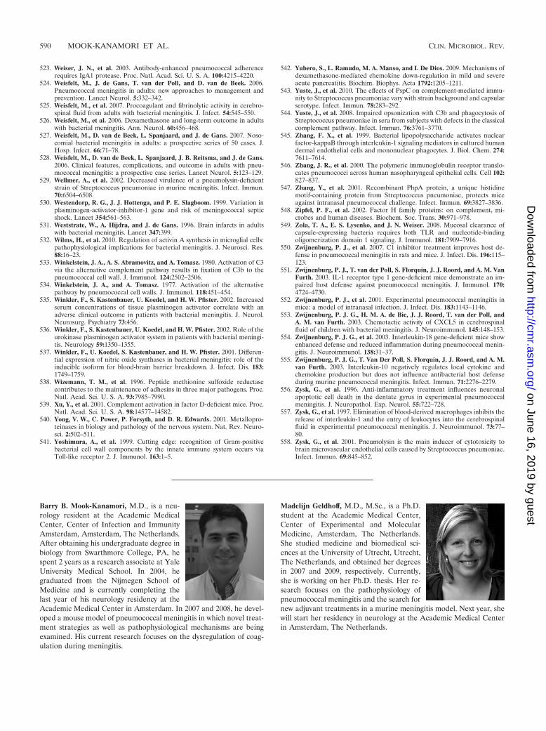
523. Weiser, J. N., et al. 2003. Antibody-enhanced pneumococcal adherencerequires IgA1 protease. Proc. Natl. Acad. Sci. U. S. A. 100:4215–4220.
524. Weisfelt, M., J. de Gans, T. van der Poll, and D. van de Beek. 2006.Pneumococcal meningitis in adults: new approaches to management andprevention. Lancet Neurol. 5:332–342.
525. Weisfelt, M., et al. 2007. Procoagulant and fibrinolytic activity in cerebro-spinal fluid from adults with bacterial meningitis. J. Infect. 54:545–550.
526. Weisfelt, M., et al. 2006. Dexamethasone and long-term outcome in adultswith bacterial meningitis. Ann. Neurol. 60:456–468.
527. Weisfelt, M., D. van de Beek, L. Spanjaard, and J. de Gans. 2007. Noso-comial bacterial meningitis in adults: a prospective series of 50 cases. J.Hosp. Infect. 66:71–78.
528. Weisfelt, M., D. van de Beek, L. Spanjaard, J. B. Reitsma, and J. de Gans.2006. Clinical features, complications, and outcome in adults with pneu-mococcal meningitis: a prospective case series. Lancet Neurol. 5:123–129.
529. Wellmer, A., et al. 2002. Decreased virulence of a pneumolysin-deficientstrain of Streptococcus pneumoniae in murine meningitis. Infect. Immun.70:6504–6508.
530. Westendorp, R. G., J. J. Hottenga, and P. E. Slagboom. 1999. Variation inplasminogen-activator-inhibitor-1 gene and risk of meningococcal septicshock. Lancet 354:561–563.
531. Weststrate, W., A. Hijdra, and J. de Gans. 1996. Brain infarcts in adultswith bacterial meningitis. Lancet 347:399.
532. Wilms, H., et al. 2010. Regulation of activin A synthesis in microglial cells:pathophysiological implications for bacterial meningitis. J. Neurosci. Res.88:16–23.
533. Winkelstein, J. A., A. S. Abramovitz, and A. Tomasz. 1980. Activation of C3via the alternative complement pathway results in fixation of C3b to thepneumococcal cell wall. J. Immunol. 124:2502–2506.
534. Winkelstein, J. A., and A. Tomasz. 1977. Activation of the alternativepathway by pneumococcal cell walls. J. Immunol. 118:451–454.
535. Winkler, F., S. Kastenbauer, U. Koedel, and H. W. Pfister. 2002. Increasedserum concentrations of tissue plasminogen activator correlate with anadverse clinical outcome in patients with bacterial meningitis. J. Neurol.Neurosurg. Psychiatry 73:456.
536. Winkler, F., S. Kastenbauer, U. Koedel, and H. W. Pfister. 2002. Role of theurokinase plasminogen activator system in patients with bacterial meningi-tis. Neurology 59:1350–1355.
537. Winkler, F., U. Koedel, S. Kastenbauer, and H. W. Pfister. 2001. Differen-tial expression of nitric oxide synthases in bacterial meningitis: role of theinducible isoform for blood-brain barrier breakdown. J. Infect. Dis. 183:1749–1759.
538. Wizemann, T. M., et al. 1996. Peptide methionine sulfoxide reductasecontributes to the maintenance of adhesins in three major pathogens. Proc.Natl. Acad. Sci. U. S. A. 93:7985–7990.
539. Xu, Y., et al. 2001. Complement activation in factor D-deficient mice. Proc.Natl. Acad. Sci. U. S. A. 98:14577–14582.
540. Yong, V. W., C. Power, P. Forsyth, and D. R. Edwards. 2001. Metallopro-teinases in biology and pathology of the nervous system. Nat. Rev. Neuro-sci. 2:502–511.
541. Yoshimura, A., et al. 1999. Cutting edge: recognition of Gram-positivebacterial cell wall components by the innate immune system occurs viaToll-like receptor 2. J. Immunol. 163:1–5.
542. Yubero, S., L. Ramudo, M. A. Manso, and I. De Dios. 2009. Mechanisms ofdexamethasone-mediated chemokine down-regulation in mild and severeacute pancreatitis. Biochim. Biophys. Acta 1792:1205–1211.
543. Yuste, J., et al. 2010. The effects of PspC on complement-mediated immu-nity to Streptococcus pneumoniae vary with strain background and capsularserotype. Infect. Immun. 78:283–292.
544. Yuste, J., et al. 2008. Impaired opsonization with C3b and phagocytosis ofStreptococcus pneumoniae in sera from subjects with defects in the classicalcomplement pathway. Infect. Immun. 76:3761–3770.
545. Zhang, F. X., et al. 1999. Bacterial lipopolysaccharide activates nuclearfactor-kappaB through interleukin-1 signaling mediators in cultured humandermal endothelial cells and mononuclear phagocytes. J. Biol. Chem. 274:7611–7614.
546. Zhang, J. R., et al. 2000. The polymeric immunoglobulin receptor translo-cates pneumococci across human nasopharyngeal epithelial cells. Cell 102:827–837.
547. Zhang, Y., et al. 2001. Recombinant PhpA protein, a unique histidinemotif-containing protein from Streptococcus pneumoniae, protects miceagainst intranasal pneumococcal challenge. Infect. Immun. 69:3827–3836.
548. Zipfel, P. F., et al. 2002. Factor H family proteins: on complement, mi-crobes and human diseases. Biochem. Soc. Trans. 30:971–978.
549. Zola, T. A., E. S. Lysenko, and J. N. Weiser. 2008. Mucosal clearance ofcapsule-expressing bacteria requires both TLR and nucleotide-bindingoligomerization domain 1 signaling. J. Immunol. 181:7909–7916.
550. Zwijnenburg, P. J., et al. 2007. C1 inhibitor treatment improves host de-fense in pneumococcal meningitis in rats and mice. J. Infect. Dis. 196:115–123.
551. Zwijnenburg, P. J., T. van der Poll, S. Florquin, J. J. Roord, and A. M. VanFurth. 2003. IL-1 receptor type 1 gene-deficient mice demonstrate an im-paired host defense against pneumococcal meningitis. J. Immunol. 170:4724–4730.
552. Zwijnenburg, P. J., et al. 2001. Experimental pneumococcal meningitis inmice: a model of intranasal infection. J. Infect. Dis. 183:1143–1146.
553. Zwijnenburg, P. J. G., H. M. A. de Bie, J. J. Roord, T. van der Poll, andA. M. van Furth. 2003. Chemotactic activity of CXCL5 in cerebrospinalfluid of children with bacterial meningitis. J. Neuroimmunol. 145:148–153.
554. Zwijnenburg, P. J. G., et al. 2003. Interleukin-18 gene-deficient mice showenhanced defense and reduced inflammation during pneumococcal menin-gitis. J. Neuroimmunol. 138:31–37.
555. Zwijnenburg, P. J. G., T. Van Der Poll, S. Florquin, J. J. Roord, and A. M.van Furth. 2003. Interleukin-10 negatively regulates local cytokine andchemokine production but does not influence antibacterial host defenseduring murine pneumococcal meningitis. Infect. Immun. 71:2276–2279.
556. Zysk, G., et al. 1996. Anti-inflammatory treatment influences neuronalapoptotic cell death in the dentate gyrus in experimental pneumococcalmeningitis. J. Neuropathol. Exp. Neurol. 55:722–728.
557. Zysk, G., et al. 1997. Elimination of blood-derived macrophages inhibits therelease of interleukin-1 and the entry of leukocytes into the cerebrospinalfluid in experimental pneumococcal meningitis. J. Neuroimmunol. 73:77–80.
558. Zysk, G., et al. 2001. Pneumolysin is the main inducer of cytotoxicity tobrain microvascular endothelial cells caused by Streptococcus pneumoniae.Infect. Immun. 69:845–852.
Barry B. Mook-Kanamori, M.D., is a neu-rology resident at the Academic MedicalCenter, Center of Infection and ImmunityAmsterdam, Amsterdam, The Netherlands.After obtaining his undergraduate degree inbiology from Swarthmore College, PA, hespent 2 years as a research associate at YaleUniversity Medical School. In 2004, hegraduated from the Nijmegen School ofMedicine and is currently completing thelast year of his neurology residency at theAcademic Medical Center in Amsterdam. In 2007 and 2008, he devel-oped a mouse model of pneumococcal meningitis in which novel treat-ment strategies as well as pathophysiological mechanisms are beingexamined. His current research focuses on the dysregulation of coag-ulation during meningitis.
Madelijn Geldhoff, M.D., M.Sc., is a Ph.D.student at the Academic Medical Center,Center of Experimental and MolecularMedicine, Amsterdam, The Netherlands.She studied medicine and biomedical sci-ences at the University of Utrecht, Utrecht,The Netherlands, and obtained her degreesin 2007 and 2009, respectively. Currently,she is working on her Ph.D. thesis. Her re-search focuses on the pathophysiology ofpneumococcal meningitis and the search fornew adjuvant treatments in a murine meningitis model. Next year, shewill start her residency in neurology at the Academic Medical Centerin Amsterdam, The Netherlands.
590 MOOK-KANAMORI ET AL. CLIN. MICROBIOL. REV.
on June 16, 2019 by guesthttp://cm
r.asm.org/
Dow
nloaded from
Tom van der Poll, M.D., Ph.D., is Professorof Medicine at the Academic Medical Cen-ter, University of Amsterdam, The Nether-lands. He is Head of the Center of Experi-mental and Molecular Medicine and Chairof the Department of Infectious Diseases atthis institution. Dr. van der Poll is boardcertified in internal medicine and infectiousdiseases. His training included a postdoc-toral research fellowship at Cornell Univer-sity Medical College in New York (1993 to1995). Dr. van der Poll is a former Fellow of the Royal Dutch Academyof Arts and Sciences (1995 to 2000). His research focuses on the innateimmune and procoagulant responses during sepsis. He has publishedmore than 450 original articles and over 60 book chapters on this topic.
Diederik van de Beek, M.D., Ph.D., is a neu-rologist at the Academic Medical Center,Center of Infection and Immunity Amster-dam, Amsterdam, The Netherlands. Hequalified in medicine at the University ofAmsterdam in 1999 and specialized in neu-rology at the Academic Medical Center(2000 to 2006). In 2004, he successfully de-fended his thesis on bacterial meningitis inadults. During his postdoctoral time at theMayo Clinics, Rochester, MN, he studiedneurological infections after solid organ transplantation and experi-mental meningitis (2006 to 2007). Currently, he holds a faculty positionas a neurologist and Academic Medical Center Fellow in Amsterdam,The Netherlands. His research program focuses on bacterial meningi-tis but also includes projects on infections after stroke and septicencephalopathy.
VOL. 24, 2011 PATHOGENESIS OF PNEUMOCOCCAL MENINGITIS 591
on June 16, 2019 by guesthttp://cm
r.asm.org/
Dow
nloaded from