Embed Size (px)
Citation preview
320 | may 2014 | volume 44 | number 5 | journal of orthopaedic & sports physical therapy
[ research report ]
Quadriceps strengthening is a common component of the rehabilitation program for persons with patellofemoral pain (PFP) and typically includes weightbearing and non–weightbearing exercises.1,9,12,18,27 Both forms of exercise
have their advantages with respect to quadriceps strengthening. Weightbearing exercises are more functional in nature7,11 and incorporate contractions of multiple agonist and antagonist muscle groups.1921,25
In contrast, non–weight-bearing knee extension exercises re-quire less cocontraction of antagonist muscles thus provide better
quadriceps muscle isolation.5
When designing a quadriceps-strengthening program for individuals with PFP, it is important to select exer-cises that promote muscle loading and adaptation and minimize patellofemoral joint (PFJ) stress and pain. Steinkamp and colleagues17 first described the in-fluence of weight-bearing status on PFJ stress. These authors reported a con-trast in the pattern of PFJ stress during weight-bearing and non–weight-bearing exercises performed at 0° to 90° of knee flexion. During the weight-bearing task (leg press), PFJ stress increased linearly from 0° to 90° of knee flexion.17 During the non–weight-bearing knee extension exercise, however, PFJ stress was greatest at 0° and decreased with knee flexion.17
The findings of Steinkamp et al17 have been challenged by Escamilla and col-leagues,6 who quantified PFJ reaction forces during similar weight-bearing and non–weight-bearing tasks (squat and knee extension, respectively). These authors reported that the PFJ reaction
TT STUDY DESIGN: Single-group, repeated-measures design.
TT OBJECTIVE: To compare patellofemoral joint (PFJ) stress among weight-bearing and non–weight-bearing quadriceps exercises.
TT BACKGROUND: An important consideration when prescribing exercises to strengthen the quad-riceps in persons with patellofemoral pain is to minimize PFJ loading. Currently, there is disagree-ment in the literature as to which exercises and ranges of motion best accomplish this goal.
TT METHODS: Ten healthy subjects participated. Lower extremity kinematics, kinetics, and electro-myography of the knee musculature were obtained during a weight-bearing squatting exercise and 2 non–weight-bearing knee extension exercises: (1) knee extension with variable resistance, and (2) knee extension with constant resistance. A previ-ously described biomechanical model was used to estimate PFJ stress at 0°, 15°, 30°, 45°, 60°, 75°, and 90° of knee flexion. PFJ stress was compared among the 3 exercises using a 2-way analysis of variance with repeated measures.
TT RESULTS: Compared to the 2 non–weight-bearing exercises, the squat exercise produced sig-nificantly higher PFJ stress at 90°, 75°, and 60° of knee flexion. Conversely, the 2 non–weight-bearing exercises produced significantly higher PFJ stress at 30°, 15°, and 0° of knee flexion when compared to the squat exercise. The knee-extension-with-variable-resistance exercise produced significantly lower PFJ stress than the knee-extension-with-constant-resistance exercise at 90°, 75°, and 60° of knee flexion.
TT CONCLUSION: To minimize PFJ stress while performing quadriceps exercises, our data suggest that the squat exercise should be performed from 45° to 0° of knee flexion and the knee-extension-with-variable-resistance exercise should be per-formed from 90° to 45° of knee flexion. J Orthop Sports Phys Ther 2014;44(5):320-327. Epub 27 March 2014. doi:10.2519/jospt.2014.4936
TT KEY WORDS: force, patella, pressure, rehabilitation
1Jacquelin Perry Musculoskeletal Biomechanics Research Laboratory, Division of Biokinesiology and Physical Therapy, University of Southern California, Los Angeles, CA. 2Department of Physical Therapy, University of Nevada, Las Vegas, Las Vegas, NV. 3School and Graduate Institute of Physical Therapy, National Taiwan University, Taipei, Taiwan, Republic of China. 4Department of Physical Therapy and Rehabilitation Science, University of California San Francisco, San Francisco, CA. 5Department of Physical Therapy, University of Pittsburgh, Pittsburgh, PA. This study was approved by the Health Science Institutional Review Board, University of Southern California, Los Angeles, CA. The authors certify that they have no affiliations with or financial involvement in any organization or entity with a direct financial interest in the subject matter or materials discussed in the article. Address correspondence to Dr Christopher M. Powers, Division of Biokinesiology and Physical Therapy, University of Southern California, 1540 East Alcazar Street, CHP-155, Los Angeles, CA 90089-9006. E-mail: [email protected] T Copyright ©2014 Journal of Orthopaedic & Sports Physical Therapy®
CHRISTOPHER M. POWERS, PT, PhD, FACSM, FAPTA1 • KAI-YU HO, PT, PhD2
YU-JEN CHEN, PT, PhD3 • RICHARD B. SOUZA, PT, ATC, PhD4 • SHAWN FARROKHI, DPT, PhD5
Patellofemoral Joint Stress During WeightBearing and Non–WeightBearing
Quadriceps Exercises
44-05 Powers.indd 320 4/16/2014 4:14:50 PM
Jour
nal o
f O
rtho
paed
ic &
Spo
rts
Phys
ical
The
rapy
®
Dow
nloa
ded
from
ww
w.jo
spt.o
rg a
t on
Nov
embe
r 18
, 201
4. F
or p
erso
nal u
se o
nly.
No
othe
r us
es w
ithou
t per
mis
sion
. C
opyr
ight
© 2
014
Jour
nal o
f O
rtho
paed
ic &
Spo
rts
Phys
ical
The
rapy
®. A
ll ri
ghts
res
erve
d.
journal of orthopaedic & sports physical therapy | volume 44 | number 5 | may 2014 | 321
force increased in both exercises as the knee flexed from 0° to 60°. Beyond 60°, however, the curves diverged. While the PFJ reaction force during the weight-bearing exercise continued to increase with increasing knee flexion, the PFJ re-action force during non–weight-bearing knee extension decreased. At 90° of knee flexion compared to 0° of flexion, the non–weight-bearing exercise exhibited higher PFJ reaction forces. Although PFJ stress was not calculated in their study, Escamilla and colleagues6 proposed that the PFJ stress curves would show similar trajectories.
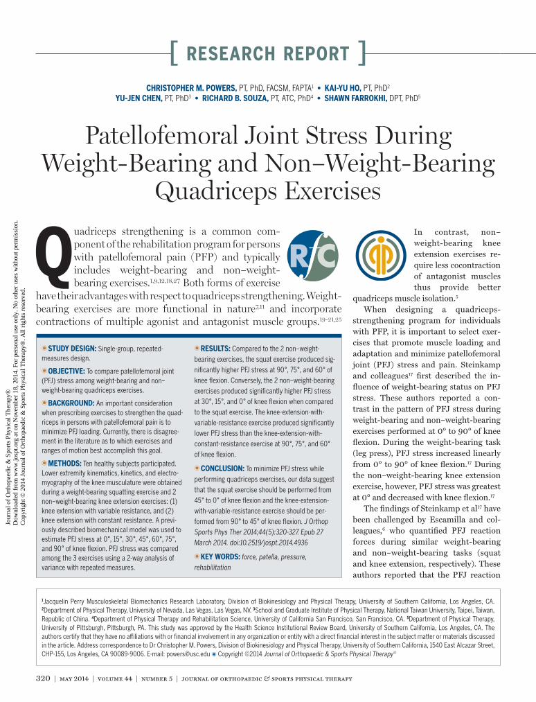
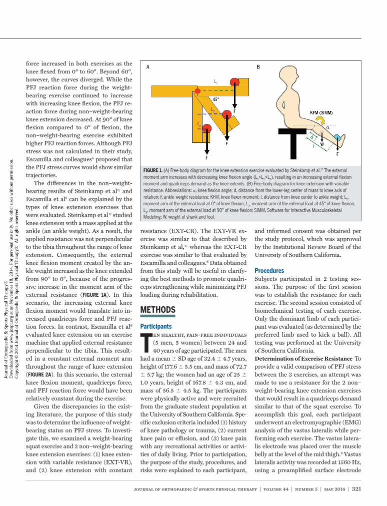
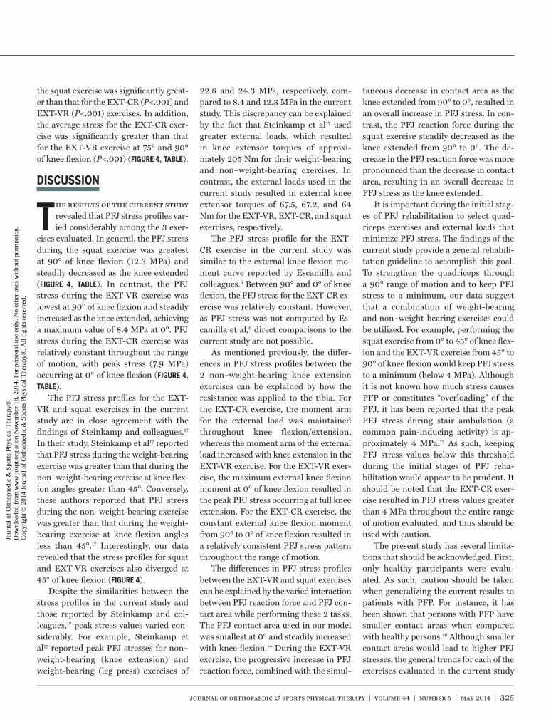
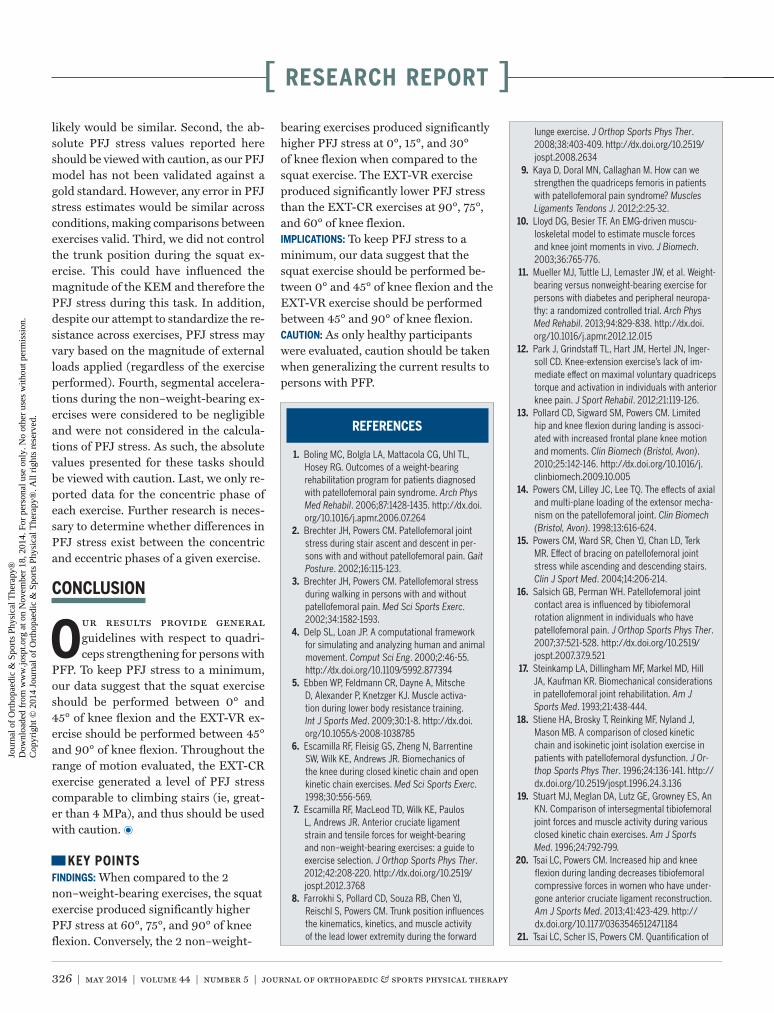
The differences in the non–weight-bearing results of Steinkamp et al17 and Escamilla et al6 can be explained by the types of knee extension exercises that were evaluated. Steinkamp et al17 studied knee extension with a mass applied at the ankle (an ankle weight). As a result, the applied resistance was not perpendicular to the tibia throughout the range of knee extension. Consequently, the external knee flexion moment created by the an-kle weight increased as the knee extended from 90° to 0°, because of the progres-sive increase in the moment arm of the external resistance (FIGURE 1A). In this scenario, the increasing external knee flexion moment would translate into in-creased quadriceps force and PFJ reac-tion forces. In contrast, Escamilla et al6 evaluated knee extension on an exercise machine that applied external resistance perpendicular to the tibia. This result-ed in a constant external moment arm throughout the range of knee extension (FIGURE 2A). In this scenario, the external knee flexion moment, quadriceps force, and PFJ reaction force would have been relatively constant during the exercise.
Given the discrepancies in the exist-ing literature, the purpose of this study was to determine the influence of weight-bearing status on PFJ stress. To investi-gate this, we examined a weight-bearing squat exercise and 2 non–weight-bearing knee extension exercises: (1) knee exten-sion with variable resistance (EXT-VR), and (2) knee extension with constant
resistance (EXT-CR). The EXT-VR ex-ercise was similar to that described by Steinkamp et al,17 whereas the EXT-CR exercise was similar to that evaluated by Escamilla and colleagues.6 Data obtained from this study will be useful in clarify-ing the best methods to promote quadri-ceps strengthening while minimizing PFJ loading during rehabilitation.
METHODS
Participants
Ten healthy, pain-free individuals (5 men, 5 women) between 24 and 40 years of age participated. The men
had a mean SD age of 32.4 4.7 years, height of 177.6 5.5 cm, and mass of 72.7 5.7 kg; the women had an age of 25 1.0 years, height of 167.8 4.3 cm, and mass of 56.5 4.5 kg. The participants were physically active and were recruited from the graduate student population at the University of Southern California. Spe-cific exclusion criteria included (1) history of knee pathology or trauma, (2) current knee pain or effusion, and (3) knee pain with any recreational activities or activi-ties of daily living. Prior to participation, the purpose of the study, procedures, and risks were explained to each participant,
and informed consent was obtained per the study protocol, which was approved by the Institutional Review Board of the University of Southern California.
ProceduresSubjects participated in 2 testing ses-sions. The purpose of the first session was to establish the resistance for each exercise. The second session consisted of biomechanical testing of each exercise. Only the dominant limb of each partici-pant was evaluated (as determined by the preferred limb used to kick a ball). All testing was performed at the University of Southern California.Determination of Exercise Resistance To provide a valid comparison of PFJ stress between the 3 exercises, an attempt was made to use a resistance for the 2 non–weight-bearing knee extension exercises that would result in a quadriceps demand similar to that of the squat exercise. To accomplish this goal, each participant underwent an electromyographic (EMG) analysis of the vastus lateralis while per-forming each exercise. The vastus latera-lis electrode was placed over the muscle belly at the level of the mid thigh.8 Vastus lateralis activity was recorded at 1560 Hz, using a preamplified surface electrode
FIGURE 1. (A) Free-body diagram for the knee extension exercise evaluated by Steinkamp et al.17 The external moment arm increases with decreasing knee flexion angle (L1>L2>L3), resulting in an increasing external flexion moment and quadriceps demand as the knee extends. (B) Free-body diagram for knee extension with variable resistance. Abbreviations: α, knee flexion angle; d, distance from the lower-leg center of mass to knee axis of rotation; F, ankle-weight resistance; KFM, knee flexor moment; l, distance from knee center to ankle weight; L1, moment arm of the external load at 0° of knee flexion; L2, moment arm of the external load at 45° of knee flexion; L3, moment arm of the external load at 90° of knee flexion; SIMM, Software for Interactive Musculoskeletal Modeling; W, weight of shank and foot.
44-05 Powers.indd 321 4/16/2014 4:14:51 PM
Jour
nal o
f O
rtho
paed
ic &
Spo
rts
Phys
ical
The
rapy
®
Dow
nloa
ded
from
ww
w.jo
spt.o
rg a
t on
Nov
embe
r 18
, 201
4. F
or p
erso
nal u
se o
nly.
No
othe
r us
es w
ithou
t per
mis
sion
. C
opyr
ight
© 2
014
Jour
nal o
f O
rtho
paed
ic &
Spo
rts
Phys
ical
The
rapy
®. A
ll ri
ghts
res
erve
d.
322 | may 2014 | volume 44 | number 5 | journal of orthopaedic & sports physical therapy
[ research report ](Motion Lab Systems, Inc, Baton Rouge, LA). The vastus lateralis EMG signal was band-pass filtered (50-200 Hz) and pro-cessed using a root-mean-square smooth-ing algorithm (75-millisecond window).
First, the level of vastus lateralis ac-tivation was established for the squat exercise. Participants assumed a comfort-able stance position (feet shoulder-width apart and toes straight ahead) and were instructed to execute the squat from a starting position of 0° of knee flexion to a depth of 90° (as determined by a plastic goniometer) and to return to the start po-sition. To ensure that 90° of knee flexion was achieved, a stool with an adjustable seat height was placed behind each par-ticipant to serve as the target for desired squat depth. Participants descended by flexing the hips and knees until the pos-terior aspect of the thighs was in con-tact with the stool. The velocity of the squatting maneuver was controlled by a metronome, such that the knee angular velocity was approximately 30°/s. Three squat trials were performed. With the participant still connected to the EMG unit, the vastus lateralis EMG time inte-gral during the concentric phase of the squat cycle was calculated for each trial and averaged.
Next, the external loads for the EXT-VR and EXT-CR exercises were deter-mined. This was done by matching the vastus lateralis EMG time integral for each non–weight-bearing exercise to that calculated for the squat. The EXT-VR exercise was performed with each participant sitting on a chair (90° of hip and knee flexion), with an ankle weight secured to the distal end of the tibia (su-perior to the malleolus). The EXT-CR ex-ercise was performed on a dynamometer (Kin-Com; Isokinetic International, Har-rison, TN). As with the EXT-VR exercise, participants were positioned in 90° of hip and knee flexion and the resistance pad was secured to the distal end of the tibia (superior to the malleolus). The Kin-Com dynamometer allows for the resistance pad to be applied perpendicular to the tibia, thus providing a constant external
moment via a fixed lever arm throughout the range of motion. The dynamometer was set to isotonic mode, allowing the re-sistance to be adjusted as necessary.
For both the EXT-VR and EXT-CR exercises, participants performed 3 knee extension trials (90°-0° of knee flexion). As with the squat exercise, the knee angu-lar velocity for both non–weight-bearing exercises was controlled by a metronome (30°/s). After each set of 3 trials, the vas-tus lateralis EMG time integral during the concentric phase of each exercise was calculated for each trial and averaged. If the calculated vastus lateralis EMG time integral did not fall within 95% to 105% of the value established during the squat exercise, the external resistance was ad-justed accordingly and 3 additional trials were collected. This process was repeated until the 5% difference threshold was achieved for both non–weight-bearing exercises. On average, the 3 trials were repeated 5 times to achieve this thresh-old. Using this procedure, the average SD resistance was 4.4 3.5 kg for the EXT-VR exercise and 4.1 2.2 kg for the EXT-CR exercise.Biomechanical Testing After determin-ing the resistance for the EXT-VR and the EXT-CR exercises, biomechanical
testing of each exercise commenced. The purpose of this testing was to calculate the knee extensor moment (KEM) dur-ing each exercise, which was the key in-put variable of a PFJ model to estimate PFJ stress. The KEM during the squat exercise was determined using inverse-dynamics equations. The KEM during the EXT-VR and EXT-CR exercises was estimated using free-body diagrams (see below for details).
Although the net KEM provides a rea-sonable estimate of the demands placed on the knee extensors, the true quadri-ceps force would be underestimated in the presence of muscle cocontraction. To account for the potential influence of muscle cocontraction, an estimate of the knee flexor moment (KFM) was obtained during each exercise by using an EMG-driven musculoskeletal model (see below for details).
Using a previously described marker set,13 lower extremity kinematics during the squat exercise was assessed with an 8-camera, Vicon motion analysis system at a sampling frequency of 250 Hz (OMG plc, Oxford, UK). Ground reaction forces were obtained with 2 force platforms at a rate of 1500 Hz (Advanced Mechanical Technology, Inc, Watertown, MA). Using
FIGURE 2. (A) Free-body diagram for the knee extension exercise used by Escamilla et al.6 The moment arm is constant throughout knee extension (L1 = L2 = L3), resulting in a consistent external flexion moment and quadriceps demand as the knee extends. (B) Free-body diagram for knee extension with constant resistance. Abbreviations: α, knee flexion angle; d, distance from the lower-leg center of mass to knee axis of rotation; F, ankle-weight resistance; KFM, knee flexor moment; l, distance from knee center to ankle weight; L1, moment arm of the external load at 0° of knee flexion; L2, moment arm of the external load at 45° of knee flexion; L3, moment arm of the external load at 90° of knee flexion; SIMM, Software for Interactive Musculoskeletal Modeling; W, weight of shank and foot.
44-05 Powers.indd 322 4/16/2014 4:14:52 PM
Jour
nal o
f O
rtho
paed
ic &
Spo
rts
Phys
ical
The
rapy
®
Dow
nloa
ded
from
ww
w.jo
spt.o
rg a
t on
Nov
embe
r 18
, 201
4. F
or p
erso
nal u
se o
nly.
No
othe
r us
es w
ithou
t per
mis
sion
. C
opyr
ight
© 2
014
Jour
nal o
f O
rtho
paed
ic &
Spo
rts
Phys
ical
The
rapy
®. A
ll ri
ghts
res
erve
d.
journal of orthopaedic & sports physical therapy | volume 44 | number 5 | may 2014 | 323
the instrumentation described above, EMG signals were obtained from the me-dial and lateral hamstrings and the medi-al and lateral heads of the gastrocnemius. The electrodes for the medial and lateral hamstrings were placed over the respec-tive muscle bellies, midway between the ischial tuberosity and the epicondyles of the femur.8 The electrodes for the medial and lateral gastrocnemius were placed over the upper one third of the respec-tive muscle bellies.8
The procedures for the squat exercise were identical to those described above. Participants were positioned such that each foot fell within the boundaries of one of the adjacent force platforms. Once 3 trials of the squat exercise had been completed, the motion markers were re-moved and 3 trials of the EXT-VR and EXT-CR exercises were obtained using the previously established resistance. A sufficient rest (approximately 10 min-utes) was provided between exercise con-ditions to minimize fatigue.
Data AnalysisKinematic and kinetic data obtained during the squat exercise were processed using Visual3D software (C-Motion, Inc, Germantown, MD). Marker trajectories were low-pass filtered at 6 Hz using a fourth-order Butterworth filter. As de-scribed above, EMG signals were band-pass filtered (50-200 Hz) and processed using a root-mean-square smoothing al-gorithm (75-millisecond window). EMG
data were normalized to the EMG data acquired during a maximal voluntary iso-metric contraction.Estimate of KFM To account for cocon-traction during the 3 exercises evaluated, an estimate of the KFM was required. The KFM was obtained from SIMM modeling software (Motion Analysis Corporation, Santa Rosa, CA). The SIMM lower-limb model contains musculotendon actuators with information about peak isometric muscle force, optimal muscle-fiber length, pennation angle, and ten-don slack length for the muscles of the lower extremity.4 In the SIMM software, muscles are represented as a series of 3-D vectors that are constrained to wrap over underlying structures. Using a Hill-based model, the SIMM software estimated the KFM based on the individual’s lower ex-tremity kinematics, speed of movement, and flexor muscle EMG. The estimated KEM derived from SIMM software has been found to be comparable to the KEM calculated with inverse-dynamics equations.10
To obtain a more accurate assessment of the KEM during the squat exercise, the KFM calculated by SIMM was added to the net KEM as estimated from the in-verse-dynamics equations. This resulted in an adjusted KEM that accounted for antagonist muscle activation through-out the squat cycle: adjusted KEM = [net KEM (inverse dynamics) + KFM (SIMM)]. The adjusted KEM during the EXT-VR exercise was calculated based on
the following equation: adjusted KEM = [(W × d + F × l)(cos α) + KFM (SIMM)], where W is weight of shank and foot (6.0% of total body weight),26 d is dis-tance from the lower-leg center of mass to the knee axis (43.3% of distance be-tween knee axis and medial malleolus),26 F is ankle-weight resistance, l is the dis-tance from knee center to ankle weight, and α is the knee flexion angle (FIGURE 1B).
The adjusted KEM during the EXT-CR exercise was calculated based on the following equation: [(W × d × cos α) + (F × l) + KFM (SIMM)], where W is weight of shank and foot (6.0% of total body weight),26 d is distance from the lower-leg center of mass to the knee axis (43.3% of distance between knee axis and medial malleolus),26 F is ankle-weight resistance, l is the distance from knee center to ankle weight, and α is the knee flexion angle (FIGURE 2B).
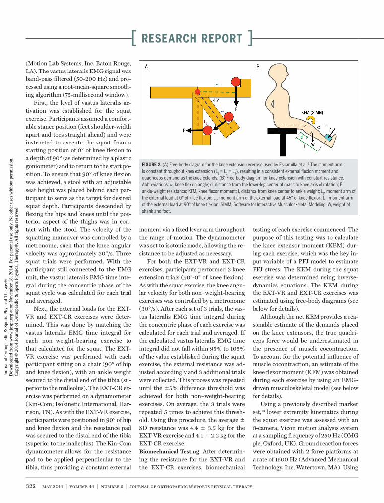
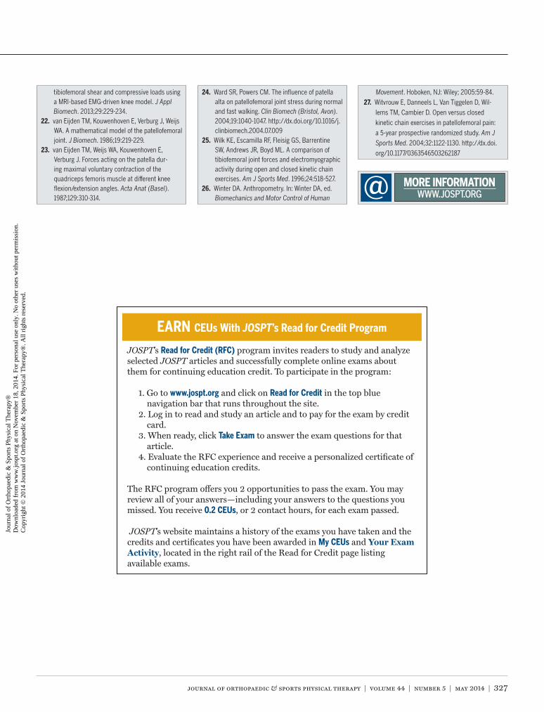
Biomechanical Model to Estimate PFJ StressA previously described model was used to quantify PFJ stress (FIGURE 3).2,3,22,24 Input variables included participant-specific parameters (ie, knee joint flexion angle and adjusted KEM) and data obtained from the literature (ie, PFJ contact area,14 quadriceps effective lever arm,14 and the relationship between quadriceps force and PFJ reaction force).23
Step 1 of the algorithm was to approxi-mate the quadriceps force. First, the effec-tive lever arm for the quadriceps muscle
Adjusted knee extensor moment
Quadriceps force
Quadriceps e�ective lever arm†
Relationship between quadriceps force and patellofemoral joint reaction force‡
Patellofemoral joint reaction force
Patellofemoral joint stress
Patellofemoral joint contact area*
Knee joint flexion angle
FIGURE 3. Flow chart of patellofemoral joint model. *Data obtained from Powers and colleagues.14 †Data obtained from van Eijden and colleagues.22 ‡Data obtained from van Eijden and colleagues.23
44-05 Powers.indd 323 4/16/2014 4:14:53 PM
Jour
nal o
f O
rtho
paed
ic &
Spo
rts
Phys
ical
The
rapy
®
Dow
nloa
ded
from
ww
w.jo
spt.o
rg a
t on
Nov
embe
r 18
, 201
4. F
or p
erso
nal u
se o
nly.
No
othe
r us
es w
ithou
t per
mis
sion
. C
opyr
ight
© 2
014
Jour
nal o
f O
rtho
paed
ic &
Spo
rts
Phys
ical
The
rapy
®. A
ll ri
ghts
res
erve
d.
324 | may 2014 | volume 44 | number 5 | journal of orthopaedic & sports physical therapy
[ research report ]
was determined at each degree of knee flexion by fitting a nonlinear equation to the data of van Eijden and colleagues.22 Next, the quadriceps force was calculated by dividing the adjusted KEM calculated during each exercise by the effective le-ver arm. Step 2 of the algorithm was to estimate the PFJ reaction force. This was accomplished by multiplying the quad-riceps force by a constant established by van Eijden and colleagues23 that defined
the relationship between quadriceps force and PFJ reaction force as a function of knee flexion angle. The third step of the algorithm was to calculate PFJ stress. The PFJ joint reaction force established in step 2 was divided by the PFJ contact area. PFJ contact area was determined for each knee flexion angle using a second-order polynomial curve fit to the data of Powers et al.14 The model output was PFJ stress as a function of knee flexion angle.
Statistical AnalysisPFJ stress was compared among the 3 exercises at 0°, 15°, 30°, 45°, 60°, 75°, and 90° of knee flexion using a 2-factor (exercise by knee flexion angle) analy-sis of variance (ANOVA) with repeated measures. If a significant interaction was found, separate 1-way ANOVAs using a Bonferroni correction were used to assess differences in PFJ stress between exercis-es at each knee flexion angle. If a post hoc ANOVA was found to be significant, then a second level of post hoc testing was em-ployed (paired t tests with a Bonferroni correction). Statistical analysis was per-formed using SPSS Version 18.0 statisti-cal software (SPSS Inc, Chicago, IL).
RESULTS
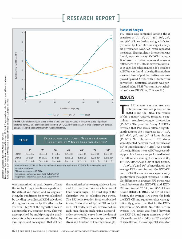
The PFJ stress results for the different exercises are presented in FIGURE 4 and the TABLE. The results
of the 2-factor ANOVA revealed a sig-nificant exercise-by-angle interaction (P<.001). The post hoc 1-way ANOVAs revealed that PFJ stress differed signifi-cantly among the 3 exercises at 0°, 15°, 30°, 60°, 75°, and 90° of knee flexion (P<.001). No differences in PFJ stress were detected between the 3 exercises at 45° of knee flexion (P = .126). As a result of the significant 1-way ANOVAs, second-ary post hoc t tests were performed to test the differences among 3 exercises at 0°, 15°, 30°, 60°, 75°, and 90° of knee flexion.
At 0°, 15°, and 30° of knee flexion, the average PFJ stress for both the EXT-VR and EXT-CR exercises was significantly greater than the squat exercise (P<.001). No difference in average PFJ stress was found between the EXT-VR and EXT-CR exercises at 0°, 15°, and 30° of knee flexion (FIGURE 4, TABLE). At 60° of knee flexion, the average PFJ stress for both the EXT-CR and squat exercises was sig-nificantly greater than that for the EXT-VR exercise (P<.001). No difference in average PFJ stress was found between the EXT-CR and squat exercises at 60° of knee flexion (P = .062). At 75° and 90° of knee flexion, the average PFJ stress for
TABLEPatellofemoral Joint Stresses Among 3 Exercises at 7 Knee Flexion Angles*
Abbreviations: EXT-CR, knee extension with constant resistance; EXT-VR, knee extension with variable resistance.*Values are mean SD MPa.†Significant difference from EXT-VR (P<.016).‡Significant difference from EXT-CR (P<.016).
0° 15° 30° 45° 60° 75° 90°
EXT-VR 8.4 1.6 5.6 1.0 4.7 0.6 4.2 0.8 3.6 0.8 2.6 0.8 0.5 0.5
EXT-CR 7.9 1.6 5.6 1.6 5.1 1.5 5.0 1.5 5.2 1.6† 6.0 2.0† 6.5 2.6†
Squat 0.3 0.8†‡ 0.9 0.9†‡ 1.9 0.9†‡ 3.9 1.3 6.7 1.4† 10.5 1.6†‡ 12.3 1.6†‡
0
0 15 30 45 60 75 90
2
4
6
8
10
12
14
Pate
llofe
mor
al J
oint
Str
ess,
MPa
*†
*†
*†
*†
*†
*
**
*
EXT-VR EXT-CR
Knee Flexion Angle, deg
Squat
FIGURE 4. Patellofemoral joint stress profiles of the 3 exercises evaluated in the current study. *Significant difference from EXT-VR. †Significant difference from EXT-CR. Abbreviations: EXT-CR, knee extension with constant resistance; EXT-VR, knee extension with variable resistance.
44-05 Powers.indd 324 4/16/2014 4:14:54 PM
Jour
nal o
f O
rtho
paed
ic &
Spo
rts
Phys
ical
The
rapy
®
Dow
nloa
ded
from
ww
w.jo
spt.o
rg a
t on
Nov
embe
r 18
, 201
4. F
or p
erso
nal u
se o
nly.
No
othe
r us
es w
ithou
t per
mis
sion
. C
opyr
ight
© 2
014
Jour
nal o
f O
rtho
paed
ic &
Spo
rts
Phys
ical
The
rapy
®. A
ll ri
ghts
res
erve
d.
journal of orthopaedic & sports physical therapy | volume 44 | number 5 | may 2014 | 325
the squat exercise was significantly great-er than that for the EXT-CR (P<.001) and EXT-VR (P<.001) exercises. In addition, the average stress for the EXT-CR exer-cise was significantly greater than that for the EXT-VR exercise at 75° and 90° of knee flexion (P<.001) (FIGURE 4, TABLE).
DISCUSSION
The results of the current study revealed that PFJ stress profiles var-ied considerably among the 3 exer-
cises evaluated. In general, the PFJ stress during the squat exercise was greatest at 90° of knee flexion (12.3 MPa) and steadily decreased as the knee extended (FIGURE 4, TABLE). In contrast, the PFJ stress during the EXT-VR exercise was lowest at 90° of knee flexion and steadily increased as the knee extended, achieving a maximum value of 8.4 MPa at 0°. PFJ stress during the EXT-CR exercise was relatively constant throughout the range of motion, with peak stress (7.9 MPa) occurring at 0° of knee flexion (FIGURE 4, TABLE).
The PFJ stress profiles for the EXT-VR and squat exercises in the current study are in close agreement with the findings of Steinkamp and colleagues.17 In their study, Steinkamp et al17 reported that PFJ stress during the weight-bearing exercise was greater than that during the non–weight-bearing exercise at knee flex-ion angles greater than 45°. Conversely, these authors reported that PFJ stress during the non–weight-bearing exercise was greater than that during the weight-bearing exercise at knee flexion angles less than 45°.17 Interestingly, our data revealed that the stress profiles for squat and EXT-VR exercises also diverged at 45° of knee flexion (FIGURE 4).
Despite the similarities between the stress profiles in the current study and those reported by Steinkamp and col-leagues,17 peak stress values varied con-siderably. For example, Steinkamp et al17 reported peak PFJ stresses for non–weight-bearing (knee extension) and weight-bearing (leg press) exercises of
22.8 and 24.3 MPa, respectively, com-pared to 8.4 and 12.3 MPa in the current study. This discrepancy can be explained by the fact that Steinkamp et al17 used greater external loads, which resulted in knee extensor torques of approxi-mately 205 Nm for their weight-bearing and non–weight-bearing exercises. In contrast, the external loads used in the current study resulted in external knee extensor torques of 67.5, 67.2, and 64 Nm for the EXT-VR, EXT-CR, and squat exercises, respectively.
The PFJ stress profile for the EXT-CR exercise in the current study was similar to the external knee flexion mo-ment curve reported by Escamilla and colleagues.6 Between 90° and 0° of knee flexion, the PFJ stress for the EXT-CR ex-ercise was relatively constant. However, as PFJ stress was not computed by Es-camilla et al,6 direct comparisons to the current study are not possible.
As mentioned previously, the differ-ences in PFJ stress profiles between the 2 non–weight-bearing knee extension exercises can be explained by how the resistance was applied to the tibia. For the EXT-CR exercise, the moment arm for the external load was maintained throughout knee flexion/extension, whereas the moment arm of the external load increased with knee extension in the EXT-VR exercise. For the EXT-VR exer-cise, the maximum external knee flexion moment at 0° of knee flexion resulted in the peak PFJ stress occurring at full knee extension. For the EXT-CR exercise, the constant external knee flexion moment from 90° to 0° of knee flexion resulted in a relatively consistent PFJ stress pattern throughout the range of motion.
The differences in PFJ stress profiles between the EXT-VR and squat exercises can be explained by the varied interaction between PFJ reaction force and PFJ con-tact area while performing these 2 tasks. The PFJ contact area used in our model was smallest at 0° and steadily increased with knee flexion.14 During the EXT-VR exercise, the progressive increase in PFJ reaction force, combined with the simul-
taneous decrease in contact area as the knee extended from 90° to 0°, resulted in an overall increase in PFJ stress. In con-trast, the PFJ reaction force during the squat exercise steadily decreased as the knee extended from 90° to 0°. The de-crease in the PFJ reaction force was more pronounced than the decrease in contact area, resulting in an overall decrease in PFJ stress as the knee extended.
It is important during the initial stag-es of PFJ rehabilitation to select quad-riceps exercises and external loads that minimize PFJ stress. The findings of the current study provide a general rehabili-tation guideline to accomplish this goal. To strengthen the quadriceps through a 90° range of motion and to keep PFJ stress to a minimum, our data suggest that a combination of weight-bearing and non–weight-bearing exercises could be utilized. For example, performing the squat exercise from 0° to 45° of knee flex-ion and the EXT-VR exercise from 45° to 90° of knee flexion would keep PFJ stress to a minimum (below 4 MPa). Although it is not known how much stress causes PFP or constitutes “overloading” of the PFJ, it has been reported that the peak PFJ stress during stair ambulation (a common pain-inducing activity) is ap-proximately 4 MPa.15 As such, keeping PFJ stress values below this threshold during the initial stages of PFJ reha-bilitation would appear to be prudent. It should be noted that the EXT-CR exer-cise resulted in PFJ stress values greater than 4 MPa throughout the entire range of motion evaluated, and thus should be used with caution.
The present study has several limita-tions that should be acknowledged. First, only healthy participants were evalu-ated. As such, caution should be taken when generalizing the current results to patients with PFP. For instance, it has been shown that persons with PFP have smaller contact areas when compared with healthy persons.16 Although smaller contact areas would lead to higher PFJ stresses, the general trends for each of the exercises evaluated in the current study
44-05 Powers.indd 325 4/16/2014 4:14:54 PM
Jour
nal o
f O
rtho
paed
ic &
Spo
rts
Phys
ical
The
rapy
®
Dow
nloa
ded
from
ww
w.jo
spt.o
rg a
t on
Nov
embe
r 18
, 201
4. F
or p
erso
nal u
se o
nly.
No
othe
r us
es w
ithou
t per
mis
sion
. C
opyr
ight
© 2
014
Jour
nal o
f O
rtho
paed
ic &
Spo
rts
Phys
ical
The
rapy
®. A
ll ri
ghts
res
erve
d.
326 | may 2014 | volume 44 | number 5 | journal of orthopaedic & sports physical therapy
[ research report ]
REFERENCES
1. Boling MC, Bolgla LA, Mattacola CG, Uhl TL, Hosey RG. Outcomes of a weight-bearing rehabilitation program for patients diagnosed with patellofemoral pain syndrome. Arch Phys Med Rehabil. 2006;87:1428-1435. http://dx.doi.org/10.1016/j.apmr.2006.07.264
2. Brechter JH, Powers CM. Patellofemoral joint stress during stair ascent and descent in per-sons with and without patellofemoral pain. Gait Posture. 2002;16:115-123.
3. Brechter JH, Powers CM. Patellofemoral stress during walking in persons with and without patellofemoral pain. Med Sci Sports Exerc. 2002;34:1582-1593.
4. Delp SL, Loan JP. A computational framework for simulating and analyzing human and animal movement. Comput Sci Eng. 2000;2:46-55. http://dx.doi.org/10.1109/5992.877394
5. Ebben WP, Feldmann CR, Dayne A, Mitsche D, Alexander P, Knetzger KJ. Muscle activa-tion during lower body resistance training. Int J Sports Med. 2009;30:1-8. http://dx.doi.org/10.1055/s-2008-1038785
6. Escamilla RF, Fleisig GS, Zheng N, Barrentine SW, Wilk KE, Andrews JR. Biomechanics of the knee during closed kinetic chain and open kinetic chain exercises. Med Sci Sports Exerc. 1998;30:556-569.
7. Escamilla RF, MacLeod TD, Wilk KE, Paulos L, Andrews JR. Anterior cruciate ligament strain and tensile forces for weight-bearing and non–weight-bearing exercises: a guide to exercise selection. J Orthop Sports Phys Ther. 2012;42:208-220. http://dx.doi.org/10.2519/jospt.2012.3768
8. Farrokhi S, Pollard CD, Souza RB, Chen YJ, Reischl S, Powers CM. Trunk position influences the kinematics, kinetics, and muscle activity of the lead lower extremity during the forward
lunge exercise. J Orthop Sports Phys Ther. 2008;38:403-409. http://dx.doi.org/10.2519/jospt.2008.2634
9. Kaya D, Doral MN, Callaghan M. How can we strengthen the quadriceps femoris in patients with patellofemoral pain syndrome? Muscles Ligaments Tendons J. 2012;2:25-32.
10. Lloyd DG, Besier TF. An EMG-driven muscu-loskeletal model to estimate muscle forces and knee joint moments in vivo. J Biomech. 2003;36:765-776.
11. Mueller MJ, Tuttle LJ, Lemaster JW, et al. Weight-bearing versus nonweight-bearing exercise for persons with diabetes and peripheral neuropa-thy: a randomized controlled trial. Arch Phys Med Rehabil. 2013;94:829-838. http://dx.doi.org/10.1016/j.apmr.2012.12.015
12. Park J, Grindstaff TL, Hart JM, Hertel JN, Inger-soll CD. Knee-extension exercise’s lack of im-mediate effect on maximal voluntary quadriceps torque and activation in individuals with anterior knee pain. J Sport Rehabil. 2012;21:119-126.
13. Pollard CD, Sigward SM, Powers CM. Limited hip and knee flexion during landing is associ-ated with increased frontal plane knee motion and moments. Clin Biomech (Bristol, Avon). 2010;25:142-146. http://dx.doi.org/10.1016/j.clinbiomech.2009.10.005
14. Powers CM, Lilley JC, Lee TQ. The effects of axial and multi-plane loading of the extensor mecha-nism on the patellofemoral joint. Clin Biomech (Bristol, Avon). 1998;13:616-624.
15. Powers CM, Ward SR, Chen YJ, Chan LD, Terk MR. Effect of bracing on patellofemoral joint stress while ascending and descending stairs. Clin J Sport Med. 2004;14:206-214.
16. Salsich GB, Perman WH. Patellofemoral joint contact area is influenced by tibiofemoral rotation alignment in individuals who have patellofemoral pain. J Orthop Sports Phys Ther. 2007;37:521-528. http://dx.doi.org/10.2519/jospt.2007.37.9.521
17. Steinkamp LA, Dillingham MF, Markel MD, Hill JA, Kaufman KR. Biomechanical considerations in patellofemoral joint rehabilitation. Am J Sports Med. 1993;21:438-444.
18. Stiene HA, Brosky T, Reinking MF, Nyland J, Mason MB. A comparison of closed kinetic chain and isokinetic joint isolation exercise in patients with patellofemoral dysfunction. J Or-thop Sports Phys Ther. 1996;24:136-141. http://dx.doi.org/10.2519/jospt.1996.24.3.136
19. Stuart MJ, Meglan DA, Lutz GE, Growney ES, An KN. Comparison of intersegmental tibiofemoral joint forces and muscle activity during various closed kinetic chain exercises. Am J Sports Med. 1996;24:792-799.
20. Tsai LC, Powers CM. Increased hip and knee flexion during landing decreases tibiofemoral compressive forces in women who have under-gone anterior cruciate ligament reconstruction. Am J Sports Med. 2013;41:423-429. http://dx.doi.org/10.1177/0363546512471184
21. Tsai LC, Scher IS, Powers CM. Quantification of
likely would be similar. Second, the ab-solute PFJ stress values reported here should be viewed with caution, as our PFJ model has not been validated against a gold standard. However, any error in PFJ stress estimates would be similar across conditions, making comparisons between exercises valid. Third, we did not control the trunk position during the squat ex-ercise. This could have influenced the magnitude of the KEM and therefore the PFJ stress during this task. In addition, despite our attempt to standardize the re-sistance across exercises, PFJ stress may vary based on the magnitude of external loads applied (regardless of the exercise performed). Fourth, segmental accelera-tions during the non–weight-bearing ex-ercises were considered to be negligible and were not considered in the calcula-tions of PFJ stress. As such, the absolute values presented for these tasks should be viewed with caution. Last, we only re-ported data for the concentric phase of each exercise. Further research is neces-sary to determine whether differences in PFJ stress exist between the concentric and eccentric phases of a given exercise.
CONCLUSION
Our results provide general guidelines with respect to quadri-ceps strengthening for persons with
PFP. To keep PFJ stress to a minimum, our data suggest that the squat exercise should be performed between 0° and 45° of knee flexion and the EXT-VR ex-ercise should be performed between 45° and 90° of knee flexion. Throughout the range of motion evaluated, the EXT-CR exercise generated a level of PFJ stress comparable to climbing stairs (ie, great-er than 4 MPa), and thus should be used with caution. t
KEY POINTSFINDINGS: When compared to the 2 non–weight-bearing exercises, the squat exercise produced significantly higher PFJ stress at 60°, 75°, and 90° of knee flexion. Conversely, the 2 non–weight-
bearing exercises produced significantly higher PFJ stress at 0°, 15°, and 30° of knee flexion when compared to the squat exercise. The EXT-VR exercise produced significantly lower PFJ stress than the EXT-CR exercises at 90°, 75°, and 60° of knee flexion.IMPLICATIONS: To keep PFJ stress to a minimum, our data suggest that the squat exercise should be performed be-tween 0° and 45° of knee flexion and the EXT-VR exercise should be performed between 45° and 90° of knee flexion.CAUTION: As only healthy participants were evaluated, caution should be taken when generalizing the current results to persons with PFP.
44-05 Powers.indd 326 4/16/2014 4:14:55 PM
Jour
nal o
f O
rtho
paed
ic &
Spo
rts
Phys
ical
The
rapy
®
Dow
nloa
ded
from
ww
w.jo
spt.o
rg a
t on
Nov
embe
r 18
, 201
4. F
or p
erso
nal u
se o
nly.
No
othe
r us
es w
ithou
t per
mis
sion
. C
opyr
ight
© 2
014
Jour
nal o
f O
rtho
paed
ic &
Spo
rts
Phys
ical
The
rapy
®. A
ll ri
ghts
res
erve
d.
journal of orthopaedic & sports physical therapy | volume 44 | number 5 | may 2014 | 327
MORE INFORMATIONWWW.JOSPT.ORG@
tibiofemoral shear and compressive loads using a MRI-based EMG-driven knee model. J Appl Biomech. 2013;29:229-234.
22. van Eijden TM, Kouwenhoven E, Verburg J, Weijs WA. A mathematical model of the patellofemoral joint. J Biomech. 1986;19:219-229.
23. van Eijden TM, Weijs WA, Kouwenhoven E, Verburg J. Forces acting on the patella dur-ing maximal voluntary contraction of the quadriceps femoris muscle at different knee flexion/extension angles. Acta Anat (Basel). 1987;129:310-314.
24. Ward SR, Powers CM. The influence of patella alta on patellofemoral joint stress during normal and fast walking. Clin Biomech (Bristol, Avon). 2004;19:1040-1047. http://dx.doi.org/10.1016/j.clinbiomech.2004.07.009
25. Wilk KE, Escamilla RF, Fleisig GS, Barrentine SW, Andrews JR, Boyd ML. A comparison of tibiofemoral joint forces and electromyographic activity during open and closed kinetic chain exercises. Am J Sports Med. 1996;24:518-527.
26. Winter DA. Anthropometry. In: Winter DA, ed. Biomechanics and Motor Control of Human
Movement. Hoboken, NJ: Wiley; 2005:59-84.
27. Witvrouw E, Danneels L, Van Tiggelen D, Wil-
lems TM, Cambier D. Open versus closed
kinetic chain exercises in patellofemoral pain:
a 5-year prospective randomized study. Am J
Sports Med. 2004;32:1122-1130. http://dx.doi.
org/10.1177/0363546503262187
EARN CEUs With JOSPT’s Read for Credit Program
JOSPT’s Read for Credit (RFC) program invites readers to study and analyze selected JOSPT articles and successfully complete online exams about them for continuing education credit. To participate in the program:
1. Go to www.jospt.org and click on Read for Credit in the top blue navigation bar that runs throughout the site.
2. Log in to read and study an article and to pay for the exam by credit card.
3. When ready, click Take Exam to answer the exam questions for that article.
4. Evaluate the RFC experience and receive a personalized certificate of continuing education credits.
The RFC program o�ers you 2 opportunities to pass the exam. You may review all of your answers—including your answers to the questions you missed. You receive 0.2 CEUs, or 2 contact hours, for each exam passed.
JOSPT’s website maintains a history of the exams you have taken and the credits and certificates you have been awarded in My CEUs and Your Exam Activity, located in the right rail of the Read for Credit page listing available exams.
44-05 Powers.indd 327 4/16/2014 4:14:55 PM
Jour
nal o
f O
rtho
paed
ic &
Spo
rts
Phys
ical
The
rapy
®
Dow
nloa
ded
from
ww
w.jo
spt.o
rg a
t on
Nov
embe
r 18
, 201
4. F
or p
erso
nal u
se o
nly.
No
othe
r us
es w
ithou
t per
mis
sion
. C
opyr
ight
© 2
014
Jour
nal o
f O
rtho
paed
ic &
Spo
rts
Phys
ical
The
rapy
®. A
ll ri
ghts
res
erve
d.