Embed Size (px)
Citation preview
Steinbrück et al. BioMedical Engineering OnLine 2013, 12:58http://www.biomedical-engineering-online.com/content/12/1/58
RESEARCH Open Access
Patellofemoral contact patterns before and aftertotal knee arthroplasty: an in vitro measurementArnd Steinbrück*†, Christian Schröder†, Matthias Woiczinski, Andreas Fottner, Peter E Müller and Volkmar Jansson
* Correspondence: [email protected]†Equal contributorsDepartment of OrthopaedicSurgery, University Hospital ofMunich (LMU), CampusGrosshadern, Marchioninistr. 15,Munich 81377, Germany
Abstract
Background: Patellofemoral complications are one of the main problems after TotalKnee Arthroplasty (TKA). Retropatellar pressure distribution after TKA can contributeto these symptoms. Therefore we evaluated retropatellar pressure distributionsubdivided on the ridge, medial and lateral surface on non-resurfaced patella beforeand after TKA. Additionally, we analyzed axial femorotibial rotation and quadricepsload before and after TKA.
Methods: Seven fresh frozen cadaver knees were tested in a force controlled kneerig before and after TKA (Aesculap, Tuttlingen, Germany, Columbus CR) whileisokinetic flexing the knee from 20° to 120° under weight bearing. Ridge, medial andlateral retropatellar surface were defined and pressure distribution was dynamicallymeasured while quadriceps muscles and hamstring forces were applied. Aside axialfemorotibial rotation and quadriceps load was recorded.
Results: There was a significant change of patella pressure distribution before andafter TKA (p = 0.004). In physiological knees pressure distribution on medial andlateral retropatellar surface was similar. After TKA the ridge of the patella wasespecially in higher flexion grades strongly loaded (6.09 +/−1.31 MPa) compared tothe natural knee (2.92 +/−1.15 MPa, p < 0.0001). Axial femorotibial rotation showedtypical internal rotation with increasing flexion both before and after TKA, butpostoperatively it was significantly lower. The average amount of axial rotation was3.5° before and after TKA 1.3° (p = 0.001). Mean quadriceps loading after implantationof knee prosthesis did not change significantly (575 N ±60 N in natural knee andafter TKA 607 N ±96 N; p = 0.28).
Conclusions: The increased retropatellar pressure especially on the ridge may beone important reason for anterior knee pain after TKA. The trochlea of the femoralcomponent might highly influence the pressure distribution of the non-resurfacedretropatellar surface. Additionally, lower axial femorotibial rotation after TKA mightlead to patella maltracking. Changing the design of the prosthesis or a special wayof patella shaping might increase the conformity of the patella to trochlea tomaintain natural contact patterns.
Keywords: Patella, Total knee arthroplasty, Patella tracking, Retropatellar pressure,Trochlear groove, Knee rig
© 2013 Steinbrück et al.; licensee BioMed Central Ltd. This is an Open Access article distributed under the terms of the CreativeCommons Attribution License (http://creativecommons.org/licenses/by/2.0), which permits unrestricted use, distribution, andreproduction in any medium, provided the original work is properly cited.
Steinbrück et al. BioMedical Engineering OnLine 2013, 12:58 Page 2 of 13http://www.biomedical-engineering-online.com/content/12/1/58
Abstract
Hintergrund: Das patellofemorale Schmerzsyndrom stellt eines der zentralenProbleme in der Knieendoprothetik dar. Dabei wird angenommen, dass dieretropatellare Druckverteilung nach Implantation einer Knietotalendoprothese (KTEP)zu dieser Symptomatik beiträgt. Wir untersuchten daher die retropatellareDruckverteilung unterteilt in First, mediale und laterale Rückfläche vor und nach KTEP.Zusätzlich analysierten wir die tibiofemorale Rotation und die Quadrizepszugkraft vorund nach KTEP.
Material und methoden: Die Simulation der Kniebewegung erfolgte an siebenhumanen fresh-frozen Kniegelenken mit einem kraftgesteuertenKniegelenkskinemator von 20° bis 120° Flexion mit programmierter Regelung unterkonstanter isokinetischer Gewichtskraft an den Sehnen des M. quadriceps, M.semitendinosus und M. biceps femoris. Nach standardisierter Flächendefinition desFirstes, der medialen und lateralen Retropatellarfläche wurde die retropatellareDruckverteilung mittels Druckmessfolie vor und nach Implantation einer KTEPdynamisch gemessen. Zusätzlich wurden die tibiofemorale Rotation, sowie dieQuadrizepszugkraft analysiert.
Ergebnisse: Nach Implantation einer KTEP zeigte sich eine signifikante Änderung derretropatellaren Druckverteilung. Im natürlichen Kniegelenk war der retropatellareDruck medial und lateral gleichmäßig verteilt. Postoperativ zeigte sich bei höherenFlexionsgraden ein maximaler retropatellarer Druck besonders im Bereich des Patellafirstes(6,09 +/−1,31 MPa) im Vergleich zum natürlichen Kniegelenk (2,92 +/−1,15 MPa, p <0,0001). Die axiale tibiofemorale Rotation zeigte die typische Innenrotation beiFlexion und Außenrotation bei Extension sowohl vor als auch nach Implantationeiner KTEP; postoperativ war diese Rotation jedoch deutlich vermindert. Diedurchschnittliche axiale Rotation betrug 3,5° vor und 1,3° nach KTEP-Implantation(p = 0,001). Die Quadrizepszugkraft änderte sich nach KTEP-Implantation nichtsignifikant (575 N ±60 N beim natürlichen Knie und nach KTEP 607 N (±96 N),p = 0,28).
Schlussfolgerung: Die Implantation einer KTEP führte in unserer Studie zu einerSteigerung des retropatellaren Drucks insbesondere im Firstbereich. Es ist davonauszugehen, dass durch die Erhöhung des retropatellaren Drucks und dieungünstigere Druckverteilung in vivo postoperative Patella Beschwerdenentstehen können. Daneben kann die postoperativ verminderte Innenrotation derTibia bei Flexion des Kniegelenkes zum Patella Maltracking führen. Wird bei derImplantation einer Knieprothese kein Retropatellarersatz durchgeführt ist einstandardisiertes knöchernes “Trimming” der Patella entsprechend dem Trochlea-Design der Prothese zu diskutieren.
BackgroundAlthough Total Knee Arthroplasty (TKA) is a successful solution for osteoarthritis of
the knee up to 19% of primary TKA patients are not satisfied with the outcome [1].
Patellofemoral complications after TKA are still a main cause of failure [2] and include
chronic anterior knee pain, fracture of the patella, patella clunk syndrome, patella lux-
ation and subluxation, as well as rupture of patella tendon [3-6]. Boyd et al. found 12%
of peripatellar complications after TKA without resurfacing [3]. Revision rates and
postoperative outcome, however, are similar for resurfaced and nonresurfaced patella
[7,8]. Regarding anterior knee pain several authors emphasize that the postoperative
outcome is rather influenced by femoral component design than by retropatellar

Steinbrück et al. BioMedical Engineering OnLine 2013, 12:58 Page 3 of 13http://www.biomedical-engineering-online.com/content/12/1/58
resurfacing [7,9,10]. It is assumed that excessive postoperative retropatellar pressure
contributes to a great extent to postoperative patella problems [9,11-13]. In vitro stud-
ies with cadaver knees are a successful method to analyze this pressure distribution
[12,14], and were also used to investigate retropatellar contact patterns in this study.
Studies with fluoroscopy also showed that patellofemoral kinematics are modified
after TKA [15]. In this context, Dennis et al. argue that femorotibial rotation is espe-
cially important for a normal patellar tracking and to prevent patellofemoral shear
forces [16]. A decrease of femorotibial rotation after TKA might be one reason for pa-
tella maltracking and anterior knee pain.
Flexion and extension moment are also altering after TKA [17]. In particular, quadriceps
strength is supposed to be an important factor for a well-functioning TKA and to prevent
postoperative patella problems [18]. Ostermeier et al. showed in their study [19] an increase
of 9% of quadriceps load for the posterior cruciate-retaining fixed-bearing TKA group.
To the authors knowledge there is no in vitro study which investigates retropatellar pres-
sure distribution subdivided in ridge, medial and lateral surface, femorotibial rotation and
quadriceps load before and after TKA with fresh-frozen specimens during a simulated squat
of 100° of range of motion. Therefore, we focused on three aspects in our knee rig study:
First we analyzed retropatellar contact patterns subdivided in ridge, medial and lateral sur-
face in natural knees and after implantation of knee prostheses to evaluate this specific
TKA design. Then we continued with investigating axial femorotibial rotation, which was
assumed to alter after TKA. Finally, flexion and extension force of the quadriceps muscle
was analyzed before and after TKA. The in vitro analysis was performed with fresh-frozen
specimens dynamically in a special knee rig under force controlled muscle loading in a
range of motion of 20° to 120° of knee flexion.
MethodsSeven fresh frozen knee specimens (age range between 38 and 68 years, 3 female, 4 male)
were resected about 20 cm proximal and 15 cm distal to the joint line, preserving articular
capsule, ligaments and tendons surrounding the knee joint. Fibula head was fixed to tibia
with a standard 4.5 diameter screw. Muscle- and fat-tissue was carefully removed from the
tendons and bones. After on metallic finger traps (Bühler-Instrumente Medizintechnik
GmbH, Tuttlingen, Germany) were connected to the tendons and suture material
(FibreWire, Arthrex, Munich, Germany) was used to fix the tendon into the metallic mesh
of the traps. The femoral and tibial bones were embedded into a metallic pot both fixed
with epoxy casting resin (Rencast FC53, Huntsman, Basel, Switzerland).
Shaping of the patella was performed by removing existing osteophytes on the patella
circumference. The patella ridge was not shaped and patella remained unresurfaced.
Afterwards a pressure sensitive film (K-Scan 4000, Tekscan Inc., Boston, USA) was at-
tached to the retropatellar surface by subcutaneous 1.0 suture material. For stable suturing
and to avoid shear forces a 0.1 mm Teflon tape (PTFE-tape) was glued on the sensor be-
fore (Figure 1). The sensor was conditioned and calibrated with a two point calibration
(error of the root mean square 2.7% [20], nonlinearity max. 3%) as recommended by the
manufacturer using a material testing machine (Z010, Zwick, Ulm, Germany). The max-
imum pressure of the sensor film was 1500 PSI (~10 MPa) with 572 total number of
sensels (62 sensel per cm2).

Figure 1 Photograph of a specimen after the finger traps were connected to the prepared tendonsand the pressure sensitive film was attached to the retropatellar surface.
Steinbrück et al. BioMedical Engineering OnLine 2013, 12:58 Page 4 of 13http://www.biomedical-engineering-online.com/content/12/1/58
For definition of the ridge of the patella first the highest point of the ridge of the pa-
tella was located with a pen also used for digital devices as smartphones. Afterwards
the course of the ridge was followed from proximal to distal. This sweeping was
recorded by the software of the sensitive film and repeated three times. Then the bor-
ders of the medial and lateral retropatellar surface were located on the retropatellar
pressure sensor. From the middle of the course of the ridge (width: 1.3 mm = 1 sensel)
we added 2.6 (2 sensels) mm medial and lateral to define the overall width of 7.5 mm
(5 sensels) of the patella ridge and the borders to medial and lateral retropatellar sur-
face to differentiate patellofemoral contact points.
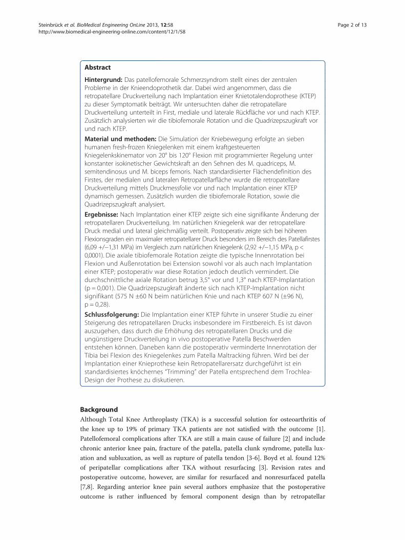
The prepared knee was then mounted into the specially built knee simulator with 6
degrees of freedom (dof ) (Figure 2a + b). This knee rig simulates a loaded squat using
two linear drives (Driveset M150/180, Systec GmbH, Muenster, Germany; max. axial
static load 2500/5000 N; precision of 0.1/0.1 mm; max. velocity 0.1/0.35 m/s). The first
actor flexes and extends the knee with a constant velocity of 3°/s. Two angle sensors
(8820 Burster, Gernsbach, Germany; capacity 350°, nonlinearity 0.5%, resolution 0.01°)
in the simulated upper “hip-assembly” and simulated lower “ankle-assembly” were used
to measure the flexion angle of the knee joint and axial femorotibial rotation. The sec-
ond drive simulates the quadriceps muscle; the actual force was measured with mini-
ature force transducers (8417–6002 Burster, Gernsbach, Germany; max. load 2000 N,
nonlinearity 0.5%) near the tendons. Additionally, muscle forces of the hamstrings
(semitendinosus and biceps femoris muscle), as well as lateral vastus and medial vastus
muscle were simulated with 2 kg weights. A 6 degrees of freedom force-moment-sensor
(FN 7325–31 FGP Sensors, Cedex, France) measured the ground reaction at the ankle
assembly. This sensor has a capacity of 5000 N (axial), 2500 N(x/y plane), 65 Nm
(torque/bending) with a nonlinearity of 1.0%. All sensors were amplified up to max 10
volts and digitalized with an 18–bit (262144 increments) analog digital converter using
8 differential analog input channels (PCI 6281 National Instruments, Texas, USA; min.
accuracy: 28 μvolts). The rig was controlled with a personal computer with a self-
programed code with LabVIEW 8.6 (National Instruments, Austin, Texas, USA). The
knee was moving with a 50 ms PID- force- control loops to hold the ground reaction
force constant to 50 N with the loaded quadriceps muscle. The data of the sensors were
collected and written in an ASCII file.
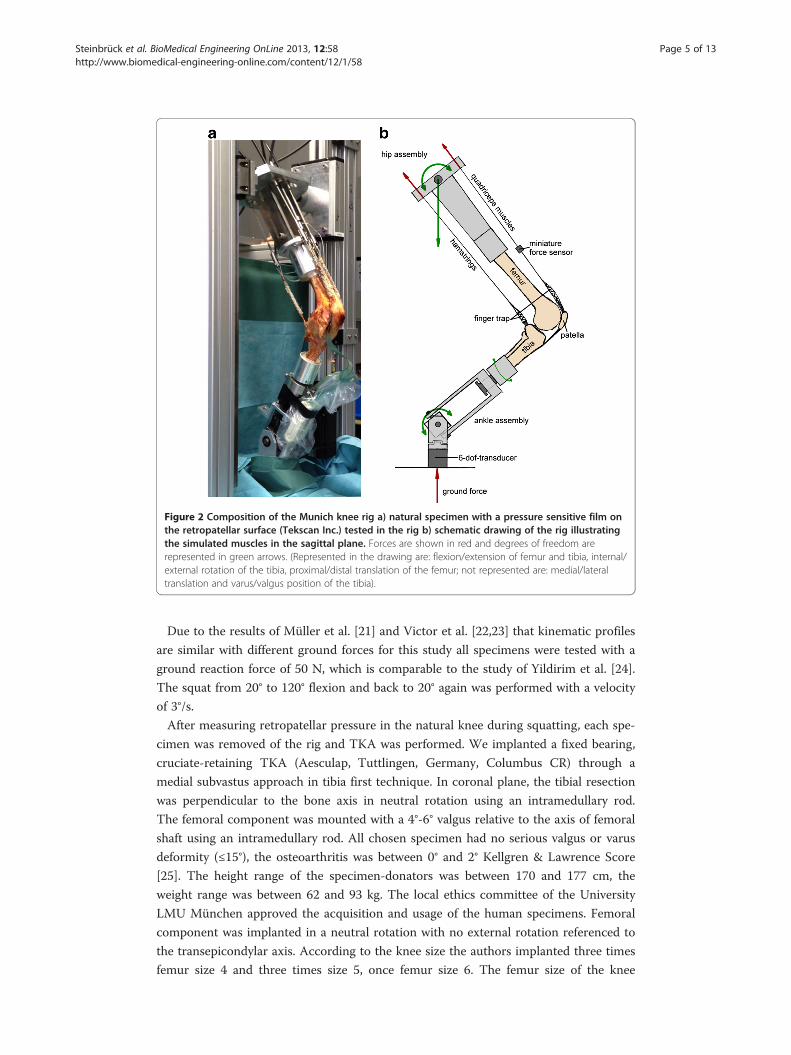
Figure 2 Composition of the Munich knee rig a) natural specimen with a pressure sensitive film onthe retropatellar surface (Tekscan Inc.) tested in the rig b) schematic drawing of the rig illustratingthe simulated muscles in the sagittal plane. Forces are shown in red and degrees of freedom arerepresented in green arrows. (Represented in the drawing are: flexion/extension of femur and tibia, internal/external rotation of the tibia, proximal/distal translation of the femur; not represented are: medial/lateraltranslation and varus/valgus position of the tibia).
Steinbrück et al. BioMedical Engineering OnLine 2013, 12:58 Page 5 of 13http://www.biomedical-engineering-online.com/content/12/1/58
Due to the results of Müller et al. [21] and Victor et al. [22,23] that kinematic profiles
are similar with different ground forces for this study all specimens were tested with a
ground reaction force of 50 N, which is comparable to the study of Yildirim et al. [24].
The squat from 20° to 120° flexion and back to 20° again was performed with a velocity
of 3°/s.
After measuring retropatellar pressure in the natural knee during squatting, each spe-
cimen was removed of the rig and TKA was performed. We implanted a fixed bearing,
cruciate-retaining TKA (Aesculap, Tuttlingen, Germany, Columbus CR) through a
medial subvastus approach in tibia first technique. In coronal plane, the tibial resection
was perpendicular to the bone axis in neutral rotation using an intramedullary rod.
The femoral component was mounted with a 4°-6° valgus relative to the axis of femoral
shaft using an intramedullary rod. All chosen specimen had no serious valgus or varus
deformity (≤15°), the osteoarthritis was between 0° and 2° Kellgren & Lawrence Score
[25]. The height range of the specimen-donators was between 170 and 177 cm, the
weight range was between 62 and 93 kg. The local ethics committee of the University
LMU München approved the acquisition and usage of the human specimens. Femoral
component was implanted in a neutral rotation with no external rotation referenced to
the transepicondylar axis. According to the knee size the authors implanted three times
femur size 4 and three times size 5, once femur size 6. The femur size of the knee

Steinbrück et al. BioMedical Engineering OnLine 2013, 12:58 Page 6 of 13http://www.biomedical-engineering-online.com/content/12/1/58
system ranges from 1 to 7. Tibial we implanted four times size 4, two times size 3, once
tibia size 2. The tibia size ranges from 1 to 5. The knees were balanced using a gauge
instrument. A 10 mm polyethylene inlay was used in every TKA. All knees were oper-
ated by the author A.S., who is in his 8th post-graduate year and a specialist for ortho-
pedic and trauma surgery. All knees were operated by the author under assistance of
author A.F, who is the senior attending of knee and hip arthroplasty in our department.
The evaluation after TKA was performed under the same conditions as the natural
knee. All specimen were x-rayed anterior-posterior, sagittal and sunrise view by fluoros-
copy before implantation of the prosthesis (to evaluate the grade of osteoarthritis and
patellaalignment) and after TKA (to control the correct implantation of TKA).
Statistical methods
All results were represented with the mean and standard deviation. The functional rela-
tion between flexion angle (FA) and the measured parameters were modeled by means
of mixed effects models using a random intercept per specimen. Fixed effects included
in the model were the cosine of the flexion angle, the squared cosine of the flexion
angle, the cubed cosine of the flexion angle, and the presence of TKA (yes = 1/no = 0).
Separate models were fitted for flexion (=0) and extension (=1). These analyses were
performed using SAS version 9.2 for Linux (SAS Institute, Cary, NC). The regression
coefficients to the statistical model including the p-values are summarized in Table 1.
ResultsThe load of quadriceps was comparable before and after TKA (p = 0.28). The mean
minimum load in natural knee and after TKA was 15 N (±12 N) near extension (20° of
flexion). The mean maximum load was 575 N (±60 N) in natural knee and after TKA
607 N (±96 N) during extension (near 120° of flexion) (Figure 3).
All seven natural knees showed typical tibial internal rotation during flexion respect-
ively external rotation with extension the so called screw-home rotation [26]. The max-
imum amount of axial rotation from 20° to 120° of knee flexion was 8.0°, the minimum
amount was 0.1° (Figure 4). The average amount of axial rotation was 3.5°.
Table 1 Regression equations and p-values of the mixed effects model
Variable Solutions for the fixed effects using the mixed effect model p-value
Intercept cosFA
cos2 FA cos3 FA Flexion/extension
Natural/TKA
Natural/TKA
Quadriceps load 52.88 −7.51 +0.23 −0.0011 +118.91 +3.79 0.28
Tibial rotation −1.00 +0.067 −0.0011 +4.75e-6 −0.38 +2.66 0.001
Peak pressure 0.89 −0.058 0.0018 −8.83e-6 +0.45 +2.66 0.004
Peak pressure(ridge)
0.99 +0.063 0.0021 −0.00001 +0.48 −0.61 0.0001
Peak pressure(medial)
−0.10 −0.015 +0.00089 −4.00e-6 +0.27 −1.24 0.064
Peak pressure(lateral)
0.42 −0.032 +0.0011 −5.09e-6 +0.30 −0.16 0.011
Contact area 285.99 −0.30 +0.0097 -0.00014 +7.36 −0.14 <0.0001
Proximalization 8.55 +0.31 −0.0058 + 0.000019 +2.51 +3.58 <0.0001

Figure 3 Charts are representing mean quadriceps load (in N) with standard deviation according toflexion and extension grade of the knee (between 20°-120°).
Steinbrück et al. BioMedical Engineering OnLine 2013, 12:58 Page 7 of 13http://www.biomedical-engineering-online.com/content/12/1/58
In contrast after TKA there was a significant lower axial tibial rotation with flexion
and extension (p = 0.001). The maximum amount of axial rotation from 20° to 120° of
knee flexion after TKA was 1.7°, the minimum amount was 0.6°. The average amount
of axial rotation was 1.3°. Five knees showed internal rotation with flexion after TKA
and two knees showed slight external rotation with flexion (average amount of reverse
axial rotation 1.6°). One knee rotated internally until 100° and then rotated slightly ex-
ternally until 120° of flexion.
There was a significant difference of mean retropatellar pressure between natural
knee and after TKA. In the natural knee there was a mean peak pressure of 4.05
(+/−1.23) MPa and after TKA of 6.19 (+/−1.27) MPa (p = 0.004). After TKA peak pres-
sure on the entire retropatellar surface was measured in 120° flexion of the knee. Nat-
ural knee in contrast had a peak pressure between 75° to 100° of flexion. Peak pressure
decreased between 100° and 120° constantly and increased again with following exten-
sion in natural knee.
Considering the subdivision of the retropatellar surface in ridge, medial and lateral
surface we obtained significant differences on the ridge of the patella during complete
flexion and extension cycle. After TKA there was a mean peak pressure of 6.09
(+/−1.31) MPa in contrast to natural knee with a peak pressure of 2.92 (+/−1.15) MPa
(p < 0.0001) (Figure 5). On the medial retropatellar surface there was a mean peak pres-
sure of 3.68 (+/−1.30) MPa preoperatively and 3.97 (+/−1.40) MPa postoperatively (p =
0.064). On the lateral retropatellar surface there was a mean peak pressure of 3.21
(+/−1.57) MPa in the natural knee and after TKA of 3.82 (+/−1.99) MPa (p = 0.011).
Figure 4 Charts are representing mean axial femorotibial rotation (in °; positive: internal rotation,negative: external rotation) with standard deviation in natural knee and after TKA.
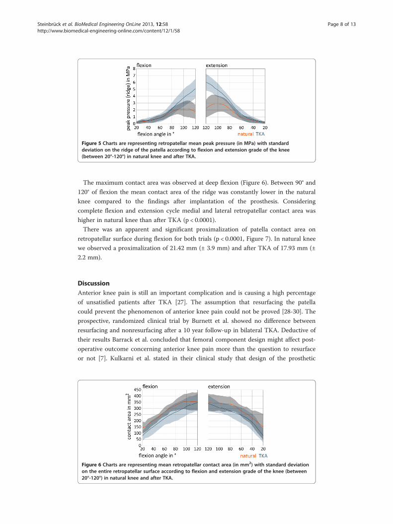
Figure 5 Charts are representing retropatellar mean peak pressure (in MPa) with standarddeviation on the ridge of the patella according to flexion and extension grade of the knee(between 20°-120°) in natural knee and after TKA.
Steinbrück et al. BioMedical Engineering OnLine 2013, 12:58 Page 8 of 13http://www.biomedical-engineering-online.com/content/12/1/58
The maximum contact area was observed at deep flexion (Figure 6). Between 90° and
120° of flexion the mean contact area of the ridge was constantly lower in the natural
knee compared to the findings after implantation of the prosthesis. Considering
complete flexion and extension cycle medial and lateral retropatellar contact area was
higher in natural knee than after TKA (p < 0.0001).
There was an apparent and significant proximalization of patella contact area on
retropatellar surface during flexion for both trials (p < 0.0001, Figure 7). In natural knee
we observed a proximalization of 21.42 mm (± 3.9 mm) and after TKA of 17.93 mm (±
2.2 mm).
DiscussionAnterior knee pain is still an important complication and is causing a high percentage
of unsatisfied patients after TKA [27]. The assumption that resurfacing the patella
could prevent the phenomenon of anterior knee pain could not be proved [28-30]. The
prospective, randomized clinical trial by Burnett et al. showed no difference between
resurfacing and nonresurfacing after a 10 year follow-up in bilateral TKA. Deductive of
their results Barrack et al. concluded that femoral component design might affect post-
operative outcome concerning anterior knee pain more than the question to resurface
or not [7]. Kulkarni et al. stated in their clinical study that design of the prosthetic
Figure 6 Charts are representing mean retropatellar contact area (in mm2) with standard deviationon the entire retropatellar surface according to flexion and extension grade of the knee (between20°-120°) in natural knee and after TKA.
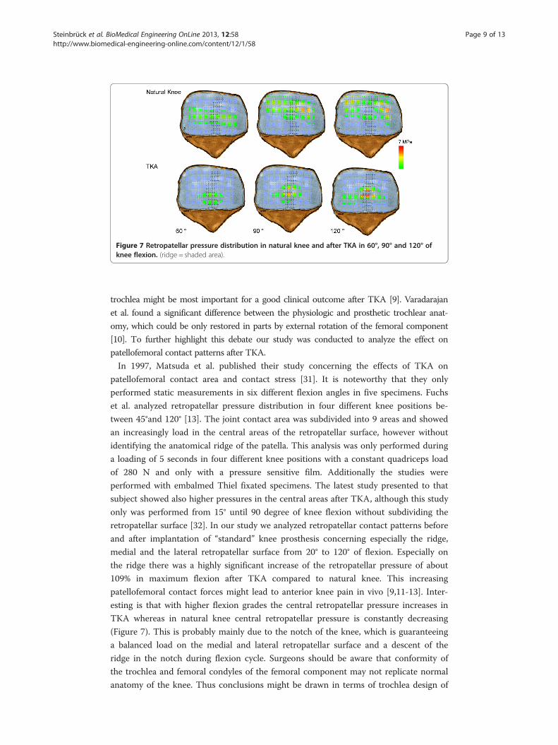
Figure 7 Retropatellar pressure distribution in natural knee and after TKA in 60°, 90° and 120° ofknee flexion. (ridge = shaded area).
Steinbrück et al. BioMedical Engineering OnLine 2013, 12:58 Page 9 of 13http://www.biomedical-engineering-online.com/content/12/1/58
trochlea might be most important for a good clinical outcome after TKA [9]. Varadarajan
et al. found a significant difference between the physiologic and prosthetic trochlear anat-
omy, which could be only restored in parts by external rotation of the femoral component
[10]. To further highlight this debate our study was conducted to analyze the effect on
patellofemoral contact patterns after TKA.
In 1997, Matsuda et al. published their study concerning the effects of TKA on
patellofemoral contact area and contact stress [31]. It is noteworthy that they only
performed static measurements in six different flexion angles in five specimens. Fuchs
et al. analyzed retropatellar pressure distribution in four different knee positions be-
tween 45°and 120° [13]. The joint contact area was subdivided into 9 areas and showed
an increasingly load in the central areas of the retropatellar surface, however without
identifying the anatomical ridge of the patella. This analysis was only performed during
a loading of 5 seconds in four different knee positions with a constant quadriceps load
of 280 N and only with a pressure sensitive film. Additionally the studies were
performed with embalmed Thiel fixated specimens. The latest study presented to that
subject showed also higher pressures in the central areas after TKA, although this study
only was performed from 15° until 90 degree of knee flexion without subdividing the
retropatellar surface [32]. In our study we analyzed retropatellar contact patterns before
and after implantation of “standard” knee prosthesis concerning especially the ridge,
medial and the lateral retropatellar surface from 20° to 120° of flexion. Especially on
the ridge there was a highly significant increase of the retropatellar pressure of about
109% in maximum flexion after TKA compared to natural knee. This increasing
patellofemoral contact forces might lead to anterior knee pain in vivo [9,11-13]. Inter-
esting is that with higher flexion grades the central retropatellar pressure increases in
TKA whereas in natural knee central retropatellar pressure is constantly decreasing
(Figure 7). This is probably mainly due to the notch of the knee, which is guaranteeing
a balanced load on the medial and lateral retropatellar surface and a descent of the
ridge in the notch during flexion cycle. Surgeons should be aware that conformity of
the trochlea and femoral condyles of the femoral component may not replicate normal
anatomy of the knee. Thus conclusions might be drawn in terms of trochlea design of
Steinbrück et al. BioMedical Engineering OnLine 2013, 12:58 Page 10 of 13http://www.biomedical-engineering-online.com/content/12/1/58
the femoral component or shaping the nonresurfaced patella. However, it has to be ad-
mitted, that other implant designs have not been tested in this setup.
Another aspect after TKA is the alternated knee kinematics compared to normal
knee [33,34]. A multicenter fluoroscopy analysis of 1,027 knees showed especially in
ACL-sacrificing TKAs a significant changing of axial femorotibial rotation patterns [16]
in deep knee bending maneuver. Their study revealed a significant less overall rotation
after TKA compared to natural knees independent of prosthesis design (mobile vs. fix
bearing). Several other studies also showed a lower femorotibial rotation after TKA and
in some cases even a reverse screw-home rotation [23,35-37]. This is similar to our re-
sults with a significant lower axial femorotibial rotation after TKA and two knees with
reverse screw-home rotation. It is assumed that the lack of the anterior cruciate liga-
ment and the failure to duplicate the geometry of the femoral and tibial condyles are
causing different femorotibial rotation patterns after TKA compared to natural knee.
Although femorotibial rotation is important for patellar tracking and to prevent
patellofemoral shear forces [16]. With less femorotibial rotation after TKA
patellofemoral problems might occur.
In addition flexion and extension moment are altering after TKA [17] and quadriceps
strength is supposed to be an important factor for a good function of a TKA and to
prevent anterior knee pain [18]. An alternated movement of the knee after TKA might
lead to a higher quadriceps load and lead to earlier fatigue under physiologic strain
such as squatting, rising from a chair or walking stairs. Indeed our results showed a
slightly higher maximum quadriceps load of 5% after TKA, although this difference did
not reach statistical significance. Ostermeier et al. showed in their study [19] an in-
crease of 9% of quadriceps load for the posterior cruciate-retaining fixed-bearing group.
Decreasing effectiveness of the lever arm as a consequence of the alternated knee kine-
matics is made responsible for this phenomenon. Hamstring muscles were although
not simulated in this study, which could change the lever arm through more paradox-
ical anterior femoral translation [38-40] and might explain the higher percentage of
quadriceps load.
In this study all specimens were tested with a ground reaction force of 50 N under a
velocity of 3°/s from 20° to 120° flexion and back to 20° extension. Results of Müller
et al. [21] and Victor et al. [22] showed that knee kinematic profiles differed between
the nonloaded and loaded simulation in the knee rig. However both authors did not
find relevant changes of the kinematic profiles with different loadings. Therefore an in-
creased ground load may not improve a qualitative better outcome. Due to the ground
force of 50 N the soft tissue of the specimens can be protected and same conditions of
the specimens are more likely during the whole experiment. Furthermore the measured
peak pressure of some specimens after TKA reached nearly the maximum capacity of
the pressure sensor. An increased ground force would have resulted in a pressure sen-
sor with an increased maximum capacity and consequential in a lower accuracy and
sensitivity of this sensor. There is no standard in literature regarding the velocity of
knee rigs. The test rig of Müller et al. performed the squat with 1°/s [21]. Another re-
search group defined the velocity of the hip-joint according to the ankle-joint to
2.75 mm/s [41]. To combine an accurate force control with a realistic squat simulation
a velocity of 3°/s was defined for this study (velocity of max. 10 mm/s; hip-joint in rela-
tion to ankle-joint).
Steinbrück et al. BioMedical Engineering OnLine 2013, 12:58 Page 11 of 13http://www.biomedical-engineering-online.com/content/12/1/58
Seven specimens were tested in this study which is a comparable number to other
studies with similar questions [12,13,18,23,31]. However due to the number of speci-
mens tested a type II error might not be fully excluded in our study. An important limi-
tation of our study is that only one particular TKA design has been tested. Hence the
results may not be transferred to all other implant designs. However, this is a world-
wide known TKA-system of one major manufacturer on the market, which is currently
used for total knee replacement [42] and our results may be applied for similar im-
plants. Retropatellar contact patterns after resurfacing was not investigated as this was
not the question of the study. As there was no patellofemoral arthritis the patellae
would not have been resurfaced in vivo as well [43]. We also could not simulate daily
activities like normal gait or walking stairs with our knee rig, but parts of a loading
squat might be transmittable in parts of some of these activities. Finally we did not
analyze modifications in implanting the femoral component e.g. external or internal
rotation.
ConclusionsIn the presented setting we found a significant increase of pressure on the ridge of the
retropatellar surface, which may be one important reason for anterior knee pain after
TKA in vivo. In contrast mean quadriceps loading after implantation of knee prosthesis
did not change significantly. The significant lower internal rotation with knee flexion
after TKA might lead to a patella maltracking. Changing the design of the prosthesis or
a special way of patella shaping might increase the conformity of the patella to trochlea
to maintain natural patellofemoral contact patterns.
Further studies must be conducted to analyze the influence of different designs of
prostheses on patellofemoral contact patterns, knee kinematics and quadriceps load
after TKA.
AbbreviationsASCII: American standard code for information interchange; DOF: Degree of freedom; FA: flexion angle; MPa: MegaPascal; TKA: Total knee arthroplasty.
Competing interestsThe authors declare no conflict of interest with the content of this study.
Authors’ contributionsAuthor AS conducted funding and designing the study, operating the knees and drafted the manuscript. Author CSconstructed the knee rig, assisted during the trials and interpreted the results and participated substantially in draftingthe manuscript. Thus author AS and CS contributed equally to this study. Author MW contributed in designing andaccompanied the proceeding of the study. Author AF assisted during Total Knee Arthroplasty and helped to draft themanuscript. Author PEM participated in the design of the study and helped to draft the manuscript. Author VJconceived of the study and participated in its design and coordination. All authors read and approved the finalmanuscript.
AcknowledgementsThe authors thank the Dr. Auguste Schaedel-Dantscher Foundation for their financial support of the study. The authorsalso thank the Aesculap Company for providing the prosthesis system and set of tools. We also thank Dr. AlexanderCrispin (Institute for Biometry and Bioinformatics, LMU München) for his help in generating the statistical tests in thisstudy. Additionally the authors thank Moritz von Holst for his technical help in preparing this paper.
Received: 9 March 2013 Accepted: 26 May 2013Published: 26 June 2013
References
1. Bourne RB, Chesworth BM, Davis AM, Mahomed NN, Charron KD: Patient satisfaction after total kneearthroplasty: who is satisfied and who is not? Clin Orthop Relat Res 2010, 468:57–63.2. Vince KG: Why knees fail. J Arthroplasty 2003, 18:39–44.

Steinbrück et al. BioMedical Engineering OnLine 2013, 12:58 Page 12 of 13http://www.biomedical-engineering-online.com/content/12/1/58
3. Boyd AD Jr, Ewald FC, Thomas WH, Poss R, Sledge CB: Long-term complications after total knee arthroplastywith or without resurfacing of the patella. J Bone Joint Surg Am 1993, 75:674–681.
4. Ip D, Ko PS, Lee OB, Wu WC, Lam JJ: Natural history and pathogenesis of the patella clunk syndrome. ArchOrthop Trauma Surg 2004, 124:597–602.
5. Scuderi GR, Insall JN, Scott NW: Patellofemoral pain after total knee arthroplasty. J Am Acad Orthop Surg 1994,2:239–246.
6. Berger RA, Crossett LS, Jacobs JJ, Rubash HE: Malrotation causing patellofemoral complications after total kneearthroplasty. Clin Orthop Relat Res 1998, 356:144–153.
7. Barrack RL, Bertot AJ, Wolfe MW, Waldman DA, Milicic M, Myers L: Patellar resurfacing in total knee arthroplasty.A prospective, randomized, double-blind study with five to seven years of follow-up. J Bone Joint Surg Am2001, 83-A:1376–1381.
8. Robertsson O, Knutson K, Lewold S, Lidgren L: The Swedish Knee Arthroplasty Register 1975–1997: an updatewith special emphasis on 41,223 knees operated on in 1988–1997. Acta Orthop Scand 2001, 72:503–513.
9. Kulkarni SK, Freeman MA, Poal-Manresa JC, Asencio JI, Rodriguez JJ: The patellofemoral joint in total kneearthroplasty: is the design of the trochlea the critical factor? J Arthroplasty 2000, 15:424–429.
10. Varadarajan KM, Rubash HE, Li G: Are current total knee arthroplasty implants designed to restore normaltrochlear groove anatomy? J Arthroplasty 2011, 26:274–281.
11. Hauf W, Mittlmeier T, Hagena FW, Plitz W: [Method for in vivo measurement of intraosseous pressure of thepatella]. Biomed Tech (Berl) 1992, 37:263–272.
12. Stukenborg-Colsman C, Ostermeier S, Burmester O, Wirth CJ: [Dynamic in vitro measurement of retropatellarpressure after knee arthroplasty]. Orthopäde 2003, 32:319–322.
13. Fuchs S, Skwara A, Tibesku CO, Rosenbaum D: Retropatellar contact characteristics before and after total kneearthroplasty. Knee 2005, 12:9–12.
14. Fuchs S, Schutte G, Witte H: [Effect of knee joint flexion and femur rotation on retropatellar contact of thehuman knee joint]. Biomed Tech (Berl) 1999, 44:334–338.
15. Komistek RD, Dennis DA, Mabe JA, Walker SA: An in vivo determination of patellofemoral contact positions.Clin Biomech (Bristol, Avon) 2000, 15:29–36.
16. Dennis DA, Komistek RD, Mahfouz MR, Walker SA, Tucker A: A multicenter analysis of axial femorotibial rotationafter total knee arthroplasty. Clin Orthop Relat Res 2004, 428:180–189.
17. Andriacchi TP, Galante JO, Fermier RW: The influence of total knee-replacement design on walking and stair-climbing. J Bone Joint Surg Am 1982, 64:1328–1335.
18. Heyse TJ, Becher C, Kron N, Ostermeier S, Hurschler C, Schofer MD, Fuchs-Winkelmann S, Tibesku CO: Quadricepsforce in relation of intrinsic anteroposterior stability of TKA design. Arch Orthop Trauma Surg 2010, 130:1–9.
19. Ostermeier S, Hurschler C, Stukenborg-Colsman C: Quadriceps function after TKA--an in vitro study in a kneekinematic simulator. Clin Biomech (Bristol, Avon) 2004, 19:270–276.
20. Brimacombe JM, Wilson DR, Hodgson AJ, Ho KC, Anglin C: Effect of calibration method on Tekscan sensoraccuracy. J Biomech Eng 2009, 131:034503.
21. Muller O, Lo J, Wunschel M, Obloh C, Wulker N: Simulation of force loaded knee movement in a newlydeveloped in vitro knee simulator. Biomedizinische Technik Biomedical engineering 2009, 54:142–149.
22. Victor J, Van Glabbeek F, Vander Sloten J, Parizel PM, Somville J, Bellemans J: An experimental model forkinematic analysis of the knee. J Bone Joint Surg Am 2009, 91(Suppl 6):150–163.
23. Victor J, Labey L, Wong P, Innocenti B, Bellemans J: The influence of muscle load on tibiofemoral kneekinematics. Journal of orthopaedic research: official publication of the Orthopaedic Research Society 2010,28:419–428.
24. Yildirim G, Walker PS, Boyer J: Total knees designed for normal kinematics evaluated in an up-and-downcrouching machine. Journal of orthopaedic research: official publication of the Orthopaedic Research Society 2009,27:1022–1027.
25. Kellgren JH, Lawrence JS: Radiological assessment of osteo-arthrosis. Ann Rheum Dis 1957, 16:494–502.26. Hallen LG, Lindahl O: The “screw-home” movement in the knee-joint. Acta Orthop Scand 1966, 37:97–106.27. Bourne RB: To resurface the patella or not? better assessments needed to address the benefits for total knee
replacement: commentary on an article by George Pavlou, BSc, MRCS, et al.: “patellar resurfacing in totalknee arthroplasty: does design matter? a meta-analysis of 7075 cases”. J Bone Joint Surg Am 2011, 20;93:e82.
28. Fu Y, Wang G, Fu Q: Patellar resurfacing in total knee arthroplasty for osteoarthritis: a meta-analysis. Knee SurgSports Traumatol Arthrosc 2011, 19:1460–1466.
29. He JY, Jiang LS, Dai LY: Is patellar resurfacing superior than nonresurfacing in total knee arthroplasty? A meta-analysis of randomized trials. Knee 2011, 18:137–144.
30. Burnett RS, Boone JL, McCarthy KP, Rosenzweig S, Barrack RL: A prospective randomized clinical trial of patellarresurfacing and nonresurfacing in bilateral TKA. Clin Orthop Relat Res 2007, 464:65–72.
31. Matsuda S, Ishinishi T, White SE, Whiteside LA: Patellofemoral joint after total knee arthroplasty. Effect oncontact area and contact stress. J Arthroplasty 1997, 12:790–797.
32. Leichtle UG, Wunschel M, Leichtle CI, Muller O, Kohler P, Wulker N, Lorenz A: Increased patellofemoral pressureafter TKA: an in vitro study. Knee surgery, sports traumatology, arthroscopy. 2013 Jan 18. [Epub ahead of print].
33. Banks SA, Markovich GD, Hodge WA: In vivo kinematics of cruciate-retaining and -substituting kneearthroplasties. J Arthroplasty 1997, 12:297–304.
34. Incavo SJ, Mullins ER, Coughlin KM, Banks S, Banks A, Beynnon BD: Tibiofemoral kinematic analysis of kneelingafter total knee arthroplasty. J Arthroplasty 2004, 19:906–910.
35. Karrholm J, Jonsson H, Nilsson KG, Soderqvist I: Kinematics of successful knee prostheses during weight-bearing: three-dimensional movements and positions of screw axes in the Tricon-M and Miller-Galantedesigns. Knee surgery, sports traumatology, arthroscopy: official journal of the ESSKA 1994, 2:50–59.
Steinbrück et al. BioMedical Engineering OnLine 2013, 12:58 Page 13 of 13http://www.biomedical-engineering-online.com/content/12/1/58
36. Nilsson KG, Karrholm J, Gadegaard P: Abnormal kinematics of the artificial knee. Roentgenstereophotogrammetric analysis of 10 Miller-Galante and five New Jersey LCS knees. Acta Orthop Scand 1991,62:440–446.
37. Nilsson KG, Karrholm J, Ekelund L: Knee motion in total knee arthroplasty. A roentgen stereophotogrammetricanalysis of the kinematics of the Tricon-M knee prosthesis. Clin Orthop Relat Res 1990, 256:147–161.
38. More RC, Karras BT, Neiman R, Fritschy D, Woo SL, Daniel DM: Hamstrings–an anterior cruciate ligamentprotagonist. An in vitro study. Am J Sports Med 1993, 21:231–237.
39. Dennis DA, Komistek RD, Colwell CE Jr, Ranawat CS, Scott RD, Thornhill TS, Lapp MA: In vivo anteroposteriorfemorotibial translation of total knee arthroplasty: a multicenter analysis. Clin Orthop Relat Res 1998, 356:47–57.
40. Dennis DA, Komistek RD, Mahfouz MR, Haas BD, Stiehl JB: Multicenter determination of in vivo kinematics aftertotal knee arthroplasty. Clin Orthop Relat Res 2003, 416:37–57.
41. Van Haver ADR K, Claessens T, De Beule M, Verdonk P, De Baets P: Pilot validation study on a quasi-staticweight-bearing knee rig. Journal of Engineering in Medicine 2013, 227:229–233.
42. Hauschild O, Muenzberg M, Knothe D, Konstantinidis L, Helwig P, Sudkamp NP, Thielemann FW: Rotational limbalignment changes following total knee arthroplasty. Knee surgery, sports traumatology, arthroscopy. 2012 Nov28. [Epub ahead of print].
43. Barrack RL: Orthopaedic crossfire–All patellae should be resurfaced during primary total knee arthroplasty: inopposition. J Arthroplasty 2003, 18(3 Suppl 1):35–38.
doi:10.1186/1475-925X-12-58Cite this article as: Steinbrück et al.: Patellofemoral contact patterns before and after total knee arthroplasty: anin vitro measurement. BioMedical Engineering OnLine 2013 12:58.
Submit your next manuscript to BioMed Centraland take full advantage of:
• Convenient online submission
• Thorough peer review
• No space constraints or color figure charges
• Immediate publication on acceptance
• Inclusion in PubMed, CAS, Scopus and Google Scholar
• Research which is freely available for redistribution
Submit your manuscript at www.biomedcentral.com/submit


![Original Article Patellofemoral contact mechanics · patellofemoral joint (PFJ) [5,6]. Whereas the normally aligned patellar in growing dogs exerts physiological pressure on the trochlear](https://img.pdfslide.us/doc/110x75/5faeb6201161442eea6324ed/original-article-patellofemoral-contact-mechanics-patellofemoral-joint-pfj-56.jpg)