Embed Size (px)
Citation preview

Partners in Global Health Education
1. How to use this module
2. Learning outcomes
3. Anatomy and function of skin
4. Local effects of burn injury
5. Systemic effects of burn injury
6. Assessing the burn surface area
7. Assessing the depth of the burn
8. Classification of burn injury
9. Information Sources
10. End of Module Quiz
Home
Burns
Welcome to the burns module!
Burns constitute a major global problem and are a leading cause of trauma deaths in children. Minor burns, if poorly treated, cause devastating complications with lifelong morbidity.
Understanding how burns cause tissue damage and how the skin heals is vitally important in ensuring that the right diagnosis is made and the right treatment given.
Typical burns from hot water in a child
For information about the authors of this module, click here

Partners in Global Health Education
1. How to use this module
2. Learning outcomes
3. Anatomy and function of skin
4. Local effects of burn injury
5. Systemic effects of burn injury
6. Assessing the burn surface area
7. Assessing the depth of the burn
8. Classification of burn injury
9. Information Sources
10. End of Module Quiz
Home
How to use this module
• This self - directed learning (SDL) module has been designed
for medical and other health care students.
• We suggest that you start with the learning objectives and try
to keep these in mind as you go through the module slide by
slide, in order and at your own pace.
• You should research any issues that you are unsure about.
Look in your textbooks, access the on-line resources
indicated at the end of the module and discuss with your
peers and teachers.
• Finally, enjoy your learning! We hope that this module will be
enjoyable to study and complement your learning about burns
from other sources.

Partners in Global Health Education
1. How to use this module
2. Learning outcomes
3. Anatomy and function of skin
4. Local effects of burn injury
5. Systemic effects of burn injury
6. Assessing the burn surface area
7. Assessing the depth of the burn
8. Classification of burn injury
9. Information Sources
10. End of Module Quiz
Home
Learning outcomes
By the end of the module, you should be able to:
• describe the structure of the skin
• outline the local and systemic effects of burn injury
• assess the size of burns accurately
• assess the depth of burns accurately and relate how
this determines the way in which it heals
• classify burn injuries according to the type of
treatment required (outpatient, inpatient or specialist
care)

Partners in Global Health Education
1. How to use this module
2. Learning outcomes
3. Anatomy and function of skin
4. Local effects of burn injury
5. Systemic effects of burn injury
6. Assessing the burn surface area
7. Assessing the depth of the burn
8. Classification of burn injury
9. Information Sources
10. End of Module Quiz
Home
Anatomy of skin (1)
Epidermis
Dermis
The skin is made up of two layers, the outer layer (epidermis) and inner layer (dermis). Between the epidermis and dermis is the basement membrane which is semi permeable and acellular. It provides support, flexibility and regulates the transfer of substances across the dermal-epidermal junction.
Under the skin is the subcutaneous layer which allows the skin to be loosely attached to the underlying fascia. It increases mobility and is especially important over joints.
basement membrane
Subcutaneous layer

Partners in Global Health Education
1. How to use this module
2. Learning outcomes
3. Anatomy and function of skin
4. Local effects of burn injury
5. Systemic effects of burn injury
6. Assessing the burn surface area
7. Assessing the depth of the burn
8. Classification of burn injury
9. Information Sources
10. End of Module Quiz
Home
Anatomy of skin (2)
Thickness of skin increases from birth until approximately 40 years of age, then it starts to thin again. It also varies over different parts of the body.
The eyelid has a thin epidermis (~0.05mm). The palm and foot have a thick epidermis (>1.5mm).
Click to Reveal AnswersClick to Reveal Answers
Which of the following areas do you think has a thin epidermis?:
a. Eyelidb. Palmc. Foot

Partners in Global Health Education
1. How to use this module
2. Learning outcomes
3. Anatomy and function of skin
4. Local effects of burn injury
5. Systemic effects of burn injury
6. Assessing the burn surface area
7. Assessing the depth of the burn
8. Classification of burn injury
9. Information Sources
10. End of Module Quiz
Home
Anatomy of skin – Epidermis (1)
A protective barrier of stratified squamous epithelium consisting of 5 layers1. Stratum corneum: 20-30 rows
of dead cells continually shed2. Stratum lucidum: 3-4 layers
clear flat dead cells3. Stratum granulosum: Cells
degenerating with production of keratin
4. Stratum spinosum: 8-10 rows of cells that produce protein but can not duplicate
5. Stratum basale: Columnar cells continually dividing, gradually migrating to surface
EPIDERMIS
There are three other cell types within the epidermis: melanocyte, Langerhan and Merkel cells

Partners in Global Health Education
1. How to use this module
2. Learning outcomes
3. Anatomy and function of skin
4. Local effects of burn injury
5. Systemic effects of burn injury
6. Assessing the burn surface area
7. Assessing the depth of the burn
8. Classification of burn injury
9. Information Sources
10. End of Module Quiz
Home
Anatomy of skin – Epidermis (2)
Other cell types within the epidermis:
1. Melanocytes: Produce melanin pigment causing brown colouration of skin and protects skin from UV light damage
2. Langerhan cells: Immune cells which help in defence. Situated in stratum spinosum, they help process and present foreign antigens to the immune system
3. Merkel cells: Within the basal layer, close to hair follicles; involved in touch sensation
Who do you think has more melanocytes (a), (b) or (c)?
Click to Reveal AnswersClick to Reveal Answers
None of them! All racial groups have the same
number of melanocytes, but dark skin individuals have more metabolically active cells which produce more
melanin.
(a) (b) (c)

Partners in Global Health Education
1. How to use this module
2. Learning outcomes
3. Anatomy and function of skin
4. Local effects of burn injury
5. Systemic effects of burn injury
6. Assessing the burn surface area
7. Assessing the depth of the burn
8. Classification of burn injury
9. Information Sources
10. End of Module Quiz
Home
Anatomy of skin – Dermis (1)
The dermis consists of 2 layers:• Papiliary dermis: The upper layer of
dermis. It has extensions protruding into the epidermis called Rete pegs which also contain small capillary loops
• Reticular dermis: The lower layer of dermis. It is made up of collagen, elastin and ground substance as well as hair follicles, sweat and sebaceous glands
Fibroblasts are the predominant cell type in the dermis and produce collagen and elastin which provide strength and flexibility to the skin.
In addition, there are blood vessels, sebaceous glands, sweat glands, hair follicles, sensory receptors and fat cells.

Partners in Global Health Education
1. How to use this module
2. Learning outcomes
3. Anatomy and function of skin
4. Local effects of burn injury
5. Systemic effects of burn injury
6. Assessing the burn surface area
7. Assessing the depth of the burn
8. Classification of burn injury
9. Information Sources
10. End of Module Quiz
Home
Anatomy of skin – Dermis (2)
There are other cell types and structures within the dermis:
• Myofibroblasts - contractile, important in healing of wounds
• Macrophages - derived from vascular leucocytes;
phagocytic and stimulate fibroblasts
• Mast cells - contain histamine
• Lymphocytes - mediate immune function
• Sensory receptors
Meisners Khause Ruffins Paccinian
Texture Cold Heat Vibration & deep
pressure

Partners in Global Health Education
1. How to use this module
2. Learning outcomes
3. Anatomy and function of skin
4. Local effects of burn injury
5. Systemic effects of burn injury
6. Assessing the burn surface area
7. Assessing the depth of the burn
8. Classification of burn injury
9. Information Sources
10. End of Module Quiz
Home
Functions of the skin
Physical barrier
Temperature control
Immunity
Sensation
Vitamin D production
Identity
Remember P V I S I T !

Partners in Global Health Education
1. How to use this module
2. Learning outcomes
3. Anatomy and function of skin
4. Local effects of burn injury
5. Systemic effects of burn injury
6. Assessing the burn surface area
7. Assessing the depth of the burn
8. Classification of burn injury
9. Information Sources
10. End of Module Quiz
Home
Local effects of burn injury (1)
Summary of local effects:– Cell death/disturbed function– Release of inflammatory mediators– Increased capillary permeability– Microvascular thrombosis
1. Cell death/disturbed function
Cellular function is disturbed when the temperature rises above 43oC. The higher
the temperature and more prolonged the contact, the more cells die. An
instantaneous full thickness burn occurs at a temperature of 700C or greater.
Due to differences in skin thickness with age, at 55C, severe damage occurs after 10 seconds in a child and 30 seconds in an adult. Skin thickness is also reduced in older people and in certain conditions (e.g. steroid therapy).

Partners in Global Health Education
1. How to use this module
2. Learning outcomes
3. Anatomy and function of skin
4. Local effects of burn injury
5. Systemic effects of burn injury
6. Assessing the burn surface area
7. Assessing the depth of the burn
8. Classification of burn injury
9. Information Sources
10. End of Module Quiz
Home
Local effects of burn injury (2)
2. Release of inflammatory mediators
Potent vasoactive mediators are released from the burn wound. These include
vasoconstrictors and vasodilators, histamine, serotonin, kinins, prostaglandins
and oxygen free radicals
• Thromboxane: causes platelet aggregation and microvascular thrombus formation
• Histamine: released by mast cells; causes increase in capillary permeability
• Prostaglandins: result in arteriolar dilatation
• Kinins: increases vascular permeability
• Serotonin: increases vascular resistance and venous hydrostatic pressure leading
to oedema
• Oxygen free radicals: increase vascular permeability

Partners in Global Health Education
1. How to use this module
2. Learning outcomes
3. Anatomy and function of skin
4. Local effects of burn injury
5. Systemic effects of burn injury
6. Assessing the burn surface area
7. Assessing the depth of the burn
8. Classification of burn injury
9. Information Sources
10. End of Module Quiz
Home
Local effects of burn injury (3)
3. Increased capillary permeabilityWhen capillaries are damaged, they leak protein-rich fluid which results in oedema.
Normal skin; normal capillary permeability
Burn wound oedema with increased capillary permeability
and protein leakage
Partners in Global Health Education
1. How to use this module
2. Learning outcomes
3. Anatomy and function of skin
4. Local effects of burn injury
5. Systemic effects of burn injury
6. Assessing the burn surface area
7. Assessing the depth of the burn
8. Classification of burn injury
9. Information Sources
10. End of Module Quiz
Home
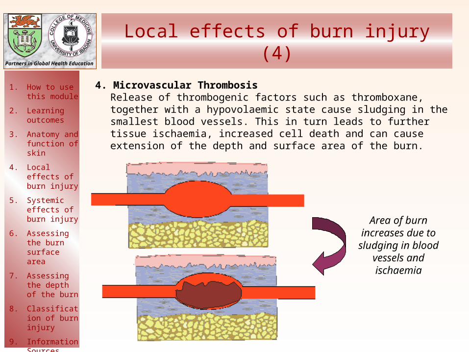
Local effects of burn injury (4)
4. Microvascular ThrombosisRelease of thrombogenic factors such as thromboxane, together with a hypovolaemic state cause sludging in the smallest blood vessels. This in turn leads to further tissue ischaemia, increased cell death and can cause extension of the depth and surface area of the burn.
Area of burn increases due to sludging in blood
vessels and ischaemia

Partners in Global Health Education
1. How to use this module
2. Learning outcomes
3. Anatomy and function of skin
4. Local effects of burn injury
5. Systemic effects of burn injury
6. Assessing the burn surface area
7. Assessing the depth of the burn
8. Classification of burn injury
9. Information Sources
10. End of Module Quiz
Home
Systemic effects of burn injury (1)
When a burn is large (>20% of total body surface area), in
addition to the local response, there is also a systemic
response
Vasoactive substances are released that act not just locally
in the burned tissue, but in non-burned tissue as well.
With large burns, the loss of
circulating blood volume
will rapidly lead to
HYPOVOLAEMIC
SHOCK, unless
resuscitation is started
Loss of circulating
blood
Vascular permeability
Ischaemia

Partners in Global Health Education
1. How to use this module
2. Learning outcomes
3. Anatomy and function of skin
4. Local effects of burn injury
5. Systemic effects of burn injury
6. Assessing the burn surface area
7. Assessing the depth of the burn
8. Classification of burn injury
9. Information Sources
10. End of Module Quiz
Home
Systemic effects of burn injury (2)
Click each box
Respiratory system
Cardiovascular system
Renal system
Haematological system
Immune system
Psychological system
Gastrointestinal system

Partners in Global Health Education
1. How to use this module
2. Learning outcomes
3. Anatomy and function of skin
4. Local effects of burn injury
5. Systemic effects of burn injury
6. Assessing the burn surface area
7. Assessing the depth of the burn
8. Classification of burn injury
9. Information Sources
10. End of Module Quiz
Home
Assessing total burn surface area (TBSA)
The area of this burn is about 3-5% of total body surface area.
How much of the body surface area is burnt?
There are several ways to assess the size of a burn. They all consider the burnt area as a percentage of the total body surface area and are supported by mapping the burnt area on a diagram. In the next couple of slides, we will be looking at the following methods of assessment:
1. The rule of 9’s
2. Lund and Browder charts
3. Palm of hand
4. Unburnt area
Click to Reveal AnswersClick to Reveal Answers

Partners in Global Health Education
1. How to use this module
2. Learning outcomes
3. Anatomy and function of skin
4. Local effects of burn injury
5. Systemic effects of burn injury
6. Assessing the burn surface area
7. Assessing the depth of the burn
8. Classification of burn injury
9. Information Sources
10. End of Module Quiz
Home
Assessing TBSA - Rule of Nines
This method divides the body into areas each of which
equates to 9% of the total body surface area:
• the whole of one arm (anterior and posterior surfaces
including the hand) is 9%, therefore 2 arms = 18%
• the entire head including face, scalp and neck is 9%
• anterior trunk is 18%
• posterior trunk including buttocks is 18%
• the whole lower limb (anterior and posterior surfaces,
including the thigh, leg and foot) is 18%; therefore both lower
limbs = 36%.
This totals 99% with the perineum making the final 1%.
Beware: this method is unreliable in young children.

Partners in Global Health Education
1. How to use this module
2. Learning outcomes
3. Anatomy and function of skin
4. Local effects of burn injury
5. Systemic effects of burn injury
6. Assessing the burn surface area
7. Assessing the depth of the burn
8. Classification of burn injury
9. Information Sources
10. End of Module Quiz
Home
Assessing TBSA in children
Why might the “rule of 9’s” be unreliable in children?
Body proportions change with age. In a child, the head represents a much greater proportion
of the total body surface area.Click to Reveal AnswersClick to Reveal Answers

Partners in Global Health Education
1. How to use this module
2. Learning outcomes
3. Anatomy and function of skin
4. Local effects of burn injury
5. Systemic effects of burn injury
6. Assessing the burn surface area
7. Assessing the depth of the burn
8. Classification of burn injury
9. Information Sources
10. End of Module Quiz
Home
Assessing TBSA - Lund and Browder charts
These take account of the
patient’s age and provide a
more detailed mapping system
for the burnt area
AREA AGE 0 1 5 10 15 ADULT
A = ½ OF HEAD 9 ½ 8 ½ 6 ½ 5 ½ 4 ½ 3 ½
B = ½ OF ONE THIGH 2 ¾ 3 ¼ 4 4 ½ 4 ½ 4 ¾
C = ½ OF ONE LEG 2 ½ 2 ½ 2 ¾ 3 3 ¼ 3 ½

Partners in Global Health Education
1. How to use this module
2. Learning outcomes
3. Anatomy and function of skin
4. Local effects of burn injury
5. Systemic effects of burn injury
6. Assessing the burn surface area
7. Assessing the depth of the burn
8. Classification of burn injury
9. Information Sources
10. End of Module Quiz
Home
Assessing TBSA - Palm size
Another useful way, especially for small burns is to use the palm of the patient’s hand (with fingers extended). This equates to approximately 1% of the body surface area.

Partners in Global Health Education
1. How to use this module
2. Learning outcomes
3. Anatomy and function of skin
4. Local effects of burn injury
5. Systemic effects of burn injury
6. Assessing the burn surface area
7. Assessing the depth of the burn
8. Classification of burn injury
9. Information Sources
10. End of Module Quiz
Home
Assessing TBSA - Unburnt area
In very large burns, it is often easier to measure the area of skin that is unburnt and then subtract this from 100%.

Partners in Global Health Education
1. How to use this module
2. Learning outcomes
3. Anatomy and function of skin
4. Local effects of burn injury
5. Systemic effects of burn injury
6. Assessing the burn surface area
7. Assessing the depth of the burn
8. Classification of burn injury
9. Information Sources
10. End of Module Quiz
Home
Circumferential burns of the limbs can cause distal ischaemia; of the chest, can compromise breathing
Area of the body involved
Not only is the surface area or size of burn important, but also the specific part of the body affected
Face: Facial oedema can lead to airway obstruction. Scarring can cause significant psychosocial problems
Perineum: problems with urogenital function and psychosexual
Hands: Problems with feeding and hygiene
Feet: Mobility problems
Eyes: Burns to the eyes (especially chemical) can cause blindness.

Partners in Global Health Education
1. How to use this module
2. Learning outcomes
3. Anatomy and function of skin
4. Local effects of burn injury
5. Systemic effects of burn injury
6. Assessing the burn surface area
7. Assessing the depth of the burn
8. Classification of burn injury
9. Information Sources
10. End of Module Quiz
Home
Depth of burn
The depth of a burn determines its treatment and how long it takes to heal.
For this reason, it is important to be able to assess the depth as:
Superficial
Partial thickness
• Superficial partial thickness
• Deep partial thickness
Full thickness

Partners in Global Health Education
1. How to use this module
2. Learning outcomes
3. Anatomy and function of skin
4. Local effects of burn injury
5. Systemic effects of burn injury
6. Assessing the burn surface area
7. Assessing the depth of the burn
8. Classification of burn injury
9. Information Sources
10. End of Module Quiz
Home
Depth of burn - Superficial (erythema)
Involves epidermis only:
• Painful
• Red
• No blistering
• Heals rapidly (reversible injury)
• No permanent scars
Note that erythema is NOT included when assessing TBSA

Partners in Global Health Education
1. How to use this module
2. Learning outcomes
3. Anatomy and function of skin
4. Local effects of burn injury
5. Systemic effects of burn injury
6. Assessing the burn surface area
7. Assessing the depth of the burn
8. Classification of burn injury
9. Information Sources
10. End of Module Quiz
Home
Depth of Burn – superficial partial thickness
Patches of skin that would come off on cleaning
Glistening moist red/pink
appearance typical of superficial injury
Typical hot water scald
Involves epidermis and upper dermis:
• Red
• Blistering, moist
• Painful
• Heals by epithelialization
• Healing complete within 14 days
• Minimal or no permanent scars
but can leave discolouration

Partners in Global Health Education
1. How to use this module
2. Learning outcomes
3. Anatomy and function of skin
4. Local effects of burn injury
5. Systemic effects of burn injury
6. Assessing the burn surface area
7. Assessing the depth of the burn
8. Classification of burn injury
9. Information Sources
10. End of Module Quiz
Home
Depth of Burn - superficial partial thickness
Blister
Pin-point bleeding
Pink surface; blanches on
pressure

Partners in Global Health Education
1. How to use this module
2. Learning outcomes
3. Anatomy and function of skin
4. Local effects of burn injury
5. Systemic effects of burn injury
6. Assessing the burn surface area
7. Assessing the depth of the burn
8. Classification of burn injury
9. Information Sources
10. End of Module Quiz
Home
Depth of Burn – deep partial thickness
Involves epidermis, upper dermis and varying degrees of lower dermis:
• Pale, mottled appearance
• Fixed staining (no blanching)
• May be painful or insensate (depending on depth)
• Heals by combination of epithilialization and wound contracture
• May take weeks to heal
• Can leave significant scars and contractures over joints depending on time taken to heal
Deep dermal area, reddish with fixed staining
Partners in Global Health Education
1. How to use this module
2. Learning outcomes
3. Anatomy and function of skin
4. Local effects of burn injury
5. Systemic effects of burn injury
6. Assessing the burn surface area
7. Assessing the depth of the burn
8. Classification of burn injury
9. Information Sources
10. End of Module Quiz
Home
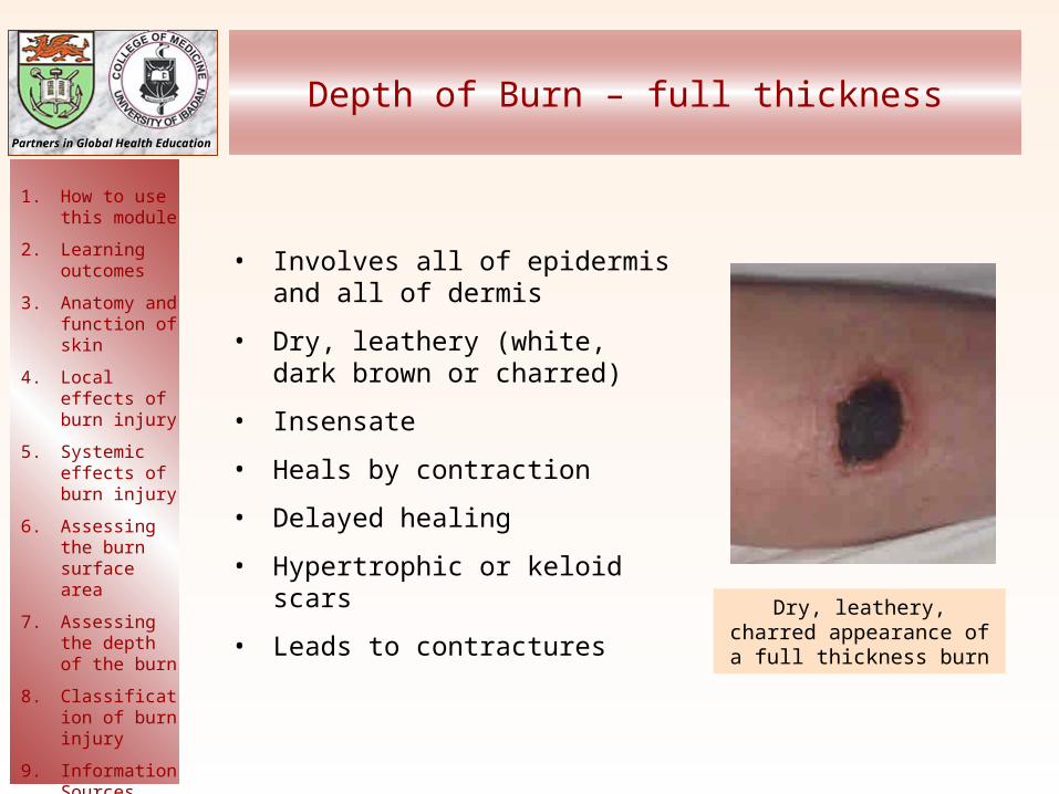
Depth of Burn – full thickness
• Involves all of epidermis and all of dermis
• Dry, leathery (white, dark brown or charred)
• Insensate
• Heals by contraction
• Delayed healing
• Hypertrophic or keloid scars
• Leads to contractures Dry, leathery, charred appearance of a full
thickness burn

Partners in Global Health Education
1. How to use this module
2. Learning outcomes
3. Anatomy and function of skin
4. Local effects of burn injury
5. Systemic effects of burn injury
6. Assessing the burn surface area
7. Assessing the depth of the burn
8. Classification of burn injury
9. Information Sources
10. End of Module Quiz
Home
Circumferential full thickness burn
Typical position of hand in full thickness
burns with metacarpophalangeal joints extended and
interphalangeal joints flexed
Black, charred skin

Partners in Global Health Education
1. How to use this module
2. Learning outcomes
3. Anatomy and function of skin
4. Local effects of burn injury
5. Systemic effects of burn injury
6. Assessing the burn surface area
7. Assessing the depth of the burn
8. Classification of burn injury
9. Information Sources
10. End of Module Quiz
Home
Depth of Burn – mixed thickness
Assess the depth of the
burn in areas A, B and C
( C )(B)
(A)
Click to Reveal AnswersClick to Reveal Answers

Partners in Global Health Education
1. How to use this module
2. Learning outcomes
3. Anatomy and function of skin
4. Local effects of burn injury
5. Systemic effects of burn injury
6. Assessing the burn surface area
7. Assessing the depth of the burn
8. Classification of burn injury
9. Information Sources
10. End of Module Quiz
Home
Depth of Burn – Mixed thickness
Deep dermal with pale pink and white patches, non blanching
Superficial partial thickness showing pink blanching
Full thickness, dry white leathery appearance

Partners in Global Health Education
1. How to use this module
2. Learning outcomes
3. Anatomy and function of skin
4. Local effects of burn injury
5. Systemic effects of burn injury
6. Assessing the burn surface area
7. Assessing the depth of the burn
8. Classification of burn injury
9. Information Sources
10. End of Module Quiz
Home
Classifying the patient
First you should assess the severity of the burn injury according to• TBSA• depth• position• presence of infection• time since the burn• presence or absence of inhalation injury
Combine this information with patient factors:• age • associated injuries• other medical problems• nutritional status
Finally consider social and family factors to classify the patient according to how and where to provide treatment.

Partners in Global Health Education
1. How to use this module
2. Learning outcomes
3. Anatomy and function of skin
4. Local effects of burn injury
5. Systemic effects of burn injury
6. Assessing the burn surface area
7. Assessing the depth of the burn
8. Classification of burn injury
9. Information Sources
10. End of Module Quiz
Home
A guideline for patient classification
significantnone• other medical problems
SpecialistIn-patientOut-patient
Social / family factors
• nutritional status
significantnone• associated injuries
Extremes of ageAdult or older child• age
Patient factors
severemildAbsent• inhalation injury
Critical areaNon-critical area• position
• presence of infection
• depth
• TBSA
Burn injury
Factors
Small Moderate Large
Superficial Partial thickness Full thickness
Absent Localised Systemic
Normal Malnourished
Able to care for oneself Unable to care for oneself

Partners in Global Health Education
1. How to use this module
2. Learning outcomes
3. Anatomy and function of skin
4. Local effects of burn injury
5. Systemic effects of burn injury
6. Assessing the burn surface area
7. Assessing the depth of the burn
8. Classification of burn injury
9. Information Sources
10. End of Module Quiz
Home
Sources of information
• Some images have been adapted from CorelDraw clipart
• See www.interburns.org for more information

Partners in Global Health Education
1. How to use this module
2. Learning outcomes
3. Anatomy and function of skin
4. Local effects of burn injury
5. Systemic effects of burn injury
6. Assessing the burn surface area
7. Assessing the depth of the burn
8. Classification of burn injury
9. Information Sources
10. End of Module Quiz
Home
End of Module Quiz
Well done!Now that you have completed the burns module you may wish to try these questions to assess
your learning.
First, print-out the questions and write down your answers to each one.
Then look at the answer sheet to assess your learning.
QuestionsQuestions AnswersAnswers