Embed Size (px)
Citation preview

Participation of Plasma Membrane Proteins in the Formation
of Tight Junctions by Cultured Epithelial Cells
EVA B. GRIEPP, WILLIAM J. DOLAN, EDITH S. ROBBINS, and DAVID D. SABATINI Department of Cell Biology, New York University Medical Center, New York 10016
ABSTRACT Measurements of the transepithelial electrical resistance correlated with freeze- fracture observations have been used to study the process of t ight junct ion formation under various experimental condit ions in monolayers of the canine kidney epithelial cell line MDCK.
Cells derived from previously conf luent cultures and plated immediately after trypsin-EDTA dissociation develop a resistance that reaches its maximum value of several hundred ohms- cm 2 after ~24 h and falls to a steady-state value of 80-150 ohms-cm 2 by 48 h. The rise in resistance and the development of t ight junctions can be completely and reversibly prevented by the addit ion of 10/~g/ml cycloheximide at the time of plating, but not when this inhibi tor is added more than 10 h after plating. Thus t ight junct ion formation consists of separable synthetic and assembly phases. These two phases can also be dissociated and the requirement for protein synthesis after plating eliminated if, fo l lowing trypsinization, the cells are maintained in spinner culture for 24 h before plating. The requirement for protein synthesis is restored, however, if cells maintained in spinner culture are treated with trypsin before plating. Actino- mycin D prevents development of resistance only in monolayers formed from cells derived from sparse rather than conf luent cultures, but new mRNA synthesis is not required if cells obtained from sparse cultures are maintained for 24 h in spinner culture before plating. Once a steady-state resistance has been reached, its maintenance does not require either mRNA or protein synthesis; in fact, inhibi t ion of protein synthesis causes a rise in the resistance over a 30-h period. Following treatments that disrupt the junctions in steady-state monolayers, recovery of resistance also does not require protein synthesis.
These observations suggest that proteins are involved in t ight junct ion formation. Such proteins, which do not turn over rapidly under steady-state condit ions, are destroyed by trypsinization and can be resynthesized in the absence of stable cell-cell or cell-substratum contact. Messenger RNA coding for proteins involved in t ight junct ion formation is stable except when cells are sparsely plated, and can also be synthesized wi thout intercellular contacts or cell-substratum attachment.
Cells in transporting epithelia form continuous monolayers which function as selective permeability barriers between com- partments. Within such monolayers tight (occluding)junctions located near the apical surface of the cells control the move- ment of substances across the intercellular spaces (8, 9). Freeze- fracture electron microscopy reveals that tight junctions consist of a complex system of anastomosing intramembranous strands involved in maintaining the close contact of the membranes (2, 10, 19, 38, 39, 42). It has been proposed that these strands contain proteins and perhaps lipids (31, 38, 41), but little direct information is available about the biochemical nature ofjunc-
tional components and their precise organization into a supra- molecular structure.
The cell line MDCK of canine kidney origin provides one of several model systems that have recently been adopted for in vitro studies of the development of epithelia and their func- tional properties (3-5, 13, 14, 21, 25, 26, 27, 30, 33, 34, 35, 40). Confluent monolayers of these cells exhibit many characteris- tics of renal tubular epithelia, including the capacity to trans- port fluid and electrolytes in an apical to basolateral direction (3, 20, 27, 37). At confluence, MDCK monolayers acquire a transepithelial resistance that is correlated with the establish-
THE JOURNAL OF CELL BIOLOGY. VOLUme 96 MARCH 1983 693-702 © The Rockefeller University Press • 0021-9525/83/03/0693/10 $1.00 693
on January 13, 2019jcb.rupress.org Downloaded from http://doi.org/10.1083/jcb.96.3.693Published Online: 1 March, 1983 | Supp Info:
m e n t o f a c o m p l e t e s y s t e m o f t igh t j u n c t i o n s de t ec t ed by t h i n
sec t ion a n d f reeze - f rac tu re e l ec t ron m i c r o s c o p y (3, 4, 27).
U s i n g t h e d e v e l o p m e n t o f t r a n s e p i t h e l i a l r e s i s t ance as a n
i n d e x o f j u n c t i o n f o r m a t i o n we h a v e s t u d i e d t h e s y n t h e s i s o f
t h e r e q u i r e d p r o t e i n s a n d t h e a s s e m b l y o f t igh t j u n c t i o n s in
c u l t u r e d M D C K cells. W e h a v e a s s e s s e d t h e s tab i l i ty o f j u n c -
t iona l c o m p o n e n t s in e s t a b l i s h e d m o n o l a y e r s , as wel l as t he
p r o t e i n a n d R N A s y n t h e t i c r e q u i r e m e n t s o f ceils d e r i ved f r o m
c o n f l u e n t a n d s p a r s e cu l tu re s for t h e s u b s e q u e n t d e v e l o p m e n t
o f j u n c t i o n s . W e h a v e f o u n d t h a t s y n t h e s i s a n d a s s e m b l y o f
j u n c t i o n a l c o m p o n e n t s a re t e m p o r a l l y s e p a r a t e d a n d t ha t b o t h
p rocesses c a n be s t u d i e d i n d e p e n d e n t l y by a p p r o p r i a t e expe r -
i m e n t a l m a n i p u l a t i o n s o f t h e c u l t u r e cond i t i ons .
T h i s w o r k s u g g e s t s t h a t f o r m a t i o n o f t igh t j u n c t i o n s r equ i r e s
t h e pa r t i c ipa t i on o f speci f ic p l a s m a m e m b r a n e p r o t e in s a n d
t h a t t h e s y n t h e s i s o f t hese p r o t e i n s is a r e g u l a t e d r a t h e r t h a n a
cons t i tu t ive process . T h e g r o w t h s ta te o f the cells a p p e a r s to
p l a y a n i m p o r t a n t role in con t ro l l i ng t h e a c c u m u l a t i o n o f
m R N A ' s for p r o t e i n s r e q u i r e d for j u n c t i o n f o r m a t i o n , b u t
p r o l o n g e d in t e rce l lu l a r c o n t a c t or a t t a c h m e n t to a s u b s t r a t u m
d o e s n o t s e e m to be r e q u i r e d for t h e s y n t h e s i s o f t h e se p ro te ins .
R e s u l t s o f th i s w o r k h a v e b e e n p r e s e n t e d in p r e l i m i n a r y
f o r m (6, 12).
M A T E R I A L S A N D M E T H O D S
MDCK ceils that had been grown or maintained under the various conditions described below were plated onto collagen-coated nylon disks used for electro- physiological measurements (3). All cells were seeded at the same density (106 cells/ml, 1 ml/well) onto disks placed in 24-well Falcon dishes (Falcon Labware, Oxnard, CA). Disks were transferred to fresh medium after 1.5 h and the development of an electrical resistance was monitored essentially as previously described (3, 4). In these experiments t = 0 is the time of plating. Studies were also carried out with monolayers that had been plated on collagen-coated disks, transferred to fresh medium at 1.5 h, and then maintained in this medium for 48 h before the start of the experiment (steady-state monolayers).
In all experiments six disks were used per time point per variable, and the mean and standard error for each point were calculated. Experiments were repeated several times, but data from single experiments were used to construct the graphs (except as noted), because of variability in the absolute values of control resistances.
The following preparations of cells were used for plating: 1. FRESrILY TRYPSINIZED CELLS: Cells were grown in disposable glass
or plastic roller bottles with complete medium consisting of Eagle's Minimal Essential Medium (MEM) (Gibco Laboratories, Grand Island Biological Co., Grand Island, NY) with 10% fetal calf serum (FCS). Cells from recently confluent cultures (3-7 d after reaching confluence) or sparsely plated cells (sparse cultures) were removed from the roller bottles by incubation at 37°C with 0.25% hog pancreas trypsin (Gibeo Laboratories) and 2 mM EDTA and plated onto disks immediately after dissociation. Sparse cultures were obtained from roller bottles in which 5 × 10⁶ cells were plated 24 h before harvesting.
2. SPINNER-MAINTAINED CELLS: Cells from sparsely plated or recently confluent cultures were removed with trypsin-EDTA from roller bottles and maintained for 20-24 h at a density of 5 × 10⁵ cells/ml in spinner flasks containing Joklik-modifted Minimum Essential Medium for suspension culture (SMEM) (Gibco Laboratories) with 10% FCS. Under these conditions cell counts were stable for several days, indicating that growth was inhibited, although viability remained excellent for at least 48 h. Cell dumps were removed by filtration through a Nitex (Tetko, Elmsford, N J) mesh with a pore size of 20 ~tm. Cells were sedimented by centrifugation at 500 g for 15 min and resuspended in complete medium before plating.
3. STEADY-STATE MONOLAYERS: After removal from roller bottles with trypsin-EDTA, cells were suspended in MEM and 10% FCS and immediately plated on collagen disks, but the monolayers were not used until 40-48 h after plating. In these experiments t = 0 is the time at which the monolayers were perturbed by the addition of EGTA or Diamide, or by a pH change.
a. EGTA: The medium was removed and replaced with a Ca ++, Mg++-fre¢ Moscona's solution containing 2 mM EGTA (ethyleneglycol-bis LS-amino-ethyl ether] N-W-tetra-acetic acid). The kinetics of loss of transepithelial resistance during l-h incubation at 37°C in this medium and the subsequent recovery of the resistance when monolayers were returned to normal medium were deter- mined.
694 THE JOURNAL OF CELL BIOLOGY-VOLUME 96, 1983
b, Diamide: Monolayers were treated for different times (15-30 min) with 0,4-0.6 mM Diamide (Sigma Chemical Co., St. Louis, MO) in Dulbecco's phosphate-buffered saline containing Ca ++, until the transepithelial resistance dropped to 20 ohms-era 2. As with EGTA treatment, changes in the resistance during this treatment and after monolayers were returned to normal medium were followed,
c. pH Changes: Disks were incubated in alkaline medium (MEM with serum adjusted to pH 10 with 1 N NaOH) for 1 h. Normal medium was restored and recovery of resistance monitored.
Inhibi t ion o f Protein and RNA Synthesis: Media containing 5-10 #g/ml cycloheximide (Sigma Chemical Co.), 2.5-10 t~g/ml puromycin (di- HCI, Sigma Chemical Co.), or I-2 #g/ml actinomycin D, (Aldrich Chemical Co., Milwaukee, WI; prepared from a 2 mg/ml stock solution in ethanol) were added at t = 0 and maintained throughout the experiment, unless otherwise indicated.
Rates of protein synthesis were monitored in monolayers developing or established on collagen-coated disks that were incubated with [aH]leucine (10 /tCi/ml) for 1 h ([aH]leucine specific activity 5 Ci/mM; New England Nuclear, Boston, MA). Disks were transferred to 10% cold TCA and the precipitated proteins were collected on glass fiber filters that were rinsed three times with 5% cold TCA before counting in a Beckman liquid scintillation counter (Beckman Instruments, Inc., Fullerton, CA). These measurements demonstrated that cyclo- heximide and puromycin inhibited protein synthesis by >90% within 10 rain of their application.
Freeze-fracture Observat ions: Cells were plated at the appropriate density onto washed, glutaraldehyde-fLxed collagen-coated 100-mm plastic tissue culture dishes each of which also contained three collagen-coated disks. After 1.5 h the dishes were gently rinsed and the medium changed. The disks were removed and their resistances measured before futation of the monolayers developing directly on the collagen-coated dish surfaces, to correlate the resistance changes with morphological observations. After fixation in situ with cold 2% glutaralde- hyde in 0.1 M sucrose and 0.1 M sodium cacodylate buffer for 2 or more hours, the monolayers were scraped off with a Teflon spatula, sedimented into a pellet, rinsed three times in the buffer containing sucrose and impregnated with 20-25% glycerol in 0.1 M sodium cacodylate. The pellets were frozen in liquid Freon 22, stored in liquid N2 and transferred to a Balzer's model #301 apparatus where they were fractured at -150°C, etched for 0-1 min at -120°C and shadowed with platinum and carbon. The organic material was digested with Clorox and the replicas mounted on grids were examined in a Phillips EM 301 electron microscope. Experimental and control samples from at least two different exper- iments were examined, with a total of 50-100 cells from each group evaluated.
Scan ning Electron Microscopy: Disks whose transepithelial resist- ance had been measured were fixed in 2% glutaraldehyde in 0.1 M sodium cacodylate buffer ± 0.1 M sucrose for at least 1 h. They were impregnated with 1% osmium tetroxide, dehydrated in alcohol, and dried at the critical point in COs. Specimens were attached to stubs with double-sided tape, coated with gold- palladium and carbon, and examined in a ETEC or AMR 1000 scanning electron microscope. Experimental and control samples for at least two separate experi- ments were examined.
RESULTS
Prev ious o b s e r v a t i o n s h a v e s h o w n tha t w h e n p l a t ed a t h i g h
d e n s i t y o n c o l l a g e n - c o a t e d disks , M D C K cells o b t a i n e d b y
t r yps in i za t i on f r o m c o n f l u e n t cu l t u r e s r ap id ly a d h e r e to the
su r f ace o f t he d i sks to f o r m c o m p a c t m o n o l a y e r s t h a t w i t h i n a
few h o u r s deve lop a s ign i f i can t t r an sep i t he l i a l e lect r ical resist-
ance (3, 4). T h i s r ise in e lect r ical r e s i s t ance is co r r e l a t ed wi th
t he f o r m a t i o n o f a n e x t e n s i v e s y s t e m o f t igh t j u n c t i o n a l c o m -
p lexes b e t w e e n the cells (6). A l t h o u g h in m o n o l a y e r s e s t ab -
l i shed by th is p r o c e d u r e t he a b s o l u t e v a l u e o f t he f ina l resist-
ance a n d t he s lope o f its in i t ia l r ise a re s o m e w h a t var iab le , in
a typ ica l e x p e r i m e n t (Fig. 1) t he r e s i s t ance r ises to a m a x i m u m ( ~ 3 0 0 o h m - c m 2) u s u a l l y a t t a i n e d by 2 0 - 2 4 h, a n d t h e n de-
c reases to s t e ady - s t a t e levels ( 80 -150 o h m - c m 2) t ha t a re r e a c h e d
by 48 h a n d h a v e p r e v i o u s l y b e e n s h o w n to be m a i n t a i n e d for
weeks t he rea f t e r (3, 4; see a l so Figs. 2 a n d 3).
Stability of Tight Junctions in Confluent Monolayers
T o d e t e r m i n e to w h a t ex t en t c o n t i n u o u s r e p l a c e m e n t o f p ro t e in s i n v o l v e d in t igh t j u n c t i o n s is n e c e s s a r y for t h e m a i n -

tenance of the resistance in established monolayers, we exam- ined the effect of cycloheximide added 40-48 h after plating. It was found that inhibition of protein synthesis for 24 h caused no decrease in the electrical resistance (Fig. 2 a). Instead, the resistance gradually increased, reaching values up to 30--40% higher than in controls. These observations suggest that junc- tional proteins critical for the maintenance of the resistance in established monolayers do not turn over rapidly and therefore need not be constantly replaced. Indeed, the rise of the resist- ance caused by cycloheximide suggests that when protein synthesis is inhibited, previously synthesized proteins necessary for junction formation may be utilized for assembly of new junctions. As expected from these results, inhibition of m-RNA synthesis with actinomycin D did not lower the steady-state value of the resistance (Fig. 2 b).
aooL
200 o
IO0
O - J i i
I0 20 30 Hours offer glaring
FIGURE 1 Development of resistance in MDCK monolayers formed by plating freshly trypsinized cells obtained from recently conf luent cultures. After trypsinization, cells were plated on collagen-coated ny(on disks at high density (10 ° cells/roll. After 1.5 h the disks were rinsed and transferred to fresh medium. Measurements of the trans- epithelial resistance were made at indicated times. Each point is the mean resistance calculated from six different disks.
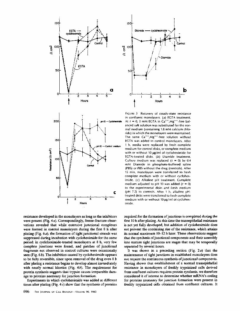
Disruption of Established Tight Junctions Several treatments, such as incubation with media containing
EGTA or the weak base diamide or a rise in the pH of the medium, cause a rapid fall in the transepithelial resistance in established monolayers (Fig. 3 a, b, and c). These effects were reversible and the steady-state values of the resistances were restored within 3-5 h after the treated monolayers were trans- ferred to normal medium (Fig. 3). Inclusion of cycloheximide in the medium did not prevent the reappearance of the resist- ance after removal of the disruptive agent (Fig. 3). Similar results (not shown) were obtained when cycloheximide was added together with EGTA. The observation that protein synthesis is not required for the reassembly of junctions in MDCK monolayers briefly treated with EGTA has been re- ported by Dolan et al. (6) and Martinez-Palomo et al. (22). It has also been previously shown (22) that the loss in resistance caused by EGTA is correlated with disruption of junctions. Regardless of the specific mechanism by which this disruption occurs, it can be inferred from the results presented in Fig. 3 that proteins necessary for junction formation synthesized be- fore and/or during the disruptive treatments--possibly includ- ing some proteins previously involved in the formation of junctional strands--were utilized in the reestablishment of normal levels of resistance.
Formation of Junctions from Newly Synthesized Components by Cells Removed from Confluent Cultures by Trypsinization
The development of electrical resistance that accompanies the formation of new junctions in monolayers made by plating freshly trypsinized cells obtained from confluent cultures is illustrated in Fig. 1. To determine whether under those condi- tions the synthesis of new proteins is required for the devel- opment of tight junctions, we plated cells in the presence of cycloheximide or puromycin (Fig. 4). Although at the concen- trations used (10 #g/ml) these inhibitors do not prevent the settling down and spreading of the cells necessary for the establishment of intercellular contacts (Fig. 5), no significant
180
120
1 } 6o
I ""I" Cycloheximide ,.. "'"'"I"'"~ Puromycin
;"...... . . . . . '"~
~ Control
150
~" I00
v
; 5c
~ rol
Actinomycin D
a b i i I 0 8 ,e 2, ,b 2'0 3'0
FIGURE 2 Protein and mRNA syn- thesis are not required for the main- tenance of the resistance in estab- lished monolayers. (a) Effect of in- hibit ion of protein synthesis. Mono- layers were prepared by heavy plat- ing of ceils as indicated in Fig. I; 40-48 h after plating, when the re- sistance had already reached a steady-state value, fresh complete medium containing 5 #g/ml cyclo- heximide or 2.5 #g/ml puromycin was added (t = 0). The presence of cycloheximide resulted in 90% in- hibit ion of protein synthesis, meas- ured by [3H]leucine incorporation. Fresh medium wi thout drugs was added to controls. Data from four experiments have been pooled. (b)
Hours Effect of inhibi t ion of mRNA syn- thesis. Steady-state monolayers were prepared as described for Fig. 2 a. 40-48 h after plating fresh medium containing actinomycin D (1/tg/ ml) was added (t -- 0) to the experimental disks and fresh medium wi thout drugs to the controls. Data from three experiments have been pooled.
GRIEPP At At. Participation of Plasma Membrane Proteins in Tight Junctions 695
150
~ 1oo
5 0
EGTA ~ " " cydd~xirr~e )
I // //
1~//11/ I I I I
I 2 3 4 5
t Hours
a
i
'°° I ... :[ ' t ..........
!il - L ~1 \ f ~O--Contr01 m~um
IOC ~ L ~
\,iJ ¢
I I 0 2 4 l Hours
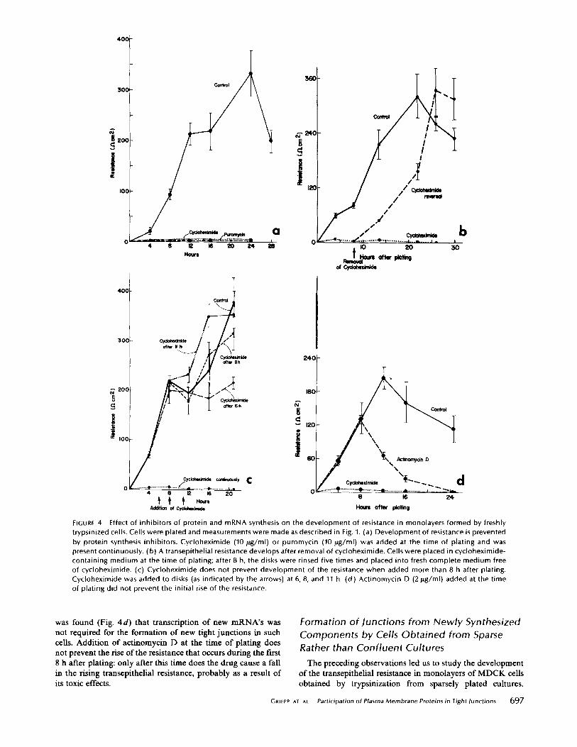
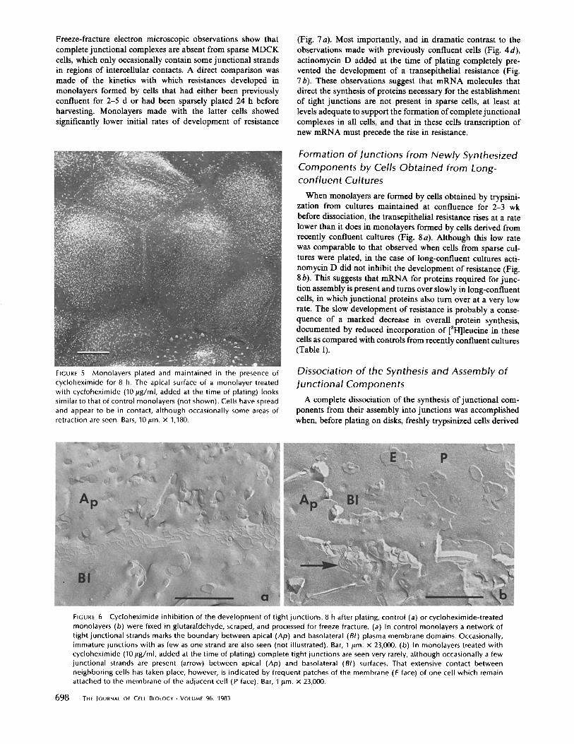
resistance developed in the monolayers as long as the inhibitors were present (Fig. 4 a). Correspondingly, freeze-fracture obser- vations revealed that while extensive junctional complexes were formed in control monolayers during the first 8 h after plating (Fig. 6 a), the formation of tight junctional strands was suppressed during incubation with cycloheximide for the same period: in cycloheximide-treated monolayers at 8 h, very few complete junctions were found, and patches of junctional fragments not observed in control cultures were occasionally seen (Fig. 6 b). The inhibition caused by cycloheximide appears to be fully reversible, since upon removal of the drug even 8 h after plating a resistance begins to develop almost immediately with nearly normal kinetics (Fig. 4b). The requirement for protein synthesis suggests that trypsin causes irreparable dam- age to proteins necessary for junction formation.
Experiments in which cycloheximide was added at different times after plating (Fig. 4 c) show that the synthesis of proteins
696 THE JOURNAL OF CELL BIOLOGY-VOLUME 96, 1983
150
~ IOC
5C
" "..."
/ .: / /
/ :/ / / I ::'~ Diamide
I I 3 4
Hours
0ntrol
b I 5
FIGURE 3 Recovery of steady-state resistance in confluent monolayers. (a) EGTA treatment. At t = 0, 2 mM EGTA in Ca++,Mg++-free bal- anced salt solution was substituted for the nor- mal medium (containing 1.8 mM calcium chlo- ride) in which the monolayers were maintained. The same Ca++,Mg++-free solution without EGTA was added to control monolayers. After 1 h, media were replaced by fresh complete medium for control disks, or complete medium with or without 10 p,g/ml of cycloheximide for EGTA-treated disks. (b) Diamide treatment. Culture medium was replaced (t = 0) by 0.4 mM Diamide in phosphate-buffered saline (PBS) or PBS without the drug (controls). After 15 min, monolayers were transferred to fresh complete medium with or wi thout cyclohex- imide. (c) Alkaline pH treatment. Complete medium adjusted to pH 10 was added (t = 0) to the experimental disks and fresh medium (pH 7.2) to controls. After ~1 h, alkaline pH- treated disks were transferred to fresh complete medium with or wi thout 10 p.g/ml of cyclohex- imide.
required for the formation of junctions is completed during the first 10 h after plating. At this time the transepithelial resistance is not yet fully developed, but addition of cycloheximide does not prevent the continuing rise of the resistance, which attains its normal maximum 10-12 h later. These observations suggest that the synthesis of junctional components and their assembly into mature tight junctions are stages that may be temporally separated by several hours.
It was shown in a preceding section (Fig. 2a) that the maintenance of tight junctions in established monolayers does not require the continuous synthesis of junctional components. Having shown that establishment of a normal transepithelial resistance in monolayers of freshly trypsinized cells derived from confluent cultures requires protein synthesis, we therefore considered it of interest to determine whether mRNA's coding for proteins necessary for junction formation were present in freshly trypsinized cells obtained from confluent cultures. It
~ c 2OO
I00 , I X
4 8 12 16 ~O 24 :!8
H~rs
4 0 (
30¢
~EE 2°0 u
l '~ ~oo
0
T I
¢
/ / l ... s
afar 6h
~clohexin~le e.,ami-L~ly C ........ • ........... 9" ......... 4 .......... • ........... I,-e
4 6 12 16 20
Addition of Cy¢lolmdr~le
Z 4 0
180
g
120
J
..... ~"'-.~...~ ...... "O"'I ........... L.-L.--. J . I io zo 3o
% \ T y c i n D
d 8 16 2 4
Hours after plating
FIGURE 4 Effect of inhib i tors of prote in and mRNA synthesis on the deve lopment o f resistance in monolayers formed by freshly trypsinized cells. Cells were plated and measurements were made as descr ibed in Fig. I. (a) Deve lopment of resistance is prevented by protein synthesis inhibi tors. Cyc lohex imide (10/~g/ml) or puromycin (10 Fg/ml) was added at the t ime of plat ing and was present cont inuously. (b) A transepithel ia l resistance develops after removal o f cycloheximide. Cells were placed in cyc lohex imide- conta in ing med ium at the t ime o f plat ing; af ter 8 h, the disks were rinsed f ive times and placed into fresh comple te med ium free of cycloheximide. (c) Cyc lohex imide does not prevent deve lopment o f the resistance when added more than 8 h after plating. Cyc lohex imide was added to disks (as indicated by the arrows) at 6, 8, and 11 h. (d ) Act inomyc in D (2 / tg /ml ) added at the t ime o f plat ing did not prevent the init ial rise o f the resistance.
was found (Fig. 4d) that transcription of new mRNA's was not required for the formation of new tight junctions in such cells. Addition of actinomycin D at the time of plating does not prevent the rise of the resistance that occurs during the first 8 h after plating: only after this time does the drug cause a fall in the rising transepithelial resistance, probably as a result of its toxic effects.
GRIEPP AT AL.
Formation of ]unctions from Newly Synthesized Components by Cells Obtained from Sparse Rather than Confluent Cultures
The preceding observations led us to study the development of the transepithelial resistance in monolayers of MDCK cells obtained by trypsinization from sparsely plated cultures.
Participation of Plasma Membrane Proteins in Tight Junctions 697
Freeze-fracture electron microscopic observations show that complete junctional complexes are absent from sparse MDCK cells, which only occasionally contain some junctional strands in regions of intercellular contacts. A direct comparison was made of the kinetics with which resistances developed in monolayers formed by cells that had either been previously confluent for 2-5 d or had been sparsely plated 24 h before harvesting. Monolayers made with the latter cells showed significantly lower initial rates of development of resistance
FIGURE 5 Monolayers plated and maintained in the presence of cycloheximide for 8 h. The apical surface of a monolayer treated with cycloheximide (10/ ig/ml, added at the time of plating) looks similar to that of control monolayers (not shown). Cells have spread and appear to be in contact, although occasionally some areas of retraction are seen. Bars, 10/~m. X 1,180.
(Fig. 7 a). Most importantly, and in dramatic contrast to the observations made with previously confluent cells (Fig. 4 d), actinomycin D added at the time of plating completely pre- vented the development of a transepithelial resistance (Fig. 7 b). These observations suggest that mRNA molecules that direct the synthesis of proteins necessary for the establishment of tight junctions are not present in sparse cells, at least at levels adequate to support the formation of complete junctional complexes in all cells, and that in these cells transcription of new mRNA must precede the rise in resistance.
Formation of Junctions from Newly Synthesized Components by C e l l s Obtained from Long- confluent Cultures
When monolayers are formed by cells obtained by trypsini- zation from cultures maintained at confluence for 2-3 wk before dissociation, the transepithelial resistance rises at a rate lower than it does in monolayers formed by cells derived from recently confluent cultures (Fig. 8 a). Although this low rate was comparable to that observed when cells from sparse cul- tures were plated, in the case of long-confluent cultures acti- nomycin D did not inhibit the development of resistance (Fig. 8 b). This suggests that mRNA for proteins required for junc- tion assembly is present and turns over slowly in long-confluent cells, in which junctional proteins also turn over at a very low rate. The slow development of resistance is probably a conse- quence of a marked decrease in overall protein synthesis, documented by reduced incorporation of [3H]leucine in these cells as compared with controls from recently confluent cultures (Table I).
Dissociation of the Synthesis and Assembly of Junctional Components
A complete dissociation of the synthesis of junctional com- ponents from their assembly into junctions was accomplished when, before plating on disks, freshly trypsinized cells derived
FIGURE 6 Cycloheximide inhibi t ion of the development of tight junctions. 8 h after plating, control (a) or cycloheximide-treated monolayers (b) were fixed in glutaraldehyde, scraped, and processed for freeze fracture. (a) In control monolayers a network of tight junctional strands marks the boundary between apical (Ap) and basolateral (BI) plasma membrane domains. Occasionally, immature junctions with as few as one strand are also seen (not illustrated). Bar, I/Lm. x 23,000. (b) In monolayers treated with cycloheximide (10 gg/ml, added at the time of plating) complete tight junctions are seen very rarely, although occasionally a few junctional strands are present (arrow) between apical (Ap) and basolateral (BI) surfaces. That extensive contact between neighboring cells has taken place, however, is indicated by frequent patches of the membrane (E face) of one cell which remain attached to the membrane of the adjacent cell (P face). Bar, 1 gm. X 23,000.
698 THE JOURNAL OF CELL BIOLOGY • VOLUME 96, 1983

T
confluent
400,
,oo / / t / / r
0 , ~ , ~ , , , a I0 20 5O Hour= afro' plotl~
300 -
200,
0, ~ ~ -- . - ~ ~ . - -E.2~ 8 16 24 1.1o~= aft" pl~nll
FIGURE 7 Development of transepi- thelial resistance in monolayers formed by cells derived by trypsini- zation from sparse compared with confluent cultures. (a) The rate of rise of the resistance is lower when cells obtained from sparse cultures (5 x 10 6 cells/roller bottle) rather than re- cently conf luent cultures (0.5-1 x 10 s cells/roller bottle) were seeded on discs at the same density (10 6 cells,/ ml). (b) Actinomycin D (2/,tg/ml) as well as cycloheximide (10 /~g/ml) added at the t ime of plating prevent the development of resistance in monolayers formed by plating freshly trypsinized cells obtained from sparse cultures.
400
~ 3 0 C o
. | ~. zoc
100
0 ~ I0
Houri after plating
newly confluent
long confluent
a
20
°I t: 50 ./2~.... Z
l ~ ' / \ A¢,,om~i, 0 /zJ \ \ //.c5~' .... c~,~e,,.~,'".,, h
o ! a ~ ~ ................ _', ,,,- to 20
Hour's after plating
FIGURI- 8 Development of transepithelial resistance in monolayers formed by freshly trypsinized cells derived from cultures maintained at confluency for varying pe- riods, Cells obtained from cultures con- f luent for 2 d (newly confluent) or for 15 d (long-confluent) were plated at high density (10 6 cells/ml) and measurements were made as described in Fig. I, (a) The resistance rises at a higher rate when cells obtained from newly confluent cultures are plated. (b) Actinomycin D does not prevent the initial rise of the resistance in monolayers formed with cells derived from long-confluent cultures. Act inomy- cin D (2/Lg/ml) and cycloheximide (10 #g/ ml) were added at the t ime of plating.
from confluent cultures were incubated for 24 h as suspension cultures in spinner flasks. Monolayers formed by these cells (Fig. 9a) develop an electrical resistance more rapidly than those made from freshly trypsinized cells and achieve earlier a peak resistance that is generally of a higher value. Most strik- ingly, treatment of spinner cells with cycloheximide continu- ously from the time of plating does not prevent the develop- ment of a resistance (Fig. 9 b), which, however, often does not reach peak values as high as those in controls. These results clearly show that the synthesis of proteins necessary for the formation of junctions can proceed in suspension cultures, in which cells do not make stable contacts with other cells or with a solid substratum. Such proteins appear to have been trans-
ferred to the cell surface, since treatment of spinner-maintained cells with trypsin-EDTA immediately before plating renders them incapable of developing a resistance in the absence of protein synthesis (Fig. 9 c).
Transcription of mRNA's required for the synthesis of junc- tional proteins also takes place when cells derived from sparse cultures are maintained in spinner conditions. Thus, while monolayers made of freshly trypsinized sparse cells do not develop a transepithelial resistance when incubated with either actinomycin D or cycloheximide (Fig. 7 b), these inhibitors do not abolish the rise of the resistance, but only partially reduce it, when the same cells are plated after having been maintained for 24 h in spinner culture (Fig. 9 d).
G~tlEt'l" h7 AL. Part ic ipat ion o f Plasma Membrane Proteins in Tight }unctions 699

]-ABLE I
Rates of Protein Synthesis in Monolayers Formed by Plating Freshly Trypsinized Cells from Newly Confluent or Long
Confluent Cultures
[3H]Leucine incorporat ion dur ing 1 h (cpm x 103 )
Time Newly conf luent Long conf luent
h 3 12.7 + 5.0 9.5 +_ 0.6 7 23.1 + 1.2 8.6 + 3.2
11 20.3 + 0.8 12,7 + 1.2
Cells were obtained from cultures confluent for 2 d (newly confluent) and 15 d (long confluent) and plated at high density as described for the measure- ment of resistance. [3H]leucine (10#Ci/ml) was added to disks for ! h pulses at 3, 7, and 11 h after plating. Total proteins on each disk were precipitated with 10% TCA, rinsed with 5% cold ]-CA and counted in a liquid scintillation counter. Each value is the mean of 4 disks +_ standard error.
DISCUSSION
The experiments described in this paper provide insights into the nature and regulatory features of the biosynthetic processes necessary for the assembly of functional tight junctions in epithelial ceils. The previously reported finding (4, 6), analyzed in more detail in this paper, that MDCK cells require protein synthesis to reestablish a transepithelial resistance after trypsin dissociation most likely indicates that proteins that are exposed on the cell surface participate in the process of junction for- mation. Our observations also suggest that confluent MDCK ceils do not contain a large intracellular pool (i.e., inaccessible to trypsin) of proteins that can be utilized injunction formation in the absence of protein synthesis. A similar conclusion has been reached from freeze-fracture observations, which indicate that junction formation does not take place when cyclohexim- ide is added to developing monolayers formed by cells disso- ciated from subconfluent cultures with EGTA (15).
Synthesis of proteins necessary for junction formation ap- pears to be completed within 8 h after trypsinized ceils are plated at high density. After this time, assembly of the junc- tions, as manifested by an increase in transepithelial resistance, is unaffected by the addition of cycloheximide and proceeds even when the drug is present for an additional 12-14 h. A complete temporal dissociation between the assembly of tight j unctions and synthesis of the required proteins can be achieved under several experimental conditions. Cells from previously confluent cultures are capable of reestablishing junctions in the absence of protein synthesis, if after trypsinization they are maintained in a spinner culture for 24 h before plating. As expected, the kinetics of junction formation by these cells are faster than with cells not incubated in spinner culture but plated immediately after trypsinization. Proteins required for junction formation are probably incorporated into the cell surface during maintenance of the cells in spinner culture, since trypsinization immediately before plating abolishes the capac- ity of such cells to form junctions in the presence of cyclohex- imide.
Assembly of junctions in the absence of protein synthesis can also be achieved in monolayers briefly treated with various agents that rapidly and reversibly lead to a loss of transepithe- lial resistance. Although it is likely that these agents only lead to junction disruption secondarily, most likely as a result of primary effects on cell shape (1, 7, 11, 24, 25, 32), the rapid recovery of the resistance in the presence of cycloheximide after removal of the perturbing agents indicates that the capac-
7 0 0 THE JOURNAL OF CELL BIOLOGY. VOLUME 96, 1983
ity of preexisting proteins to participate in junction formation is not affected. Using freeze-fracture electron microscopy, Mel- dolesi et al. (23) drew similar conclusions after examining the effects of cycloheximide on guinea pig pancreatic acinar cells whose junctions were disrupted by Ca ++ chelation.
It is known that the plasma membrane is a dynamic structure in which protein components may turn over with independent rates (16, 18, 29, 36). The possibility that junctional structures may undergo remodeling is suggested by the observation that peak levels of resistance, usually attained 24 h after plating of trypsinized cells, decrease during the next 24 h to steady-state levels, which are often >50% lower (3). The extent to which synthesis of new proteins is required for the maintenance of established tight junctions was examined in mature mono- layers, in which protein synthesis was inhibited during long periods of incubation with cycloheximide. The finding that prolonged treatment with cycloheximide does not lead to a decay in resistance suggests that, if proteins are involved in maintaining the junctional complexes, such proteins do not normally turn over with a high rate.
The unexpected observation that cycloheximide treatment of mature monolayers leads to a 30-40% increase in resistance within 24 h may reflect a reorganization of junctional elements that takes place during this period and renders the junctions more impermeable to ions. This could possibly involve the recruitment of a pool of plasma membrane components that for some reason had not previously been integrated into func- tional junctions. It has been observed that different treatments can induce a proliferation of tight junction strands in natural epithelia (23, 28), and it has recently been shown that a massive increase in the total length of tight junction strands between prostatic epithelial cells takes place during incubation of the tissue at 37°C (17). This effect was not prevented by cyclohex- imide and it was therefore suggested that it reflected the assembly of preexisting components into new junctions (31).
An increase in resistance during incubation with cyclohex- imide is also consistent with more speculative interpretations in which the suppression of the synthesis of a labile inhibitor of junction formation leads to a decrease in transepithelial permeability. If such a feedback mechanism to maintain tight junction permeability exists, it might also explain why trans- epithelial resistance reaches a peak before assuming steady- state values (Fig. 1) and also why, after disruption of estab- lished monolayers, the resistance initially rebounds to higher than steady-state values (Fig. 3).
In the experiments presented in this paper, ceils were always plated on collagen disks at a sufficiently high density that formation of a confluent monolayer and development of tight junctions could proceed without cell division. This allowed us to compare the readiness of cells previously found in different growth states to form junctions. Ceils derived from cultures maintained at confluence for 2-5 d or even longer contained a level of mRNA adequate for the synthesis of proteins necessary for junction formation, which therefore proceeded even in the presence of actinomycin D. On the other hand, cells that had not yet reached confluence at the time of trypsinization because they had been sparsely plated and cultured for only 24 h did not contain sufficient levels o fmRNA to synthesize the proteins necessary for junction formation in the presence of actinomycin D. If one discounts the possibility of greater toxicity of acti- nomycin D on sparse than on confluent cultures, these results suggest that expression of genes involved in tight junction formation may be regulated by the growth state of the cells so

60C
40(
N
20C
~" 160
8
ll II
IJ_
' ~ ' 2'o oftllr lllaling
fr~ly \ ce~ls
\ \
.28C
240
210
E 1 4 0
I l
a : 70
a
3O
300
Trypsin - treated k
200
-~ I00
Trypsin* cyclohexirnide ¢
4 8 12 16 2 0
T
I I I I I x _ . 8 16 24
Hours after plating
Hours
-,.. ,S "<
8 16 24
Hours after plating
FIGURE 9 Development of the transepithelial resistance in monolayers formed by cells that after trypsinization are maintained in spinner culture for 20-24 h before plating. Cells obtained by trypsinization from confluent (a, b, and c) or sparse (d) cultures were maintained for 20-24 h in spinner culture before plating on disks at high density. (a) The resistance develops faster when cells were maintained in spinner medium fol lowing trypsinization. (b) Cycloheximide (10 #g/ml) added at the t ime of plating does not prevent the development of a resistance when cells previously maintained in spinner culture were plated. (c) Treatment with trypsin before plating restores the requirement for protein synthesis when cells previously maintained in spinner culture were plated. Cells were treated with trypsin-EDTA in a Ca ++, Mg++-free salt solution for 15 rain before plating on disks. Cycloheximide (10 p,g/ml) was added at the time of plating. (d) Maintenance in spinner cultures allows the transcription of messengers and the synthesis of proteins required for the development of a transepithelial resistance in cells obtained from sparse cultures. Actinomycin D (2/tg/ml) and cycloheximide (10/~g/ml) were added at the t ime of plating.
that adequate levels of the corresponding mRNA's normally accumulate only after confluence. However, the finding that, in cells that were obtained from sparse cultures by trypsiniza- tion and maintained in suspension for 24 h before plating, the necessary mRNA's also accumulate in levels sufficient to sup- port junction formation indicates that neither attachment to a substratum nor extensive intercellular contacts are required for messenger accumulation. Readiness to establish junctions may
follow the cessation of growth that occurs after confluence in monolayer cultures or during maintenance in suspension.
The experiments presented in this paper emphasize the role of some plasma membrane proteins in the process of junction formation. They do not, however, demonstrate that these pro- teins are structural components of the junction. An understand- ing of the ability of ceils to regulate the synthesis of required components and their assembly into tight junctions may facil-
GRIEPP AT AL. Participation of Plasma Membrane Proteins in Tight)unctions 701

itate the identification of those proteins that play a specific role in the process o f junction formation.
We acknowledge with gratitude the expert technical assistance of Harriet Snitkin, Susan Malamet, and Heide Plesken. We wish to thank Joyce Mixon, Sonia Martinez, and Myma Chung for their help with the typing of the manuscript, and Jody Culkin and Brian Zietlow for photographic work.
E. B. Griepp was supported by National Institutes of Health (NIH) postdoctoral fellowship HD 05076 and subsequently by an Investiga- torship from the New York Heart Association. This work was sup- ported by NIH grant AG 00378.
Received for publication 28 September 1982, and in revised form 7 December 1982.
REFERENCES
1. Britch, M., and T. D. Allen. 1980. The response of the cellular contractile system to EGTA. Cell Biol. Int. Rep. 4:760.
2. Bullivant, S. 1978. The structure of tight junctions. Ninth International Congress on Eioctron Microscopy, Toronto. 3:659-672.
3. Cereijido, M., E. S. Robbins, W. J. Dolan, C. A. Rotunno, and D. D. Sabatini. 1978. Polarized monolayers formed by epithehal cells on a permeable and translucent support. J. Cell Biol. 77:853-880.
4. Cereijido, M., C. A. Rotunno, E. S. Robbins, and D. D. Sabatini. 1978. Polarized epithelial membranes produced in vitro. In Membrane Transport Processes. J. E Hoffman, editor. Raven Press, New York. 433.
5. Cereijido, M., J. Ehrenfeld, S. Fernandez-Castels, and I. Meza. 1981. Fluxes, junctions, and blisters in coRured monolayers of epithaloid ceils MDCK). In Hormonal Regulation of Epithehal Transport of Ions and Water. W. N. Scot and, D. B. Goodman, editors. NY. Acad. of Sci. 372:422~40.
6. Dolan, W., E. B. Oriepp, E. S. Robbins, and D. D. SabatinL 1978. Synthesis and assembly of tight junction components of epithelial cells in vitro. £ Cell Biol. 79:220a. (Abstr.).
7. Edelhauser, H. F., D. L. Van Horn, P. Miller, and H. J. Pederson. 1976. Effect of thiol- oxidation of glutathione with diamide on corneal endothelial function, junctional com- plexus, and micro filaments. J. Cell Biol. 68:567-578.
8. Farquhar, M. G., and G. Palade. 1963. Junctional complexes in various epithelia. £ Cell Biol. 17:375~,12.
9. Farquhar, M. G., and G. Palade. 1965. Cell junctions in amphibian skin. ,L Cell Biol. 26:263-29l.
10. Friend, D. S., and N. B. Gilula. 1972. Variations in tight and gap junctions in mammalian tissues. J. Cell Biol. 53:758-776.
I1. Fujimoto, T., and K. Ogawa. 1982. Energy dependent transformation of mouse gall bladder epithelial cells in a Ca z* -depleted medium..L Ultrastruct. Res. 79:327-340.
12. Griepp, E., E~ Robbins, S. Malamet, W. Dolan, and D. D. SabatinL 1979. Studies on the role of glycoproteins in tight junction formation. J. Cell Biol. 83:88a. (Abstr.).
13. Handier, J. R., F. M. Perkins, and J. P. Johnson. 1980. Studies of renal ceil function using cell culture techniques. Am. J. Physiol. 238:FI-F9.
14. Handler, J. S., R. F. Steele, J. B. Wade, A. S. Peterson, N. L. Lawson, and J. P. Johnson. 1979. Toad urinary bladder epithelial cells in culture: maintenance of epithehal structure, sodium transport, and response to hormones. Proc. Natl. Acad. Sci. USA. 76:4151-4155.
15. Hal Sang, U., M. H. Saiar, M. H. Ellisman. 1979. Tight junction formation is closely linked to the polar redistribution of intramembrunous particles in aggregating MDCK epithelia. Exp. Cell Res. 122:384-391.
16. Hubbard, A. L. 1978. Turnover of Membrane Polypeplldes. In Transport of Macromole- cules in Cellular Systems. S. C. Silverstein, editor. Daklem Konferenzen, Berlin. 363-390.
17. Kachar, B., and P. Pinto da Silva. 1981. Rapid massive assembly of tight junction strands. Science (Wash. DC). 213:541-544.
18. Kaplan, L, and M. Moskowitz. 1975. Studies on the turnover of plasma membranes in cultural mannnahan cells. II. Demonstration of heterogeneous rates of turnover for plasma membrane proteins and glycoproteins. Biochim Biophys. Acts. 389:306-313.
19. Kreutziger, G. O. 1968. Freeze-etching of intercellular junctions of mouse liver. In Proceedings of the 26th Annual Meeting of the Electron Microscopic Society of America. C. J. Arceneaux, editor. Baton Rouge, Claitor's Publishing Division, Baton Rouge, LA. 234-235.
20. Leighton, L, Z. Brada, L. W. Estes, and G. Jnsth. 1963. Secretory activity and oocogenicily of a cell line (MDCK) derived from canine kidney. Science (Wash. DC). 163:472-473.
21. Louvard, D. 1980. Apical membrane aminopeptidase appears at site of cell-ceU contact in cultured kidney epithelial cells. Proc. Natl. Acad. Sci. USA. 77:4132-4136.
22. Martincz-Palomo, A., I. Meza, G. Beaty, and M. Cereijido. 1980. Experimental modulation of occluding junctions in a cultured transporting epithelium. J. Cell Biol. 87:736-745.
23. Maldolesi, J , G. Castiglioni, R. Parma, N. Nassivera, and P. De Camilli. 1978. Ca *+- dependent disassembly and reassembly of occinding junctions in guinea pancreatic acinar cells. J. Cell Biol. 79:156-172.
24. Meza, 1., G. Ibarra, M. Sabanero, A. Martinez-Palomo, and M. Cereijido. 1980. Occluding junctions and cytoskaletal components m a cultural transporting epithehum. J. Cell BioL 87:746-754.
25. Meza, I., M. Sabanero, E. Stefani, and M. Cereijido. 1982. Occluding junctions in MDCK cells: modulation of transepithelial permeability by the cytoskeleton. J. Cell Biochem. 18:407-42 I.
26. Mills, J. W., A. D. C. MacKnight, J.-M. Dayer, and D. A. Ausiello. 1979. Localization of aH-ouabain-sgnsitive Na%pump sites in cultured pig kidney ceils. Am. .7 . Physiol. 236:C 157-C 162.
27. Misfeldt, D. S., S. T. Hamamoto, and D. R. Pitelka. 1976. Transepithelial transport in celt culture. Proc. NatL Acad. Sci. USA. 73:1212-1216.
28. Montesano, R., G. Gabbiani, A. Perrelet, and L. OrcL 1976. In vivo induction of tight junction proliferation in rat liver. J. Cell BioL 68:793-798.
29. Parry, G. 1978. Membrane assembly and turnover. In Subcellular Biochemistry. D B. Roodyn, editor. Plenum Press, NY. 261-326.
30. Perkins, F , and J. S. Handier. 1981. Transport properties of toad kidney epithelia in culture. Am. J. PhysioL 241:C154-CI59.
31. Pinto da Silva, P., and B. Kachar. 1982. On tight-junction structure. Cell. 28:4al-450. 32. Pilelka, D. R., and S. T. Hamamoto. 1977. Calcium-chelation induced disruption of
occluding junctions by cultured mammary epithelial cells..L Cell Biol. 75:69a. (Abstr.). 33. Rabilo, C. A., R., Tchao, J. Valentich, and J. Leighton. 1978. Distribution and character-
istics of the occluding junctions in a monolayer of a cell line (MDCK) derived from canine kidney. ,L Membr. Biol. 43:351-365.
34. Richardson, J. C. W., and N. L. Simmons. 1979. Demonstration of protein asymmetries in the plasma membrane of cultured renal (MDCK) epithelial cells by lactoperoxidase- mediated iodination. FEBS (Fed. Eur. Biochem. Soc.) Lett. 105:201-204.
35. Rindler, M. L, M. Chuman, L. Shaffer, and M. H Saier. 1979. Retention ofdifferentiated properties in an established dog kidney epithelial cell line (MDCK). ,L Cell BioL 81:635 648.
36. Schimke, R. T. 1975. Turnover of Membrane Proteins in Animal Cells. In Methods Membr. BioL 3:201-236.
37. Simmons, N. L. 1981. Ion transport in 'tight' epithelial monolayers of MDCK cells. Z Membrane Biol. 59:105-114.
38. Staehelin, L A. 1974. Structure and function of intercellular junctions. Int. Rev. CytoL 39:191-283.
39. Staehetin, L. A., T. M. Mukherjhce, and A. W. Wiihams. 1969. Freeze-etch appearance of the tight junctions in the epithelium of small and large intestines of mice. Protoplasms. 67:165-184.
40. Taub, M., and M. H. Saier. 1979. Regulation of Z~Na+ transport by calcium in an established kidney epithelial cell line. J. Biol. Chem. 254: 11440-11444.
41. Van Deurs, B., and J. H. Luff. 1975. Effects of glutaraldehyde rotation on the structure of tight junctions: a quantitative freeze-fracture analysis. £ UItrastruct. Res. 68:160-172.
42. Wade, J. B., and M. J. Karnovsky. 1974. The structure of the zonula occludens: a single fibril model based on freeze fracture. ,£ Cell Biol. 60:168 180.
702 TH[ )OURNAL OF C[l_[ BIOLOGY -VOLUME 96, 1983