Embed Size (px)
Citation preview
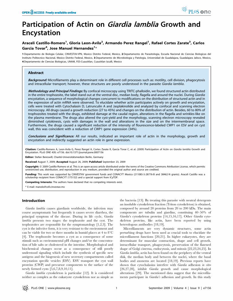
Participation of Actin on Giardia lamblia Growth andEncystationAraceli Castillo-Romero1, Gloria Leon-Avila2, Armando Perez Rangel1, Rafael Cortes Zarate3, Carlos
Garcia Tovar4, Jose Manuel Hernandez1*
1 Departamento de Biologia Celular, CINVESTAV-IPN, Mexico Distrito Federal, Mexico, 2 Departamento de Parasitologia, Escuela Nacional de Ciencias Biologicas del
Instituto Politecnico Nacional, Mexico Distrito Federal, Mexico, 3 Departamento de Microbiologia y Patologia, Universidad de Guadalajara, Guadalajara Jalisco, Mexico,
4 Departamento de Ciencias Biologicas, UNAM, FES-Cuautitlan, Cuautitlan Izcalli, Mexico
Abstract
Background: Microfilaments play a determinant role in different cell processes such as: motility, cell division, phagocytosisand intracellular transport; however, these structures are poorly understood in the parasite Giardia lamblia.
Methodology and Principal Findings: By confocal microscopy using TRITC-phalloidin, we found structured actin distributedin the entire trophozoite, the label stand out at the ventral disc, median body, flagella and around the nuclei. During Giardiaencystation, a sequence of morphological changes concurrent to modifications on the distribution of structured actin and inthe expression of actin mRNA were observed. To elucidate whether actin participates actively on growth and encystation,cells were treated with Cytochalasin D, Latrunculin A and Jasplakinolide and analyzed by confocal and scanning electronmicroscopy. All drugs caused a growth reduction (27 to 45%) and changes on the distribution of actin. Besides, 60 to 80% oftrophozoites treated with the drugs, exhibited damage at the caudal region, alterations in the flagella and wrinkles-like onthe plasma membrane. The drugs also altered the cyst-yield and the morphology, scanning electron microscopy revealeddiminished cytokinesis, cysts with damages in the wall and alterations in the size and on the intermembranal space.Furthermore, the drugs caused a significant reduction of the intensity of flourescence-labeled CWP1 on ESV and on cystwall, this was coincident with a reduction of CWP1 gene expression (34%).
Conclusions and Significance: All our results, indicated an important role of actin in the morphology, growth andencystation and indirectly suggested an actin role in gene expression.
Citation: Castillo-Romero A, Leon-Avila G, Perez Rangel A, Cortes Zarate R, Garcia Tovar C, et al. (2009) Participation of Actin on Giardia lamblia Growth andEncystation. PLoS ONE 4(9): e7156. doi:10.1371/journal.pone.0007156
Editor: Stefan Bereswill, Charite-Universitatsmedizin Berlin, Germany
Received August 7, 2009; Accepted August 26, 2009; Published September 23, 2009
Copyright: � 2009 Castillo-Romero et al. This is an open-access article distributed under the terms of the Creative Commons Attribution License, which permitsunrestricted use, distribution, and reproduction in any medium, provided the original author and source are credited.
Funding: This work was supported by CINVESTAV government funds and CONACYT Mexico (211085-5-28776-B and 38462-N grants). Araceli Castillo was ascholarship recipient from CONACYT (175132) and a special support from ICyTDF.
Competing Interests: The authors have declared that no competing interests exist.
* E-mail: [email protected]
Introduction
Giardia lamblia causes giardiasis worldwide, the infection may
course asymptomatic but frequently it causes severe diarrhea, the
principal symptom of the disease. During its life cycle, Giardia
lamblia presents two stages: the trophozoite and the cyst. The
trophozoites are responsible for the symptomatology [1,2,3]. The
cyst is the infective form, it is very resistant to the environment and
can be viable for two or three months in humid places at 4 to 8uC[4]. The trophozoite becomes a cyst as a consequence of some
stimuli such as environmental pH changes and/or the concentra-
tion of bile salts or cholesterol in the intestine. Morphological and
biochemical changes occur as a consequence of still poorly
understood signaling which include the synthesis of specific new
antigens and the biogenesis of new secretory compartments called
encystation specific vesicles (ESV). ESV transport the cyst wall
proteins (CWP) and precursor components to the surface of the
newly formed cysts [5,6,7,8,9,10,11].
Giardia lamblia cytoskeleton is particular [12]. It is considered
neither as complex as the eukaryote cytoskeleton nor as simple as
the bacteria [13]. By treating this parasite with neutral detergents
an insoluble cytoskeleton fraction (Triton cytoskeleton) is obtained,
composed by around 20 proteins from 20 to 200 kDa. The main
components are tubulin and giardins, constituing 40–50% of
Giardia’s cytoskeleton proteins [14,15,16,17]. Other Giardia cyto-
skeleton proteins, like actin, have been reported by using
heterologous antibodies [18,19].
Microfilaments are very dynamic structures, some actin
perturbing drugs have been used as crucial tools to elucidate the
microfilament functions [20,21]. In higher eukaryotes, they are
determinant for muscular contraction, shape and cell growth,
intracellular transport, phagocytosis, preservation of the flatened
shape of Golgi cisterna, endocytosis, and mitosis [22,23,24,25]. In
Giardia lamblia, actin has been found on the periphery of the ventral
disk, the median body and between the nuclei, where the basal
bodies and axonems are located [18,19]. Previous reports have
shown that cytochalasins interfere with Giardia adhesion in vitro
[26,27,28], inhibit Giardia growth and cause morphological
alterations [29]. The mentioned data suggest that the microfila-
ments participate in Giardia’s adhesion and growth. In this study
PLoS ONE | www.plosone.org 1 September 2009 | Volume 4 | Issue 9 | e7156
we demonstrated, by using microfilament disturbing drugs, that
actin plays a critical role in growth and encystation, it is important
in morphology and indirectly regulates CWP1 gene expression.
Our results could aim to establish new strategies focused to find
specific targets to avoid the formation of cysts, the infective form of
Giardia.
Methods
Culture of Giardia lamblia and in vitro encystationAxenic cultures of Giardia lamblia (WB strain) trophozoites were
grown at 37uC in Diamond’s TYI-S-33 medium pH 7.1
supplemented with 10% bovine serum and 0.5 mg/ml bovine
bile [30]. For in vitro encystation, 66105 trophozoites/ml were
cultured at 37uC for 24 h in TYI-S-33 medium pH 7.8,
supplemented with 10% bovine serum and 10 mg/ml bovine
bile. Trophozoites and cysts were diluted in phosphate-buffered
saline (PBS) and counted in a hemocytometer.
Analysis of actin distribution on trophozoites and duringin vitro encystation
Giardia lamblia trophozoites (66105 cells/ml) were cultured at
37uC for 24 h in TYI-S-33 encystation medium. The distribution
of actin was analyzed at the begining of encystation (time 0,
trophozoites) and during the encystation process at 6, 12, 18 and
24 h by using TRITC-phalloidin. The stained samples were
analyzed by confocal microscopy.
In vitro experiments with drugsIn order to evaluate the effect of microfilament disturbing
drugs on Giardia lamblia growth, 103 trophozoites were grown
for 24 h, then, cells were incubated with 10 mM Cytochalasin D
(CD; Sigma-Aldrich St. Louis, Mo. USA) or 1 mM Latrunculin A
(LA, ; Sigma-Aldrich St. Louis, Mo. USA) or 1 mM Jasplakinolide
(JAS; Calbiochem-Novabiochem La Jolla, CA. USA) disolved
in 0.1% DMSO. Cultures were monitored for 24 to 72 h. 0.1%
DMSO, the drugs diluent was used as a negative control. To prove
the effect of the same drugs on the encystation process, troph-
ozoites (66105) were cultured for 24 h in encystation medium
containing 10 mM CD, 1 mM LA, or 1 mM JAS. The doses used
for each compound were selected accordingly to previous studies
in Giardia, some other parasites and mammalian cells. [20,21,
26,29,31,32].
Expression and purification of CWP1The cyst wall protein 1 (CWP1, GenBank Accession
No. U09330 ) gene was amplified from genomic DNA by PCR
using the primers: forward 59-AGA AGA GAA TTC AAA TGA
TGC TCG CTC TCC TTG C-39 and reverse 59-TCT TCT
GCG GCC GCT TTC AAG GCG GGG TGA GGC AG-39
(EcoRI and NotI restriction sites are underlined). The product was
cloned into pPROEX-1 expression vector in order to get
pPROEX-1-CWP1 and after that DH5a E. coli was transformed
with this construction. The construction was verified by DNA
sequencing (Automated sequencer ABI Prism 310, Perkin-Elmer,
Applied Biosystems) and the overexpression of the fusion protein
was induced by adding 1 mM isopropyl-D-thiogalactopyranoside
IPTG (Invitrogen Carlsbad, CA. USA) to the transformed cells
culture. Fusion protein was purified by Ni-NTA Agarose affinity
chomatography (Qiagen; Valencia, CA USA), following the
manufacturer instructions. Protein concentration was determined
by Bradford assay [33] and purity was analyzed by 12% SDS-
PAGE [34].
Production of policlonal antibodiesBALB/c mice and Wistar rats were immunized with 100 mg
and 500 mg of recombinant CWP1, respectively, by intraperito-
neal route at 0, 7 and 15 days. First immunizations were applied as
an emulsified mixture 10:1 protein:TiterMaxH (Sigma) [35]; the
following challenges were done with a mixture 1:1 protein:alumi-
num hydroxide. Pre-immune and immune sera were assayed by
ELISA and Western blot [36,37]. All the procedures involving
animals were carried on following federal and local regulations for
animal care and use (CINVESTAV-IACUC, approved by the
Mexican Oficial Norm: NOM-062-ZOO-1999).
ImmunofluorescenceCells were washed twice with PBS and fixed for 1 h at 37uC
with 1% paraformaldehyde in PBS. Fixed cells were washed twice
with PBS, applied on coverslips precoated with poly-L-lysine and
were allowed to adhere at room temperature. Then, cells were
permeabilized with 0.5% Triton X100-SDS for 30 min, washed
three times with PBS and incubated at room temperature for
30 min with 1% bovine serum albumin. Then, cells were labeled
with diluted 1:100 TRITC-phalloidin (Sigma) for 1 h. Cysts were
processed as trophozoites, but they were incubated for 1 h with
primary polyclonal anti-CWP antibody (1:300) before phalloidin
labeling. Then, the cells were washed twice with PBS and
incubated for 1 h with diluted 1:100 TRITC-phalloidin and 1:100
Cy5 conjugated anti-mouse (Jackson ImmunoResearch Laborato-
ries). Finally, coverslips were mounted on glass slides with
Vectashield mounting medium (Vector Laboratories) and ana-
lyzed by Confocal Microscopy (Leica DMIRE2 and TCS-SPE).
The images were processed with the Leica Lite and the Leica
Application Suite softwares and transformed to the apropriate
quality format.
Relative-quantitative RT-PCRThe actin (GenBank accession no. AAA99305.1 and CWP1
(GenBank accession no. U09330) cDNAs were synthesized, by a
reverse transcriptase reaction (INVITROGEN), using 2 mg of
RNA (purified by Trizol method) from trophozoites and cysts and
the following primers: actin sense 59-AGA AGA GAA TTC AAA
TGA CAG ACG ACA ACC CTG CCA TAG-39 and actin
antisense 59-TCT TCT GCG GCC GCT TCA CAT ACA CTT
ACG GTT TGC AAT G-39.and CWP1 sense 59-TCT TCT
GCG GCC GCT TCA CAT ACA CTT ACG GTT TGC AAT
G-39 and CWP1 antisense 59-TCT TCT CCA TGG TAG GCG
GGG TGA GGC AGT ACT CTC CGC AGT CCG-39 Relative-
quantitative RT-PCRs were performed in a 7500 Real Time PCR
System (Applied Biosystems, Foster City CA. USA), using
MaximaTM SYBR Green qPCR Master Mix (2X) (Fermentas
Life Sciences) to monitor the amplification reactions. The
expression of both genes were normalized to the expression of
Giardia glyceraldehyde 3-phosphate dehydrogenase (gap1) gene
(GenBank accession no. M88062) using gap1 sense primer 59-
GCA AGC GTG TCA TCA TCT CCG CTC CG-39 and gap1
antisense primer 59-AAG GAC CTT CCC GAC AGC CTT
TGC G-39. Also a melting curve was performed to confirm the
absence of primer dimerization. The relative-quantitative RT-
PCR conditions were: hot start 95uC for 10 min, 40 cycles at 95uCfor 30 s, 65uC for 30 s and 72uC for 1 min. Expression data were
determined by using the comparative DDCt method. Significant
differences (p,0.05) (calculated by t–Student and ANOVA tests
using the program GraphPad Prism 5.02) are indicated by
asterisks in figures. Error bars indicate standard deviation for
experiments with more than one trial.
Cytoskeleton Actin and Giardia
PLoS ONE | www.plosone.org 2 September 2009 | Volume 4 | Issue 9 | e7156
Scanning electron microscopyFor scanning electron microscopy (SEM) analysis, trophozoites
and cysts were washed and fixed with 2.5% glutaraldehyde in PBS
for 1 h. Then, cells were adhered on poly-L-lysine pretreated
coverlips, washed three times with PBS and post-fixed in 1%
osmium tetroxide for 1 h. Next, cells were washed with PBS,
dehydrated in alcohol, dried to critical-point with CO2, coated
with gold and analyzed in a SEM (JSM-35C).
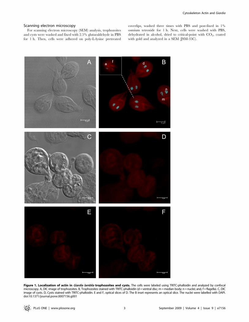
Figure 1. Localization of actin in Giardia lamblia trophozoites and cysts. The cells were labeled using TRITC-phalloidin and analyzed by confocalmicroscopy. A, DIC image of trophozoites. B, Trophozoites stained with TRITC-phalloidin (d = ventral disc; m = median body; n = nuclei; and, f = flagella). C, DICimage of cysts. D, Cysts stained with TRITC-phalloidin. E and F, optical slices of D. The B inset represents an optical slice. The nuclei were labelled with DAPI.doi:10.1371/journal.pone.0007156.g001
Cytoskeleton Actin and Giardia
PLoS ONE | www.plosone.org 3 September 2009 | Volume 4 | Issue 9 | e7156
Results
Actin is distributed in the entire trophozoite, medianbody, disc, flagella and concentrated into threads at thecentral part of the cyst
Even though there are no available homologous antibodies
against Giardia actin, some authors have revealed the localization
of this protein in Giardia trophozoites by using heterologous
antibodies [18,19], but there is no a specific analysis that shows the
distribution of actin during the encystation process.
In order to detect actin in both stages of Giardia, we first
analyzed its distribution using TRITC-phalloidin –a well accepted
specific tool for structured actin identification–. Confocal micros-
copy images of trophozoites (Figure 1A) demonstrated actin
distributed like small spots in the entire trophozoites and in nuclei.
However an intense stain was evident, in median body, on the
periphery of the ventral disc and flagella (Figure 1B). In cysts
(Figure 1C), actin was distributed as thick ribbons forming a
compact oval structure, also some patches were evidents
(Figure 1D–F).
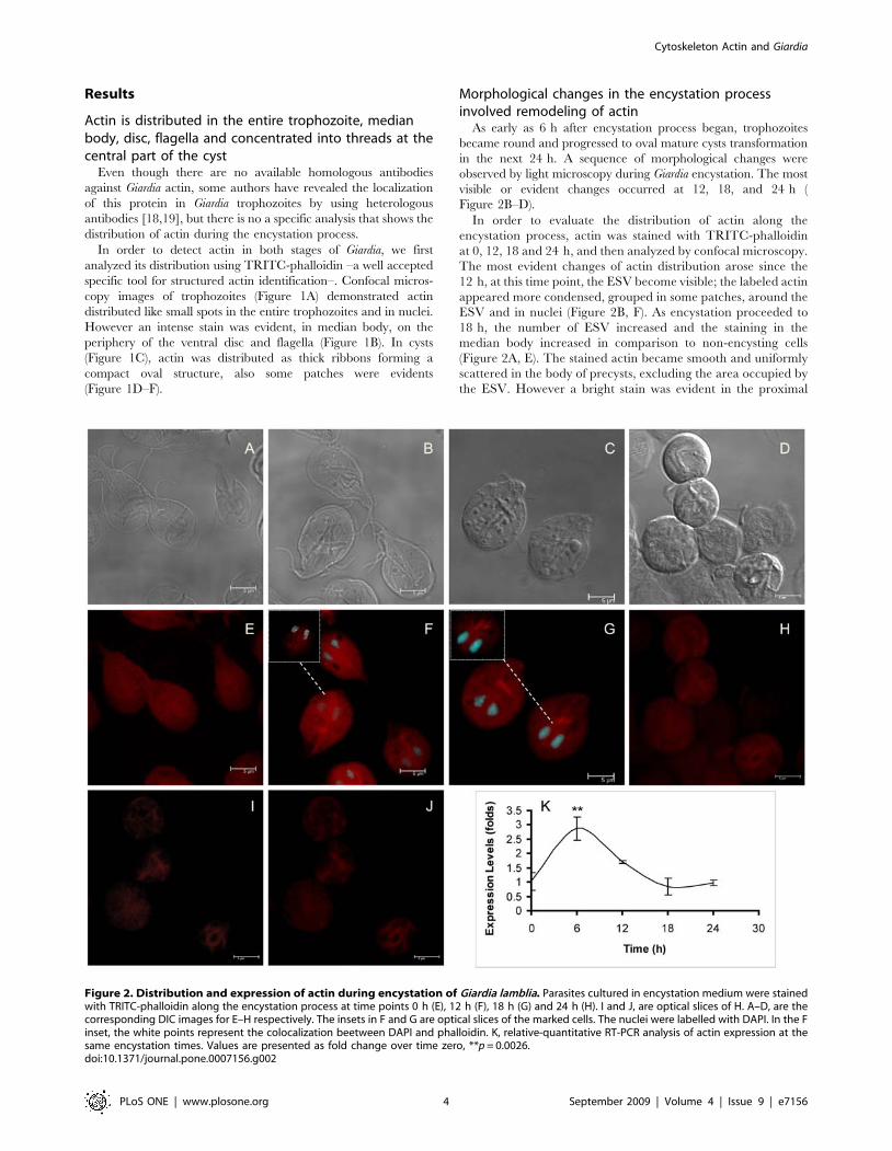
Morphological changes in the encystation processinvolved remodeling of actin
As early as 6 h after encystation process began, trophozoites
became round and progressed to oval mature cysts transformation
in the next 24 h. A sequence of morphological changes were
observed by light microscopy during Giardia encystation. The most
visible or evident changes occurred at 12, 18, and 24 h (
Figure 2B–D).
In order to evaluate the distribution of actin along the
encystation process, actin was stained with TRITC-phalloidin
at 0, 12, 18 and 24 h, and then analyzed by confocal microscopy.
The most evident changes of actin distribution arose since the
12 h, at this time point, the ESV become visible; the labeled actin
appeared more condensed, grouped in some patches, around the
ESV and in nuclei (Figure 2B, F). As encystation proceeded to
18 h, the number of ESV increased and the staining in the
median body increased in comparison to non-encysting cells
(Figure 2A, E). The stained actin became smooth and uniformly
scattered in the body of precysts, excluding the area occupied by
the ESV. However a bright stain was evident in the proximal
Figure 2. Distribution and expression of actin during encystation of Giardia lamblia. Parasites cultured in encystation medium were stainedwith TRITC-phalloidin along the encystation process at time points 0 h (E), 12 h (F), 18 h (G) and 24 h (H). I and J, are optical slices of H. A–D, are thecorresponding DIC images for E–H respectively. The insets in F and G are optical slices of the marked cells. The nuclei were labelled with DAPI. In the Finset, the white points represent the colocalization beetween DAPI and phalloidin. K, relative-quantitative RT-PCR analysis of actin expression at thesame encystation times. Values are presented as fold change over time zero, **p = 0.0026.doi:10.1371/journal.pone.0007156.g002
Cytoskeleton Actin and Giardia
PLoS ONE | www.plosone.org 4 September 2009 | Volume 4 | Issue 9 | e7156
region of the ventral axonems. (Figure 2C, G). After 24 h, actin
was irregularly distributed in the core of mature cysts (Figure 2H),
it was found forming patches (Figure 2I, J). Besides, during
Giardia encystation the relative-quantitative RT-PCR analysis
demostrated variations of actin mRNA expression; at 6 h of
encystation, actin mRNA increased almost two folds in
comparison to non encysting cells, after, the expression decreased
and remaining almost constant from 18 to 24 h of encystation
(Figure 2K).
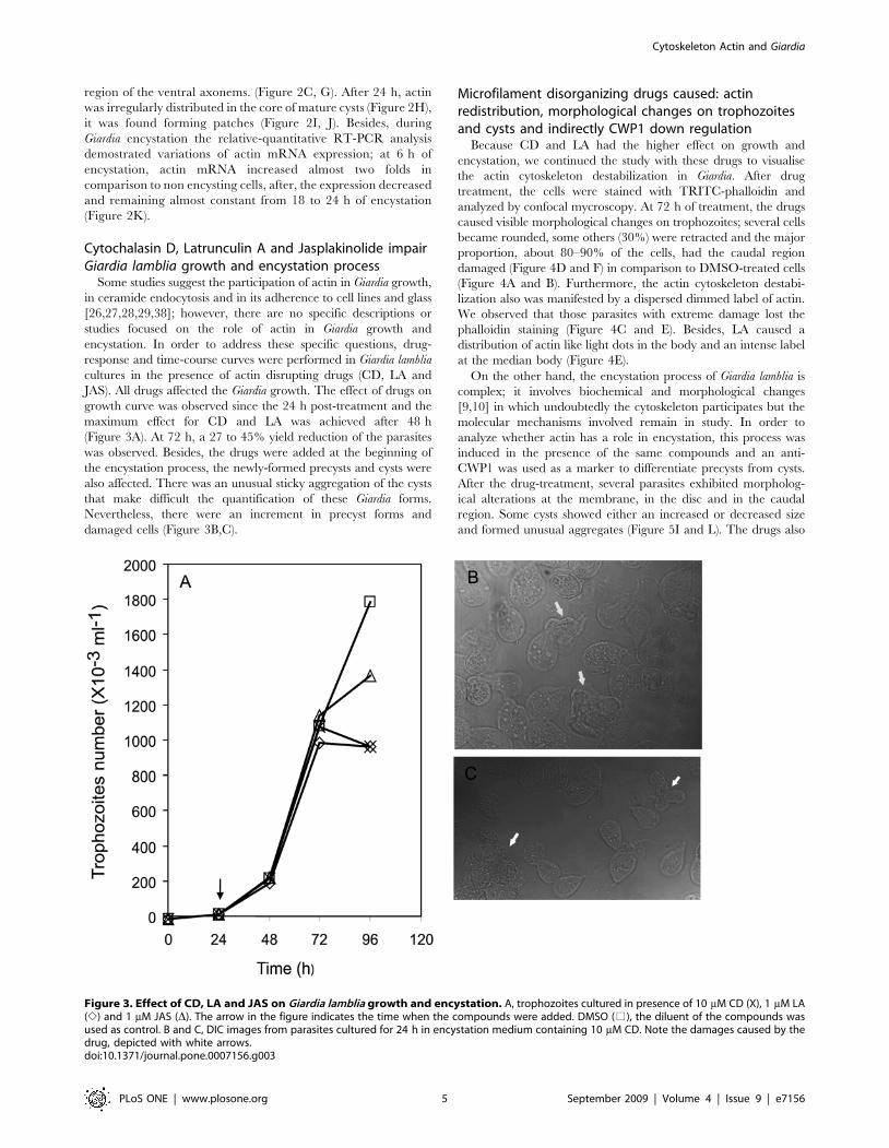
Cytochalasin D, Latrunculin A and Jasplakinolide impairGiardia lamblia growth and encystation process
Some studies suggest the participation of actin in Giardia growth,
in ceramide endocytosis and in its adherence to cell lines and glass
[26,27,28,29,38]; however, there are no specific descriptions or
studies focused on the role of actin in Giardia growth and
encystation. In order to address these specific questions, drug-
response and time-course curves were performed in Giardia lamblia
cultures in the presence of actin disrupting drugs (CD, LA and
JAS). All drugs affected the Giardia growth. The effect of drugs on
growth curve was observed since the 24 h post-treatment and the
maximum effect for CD and LA was achieved after 48 h
(Figure 3A). At 72 h, a 27 to 45% yield reduction of the parasites
was observed. Besides, the drugs were added at the beginning of
the encystation process, the newly-formed precysts and cysts were
also affected. There was an unusual sticky aggregation of the cysts
that make difficult the quantification of these Giardia forms.
Nevertheless, there were an increment in precyst forms and
damaged cells (Figure 3B,C).
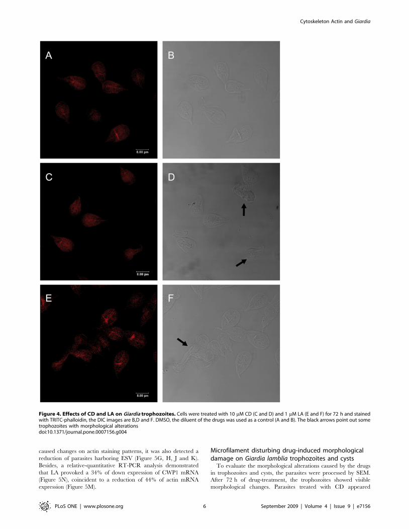
Microfilament disorganizing drugs caused: actinredistribution, morphological changes on trophozoitesand cysts and indirectly CWP1 down regulation
Because CD and LA had the higher effect on growth and
encystation, we continued the study with these drugs to visualise
the actin cytoskeleton destabilization in Giardia. After drug
treatment, the cells were stained with TRITC-phalloidin and
analyzed by confocal mycroscopy. At 72 h of treatment, the drugs
caused visible morphological changes on trophozoites; several cells
became rounded, some others (30%) were retracted and the major
proportion, about 80–90% of the cells, had the caudal region
damaged (Figure 4D and F) in comparison to DMSO-treated cells
(Figure 4A and B). Furthermore, the actin cytoskeleton destabi-
lization also was manifested by a dispersed dimmed label of actin.
We observed that those parasites with extreme damage lost the
phalloidin staining (Figure 4C and E). Besides, LA caused a
distribution of actin like light dots in the body and an intense label
at the median body (Figure 4E).
On the other hand, the encystation process of Giardia lamblia is
complex; it involves biochemical and morphological changes
[9,10] in which undoubtedly the cytoskeleton participates but the
molecular mechanisms involved remain in study. In order to
analyze whether actin has a role in encystation, this process was
induced in the presence of the same compounds and an anti-
CWP1 was used as a marker to differentiate precysts from cysts.
After the drug-treatment, several parasites exhibited morpholog-
ical alterations at the membrane, in the disc and in the caudal
region. Some cysts showed either an increased or decreased size
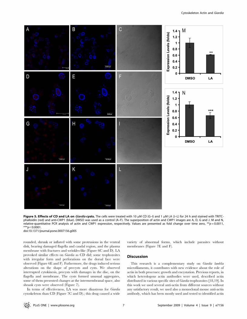
and formed unusual aggregates (Figure 5I and L). The drugs also
Figure 3. Effect of CD, LA and JAS on Giardia lamblia growth and encystation. A, trophozoites cultured in presence of 10 mM CD (X), 1 mM LA(e) and 1 mM JAS (D). The arrow in the figure indicates the time when the compounds were added. DMSO (%), the diluent of the compounds wasused as control. B and C, DIC images from parasites cultured for 24 h in encystation medium containing 10 mM CD. Note the damages caused by thedrug, depicted with white arrows.doi:10.1371/journal.pone.0007156.g003
Cytoskeleton Actin and Giardia
PLoS ONE | www.plosone.org 5 September 2009 | Volume 4 | Issue 9 | e7156
caused changes on actin staining patterns, it was also detected a
reduction of parasites harboring ESV (Figure 5G, H, J and K).
Besides, a relative-quantitative RT-PCR analysis demonstrated
that LA provoked a 34% of down expression of CWP1 mRNA
(Figure 5N), coincident to a reduction of 44% of actin mRNA
expression (Figure 5M).
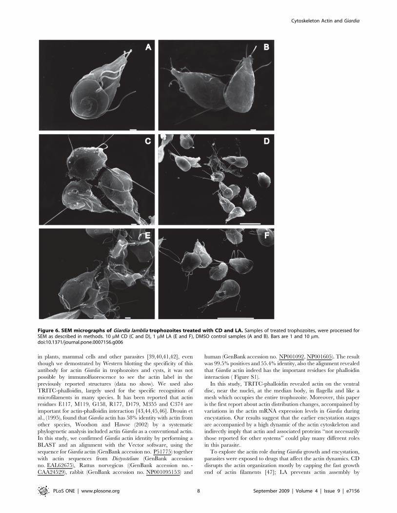
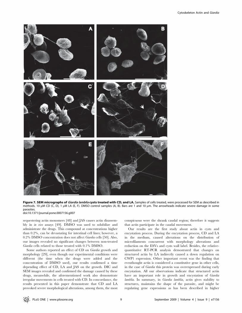
Microfilament disturbing drug-induced morphologicaldamage on Giardia lamblia trophozoites and cysts
To evaluate the morphological alterations caused by the drugs
in trophozoites and cysts, the parasites were processed by SEM.
After 72 h of drug-treatment, the trophozoites showed visible
morphological changes. Parasites treated with CD appeared
Figure 4. Effects of CD and LA on Giardia trophozoites. Cells were treated with 10 mM CD (C and D) and 1 mM LA (E and F) for 72 h and stainedwith TRITC-phalloidin, the DIC images are B,D and F. DMSO, the diluent of the drugs was used as a control (A and B). The black arrows point out sometrophozoites with morphological alterationsdoi:10.1371/journal.pone.0007156.g004
Cytoskeleton Actin and Giardia
PLoS ONE | www.plosone.org 6 September 2009 | Volume 4 | Issue 9 | e7156
rounded, shrunk or inflated with some protrusions in the ventral
disk, bearing damaged flagella and caudal region, and the plasma
membrane with fractures and wrinkles-like (Figure 6C and D). LA
provoked similar effects on Giardia as CD did; some trophozoites
with irregular form and perforations on the dorsal face were
observed (Figure 6E and F). Futhermore, the drugs induced serious
alterations on the shape of precysts and cysts. We observed
interrupted cytokinesis, precysts with damages in the disc, on the
flagella and membrane. The cysts formed unusual aggregates,
some of them presented changes at the intermembranal space, also
shrank cysts were observed (Figure 7).
In terms of effectiveness, LA was more disastrous for Giardia
cytoskeleton than CD (Figure 7C and D).; this drug caused a wide
variety of abnormal forms, which include parasites without
membranes (Figure 7E and F).
Discussion
This research is a complementary study on Giardia lamblia
microfilaments, it contributes with new evidence about the role of
actin in both processes: growth and encystation. Previous reports, in
which heterologous actin antibodies were used, described actin
distributed in various specific sites of Giardia trophozoites [18,19]. In
this work we used several anti-actin from different sources without
any satisfactory result, we used also a monoclonal mouse anti-actin
antibody, which has been mostly used and tested to identified actin
Figure 5. Effects of CD and LA on Giardia cysts. The cells were treated with 10 mM CD (G–I) and 1 mM LA (J–L) for 24 h and stained with TRITC-phalloidin (red) and anti-CWP1 (blue). DMSO was used as a control (A–F). The superposition of actin and CWP1 images are A, D, G and J. M and N,relative-quantitative PCR analysis of actin and CWP1 expression, respectively. Values are presented as fold change over time zero, **p = 0.0011,***p,0.0001.doi:10.1371/journal.pone.0007156.g005
Cytoskeleton Actin and Giardia
PLoS ONE | www.plosone.org 7 September 2009 | Volume 4 | Issue 9 | e7156
in plants, mammal cells and other parasites [39,40,41,42], even
though we demostrated by Western blotting the specificity of this
antibody for actin Giardia in trophozoites and cysts, it was not
possible by immunolfuorescence to see the actin label in the
previously reported structures (data no show). We used also
TRITC-phalloidin, largely used for the specific recognition of
microfilaments in many species. It has been reported that actin
residues E117, M119, G158, R177, D179, M355 and C374 are
important for actin-phalloidin interaction [43,44,45,46]. Drouin et
al., (1995), found that Giardia actin has 58% identity with actin from
other species, Woodson and Hawse (2002) by a systematic
phylogenetic analysis included actin Giardia as a conventional actin.
In this study, we confirmed Giardia actin identity by performing a
BLAST and an alignment with the Vector software, using the
sequence for Giardia actin (GenBank accession no. P51775) together
with actin sequences from Dictyostelium (GenBank accession
no. EAL62675), Rattus norvegicus ((GenBank accession no. -
CAA24529), rabbit (GenBank accession no. NP001095153) and
human (GenBank accession no. NP001092, NP001605). The result
was 99.5% positives and 55.4% identity, also the alignment revealed
that Giardia actin indeed has the important residues for phalloidin
interaction ( Figure S1).
In this study, TRITC-phalloidin revealed actin on the ventral
disc, near the nuclei, at the median body, in flagella and like a
mesh which occupies the entire trophozoite. Moreover, this paper
is the first report about actin distribution changes, accompained by
variations in the actin mRNA expression levels in Giardia during
encystation. Our results suggest that the earlier encystation stages
are accompanied by a high dynamic of the actin cytoskeleton and
indirectly imply that actin and associated proteins ‘‘not necessarily
those reported for other systems’’ could play many different roles
in this parasite.
To explore the actin role during Giardia growth and encystation,
parasites were exposed to drugs that affect the actin dynamics. CD
disrupts the actin organization mostly by capping the fast growth
end of actin filaments [47]; LA prevents actin assembly by
Figure 6. SEM micrographs of Giardia lamblia trophozoites treated with CD and LA. Samples of treated trophozoites, were processed forSEM as described in methods. 10 mM CD (C and D), 1 mM LA (E and F), DMSO control samples (A and B). Bars are 1 and 10 mm.doi:10.1371/journal.pone.0007156.g006
Cytoskeleton Actin and Giardia
PLoS ONE | www.plosone.org 8 September 2009 | Volume 4 | Issue 9 | e7156
sequestering actin monomers [48] and JAS causes actin disassem-
bly in in vivo assays [49]. DMSO was used to solubilize and
administrate the drugs. This compound at concentrations higher
than 0.2%, can be devastating for intestinal cell lines; however, a
0.2% DMSO concentration does not affect Giardia cells [50]. Also,
our images revealed no significant changes between non-treated
Giardia cells related to those treated with 0.1% DMSO.
Some authors reported an effect of CD on Giardia growth and
morphology [29], even though our experimental conditions were
different (the time when the drugs were added and the
concentration of DMSO used), our results confirmed a time
depending effect of CD, LA and JAS on the growth. DIC and
SEM images revealed and confirmed the damage caused by these
drugs, meanwhile, the aforementioned work also demonstrate
irregular movements in cells treated with CD. In concordance, the
results presented in this paper demonstrate that CD and LA
provoked severe morphological alterations, among them, the most
conspicuous were the shrank caudal region; therefore it suggests
that actin participate in the caudal movement.
Our results are the first study about actin in cysts and
encystation process. During the encystation process, CD and LA
in the medium, caused alterations on the distribution of
microfilaments concurrent with morphology alterations and
reduction on the ESVs and cysts wall label. Besides, the relative-
quantitative RT-PCR analysis demostrated that changes on
structured actin by LA indirectly caused a down regulation on
CWP1 expression. Other important event was the finding that
eventhought actin is considered a constitutive gene in other cells,
in the case of Giardia this protein was overexpressed during early
encystation. All our observations indicate that structured actin
have an important role in growth and encystation of Giardia
lamblia. In summary, in Giardia lamblia, actin gives stability to
structures, maintains the shape of the parasite, and might be
regulating gene expressions as has been described in higher
Figure 7. SEM micrographs of Giardia lamblia cysts treated with CD, and LA. Samples of cells treated, were processed for SEM as described inmethods. 10 mM CD (C, D), 1 mM LA (E, F). DMSO control samples (A, B). Bars are 1 and 10 mm. The arrowheads indicate severe damage in someparasites.doi:10.1371/journal.pone.0007156.g007
Cytoskeleton Actin and Giardia
PLoS ONE | www.plosone.org 9 September 2009 | Volume 4 | Issue 9 | e7156
eukaryotes and other parasites [51,52,53], also it must have a role
in intracellular transport. Obviously, particular ABPs and
microtubular associated proteins MAPs must be involved in those
processes, and currently our group is addressing that item.
Supporting Information
Figure S1 Alignment of Giardia actin against other-species
actins. Alignment of Giardia actin against other-species actins.
The boxes depict the residues involved in actin-phalloidin
interaction identificated on actin Giardia sequence.
Found at: doi:10.1371/journal.pone.0007156.s001 (5.37 MB TIF)
Acknowledgments
We thank Dr Isabel Salazar for critical review of the manuscript, to Blanca
Reyes and Ivan J. Galvan M. for technical support in Confocal
Microscopy. We thank Guadalupe Aguilar for assistance with DNA
sequencing. We also thank to General Services CINVESTAV specially
Sirenia Gonzalez Pozos for technical assistance in SEM and to Jose Molina
for photography assistance. We express our gratitude to Fortunato Mote
and Armando Sanchez for technical assistance as well as to Esther Cid,
Maria del Refugio Contreras, Lucero Gonzalez, Clara Hernandez and
Marisol Camba for excellent administrative assistance.
Author Contributions
Conceived and designed the experiments: ACR GL MH. Performed the
experiments: ACR APR. Analyzed the data: ACR GL APR RCZ CGT
MH. Contributed reagents/materials/analysis tools: GL RCZ CGT MH.
Wrote the paper: ACR MH.
References
1. Adam RD (1991) The biology of Giardia spp. Microbiol Rev 55: 706–732.
2. Adam RD (2001) Biology of Giardia lamblia. Clin Microbiol Rev 14: 447–475.
3. Lujan HD (2006) [Giardia and giardiasis]. Medicina (B Aires) 66: 70–74.
4. Bingham AK, Jarroll EL Jr, Meyer EA, Radulescu S (1979) Giardia sp.: physical
factors of excystation in vitro, and excystation vs eosin exclusion as determinants
of viability. Exp Parasitol 47: 284–291.
5. Gillin FD, Reiner DS, Gault MJ, Douglas H, Das S, et al. (1987) Encystation
and expression of cyst antigens by Giardia lamblia in vitro. Science 235:
1040–1043.
6. Reiner DS, Douglas H, Gillin FD (1989) Identification and localization of cyst-
specific antigens of Giardia lamblia. Infect Immun 57: 963–968.
7. Reiner DS, McCaffery M, Gillin FD (1990) Sorting of cyst wall proteins to a
regulated secretory pathway during differentiation of the primitive eukaryote,
Giardia lamblia. Eur J Cell Biol 53: 142–153.
8. Gillin FD, Reiner DS, McCaffery JM (1996) Cell biology of the primitive
eukaryote Giardia lamblia. Annu Rev Microbiol 50: 679–705.
9. Lujan HD, Mowatt MR, Nash TE (1997) Mechanisms of Giardia lamblia
differentiation into cysts. Microbiol Mol Biol Rev 61: 294–304.
10. Lujan HD, Mowatt MR, Nash TE (1998) The Molecular Mechanisms of
Giardia Encystation. Parasitol Today 14: 446–450.
11. Eichinger D (2001) Encystation in parasitic protozoa. Curr Opin Microbiol 4:
421–426.
12. Elmendorf HG, Dawson SC, McCaffery JM (2003) The cytoskeleton of Giardia
lamblia. Int J Parasitol 33: 3–28.
13. Pogliano J (2008) The bacterial cytoskeleton. Curr Opin Cell Biol 20: 19–27.
14. Holberton DV, Ward AP (1981) Isolation of the cytoskeleton from Giardia.
Tubulin and a low-molecular-weight protein associated with microribbon
structures. J Cell Sci 47: 139–166.
15. Crossley R, Holberton DV (1983) Characterization of proteins from the
cytoskeleton of Giardia lamblia. J Cell Sci 59: 81–103.
16. Crossley R, Holberton DV (1983) Selective extraction with Sarkosyl and
repolymerization in vitro of cytoskeleton proteins from Giardia. J Cell Sci 62:
419–438.
17. Jimenez-Cardoso E, Crisostomo-Vazquez P, Suarez-Souto A (1997) [Separation
of the ventral disc proteins of Giardia intestinalis]. Rev Invest Clin 49: 123–128.
18. Feely DE, Schollmeyer JV, Erlandsen SL (1982) Giardia spp.: distribution of
contractile proteins in the attachment organelle. Exp Parasitol 53: 145–154.
19. Narcisi EM, Paulin JJ, Fechheimer M (1994) Presence and localization of
vinculin in Giardia. J Parasitol 80: 468–473.
20. Spector I, Shochet NR, Blasberger D, Kashman Y (1989) Latrunculins–novel
marine macrolides that disrupt microfilament organization and affect cell
growth: I. Comparison with cytochalasin D. Cell Motil Cytoskeleton 13:
127–144.
21. Wakatsuki T, Schwab B, Thompson NC, Elson EL (2001) Effects of cytochalasin
D and latrunculin B on mechanical properties of cells. J Cell Sci 114:
1025–1036.
22. Lamaze C, Fujimoto LM, Yin HL, Schmid SL (1997) The actin cytoskeleton is
required for receptor-mediated endocytosis in mammalian cells. J Biol Chem
272: 20332–20335.
23. Cordonnier MN, Dauzonne D, Louvard D, Coudrier E (2001) Actin filaments
and myosin I alpha cooperate with microtubules for the movement of lysosomes.
Mol Biol Cell 12: 4013–4029.
24. Lazaro-Dieguez F, Jimenez N, Barth H, Koster AJ, Renau-Piqueras J, et al.
(2006) Actin filaments are involved in the maintenance of Golgi cisternae
morphology and intra-Golgi pH. Cell Motil Cytoskeleton 63: 778–791.
25. Lovy-Wheeler A, Cardenas L, Kunkel JG, Hepler PK (2007) Differential
organelle movement on the actin cytoskeleton in lily pollen tubes. Cell Motil
Cytoskeleton 64: 217–232.
26. Feely DE, Erlandsen SL (1982) Effect of cytochalasin-B, low Ca++ concentra-
tion, iodoacetic acid, and quinacrine-HCl on the attachment of Giardiatrophozoites in vitro. J Parasitol 68: 869–873.
27. Katelaris PH, Naeem A, Farthing MJ (1995) Attachment of Giardia lambliatrophozoites to a cultured human intestinal cell line. Gut 37: 512–518.
28. Sousa MC, Goncalves CA, Bairos VA, Poiares-Da-Silva J (2001) Adherence ofGiardia lamblia trophozoites to Int-407 human intestinal cells. Clin Diagn Lab
Immunol 8: 258–265.
29. Correa G, Benchimol M (2006) Giardia lamblia behavior under cytochalasins
treatment. Parasitol Res 98: 250–256.
30. Keister DB (1983) Axenic culture of Giardia lamblia in TYI-S-33 medium
supplemented with bile. Trans R Soc Trop Med Hyg 77: 487–488.
31. Makioka A, Kumagai M, Ohtomo H, Kobayashi S, Takeuchi T (2001)
Entamoeba invadens: enhancement of excystation and metacystic developmentby cytochalasin D. Exp Parasitol 98: 145–151.
32. Makioka A, Kumagai M, Ohtomo H, Kobayashi S, Takeuchi T (2001) Effect ofjasplakinolide on the growth, encystation, and actin cytoskeleton of Entamoeba
histolytica and Entamoeba invadens. J Parasitol 87: 399–405.
33. Bradford MM (1976) A rapid and sensitive method for the quantitation of
microgram quantities of protein utilizing the principle of protein-dye binding.
Anal Biochem 72: 248–254.
34. Laemmli UK (1970) Cleavage of structural proteins during the assembly of the
head of bacteriophage T4. Nature 227: 680–685.
35. Svendsen Bollen L, Crowley A, Stodulski G, Hau J (1996) Antibody production
in rabbits and chickens immunized with human IgG. A comparison of titre andavidity development in rabbit serum, chicken serum and egg yolk using three
different adjuvants. J Immunol Methods 191: 113–120.
36. Clem TR, Yolken RH (1978) Practical colorimeter for direct measurement of
microplates in enzyme immunoassay systems. J Clin Microbiol 7: 55–58.
37. Towbin H, Staehelin T, Gordon J (1992) Electrophoretic transfer of proteins
from polyacrylamide gels to nitrocellulose sheets: procedure and someapplications. 1979. Biotechnology 24: 145–149.
38. Hernandez Y, Castillo C, Roychowdhury S, Hehl A, Aley SB, et al. (2007)Clathrin-dependent pathways and the cytoskeleton network are involved in
ceramide endocytosis by a parasitic protozoan, Giardia lamblia. Int J Parasitol37: 21–32.
39. Guerrero-Barrera AL, Garcia-Cuellar CM, Villalba JD, Segura-Nieto M,Gomez-Lojero C, et al. (1996) Actin-related proteins in Anabaena spp. and
Escherichia coli. Microbiology 142 (Pt 5): 1133–1140.
40. Hernandez-Gonzalez EO, Lecona-Valera AN, Escobar-Herrera J, Mujica A
(2000) Involvement of an F-actin skeleton on the acrosome reaction in guinea pig
spermatozoa. Cell Motil Cytoskeleton 46: 43–58.
41. Ocampo J, Mondragon R, Roa-Espitia AL, Chiquete-Felix N, Salgado ZO, et
al. (2005) Actin, myosin, cytokeratins and spectrin are components of the guineapig sperm nuclear matrix. Tissue Cell 37: 293–308.
42. Pasten-Hidalgo K, Hernandez-Rivas R, Roa-Espitia AL, Sanchez-Gutierrez M,Martinez-Perez F, et al. (2008) Presence, processing, and localization of mouse
ADAM15 during sperm maturation and the role of its disintegrin domain duringsperm-egg binding. Reproduction 136: 41–51.
43. Vandekerckhove J, Deboben A, Nassal M, Wieland T (1985) The phalloidinbinding site of F-actin. Embo J 4: 2815–2818.
44. Jahraus A, Egeberg M, Hinner B, Habermann A, Sackman E, et al. (2001) ATP-dependent membrane assembly of F-actin facilitates membrane fusion. Mol Biol
Cell 12: 155–170.
45. Belmont LD, Patterson GM, Drubin DG (1999) New actin mutants allow further
characterization of the nucleotide binding cleft and drug binding sites. J Cell Sci112 (Pt 9): 1325–1336.
46. Oda T, Namba K, Maeda Y (2005) Position and orientation of phalloidin in F-actin determined by X-ray fiber diffraction analysis. Biophys J 88: 2727–2736.
Cytoskeleton Actin and Giardia
PLoS ONE | www.plosone.org 10 September 2009 | Volume 4 | Issue 9 | e7156
47. Flanagan MD, Lin S (1980) Cytochalasins block actin filament elongation by
binding to high affinity sites associated with F-actin. J Biol Chem 255: 835–838.48. Morton WM, Ayscough KR, McLaughlin PJ (2000) Latrunculin alters the actin-
monomer subunit interface to prevent polymerization. Nat Cell Biol 2: 376–378.
49. Bubb MR, Spector I, Beyer BB, Fosen KM (2000) Effects of jasplakinolide onthe kinetics of actin polymerization. An explanation for certain in vivo
observations. J Biol Chem 275: 5163–5170.50. Gadelha AP, Travassos R, Monteiro-Leal LH (2007) The evaluation of a
semiautomated computer method to determine the effects of DMSO on Giardia
lamblia-intestinal cell interaction. Parasitol Res 101: 1401–1406.
51. Witteck A, Yao Y, Fechir M, Forstermann U, Kleinert H (2003) Rho protein-
mediated changes in the structure of the actin cytoskeleton regulate humaninducible NO synthase gene expression. Exp Cell Res 287: 106–115.
52. Nemeth ZH, Deitch EA, Davidson MT, Szabo C, Vizi ES, et al. (2004)
Disruption of the actin cytoskeleton results in nuclear factor-kappaB activationand inflammatory mediator production in cultured human intestinal epithelial
cells. J Cell Physiol 200: 71–81.53. Wolyniak MJ, Sundstrom P (2007) Role of actin cytoskeletal dynamics in
activation of the cyclic AMP pathway and HWP1 gene expression in Candida
albicans. Eukaryot Cell 6: 1824–1840.
Cytoskeleton Actin and Giardia
PLoS ONE | www.plosone.org 11 September 2009 | Volume 4 | Issue 9 | e7156
![RESEARCH ARTICLE Open Access Identification of ... · agricultural and industrial applications [1]. * Correspondence: ulhoa@icb.ufg.br 1Departamento de Bioquímica e Biologia Molecular,](https://img.pdfslide.us/doc/110x75/5f0381717e708231d409656f/research-article-open-access-identification-of-agricultural-and-industrial-applications.jpg)