Embed Size (px)
Citation preview
On November 1, 2002, John Quigley climbed into the branches of a 70-foot-tall oak tree estimated to be 150 to 400 years old. He stayed perched thereuntil he was removed, 71 days later, to allow a housing developer to cutdown the tree. That was a short stay, however, compared with Julia But-
terfly Hill’s sojourn in a 600-year-old redwood. In the year 2000, Hill created a perch 180feet above ground and didn’t come down to Earth until just over 2 years later, when thePacific Lumber Company agreed to spare that tree and others in its immediate vicinity.
What prompts some people to tree-sit or protest in other ways against the removalof trees? Clearly, one motivation is their admiration for the sheer longevity of these or-ganisms, which have survived in their environmentsfor decades and even centuries, during which a greatdeal of human history has taken place. The oldestknown individual plant is a bristlecone pine that haslived for more than 4,900 years—almost 50 centuries.In contrast, it is doubtful that any animal has everlived much longer than 2 centuries.
This longevity is even more impressive when it isunderstood that plants cannot move from site to siteto avoid danger or environmental challenges. Eventhough plants are not motile, the extreme agesachieved by some trees prove that plants can never-theless cope successfully with their environment. Theplant body creates and maintains an internal envi-ronment that differs from the external environment.
Plants accomplish through growth some of the samethings that animals achieve through mobility. Growingroots, for example, can reach into new supplies of wa-ter and nutrients. By growing, stems and leaves rise out of shaded areas into the sun to obtain energy.
Although plants do not need to obtain complexsubstances like vitamins from their environments asanimals do, they must nevertheless obtain nutri-ents—not only the raw materials of photosynthesis(carbon dioxide and water), but also mineral ele-ments such as nitrogen, potassium, and calcium.Seed plants—even the tallest trees—transport waterand minerals from the soil to their tops, and theytransport the products of photosynthesis from theleaves to their roots and other parts.
The Plant Body
An Ancient Individual Bristleconepines (Pinus longaeva) can live for cen-turies. The oldest known living organismis a bristlecone pine that has been alivefor almost 5,000 years—long enough tohave witnessed all of recorded humanhistory.
35
Part Six • THE BIOLOGY OF FLOWERING PLANTS

Plants also interact with their living and nonliving envi-ronments. They respond to environmental cues as they growand develop. Their responses are mediated by chemical sig-nals that move within cells and throughout the plant body.Among the resulting changes are ones that lead to growth,development, and reproduction.
Because we can understand the functioning of plants onlyin terms of their underlying structure, this chapter focuses onthe structure of the plant body, with a primary emphasis onflowering plants. We’ll examine plant structure at the levelsof organs, cells, tissues, and tissue systems. Then we’ll seehow organized groups of dividing cells, called meristems,contribute to the growth of the plant body, both in length and,in woody plants, in width. The chapter concludes with a con-sideration of how leaf structure supports photosynthesis.
Vegetative Organs of the Flowering Plant Body
You will recall from Chapter 30 that flowering plants (an-giosperms) are tracheophytes that are characterized by doublefertilization, a triploid endosperm, and seeds enclosed inmodified leaves called carpels. Their xylem contains cellscalled vessel elements and fibers, and their phloem containssieve tube elements and companion cells.
Flowering plants possess three kinds of vegetative (nonre-productive) organs: roots, stems, and leaves. Flowers, whichare the plant’s devices for sexual reproduction, consist ofmodified leaves and stems; flowers will be considered in de-tail in a later chapter.
Most flowering plants belong to one of two major line-ages. Monocots are generally narrow-leaved flowering plantssuch as grasses, lilies, orchids, and palms. Eudicots arebroad-leaved flowering plants such as soybeans, roses, sun-flowers, and maples. These two lineages account for 97 per-
cent of flowering plant species (Figure 35.1). Most of the re-maining species (including water lilies and magnoliids) arestructurally similar to the eudicots.*
The basic body plans of a generalized monocot and a gen-eralized eudicot are shown in Figure 35.2. In both lineages,the vegetative plant body consists of two systems: the shootsystem and the root system.
The shoot system of a plant consists of the stems, leaves,and flowers. Broadly speaking, the leaves are the chief or-gans of photosynthesis. The stems hold and display theleaves to the sun and provide connections for the transportof materials between roots and leaves. The locations whereleaves attach to a stem are called nodes, and the stem regionsbetween successive nodes are internodes.
The root system anchors the plant in place and providesnutrition. The extreme branching of plant roots and theirhigh surface area-to-volume ratio allow them to absorb wa-ter and mineral nutrients from the soil.
Each of the vegetative organs can be understood in termsof its structure. By structure we mean both its overall form,called its morphology, and its component cells and tissues andtheir arrangement, called its anatomy. Let’s first consider theoverall forms of roots, stems, and leaves.
Roots anchor the plant and take up water and mineralsWater and minerals usually enter the plant through the rootsystem, which usually lies in the soil, where light does notpenetrate. Roots typically lack the capacity for photosynthe-sis even when removed from the soil and placed in light.
THE PLANT BODY 683
Monocots
Cotyledons Veins in leaves Flower parts
Arrangement ofprimary vascular bundles in stem
Two Usually netlike Usually in fours or fives
In a ring
One Usually parallel Usually in multiples of three
Scattered
Eudicots35.1 Monocots versus Eudicots The possessionof a single cotyledon clearly distinguishes themonocots from the other angiosperms. Severalother anatomical characteristics also differ betweenthe monocots and the eudicots. Most angiospermsthat do not belong to either lineage resemble eudi-cots in the characteristics shown here.
*Botanists traditionally have referred to all flowering plants other thanmonocots as dicots. However, the dicots do not constitute a monophylet-ic lineage (see Figure 30.13). Because we wish to emphasize lineages, wedo not use the term dicot here.

There are two principal types of root systems. Many eu-dicots have a taproot system: a single, large, deep-growing pri-mary root accompanied by less prominent lateral roots. Thetaproot itself often functions as a nutrient storage organ, asin carrots (Figure 35.3a).
By contrast, monocots and some eudicots have a fibrousroot system, which is composed of numerous thin roots thatare all roughly equal in diameter (Figure 35.3b). Many fibrousroot systems have a large surface area for the absorption ofwater and minerals. A fibrous root system clings to soil verywell. Grasses with fibrous root systems, for example, mayprotect steep hillsides where runoff from rain would other-wise cause erosion.
Some plants have adventitious roots. These roots arise aboveground from points along the stem; some even arise from theleaves. In many species, adventitious roots can form when apiece of shoot is cut from the plant and placed in water or soil.Adventitious rooting enables the cutting to establish itself inthe soil as a new plant. Such a cutting is a form of vegetativereproduction, which we will discuss in a later chapter. Someplants—corn, banyan trees, and some palms, for example—use adventitious roots as props to help support the shoot.
Stems bear buds, leaves, and flowersUnlike roots, stems bear buds of various types. A bud is anembryonic shoot. A stem bears leaves at its nodes, and whereeach leaf meets the stem there is a lateral bud (see Figure
35.2). If it becomes active, the lateral bud can develop into anew branch, or extension of the shoot system. The branchingpatterns of plants are highly variable, depending on thespecies, environmental conditions, and a gardener’s pruningactivities.
684 CHAPTER THIRT Y-FIVE
Monocot Eudicot
The shoot system consists of stems and leaves, in which photosynthesis takes place.
The root system anchors and provides nutrients for the shoot system.
Flowers, made up of specialized leaflikestructures, are adapted for sexual reproduction.
Flower Apicalbud
LateralbudLeaf:
PetioleBlade
Node
Internode
Stem
Roots
35.2 Vegetative Organs and Systems The basic plantbody plan and the principal vegetative organs are similarin monocots and eudicots.
35.3 Root Systems The taproot system of a carrot (a) contrastswith the fibrous root system of a grass (b).
(a) (b)
At the tip of each stem or branch is an apical bud, whichproduces the cells for the upward and outward growth anddevelopment of that shoot. Under appropriate conditions,other buds form that develop into flowers.
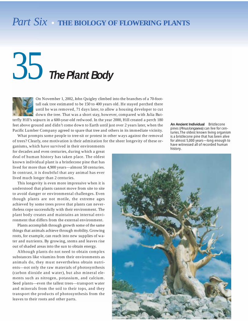
Some stems are highly modified. The tuber of a potato, forexample—the part of the plant eaten by humans—is an un-derground stem rather than a root. Its “eyes” contain lateralbuds; thus, a sprouting potato is just a branching stem (Fig-ure 35.4a). The runners of strawberry plants and Bermudagrass are horizontal stems from which roots grow at frequentintervals (Figure 35.4b). If the links between the rooted por-tions are broken, independent plants can develop on eachside of the break. This phenomenon is a form of vegetativereproduction.
Although stems are usually green and capable of photo-synthesis, they usually are not the principal sites of photo-synthesis. Most photosynthesis takes place in leaves.
Leaves are the primary sites of photosynthesisIn gymnosperms and most flowering plants, the leaves areresponsible for most of the plant’s photosynthesis, produc-ing energy-rich organic molecules and releasing oxygen gas.
In certain plants, the leaves are highly modified for more spe-cialized functions, as we will see below.
As photosynthetic organs, leaves are marvelously adaptedfor gathering light. Typically, the blade of a leaf is a thin, flatstructure attached to the stem by a stalk called a petiole. Dur-ing the daytime, the leaf blade is held by its petiole at an an-gle almost perpendicular to the rays of the sun. This orienta-tion, with the leaf surface facing the sun, maximizes theamount of light available for photosynthesis. Some leavestrack the sun, moving so that they constantly face it.
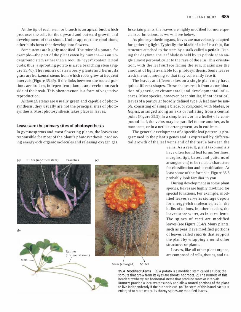
The leaves at different sites on a single plant may havequite different shapes. These shapes result from a combina-tion of genetic, environmental, and developmental influ-ences. Most species, however, bear similar, if not identical,leaves of a particular broadly defined type. A leaf may be sim-ple, consisting of a single blade, or compound, with blades, orleaflets, arranged along an axis or radiating from a centralpoint (Figure 35.5). In a simple leaf, or in a leaflet of a com-pound leaf, the veins may be parallel to one another, as inmonocots, or in a netlike arrangement, as in eudicots.
The general development of a specific leaf pattern is pro-grammed in the plant’s genes and is expressed by differen-tial growth of the leaf veins and of the tissue between the
veins. As a result, plant taxonomistshave often found leaf forms (outlines,margins, tips, bases, and patterns ofarrangement) to be reliable charactersfor classification and identification. Atleast some of the forms in Figure 35.5probably look familiar to you.
During development in some plantspecies, leaves are highly modified forspecial functions. For example, mod-ified leaves serve as storage depotsfor energy-rich molecules, as in thebulbs of onions. In other species, theleaves store water, as in succulents.The spines of cacti are modifiedleaves (see Figure 35.4c). Many plants,such as peas, have modified portionsof leaves called tendrils that supportthe plant by wrapping around otherstructures or plants.
Leaves, like all other plant organs,are composed of cells, tissues, and tis-
THE PLANT BODY 685
(a)
(b)
(c)
Stem
Runner(horizontal stem)
Spines
Tuber (modified stem) Branches
Stem (enlarged)
35.4 Modified Stems (a) A potato is a modified stem called a tuber; thesprouts that grow from its eyes are shoots, not roots. (b) The runners of thisbeach strawberry are horizontal stems that produce roots at intervals.Runners provide a local water supply and allow rooted portions of the plantto live independently if the runner is cut. (c) The stem of this barrel cactus isenlarged to store water. Its thorny spines are modified leaves.
sue systems. Let’s now consider plant cells—the basic struc-tural and functional units of plant organs.
Plant Cells
Plant cells have all the essential organelles common to eu-karyotes (see Figure 4.7). In addition, they have certain struc-tures and organelles that distinguish them from many othereukaryotes:
� They contain chloroplasts or other plastids.� They contain vacuoles.� They possess cellulose-containing cell walls.
Plant cells are alive when they divide and grow, but certaincells function only after their living parts have died and dis-integrated. Other plant cells develop specialized metaboliccapabilities; for example, some can perform photosynthesis,and others produce and secrete waterproofing materials.There are several different types of plant cells, which differdramatically in the composition and structure of their cellwalls. The walls of each cell type have a composition andstructure that corresponds to its special functions.
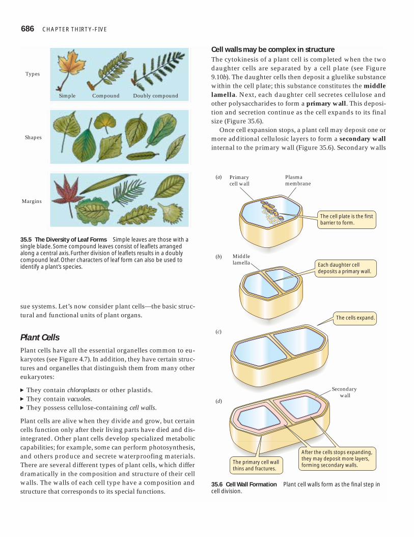
Cell walls may be complex in structureThe cytokinesis of a plant cell is completed when the twodaughter cells are separated by a cell plate (see Figure9.10b). The daughter cells then deposit a gluelike substancewithin the cell plate; this substance constitutes the middlelamella. Next, each daughter cell secretes cellulose andother polysaccharides to form a primary wall. This deposi-tion and secretion continue as the cell expands to its finalsize (Figure 35.6).
Once cell expansion stops, a plant cell may deposit one ormore additional cellulosic layers to form a secondary wallinternal to the primary wall (Figure 35.6). Secondary walls
686 CHAPTER THIRT Y-FIVE
Shapes
Margins
Types
Simple Compound Doubly compound
35.5 The Diversity of Leaf Forms Simple leaves are those with asingle blade. Some compound leaves consist of leaflets arrangedalong a central axis. Further division of leaflets results in a doublycompound leaf. Other characters of leaf form can also be used toidentify a plant’s species.
Middle lamella
Primary cell wall
(a)
(b)
(c)
(d)
Plasmamembrane
Secondary wall
The cell plate is the first barrier to form.
Each daughter cell deposits a primary wall.
The cells expand.
After the cells stops expanding, they may deposit more layers, forming secondary walls.
The primary cell wall thins and fractures.
35.6 Cell Wall Formation Plant cell walls form as the final step incell division.
are often impregnated with unique substances that give themspecial properties. Those impregnated with the polymerlignin become strong, as in wood cells. Walls to which thecomplex lipid suberin are added become waterproof.
Although it lies outside the plasma membrane, the cellwall is not a chemically inactive region. In addition to cellu-lose and other polysaccharides, the cell wall contains pro-teins, some of which are enzymes. Chemical reactions in thewall play important roles in cell expansion and in defenseagainst invading organisms. Cell walls may thicken or besculpted or perforated as cells differentiate into specializedcell types. Except where the secondary wall is waterproofed,the cell wall is permeable to water, small molecules, andmineral ions.
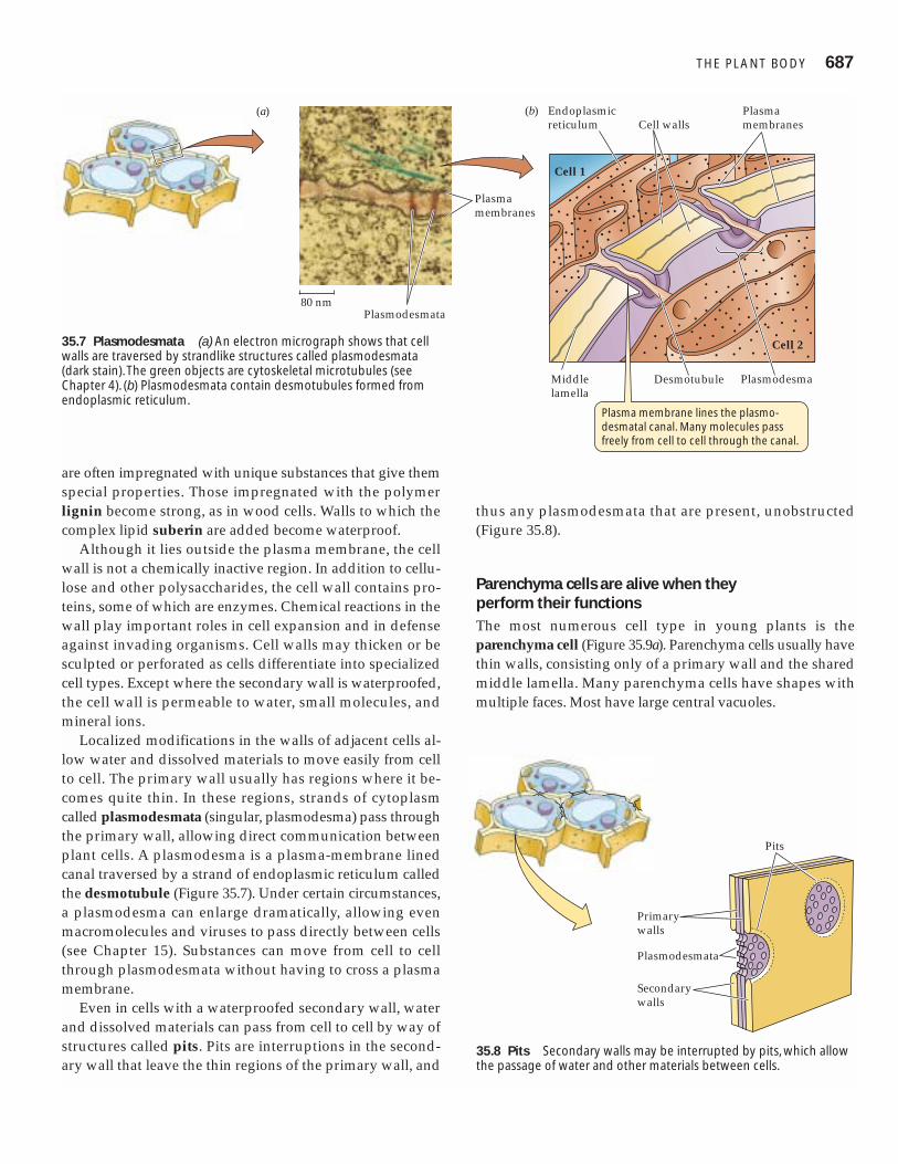
Localized modifications in the walls of adjacent cells al-low water and dissolved materials to move easily from cellto cell. The primary wall usually has regions where it be-comes quite thin. In these regions, strands of cytoplasmcalled plasmodesmata (singular, plasmodesma) pass throughthe primary wall, allowing direct communication betweenplant cells. A plasmodesma is a plasma-membrane linedcanal traversed by a strand of endoplasmic reticulum calledthe desmotubule (Figure 35.7). Under certain circumstances,a plasmodesma can enlarge dramatically, allowing evenmacromolecules and viruses to pass directly between cells(see Chapter 15). Substances can move from cell to cellthrough plasmodesmata without having to cross a plasmamembrane.
Even in cells with a waterproofed secondary wall, waterand dissolved materials can pass from cell to cell by way ofstructures called pits. Pits are interruptions in the second-ary wall that leave the thin regions of the primary wall, and
thus any plasmodesmata that are present, unobstructed(Figure 35.8).
Parenchyma cells are alive when they perform their functionsThe most numerous cell type in young plants is theparenchyma cell (Figure 35.9a). Parenchyma cells usually havethin walls, consisting only of a primary wall and the sharedmiddle lamella. Many parenchyma cells have shapes withmultiple faces. Most have large central vacuoles.
THE PLANT BODY 687
Plasmodesma
Cell wallsPlasma membranes
Cell 1
Endoplasmic reticulum
Middlelamella
Cell 2
Plasma membrane lines the plasmo-desmatal canal. Many molecules pass freely from cell to cell through the canal.
Plasmodesmata
Plasmamembranes
(a)
Desmotubule
80 nm
(b)
35.7 Plasmodesmata (a) An electron micrograph shows that cellwalls are traversed by strandlike structures called plasmodesmata(dark stain). The green objects are cytoskeletal microtubules (seeChapter 4). (b) Plasmodesmata contain desmotubules formed fromendoplasmic reticulum.
Primary walls
Plasmodesmata
Secondary walls
Pits
35.8 Pits Secondary walls may be interrupted by pits, which allowthe passage of water and other materials between cells.

The photosynthetic cells in leaves are parenchyma cellsthat contain numerous chloroplasts. Some nonphotosyntheticparenchyma cells store substances such as starch or lipids. Inthe cytoplasm of these cells, starch is often stored in special-ized plastids called leucoplasts (see Figure 4.17b). Lipids may
be stored as oil droplets, also in the cytoplasm. Someparenchyma cells appear to serve as “packing material” andplay a vital role in supporting the stem. Many retain the ca-pacity to divide and hence may give rise to new cells, aswhen a wound results in cell proliferation.
688 CHAPTER THIRT Y-FIVE
(a) Parenchyma cells Cell walls (b) Collenchyma cells Cell walls
(c) Sclerenchyma: Fibers Cell walls (d) Sclerenchyma: Sclereids Secondary cell walls
(e) (f)
Vessel elements
Secondary cell wall
Tracheids
Cell walls
Pits
50 µm 50 µm
50 µm 50 µm
50 µm 50 µm
35.9 Plant Cell Types (a) Parenchymacells in the leaf stem of Coleus. Note thethin, uniform cell walls. (b) Collenchymacells make up the five outer cell layers ofthis spinach leaf vein. Their cell walls arethick at the corners of the cells and thinelsewhere. (c) Sclerenchyma: Fibers in asunflower plant (Helianthus). The thicksecondary walls are stained red.(d ) Sclerenchyma: Sclereids. Theextremely thick secondary walls of scle-reids are laid down in layers. They pro-vide support and a hard texture tostructures such as nuts and seeds. (e)Water-conducting tracheids in pinewood. The thick cell walls are staineddark red. (f ) Vessel elements in the stemof a squash. The secondary walls arestained red; note the different patternsof thickening, including rings and spirals.
Collenchyma cells provide flexible support while aliveCollenchyma cells are supporting cells. Their primary wallsare characteristically thick at the corners of the cells (Figure35.9b). Collenchyma cells are generally elongated. In thesecells, the primary wall thickens, but no secondary wall forms.Collenchyma provides support to leaf petioles, nonwoodystems, and growing organs. Tissue made of collenchyma cellsis flexible, permitting stems and petioles to sway in the windwithout snapping. The familiar “strings” in celery consist pri-marily of collenchyma cells.
Sclerenchyma cells provide rigid supportIn contrast to collenchyma cells, sclerenchyma cells have athickened secondary wall that performs their major function:support. Many sclerenchyma cells function when dead. Thereare two types of sclerenchyma cells: elongated fibers and var-iously shaped sclereids. Fibers provide relatively rigid sup-port in wood and other parts of the plant, where they are of-ten organized into bundles (Figure 35.9c). The bark of treesowes much of its mechanical strength to long fibers. Sclereidsmay pack together densely, as in a nut’s shell or in some seedcoats (Figure 35.9d). Isolated clumps of sclereids, called stonecells, in pears and some other fruits give them their character-istic gritty texture.
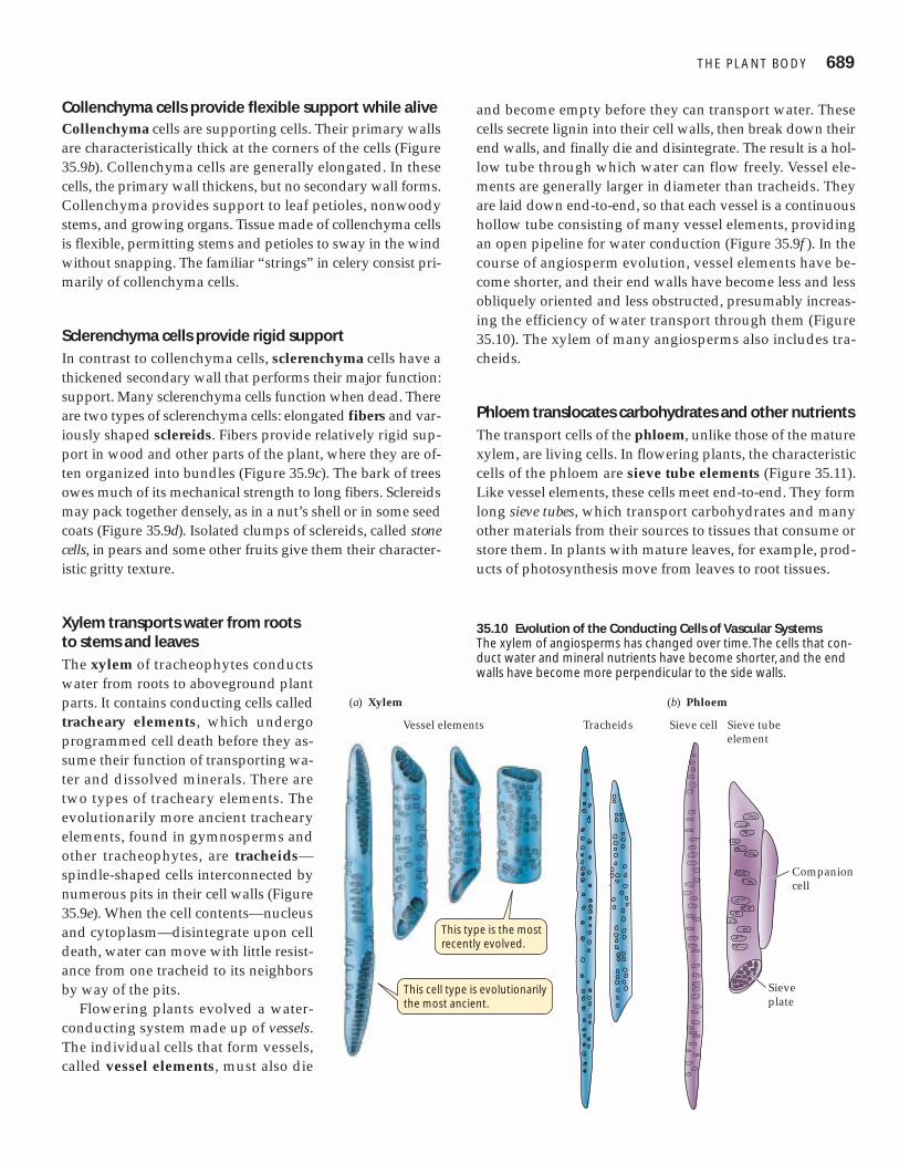
Xylem transports water from rootsto stems and leavesThe xylem of tracheophytes conductswater from roots to aboveground plantparts. It contains conducting cells calledtracheary elements, which undergoprogrammed cell death before they as-sume their function of transporting wa-ter and dissolved minerals. There aretwo types of tracheary elements. Theevolutionarily more ancient trachearyelements, found in gymnosperms andother tracheophytes, are tracheids—spindle-shaped cells interconnected bynumerous pits in their cell walls (Figure35.9e). When the cell contents—nucleusand cytoplasm—disintegrate upon celldeath, water can move with little resist-ance from one tracheid to its neighborsby way of the pits.
Flowering plants evolved a water-conducting system made up of vessels.The individual cells that form vessels,called vessel elements, must also die
and become empty before they can transport water. Thesecells secrete lignin into their cell walls, then break down theirend walls, and finally die and disintegrate. The result is a hol-low tube through which water can flow freely. Vessel ele-ments are generally larger in diameter than tracheids. Theyare laid down end-to-end, so that each vessel is a continuoushollow tube consisting of many vessel elements, providingan open pipeline for water conduction (Figure 35.9f ). In thecourse of angiosperm evolution, vessel elements have be-come shorter, and their end walls have become less and lessobliquely oriented and less obstructed, presumably increas-ing the efficiency of water transport through them (Figure35.10). The xylem of many angiosperms also includes tra-cheids.
Phloem translocates carbohydrates and other nutrientsThe transport cells of the phloem, unlike those of the maturexylem, are living cells. In flowering plants, the characteristiccells of the phloem are sieve tube elements (Figure 35.11).Like vessel elements, these cells meet end-to-end. They formlong sieve tubes, which transport carbohydrates and manyother materials from their sources to tissues that consume orstore them. In plants with mature leaves, for example, prod-ucts of photosynthesis move from leaves to root tissues.
THE PLANT BODY 689
This cell type is evolutionarily the most ancient.
This type is the most recently evolved.
(a) Xylem
Vessel elements Tracheids
(b) Phloem
Sieve cell Sieve tube element
Companion cell
Sieveplate
35.10 Evolution of the Conducting Cells of Vascular SystemsThe xylem of angiosperms has changed over time. The cells that con-duct water and mineral nutrients have become shorter, and the endwalls have become more perpendicular to the side walls.
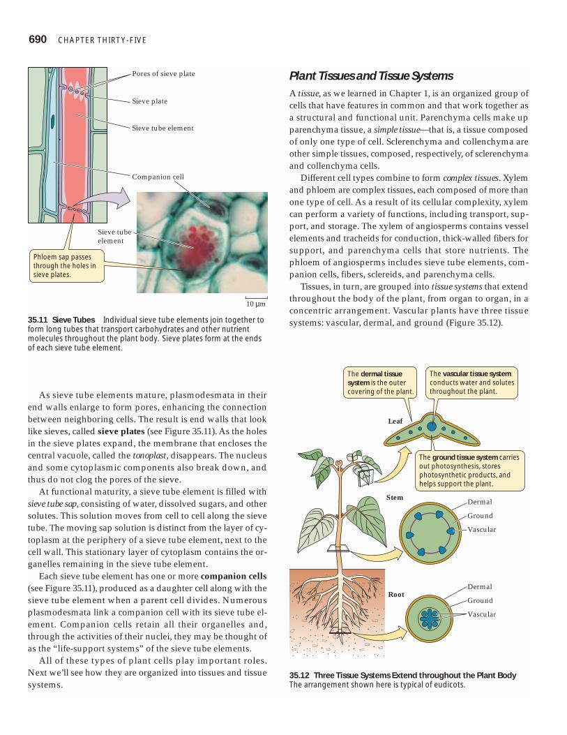
As sieve tube elements mature, plasmodesmata in theirend walls enlarge to form pores, enhancing the connectionbetween neighboring cells. The result is end walls that looklike sieves, called sieve plates (see Figure 35.11). As the holesin the sieve plates expand, the membrane that encloses thecentral vacuole, called the tonoplast, disappears. The nucleusand some cytoplasmic components also break down, andthus do not clog the pores of the sieve.
At functional maturity, a sieve tube element is filled withsieve tube sap, consisting of water, dissolved sugars, and othersolutes. This solution moves from cell to cell along the sievetube. The moving sap solution is distinct from the layer of cy-toplasm at the periphery of a sieve tube element, next to thecell wall. This stationary layer of cytoplasm contains the or-ganelles remaining in the sieve tube element.
Each sieve tube element has one or more companion cells(see Figure 35.11), produced as a daughter cell along with thesieve tube element when a parent cell divides. Numerousplasmodesmata link a companion cell with its sieve tube el-ement. Companion cells retain all their organelles and,through the activities of their nuclei, they may be thought ofas the “life-support systems” of the sieve tube elements.
All of these types of plant cells play important roles.Next we’ll see how they are organized into tissues and tissuesystems.
Plant Tissues and Tissue SystemsA tissue, as we learned in Chapter 1, is an organized group ofcells that have features in common and that work together asa structural and functional unit. Parenchyma cells make upparenchyma tissue, a simple tissue—that is, a tissue composedof only one type of cell. Sclerenchyma and collenchyma areother simple tissues, composed, respectively, of sclerenchymaand collenchyma cells.
Different cell types combine to form complex tissues. Xylemand phloem are complex tissues, each composed of more thanone type of cell. As a result of its cellular complexity, xylemcan perform a variety of functions, including transport, sup-port, and storage. The xylem of angiosperms contains vesselelements and tracheids for conduction, thick-walled fibers forsupport, and parenchyma cells that store nutrients. Thephloem of angiosperms includes sieve tube elements, com-panion cells, fibers, sclereids, and parenchyma cells.
Tissues, in turn, are grouped into tissue systems that extendthroughout the body of the plant, from organ to organ, in aconcentric arrangement. Vascular plants have three tissuesystems: vascular, dermal, and ground (Figure 35.12).
690 CHAPTER THIRT Y-FIVE
Phloem sap passes through the holes in sieve plates.
Pores of sieve plate
Sieve plate
Sieve tube element
Companion cell
10 µm
Sieve tube element
35.11 Sieve Tubes Individual sieve tube elements join together toform long tubes that transport carbohydrates and other nutrientmolecules throughout the plant body. Sieve plates form at the endsof each sieve tube element.
The dermal tissue system is the outer covering of the plant.
The vascular tissue system conducts water and solutes throughout the plant.
The ground tissue system carries out photosynthesis, stores photosynthetic products, and helps support the plant.
Dermal
Leaf
Stem
Root
Ground
Vascular
Dermal
Ground
Vascular
35.12 Three Tissue Systems Extend throughout the Plant BodyThe arrangement shown here is typical of eudicots.
The vascular tissue system, which includes the xylem andphloem, is the plant’s plumbing or transport system. All theliving cells of the plant body require a source of energy andchemical building blocks. The phloem transports carbohy-drates from sites of production (called sources, primarilyleaves) to sites of utilization or storage (called sinks, such asgrowing tissue, storage tubers, and developing flowers). Thexylem distributes water and mineral ions taken up by theroots to all the cells of the stem and leaves.
The dermal tissue system is the outer covering of theplant. All parts of the young plant body are covered by anepidermis, which may be a single layer of cells or several lay-ers. The epidermis contains epidermal cells and may also in-clude specialized cell types, such as the guard cells that formstomata (pores) in leaves. The shoot epidermis secretes alayer of wax-covered cutin, the cuticle, that helps retard wa-ter loss from stems and leaves. The stems and roots of woodyplants have a protective covering called the periderm, whichwill be discussed later in this chapter.
The ground tissue system makes up the rest of the plant.It consists primarily of parenchyma tissue, often supple-mented by collenchyma or sclerenchyma. Ground tissuefunctions primarily in storage, support, photosynthesis, andthe production of defensive and attractive substances.
In the discussions that follow, we’ll examine how the tis-sue systems are organized in the different organs of a flow-ering plant. Let’s begin by seeing how this organization de-velops as the plant grows.
Forming the Plant Body
In its early embryonic stages, a plant establishes the basicbody plan for its mature form. Two patterns contribute to theplant body plan:
� The arrangement of cells and tissues along the main axisfrom root to shoot
� The concentric arrangement of the tissue systems
Both patterns arise through orderly development and arebest understood in developmental terms.
Plants and animals grow differentlyAs the plant body grows, it may lose parts, and it forms newparts that may grow at different rates. The growing stem con-sists of modules or units, laid down one after another. Eachmodule consists of a node with its attached leaf or leaves, theinternode below that node, and the lateral bud or buds at thebase of that internode (see Figure 35.2). New modules areformed as long as the stem continues to grow.
Each branch of a plant may be thought of as a unit that isin some ways independent of the other branches. A branch
of a plant does not bear the same relationship to the remain-der of the plant body as a limb does to the remainder of ananimal body. Among other things, branches form one afteranother (unlike limbs, which form simultaneously duringembryonic development). Also, branches often differ fromone another in their number of leaves and in the degree towhich they themselves branch.
Leaves are units of another sort. They are usually short-lived, lasting weeks to a few years. Branches and stems arelonger-lived, lasting from years to centuries.
Root systems are also branching structures, and lateralroots are semi-independent units. As the root system grows,penetrating and exploring the soil environment, many rootsdie and are replaced by new ones.
All parts of the animal body grow as an individual devel-ops from embryo to adult, but in most animals, this growthis determinate. That is, the growth of the individual and all itsparts ceases when the adult state is reached. Determinategrowth is also characteristic of some plant parts, such asleaves, flowers, and fruits. The growth of stems and roots, bycontrast, is indeterminate, and it is generated from specific re-gions of active cell division and cell expansion.
The localized regions of cell division in plants are calledmeristems. Meristems are forever young, retaining the abil-ity to produce new cells indefinitely. The cells that perpetu-ate the meristems, called initials, are comparable to the stemcells found in animals (discussed in Chapter 19). When aninitial divides, one daughter cell develops into another meri-stem cell the size of its parent, while the other daughter celldifferentiates into a more specialized cell.
A hierarchy of meristems generates a plant’s bodyThere are two types of meristems:
� Apical meristems give rise to the primary plant body,which is the entire body of many plants.
� Lateral meristems give rise to the secondary plant body.The stems and roots of some plants (most obviously trees)form wood and become thick; it is the lateral meristemsthat give rise to the tissues responsible for this thickening.
APICAL MERISTEMS. Apical meristems are located at the tipsof roots and stems and in buds. They extend the plant bodyby producing the cells that subsequently expand and dif-ferentiate to form all plant organs (Figure 35.13).
� Shoot apical meristems supply the cells that extend stemsand branches, allowing more leaves to form and photo-synthesize.
� Root apical meristems supply the cells that extend roots,enabling the plant to “forage” for water and minerals.
THE PLANT BODY 691

Both root and shoot apical meristems give rise to a set of cylin-drical primary meristems that produce the primary tissues ofthe plant body. From the outside to the inside of the root orshoot, which are both cylindrical organs, the primary meris-tems are the protoderm, the ground meristem, and the pro-cambium. These in turn give rise to the three tissue systems:
Apical meristems are responsible for primary growth, whichleads to lengthening of the plant body and organ formation.All plant organs arise ultimately from cell divisions in the api-cal meristems, followed by cell expansion and differentiation.Primary growth gives rise to the entire body of many plants.
Because meristems can continue to produce new organsthroughout the lifetime of the plant, the plant body is muchmore variable in form than the animal body, whose organsare produced only once.
LATERAL MERISTEMS. Some roots and stems develop a sec-ondary body, the tissues of which we commonly refer to as
wood and bark. These complex tissues are derived fromtwo lateral meristems: the vascular cambium and the corkcambium (Figure 35.14).
The vascular cambium is a cylindrical tissue consistingpredominantly of vertically elongated cells that divide fre-quently. Toward the inside of the stem or root, the dividingcells form new xylem, the secondary xylem, and toward theoutside they form new phloem, the secondary phloem.
As a tree trunk grows in diameter, the outermost layers ofthe stem crack and fall off. Without the activity of the corkcambium, this sloughing off of tissues, including the epider-mis, would expose the tree to potential damage, includingexcessive water loss or invasion by microorganisms. The corkcambium produces new protective cells, primarily in the out-ward direction. The walls of these cork cells become impreg-nated with suberin. The mass of waterproofed cells producedby the cork cambium is called the periderm.
Growth in the diameter of stems and roots, produced bythe vascular and cork cambia, is called secondary growth. Itis the source of wood and bark. Wood is secondary xylem.Bark is everything external to the vascular cambium (peri-derm plus secondary phloem).
Each year, deciduous trees lose their leaves, leaving barebranches and twigs in winter. These twigs illustrate both pri-
Apical meristems Primary meristems
Root or shoot apical meristem
Tissue systems
Procambium
Protoderm Dermal tissue system
Ground meristem Ground tissue system
Vascular tissue system
692 CHAPTER THIRT Y-FIVE
The apical bud contains a shoot apical meristem.
Root apical meristem
Leaf primordia
Lateral budprimordia
Lateral bud
Root hairs
Cork cambium
Root cap
Root apical meristem
Vascular cambium
In woody plants the vascular cambium and cork cambium thicken the stem and root.
50 µm
100 µm
35.13 Apical and Lateral Meristems Apical meri-stems produce the primary plant body; lateral meri-stems produce the secondary plant body.
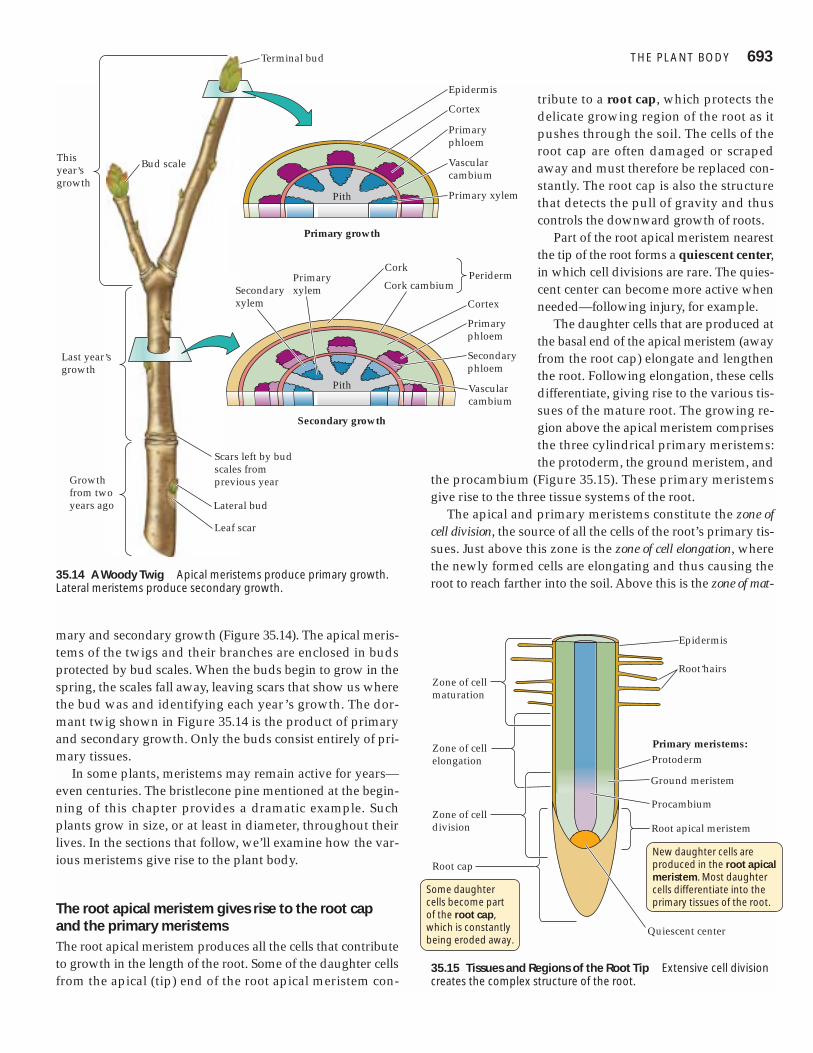
mary and secondary growth (Figure 35.14). The apical meris-tems of the twigs and their branches are enclosed in budsprotected by bud scales. When the buds begin to grow in thespring, the scales fall away, leaving scars that show us wherethe bud was and identifying each year’s growth. The dor-mant twig shown in Figure 35.14 is the product of primaryand secondary growth. Only the buds consist entirely of pri-mary tissues.
In some plants, meristems may remain active for years—even centuries. The bristlecone pine mentioned at the begin-ning of this chapter provides a dramatic example. Suchplants grow in size, or at least in diameter, throughout theirlives. In the sections that follow, we’ll examine how the var-ious meristems give rise to the plant body.
The root apical meristem gives rise to the root capand the primary meristemsThe root apical meristem produces all the cells that contributeto growth in the length of the root. Some of the daughter cellsfrom the apical (tip) end of the root apical meristem con-
tribute to a root cap, which protects thedelicate growing region of the root as itpushes through the soil. The cells of theroot cap are often damaged or scrapedaway and must therefore be replaced con-stantly. The root cap is also the structurethat detects the pull of gravity and thuscontrols the downward growth of roots.
Part of the root apical meristem nearestthe tip of the root forms a quiescent center,in which cell divisions are rare. The quies-cent center can become more active whenneeded—following injury, for example.
The daughter cells that are produced atthe basal end of the apical meristem (awayfrom the root cap) elongate and lengthenthe root. Following elongation, these cellsdifferentiate, giving rise to the various tis-sues of the mature root. The growing re-gion above the apical meristem comprisesthe three cylindrical primary meristems:the protoderm, the ground meristem, and
the procambium (Figure 35.15). These primary meristemsgive rise to the three tissue systems of the root.
The apical and primary meristems constitute the zone ofcell division, the source of all the cells of the root’s primary tis-sues. Just above this zone is the zone of cell elongation, wherethe newly formed cells are elongating and thus causing theroot to reach farther into the soil. Above this is the zone of mat-
THE PLANT BODY 693Terminal bud
Bud scale
Lateral bud
Leaf scar
Scars left by bud scales from previous year
This year,s growth
Last year,s growth
Growth from twoyears ago
Epidermis
Cortex
Primary phloem
Vascular cambium
Primary xylem
Vascular cambium
Cork cambium
Cortex
Primary phloem
Cork
Secondary phloem
Pith
Pith
Secondary xylem
Primary xylem
Periderm
Primary growth
Secondary growth
35.14 A Woody Twig Apical meristems produce primary growth.Lateral meristems produce secondary growth.
Zone of cellmaturation
Zone of cellelongation
Zone of celldivision
Root cap
Root hairs
Epidermis
Primary meristems:Protoderm
Ground meristem
Procambium
Root apical meristem
Quiescent center
New daughter cells are produced in the root apical meristem. Most daughter cells differentiate into the primary tissues of the root.
Some daughter cells become part of the root cap, which is constantly being eroded away.
35.15 Tissues and Regions of the Root Tip Extensive cell divisioncreates the complex structure of the root.

uration, where the cells are differentiating, taking on special-ized forms and functions such as water transport or mineraluptake. These three zones grade imperceptibly into one an-other; there is no abrupt line of demarcation.
The products of the root’s primary meristems become root tissuesWhat are the products of the three primary meristems? Theprotoderm gives rise to the outer layer of cells—the epider-mis—which is adapted for protection of the root and for theabsorption of mineral ions and water (Figure 35.16). In thezone of maturation, many of the epidermal cells produceamazingly long, delicate root hairs, which vastly increase thesurface area of the root (Figure 35.16b). It has been estimatedthat the root system of a mature rye plant has a total absorp-tive surface of more than 600 square meters (almost halfagain the area of a basketball court). Root hairs grow outamong the soil particles, probing nooks and crannies and tak-ing up water and minerals.
Internal to the epidermis, the ground meristem gives riseto a region of ground tissue that is many cells thick, calledthe cortex. The cells of the cortex are relatively unspecializedand often function in nutrient storage.
In the great majority of plants, especially in trees, a fungusis closely associated with the root tips. This association, calleda mycorrhiza, increases the plant’s absorption of minerals andwater (see Figure 31.16). Such roots have poorly developed
or no root hairs. These plants cannot survive without the my-corrhizae that help them absorb minerals.
Proceeding inward, we come to the endodermis of theroot, a single cylindrical layer of cells that is the innermostcell layer of the cortex. Unlike those of other cortical cells, thecell walls of the endodermal cells contain suberin. The place-ment of this waterproofing substance in only certain parts ofthe cell wall enables the cylindrical ring of endodermal cellsto control the access of water and dissolved ions to the vas-cular tissues.
Moving inward past the endodermis, we enter the vascu-lar cylinder, or stele, produced by the procambium. The steleconsists of three tissues: pericycle, xylem, and phloem (Fig-ure 35.17).
The pericycle consists of one or more layers of relativelyundifferentiated cells. It has three important functions:
� It is the tissue within which lateral roots arise (see Figure35.16a).
� It can contribute to secondary growth by giving rise to lat-eral meristems that thicken the root.
� Its cells contain membrane transport proteins that exportnutrient ions into the cells of the xylem.
694 CHAPTER THIRT Y-FIVE
EndodermisRoothairs
Root apicalmeristem
Cortex
Stele
Epidermis
Lateralroot
Root cap
(a) Developing lateral root
(b) Root hairs
(c) Eudicot root
(d) Monocot root
Endodermis Pericycle
Pith
Endodermis Phloem Xylem
35.16 Root Anatomy The drawing at the left shows a gen-eralized root structure. (a) Cross section through the tip of alateral root. Cells in the pericycle divide and the products dif-ferentiate, forming the tissues of a lateral root. (b) Root hairs,
seen with a scanning electron microscope. (c, d) Cross sections show-ing the primary root tissues of (c) a eudicot and (d) a monocot.Themonocot has a central pith region; the eudicot does not.
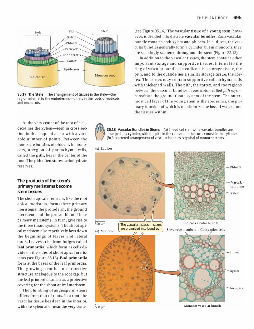
At the very center of the root of a eu-dicot lies the xylem—seen in cross sec-tion in the shape of a star with a vari-able number of points. Between thepoints are bundles of phloem. In mono-cots, a region of parenchyma cells,called the pith, lies in the center of theroot. The pith often stores carbohydratereserves.
The products of the stem’s primary meristems become stem tissues
The shoot apical meristem, like the rootapical meristem, forms three primarymeristems: the protoderm, the groundmeristem, and the procambium. Theseprimary meristems, in turn, give rise tothe three tissue systems. The shoot api-cal meristem also repetitively lays downthe beginnings of leaves and lateralbuds. Leaves arise from bulges calledleaf primordia, which form as cells di-vide on the sides of shoot apical meris-tems (see Figure 35.13). Bud primordiaform at the bases of the leaf primordia.The growing stem has no protectivestructure analogous to the root cap, butthe leaf primordia can act as a protectivecovering for the shoot apical meristem.
The plumbing of angiosperm stemsdiffers from that of roots. In a root, thevascular tissue lies deep in the interior,with the xylem at or near the very center
(see Figure 35.16). The vascular tissue of a young stem, how-ever, is divided into discrete vascular bundles. Each vascularbundle contains both xylem and phloem. In eudicots, the vas-cular bundles generally form a cylinder, but in monocots, theyare seemingly scattered throughout the stem (Figure 35.18).
In addition to the vascular tissues, the stem contains otherimportant storage and supportive tissues. Internal to thering of vascular bundles in eudicots is a storage tissue, thepith, and to the outside lies a similar storage tissue, the cor-tex. The cortex may contain supportive collenchyma cellswith thickened walls. The pith, the cortex, and the regionsbetween the vascular bundles in eudicots—called pith rays—constitute the ground tissue system of the stem. The outer-most cell layer of the young stem is the epidermis, the pri-mary function of which is to minimize the loss of water fromthe tissues within.
THE PLANT BODY 695
Stele
Eudicot root
Stele
Monocot root
Pith
Xylem
Phloem
Pericycle
Endodermis
Cortex
Epidermis
35.17 The Stele The arrangement of tissues in the stele—theregion internal to the endodermis—differs in the roots of eudicotsand monocots.
(a) Eudicot
(b) Monocot
Monocot vascular bundle
Phloem
Sieve tube members Companion cells
Xylem
Air space
Eudicot vascular bundle
Phloem
Vascularcambium
Xylem
The vascular tissues in stems are organized into bundles.
500 µm
500 µm
35.18 Vascular Bundles in Stems (a) In eudicot stems, the vascular bundles arearranged in a cylinder, with the pith in the center and the cortex outside the cylinder.(b) A scattered arrangement of vascular bundles is typical of monocot stems.

Many stems and roots undergo secondary growthSome stems and roots remain slender and show little or nosecondary growth. However, in many eudicots, secondarygrowth thickens stems and roots considerably. This processgives rise to wood and bark, and it makes the support of talltrees possible.
Secondary growth results from the activity of the two lat-eral meristems: vascular cambium and cork cambium (seeFigure 35.13). Vascular cambium consists of cells that divideto produce secondary xylem and phloem cells, while corkcambium produces mainly waxy-walled cork cells.
Initially, the vascular cambium is a single layer of cells ly-ing between the primary xylem and the primary phloem (seeFigure 35.18a). The root or stem increases in diameter whenthe cells of the vascular cambium divide, producing second-ary xylem cells toward the inside of the root or stem and pro-ducing secondary phloem cells toward the outside (Figure35.19). In the stems of woody plants, cells in the pith rays be-tween the vascular bundles also divide, forming a continu-ous cylinder of vascular cambium running the length of thestem. This cylinder, in turn, gives rise to complete cylindersof secondary xylem (wood) and secondary phloem, whichcontributes to the bark.
As the vascular cambium produces secondary xylem andphloem, its principal cell products are vessel elements, sup-portive fibers, and parenchyma cells in the xylem and sievetube elements, companion cells, fibers, and parenchyma cellsin the phloem. The parenchyma cells in the xylem andphloem store carbohydrate reserves in the stem and root.
Living tissues such as this storage parenchyma must beconnected to the sieve tubes of the phloem, or they willstarve to death. These connections are provided by vascularrays, which are composed of cells derived from the vascularcambium. These rays, laid down progressively as the cam-bium divides, are rows of living parenchyma cells that runperpendicular to the xylem vessels and phloem sieve tubes(Figure 35.20). As the root or stem continues to increase in di-ameter, new vascular rays are initiated so that this storageand transport tissue continues to meet the needs of both thebark and the living cells in the xylem.
The vascular cambium itself increases in circumferencewith the growth of the root or stem. To do this, some of itscells divide in a plane at right angles to the plane that givesrise to secondary xylem and phloem. The products of each ofthese divisions lie within the vascular cambium itself and in-crease its circumference.
Only eudicots and other non-monocot angiosperms havea vascular cambium and a cork cambium and thus undergosecondary growth. The few monocots that form thickenedstems—palm trees, for example—do so without using vas-cular cambium or cork cambium. Palm trees have a verywide apical meristem that produces a wide stem, and deadleaf bases also add to the diameter of the stem. Basically,monocots grow in the same way as do other angiospermsthat lack secondary growth.
Wood and bark, consisting of secondary phloem, areunique to plants showing secondary growth. These tissueshave their own patterns of organization and development.
696 CHAPTER THIRT Y-FIVE
TimeOutward growth
The vascular cambium thickens the stem by pro-ducing secondary xylem and secondary phloem.
New secondary phloem cell
Next new secondary phloem cell
Vascular cambium cell
Outer margin of primary xylem
p
New secondaryxylem cell
Next new secondaryxylem cell
When a vascular cambium cell divides, it produces either a new xylem cell toward the inside of the stem or root, or a new phloem cell toward the outside.
cc
cc
cp
pp
xx
xx
xx
Pith
Primary xylem
Vascular cambium
Primary phloem
Secondary phloem
Secondary xylem
Woody stem
35.19 Vascular Cambium Thickens Stems and RootsStems and roots grow thicker because a thin layer of cells,the vascular cambium, remains meristematic.

WOOD. Cross sections of most tree trunks (mature stems)in temperate-zone forests show annual rings (Figure 35.21),which result from seasonal environmental conditions. Inspring, when water is relatively plentiful, the tracheids orvessel elements produced by the vascular cambium tend tobe large in diameter and thin-walled. Such wood is welladapted for transporting water and minerals. As waterbecomes less available during the summer, narrower cellswith thicker walls are produced, making this summerwood darker and perhaps more dense than the woodformed in spring. Thus each growing season is usuallyrecorded in a tree trunk by a clearly visible annual ring.Trees in the moist Tropics do not undergo seasonal growth,so they do not lay down such obvious regular rings.Variations in temperature or water supply can lead to theformation of more than one “annual” ring in a single year.
The difference between old and new regions of wood alsocontributes to its appearance. As a tree grows in diameter, thexylem toward the center becomes clogged with water-insol-uble substances and ceases to conduct water and minerals;this heartwood appears darker in color. The portion of thexylem that is actively conducting water and mineralsthroughout the tree is called sapwood and is lighter in colorand more porous than heartwood.
The knots that we find attractive in knotty pine but regardas a defect in structural timbers are cross sections of branches.
As a trunk grows, the bases of branches become buried in thetrunk’s new wood and appear as knots when the trunk is cutlengthwise.
BARK. As secondary growth of stems or roots continues,the expanding vascular tissue stretches and breaks the epi-dermis and cortex, which ultimately flake away. Tissuederived from the secondary phloem then becomes the out-ermost part of the stem. Before the dermal tissues are bro-ken away, cells lying near the surface of the secondaryphloem begin to divide and produce layers of cork, a tissuecomposed of cells with thick walls, waterproofed withsuberin. The cork soon becomes the outermost tissue of thestem or root (see Figure 35.14). The dividing cells, derivedfrom the secondary phloem, form a cork cambium. Some-times the cork cambium produces cells to the inside as wellas to the outside; these cells constitute what is known as thephelloderm.
Cork, cork cambium, and phelloderm make up the peri-derm of the secondary plant body. As the vascular cambiumcontinues to produce secondary vascular tissue, the corkylayers are in turn lost, but the continuous formation of newcork cambia in the underlying phloem gives rise to newcorky layers.
When periderm forms on stems and roots, the underlyingtissues still need to release carbon dioxide and take up oxy-gen. Lenticels are spongy regions in the periderm of stemsand roots that allow such gas exchange (Figure 35.22).
THE PLANT BODY 697
Vascular rays conduct nutrients horizontally.
Vessel elements conduct water vertically.
Vascular ray Vessel element
60 µm
Vessel ray
Vascularelement
35.20 Vascular Rays and Vessel Elements In this sample of woodfrom the tulip poplar, the orientation of vascular rays is perpendicularto that of the vessel elements. The vascular rays transport sieve tubesap horizontally from the phloem to storage parenchyma cells.
Pith
Secondaryphloem
Secondaryxylem
Annual ring
35.21 Annual Rings Rings of secondary xylem are the mostnoticeable feature of this cross section from a 3-year-old basswoodstem.
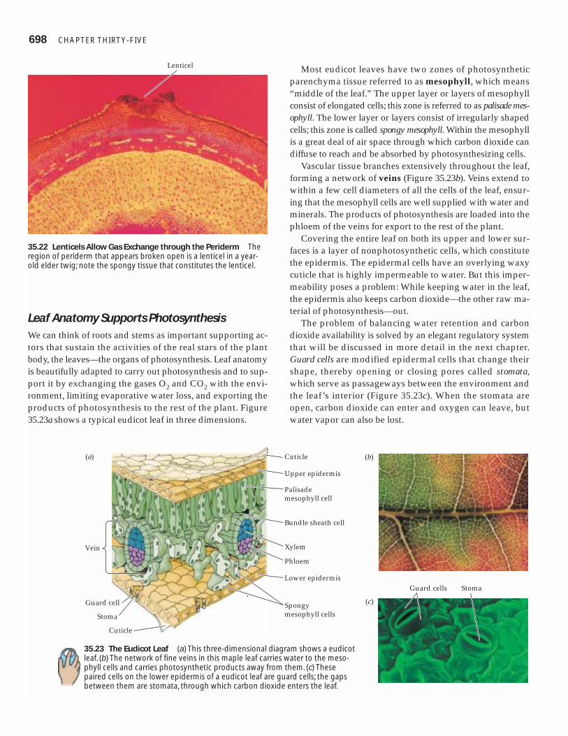
Leaf Anatomy Supports PhotosynthesisWe can think of roots and stems as important supporting ac-tors that sustain the activities of the real stars of the plantbody, the leaves—the organs of photosynthesis. Leaf anatomyis beautifully adapted to carry out photosynthesis and to sup-port it by exchanging the gases O2 and CO2 with the envi-ronment, limiting evaporative water loss, and exporting theproducts of photosynthesis to the rest of the plant. Figure35.23a shows a typical eudicot leaf in three dimensions.
Most eudicot leaves have two zones of photosyntheticparenchyma tissue referred to as mesophyll, which means“middle of the leaf.” The upper layer or layers of mesophyllconsist of elongated cells; this zone is referred to as palisade mes-ophyll. The lower layer or layers consist of irregularly shapedcells; this zone is called spongy mesophyll. Within the mesophyllis a great deal of air space through which carbon dioxide candiffuse to reach and be absorbed by photosynthesizing cells.
Vascular tissue branches extensively throughout the leaf,forming a network of veins (Figure 35.23b). Veins extend towithin a few cell diameters of all the cells of the leaf, ensur-ing that the mesophyll cells are well supplied with water andminerals. The products of photosynthesis are loaded into thephloem of the veins for export to the rest of the plant.
Covering the entire leaf on both its upper and lower sur-faces is a layer of nonphotosynthetic cells, which constitutethe epidermis. The epidermal cells have an overlying waxycuticle that is highly impermeable to water. But this imper-meability poses a problem: While keeping water in the leaf,the epidermis also keeps carbon dioxide—the other raw ma-terial of photosynthesis—out.
The problem of balancing water retention and carbondioxide availability is solved by an elegant regulatory systemthat will be discussed in more detail in the next chapter.Guard cells are modified epidermal cells that change theirshape, thereby opening or closing pores called stomata,which serve as passageways between the environment andthe leaf’s interior (Figure 35.23c). When the stomata areopen, carbon dioxide can enter and oxygen can leave, butwater vapor can also be lost.
698 CHAPTER THIRT Y-FIVE
Lenticel
35.22 Lenticels Allow Gas Exchange through the Periderm Theregion of periderm that appears broken open is a lenticel in a year-old elder twig; note the spongy tissue that constitutes the lenticel.
Vein
(b)(a)
(c)
Cuticle
Upper epidermis
Palisade mesophyll cell
Bundle sheath cell
Xylem
Phloem
Lower epidermis
Spongy mesophyll cells
Guard cell
Stoma
Cuticle
Guard cells Stoma
35.23 The Eudicot Leaf (a) This three-dimensional diagram shows a eudicotleaf. (b) The network of fine veins in this maple leaf carries water to the meso-phyll cells and carries photosynthetic products away from them. (c) Thesepaired cells on the lower epidermis of a eudicot leaf are guard cells; the gapsbetween them are stomata, through which carbon dioxide enters the leaf.
In Chapter 8 we described C4 plants, which can fix carbondioxide efficiently even when the carbon dioxide supply inthe leaf decreases to a level at which the photosynthesis of C3plants is inefficient. One adaptation that helps C4 plants dothis is their modified leaf anatomy (see Figure 8.16). The pho-tosynthetic cells in the C4 leaf are grouped around the veinsin concentric layers, forming an outer mesophyll layer andan inner bundle sheath. These layers each contain differenttypes of chloroplasts, leading to the biochemical division oflabor illustrated in Figure 8.17.
Leaves receive water and mineral nutrients from the rootsby way of the stems. In return, the leaves export products ofphotosynthesis, providing a supply of chemical energy to therest of the plant body. And, as we have just seen, leaves ex-change gases, including water vapor, with the environmentby way of the stomata. All three of these processes will beconsidered in detail in the next chapter.
Chapter Summary
Vegetative Organs of the Flowering Plant Body� Monocots typically have a single cotyledon, narrow leaveswith parallel veins, flower parts in threes or multiples of three,and stems with scattered vascular bundles. Review Figure 35.1� Eudicots typically have two cotyledons, broad leaves withnetlike veins, flower parts in fours or fives, and vascular bun-dles in a ring. Review Figure 35.1� Flowering plants that are neither monocots nor eudicots aregenerally similar in structure to eudicots.� The vegetative organs of flowering plants are roots, whichform a root system, and stems and leaves, which form a shootsystem. Review Figure 35.2� Roots anchor the plant and take up water and minerals.� Stems bear leaves and buds. Lateral buds form branches. Apicalbuds produce cells that contribute to the elongation of the stem.� Leaves are responsible for most photosynthesis, for whichtheir flat blades, held perpendicular to the sun’s rays, are welladapted. Review Figure 35.5
Plant Cells� The walls of plant cells have a structure that often corre-sponds to the special functions of the cell.� The walls of individual cells are separated by a middle lamel-la common to two neighboring cells; each cell also has its ownprimary wall. Review Figure 35.6� Some cells produce a thick secondary wall. Adjacent cells areconnected by plasmodesmata. Review Figures 35.7, 35.8� Parenchyma cells have thin walls. Many parenchyma cellsstore starch or lipids; some others carry out photosynthesis.Review Figure 35.9a� Collenchyma cells provide flexible support. Review Figure35.9b� Sclerenchyma cells provide strength and often function whendead. Review Figure 35.9c, d� Tracheids and vessel elements are xylem cells that conductwater and minerals after the cells die. Review Figures 35.9e, f,35.10
� Sieve tube elements are the conducting cells of the phloem.Their activities are often controlled by companion cells. ReviewFigure 35.11
Plant Tissues and Tissue Systems� Three tissue systems extend throughout the plant body.� The vascular tissue system, consisting of xylem and phloem,conducts water, minerals, and the products of photosynthesisthroughout the plant body.� The dermal tissue system protects the body surface.� The ground tissue system produces and stores nutrient mate-rials and performs other functions. Review Figure 35.12
Forming the Plant Body� The pattern of cells and tissues along the long axis and theconcentric arrangement of the tissue systems are parts of theplant body plan; they arise through orderly development.� The plant body consists of semi-independent modules orunits. The growth of stems and roots is indeterminate. Leaves,flowers, and fruits show determinate growth.� Meristems are localized regions of cell division. A hierarchyof meristems generates the plant body.� Apical meristems at the tips of stems and roots produce theprimary tissues of those organs. Review Figure 35.13� Shoot apical meristems and root apical meristems give rise toprimary meristems: the protoderm, the ground meristem, andthe procambium. The protoderm produces the dermal tissuesystem, the ground meristem produces the ground tissue sys-tem, and the procambium produces the vascular tissue system.� In some plants, the products of primary growth constitute theentire plant body. Many other plants show secondary growth.Two lateral meristems, the vascular cambium and cork cambi-um, are responsible for secondary growth. Review Figure 35.13� The structure of a winter woody twig reflects both primaryand secondary growth. Review Figure 35.14� The young root has an apical meristem that gives rise to theroot cap and to the three primary meristems, which in turn pro-duce the three tissue systems. Root tips have three overlappingzones: the zone of cell division, the zone of cell elongation, andthe zone of maturation. Review Figure 35.15� The protoderm gives rise to the epidermis, part of whichforms the root hairs that are responsible for absorbing waterand minerals. Review Figure 35.16� The ground tissue system of a young root is the cortex, whoseinnermost cell layer, the endodermis, controls access to the stele.� The stele, consisting of the pericycle, xylem, and phloem, isthe root’s vascular tissue system. Lateral roots arise in the peri-cycle. Review Figure 35.17. See Web/CD Activities 35.1 and 35.2� The shoot apical meristem also gives rise to three primarymeristems, with roles similar to their counterparts in the root.Leaf primordia on the sides of the apical meristem develop intoleaves.� The vascular tissue in young stems is divided into vascularbundles, each containing both xylem and phloem. Pith occupiesthe center of the eudicot stem, and cortex lies outside the ring ofvascular bundles, with pith rays lying between the vascularbundles. Review Figure 35.18. See Web/CD Activities 35.3 and35.4� Many eudicot stems and roots show secondary growth inwhich vascular cambia and cork cambia give rise, respectively,to secondary xylem (wood) and secondary phloem and to cork.Review Figure 35.19. See Web/CD Tutorial 35.1
THE PLANT BODY 699

� The vascular cambium lays down layers of secondary xylemand phloem. Living cells within these tissues are nourished byvascular rays. Review Figure 35.20� The periderm consists of cork, cork cambium, and phello-derm, all pierced at intervals by lenticels that allow gasexchange.
Leaf Anatomy Supports Photosynthesis� The photosynthetic tissue of a leaf is called mesophyll. Veinsbring water and minerals to the mesophyll and carry the prod-ucts of photosynthesis to other parts of the plant body.� A waxy cuticle retards water loss from the leaf and is imper-meable to carbon dioxide. Guard cells control the opening ofstomata, openings in the leaf that allow CO2 to enter, but alsoallow some water to escape. Review Figure 35.23. See Web/CDActivity 35.5
Self-Quiz1. Which of the following is not a difference between monocots
and eudicots?a. Eudicots more frequently have broad leaves.b. Monocots commonly have flower parts in multiples of
three.c. Monocot stems do not generally undergo secondary
thickening.d. The vascular bundles of monocots are commonly
arranged as a cylinder.e. Eudicot embryos commonly have two cotyledons.
2. Rootsa. always form a fibrous root system that holds the soil.b. possess a root cap at their tip.c. form branches from lateral buds.d. are commonly photosynthetic.e. do not show secondary growth.
3. The plant cell walla. lies immediately inside the plasma membrane.b. is an impermeable barrier between cells.c. is always waterproofed with either lignin or suberin.d. always consists of a primary wall and a secondary wall,
separated by a middle lamella.e. contains cellulose and other polysaccharides.
4. Which statement about parenchyma cells is not true?a. They are alive when they perform their functions.b. They typically lack a secondary wall.c. They often function as storage depots.d. They are the most numerous cells in the primary plant
body.e. They are found only in stems and roots.
5. Tracheids and vessel elementsa. die before they become functional.b. are important constituents of all plants.c. have walls consisting of middle lamella and primary wall.d. are always accompanied by companion cells.e. are found only in the secondary plant body.
6. Which statement about sieve tube elements is not true?a. Their end walls are called sieve plates.b. They die before they become functional.c. They link end-to-end, forming sieve tubes.d. They form the system for translocation of organic
nutrients.e. They lose the membrane that surrounds their central
vacuole.
7. The pericyclea. separates the stele from the cortex.b. is the tissue within which branch roots arise.c. consists of highly differentiated cells.d. forms a star-shaped structure at the very center of the root.e. is waterproofed by Casparian strips.
8. Secondary growth of stems and rootsa. is brought about by the apical meristems.b. is common in both monocots and eudicots.c. is brought about by vascular and cork cambia.d. produces only xylem and phloem.e. is brought about by vascular rays.
9. Periderma. contains lenticels that allow for gas exchange.b. is produced during primary growth.c. is permanent; it lasts as long as the plant does.d. is the innermost part of the plant.e. contains vascular bundles.
10. Which statement about leaf anatomy is not true?a. Stomata are controlled by paired guard cells.b. The cuticle is secreted by the epidermis.c. The veins contain xylem and phloem.d. The cells of the mesophyll are packed together,
minimizing air space.e. C3 and C4 plants differ in leaf anatomy.
For Discussion1. When a young oak was 5 m tall, a thoughtless person carved
his initials in its trunk at a height of 1.5 m above the ground.Today that tree is 10 m tall. How high above the ground arethose initials? Explain your answer in terms of the manner ofplant growth.
2. Consider a newly formed sieve tube element in the secondaryphloem of an oak tree. What kind of cell divided to producethe sieve tube element? What kind of cell divided to producethat parent cell? Keep tracing back until you arrive at a cell inthe apical meristem.
3. Distinguish between sclerenchyma cells and collenchymacells in terms of structure and function.
4. Distinguish between primary and secondary growth. Do allangiosperms undergo secondary growth? Explain.
5. What anatomical features make it possible for a plant toretain water as it grows? Describe the plant tissues and howand when they form.
700 CHAPTER THIRT Y-FIVE


![Research Article - Hindawi Publishing Corporationdownloads.hindawi.com/journals/tswj/2012/648085.pdfplants, and animals [1, 2]. Beside cyanide detoxification, many other physiological](https://img.pdfslide.us/doc/110x75/5e4dc6da0791762a58202ba0/research-article-hindawi-publishing-plants-and-animals-1-2-beside-cyanide.jpg)