Embed Size (px)
Citation preview

Part III Physics: Medical Physics Magnetic
Resonance Imaging1999
Part III Physics: Medical Physics Option
Magnetic Resonance Imaging
Dr T A Carpenterhttp://www.wbic.cam.ac.uk/~tac12

Part III Physics: Medical Physics Option Magnetic Resonance Imaging
Lecture Content
Lecture I– Overview of Nuclear Magnetic Resonance
– Excitation and Signal detection– One pulse and Two pulse
experiments– Hardware

Part III Physics: Medical Physics Option Magnetic Resonance Imaging
Lecture Content
Lecture II– How does NMR become MRI – Effects of Magnetic Field
Gradients– Imaging pulse sequences– contrast– examples

Part III Physics: Medical Physics Option Magnetic Resonance Imaging
Lecture Content
Lecture III– functional MRI– Diffusion MRI– interventional MRI– examples
Part III Physics: Medical Physics Option Magnetic Resonance Imaging
Useful Web Sites
Rochester Institute:
http://www.cis.rit.edu/htbooks/mri/mri-main.htm
UCLA Brain Mapping Centre:
http://brainmapping.loni.ucla.edu/BMD_HTML/SharedCode/Shared.htm
Part III Physics: Medical Physics Option Magnetic Resonance Imaging
Part III Physics: Medical Physics Option Magnetic Resonance Imaging

Part III Physics: Medical Physics Option Magnetic Resonance Imaging

Part III Physics: Medical Physics Option Magnetic Resonance Imaging

Part III Physics: Medical Physics Option Magnetic Resonance Imaging

Part III Physics: Medical Physics Option Magnetic Resonance Imaging
NMR History
1921: Compton: electron spin 1924: Pauli: Proposes nuclear spin 1946: Stanford/Harvard group
detect first NMR signal mid -50 to mid 70’s NMR become
powerful tool for structural analysis mid-70 first superconducting
magnets
Part III Physics: Medical Physics Option Magnetic Resonance Imaging
NMR History
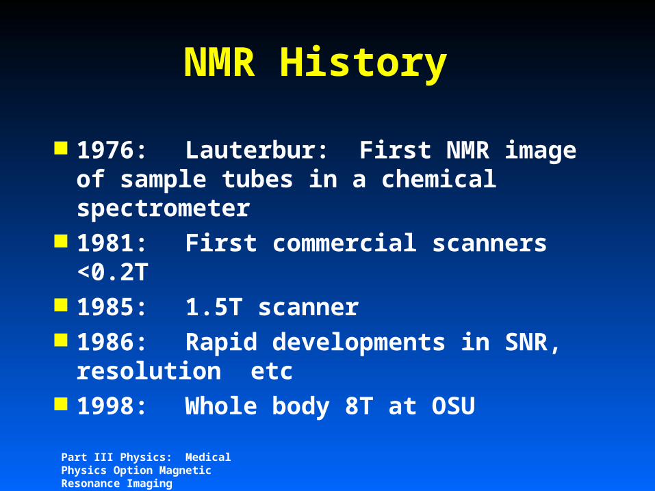
1976: Lauterbur: First NMR image of sample tubes in a chemical spectrometer
1981: First commercial scanners <0.2T
1985: 1.5T scanner 1986: Rapid developments in SNR,
resolution etc 1998: Whole body 8T at OSU
Part III Physics: Medical Physics Option Magnetic Resonance Imaging
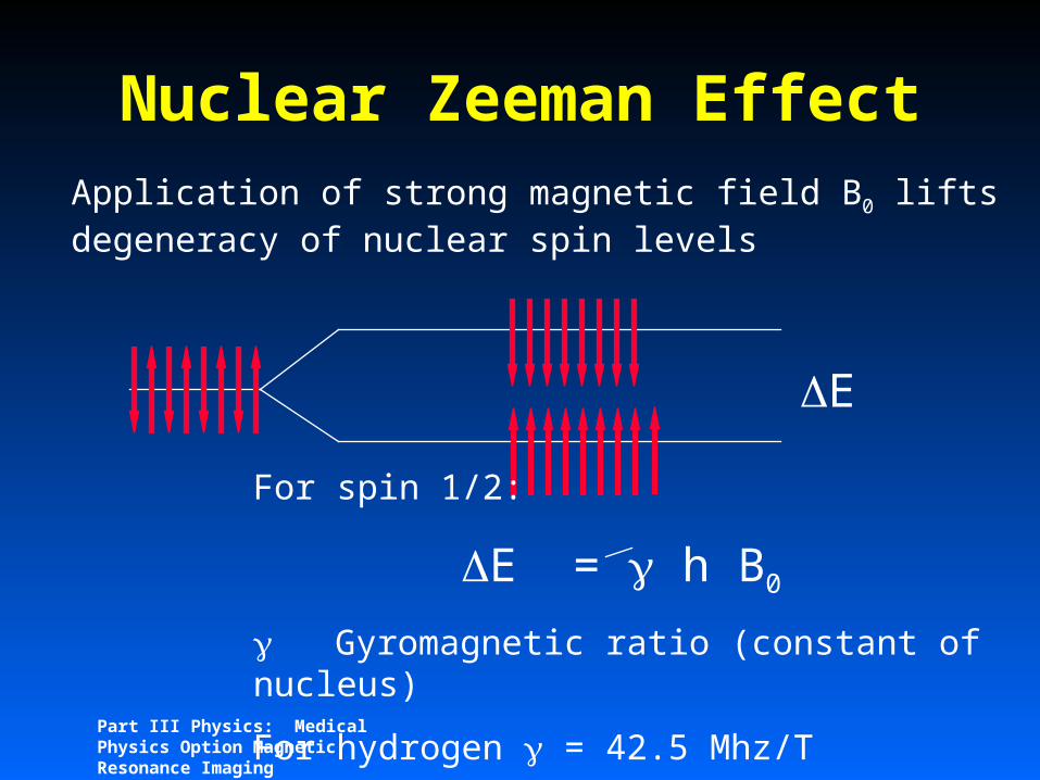
Nuclear Zeeman EffectApplication of strong magnetic field B0 lifts degeneracy of nuclear spin levels
For spin 1/2:
E = h B0
Gyromagnetic ratio (constant of nucleus)
For hydrogen = 42.5 Mhz/T
E
Part III Physics: Medical Physics Option Magnetic Resonance Imaging
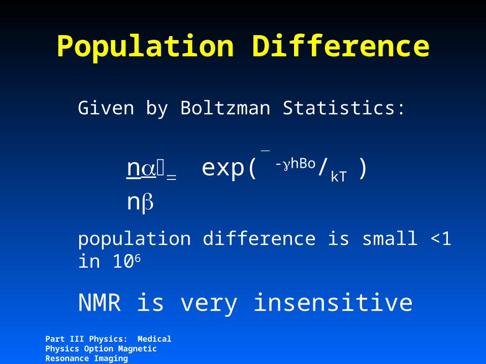
Population Difference
Given by Boltzman Statistics:
nexp( -hBo/kT )n
population difference is small <1 in 106
NMR is very insensitive
Part III Physics: Medical Physics Option Magnetic Resonance Imaging
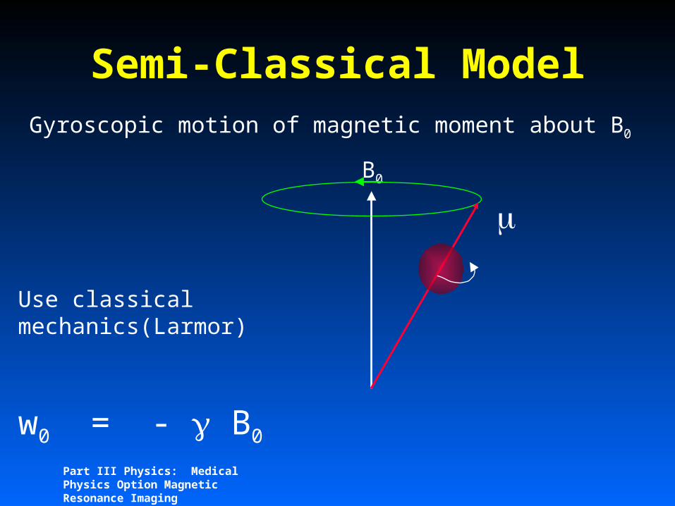
Semi-Classical ModelGyroscopic motion of magnetic moment about B0
B0
Use classical mechanics(Larmor)
w0 = - B0

Part III Physics: Medical Physics Option Magnetic Resonance Imaging
Ensemble Average
M

Part III Physics: Medical Physics Option Magnetic Resonance Imaging
Rotating FrameConsider precessing moment in a frame of reference rotating at the larmor frequency around B0
xy
= Bo
X’ Y’

Part III Physics: Medical Physics Option Magnetic Resonance Imaging
Rotating FrameClassical treatment of M
Effect of RF in laboratory Frame:
Y
XEquivalent to sinusoidal Brf

Part III Physics: Medical Physics Option Magnetic Resonance Imaging
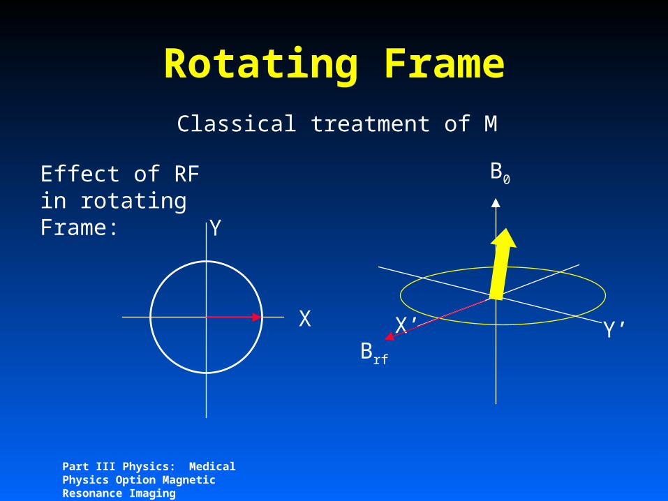
Rotating FrameClassical treatment of M
Effect of RF in rotating Frame:
Y
X X’ Y’
B0
Brf
Part III Physics: Medical Physics Option Magnetic Resonance Imaging
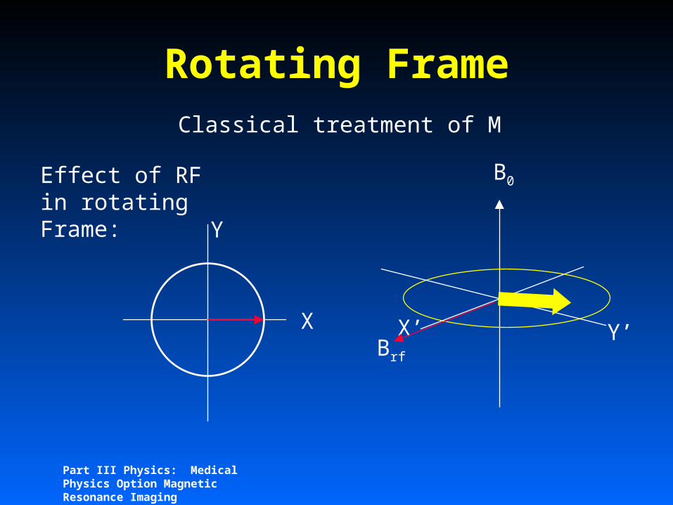
Rotating FrameClassical treatment of M
Effect of RF in rotating Frame:
Y
X X’ Y’
B0
Brf

Part III Physics: Medical Physics Option Magnetic Resonance Imaging
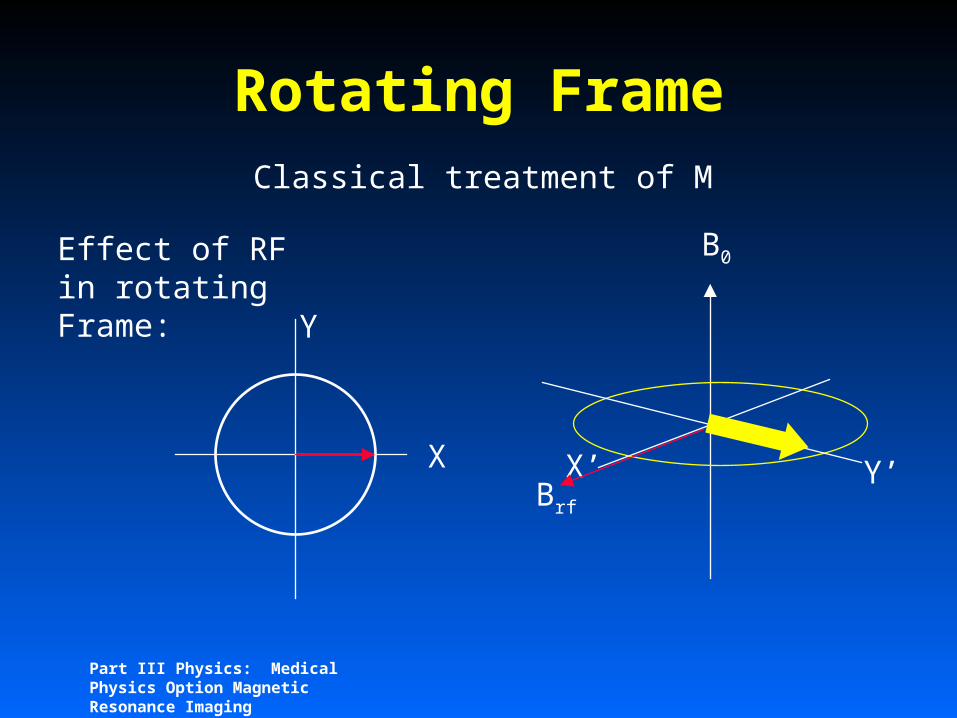
Rotating FrameClassical treatment of M
Effect of RF in rotating Frame:
Y
X X’ Y’
B0
Brf

Part III Physics: Medical Physics Option Magnetic Resonance Imaging
Rotating FrameClassical treatment of M
Effect of RF in rotating Frame:
Y
X X’ Y’
B0
Brf

Part III Physics: Medical Physics Option Magnetic Resonance Imaging
Rotating FrameClassical treatment of M
Effect of RF in rotating Frame:
Y
X X’ Y’
B0
Brf
Part III Physics: Medical Physics Option Magnetic Resonance Imaging
Rotating FrameClassical treatment of M
Effect of RF in rotating Frame:
Y
X X’ Y’
B0
Brf
Part III Physics: Medical Physics Option Magnetic Resonance Imaging
Rotating FrameClassical treatment of M
Effect of RF in rotating Frame:
Y
X X’ Y’
B0
Brf
Part III Physics: Medical Physics Option Magnetic Resonance Imaging
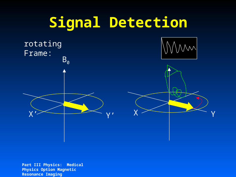
Signal Detectionrotating Frame:
Y’X’
B0
YX

Part III Physics: Medical Physics Option Magnetic Resonance Imaging
Fourier Transformation
FT
Sampling frequency = 2 expected frequency spread (Nyquist)

Basic Spin Echo Imaging 28Part III Physics: Medical Physics Option Magnetic Resonance Imaging
Effect of RF pulses:
B1 (rf)
y’ y’
x’ x’
z zB0
90o degree pulse

Basic Spin Echo Imaging 29Part III Physics: Medical Physics Option Magnetic Resonance Imaging
Effect of RF pulses:
B1 (rf)
y’ y’
x’ x’
z zB0
90o degre
e pulse

Basic Spin Echo Imaging 30Part III Physics: Medical Physics Option Magnetic Resonance Imaging
Effect of RF pulses:
B1 (rf)
y’ y’
x’ x’
z zB0
180o pulse
(inverting pulse)
Basic Spin Echo Imaging 31Part III Physics: Medical Physics Option Magnetic Resonance Imaging
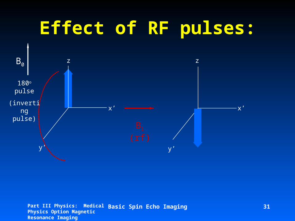
Effect of RF pulses:
B1 (rf)
y’ y’
x’ x’
z zB0
180o pulse
(inverting pulse)
Basic Spin Echo Imaging 32Part III Physics: Medical Physics Option Magnetic Resonance Imaging
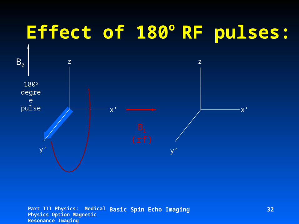
Effect of 180o RF pulses:
B1 (rf)
y’ y’
x’ x’
z zB0
180o degre
e pulse
Basic Spin Echo Imaging 33Part III Physics: Medical Physics Option Magnetic Resonance Imaging
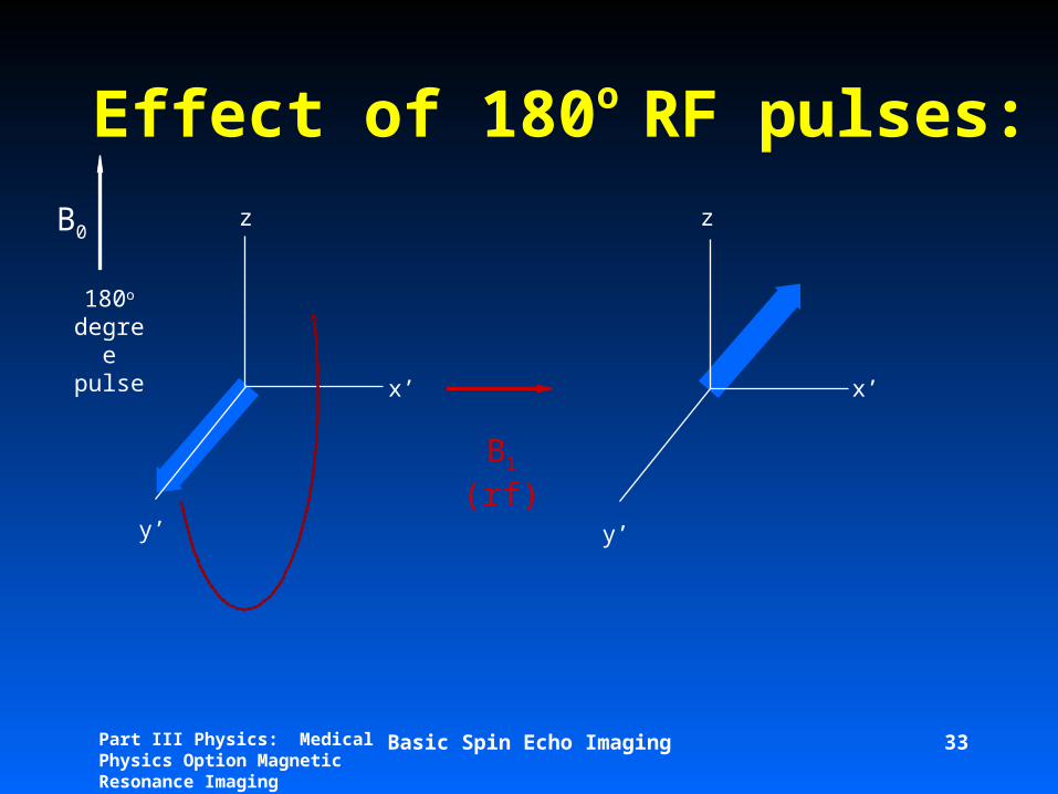
Effect of 180o RF pulses:
B1 (rf)
y’ y’
x’ x’
z zB0
180o degre
e pulse
Basic Spin Echo Imaging 34Part III Physics: Medical Physics Option Magnetic Resonance Imaging
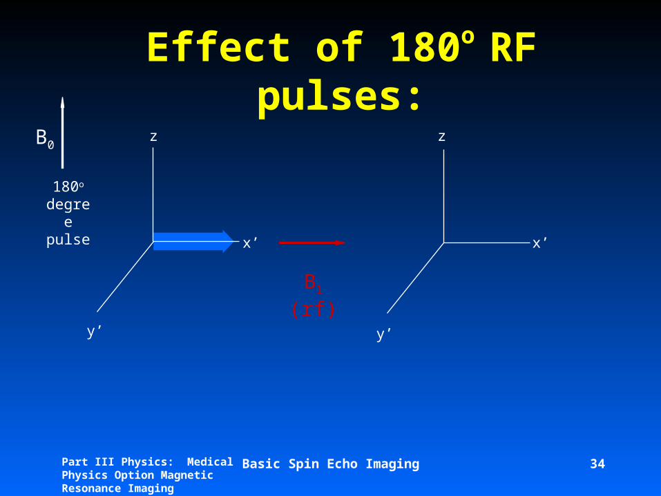
Effect of 180o RF pulses:
B1 (rf)
y’ y’
x’ x’
z zB0
180o degre
e pulse

Basic Spin Echo Imaging 35Part III Physics: Medical Physics Option Magnetic Resonance Imaging
Effect of 180o RF pulses:
B1 (rf)
y’ y’
x’ x’
z zB0
180o degre
e pulse
Basic Spin Echo Imaging 36Part III Physics: Medical Physics Option Magnetic Resonance Imaging
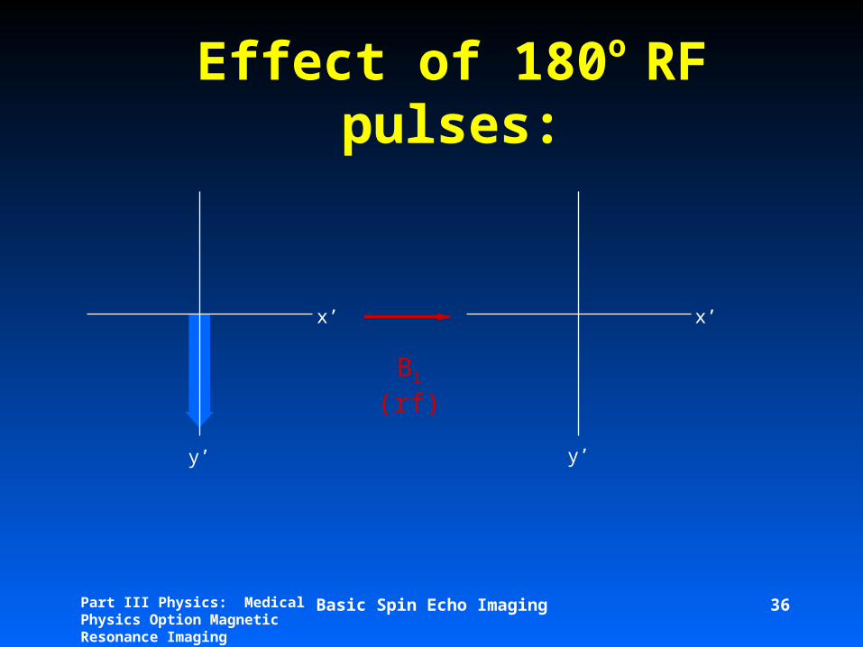
Effect of 180o RF pulses:
B1 (rf)
y’ y’
x’ x’
Basic Spin Echo Imaging 37Part III Physics: Medical Physics Option Magnetic Resonance Imaging
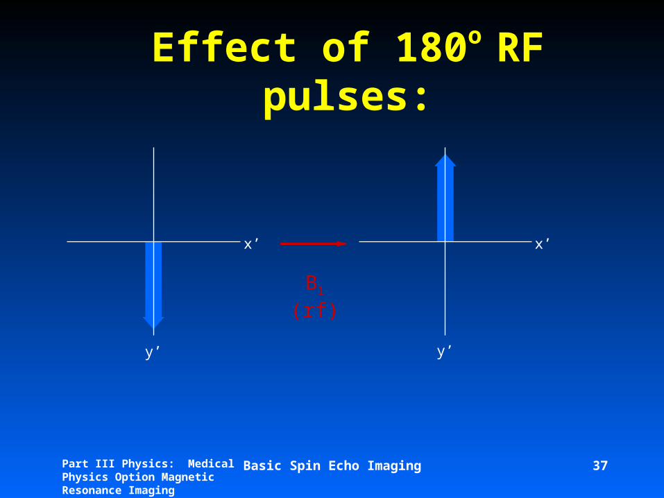
Effect of 180o RF pulses:
B1 (rf)
y’ y’
x’ x’

Basic Spin Echo Imaging 38Part III Physics: Medical Physics Option Magnetic Resonance Imaging
Effect of 180o RF pulses:
B1 (rf)
y’ y’
x’ x’
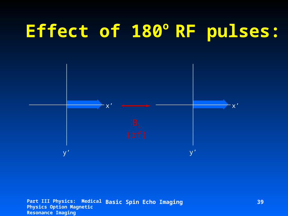
Basic Spin Echo Imaging 39Part III Physics: Medical Physics Option Magnetic Resonance Imaging
Effect of 180o RF pulses:
B1 (rf)
y’ y’
x’ x’

Basic Spin Echo Imaging 40Part III Physics: Medical Physics Option Magnetic Resonance Imaging
Effect of 180o RF pulses:
B1 (rf)
y’
x’
y’
x’
y’
x’
y’
x’
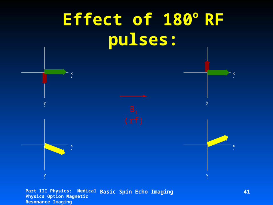
Basic Spin Echo Imaging 41Part III Physics: Medical Physics Option Magnetic Resonance Imaging
Effect of 180o RF pulses:
B1 (rf)
y’
x’
y’
x’
y’
x’
y’
x’

Part III Physics: Medical Physics Option Magnetic Resonance Imaging
Two Pulse sequences (I)
90—— ——90
Saturation recovery
Two Pulse sequences (I)
180—— ——90
Inversion recovery
1 2 3 4 5 6
T1
1 2 3 4 5 6
T1
Part III Physics: Medical Physics Option Magnetic Resonance Imaging
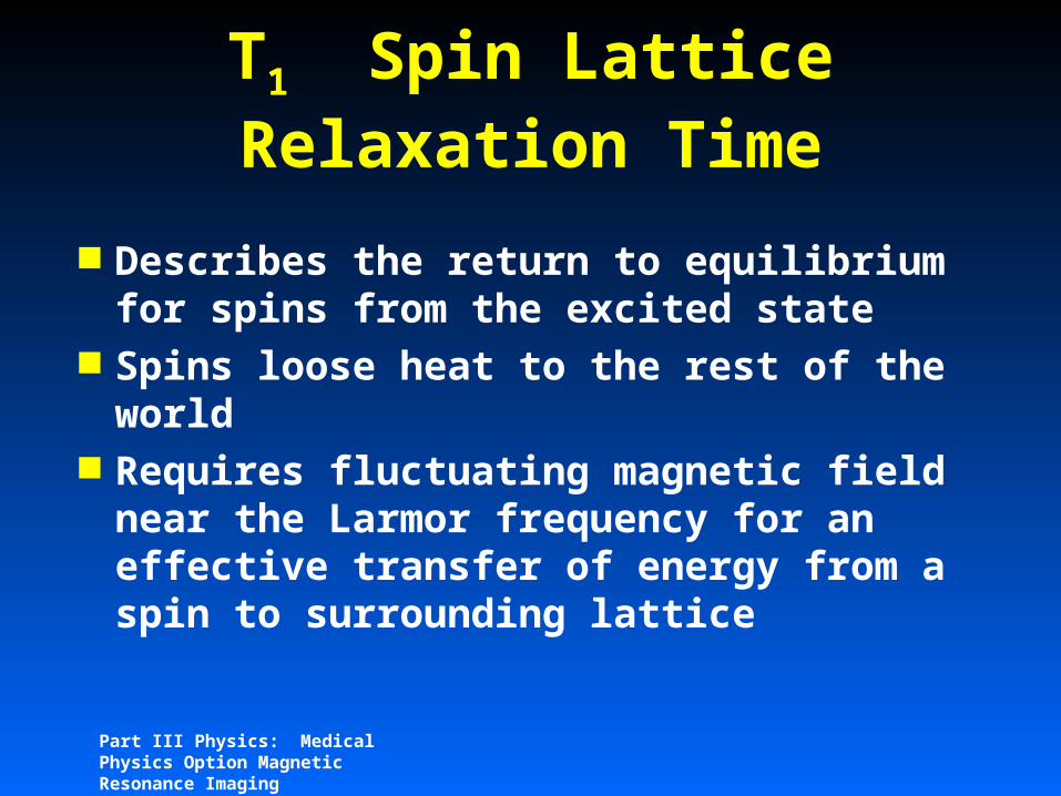
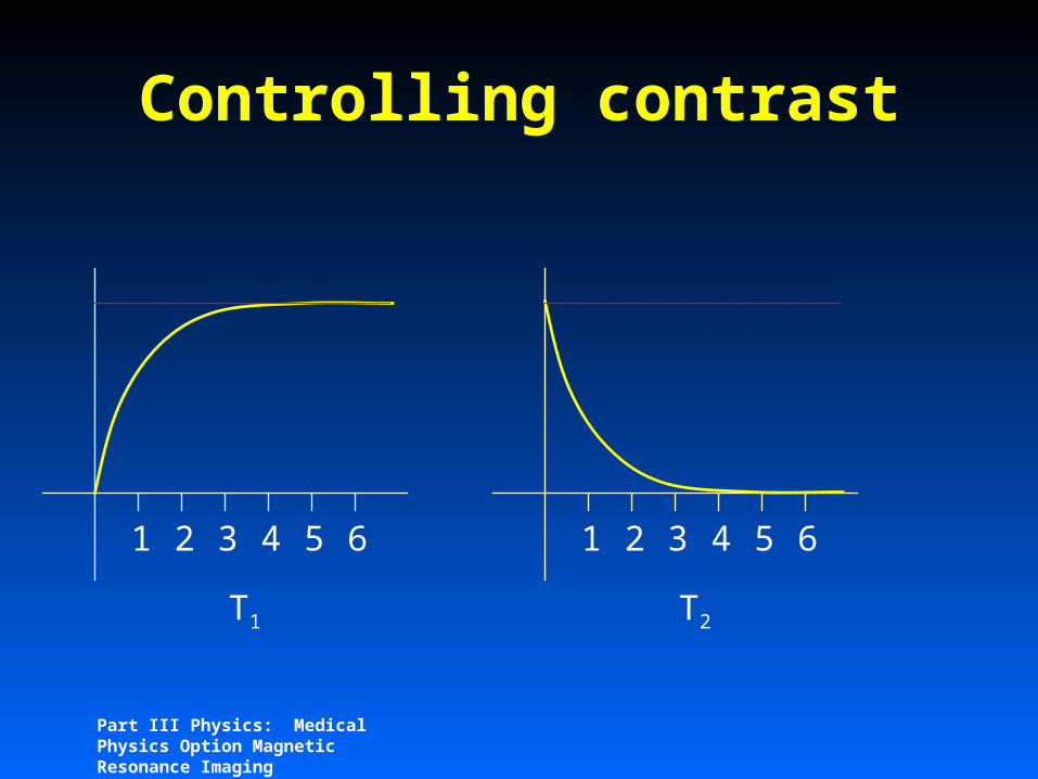
T1 Spin Lattice Relaxation Time
Describes the return to equilibrium for spins from the excited state
Spins loose heat to the rest of the world
Requires fluctuating magnetic field near the Larmor frequency for an effective transfer of energy from a spin to surrounding lattice
Part III Physics: Medical Physics Option Magnetic Resonance Imaging
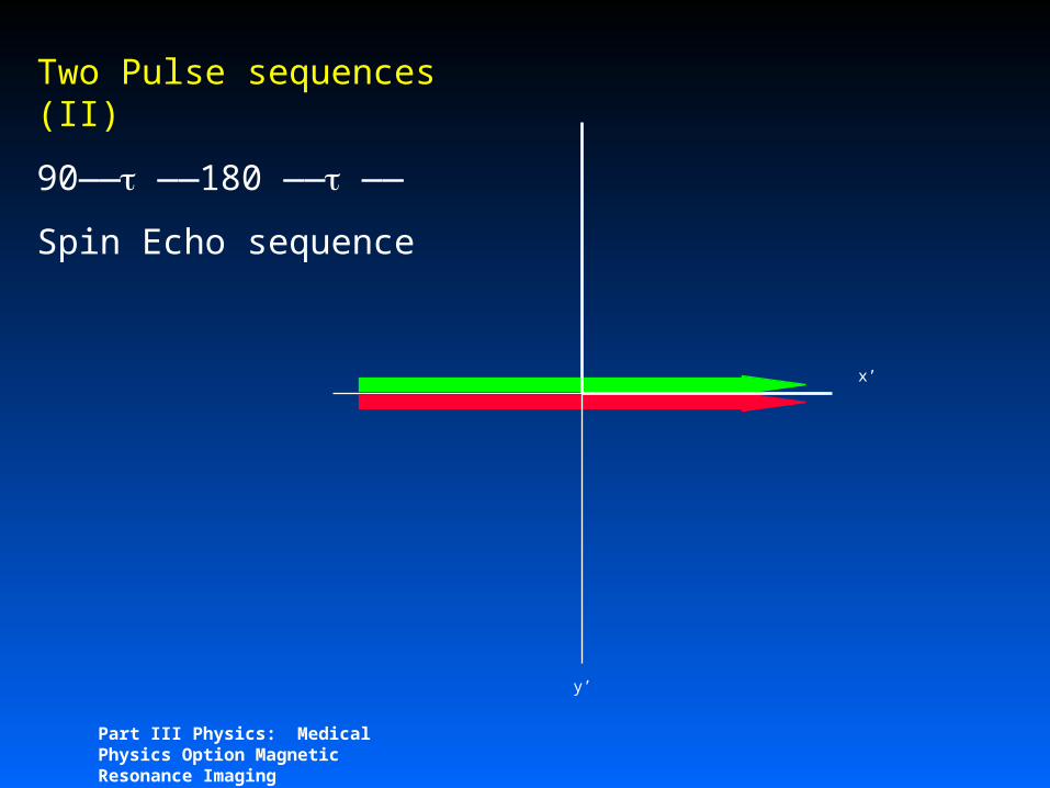
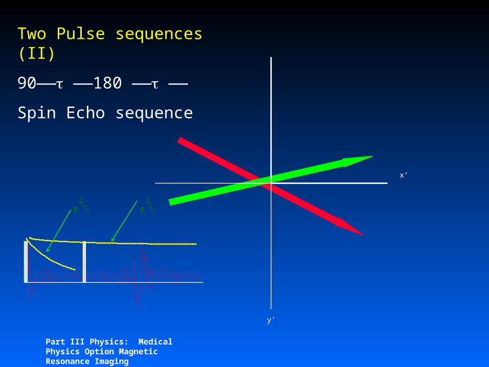
Two Pulse sequences (II)
90—— ——180 —— ——
Spin Echo sequence
y’
x’
Part III Physics: Medical Physics Option Magnetic Resonance Imaging
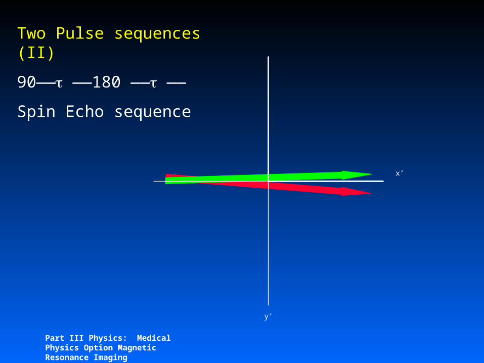
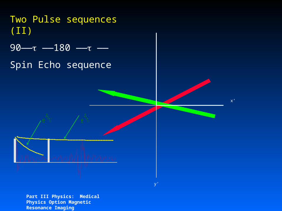
Two Pulse sequences (II)
90—— ——180 —— ——
Spin Echo sequence
y’
x’
Part III Physics: Medical Physics Option Magnetic Resonance Imaging
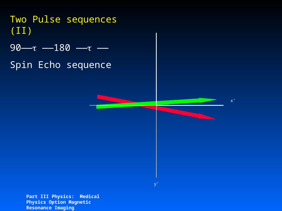
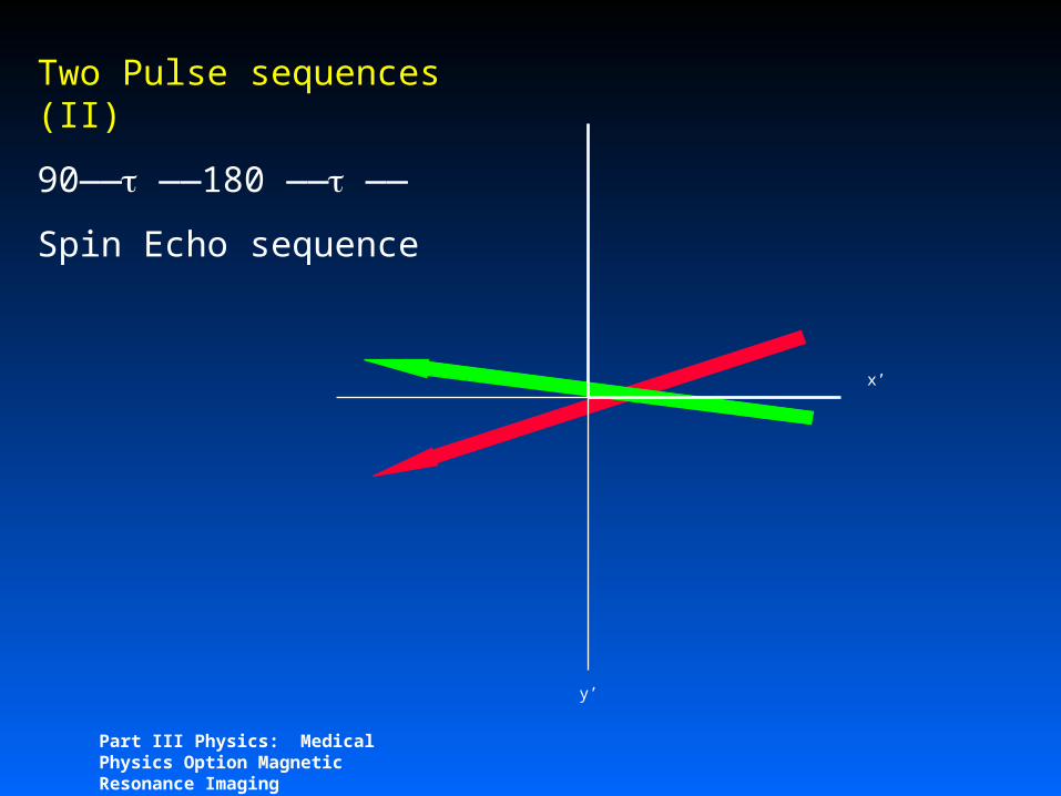
Two Pulse sequences (II)
90—— ——180 —— ——
Spin Echo sequence
y’
x’

Part III Physics: Medical Physics Option Magnetic Resonance Imaging
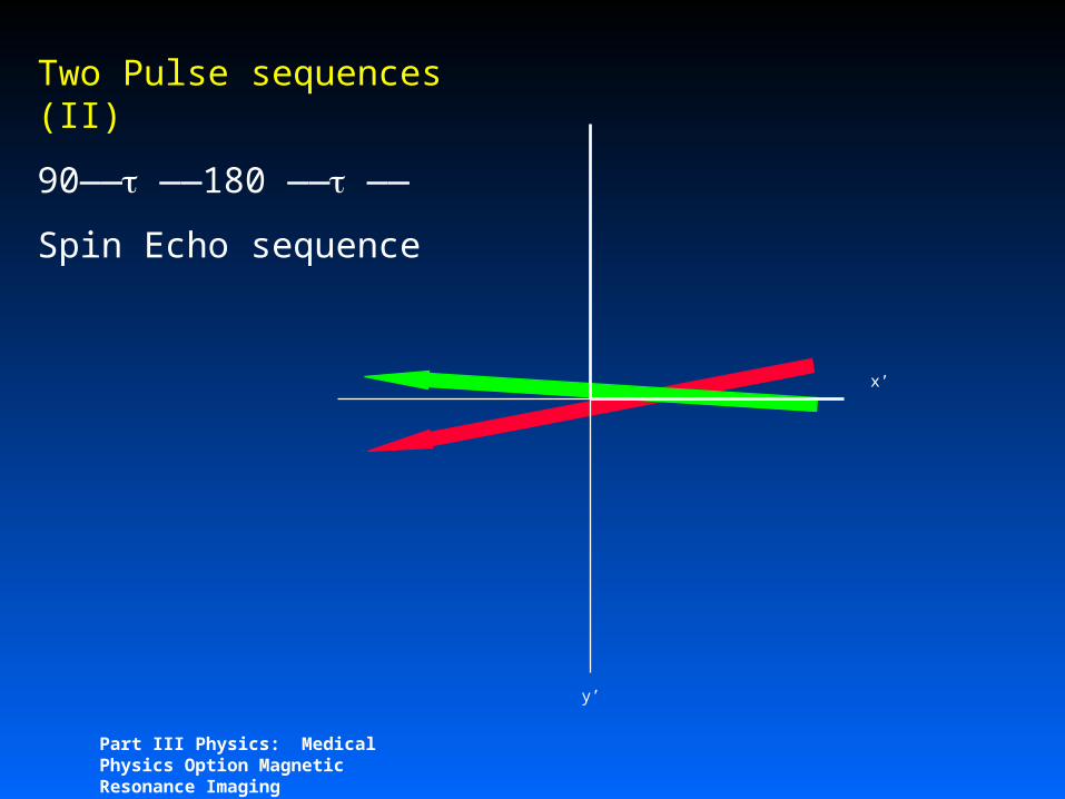
Two Pulse sequences (II)
90—— ——180 —— ——
Spin Echo sequence
y’
x’
Part III Physics: Medical Physics Option Magnetic Resonance Imaging
Two Pulse sequences (II)
90—— ——180 —— ——
Spin Echo sequence
y’
x’
e -t/T2* e -t/T2
Part III Physics: Medical Physics Option Magnetic Resonance Imaging
Two Pulse sequences (II)
90—— ——180 —— ——
Spin Echo sequence
y’
x’
e -t/T2* e -t/T2
Part III Physics: Medical Physics Option Magnetic Resonance Imaging
Two Pulse sequences (II)
90—— ——180 —— ——
Spin Echo sequence
y’
x’
Part III Physics: Medical Physics Option Magnetic Resonance Imaging
Two Pulse sequences (II)
90—— ——180 —— ——
Spin Echo sequence
y’
x’

Part III Physics: Medical Physics Option Magnetic Resonance Imaging
Two Pulse sequences (II)
90—— ——180 —— ——
Spin Echo sequence
y’
x’

Part III Physics: Medical Physics Option Magnetic Resonance Imaging
Two Pulse sequences (II)
90—— ——180 —— ——
Spin Echo sequence
y’
x’

Basic Spin Echo Imaging 54Part III Physics: Medical Physics Option Magnetic Resonance Imaging
T2 and T2*
e -t/T2* e -t/T2
H
O
H H
O
H
Part III Physics: Medical Physics Option Magnetic Resonance Imaging
Spin-Spin Relaxation Time
Static inhomogeneities refocussed by 180 pulse
Time varying imhomogeneity are not
T2 changes in disease give rise to diagnostic value of MRI

Part III Physics: Medical Physics Option Magnetic Resonance Imaging
Superconducting Magnet
Helium vessel containing super-con coil
Vacuum

Part III Physics: Medical Physics Option Magnetic Resonance Imaging
Superconducting Magnet
Bore B0
100cm 0 4T80cm 0 8T

Part III Physics: Medical Physics Option Magnetic Resonance Imaging
Shimming

Part III Physics: Medical Physics Option Magnetic Resonance Imaging
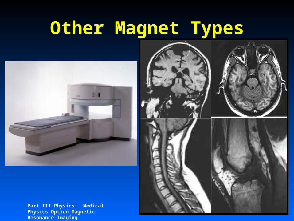
Other Magnet Types
Permanent magnet, e.g. light weight rare earth magnets, <0.3T
Part III Physics: Medical Physics Option Magnetic Resonance Imaging
Other Magnet Types

Part III Physics: Medical Physics Option Magnetic Resonance Imaging
Other Magnet Types
Electromagnet <0.3T

Part III Physics: Medical Physics Option Magnetic Resonance Imaging
Special Superconducting
Magnets Active Shielding
– Extra coils reduce stray field
– Improves siting
10
12
4
2
5mT contour
0.5T wholebody 3T AS wholebody

Part III Physics: Medical Physics Option Magnetic Resonance Imaging
RF CoilsRemember Brf must be B0
Field is subject, can use solenoid.
Part III Physics: Medical Physics Option Magnetic Resonance Imaging
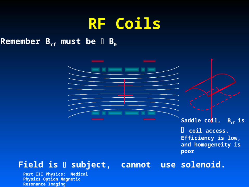
RF CoilsRemember Brf must be B0
Field is subject, cannot use solenoid.
Saddle coil, Brf is coil access. Efficiency is low, and homogeneity is poor

Part III Physics: Medical Physics Option Magnetic Resonance Imaging
Part III Physics: Medical Physics Option Magnetic Resonance Imaging
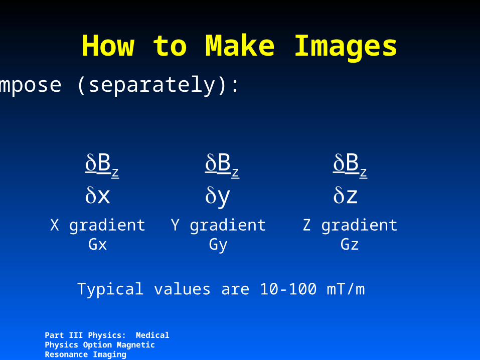
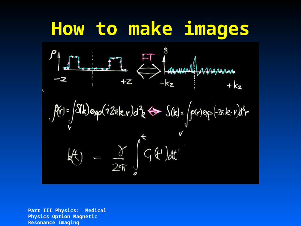
How to Make ImagesImpose (separately):
Bz
xBz
yBz
zX gradient
GxY gradient
GyZ gradient
Gz
Typical values are 10-100 mT/m

Part III Physics: Medical Physics Option Magnetic Resonance Imaging
How to make images
For a Z gradient
-hz +hz
z = -(B0 + Gz.z)
Part III Physics: Medical Physics Option Magnetic Resonance Imaging
How to make images
Part III Physics: Medical Physics Option Magnetic Resonance Imaging
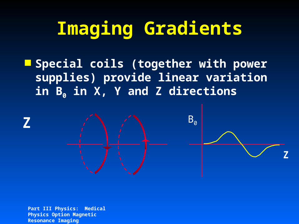
Imaging Gradients
Special coils (together with power supplies) provide linear variation in B0 in X, Y and Z directions
Z
B0Z

Part III Physics: Medical Physics Option Magnetic Resonance Imaging
Imaging Gradients
Special coils (together with power supplies) provide linear variation in B0 in X, Y and Z directions
X,Y

Part III Physics: Medical Physics Option Magnetic Resonance Imaging
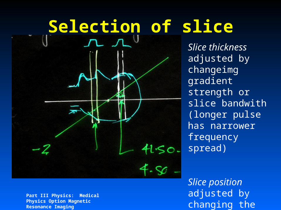
Selection of SliceUse Fourier relationship:
RF Amplitude (volts)
Part III Physics: Medical Physics Option Magnetic Resonance Imaging
Selection of sliceSlice thickness adjusted by changeimg gradient strength or slice bandwith (longer pulse has narrower frequency spread)
Slice position adjusted by changing the centre frequency of the pulse

Part III Physics: Medical Physics Option Magnetic Resonance Imaging
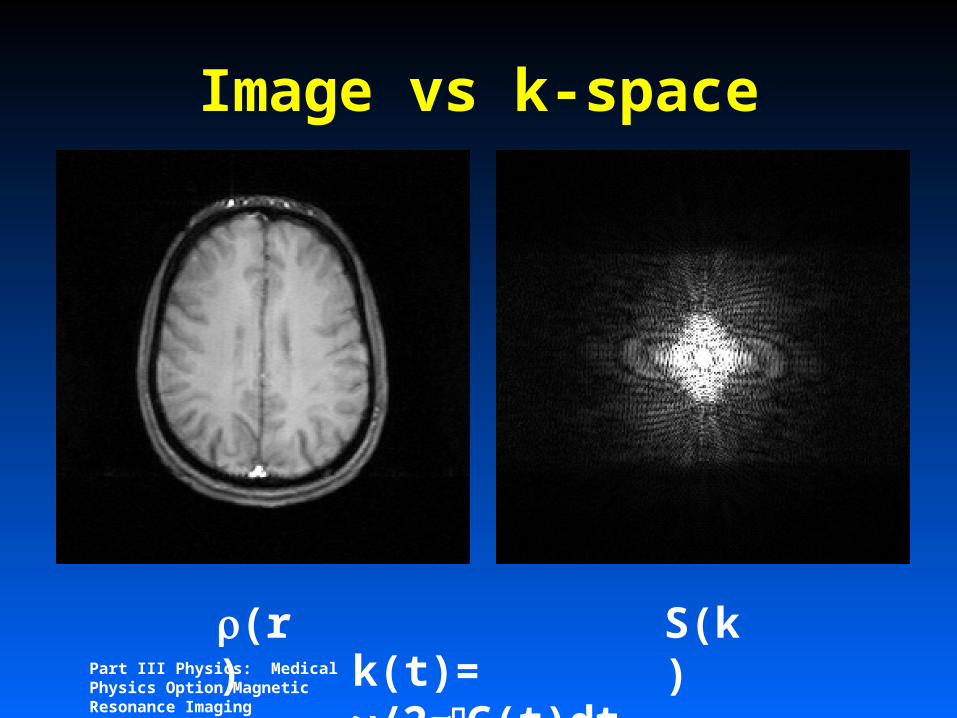
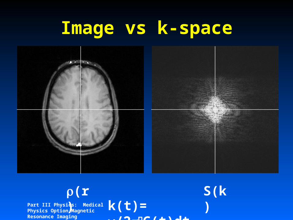
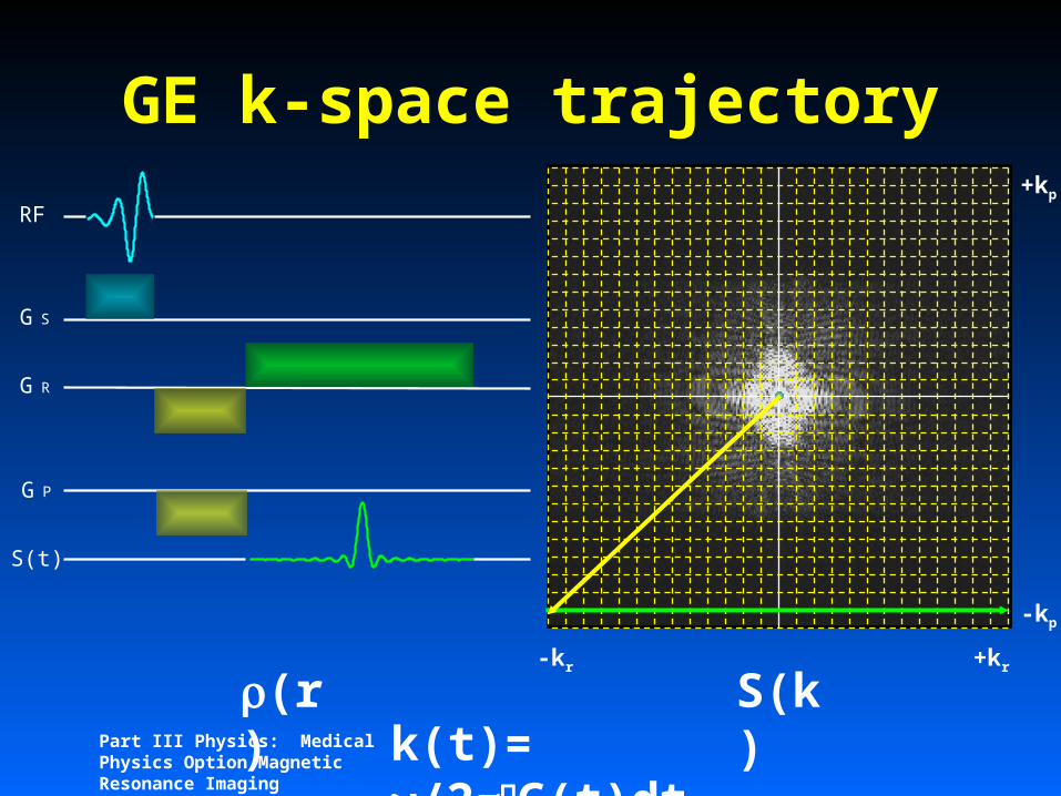
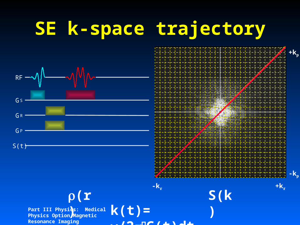
k-space
k-space is the raw data space before fourier transformation into the image
2D image will be represented by a 2D array of data points spread throughout k-space
Differing the k-space trajectory will alter image contrast
Part III Physics: Medical Physics Option Magnetic Resonance Imaging
Image vs k-space
(r) S(k)k(t)=
/2G(t)dt
Part III Physics: Medical Physics Option Magnetic Resonance Imaging
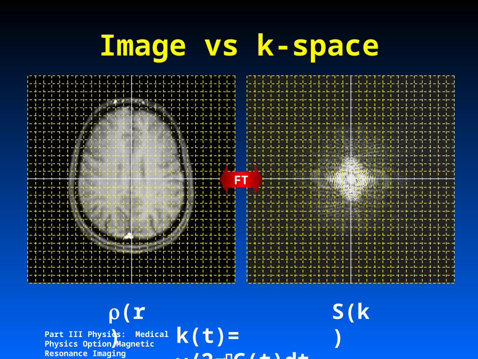
Image vs k-space
(r) S(k)k(t)=
/2G(t)dt

Part III Physics: Medical Physics Option Magnetic Resonance Imaging
Image vs k-space
(r) S(k)k(t)=
/2G(t)dt

Part III Physics: Medical Physics Option Magnetic Resonance Imaging
Image vs k-space
(r) S(k)k(t)=
/2G(t)dt
Part III Physics: Medical Physics Option Magnetic Resonance Imaging
Image vs k-space
(r) S(k)k(t)=
/2G(t)dt
FT

Part III Physics: Medical Physics Option Magnetic Resonance Imaging
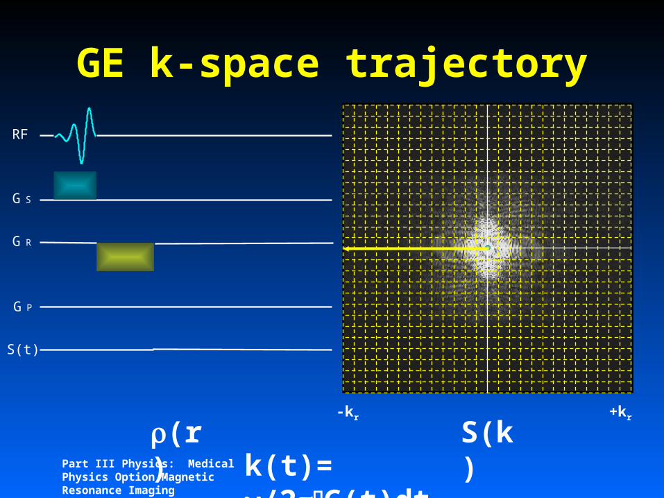
GE k-space trajectory
(r) S(k)k(t)=
/2G(t)dt
RF
G S
G R
G P
S(t)
Part III Physics: Medical Physics Option Magnetic Resonance Imaging
(r) S(k)k(t)=
/2G(t)dt
RF
G S
G R
G P
S(t)
-kr +kr
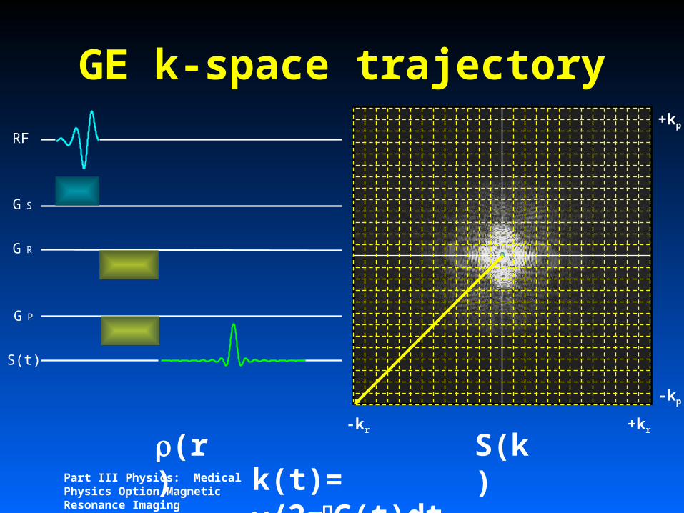
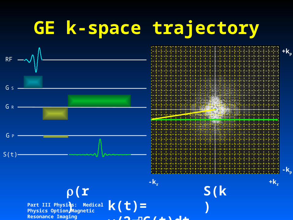
GE k-space trajectory

Part III Physics: Medical Physics Option Magnetic Resonance Imaging
(r) S(k)k(t)=
/2G(t)dt
RF
G S
G R
G P
S(t)
-kr +kr
GE k-space trajectory

Part III Physics: Medical Physics Option Magnetic Resonance Imaging
(r) S(k)k(t)=
/2G(t)dt
RF
G S
G R
G P
S(t)
-kr +kr
-kp
+kp
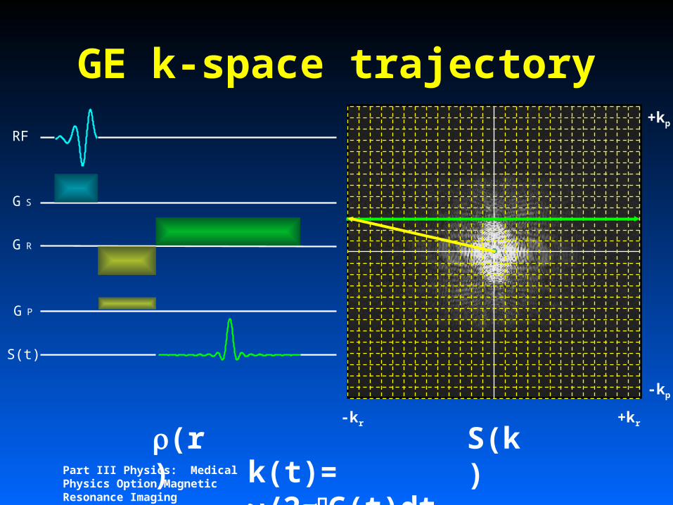
GE k-space trajectory
Part III Physics: Medical Physics Option Magnetic Resonance Imaging
(r) S(k)k(t)=
/2G(t)dt
RF
G S
G R
G P
S(t)
-kr +kr
-kp
+kp
GE k-space trajectory

Part III Physics: Medical Physics Option Magnetic Resonance Imaging
(r) S(k)k(t)=
/2G(t)dt
RF
G S
G R
G P
S(t)
-kr +kr
-kp
+kp
GE k-space trajectory

Part III Physics: Medical Physics Option Magnetic Resonance Imaging
(r) S(k)k(t)=
/2G(t)dt
RF
G S
G R
G P
S(t)
-kr +kr
-kp
+kp
GE k-space trajectory
Part III Physics: Medical Physics Option Magnetic Resonance Imaging
(r) S(k)k(t)=
/2G(t)dt
RF
G S
G R
G P
S(t)
-kr +kr
-kp
+kp
GE k-space trajectory

Part III Physics: Medical Physics Option Magnetic Resonance Imaging
(r) S(k)k(t)=
/2G(t)dt
RF
G S
G R
G P
S(t)
-kr +kr
-kp
+kp
GE k-space trajectory

Part III Physics: Medical Physics Option Magnetic Resonance Imaging
(r) S(k)k(t)=
/2G(t)dt
RF
G S
G R
G P
S(t)
-kr +kr
-kp
+kp
GE k-space trajectory

Part III Physics: Medical Physics Option Magnetic Resonance Imaging
(r) S(k)k(t)=
/2G(t)dt
RF
G S
G R
G P
S(t)
-kr +kr
-kp
+kp
GE k-space trajectory

Part III Physics: Medical Physics Option Magnetic Resonance Imaging
(r) S(k)k(t)=
/2G(t)dt
RF
G S
G R
G P
S(t)
-kr +kr
-kp
+kp
GE k-space trajectory

Part III Physics: Medical Physics Option Magnetic Resonance Imaging
(r) S(k)k(t)=
/2G(t)dt
RF
G S
G R
G P
S(t)
-kr +kr
-kp
+kp
GE k-space trajectory

Part III Physics: Medical Physics Option Magnetic Resonance Imaging
(r) S(k)k(t)=
/2G(t)dt
RF
G S
G R
G P
S(t)
-kr +kr
-kp
+kp
GE k-space trajectory
Part III Physics: Medical Physics Option Magnetic Resonance Imaging
(r) S(k)k(t)=
/2G(t)dt
RF
G S
G R
G P
S(t)
-kr +kr
-kp
+kp
GE k-space trajectory

Part III Physics: Medical Physics Option Magnetic Resonance Imaging
(r) S(k)k(t)=
/2G(t)dt
RF
G S
G R
G P
S(t)
-kr +kr
-kp
+kp
GE k-space trajectory

Part III Physics: Medical Physics Option Magnetic Resonance Imaging
(r) S(k)k(t)=
/2G(t)dt
RF
G S
G R
G P
S(t)
-kr +kr
-kp
+kp
GE k-space trajectory
Part III Physics: Medical Physics Option Magnetic Resonance Imaging
(r) S(k)k(t)=
/2G(t)dt
RF
G S
G R
G P
S(t)
-kr +kr
-kp
+kp
GE k-space trajectory

Part III Physics: Medical Physics Option Magnetic Resonance Imaging
(r) S(k)k(t)=
/2G(t)dt
RF
G S
G R
G P
S(t)
-kr +kr
-kp
+kp
GE k-space trajectory

Part III Physics: Medical Physics Option Magnetic Resonance Imaging
(r) S(k)k(t)=
/2G(t)dt
-kr +kr
-kp
+kp
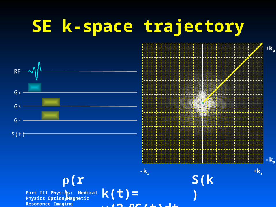
SE k-space trajectory
RF
GS
GR
GP
S(t)
Part III Physics: Medical Physics Option Magnetic Resonance Imaging
(r) S(k)k(t)=
/2G(t)dt
-kr +kr
-kp
+kp
SE k-space trajectory
RF
GS
GR
GP
S(t)

Part III Physics: Medical Physics Option Magnetic Resonance Imaging
(r) S(k)k(t)=
/2G(t)dt
-kr +kr
-kp
+kp
SE k-space trajectory
RF
GS
GR
GP
S(t)
Part III Physics: Medical Physics Option Magnetic Resonance Imaging
(r) S(k)k(t)=
/2G(t)dt
-kr +kr
-kp
+kp
SE k-space trajectory
RF
GS
GR
GP
S(t)

Part III Physics: Medical Physics Option Magnetic Resonance Imaging
(r) S(k)k(t)=
/2G(t)dt
-kr +kr
-kp
+kp
SE k-space trajectory
RF
GS
GR
GP
S(t)

Part III Physics: Medical Physics Option Magnetic Resonance Imaging
(r) S(k)k(t)=
/2G(t)dt
-kr +kr
-kp
+kp
SE k-space trajectory
RF
GS
GR
GP
S(t)
Part III Physics: Medical Physics Option Magnetic Resonance Imaging
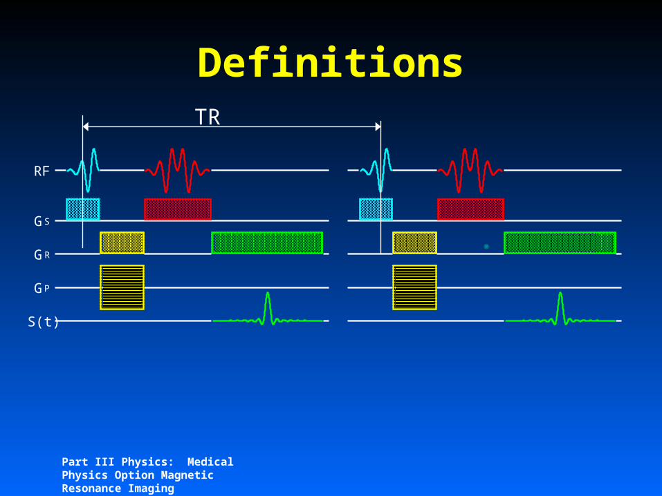
Definitions
RF
GS
GR
GP
S(t)
TR

Part III Physics: Medical Physics Option Magnetic Resonance Imaging
Definitions
RF
GS
GR
GP
S(t)
TE
Part III Physics: Medical Physics Option Magnetic Resonance Imaging
1 2 3 4 5 6
T1
1 2 3 4 5 6
T2
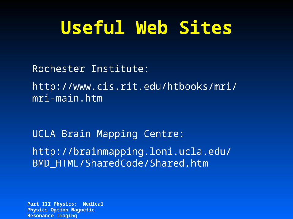
Controlling contrast
Part III Physics: Medical Physics Option Magnetic Resonance Imaging
1 2 3 4 5 6
T1
1 2 3 4 5 6
T2
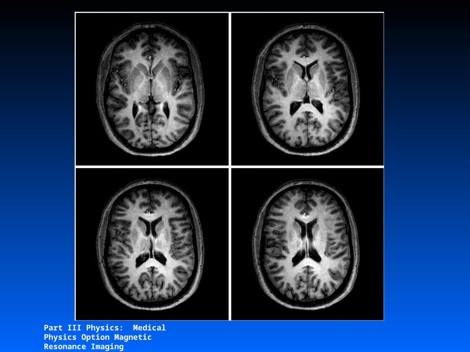
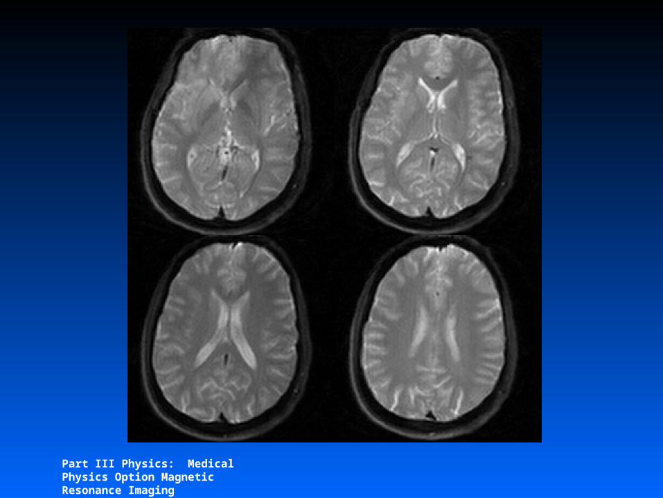
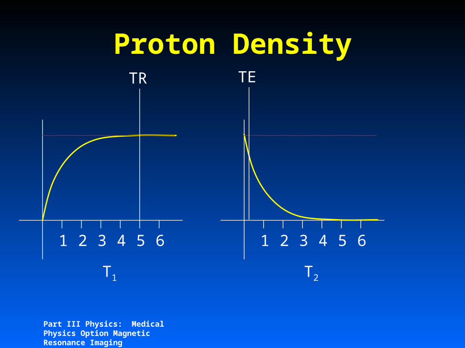
Proton DensityTR TE
Part III Physics: Medical Physics Option Magnetic Resonance Imaging
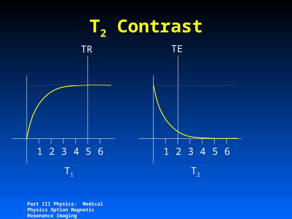
1 2 3 4 5 6
T1
1 2 3 4 5 6
T2
T2 ContrastTR TE
Part III Physics: Medical Physics Option Magnetic Resonance Imaging
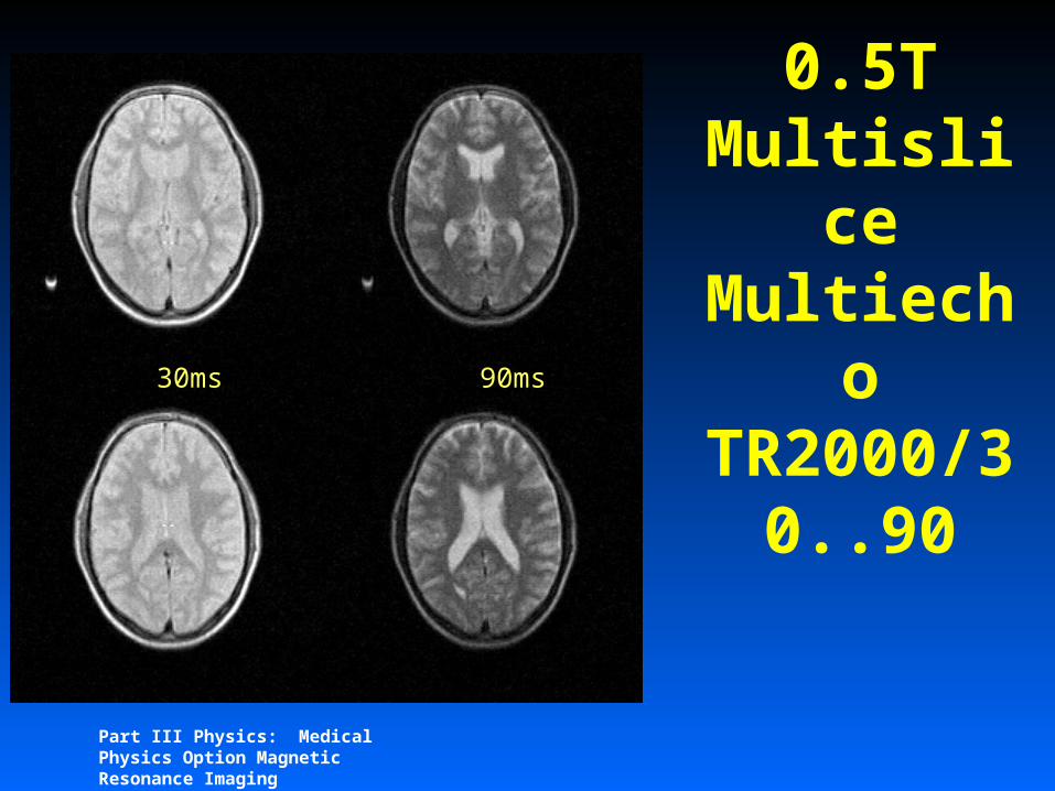
0.5T Multislic
e Multiech
oTR2000/30..90
30ms 90ms

Part III Physics: Medical Physics Option Magnetic Resonance Imaging
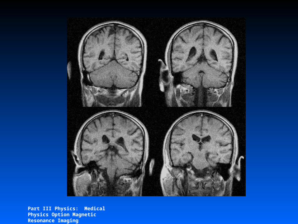
1 2 3 4 5 6
T1
1 2 3 4 5 6
T2
T1 ContrastTR TE
Part III Physics: Medical Physics Option Magnetic Resonance Imaging
Part III Physics: Medical Physics Option Magnetic Resonance Imaging
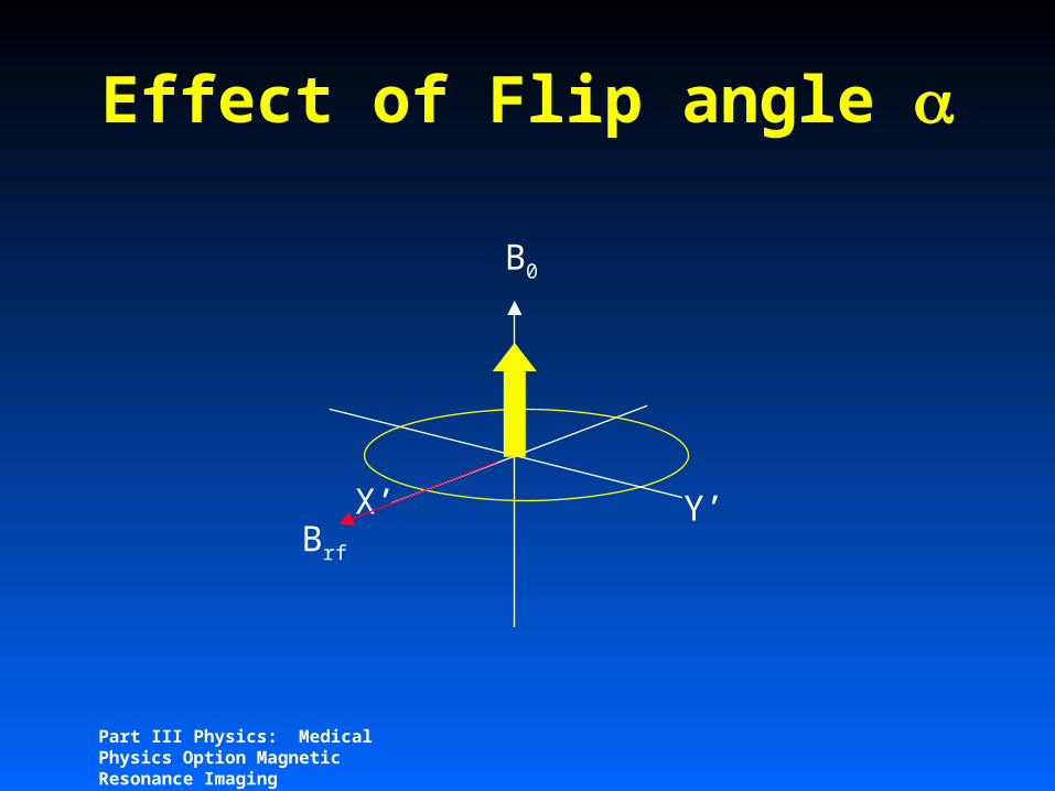
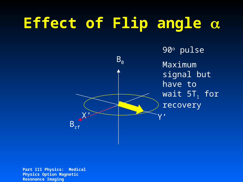
Effect of Flip angle
X’ Y’
B0
Brf
Part III Physics: Medical Physics Option Magnetic Resonance Imaging
Effect of Flip angle
X’ Y’
B0
Brf
90o pulse
Maximum signal but have to wait 5T1 for recovery

Part III Physics: Medical Physics Option Magnetic Resonance Imaging
Effect of Flip angle
X’ Y’
B0
Brf

Part III Physics: Medical Physics Option Magnetic Resonance Imaging
Effect of Flip angle
X’ Y’
B0
Brf
Flip angle 30o:
detect M0sin = 0.5 M0 remaining M0cos = 0.87 M0
Part III Physics: Medical Physics Option Magnetic Resonance Imaging
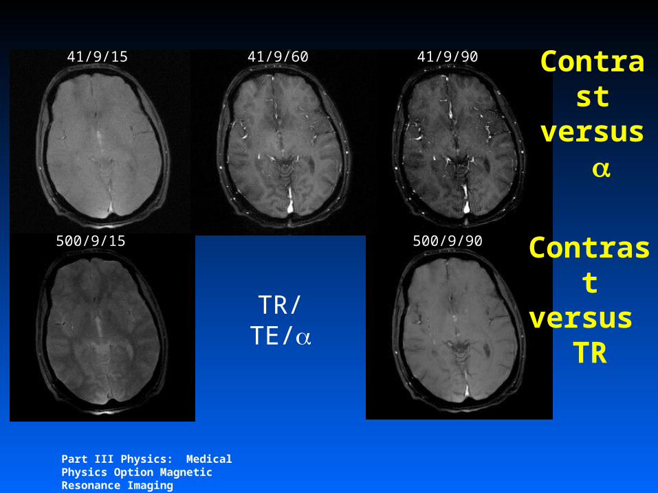
TR/TE/
41/9/15
500/9/15
41/9/9041/9/60
500/9/90
Contrast
versus
Contrast
versus TR
Part III Physics: Medical Physics Option Magnetic Resonance Imaging
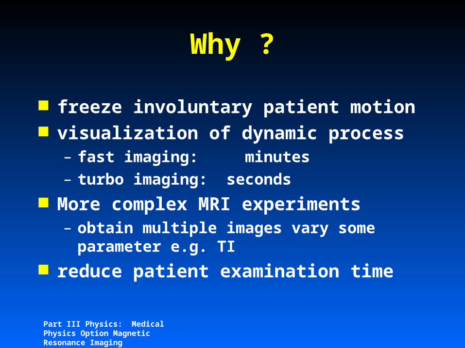
Why ?
freeze involuntary patient motion visualization of dynamic process
– fast imaging: minutes– turbo imaging: seconds
More complex MRI experiments– obtain multiple images vary some
parameter e.g. TI reduce patient examination time

Part III Physics: Medical Physics Option Magnetic Resonance Imaging
Why does MRI take so long
Answer– Only one phase encode line acquired
per excitation– Spin Echo: 256*3s for T2, 256*0.6s for
T1– Gradient Echo: 256*35ms (but have to
do 3D Solution
– get more phase encode lines per excitation
Part III Physics: Medical Physics Option Magnetic Resonance Imaging
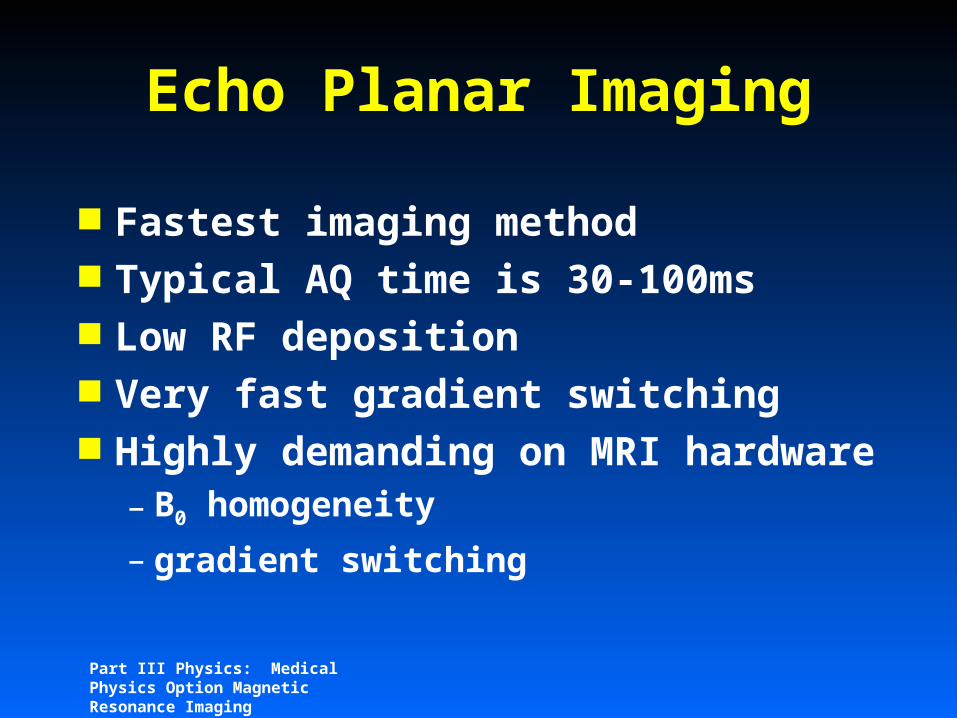
Echo Planar Imaging
Fastest imaging method Typical AQ time is 30-100ms Low RF deposition Very fast gradient switching Highly demanding on MRI
hardware– B0 homogeneity
– gradient switching

Part III Physics: Medical Physics Option Magnetic Resonance Imaging
(r) S(k)k(t)=
/2G(t)dt
RF
G S
G R
G P
S(t)
-kr +kr
-kp
+kp
GE-PEI k-space trajectory

Part III Physics: Medical Physics Option Magnetic Resonance Imaging
(r) S(k)k(t)=
/2G(t)dt
RF
G S
G R
G P
S(t)
-kr +kr
-kp
+kp
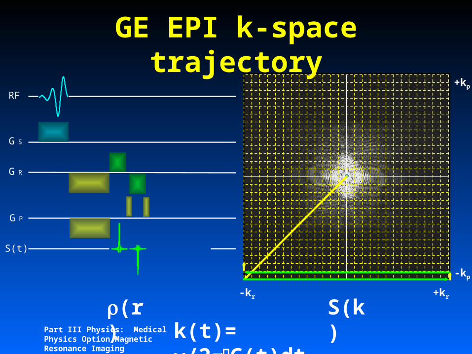
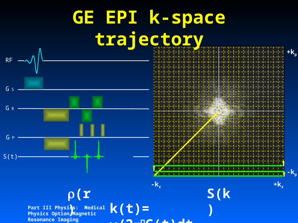
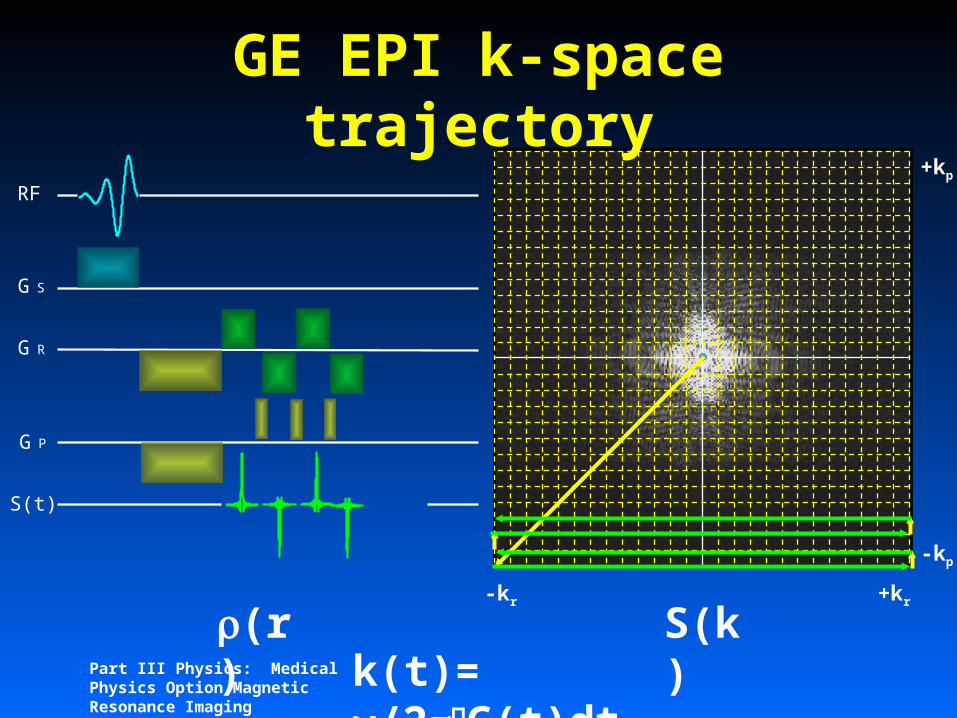
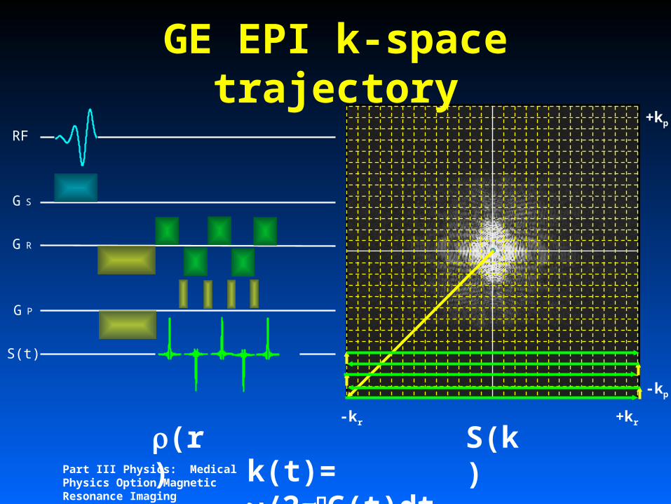
GE EPI k-space trajectory

Part III Physics: Medical Physics Option Magnetic Resonance Imaging
(r) S(k)k(t)=
/2G(t)dt
RF
G S
G R
G P
S(t)
-kr +kr
-kp
+kp
GE EPI k-space trajectory

Part III Physics: Medical Physics Option Magnetic Resonance Imaging
(r) S(k)k(t)=
/2G(t)dt
RF
G S
G R
G P
S(t)
-kr +kr
-kp
+kp
GE EPI k-space trajectory

Part III Physics: Medical Physics Option Magnetic Resonance Imaging
(r) S(k)k(t)=
/2G(t)dt
RF
G S
G R
G P
S(t)
-kr +kr
-kp
+kp
GE EPI k-space trajectory
Part III Physics: Medical Physics Option Magnetic Resonance Imaging
(r) S(k)k(t)=
/2G(t)dt
RF
G S
G R
G P
S(t)
-kr +kr
-kp
+kp
GE EPI k-space trajectory

Part III Physics: Medical Physics Option Magnetic Resonance Imaging
(r) S(k)k(t)=
/2G(t)dt
RF
G S
G R
G P
S(t)
-kr +kr
-kp
+kp
GE EPI k-space trajectory
Part III Physics: Medical Physics Option Magnetic Resonance Imaging
(r) S(k)k(t)=
/2G(t)dt
RF
G S
G R
G P
S(t)
-kr +kr
-kp
+kp
GE EPI k-space trajectory
Part III Physics: Medical Physics Option Magnetic Resonance Imaging
(r) S(k)k(t)=
/2G(t)dt
RF
G S
G R
G P
S(t)
-kr +kr
-kp
+kp
GE EPI k-space trajectory

Part III Physics: Medical Physics Option Magnetic Resonance Imaging
(r) S(k)k(t)=
/2G(t)dt
RF
G S
G R
G P
S(t)
-kr +kr
-kp
+kp
GE EPI k-space trajectory
Part III Physics: Medical Physics Option Magnetic Resonance Imaging
(r) S(k)k(t)=
/2G(t)dt
RF
G S
G R
G P
S(t)
-kr +kr
-kp
+kp
GE EPI k-space trajectory
Part III Physics: Medical Physics Option Magnetic Resonance Imaging
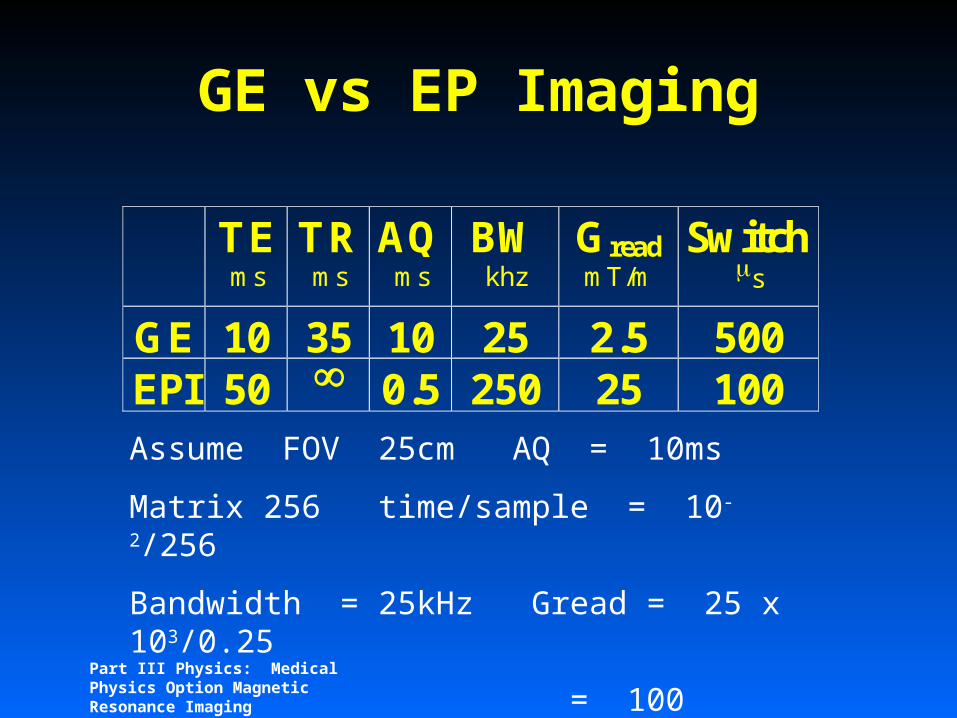
GE vs EP Imaging
TEms
TRms
AQms
BWkhz
GreadmT/m
Switchs
GE 10 35 10 25 2.5 500EPI 50 0.5 250 25 100Assume FOV 25cm AQ = 10ms
Matrix 256 time/sample = 10-2/256
Bandwidth = 25kHz Gread = 25 x 103/0.25
= 100 000Hz/m
= ~ 2.5 mT/m

Part III Physics: Medical Physics Option Magnetic Resonance Imaging
GE vs EP Imaging
TEms
TRms
AQms
BWkhz
GreadmT/m
Switchs
GE 10 35 10 25 2.5 500EPI 50 0.5 250 25 100Assume FOV 25cm AQ = 0.5ms
Matrix 128 time/sample = 5x10⁻⁴/128
Bandwidth = 250kHz Gread = 250 x 103/0.25
= 1 000 000Hz/m
= ~ 25 mT/m

Part III Physics: Medical Physics Option Magnetic Resonance Imaging
MRI at 3T
128x128 single shot, GE echo planar.
X,Y,Z shim only (~30s)
No template or navigator correction
Straight FFT after row reversal
Part III Physics: Medical Physics Option Magnetic Resonance Imaging
fMRI (functional MRI)
Monitor T2 or T2* contrast during cognitive task
eg acquire 20-30 slices every 4 seconds
Design experiment to have alternating blocks of task and control condition
Look for statistically significant signal intenisty changes correlated with task blocks
Part III Physics: Medical Physics Option Magnetic Resonance Imaging
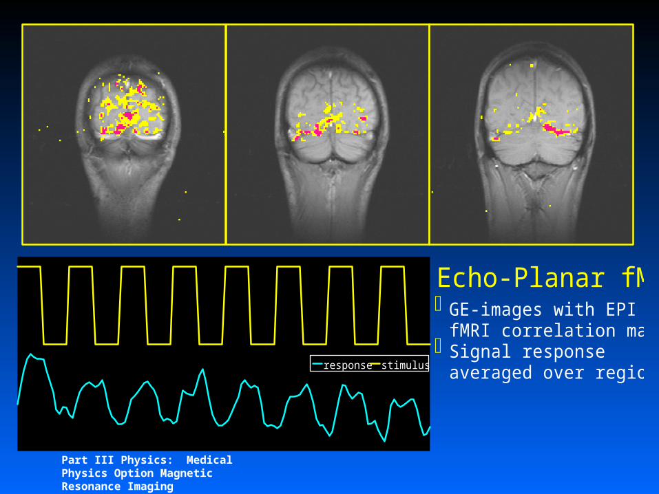
Echo-Planar fMRI
response stimulus
GE-images with EPI fMRI correlation mapsSignal response averaged over region
Part III Physics: Medical Physics Option Magnetic Resonance Imaging
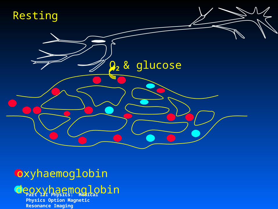
oxyhaemoglobin
deoxyhaemoglobin
Resting
O2 & glucose
Part III Physics: Medical Physics Option Magnetic Resonance Imaging
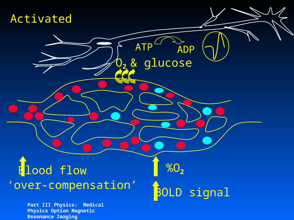
O2 & glucose
Blood flow‘over-compensation’
%O2
Activated
ATP ADP
BOLD signal
Part III Physics: Medical Physics Option Magnetic Resonance Imaging
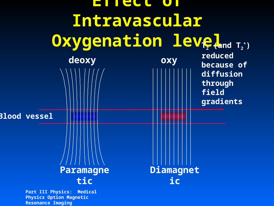
Effect of Intravascular Oxygenation level
Blood vessel
Paramagnetic
T2 (and T2*)
reduced because of diffusion through field gradients
Diamagnetic
deoxy oxy

Part III Physics: Medical Physics Option Magnetic Resonance Imaging
T2* curves activated and rest
time (ms)
signal
activated
rest
TE Signal difference ~ 1-5 %
oxyhaemoglobin
deoxyhaemoglobin
resting activated

Part III Physics: Medical Physics Option Magnetic Resonance Imaging
Unilateral Finger Opposition (high res)

Part III Physics: Medical Physics Option Magnetic Resonance Imaging
Definitions
Diffusion relates to the microscopic Brownian thermal motion of molecules
Perfusion, classically is defined as that process that results in the delivery of nutrients to cells, normally expressed as ml/min/100g wet weight of tissue

Part III Physics: Medical Physics Option Magnetic Resonance Imaging
Effect of Diffusion on NMR
Rms. of an ensemble is zero For a single molecule diffusion results
in a gaussian distribution of displacements
r

Part III Physics: Medical Physics Option Magnetic Resonance Imaging
Diffusion and Spin echoes

Part III Physics: Medical Physics Option Magnetic Resonance Imaging
Diffusion and Spin echoes
I/I0 = e -bD
b = 2g22(-/3)

Part III Physics: Medical Physics Option Magnetic Resonance Imaging
D and ADC
0
2
4
6
8
10
0 500 1000 1500
water
DMSO
I/I0 = e -bD
b = 2g22(-/3)
H2O = 2.1 x 10 -3 mm2s-1
DMSO = 0.55 x 10 -3 mm2s-1
normal = 0.71 x 10 -3 mm2s-1
ischaemic = 0.55 x 10 -3 mm2s-1
b
Log
(I/
I 0)
Part III Physics: Medical Physics Option Magnetic Resonance Imaging
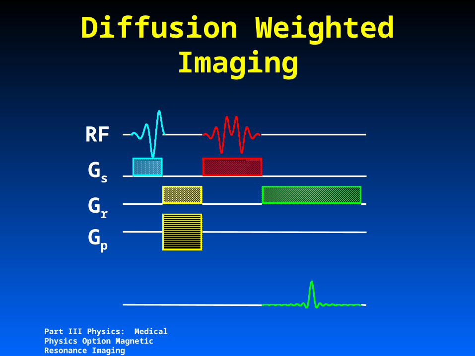
Diffusion Weighted Imaging
RF
GsGrGp

Part III Physics: Medical Physics Option Magnetic Resonance Imaging
Diffusion Weighted Imaging
RF
GsGrGp
Gdiffusion

Part III Physics: Medical Physics Option Magnetic Resonance Imaging
0
2
4
6
8
10
0 500 1000 1500
water
DMSO
Typical Values: = 20, = 50
Gmax b0.5 311 1245 3104
10 12418b
Log
(I/
I 0)
Part III Physics: Medical Physics Option Magnetic Resonance Imaging
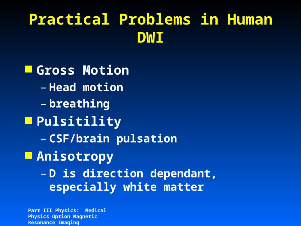
Practical Problems in Human DWI
Gross Motion– Head motion – breathing
Pulsitility– CSF/brain pulsation
Anisotropy– D is direction dependant,
especially white matter

Part III Physics: Medical Physics Option Magnetic Resonance Imaging
Practical Problems in Human DWI
Gross Motion– Echo Planar Imaging – navigator echoes
Pulsitility– gating plus navigator echoes
Anisotropy– Measure trace, Dxx + Dyy + Dzz– Measure full tensor (all matrix
elements)

Part III Physics: Medical Physics Option Magnetic Resonance Imaging
Diffusion Weighted EPI (b=1570 s/mm2)
READ PHASE SLICE
FOV 25cm, TE 118ms TYDW-EPI 128x128 interpolated to 256x256Partial k-acquisition (62.5%)4 interleaves, = 28ms ; = 66 ms

Part III Physics: Medical Physics Option Magnetic Resonance Imaging
Cambridge NIH van Zijl
AD
C t
race
Diffusion Weighted EPI (b=1570 s/mm2)

Part III Physics: Medical Physics Option Magnetic Resonance Imaging
Diffusion Weighted EPI (b=1570 s/mm2)
An
istr
op
y In
dex

Part III Physics: Medical Physics Option Magnetic Resonance Imaging
MRI and O15 water PET

Part III Physics: Medical Physics Option Magnetic Resonance Imaging
Gadolinium blous experiment in rat brain
Image number (relative to blous injection)
-20 -10 0 10 20 30 40 50 60
Rel
axat
ion
rate
cha
nge
(s-1
)
-1
0
1
2
3
4
5
6
Part III Physics: Medical Physics Option Magnetic Resonance Imaging
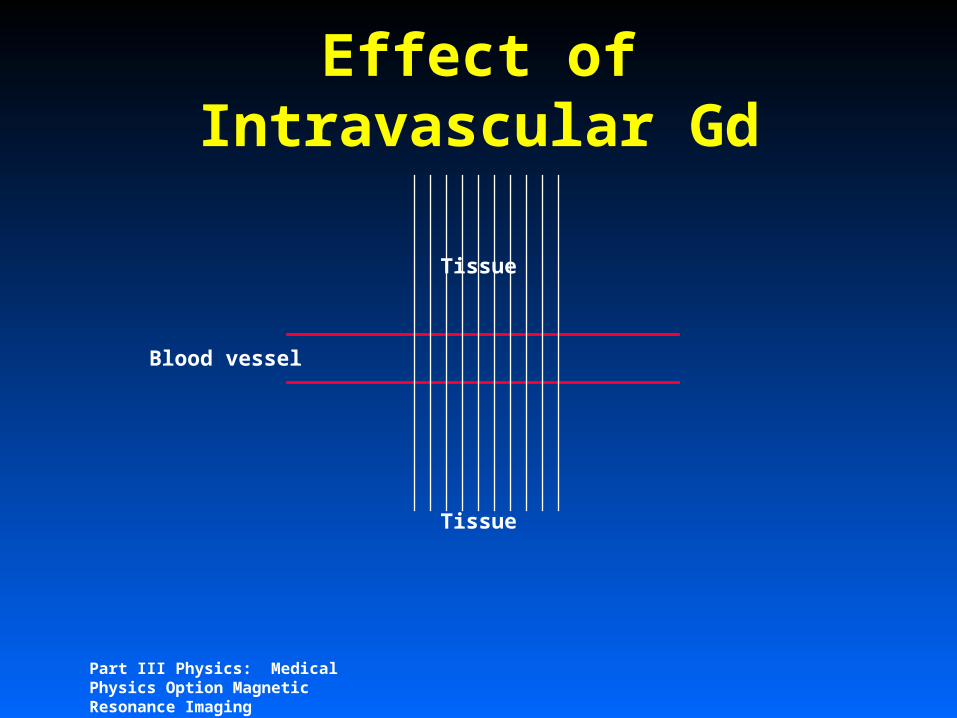
Effect of Intravascular Gd
Blood vessel
Tissue
Tissue

Part III Physics: Medical Physics Option Magnetic Resonance Imaging
Effect of Intravascular Gd
Blood vessel
Tissue
Tissue
T2 (and T2*)
reduced because of difussion through field gradients

Part III Physics: Medical Physics Option Magnetic Resonance Imaging
Gadolinium blous experiment in rat brain
Image number (relative to blous injection)
-20 -10 0 10 20 30 40 50 60
Rel
axat
ion
rate
cha
nge
(s-1
)
-1
0
1
2
3
4
5
6
Part III Physics: Medical Physics Option Magnetic Resonance Imaging
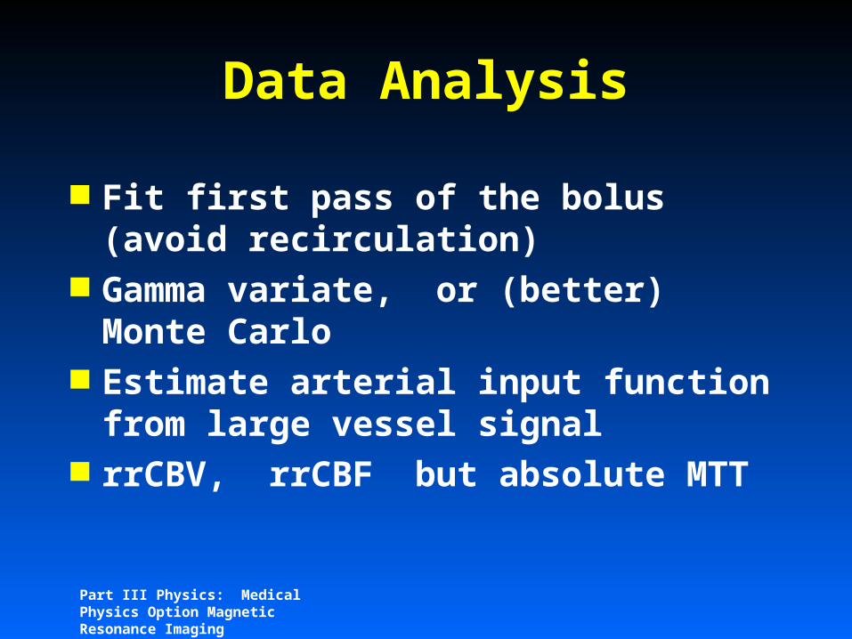
Data Analysis
Fit first pass of the bolus (avoid recirculation)
Gamma variate, or (better) Monte Carlo
Estimate arterial input function from large vessel signal
rrCBV, rrCBF but absolute MTT
Part III Physics: Medical Physics Option Magnetic Resonance Imaging
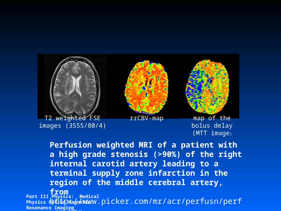
Perfusion weighted MRI of a patient with a high grade stenosis (>90%) of the right internal carotid artery leading to a terminal supply zone infarction in the region of the middle cerebral artery, from http://www.picker.com/mr/acr/perfusn/perfusn.htm
T2 weighted FSE images (3555/80/4)
rrCBV-map map of the bolus delay (MTT image)
Part III Physics: Medical Physics Option Magnetic Resonance Imaging
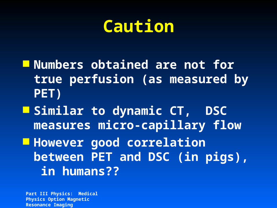
Caution
Numbers obtained are not for true perfusion (as measured by PET)
Similar to dynamic CT, DSC measures micro-capillary flow
However good correlation between PET and DSC (in pigs), in humans??

Part III Physics: Medical Physics Option Magnetic Resonance Imaging
True Perfusion by MRI
Arterial spin labeling– EPISTAR, ASL, QUIPS– label arterial blood on the way
into brain– subtract images with and without
labelling– difference is due to arterial water
that has entered tissue, i.e. perfusion
Part III Physics: Medical Physics Option Magnetic Resonance Imaging
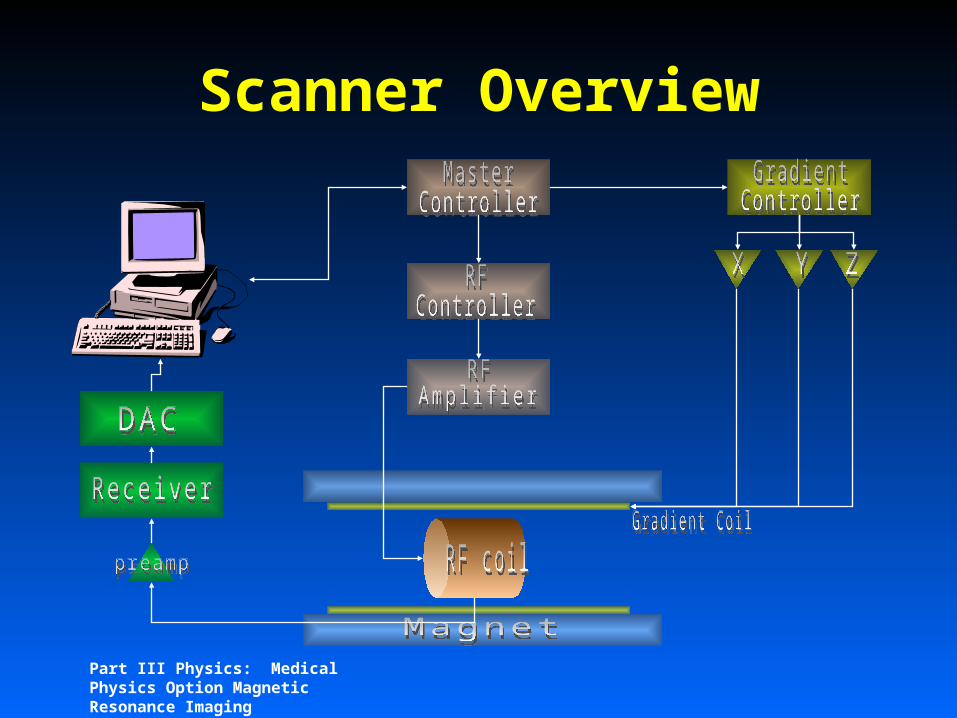
Scanner Overview