Embed Size (px)
Citation preview
EssEntial QuEstionMolecules are lifeless. Yet, the properties of living things derive from the properties of molecules.
Despite the spectacular diversity of life, the elaborate structure of biological mole-cules, and the complexity of vital mechanisms, are life functions ultimately inter-pretable in chemical terms?
the Facts of life: Chemistry is the logic of Biological Phenomena
Part i MolECular CoMPonEnts oF CElls
Molecules are lifeless. Yet, in appropriate complexity and number, molecules compose liv-ing things. These living systems are distinct from the inanimate world because they have certain extraordinary properties. They can grow, move, perform the incredible chemistry of metabolism, respond to stimuli from the environment, and most significantly, replicate themselves with exceptional fidelity. The complex structure and behavior of living organ-isms veil the basic truth that their molecular constitution can be described and understood. The chemistry of the living cell resembles the chemistry of organic reactions. Indeed, cel-lular constituents or biomolecules must conform to the chemical and physical principles that govern all matter. Despite the spectacular diversity of life, the intricacy of biological structures, and the complexity of vital mechanisms, life functions are ultimately interpre-table in chemical terms. Chemistry is the logic of biological phenomena.
1.1 What Are the Distinctive Properties of Living Systems?
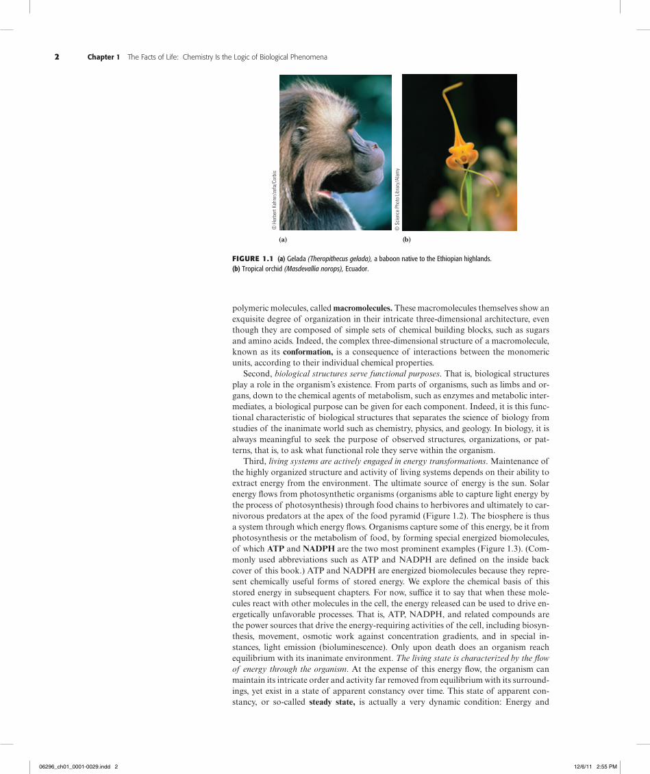
First, the most obvious quality of living organisms is that they are complicated and highly organized (Figure 1.1). For example, organisms large enough to be seen with the naked eye are composed of many cells, typically of many types. In turn, these cells pos-sess subcellular structures, called organelles, which are complex assemblies of very large
KEY QuEstions
1.1 What are the Distinctive Properties of living systems?
1.2 What Kinds of Molecules are Biomolecules?
1.3 What is the structural organization of Complex Biomolecules?
1.4 How Do the Properties of Biomolecules reflect their Fitness to the living Condition?
1.5 What is the organization and structure of Cells?
1.6 What are Viruses?
© D
enni
s W
ilson
/COR
BIS
1
“…everything that living things do can be understood in terms of the jigglings and wigglings of atoms.”
Richard P. FeynmanLectures on Physics, Addison-Wesley, 1963
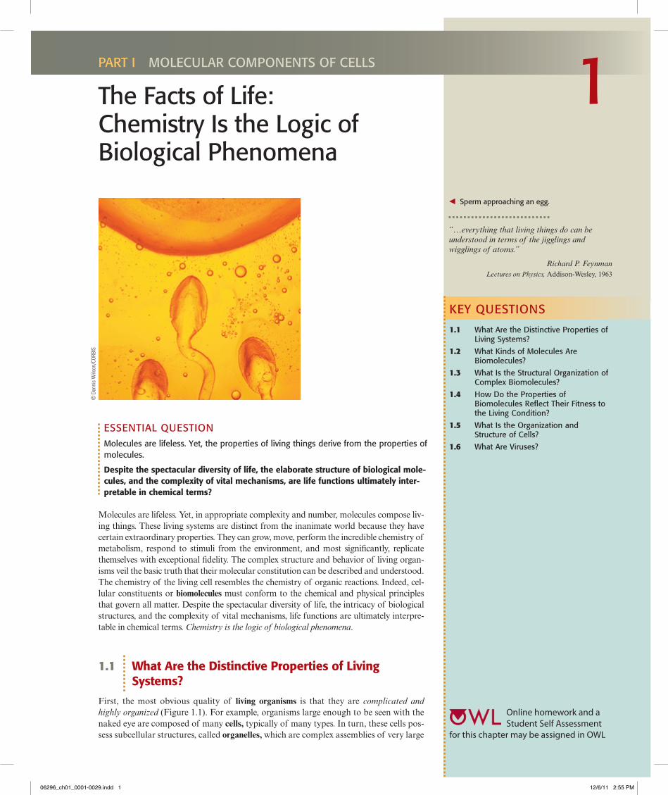
b sperm approaching an egg.
Online homework and a Student Self Assessment
for this chapter may be assigned in OWL
06296_ch01_0001-0029.indd 1 12/6/11 2:55 PM
2 Chapter 1 The Facts of Life: Chemistry Is the Logic of Biological Phenomena
polymeric molecules, called macromolecules. These macromolecules themselves show an exquisite degree of organization in their intricate three-dimensional architecture, even though they are composed of simple sets of chemical building blocks, such as sugars and amino acids. Indeed, the complex three-dimensional structure of a macromolecule, known as its conformation, is a consequence of interactions between the monomeric units, according to their individual chemical properties.
Second, biological structures serve functional purposes. That is, biological structures play a role in the organism’s existence. From parts of organisms, such as limbs and or-gans, down to the chemical agents of metabolism, such as enzymes and metabolic inter-mediates, a biological purpose can be given for each component. Indeed, it is this func-tional characteristic of biological structures that separates the science of biology from studies of the inanimate world such as chemistry, physics, and geology. In biology, it is always meaningful to seek the purpose of observed structures, organizations, or pat-terns, that is, to ask what functional role they serve within the organism.
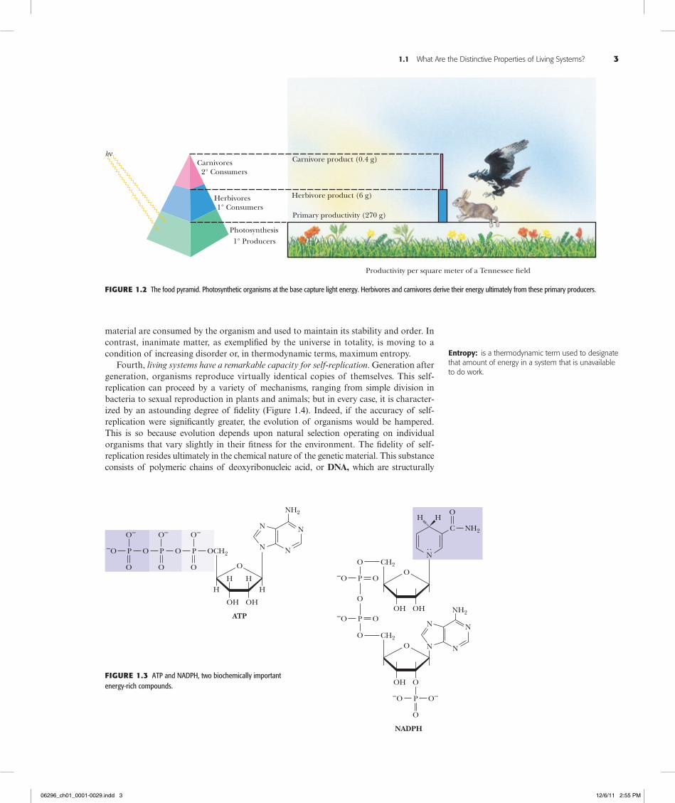
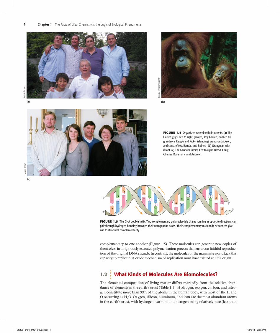
Third, living systems are actively engaged in energy transformations. Maintenance of the highly organized structure and activity of living systems depends on their ability to extract energy from the environment. The ultimate source of energy is the sun. Solar energy flows from photosynthetic organisms (organisms able to capture light energy by the process of photosynthesis) through food chains to herbivores and ultimately to car-nivorous predators at the apex of the food pyramid (Figure 1.2). The biosphere is thus a system through which energy flows. Organisms capture some of this energy, be it from photosynthesis or the metabolism of food, by forming special energized biomolecules, of which ATP and NADPH are the two most prominent examples (Figure 1.3). (Com-monly used abbreviations such as ATP and NADPH are defined on the inside back cover of this book.) ATP and NADPH are energized biomolecules because they repre-sent chemically useful forms of stored energy. We explore the chemical basis of this stored energy in subsequent chapters. For now, suffice it to say that when these mole-cules react with other molecules in the cell, the energy released can be used to drive en-ergetically unfavorable processes. That is, ATP, NADPH, and related compounds are the power sources that drive the energy-requiring activities of the cell, including biosyn-thesis, movement, osmotic work against concentration gradients, and in special in-stances, light emission (bioluminescence). Only upon death does an organism reach equilibrium with its inanimate environment. The living state is characterized by the flow of energy through the organism. At the expense of this energy flow, the organism can maintain its intricate order and activity far removed from equilibrium with its surround-ings, yet exist in a state of apparent constancy over time. This state of apparent con-stancy, or so-called steady state, is actually a very dynamic condition: Energy and
(a)
Figure1.1 (a) Gelada (Theropithecus gelada), a baboon native to the Ethiopian highlands. (b) Tropical orchid (Masdevallia norops), Ecuador.
© H
erbe
rt Ke
hrer
/zef
a/Co
rbis
© S
cien
ce P
hoto
Lib
rary
/Ala
my
(b)
06296_ch01_0001-0029.indd 2 12/6/11 2:55 PM
1.1 What Are the Distinctive Properties of Living Systems? 3
material are consumed by the organism and used to maintain its stability and order. In contrast, inanimate matter, as exemplified by the universe in totality, is moving to a condition of increasing disorder or, in thermodynamic terms, maximum entropy.
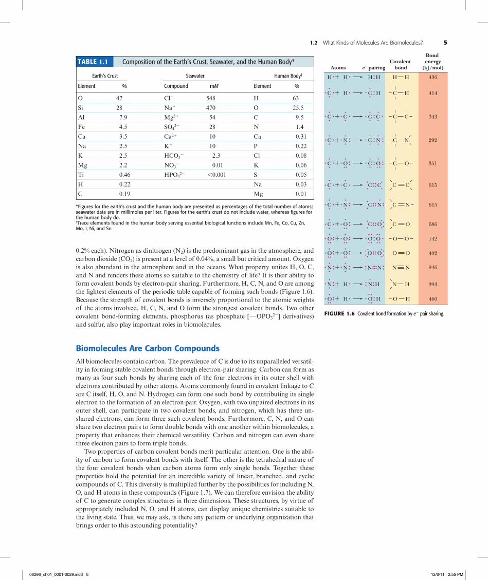
Fourth, living systems have a remarkable capacity for self-replication. Generation after generation, organisms reproduce virtually identical copies of themselves. This self- replication can proceed by a variety of mechanisms, ranging from simple division in bacteria to sexual reproduction in plants and animals; but in every case, it is character-ized by an astounding degree of fidelity (Figure 1.4). Indeed, if the accuracy of self- replication were significantly greater, the evolution of organisms would be hampered. This is so because evolution depends upon natural selection operating on individual organisms that vary slightly in their fitness for the environment. The fidelity of self-replication resides ultimately in the chemical nature of the genetic material. This substance consists of polymeric chains of deoxyribonucleic acid, or DNA, which are structurally
hνCarnivores
2° Consumers
1° Consumers
1° Producers
Carnivore product (0.4 g)
Herbivore product (6 g)
Primary productivity (270 g)
Herbivores
Photosynthesis
Productivity per square meter of a Tennessee �eld
Figure1.2 The food pyramid. Photosynthetic organisms at the base capture light energy. Herbivores and carnivores derive their energy ultimately from these primary producers.
OCH2
O
H H
N
H H
OH OH
O
O
P
O–
–O
N
N
N
NH2
ATP
N
NADPH
O
O
P
O–
O
P
O–
OHOH
OP
O
HH
–O O
O
P–O O
CH2
O
P–O O–
O
CH2
NH2C
O
N
N
NH2
N
N
OOH
O
Figure1.3 ATP and NADPH, two biochemically important energy-rich compounds.
Entropy: is a thermodynamic term used to designate that amount of energy in a system that is unavailable to do work.
06296_ch01_0001-0029.indd 3 12/6/11 2:55 PM
4 Chapter 1 The Facts of Life: Chemistry Is the Logic of Biological Phenomena
complementary to one another (Figure 1.5). These molecules can generate new copies of themselves in a rigorously executed polymerization process that ensures a faithful reproduc-tion of the original DNA strands. In contrast, the molecules of the inanimate world lack this capacity to replicate. A crude mechanism of replication must have existed at life’s origin.
1.2 What Kinds of Molecules Are Biomolecules?
The elemental composition of living matter differs markedly from the relative abun-dance of elements in the earth’s crust (Table 1.1). Hydrogen, oxygen, carbon, and nitro-gen constitute more than 99% of the atoms in the human body, with most of the H and O occurring as H2O. Oxygen, silicon, aluminum, and iron are the most abundant atoms in the earth’s crust, with hydrogen, carbon, and nitrogen being relatively rare (less than
(b)
Kris
tin G
arre
ttTh
omas
Coo
ke
Rand
al H
arris
on G
arre
tt
Figure1.4 Organisms resemble their parents. (a) The Garrett guys. Left to right: (seated) Reg Garrett, flanked by grandsons Reggie and Ricky; (standing) grandson Jackson, and sons Jeffrey, Randal, and Robert. (b) Orangutan with infant. (c) The Grisham family. Left to right: David, Emily, Charles, Rosemary, and Andrew.
AG
C
A
AA
A
A3'
5'
5'
3'
T
TT
T
T TC C CC
CG G
G
G G
Figure1.5 The DNA double helix. Two complementary polynucleotide chains running in opposite directions can pair through hydrogen bonding between their nitrogenous bases. Their complementary nucleotide sequences give rise to structural complementarity.
(a)
(c)
06296_ch01_0001-0029.indd 4 12/6/11 2:55 PM
1.2 What Kinds of Molecules Are Biomolecules? 5
0.2% each). Nitrogen as dinitrogen (N2) is the predominant gas in the atmosphere, and carbon dioxide (CO2) is present at a level of 0.04%, a small but critical amount. Oxygen is also abundant in the atmosphere and in the oceans. What property unites H, O, C, and N and renders these atoms so suitable to the chemistry of life? It is their ability to form covalent bonds by electron-pair sharing. Furthermore, H, C, N, and O are among the lightest elements of the periodic table capable of forming such bonds (Figure 1.6). Because the strength of covalent bonds is inversely proportional to the atomic weights of the atoms involved, H, C, N, and O form the strongest covalent bonds. Two other covalent bond-forming elements, phosphorus (as phosphate [ h OPO3
22] derivatives) and sulfur, also play important roles in biomolecules.
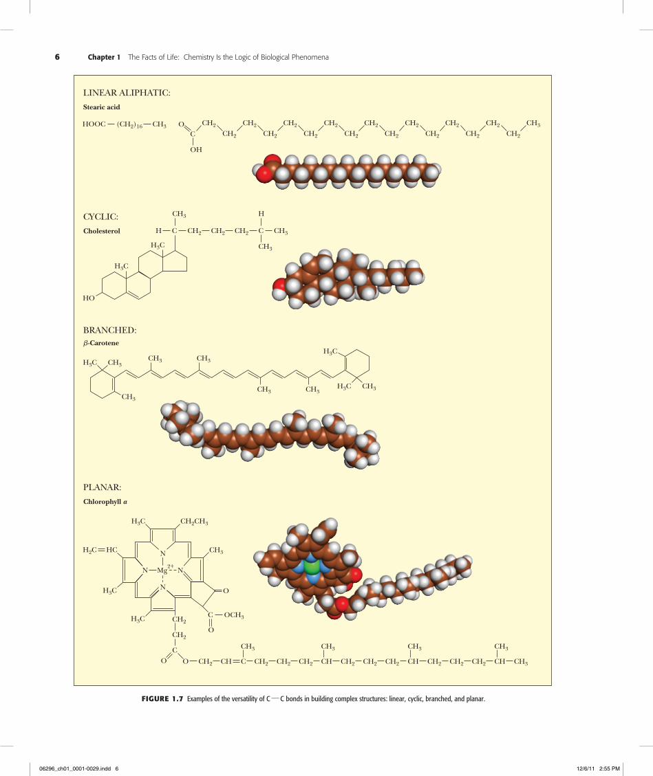
Biomolecules Are Carbon CompoundsAll biomolecules contain carbon. The prevalence of C is due to its unparalleled versatil-ity in forming stable covalent bonds through electron-pair sharing. Carbon can form as many as four such bonds by sharing each of the four electrons in its outer shell with electrons contributed by other atoms. Atoms commonly found in covalent linkage to C are C itself, H, O, and N. Hydrogen can form one such bond by contributing its single electron to the formation of an electron pair. Oxygen, with two unpaired electrons in its outer shell, can participate in two covalent bonds, and nitrogen, which has three un-shared electrons, can form three such covalent bonds. Furthermore, C, N, and O can share two electron pairs to form double bonds with one another within biomolecules, a property that enhances their chemical versatility. Carbon and nitrogen can even share three electron pairs to form triple bonds.
Two properties of carbon covalent bonds merit particular attention. One is the abil-ity of carbon to form covalent bonds with itself. The other is the tetrahedral nature of the four covalent bonds when carbon atoms form only single bonds. Together these properties hold the potential for an incredible variety of linear, branched, and cyclic compounds of C. This diversity is multiplied further by the possibilities for including N, O, and H atoms in these compounds (Figure 1.7). We can therefore envision the ability of C to generate complex structures in three dimensions. These structures, by virtue of appropriately included N, O, and H atoms, can display unique chemistries suitable to the living state. Thus, we may ask, is there any pattern or underlying organization that brings order to this astounding potentiality?
H + H H H
Atoms e– pairingCovalent
bond
Bond energy
(kJ/mol)
C + H C
+ C
+ N N
+ O O O
+ CC C
N N
O O
+ O
O
+ N N
+ N HH
+ O HH
414
343
292
351
615
615
686
142
402
946
393
460
H
H
C C
C
C
C C
+ NC
+ OC
OO
+ OO
NN
N H
HO
C
C
C
C
C
C
C
O
OO
NN
N
O
H
H
C C
C N
C
C
C
OO
O
436
Table 1.1 Composition of the Earth’s Crust, Seawater, and the Human Body*
Earth’s Crust Seawater Human Body†
Element % Compound mM Element %
O 47 Cl2 548 H 63
Si 28 Na1 470 O 25.5
Al 7.9 Mg21 54 C 9.5
Fe 4.5 SO422 28 N 1.4
Ca 3.5 Ca21 10 Ca 0.31
Na 2.5 K1 10 P 0.22
K 2.5 HCO32 2.3 Cl 0.08
Mg 2.2 NO32 0.01 K 0.06
Ti 0.46 HPO422 ,0.001 S 0.05
H 0.22 Na 0.03
C 0.19 Mg 0.01
*Figures for the earth’s crust and the human body are presented as percentages of the total number of atoms; seawater data are in millimoles per liter. Figures for the earth’s crust do not include water, whereas figures for the human body do.†trace elements found in the human body serving essential biological functions include Mn, Fe, Co, Cu, Zn, Mo, i, ni, and se.
Figure1.6 Covalent bond formation by e2 pair sharing.
06296_ch01_0001-0029.indd 5 12/6/11 2:55 PM
6 Chapter 1 The Facts of Life: Chemistry Is the Logic of Biological Phenomena
LINEAR ALIPHATIC:
Stearic acid
HOOC (CH2)16 CH3 O CH2
C CH2
CH2
CH2
CH2
CH2
CH2
CH2
CH2
CH2
CH2
CH2
CH2
CH2
CH2 CH3
CH2
OH
BRANCHED:�-Carotene
H3C CH3
CH3
CH3 CH3
CH3 CH3H3C CH3
H3C
CYCLIC:
Cholesterol H C CH2
CH3
HO
H3C
H3C
CH2 CH2 C CH3
H
CH3
PLANAR:
Chlorophyll a
N
N
N Mg2+
H3C CH2CH3
CH3
O
C OCH3
OCH2
H3C
H3C
HCH2C
CH2
C
O CH2 CH CH2 CH2 CH2C
CH3
CH2 CH2 CH2CH
CH3
CH2 CH2 CH2CH
CH3
CH3CH
CH3
O
N
Figure1.7 Examples of the versatility of C h C bonds in building complex structures: linear, cyclic, branched, and planar.
06296_ch01_0001-0029.indd 6 12/6/11 2:55 PM
1.3 What Is the Structural Organization of Complex Biomolecules? 7
1.3 What Is the Structural Organization of Complex Biomolecules?
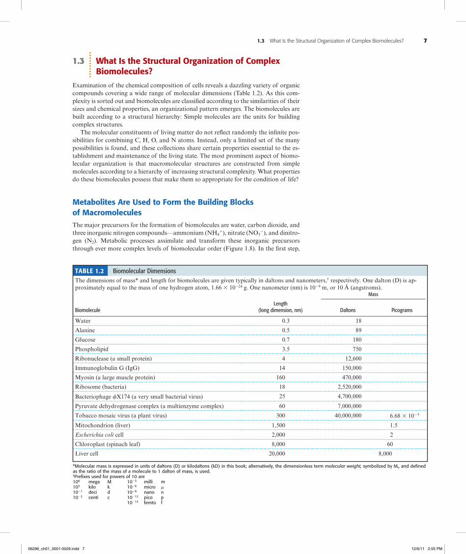
Examination of the chemical composition of cells reveals a dazzling variety of organic compounds covering a wide range of molecular dimensions (Table 1.2). As this com-plexity is sorted out and biomolecules are classified according to the similarities of their sizes and chemical properties, an organizational pattern emerges. The biomolecules are built according to a structural hierarchy: Simple molecules are the units for building complex structures.
The molecular constituents of living matter do not reflect randomly the infinite pos-sibilities for combining C, H, O, and N atoms. Instead, only a limited set of the many possibilities is found, and these collections share certain properties essential to the es-tablishment and maintenance of the living state. The most prominent aspect of biomo-lecular organization is that macromolecular structures are constructed from simple molecules according to a hierarchy of increasing structural complexity. What properties do these biomolecules possess that make them so appropriate for the condition of life?
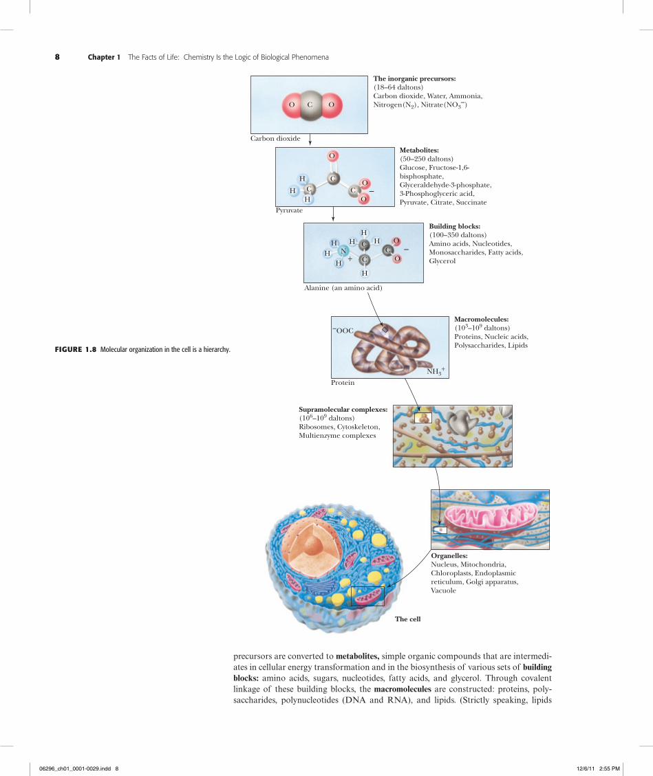
Metabolites Are Used to Form the Building Blocks of Macromolecules The major precursors for the formation of biomolecules are water, carbon dioxide, and three inorganic nitrogen compounds—ammonium (NH4
1), nitrate (NO32), and dinitro-
gen (N2). Metabolic processes assimilate and transform these inorganic precursors through ever more complex levels of biomolecular order (Figure 1.8). In the first step,
Table 1.2 Biomolecular DimensionsThe dimensions of mass* and length for biomolecules are given typically in daltons and nanometers,† respectively. One dalton (D) is ap-proximately equal to the mass of one hydrogen atom, 1.66 3 10224 g. One nanometer (nm) is 1029 m, or 10 Å (angstroms).
Mass
Biomolecule
Length (long dimension, nm)
Daltons
Picograms
Water 0.3 18
Alanine 0.5 89
Glucose 0.7 180
Phospholipid 3.5 750
Ribonuclease (a small protein) 4 12,600
Immunoglobulin G (IgG) 14 150,000
Myosin (a large muscle protein) 160 470,000
Ribosome (bacteria) 18 2,520,000
Bacteriophage fX174 (a very small bacterial virus) 25 4,700,000
Pyruvate dehydrogenase complex (a multienzyme complex) 60 7,000,000
Tobacco mosaic virus (a plant virus) 300 40,000,000 6.68 3 1025
Mitochondrion (liver) 1,500 1.5
Escherichia coli cell 2,000 2
Chloroplast (spinach leaf) 8,000 60
Liver cell 20,000 8,000
*Molecular mass is expressed in units of daltons (D) or kilodaltons (kD) in this book; alternatively, the dimensionless term molecular weight, symbolized by Mr, and defined as the ratio of the mass of a molecule to 1 dalton of mass, is used.†Prefixes used for powers of 10 are106 mega M 1023 milli m103 kilo k 1026 micro m1021 deci d 1029 nano n1022 centi c 10212 pico p 10215 femto f
06296_ch01_0001-0029.indd 7 12/6/11 2:55 PM
8 Chapter 1 The Facts of Life: Chemistry Is the Logic of Biological Phenomena
precursors are converted to metabolites, simple organic compounds that are intermedi-ates in cellular energy transformation and in the biosynthesis of various sets of building blocks: amino acids, sugars, nucleotides, fatty acids, and glycerol. Through covalent linkage of these building blocks, the macromolecules are constructed: proteins, poly-saccharides, polynucleotides (DNA and RNA), and lipids. (Strictly speaking, lipids
The inorganic precursors:(18–64 daltons)Carbon dioxide, Water, Ammonia,Nitrogen(N2), Nitrate(NO3
–)
Carbon dioxide
Pyruvate
Alanine (an amino acid)
Protein
Metabolites:(50–250 daltons)Glucose, Fructose-1,6-bisphosphate,Glyceraldehyde-3-phosphate,3-Phosphoglyceric acid,Pyruvate, Citrate, Succinate
Building blocks:(100–350 daltons)Amino acids, Nucleotides,Monosaccharides, Fatty acids,Glycerol
Macromolecules:(103–109 daltons)Proteins, Nucleic acids,Polysaccharides, Lipids
Supramolecular complexes:(106–109 daltons)Ribosomes, Cytoskeleton,Multienzyme complexes
Organelles:Nucleus, Mitochondria,Chloroplasts, Endoplasmicreticulum, Golgi apparatus,Vacuole
The cell
–OOC
NH3+
H
H
HH
HH
HH
H
H
NC
C C
C
C
CC
O
O
O O
O
O
O
–
–+
Figure1.8 Molecular organization in the cell is a hierarchy.
06296_ch01_0001-0029.indd 8 12/6/11 2:55 PM
1.3 What Is the Structural Organization of Complex Biomolecules? 9
contain relatively few building blocks and are therefore not really polymeric like other macromolecules; however, lipids are important contributors to higher levels of com-plexity.) Interactions among macromolecules lead to the next level of structural organi-zation, supramolecular complexes. Here, various members of one or more of the classes of macromolecules come together to form specific assemblies that serve important sub-cellular functions. Examples of these supramolecular assemblies are multifunctional enzyme complexes, ribosomes, chromosomes, and cytoskeletal elements. For example, a eukaryotic ribosome contains four different RNA molecules and at least 70 unique pro-teins. These supramolecular assemblies are an interesting contrast to their components because their structural integrity is maintained by noncovalent forces, not by covalent bonds. These noncovalent forces include hydrogen bonds, ionic attractions, van der Waals forces, and hydrophobic interactions between macromolecules. Such forces main-tain these supramolecular assemblies in a highly ordered functional state. Although noncovalent forces are weak (less than 40 kJ/mol), they are numerous in these assem-blies and thus can collectively maintain the essential architecture of the supramolecular complex under conditions of temperature, pH, and ionic strength that are consistent with cell life.
Organelles Represent a Higher Order in Biomolecular OrganizationThe next higher rung in the hierarchical ladder is occupied by the organelles, entities of considerable dimensions compared with the cell itself. Organelles are found only in eu-karyotic cells, that is, the cells of “higher” organisms (eukaryotic cells are described in Section 1.5). Several kinds, such as mitochondria and chloroplasts, evolved from bacte-ria that gained entry to the cytoplasm of early eukaryotic cells. Organelles share two attributes: They are cellular inclusions, usually membrane bounded, and they are dedi-cated to important cellular tasks. Organelles include the nucleus, mitochondria, chloro-plasts, endoplasmic reticulum, Golgi apparatus, and vacuoles, as well as other relatively small cellular inclusions, such as peroxisomes, lysosomes, and chromoplasts. The nu-cleus is the repository of genetic information as contained within the linear sequences of nucleotides in the DNA of chromosomes. Mitochondria are the “power plants” of cells by virtue of their ability to carry out the energy-releasing aerobic metabolism of carbohydrates and fatty acids, capturing the energy in metabolically useful forms such as ATP. Chloroplasts endow cells with the ability to carry out photosynthesis. They are the biological agents for harvesting light energy and transforming it into metabolically useful chemical forms.
Membranes Are Supramolecular Assemblies That Define the Boundaries of CellsMembranes define the boundaries of cells and organelles. As such, they are not easily clas-sified as supramolecular assemblies or organelles, although they share the properties of both. Membranes resemble supramolecular complexes in their construction because they are complexes of proteins and lipids maintained by noncovalent forces. Hydrophobic inter-actions are particularly important in maintaining membrane structure. Hydrophobic inter-actions arise because water molecules prefer to interact with each other rather than with nonpolar substances. The presence of nonpolar molecules lessens the range of opportuni-ties for water–water interaction by forcing the water molecules into ordered arrays around the nonpolar groups. Such ordering can be minimized if the individual nonpolar molecules redistribute from a dispersed state in the water into an aggregated organic phase sur-rounded by water. The spontaneous assembly of membranes in the aqueous environment where life arose and exists is the natural result of the hydrophobic (“water-fearing”) char-acter of their lipids and proteins. Hydrophobic interactions are the creative means of mem-brane formation and the driving force that presumably established the boundary of the first cell. The membranes of organelles, such as nuclei, mitochondria, and chloroplasts, differ
06296_ch01_0001-0029.indd 9 12/6/11 2:55 PM
10 Chapter 1 The Facts of Life: Chemistry Is the Logic of Biological Phenomena
from one another, with each having a characteristic protein and lipid composition tailored to the organelle’s function. Furthermore, the creation of discrete volumes or compartments within cells is not only an inevitable consequence of the presence of membranes but usually an essential condition for proper organellar function.
The Unit of Life Is the CellThe cell is characterized as the unit of life, the smallest entity capable of displaying the attributes associated uniquely with the living state: growth, metabolism, stimulus re-sponse, and replication. In the previous discussions, we explicitly narrowed the infinity of chemical complexity potentially available to organic life and we previewed an orga-nizational arrangement, moving from simple to complex, that provides interesting in-sights into the functional and structural plan of the cell. Nevertheless, we find no obvi-ous explanation within these features for the living characteristics of cells. Can we find other themes represented within biomolecules that are explicitly chemical yet anticipate or illuminate the living condition?
1.4 How Do the Properties of Biomolecules Reflect Their Fitness to the Living Condition?
If we consider what attributes of biomolecules render them so fit as components of growing, replicating systems, several biologically relevant themes of structure and orga-nization emerge. Furthermore, as we study biochemistry, we will see that these themes serve as principles of biochemistry. Prominent among them is the necessity for informa-tion and energy in the maintenance of the living state. Some biomolecules must have the capacity to contain the information, or “recipe,” of life. Other biomolecules must have the capacity to translate this information so that the organized structures essential to life are synthesized. Interactions between these structures are the processes of life. An orderly mechanism for abstracting energy from the environment must also exist in order to obtain the energy needed to drive these processes. What properties of biomolecules endow them with the potential for such remarkable qualities?
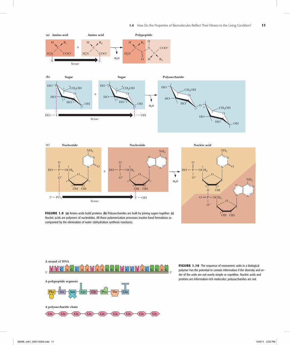
Biological Macromolecules and Their Building Blocks Have a “Sense” or DirectionalityThe macromolecules of cells are built of units—amino acids in proteins, nucleotides in nucleic acids, and carbohydrates in polysaccharides—that have structural polarity. That is, these molecules are not symmetrical, and so they can be thought of as having a “head” and a “tail.” Polymerization of these units to form macromolecules occurs by head-to-tail linear connections. Because of this, the polymer also has a head and a tail, and hence, the macromolecule has a “sense” or direction to its structure (Figure 1.9).
Biological Macromolecules Are InformationalBecause biological macromolecules have a sense to their structure, the sequential order of their component building blocks, when read along the length of the molecule, has the capacity to specify information in the same manner that the letters of the alphabet can form words when arranged in a linear sequence (Figure 1.10). Not all biological macro-molecules are rich in information. Polysaccharides are often composed of the same sugar unit repeated over and over, as in cellulose or starch, which are homopolymers of many glucose units. On the other hand, proteins and polynucleotides are typically com-posed of building blocks arranged in no obvious repetitive way; that is, their sequences are unique, akin to the letters and punctuation that form this descriptive sentence. In these unique sequences lies meaning. Discerning the meaning, however, requires some mechanism for recognition.
06296_ch01_0001-0029.indd 10 12/6/11 2:55 PM
1.4 How Do the Properties of Biomolecules Reflect Their Fitness to the Living Condition? 11
......................
.....
...........
....
Polysaccharide
COO–
+C
H3N
H R1
O
Amino acid Polypeptide
N CSense
HOCH2OH
OH
O
1
23
45
6
Sugar
+
4 1Sense
HO OH
OOCH2P
O–
N
N
NH2
OH
5'
4'
3' 2'
1'
+HO
Nucleotide
P
O–
O OH
O
Nucleic acid
PO4Sense
5' 3'
COO–H3N
Amino acid
CN
H R2
COO–
CH2OH
OH
O
1
23
45
6
Sugar
HOCH2OH
O
O
1 CH2OH
OH
O
4
O
OOCH2P
O–
N
N
NH2
OH
5'
4'
3' 2'
1'
HO
Nucleotide
ON
N
OOCH2P
O–
N O
N
NH2
5'
3' 2'
HO
O
OOCH2
N
N
NH2
OH3'
N
N
C
H R2
H3N
C
C
H R1H
HO
H2O
H2O
H2O
HO HO
HOHO
HO
HO
HO
O
OH OH
OH
(a)
(b)
(c)
OH
HO
+++
5' 3'T C C T A G AG G G G GC C CT T TAA AA G A
A strand of DNA
A polypeptide segment
Phe Ser LysAsn Gly Pro Thr Glu
A polysaccharide chain
Glc Glc Glc Glc Glc Glc Glc Glc Glc
Figure1.9 (a) Amino acids build proteins. (b) Polysaccharides are built by joining sugars together. (c) Nucleic acids are polymers of nucleotides. All these polymerization processes involve bond formations ac-companied by the elimination of water (dehydration synthesis reactions).
Figure1.10 The sequence of monomeric units in a biological polymer has the potential to contain information if the diversity and or-der of the units are not overly simple or repetitive. Nucleic acids and proteins are information-rich molecules; polysaccharides are not.
06296_ch01_0001-0029.indd 11 12/6/11 2:55 PM
12 Chapter 1 The Facts of Life: Chemistry Is the Logic of Biological Phenomena
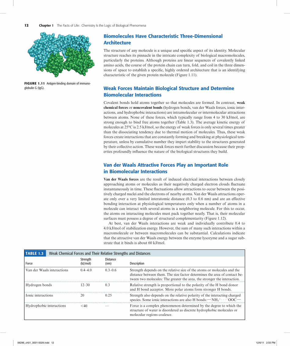
Biomolecules Have Characteristic Three-Dimensional ArchitectureThe structure of any molecule is a unique and specific aspect of its identity. Molecular structure reaches its pinnacle in the intricate complexity of biological macromolecules, particularly the proteins. Although proteins are linear sequences of covalently linked amino acids, the course of the protein chain can turn, fold, and coil in the three dimen-sions of space to establish a specific, highly ordered architecture that is an identifying characteristic of the given protein molecule (Figure 1.11).
Weak Forces Maintain Biological Structure and Determine Biomolecular InteractionsCovalent bonds hold atoms together so that molecules are formed. In contrast, weak chemical forces or noncovalent bonds (hydrogen bonds, van der Waals forces, ionic inter-actions, and hydrophobic interactions) are intramolecular or intermolecular attractions between atoms. None of these forces, which typically range from 4 to 30 kJ/mol, are strong enough to bind free atoms together (Table 1.3). The average kinetic energy of molecules at 25°C is 2.5 kJ/mol, so the energy of weak forces is only several times greater than the dissociating tendency due to thermal motion of molecules. Thus, these weak forces create interactions that are constantly forming and breaking at physiological tem-perature, unless by cumulative number they impart stability to the structures generated by their collective action. These weak forces merit further discussion because their prop-erties profoundly influence the nature of the biological structures they build.
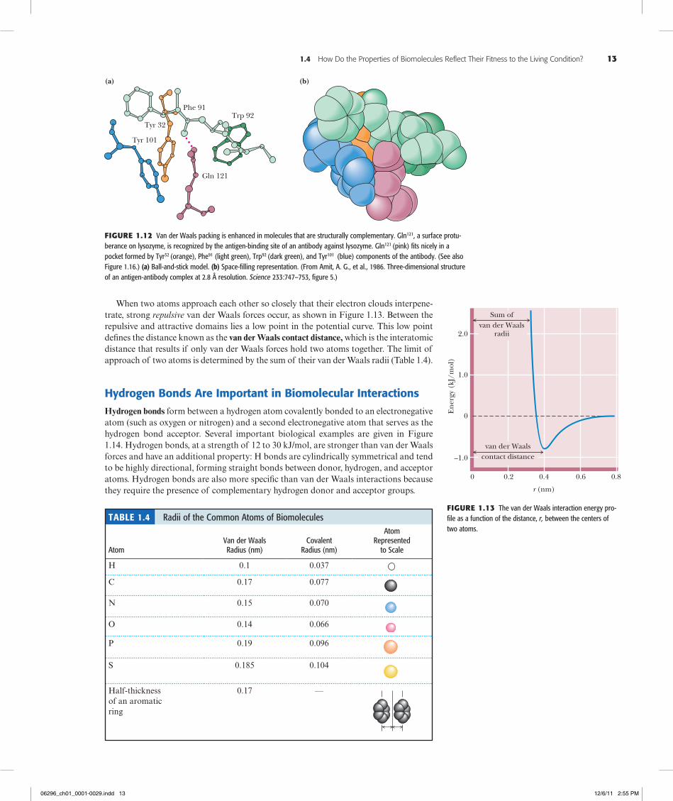
Van der Waals Attractive Forces Play an Important Role in Biomolecular InteractionsVan der Waals forces are the result of induced electrical interactions between closely approaching atoms or molecules as their negatively charged electron clouds fluctuate instantaneously in time. These fluctuations allow attractions to occur between the posi-tively charged nuclei and the electrons of nearby atoms. Van der Waals attractions oper-ate only over a very limited interatomic distance (0.3 to 0.6 nm) and are an effective bonding interaction at physiological temperatures only when a number of atoms in a molecule can interact with several atoms in a neighboring molecule. For this to occur, the atoms on interacting molecules must pack together neatly. That is, their molecular surfaces must possess a degree of structural complementarity (Figure 1.12).
At best, van der Waals interactions are weak and individually contribute 0.4 to 4.0 kJ/mol of stabilization energy. However, the sum of many such interactions within a macromolecule or between macromolecules can be substantial. Calculations indicate that the attractive van der Waals energy between the enzyme lysozyme and a sugar sub-strate that it binds is about 60 kJ/mol.
Figure1.11 Antigen-binding domain of immuno-globulin G (IgG).
Table 1.3 Weak Chemical Forces and Their Relative Strengths and Distances Force
Strength (kJ/mol)
Distance (nm)
Description
Van der Waals interactions 0.4–4.0 0.3–0.6 Strength depends on the relative size of the atoms or molecules and the distance between them. The size factor determines the area of contact be-tween two molecules: The greater the area, the stronger the interaction.
Hydrogen bonds 12–30 0.3 Relative strength is proportional to the polarity of the H bond donor and H bond acceptor. More polar atoms form stronger H bonds.
Ionic interactions 20 0.25 Strength also depends on the relative polarity of the interacting charged species. Some ionic interactions are also H bonds: h NH3
1 . . . 2OOC hHydrophobic interactions ,40 — Force is a complex phenomenon determined by the degree to which the
structure of water is disordered as discrete hydrophobic molecules or molecular regions coalesce.
06296_ch01_0001-0029.indd 12 12/6/11 2:55 PM
1.4 How Do the Properties of Biomolecules Reflect Their Fitness to the Living Condition? 13
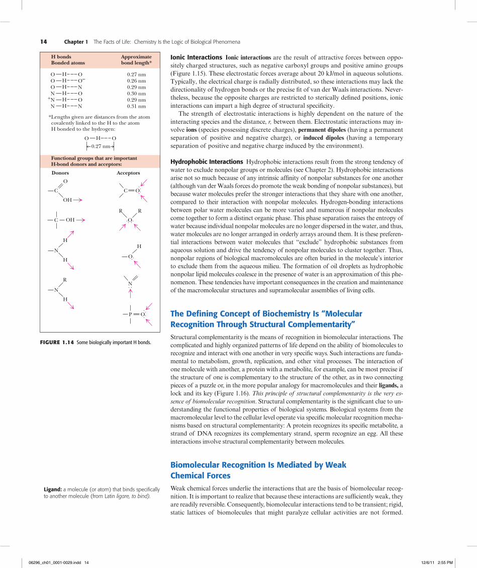
When two atoms approach each other so closely that their electron clouds interpene-trate, strong repulsive van der Waals forces occur, as shown in Figure 1.13. Between the repulsive and attractive domains lies a low point in the potential curve. This low point defines the distance known as the van der Waals contact distance, which is the interatomic distance that results if only van der Waals forces hold two atoms together. The limit of approach of two atoms is determined by the sum of their van der Waals radii (Table 1.4).
Hydrogen Bonds Are Important in Biomolecular InteractionsHydrogen bonds form between a hydrogen atom covalently bonded to an electronegative atom (such as oxygen or nitrogen) and a second electronegative atom that serves as the hydrogen bond acceptor. Several important biological examples are given in Figure 1.14. Hydrogen bonds, at a strength of 12 to 30 kJ/mol, are stronger than van der Waals forces and have an additional property: H bonds are cylindrically symmetrical and tend to be highly directional, forming straight bonds between donor, hydrogen, and acceptor atoms. Hydrogen bonds are also more specific than van der Waals interactions because they require the presence of complementary hydrogen donor and acceptor groups.
...
(b)(a)
Tyr 32
Phe 91Trp 92
Gln 121
Tyr 101
Figure1.12 Van der Waals packing is enhanced in molecules that are structurally complementary. Gln121, a surface protu-berance on lysozyme, is recognized by the antigen-binding site of an antibody against lysozyme. Gln121 (pink) fits nicely in a pocket formed by Tyr32 (orange), Phe91 (light green), Trp92 (dark green), and Tyr101 (blue) components of the antibody. (See also Figure 1.16.) (a) Ball-and-stick model. (b) Space-filling representation. (From Amit, A. G., et al., 1986. Three-dimensional structure of an antigen-antibody complex at 2.8 Å resolution. Science 233:747–753, figure 5.)
0
r (nm)
–1.0E
ner
gy (
kJ/m
ol)
0
1.0
2.0
0.2 0.4 0.6 0.8
van der Waalscontact distance
Sum ofvan der Waals
radii
Figure1.13 The van der Waals interaction energy pro-file as a function of the distance, r, between the centers of two atoms.
Table 1.4 Radii of the Common Atoms of Biomolecules Atom
Van der Waals Radius (nm)
Covalent
Radius (nm)
Atom Represented
to Scale
H 0.1 0.037
C 0.17 0.077
N 0.15 0.070
O 0.14 0.066
P 0.19 0.096
S 0.185 0.104
Half-thickness of an aromatic ring
0.17 —
06296_ch01_0001-0029.indd 13 12/6/11 2:55 PM
14 Chapter 1 The Facts of Life: Chemistry Is the Logic of Biological Phenomena
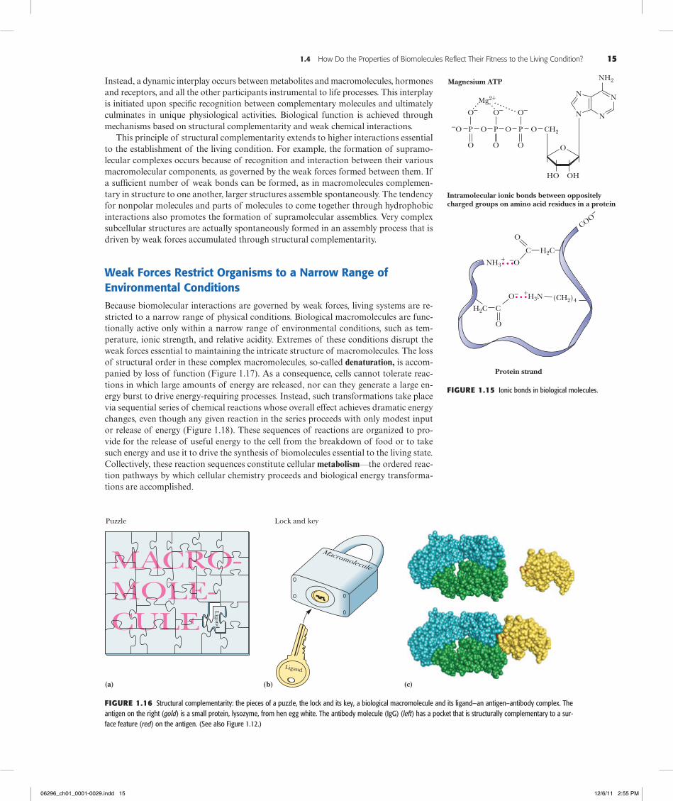
Ionic Interactions Ionic interactions are the result of attractive forces between oppo-sitely charged structures, such as negative carboxyl groups and positive amino groups (Figure 1.15). These electrostatic forces average about 20 kJ/mol in aqueous solutions. Typically, the electrical charge is radially distributed, so these interactions may lack the directionality of hydrogen bonds or the precise fit of van der Waals interactions. Never-theless, because the opposite charges are restricted to sterically defined positions, ionic interactions can impart a high degree of structural specificity.
The strength of electrostatic interactions is highly dependent on the nature of the interacting species and the distance, r, between them. Electrostatic interactions may in-volve ions (species possessing discrete charges), permanent dipoles (having a permanent separation of positive and negative charge), or induced dipoles (having a temporary separation of positive and negative charge induced by the environment).
Hydrophobic Interactions Hydrophobic interactions result from the strong tendency of water to exclude nonpolar groups or molecules (see Chapter 2). Hydrophobic interactions arise not so much because of any intrinsic affinity of nonpolar substances for one another (although van der Waals forces do promote the weak bonding of nonpolar substances), but because water molecules prefer the stronger interactions that they share with one another, compared to their interaction with nonpolar molecules. Hydrogen-bonding interactions between polar water molecules can be more varied and numerous if nonpolar molecules come together to form a distinct organic phase. This phase separation raises the entropy of water because individual nonpolar molecules are no longer dispersed in the water, and thus, water molecules are no longer arranged in orderly arrays around them. It is these preferen-tial interactions between water molecules that “exclude” hydrophobic substances from aqueous solution and drive the tendency of nonpolar molecules to cluster together. Thus, nonpolar regions of biological macromolecules are often buried in the molecule’s interior to exclude them from the aqueous milieu. The formation of oil droplets as hydrophobic nonpolar lipid molecules coalesce in the presence of water is an approximation of this phe-nomenon. These tendencies have important consequences in the creation and maintenance of the macromolecular structures and supramolecular assemblies of living cells.
The Defining Concept of Biochemistry Is “Molecular Recognition Through Structural Complementarity”Structural complementarity is the means of recognition in biomolecular interactions. The complicated and highly organized patterns of life depend on the ability of biomolecules to recognize and interact with one another in very specific ways. Such interactions are funda-mental to metabolism, growth, replication, and other vital processes. The interaction of one molecule with another, a protein with a metabolite, for example, can be most precise if the structure of one is complementary to the structure of the other, as in two connecting pieces of a puzzle or, in the more popular analogy for macromolecules and their ligands, a lock and its key (Figure 1.16). This principle of structural complementarity is the very es-sence of biomolecular recognition. Structural complementarity is the significant clue to un-derstanding the functional properties of biological systems. Biological systems from the macromolecular level to the cellular level operate via specific molecular recognition mecha-nisms based on structural complementarity: A protein recognizes its specific metabolite, a strand of DNA recognizes its complementary strand, sperm recognize an egg. All these interactions involve structural complementarity between molecules.
Biomolecular Recognition Is Mediated by Weak Chemical ForcesWeak chemical forces underlie the interactions that are the basis of biomolecular recog-nition. It is important to realize that because these interactions are sufficiently weak, they are readily reversible. Consequently, biomolecular interactions tend to be transient; rigid, static lattices of biomolecules that might paralyze cellular activities are not formed.
Ligand: a molecule (or atom) that binds specifically to another molecule (from Latin ligare, to bind).
C OH
OO H O–
O H NN H O
+N H ON H N
H bondsBonded atoms
0.27 nm0.26 nm0.29 nm0.30 nm0.29 nm0.31 nm
Approximatebond length*
Lengths given are distances from the atom covalently linked to the H to the atomH bonded to the hydrogen:
O H O
0.27 nm
Functional groups that are importantH-bond donors and acceptors:
C
OH
O
N
H
H
N
H
R
Donors Acceptors
C O
O
R R
O
H
N
P O
O H
*
Figure1.14 Some biologically important H bonds.
06296_ch01_0001-0029.indd 14 12/6/11 2:55 PM
1.4 How Do the Properties of Biomolecules Reflect Their Fitness to the Living Condition? 15
Instead, a dynamic interplay occurs between metabolites and macromolecules, hormones and receptors, and all the other participants instrumental to life processes. This interplay is initiated upon specific recognition between complementary molecules and ultimately culminates in unique physiological activities. Biological function is achieved through mechanisms based on structural complementarity and weak chemical interactions.
This principle of structural complementarity extends to higher interactions essential to the establishment of the living condition. For example, the formation of supramo-lecular complexes occurs because of recognition and interaction between their various macromolecular components, as governed by the weak forces formed between them. If a sufficient number of weak bonds can be formed, as in macromolecules complemen-tary in structure to one another, larger structures assemble spontaneously. The tendency for nonpolar molecules and parts of molecules to come together through hydrophobic interactions also promotes the formation of supramolecular assemblies. Very complex subcellular structures are actually spontaneously formed in an assembly process that is driven by weak forces accumulated through structural complementarity.
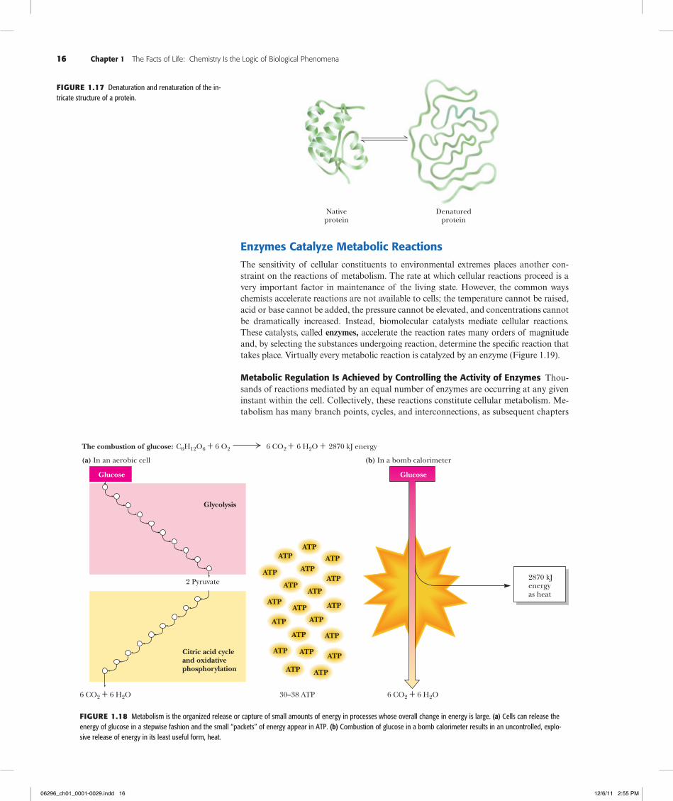
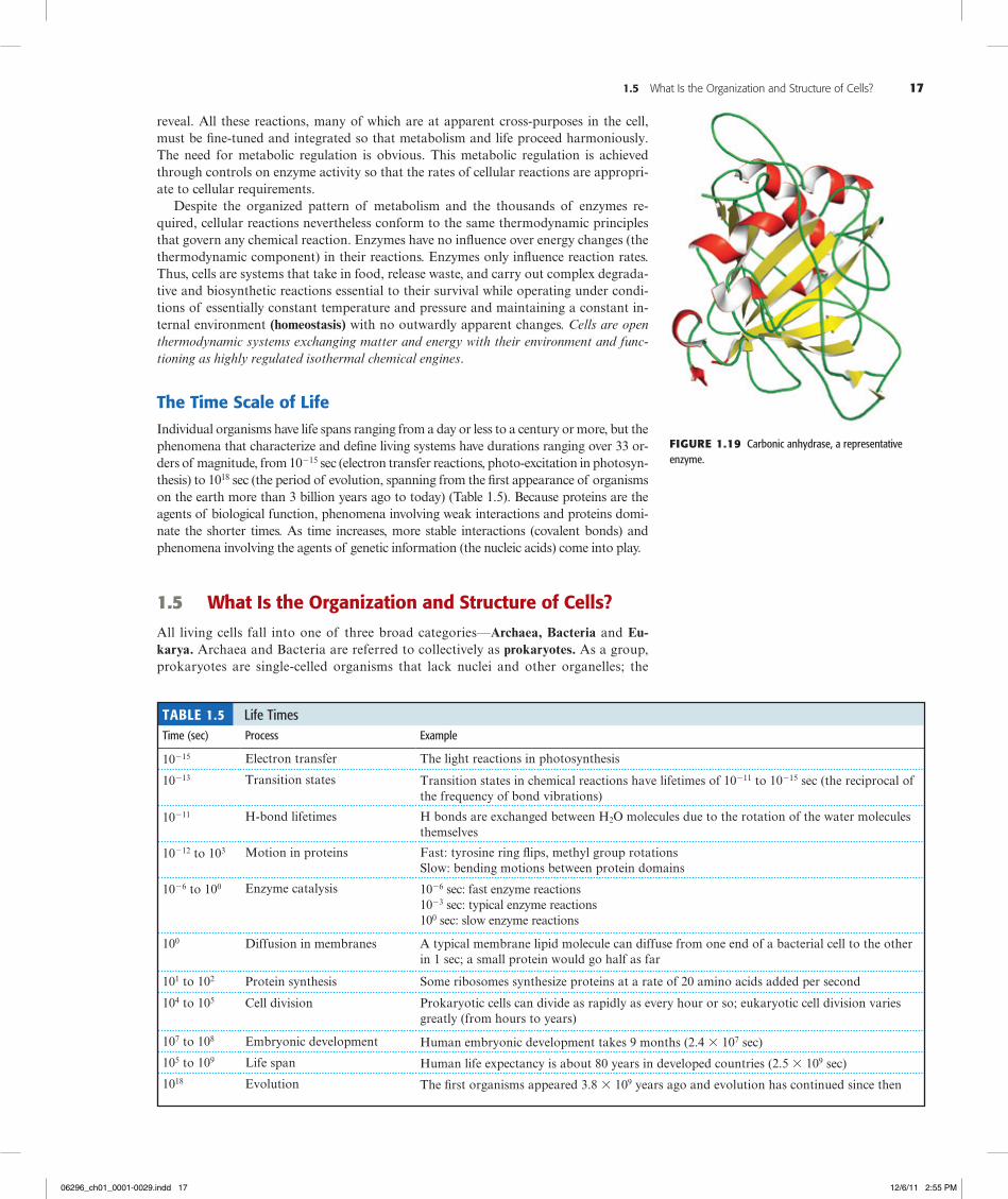
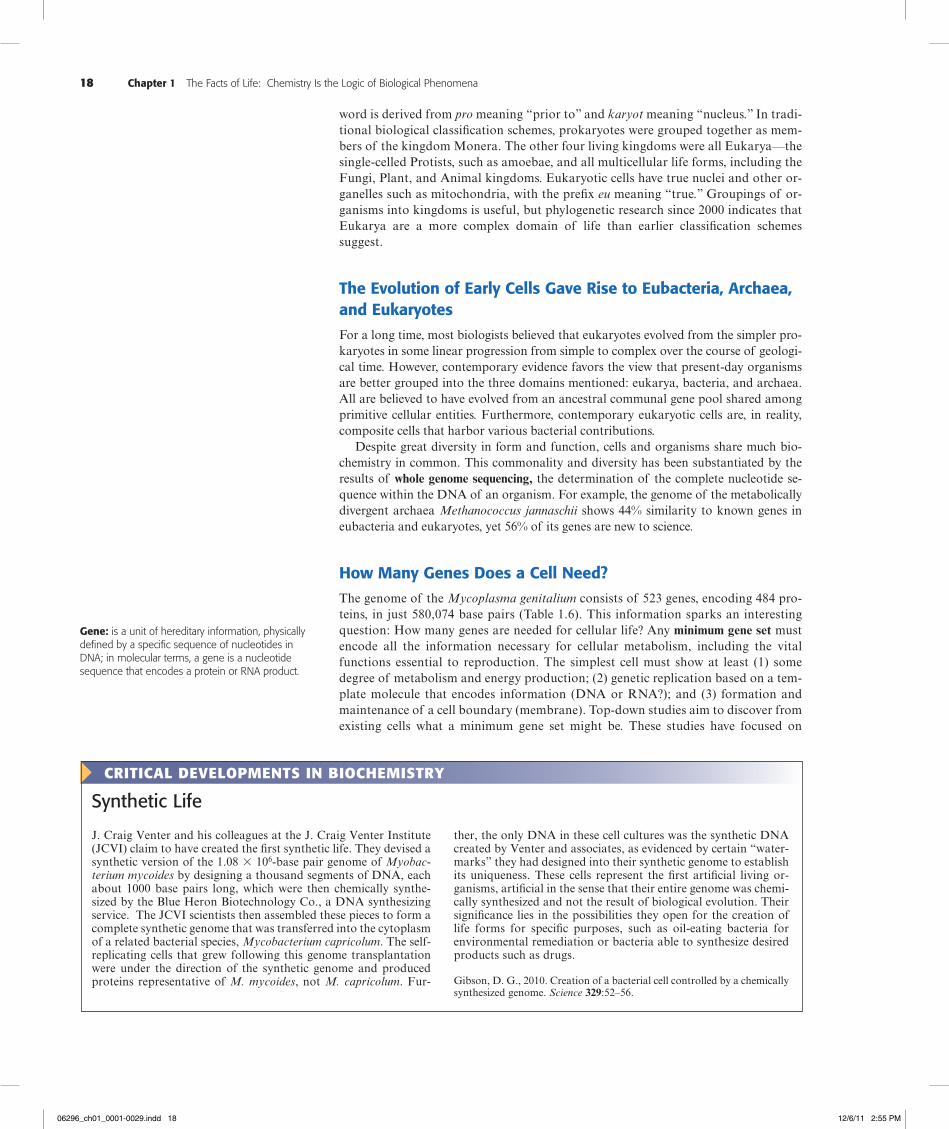
Weak Forces Restrict Organisms to a Narrow Range of Environmental ConditionsBecause biomolecular interactions are governed by weak forces, living systems are re-stricted to a narrow range of physical conditions. Biological macromolecules are func-tionally active only within a narrow range of environmental conditions, such as tem-perature, ionic strength, and relative acidity. Extremes of these conditions disrupt the weak forces essential to maintaining the intricate structure of macromolecules. The loss of structural order in these complex macromolecules, so-called denaturation, is accom-panied by loss of function (Figure 1.17). As a consequence, cells cannot tolerate reac-tions in which large amounts of energy are released, nor can they generate a large en-ergy burst to drive energy-requiring processes. Instead, such transformations take place via sequential series of chemical reactions whose overall effect achieves dramatic energy changes, even though any given reaction in the series proceeds with only modest input or release of energy (Figure 1.18). These sequences of reactions are organized to pro-vide for the release of useful energy to the cell from the breakdown of food or to take such energy and use it to drive the synthesis of biomolecules essential to the living state. Collectively, these reaction sequences constitute cellular metabolism—the ordered reac-tion pathways by which cellular chemistry proceeds and biological energy transforma-tions are accomplished.
...
...
Protein strand
Magnesium ATP
–O P O
Mg2+
O–
O
P O
O–
O
CH2P O
O–
O
..............
O
OHHO
N
N
N
N
NH2
Intramolecular ionic bonds between oppositelycharged groups on amino acid residues in a protein
NH3+
H2CC
O
–O
H2C C
O
O– +H3N (CH2)4
COO–
Figure1.15 Ionic bonds in biological molecules.
Puzzle Lock and key
Ligan
d
Ligand
(a) (b) (c)
Figure1.16 Structural complementarity: the pieces of a puzzle, the lock and its key, a biological macromolecule and its ligand—an antigen–antibody complex. The antigen on the right (gold) is a small protein, lysozyme, from hen egg white. The antibody molecule (IgG) (left) has a pocket that is structurally complementary to a sur-face feature (red) on the antigen. (See also Figure 1.12.)
06296_ch01_0001-0029.indd 15 12/6/11 2:55 PM
16 Chapter 1 The Facts of Life: Chemistry Is the Logic of Biological Phenomena
Enzymes Catalyze Metabolic ReactionsThe sensitivity of cellular constituents to environmental extremes places another con-straint on the reactions of metabolism. The rate at which cellular reactions proceed is a very important factor in maintenance of the living state. However, the common ways chemists accelerate reactions are not available to cells; the temperature cannot be raised, acid or base cannot be added, the pressure cannot be elevated, and concentrations cannot be dramatically increased. Instead, biomolecular catalysts mediate cellular reactions. These catalysts, called enzymes, accelerate the reaction rates many orders of magnitude and, by selecting the substances undergoing reaction, determine the specific reaction that takes place. Virtually every metabolic reaction is catalyzed by an enzyme (Figure 1.19).
Metabolic Regulation Is Achieved by Controlling the Activity of Enzymes Thou-sands of reactions mediated by an equal number of enzymes are occurring at any given instant within the cell. Collectively, these reactions constitute cellular metabolism. Me-tabolism has many branch points, cycles, and interconnections, as subsequent chapters
Nativeprotein
Denaturedprotein
Figure1.17 Denaturation and renaturation of the in-tricate structure of a protein.
The combustion of glucose: C6H12O6 + 6 O2 6 CO2 + 6 H2O + 2870 kJ energy
(a) In an aerobic cell
2 Pyruvate
6 CO2 + 6 H2O
Citric acid cycleand oxidativephosphorylation
Glycolysis
30–38 ATP
(b) In a bomb calorimeter
2870 kJenergyas heat
6 CO2 + 6 H2O
ATPATP
ATP
ATP
ATPATP
ATPATP
ATP
ATPATP
ATP ATP
ATPATP
ATP ATP ATP
ATPATP
Glucose Glucose
Figure1.18 Metabolism is the organized release or capture of small amounts of energy in processes whose overall change in energy is large. (a) Cells can release the energy of glucose in a stepwise fashion and the small “packets” of energy appear in ATP. (b) Combustion of glucose in a bomb calorimeter results in an uncontrolled, explo-sive release of energy in its least useful form, heat.
06296_ch01_0001-0029.indd 16 12/6/11 2:55 PM
1.5 What Is the Organization and Structure of Cells? 17
reveal. All these reactions, many of which are at apparent cross-purposes in the cell, must be fine-tuned and integrated so that metabolism and life proceed harmoniously. The need for metabolic regulation is obvious. This metabolic regulation is achieved through controls on enzyme activity so that the rates of cellular reactions are appropri-ate to cellular requirements.
Despite the organized pattern of metabolism and the thousands of enzymes re-quired, cellular reactions nevertheless conform to the same thermodynamic principles that govern any chemical reaction. Enzymes have no influence over energy changes (the thermodynamic component) in their reactions. Enzymes only influence reaction rates. Thus, cells are systems that take in food, release waste, and carry out complex degrada-tive and biosynthetic reactions essential to their survival while operating under condi-tions of essentially constant temperature and pressure and maintaining a constant in-ternal environment (homeostasis) with no outwardly apparent changes. Cells are open thermodynamic systems exchanging matter and energy with their environment and func-tioning as highly regulated isothermal chemical engines.
The Time Scale of LifeIndividual organisms have life spans ranging from a day or less to a century or more, but the phenomena that characterize and define living systems have durations ranging over 33 or-ders of magnitude, from 10215 sec (electron transfer reactions, photo-excitation in photosyn-thesis) to 1018 sec (the period of evolution, spanning from the first appearance of organisms on the earth more than 3 billion years ago to today) (Table 1.5). Because proteins are the agents of biological function, phenomena involving weak interactions and proteins domi-nate the shorter times. As time increases, more stable interactions (covalent bonds) and phenomena involving the agents of genetic information (the nucleic acids) come into play.
1.5 What Is the Organization and Structure of Cells?
All living cells fall into one of three broad categories—Archaea, Bacteria and Eu-karya. Archaea and Bacteria are referred to collectively as prokaryotes. As a group, prokaryotes are single-celled organisms that lack nuclei and other organelles; the
Figure1.19 Carbonic anhydrase, a representative enzyme.
Table 1.5 Life TimesTime (sec) Process Example
10215 Electron transfer The light reactions in photosynthesis
10213 Transition states Transition states in chemical reactions have lifetimes of 10211 to 10215 sec (the reciprocal of the frequency of bond vibrations)
10211 H-bond lifetimes H bonds are exchanged between H2O molecules due to the rotation of the water molecules themselves
10212 to 103 Motion in proteins Fast: tyrosine ring flips, methyl group rotationsSlow: bending motions between protein domains
1026 to 100 Enzyme catalysis 1026 sec: fast enzyme reactions1023 sec: typical enzyme reactions100 sec: slow enzyme reactions
100 Diffusion in membranes A typical membrane lipid molecule can diffuse from one end of a bacterial cell to the other in 1 sec; a small protein would go half as far
101 to 102 Protein synthesis Some ribosomes synthesize proteins at a rate of 20 amino acids added per second
104 to 105 Cell division Prokaryotic cells can divide as rapidly as every hour or so; eukaryotic cell division varies greatly (from hours to years)
107 to 108 Embryonic development Human embryonic development takes 9 months (2.4 3 107 sec)
105 to 109 Life span Human life expectancy is about 80 years in developed countries (2.5 3 109 sec)
1018 Evolution The first organisms appeared 3.8 3 109 years ago and evolution has continued since then
06296_ch01_0001-0029.indd 17 12/6/11 2:55 PM
18 Chapter 1 The Facts of Life: Chemistry Is the Logic of Biological Phenomena
word is derived from pro meaning “prior to” and karyot meaning “nucleus.” In tradi-tional biological classification schemes, prokaryotes were grouped together as mem-bers of the kingdom Monera. The other four living kingdoms were all Eukarya—the single-celled Protists, such as amoebae, and all multicellular life forms, including the Fungi, Plant, and Animal kingdoms. Eukaryotic cells have true nuclei and other or-ganelles such as mitochondria, with the prefix eu meaning “true.” Groupings of or-ganisms into kingdoms is useful, but phylogenetic research since 2000 indicates that Eukarya are a more complex domain of life than earlier classification schemes suggest.
The Evolution of Early Cells Gave Rise to Eubacteria, Archaea, and EukaryotesFor a long time, most biologists believed that eukaryotes evolved from the simpler pro-karyotes in some linear progression from simple to complex over the course of geologi-cal time. However, contemporary evidence favors the view that present-day organisms are better grouped into the three domains mentioned: eukarya, bacteria, and archaea. All are believed to have evolved from an ancestral communal gene pool shared among primitive cellular entities. Furthermore, contemporary eukaryotic cells are, in reality, composite cells that harbor various bacterial contributions.
Despite great diversity in form and function, cells and organisms share much bio-chemistry in common. This commonality and diversity has been substantiated by the results of whole genome sequencing, the determination of the complete nucleotide se-quence within the DNA of an organism. For example, the genome of the metabolically divergent archaea Methanococcus jannaschii shows 44% similarity to known genes in eubacteria and eukaryotes, yet 56% of its genes are new to science.
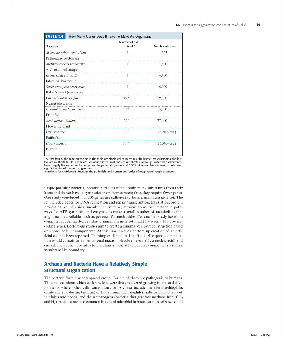
How Many Genes Does a Cell Need?The genome of the Mycoplasma genitalium consists of 523 genes, encoding 484 pro-teins, in just 580,074 base pairs (Table 1.6). This information sparks an interesting question: How many genes are needed for cellular life? Any minimum gene set must encode all the information necessary for cellular metabolism, including the vital functions essential to reproduction. The simplest cell must show at least (1) some degree of metabolism and energy production; (2) genetic replication based on a tem-plate molecule that encodes information (DNA or RNA?); and (3) formation and maintenance of a cell boundary (membrane). Top-down studies aim to discover from existing cells what a minimum gene set might be. These studies have focused on
Gene: is a unit of hereditary information, physically defined by a specific sequence of nucleotides in DNA; in molecular terms, a gene is a nucleotide sequence that encodes a protein or RNA product.
ther, the only DNA in these cell cultures was the synthetic DNA created by Venter and associates, as evidenced by certain “water-marks” they had designed into their synthetic genome to establish its uniqueness. These cells represent the first artificial living or-ganisms, artificial in the sense that their entire genome was chemi-cally synthesized and not the result of biological evolution. Their significance lies in the possibilities they open for the creation of life forms for specific purposes, such as oil-eating bacteria for environmental remediation or bacteria able to synthesize desired products such as drugs.
Gibson, D. G., 2010. Creation of a bacterial cell controlled by a chemically synthesized genome. Science 329:52–56.
J. Craig Venter and his colleagues at the J. Craig Venter Institute (JCVI) claim to have created the first synthetic life. They devised a synthetic version of the 1.08 3 106-base pair genome of Myobac-terium mycoides by designing a thousand segments of DNA, each about 1000 base pairs long, which were then chemically synthe-sized by the Blue Heron Biotechnology Co., a DNA synthesizing service. The JCVI scientists then assembled these pieces to form a complete synthetic genome that was transferred into the cytoplasm of a related bacterial species, Mycobacterium capricolum. The self-replicating cells that grew following this genome transplantation were under the direction of the synthetic genome and produced proteins representative of M. mycoides, not M. capricolum. Fur-
CriTiCALDeVeLOPMeNTSiNBiOCHeMiSTrY
synthetic life
06296_ch01_0001-0029.indd 18 12/6/11 2:55 PM
1.5 What Is the Organization and Structure of Cells? 19
simple parasitic bacteria, because parasites often obtain many substances from their hosts and do not have to synthesize them from scratch; thus, they require fewer genes. One study concluded that 206 genes are sufficient to form a minimum gene set. The set included genes for DNA replication and repair, transcription, translation, protein processing, cell division, membrane structure, nutrient transport, metabolic path-ways for ATP synthesis, and enzymes to make a small number of metabolites that might not be available, such as pentoses for nucleotides. Yet another study based on computer modeling decided that a minimum gene set might have only 105 protein-coding genes. Bottom-up studies aim to create a minimal cell by reconstruction based on known cellular components. At this time, no such bottom-up creation of an arti-ficial cell has been reported. The simplest functional artificial cell capable of replica-tion would contain an informational macromolecule (presumably a nucleic acid) and enough metabolic apparatus to maintain a basic set of cellular components within a membranelike boundary.
Archaea and Bacteria Have a Relatively Simple Structural OrganizationThe bacteria form a widely spread group. Certain of them are pathogenic to humans. The archaea, about which we know less, were first discovered growing in unusual envi-ronments where other cells cannot survive. Archaea include the thermoacidophiles (heat- and acid-loving bacteria) of hot springs, the halophiles (salt-loving bacteria) of salt lakes and ponds, and the methanogens (bacteria that generate methane from CO2 and H2). Archaea are also common in typical microbial habitats, such as soils, seas, and
the first four of the nine organisms in the table are single-celled microbes; the last six are eukaryotes; the last five are multicellular, four of which are animals; the final two are vertebrates. although pufferfish and humans have roughly the same number of genes, the pufferfish genome, at 0.365 billion nucleotide pairs, is only one-eighth the size of the human genome.*numbers for Arabidopsis thaliana, the pufferfish, and human are “order-of-magnitude” rough estimates.
Table 1.6 How Many Genes Does It Take To Make An Organism? Organism
Number of Cells in Adult*
Number of Genes
Mycobacterium genitalium
Pathogenic bacterium
1 523
Methanococcus jannaschii
Archaeal methanogen
1 1,800
Escherichia coli K12
Intestinal bacterium
1 4,400
Saccharomyces cereviseae
Baker’s yeast (eukaryote)
1 6,000
Caenorhabditis elegans
Nematode worm
959 19,000
Drosophila melanogaster
Fruit fly
104 13,500
Arabidopsis thaliana
Flowering plant
107 27,000
Fugu rubripes
Pufferfish
1012 26,700 (est.)
Homo sapiens
Human
1014 20,500 (est.)
06296_ch01_0001-0029.indd 19 12/6/11 2:55 PM
20 Chapter 1 The Facts of Life: Chemistry Is the Logic of Biological Phenomena
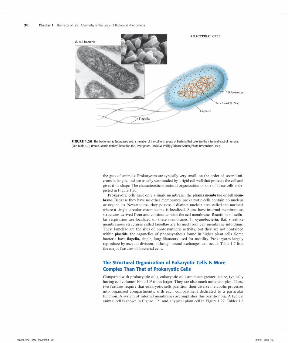
the guts of animals. Prokaryotes are typically very small, on the order of several mi-crons in length, and are usually surrounded by a rigid cell wall that protects the cell and gives it its shape. The characteristic structural organization of one of these cells is de-picted in Figure 1.20.
Prokaryotic cells have only a single membrane, the plasma membrane or cell mem-brane. Because they have no other membranes, prokaryotic cells contain no nucleus or organelles. Nevertheless, they possess a distinct nuclear area called the nucleoid where a single circular chromosome is localized. Some have internal membranous structures derived from and continuous with the cell membrane. Reactions of cellu-lar respiration are localized on these membranes. In cyanobacteria, flat, sheetlike membranous structures called lamellae are formed from cell membrane infoldings. These lamellae are the sites of photosynthetic activity, but they are not contained within plastids, the organelles of photosynthesis found in higher plant cells. Some bacteria have flagella, single, long filaments used for motility. Prokaryotes largely reproduce by asexual division, although sexual exchanges can occur. Table 1.7 lists the major features of bacterial cells.
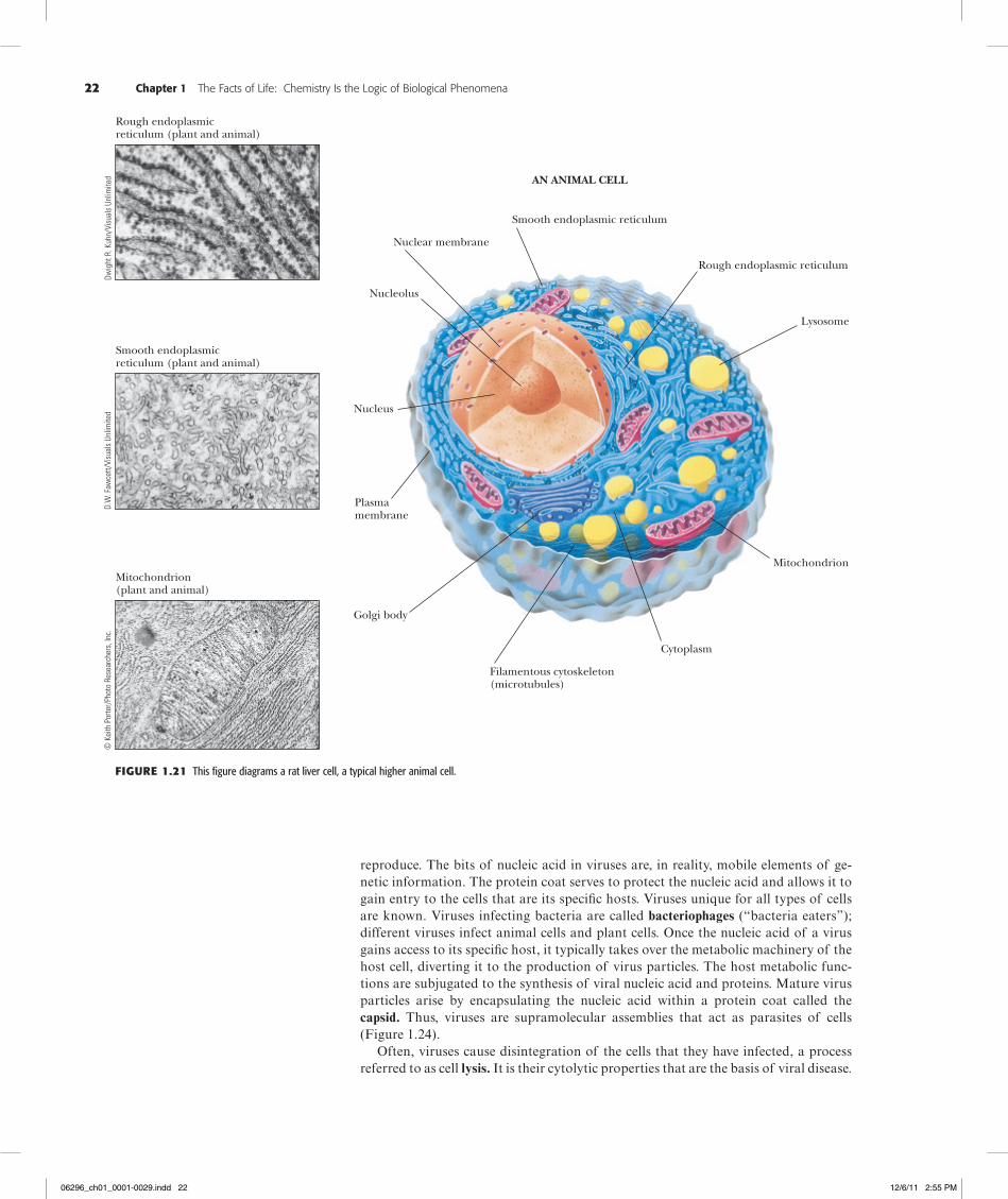
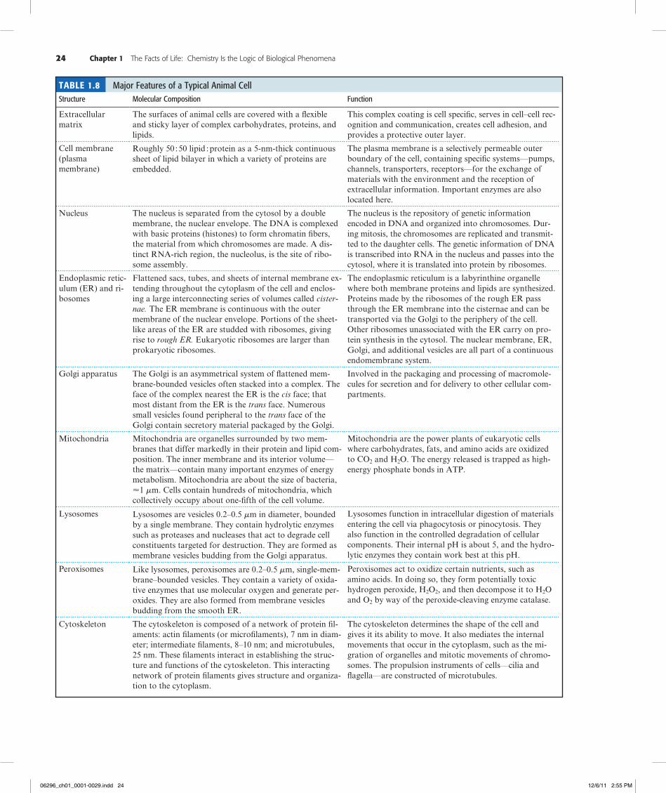
The Structural Organization of Eukaryotic Cells Is More Complex Than That of Prokaryotic CellsCompared with prokaryotic cells, eukaryotic cells are much greater in size, typically having cell volumes 103 to 104 times larger. They are also much more complex. These two features require that eukaryotic cells partition their diverse metabolic processes into organized compartments, with each compartment dedicated to a particular function. A system of internal membranes accomplishes this partitioning. A typical animal cell is shown in Figure 1.21 and a typical plant cell in Figure 1.22. Tables 1.8
Flagella
Capsule
Nucleoid (DNA)
Ribosomes
E. coli bacteria
A BACTERIAL CELL
Figure1.20 This bacterium is Escherichia coli, a member of the coliform group of bacteria that colonize the intestinal tract of humans. (See Table 1.7.) (Photo, Martin Rotker/Phototake, Inc.; inset photo, David M. Phillips/Science Source/Photo Researchers, Inc.)
06296_ch01_0001-0029.indd 20 12/6/11 2:55 PM
1.6 What Are Viruses? 21
and 1.9 list the major features of a typical animal cell and a higher plant cell, respectively.
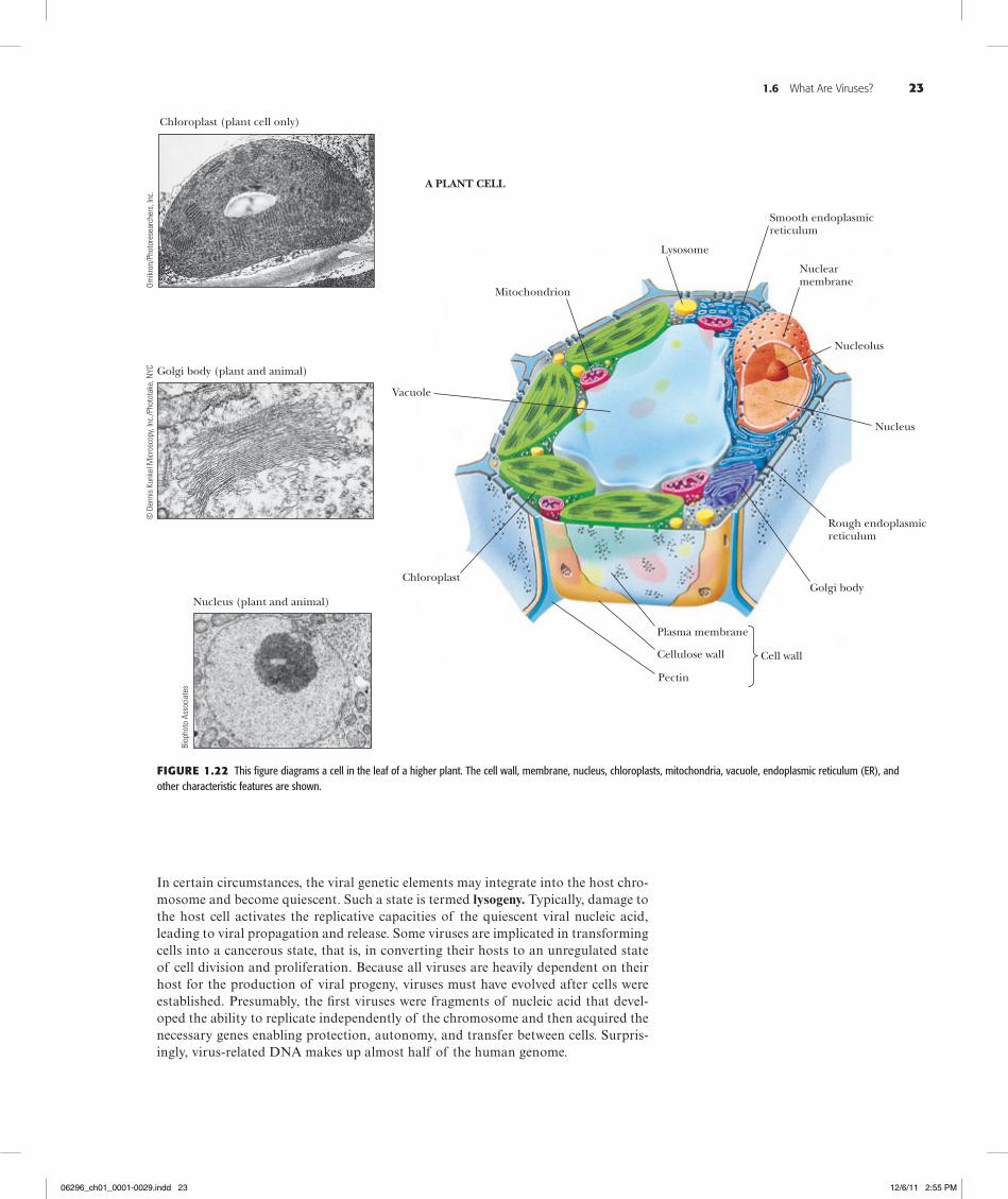
Eukaryotic cells possess a discrete, membrane-bounded nucleus, the repository of the cell’s genetic material, which is distributed among a few or many chromosomes. During cell division, equivalent copies of this genetic material must be passed to both daughter cells through duplication and orderly partitioning of the chromosomes by the process known as mitosis. Like prokaryotic cells, eukaryotic cells are surrounded by a plasma membrane. Unlike prokaryotic cells, eukaryotic cells are rich in internal membranes that are differentiated into specialized structures such as the endoplasmic reticulum (ER) and the Golgi apparatus. Membranes also surround certain organelles (mitochondria and chloroplasts, for example) and various vesicles, including vacuoles, lysosomes, and peroxisomes. The common purpose of these membranous partition-ings is the creation of cellular compartments that have specific, organized metabolic functions, such as the mitochondrion’s role as the principal site of cellular energy production. Eukaryotic cells also have a cytoskeleton composed of arrays of filaments that give the cell its shape and its capacity to move. Some eukaryotic cells also have long projections on their surface—cilia or flagella—which provide propulsion.
1.6 What Are Viruses?
Viruses are supramolecular complexes of nucleic acid, either DNA or RNA, encap-sulated in a protein coat and, in some instances, surrounded by a membrane enve-lope (Figure 1.23). Viruses are acellular, but they act as cellular parasites in order to
Table 1.7 Major Features of Prokaryotic CellsStructure Molecular Composition Function
Cell wall Peptidoglycan: a rigid framework of polysaccha-ride crosslinked by short peptide chains. Some bac-teria possess a lipopolysaccharide- and protein-rich outer membrane.
Mechanical support, shape, and protection against swell-ing in hypotonic media. The cell wall is a porous nonse-lective barrier that allows most small molecules to pass.
Cell membrane The cell membrane is composed of about 45% lipid and 55% protein. The lipids form a bilayer that is a continuous nonpolar hydrophobic phase in which the proteins are embedded.
The cell membrane is a highly selective permeability bar-rier that controls the entry of most substances into the cell. Important enzymes in the generation of cellular en-ergy are located in the membrane.
Nuclear area or nucleoid The genetic material is a single, tightly coiled DNA molecule 2 nm in diameter but more than 1 mm in length (molecular mass of E. coli DNA is 33109 daltons; 4.643106 nucleotide pairs).
DNA provides the operating instructions for the cell; it is the repository of the cell’s genetic information. During cell division, each strand of the double-stranded DNA molecule is replicated to yield two double-helical daugh-ter molecules. Messenger RNA (mRNA) is transcribed from DNA to direct the synthesis of cellular proteins.
Ribosomes Bacterial cells contain about 15,000 ribosomes. Each is composed of a small (30S) subunit and a large (50S) subunit. The mass of a single ribosome is 2.33106 daltons. It consists of 65% RNA and 35% protein.
Ribosomes are the sites of protein synthesis. The mRNA binds to ribosomes, and the mRNA nucleotide sequence specifies the protein that is synthesized.
Storage granules Bacteria contain granules that represent storage forms of polymerized metabolites such as sugars or b-hydroxybutyric acid.
When needed as metabolic fuel, the monomeric units of the polymer are liberated and degraded by energy-yield-ing pathways in the cell.
Cytosol Despite its amorphous appearance, the cytosol is an organized gelatinous compartment that is 20% protein by weight and rich in the organic molecules that are the intermediates in metabolism.
The cytosol is the site of intermediary metabolism, the interconnecting sets of chemical reactions by which cells generate energy and form the precursors necessary for biosynthesis of macromolecules essential to cell growth and function.
06296_ch01_0001-0029.indd 21 12/6/11 2:55 PM
22 Chapter 1 The Facts of Life: Chemistry Is the Logic of Biological Phenomena
reproduce. The bits of nucleic acid in viruses are, in reality, mobile elements of ge-netic information. The protein coat serves to protect the nucleic acid and allows it to gain entry to the cells that are its specific hosts. Viruses unique for all types of cells are known. Viruses infecting bacteria are called bacteriophages (“bacteria eaters”); different viruses infect animal cells and plant cells. Once the nucleic acid of a virus gains access to its specific host, it typically takes over the metabolic machinery of the host cell, diverting it to the production of virus particles. The host metabolic func-tions are subjugated to the synthesis of viral nucleic acid and proteins. Mature virus particles arise by encapsulating the nucleic acid within a protein coat called the capsid. Thus, viruses are supramolecular assemblies that act as parasites of cells (Figure 1.24).
Often, viruses cause disintegration of the cells that they have infected, a process referred to as cell lysis. It is their cytolytic properties that are the basis of viral disease.
Rough endoplasmicreticulum (plant and animal)
Smooth endoplasmicreticulum (plant and animal)
Mitochondrion(plant and animal)
Smooth endoplasmic reticulum
Nuclear membrane
Nucleolus
Nucleus
Plasmamembrane
Golgi body
Filamentous cytoskeleton(microtubules)
Cytoplasm
Mitochondrion
Lysosome
Rough endoplasmic reticulum
AN ANIMAL CELL
© K
eith
Por
ter/
Phot
o Re
sear
cher
s, In
c.D.
W. F
awce
tt/Vi
sual
s Un
limite
dDw
ight
R. K
uhn/
Visu
als
Unlim
ited
Figure1.21 This figure diagrams a rat liver cell, a typical higher animal cell.
06296_ch01_0001-0029.indd 22 12/6/11 2:55 PM
1.6 What Are Viruses? 23
In certain circumstances, the viral genetic elements may integrate into the host chro-mosome and become quiescent. Such a state is termed lysogeny. Typically, damage to the host cell activates the replicative capacities of the quiescent viral nucleic acid, leading to viral propagation and release. Some viruses are implicated in transforming cells into a cancerous state, that is, in converting their hosts to an unregulated state of cell division and proliferation. Because all viruses are heavily dependent on their host for the production of viral progeny, viruses must have evolved after cells were established. Presumably, the first viruses were fragments of nucleic acid that devel-oped the ability to replicate independently of the chromosome and then acquired the necessary genes enabling protection, autonomy, and transfer between cells. Surpris-ingly, virus-related DNA makes up almost half of the human genome.
Figure1.22 This figure diagrams a cell in the leaf of a higher plant. The cell wall, membrane, nucleus, chloroplasts, mitochondria, vacuole, endoplasmic reticulum (ER), and other characteristic features are shown.
Chloroplast (plant cell only)
Golgi body (plant and animal)
Nucleus (plant and animal)
Mitochondrion
Lysosome
Smooth endoplasmicreticulum
Nuclearmembrane
Nucleolus
Nucleus
Rough endoplasmicreticulum
Golgi body
Plasma membrane
Cellulose wall
Pectin
Cell wall
Chloroplast
Vacuole
A PLANT CELLBi
opho
to A
ssoc
iate
s
Omik
ron/
Phot
ores
earc
hers
, Inc
.©
Den
nis
Kunk
el M
icro
scop
y, In
c./P
hoto
take
, NYC
06296_ch01_0001-0029.indd 23 12/6/11 2:55 PM
24 Chapter 1 The Facts of Life: Chemistry Is the Logic of Biological Phenomena
Table 1.8 Major Features of a Typical Animal CellStructure Molecular Composition Function
Extracellular matrix
The surfaces of animal cells are covered with a flexible and sticky layer of complex carbohydrates, proteins, and lipids.
This complex coating is cell specific, serves in cell–cell rec-ognition and communication, creates cell adhesion, and provides a protective outer layer.
Cell membrane (plasma membrane)
Roughly 50;50 lipid;protein as a 5-nm-thick continuous sheet of lipid bilayer in which a variety of proteins are embedded.
The plasma membrane is a selectively permeable outer boundary of the cell, containing specific systems—pumps, channels, transporters, receptors—for the exchange of materials with the environment and the reception of extracellular information. Important enzymes are also located here.
Nucleus The nucleus is separated from the cytosol by a double membrane, the nuclear envelope. The DNA is complexed with basic proteins (histones) to form chromatin fibers, the material from which chromosomes are made. A dis-tinct RNA-rich region, the nucleolus, is the site of ribo-some assembly.
The nucleus is the repository of genetic information encoded in DNA and organized into chromosomes. Dur-ing mitosis, the chromosomes are replicated and transmit-ted to the daughter cells. The genetic information of DNA is transcribed into RNA in the nucleus and passes into the cytosol, where it is translated into protein by ribosomes.
Endoplasmic retic-ulum (ER) and ri-bosomes
Flattened sacs, tubes, and sheets of internal membrane ex-tending throughout the cytoplasm of the cell and enclos-ing a large interconnecting series of volumes called cister-nae. The ER membrane is continuous with the outer membrane of the nuclear envelope. Portions of the sheet-like areas of the ER are studded with ribosomes, giving rise to rough ER. Eukaryotic ribosomes are larger than prokaryotic ribosomes.
The endoplasmic reticulum is a labyrinthine organelle where both membrane proteins and lipids are synthesized. Proteins made by the ribosomes of the rough ER pass through the ER membrane into the cisternae and can be transported via the Golgi to the periphery of the cell. Other ribosomes unassociated with the ER carry on pro-tein synthesis in the cytosol. The nuclear membrane, ER, Golgi, and additional vesicles are all part of a continuous endomembrane system.
Golgi apparatus The Golgi is an asymmetrical system of flattened mem-brane-bounded vesicles often stacked into a complex. The face of the complex nearest the ER is the cis face; that most distant from the ER is the trans face. Numerous small vesicles found peripheral to the trans face of the Golgi contain secretory material packaged by the Golgi.
Involved in the packaging and processing of macromole-cules for secretion and for delivery to other cellular com-partments.
Mitochondria Mitochondria are organelles surrounded by two mem-branes that differ markedly in their protein and lipid com-position. The inner membrane and its interior volume—the matrix—contain many important enzymes of energy metabolism. Mitochondria are about the size of bacteria, <1 mm. Cells contain hundreds of mitochondria, which collectively occupy about one-fifth of the cell volume.
Mitochondria are the power plants of eukaryotic cells where carbohydrates, fats, and amino acids are oxidized to CO2 and H2O. The energy released is trapped as high-energy phosphate bonds in ATP.
Lysosomes Lysosomes are vesicles 0.2–0.5 mm in diameter, bounded by a single membrane. They contain hydrolytic enzymes such as proteases and nucleases that act to degrade cell constituents targeted for destruction. They are formed as membrane vesicles budding from the Golgi apparatus.
Lysosomes function in intracellular digestion of materials entering the cell via phagocytosis or pinocytosis. They also function in the controlled degradation of cellular components. Their internal pH is about 5, and the hydro-lytic enzymes they contain work best at this pH.
Peroxisomes Like lysosomes, peroxisomes are 0.2–0.5 mm, single-mem-brane–bounded vesicles. They contain a variety of oxida-tive enzymes that use molecular oxygen and generate per-oxides. They are also formed from membrane vesicles budding from the smooth ER.
Peroxisomes act to oxidize certain nutrients, such as amino acids. In doing so, they form potentially toxic hydrogen peroxide, H2O2, and then decompose it to H2O and O2 by way of the peroxide-cleaving enzyme catalase.
Cytoskeleton The cytoskeleton is composed of a network of protein fil-aments: actin filaments (or microfilaments), 7 nm in diam-eter; intermediate filaments, 8–10 nm; and microtubules, 25 nm. These filaments interact in establishing the struc-ture and functions of the cytoskeleton. This interacting network of protein filaments gives structure and organiza-tion to the cytoplasm.
The cytoskeleton determines the shape of the cell and gives it its ability to move. It also mediates the internal movements that occur in the cytoplasm, such as the mi-gration of organelles and mitotic movements of chromo-somes. The propulsion instruments of cells—cilia and flagella—are constructed of microtubules.
06296_ch01_0001-0029.indd 24 12/6/11 2:55 PM
1.6 What Are Viruses? 25
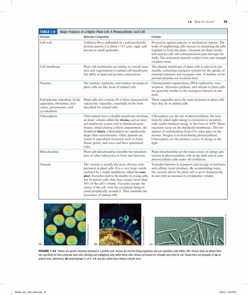
Table 1.9 Major Features of a Higher Plant Cell: A Photosynthetic Leaf CellStructure Molecular Composition Function
Cell wall Cellulose fibers embedded in a polysaccharide/protein matrix; it is thick (.0.1 mm), rigid, and porous to small molecules.
Protection against osmotic or mechanical rupture. The walls of neighboring cells interact in cementing the cells together to form the plant. Channels for fluid circula-tion and for cell–cell communication pass through the walls. The structural material confers form and strength on plant tissue.
Cell membrane Plant cell membranes are similar in overall struc-ture and organization to animal cell membranes but differ in lipid and protein composition.
The plasma membrane of plant cells is selectively per-meable, containing transport systems for the uptake of essential nutrients and inorganic ions. A number of im-portant enzymes are localized here.
Nucleus The nucleus, nucleolus, and nuclear envelope of plant cells are like those of animal cells.
Chromosomal organization, DNA replication, tran-scription, ribosome synthesis, and mitosis in plant cells are generally similar to the analogous features in ani-mals.
Endoplasmic reticulum, Golgi apparatus, ribosomes, lyso-somes, peroxisomes, and cytoskeleton
Plant cells also contain all of these characteristic eukaryotic organelles, essentially in the form described for animal cells.
These organelles serve the same purposes in plant cells that they do in animal cells.
Chloroplasts Chloroplasts have a double-membrane envelope, an inner volume called the stroma, and an inter-nal membrane system rich in thylakoid mem-branes, which enclose a third compartment, the thylakoid lumen. Chloroplasts are significantly larger than mitochondria. Other plastids are found in specialized structures such as fruits, flower petals, and roots and have specialized roles.
Chloroplasts are the site of photosynthesis, the reac-tions by which light energy is converted to metaboli-cally useful chemical energy in the form of ATP. These reactions occur on the thylakoid membranes. The for-mation of carbohydrate from CO2 takes place in the stroma. Oxygen is evolved during photosynthesis. Chloroplasts are the primary source of energy in the light.
Mitochondria Plant cell mitochondria resemble the mitochon-dria of other eukaryotes in form and function.
Plant mitochondria are the main source of energy gen-eration in photosynthetic cells in the dark and in non-photosynthetic cells under all conditions.
Vacuole The vacuole is usually the most obvious com-partment in plant cells. It is a very large vesicle enclosed by a single membrane called the tono-plast. Vacuoles tend to be smaller in young cells, but in mature cells, they may occupy more than 50% of the cell’s volume. Vacuoles occupy the center of the cell, with the cytoplasm being lo-cated peripherally around it. They resemble the lysosomes of animal cells.
Vacuoles function in transport and storage of nutrients and cellular waste products. By accumulating water, the vacuole allows the plant cell to grow dramatically in size with no increase in cytoplasmic volume.
(c)(a) (b)
Figure1.23 Viruses are genetic elements enclosed in a protein coat. Viruses are not free-living organisms and can reproduce only within cells. Viruses show an almost abso-lute specificity for their particular host cells, infecting and multiplying only within those cells. Viruses are known for virtually every kind of cell. Shown here are examples of (a) an animal virus, adenovirus; (b) bacteriophage T4 on E. coli; and (c) a plant virus, tobacco mosaic virus.
BSIP
/Pho
to R
esea
rche
rs, I
nc.
Eye
of S
cien
ce/P
hoto
Res
earc
hers
, Inc
.
© S
cien
ce S
ourc
e/Ph
oto
Rese
arch
ers,
Inc.
06296_ch01_0001-0029.indd 25 12/6/11 2:55 PM
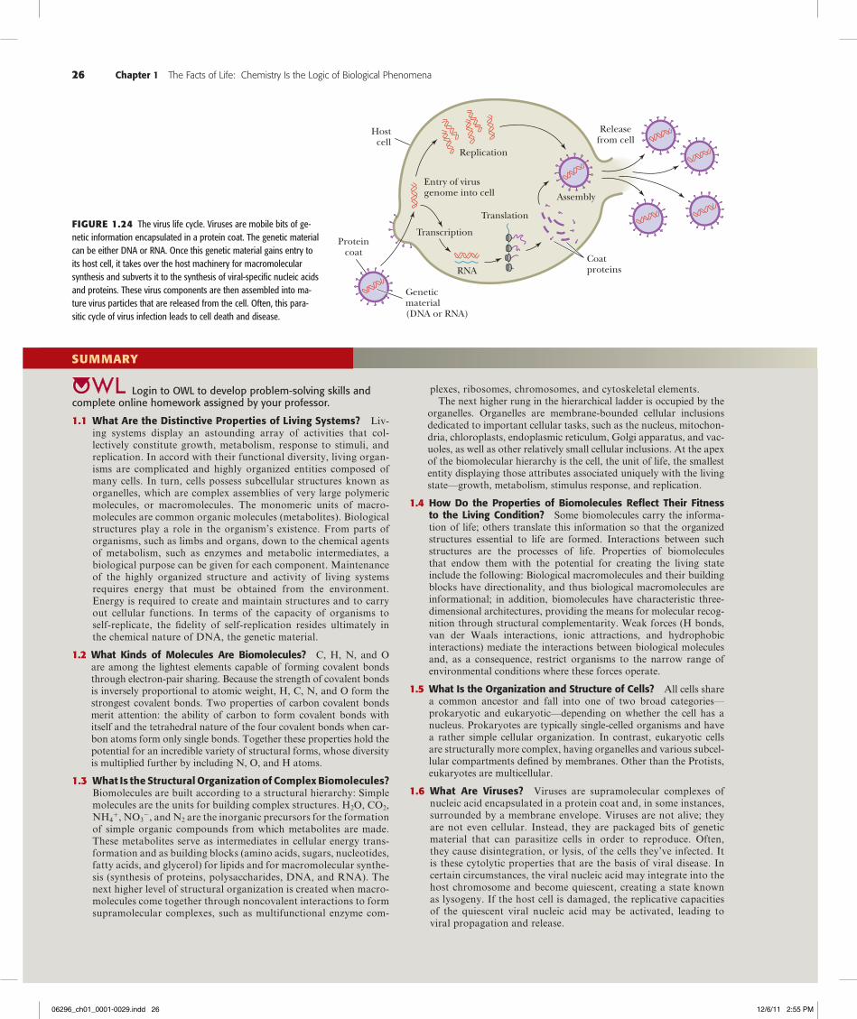
26 Chapter 1 The Facts of Life: Chemistry Is the Logic of Biological Phenomena
Proteincoat
Geneticmaterial(DNA or RNA)
Entry of virusgenome into cell
Transcription
RNA
Translation
Assembly
Replication
Hostcell
Coatproteins
Releasefrom cell
Figure1.24 The virus life cycle. Viruses are mobile bits of ge-netic information encapsulated in a protein coat. The genetic material can be either DNA or RNA. Once this genetic material gains entry to its host cell, it takes over the host machinery for macromolecular synthesis and subverts it to the synthesis of viral-specific nucleic acids and proteins. These virus components are then assembled into ma-ture virus particles that are released from the cell. Often, this para-sitic cycle of virus infection leads to cell death and disease.
login to oWl to develop problem-solving skills and complete online homework assigned by your professor.
1.1 What Are the Distinctive Properties of Living Systems? Liv-ing systems display an astounding array of activities that col-lectively constitute growth, metabolism, response to stimuli, and replication. In accord with their functional diversity, living organ-isms are complicated and highly organized entities composed of many cells. In turn, cells possess subcellular structures known as organelles, which are complex assemblies of very large polymeric molecules, or macromolecules. The monomeric units of macro-molecules are common organic molecules (metabolites). Biological structures play a role in the organism’s existence. From parts of organisms, such as limbs and organs, down to the chemical agents of metabolism, such as enzymes and metabolic intermediates, a biological purpose can be given for each component. Maintenance of the highly organized structure and activity of living systems requires energy that must be obtained from the environment. Energy is required to create and maintain structures and to carry out cellular functions. In terms of the capacity of organisms to self-replicate, the fidelity of self-replication resides ultimately in the chemical nature of DNA, the genetic material.
1.2 What Kinds of Molecules Are Biomolecules? C, H, N, and O are among the lightest elements capable of forming covalent bonds through electron-pair sharing. Because the strength of covalent bonds is inversely proportional to atomic weight, H, C, N, and O form the strongest covalent bonds. Two properties of carbon covalent bonds merit attention: the ability of carbon to form covalent bonds with itself and the tetrahedral nature of the four covalent bonds when car-bon atoms form only single bonds. Together these properties hold the potential for an incredible variety of structural forms, whose diversity is multiplied further by including N, O, and H atoms.
1.3 What Is the Structural Organization of Complex Biomolecules? Biomolecules are built according to a structural hierarchy: Simple molecules are the units for building complex structures. H2O, CO2, NH4
1, NO32, and N2 are the inorganic precursors for the formation
of simple organic compounds from which metabolites are made. These metabolites serve as intermediates in cellular energy trans-formation and as building blocks (amino acids, sugars, nucleotides, fatty acids, and glycerol) for lipids and for macromolecular synthe-sis (synthesis of proteins, polysaccharides, DNA, and RNA). The next higher level of structural organization is created when macro-molecules come together through noncovalent interactions to form supramolecular complexes, such as multifunctional enzyme com-
plexes, ribosomes, chromosomes, and cytoskeletal elements. The next higher rung in the hierarchical ladder is occupied by the
organelles. Organelles are membrane-bounded cellular inclusions dedicated to important cellular tasks, such as the nucleus, mitochon-dria, chloroplasts, endoplasmic reticulum, Golgi apparatus, and vac-uoles, as well as other relatively small cellular inclusions. At the apex of the biomolecular hierarchy is the cell, the unit of life, the smallest entity displaying those attributes associated uniquely with the living state—growth, metabolism, stimulus response, and replication.
1.4 How Do the Properties of Biomolecules Reflect Their Fitness to the Living Condition? Some biomolecules carry the informa-tion of life; others translate this information so that the organized structures essential to life are formed. Interactions between such structures are the processes of life. Properties of biomolecules that endow them with the potential for creating the living state include the following: Biological macromolecules and their building blocks have directionality, and thus biological macromolecules are informational; in addition, biomolecules have characteristic three-dimensional architectures, providing the means for molecular recog-nition through structural complementarity. Weak forces (H bonds, van der Waals interactions, ionic attractions, and hydrophobic interactions) mediate the interactions between biological molecules and, as a consequence, restrict organisms to the narrow range of environmental conditions where these forces operate.
1.5 What Is the Organization and Structure of Cells? All cells share a common ancestor and fall into one of two broad categories—prokaryotic and eukaryotic—depending on whether the cell has a nucleus. Prokaryotes are typically single-celled organisms and have a rather simple cellular organization. In contrast, eukaryotic cells are structurally more complex, having organelles and various subcel-lular compartments defined by membranes. Other than the Protists, eukaryotes are multicellular.
1.6 What Are Viruses? Viruses are supramolecular complexes of nucleic acid encapsulated in a protein coat and, in some instances, surrounded by a membrane envelope. Viruses are not alive; they are not even cellular. Instead, they are packaged bits of genetic material that can parasitize cells in order to reproduce. Often, they cause disintegration, or lysis, of the cells they’ve infected. It is these cytolytic properties that are the basis of viral disease. In certain circumstances, the viral nucleic acid may integrate into the host chromosome and become quiescent, creating a state known as lysogeny. If the host cell is damaged, the replicative capacities of the quiescent viral nucleic acid may be activated, leading to viral propagation and release.
SuMMArY
06296_ch01_0001-0029.indd 26 12/6/11 2:55 PM
Problems 27
FOuNDATiONALBiOCHeMiSTrYThings You Should Know after Reading Chapter 1.
• Chemistry is the logic of biological phenomena.
• Biological molecules serve functional purposes.
• The living state is characterized by the flow of energy through the organism.
• Biomolecules are compounds of carbon.
• Cellular macromolecules and structures are formed from simple mol-ecules according to a hierarchy of increasing structural complexity.
• Metabolites are used to form the building blocks of macromolecules.
• Membranes are supramolecular assemblies that define the boundaries of cells.
• Biological macromolecules and their building blocks have a “sense,” or directionality.
• Biological macromolecules are informational.
• Macromolecules have a defining 3-dimensional architecture.
• Weak forces important in biochemistry include hydrogen bonds, electrostatic (ionic) interactions, van der Waals interactions, and hydrophobic interactions.
• Weak forces maintain biological structure and determine biomolecu-lar interactions.
• The defining concept in biochemistry is “molecular recognition through structural complementarity.”
• Biomolecular recognition is mediated by weak chemical forces.
• The importance of weak forces restricts organisms to a narrow range of environmental conditions.
• Enzymes catalyze metabolic reactions.
• Metabolic regulation is achieved by controlling the activity of enzymes.
• The time scale of life ranges from 10215 sec (the speed of electron transfer processes) to 1018 sec, the time span that life has been evolv-ing on earth.
• The structural and functional organization of prokaryotic cells.
• The structural and functional organization of animal cells and plant cells, including the functions of the various organelles.
• Viruses are lifeless complexes of nucleic acid and protein that act as cellular parasites in order to reproduce.
PrOBLeMS
answers to all problems are at the end of this book. Detailed solutions are available in the Student Solutions Manual, Study Guide, and Prob-lems Book.
1. The Biosynthetic Capacity of Cells The nutritional requirements of Escherichia coli cells are far simpler than those of humans, yet the macromolecules found in bacteria are about as complex as those of animals. Because bacteria can make all their essential biomolecules while subsisting on a simpler diet, do you think bacteria may have more biosynthetic capacity and hence more metabolic complexity than animals? Organize your thoughts on this question, pro and con, into a rational argument.
2. Cell Structure Without consulting the figures in this chapter, sketch the characteristic prokaryotic and eukaryotic cell types and label their pertinent organelle and membrane systems.
3. The Dimensions of Prokaryotic Cells and Their Constituents Escherichia coli cells are about 2 mm (microns) long and 0.8 mm in diameter.a. How many E. coli cells laid end to end would fit across the diam-
eter of a pinhead? (Assume a pinhead diameter of 0.5 mm.)b. What is the volume of an E. coli cell? (Assume it is a cylinder, with
the volume of a cylinder given by V5pr2h, where p53.14.)c. What is the surface area of an E. coli cell? What is the surface-to-
volume ratio of an E. coli cell?d. Glucose, a major energy-yielding nutrient, is present in bacterial
cells at a concentration of about 1 mM. What is the concentration of glucose, expressed as mg/mL? How many glucose molecules are contained in a typical E. coli cell? (Recall that Avogadro’s number56.02331023.)
e. A number of regulatory proteins are present in E. coli at only one or two molecules per cell. If we assume that an E. coli cell contains just one molecule of a particular protein, what is the molar concen-tration of this protein in the cell? If the molecular weight of this protein is 40 kD, what is its concentration, expressed as mg/mL?
f. An E. coli cell contains about 15,000 ribosomes, which carry out protein synthesis. Assuming ribosomes are spherical and have a diameter of 20 nm (nanometers), what fraction of the E. coli cell volume is occupied by ribosomes?
g. The E. coli chromosome is a single DNA molecule whose mass is about 33109 daltons. This macromolecule is actually a linear array of nucleotide pairs. The average molecular weight of a nucleotide pair is 660, and each pair imparts 0.34 nm to the length of the DNA molecule. What is the total length of the E. coli chromosome? How does this length compare with the overall di-mensions of an E. coli cell? How many nucleotide pairs does this DNA contain? The average E. coli protein is a linear chain of 360 amino acids. If three nucleotide pairs in a gene encode one amino acid in a protein, how many different proteins can the E. coli chromosome encode? (The answer to this question is a reasonable approximation of the maximum number of different kinds of proteins that can be expected in bacteria.)
4. The Dimensions of Mitochondria and Their Constituents Assume that mitochondria are cylinders 1.5 mm in length and 0.6 mm in diameter.a. What is the volume of a single mitochondrion?b. Oxaloacetate is an intermediate in the citric acid cycle, an
important metabolic pathway localized in the mitochondria of eu-karyotic cells. The concentration of oxaloacetate in mitochondria is about 0.03 mM. How many molecules of oxaloacetate are in a single mitochondrion?
5. The Dimensions of Eukaryotic Cells and Their Constituents Assume that liver cells are cuboidal in shape, 20 mm on a side.a. How many liver cells laid end to end would fit across the diameter
of a pinhead? (Assume a pinhead diameter of 0.5 mm.)b. What is the volume of a liver cell? (Assume it is a cube.)c. What is the surface area of a liver cell? What is the surface-
to-volume ratio of a liver cell? How does this compare to the surface-to-volume ratio of an E. coli cell (compare this answer
06296_ch01_0001-0029.indd 27 12/6/11 2:55 PM
with that of problem 3c)? What problems must cells with low surface-to-volume ratios confront that do not occur in cells with high surface-to-volume ratios?
d. A human liver cell contains two sets of 23 chromosomes, each set being roughly equivalent in information content. The total mass of DNA contained in these 46 enormous DNA molecules is 431012 daltons. Because each nucleotide pair contributes 660 daltons to the mass of DNA and 0.34 nm to the length of DNA, what is the total number of nucleotide pairs and the complete length of the DNA in a liver cell? How does this length compare with the overall dimensions of a liver cell? The maximal informa-tion in each set of liver cell chromosomes should be related to the number of nucleotide pairs in the chromosome set’s DNA. This number can be obtained by dividing the total number of nucleotide pairs just calculated by 2. What is this value? If this information is expressed in proteins that average 400 amino acids in length and three nucleotide pairs encode one amino acid in a protein, how many different kinds of proteins might a liver cell be able to produce? (In reality, liver cell DNA encodes approximately 20,000 different proteins. Thus, a large discrepancy exists between the theoretical information content of DNA in liver cells and the amount of information actually expressed.)
6. The Principle of Molecular Recognition Through Structural Complementarity Biomolecules interact with one another through molecular surfaces that are structurally complementary. How can various proteins interact with molecules as different as simple ions, hydrophobic lipids, polar but uncharged carbohydrates, and even nucleic acids?
7. The Properties of Informational Macromolecules What structural features allow biological polymers to be informational macromolecules? Is it possible for polysaccharides to be informational macromolecules?
8. The Importance of Weak Forces in Biomolecular Recognition Why is it important that weak forces, not strong forces, mediate biomolecular recognition?
9. Interatomic Distances in Weak Forces versus Chemical Bonds What is the distance between the centers of two carbon atoms (their limit of approach) that are interacting through van der Waals forces? What is the distance between the centers of two carbon atoms joined in a covalent bond? (See Table 1.4.)
10. The Strength of Weak Forces Determines the Environmental Sensitivity of Living Cells Why does the central role of weak forces in biomolecular interactions restrict living systems to a narrow range of environmental conditions?
11. Cells as Steady-State Systems Describe what is meant by the phrase “cells are steady-state systems.”
12. A Simple Genome and Its Protein-Encoding Capacity The genome of the Mycoplasma genitalium consists of 523 genes, encoding 484 proteins, in just 580,074 base pairs (Table 1.6). What fraction of the M. genitalium genes encode proteins? What do you think the other genes encode? If the fraction of base pairs devoted to protein-coding genes is the same as the fraction of the total genes that they represent, what is the average number of base pairs per protein-coding gene? If it takes 3 base pairs to specify an amino acid in a protein, how many amino acids are found in the average M. genitali-um protein? If each amino acid contributes on average 120 daltons to the mass of a protein, what is the mass of an average M. genitalium protein?
13. An Estimation of Minimal Genome Size for a Living Cell Studies of existing cells to determine the minimum number of genes for a living cell have suggested that 206 genes are sufficient. If the ratio of protein-coding genes to non–protein-coding genes is the same in this minimal organism as the genes of Mycoplasma genitalium, how many proteins are represented in these 206 genes? How many base pairs would be required to form the genome of this minimal organism if the genes are the same size as M. genitalium genes?
14. An Estimation of the Number of Genes in a Virus Virus genomes range in size from approximately 3500 nucleotides to approximately 280,000 base pairs. If viral genes are about the same size as M. genitalium genes, what is the minimum and maximum number of genes in viruses?
15. Intracellular Transport of Proteins The endoplasmic reticulum (ER) is a site of protein synthesis. Proteins made by ribosomes associated with the ER may pass into the ER membrane or enter the lumen of the ER. Devise a pathway by which:a. a plasma membrane protein may reach the plasma membrane.b. a secreted protein may be deposited outside the cell.
Preparing for the MCAT® Exam
16. Biological molecules often interact via weak forces (H bonds, van der Waals interactions, etc.). What would be the effect of an increase in kinetic energy on such interactions?
17. Proteins and nucleic acids are informational macromolecules. What are the two minimal criteria for a linear informational polymer?
FurTHerreADiNg
General Biology Textbooks
Campbell, N. A., and Reece, J. B., 2007. Biology, 8th ed. San Francisco: Benjamin/Cummings.
Russell, P. J., Hertz P. E., and McMillan, B., 2012. Biology: The Dynamic Science, 2nd ed. Pacific Grove, CA: Brooks/Cole.
Cell and Molecular Biology Textbooks
Alberts, B., Johnson, A., Lewis, J., Raff, M., et al., 2007. Molecular Biol-ogy of the Cell, 5th ed. New York: Garland Press.
Cavicchioli, R., 2007. Archaea: Cellular and Molecular Biology. Herndon, VA: ASM Press.
Lewin, B., Cassimeris, L., Plopper, G., and Lingappa, V. R., 2011. Cells. Boston, MA: Jones and Bartlett.
Lodish, H., Berk, A., Kaiser, C. A., Kreiger, M., et al., 2008. Molecular Cell Biology, 6th ed. New York: W. H. Freeman.
Snyder, L., and Champness, W., 2002. Molecular Genetics of Bacteria, 2nd ed. Herndon, VA: ASM Press.
Watson, J. D., Baker, T. A., Bell, S. T., Gann, A., et al., 2007. Molecular Biology of the Gene, 6th ed. Menlo Park, CA: Benjamin/Cummings.
Papers on Cell Structure
Gil, R., Silva, F. J., Pereto, J., and Moya, A., 2004. Determination of the core of a minimal bacterial gene set. Microbiology and Molecular Bi-ology Reviews 68:518–537.
Goodsell, D. S., 1991. Inside a living cell. Trends in Biochemical Sciences 16:203–206.
Lewis, P. J., 2004. Bacterial subcellular architecture: Recent advances and future prospects. Molecular Microbiology 54:1135–1150.
Lloyd, C., ed., 1986. Cell organization. Trends in Biochemical Sciences 11:437–485.
28 Chapter 1 The Facts of Life: Chemistry Is the Logic of Biological Phenomena
06296_ch01_0001-0029.indd 28 12/6/11 2:55 PM
Papers on Genomes
Cho, M. K., et al., 1999. Ethical considerations in synthesizing a minimal genome. Science 286:2087–2090.
Gibson, D. G., 2010. Creation of a bacterial cell controlled by a chemi-cally synthesized genome. Science 329:52–56.
Kobayashi, K., Ehrlich, S. D., Albertini, A., Amati, G., et al., 2003. Es-sential Bacillus subtilis genes. Proceedings of the National Academy of Science, U.S.A. 100:4678–4683.
Lartigue, C., Glass, J. I., Alperovich, N., Pieper, R., et al., 2007. Genome transplantation in bacteria: changing one species to another. Science 317:632–638.
Ryan, F., 2010. I, virus: Why you are only half human. New Scientist 205:32–35 (January 27, 2010 issue).
Szathmary, E., 2005. In search of the simplest cell. Nature 433:469–470.
Papers on Early Cell Evolution
Cavalier-Smith, T., 2010. Origin of the cell nucleus, mitosis and sex: roles of intracellular coevolution. Biology Direct 5:7 (78 pages).
Margulis, L., 1996. Archaeal-eubacterial mergers in the origin of Eu-karya: Phylogenetic classification of life. Proceedings of the National Academy of Science, U.S.A. 93:1071–1076.
Pace, N. R., 2006. Time for a change. Nature 441:289.
Service, R. F., 1997. Microbiologists explore life’s rich, hidden kingdoms. Science 275:1740–1742.
Wald, G., 1964. The origins of life. Proceedings of the National Academy of Science, U.S.A. 52:595–611.
Whitfield, J., 2004. Born in a watery commune. Nature 427:674–676.
Woese, C. R., 2002. On the creation of cells. Proceedings of the National Academy of Science, U.S.A. 99:8742–8747.
A Brief History of Life
De Duve, C., 2002. Life-Evolving: Molecules, Mind, and Meaning. New York: Oxford University Press.
Morowitz, H., and Smith, E., 2007. Energy flow and the organization of life. Complexity 13:51–59.
Further Readings 29
06296_ch01_0001-0029.indd 29 12/6/11 2:55 PM