Embed Size (px)
Citation preview
PART 1
Adverse Reactions to Food Antigens: Basic Science

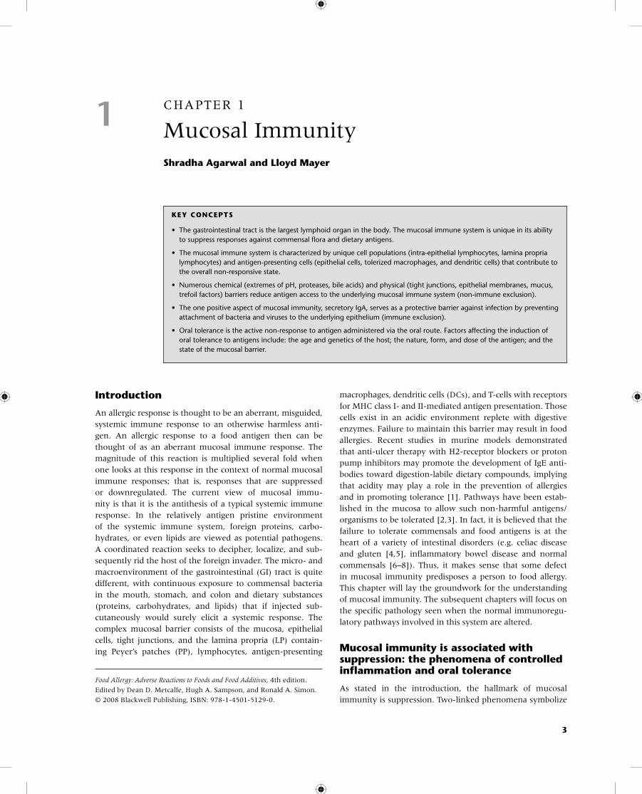
1 CHAPTER 1
Mucosal ImmunityShradha Agarwal and Lloyd Mayer
KEY CONCEPTS
• The gastrointestinal tract is the largest lymphoid organ in the body. The mucosal immune system is unique in its ability to suppress responses against commensal flora and dietary antigens.
• The mucosal immune system is characterized by unique cell populations (intra-epithelial lymphocytes, lamina propria lymphocytes) and antigen-presenting cells (epithelial cells, tolerized macrophages, and dendritic cells) that contribute to the overall non-responsive state.
• Numerous chemical (extremes of pH, proteases, bile acids) and physical (tight junctions, epithelial membranes, mucus, trefoil factors) barriers reduce antigen access to the underlying mucosal immune system (non-immune exclusion).
• The one positive aspect of mucosal immunity, secretory IgA, serves as a protective barrier against infection by preventing attachment of bacteria and viruses to the underlying epithelium (immune exclusion).
• Oral tolerance is the active non-response to antigen administered via the oral route. Factors affecting the induction of oral tolerance to antigens include: the age and genetics of the host; the nature, form, and dose of the antigen; and the state of the mucosal barrier.
Food Allergy: Adverse Reactions to Foods and Food Additives, 4th edition.
Edited by Dean D. Metcalfe, Hugh A. Sampson, and Ronald A. Simon.
© 2008 Blackwell Publishing, ISBN: 978-1-4501-5129-0.
3
Introduction
An allergic response is thought to be an aberrant, misguided, systemic immune response to an otherwise harmless anti-gen. An allergic response to a food antigen then can be thought of as an aberrant mucosal immune response. The magnitude of this reaction is multiplied several fold when one looks at this response in the context of normal mucosal immune responses; that is, responses that are suppressed or downregulated. The current view of mucosal immu-nity is that it is the antithesis of a typical systemic immune response. In the relatively antigen pristine environment of the systemic immune system, foreign proteins, carbo-hydrates, or even lipids are viewed as potential pathogens. A coordinated reaction seeks to decipher, localize, and sub-sequently rid the host of the foreign invader. The micro- and macroenvironment of the gastrointestinal (GI) tract is quite different, with continuous exposure to commensal bacteria in the mouth, stomach, and colon and dietary substances (proteins, carbohydrates, and lipids) that if injected sub-cutaneously would surely elicit a systemic response. The complex mucosal barrier consists of the mucosa, epithelial cells, tight junctions, and the lamina propria (LP) contain-ing Peyer’s patches (PP), lymphocytes, antigen-presenting
macrophages, dendritic cells (DCs), and T-cells with receptors for MHC class I- and II-mediated antigen presentation. Those cells exist in an acidic environment replete with digestive enzymes. Failure to maintain this barrier may result in food allergies. Recent studies in murine models demonstrated that anti-ulcer therapy with H2-receptor blockers or proton pump inhibitors may promote the development of IgE anti-bodies toward digestion-labile dietary compounds, implying that acidity may play a role in the prevention of allergies and in promoting tolerance [1]. Pathways have been estab-lished in the mucosa to allow such non-harmful antigens/organisms to be tolerated [2,3]. In fact, it is believed that the failure to tolerate commensals and food antigens is at the heart of a variety of intestinal disorders (e.g. celiac disease and gluten [4,5], inflammatory bowel disease and normal commensals [6–8]). Thus, it makes sense that some defect in mucosal immunity predisposes a person to food allergy. This chapter will lay the groundwork for the understanding of mucosal immunity. The subsequent chapters will focus on the specific pathology seen when the normal immunoregu-latory pathways involved in this system are altered.
Mucosal immunity is associated with suppression: the phenomena of controlled inflammation and oral tolerance
As stated in the introduction, the hallmark of mucosal immunity is suppression. Two-linked phenomena symbolize
4 Chapter 1
this state: controlled/physiologic inflammation and oral tol-erance. The mechanisms governing these phenomena are not completely understood, as the dissection of factors gov-erning mucosal immunoregulation is still evolving. It has become quite evident that the systems involved are complex and that the rules governing systemic immunity frequently do not apply in the mucosa. There is unique compartmen-talization, cell types, and routes of antigen trafficking which come together to produce the immunosuppressed state.
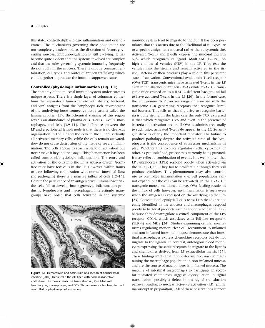
Controlled/physiologic inflammation (Fig. 1.1)The anatomy of the mucosal immune system underscores its unique aspects. There is a single layer of columnar epithe-lium that separates a lumen replete with dietary, bacterial, and viral antigens from the lymphocyte-rich environment of the underlying loose connective tissue stroma called the lamina propria (LP). Histochemical staining of this region reveals an abundance of plasma cells, T-cells, B-cells, mac-rophages, and DCs [3,9–11]. The difference between the LP and a peripheral lymph node is that there is no clear-cut organization in the LP and the cells in the LP are virtually all activated memory cells. While the cells remain activated, they do not cause destruction of the tissue or severe inflam-mation. The cells appear to reach a stage of activation but never make it beyond that stage. This phenomenon has been called controlled/physiologic inflammation. The entry and activation of the cells into the LP is antigen driven. Germ-free mice have few cells in the LP. However, within hours to days following colonization with normal intestinal flora (no pathogens) there is a massive influx of cells [12–15]. Despite the persistence of an antigen drive (luminal bacteria), the cells fail to develop into aggressive, inflammation pro-ducing lymphocytes and macrophages. Interestingly, many groups have noted that cells activated in the systemic
immune system tend to migrate to the gut. It has been pos-tulated that this occurs due to the likelihood of re-exposure to a specific antigen at a mucosal rather than a systemic site. Activated T-cells and B-cells express the mucosal integrin α4β7 which recognizes its ligand, MadCAM [12–19], on high endothelial venules (HEV) in the LP. They exit the venules into the stroma and remain activated in the tis-sue. Bacteria or their products play a role in this persistent state of activation. Conventional ovalbumin–T-cell receptor (OVA-TCR) transgenic mice have activated T-cells in the LP even in the absence of antigen (OVA) while OVA-TCR trans-genic mice crossed on to a RAG-2 deficient background fail to have activated T-cells in the LP [20]. In the former case, the endogenous TCR can rearrange or associate with the transgenic TCR generating receptors that recognize lumi-nal bacteria. This tells us that the drive to recognize bacte-ria is quite strong. In the latter case the only TCR expressed is that which recognizes OVA and even in the presence of bacteria no activation occurs. If OVA is administered orally to such mice, activated T-cells do appear in the LP. So anti-gen drive is clearly the important mediator. The failure to produce pathology despite the activated state of the lym-phocytes is the consequence of suppressor mechanisms in play. Whether this involves regulatory cells, cytokines, or other, as yet undefined, processes is currently being pursued. It may reflect a combination of events. It is well known that LP lymphocytes (LPLs) respond poorly when activated via the TCR [21,22]. They fail to proliferate although they still produce cytokines. This phenomenon may also contrib-ute to controlled inflammation (i.e. cell populations can-not expand, but the cells can be activated). In the OVA-TCR transgenic mouse mentioned above, OVA feeding results in the influx of cells however, no inflammation is seen even when the antigen is expressed on the overlying epithelium [23]. Conventional cytolytic T-cells (class I restricted) are not easily identified in the mucosa and macrophages respond poorly to bacterial products such as lipopolysaccharide (LPS) because they downregulate a critical component of the LPS receptor, CD14, which associates with Toll-like receptor-4 (TLR-4) and MD2 [24]. Studies examining cellular mecha-nisms regulating mononuclear cell recruitment to inflamed and non-inflamed intestinal mucosa demonstrate that intes-tinal macrophages express chemokine receptors but do not migrate to the ligands. In contrast, autologous blood mono-cytes expressing the same receptors do migrate to the ligands and chemokines derived from LP extracellular matrix [25]. These findings imply that monocytes are necessary in main-taining the macrophage population in non-inflamed mucosa and are the source of macrophages in inflamed mucosa. The inability of intestinal macrophages to participate in recep-tor-mediated chemotaxis suggests dysregulation in signal transduction, possibly a defect in the signal transduction pathway leading to nuclear factor-κB activation (P.D. Smith, manuscript in preparation). All of these observations support
Lumen
Figure 1.1 Hematoxylin and eosin stain of a section of normal small intestine (20�). Depicted is the villi lined with normal absorptive epithelium. The loose connective tissue stroma (LP) is filled with lymphocytes, macrophages, and DCs. This appearance has been termed controlled or physiologic inflammation.
Mucosal Immunity 5
PP: CD4+ T-cells(Th3)
(Strober, Weiner)
Spleen: CD8+ T-cells(Waksman)
CD4+ T-cells (Weiner,Strober)
OVA antigen OVA antigen
Immune Response
Tolerance Tolerance
OVA antigen OVA antigen
T-cell transfer
OVA antigen
A B C
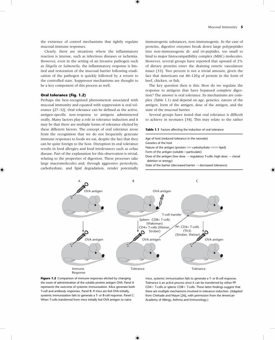
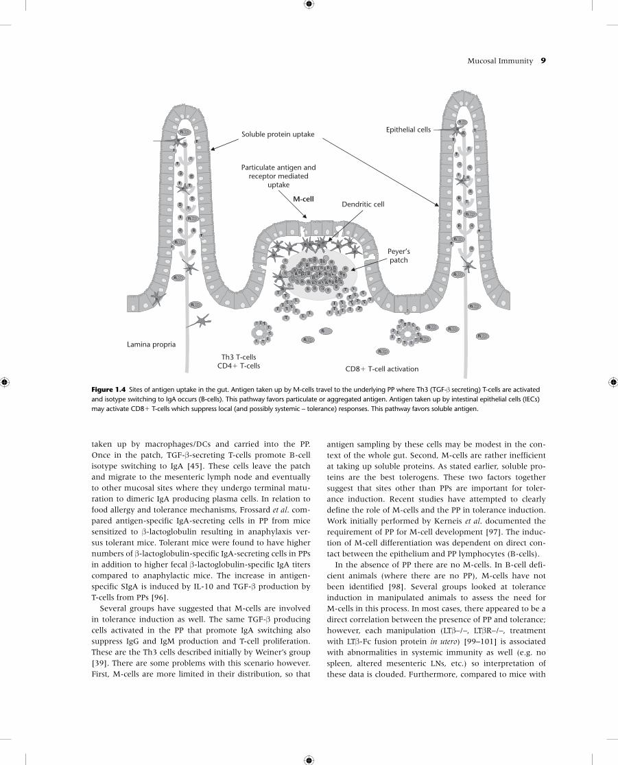
Figure 1.2 Comparison of immune responses elicited by changing the route of administration of the soluble protein antigen OVA. Panel A represents the outcome of systemic immunization. Mice generate both T-cell and antibody responses. Panel B: If mice are fed OVA initially, systemic immunization fails to generate a T- or B-cell response. Panel C: When T-cells transferred from mice initially fed OVA antigen to naïve
mice, systemic immunization fails to generate a T- or B-cell response. Tolerance is an active process since it can be transferred by either PP CD4� T-cells or splenic CD8� T-cells. These latter findings suggest that there are multiple mechanisms involved in tolerance induction. (Adapted from Chehade and Mayer [26], with permission from the American Academy of Allergy, Asthma and Immunology.)
the existence of control mechanisms that tightly regulate mucosal immune responses.
Clearly, there are situations where the inflammatory reaction is intense, such as infectious diseases or ischemia. However, even in the setting of an invasive pathogen such as Shigella or Salmonella, the inflammatory response is lim-ited and restoration of the mucosal barrier following eradi-cation of the pathogen is quickly followed by a return to the controlled state. Suppressor mechanisms are thought to be a key component of this process as well.
Oral tolerance (Fig. 1.2) Perhaps the best-recognized phenomenon associated with mucosal immunity and equated with suppression is oral tol-erance [27–32]. Oral tolerance can be defined as the active, antigen-specific non-response to antigens administered orally. Many factors play a role in tolerance induction and it may be that there are multiple forms of tolerance elicited by these different factors. The concept of oral tolerance arose from the recognition that we do not frequently generate immune responses to foods we eat, despite the fact that they can be quite foreign to the host. Disruption in oral tolerance results in food allergies and food intolerances such as celiac disease. Part of the explanation for this observation is trivial, relating to the properties of digestion. These processes take large macromolecules and, through aggressive proteolysis, carbohydrate, and lipid degradation, render potentially
immunogenic substances, non-immunogenic. In the case of proteins, digestive enzymes break down large polypeptides into non-immunogenic di- and tri-peptides, too small to bind to major histocompatibility complex (MHC) molecules. However, several groups have reported that upward of 2% of dietary proteins enter the draining enteric vasculature intact [33]. Two percent is not a trivial amount, given the fact that Americans eat 40–120 g of protein in the form of beef, chicken, or fish.
The key question then is this: How do we regulate the response to antigens that have bypassed complete diges-tion? The answer is oral tolerance. Its mechanisms are com-plex (Table 1.1) and depend on age, genetics, nature of the antigen, form of the antigen, dose of the antigen, and the state of the mucosal barrier.
Several groups have noted that oral tolerance is difficult to achieve in neonates [34]. This may relate to the rather
Table 1.1 Factors affecting the induction of oral tolerance
Age of host (reduced tolerance in the neonate)Genetics of the hostNature of the antigen (protein ��� carbohydrate ����� lipid)Form of the antigen (soluble � particulate)Dose of the antigen (low dose → regulatory T-cells: high dose → clonal deletion or anergy)State of the barrier (decreased barrier → decreased tolerance)
6 Chapter 1
permeable barrier that exists in the newborn or the imma-turity of the mucosal immune system. Within 3 weeks of age (in mice), oral tolerance can be induced, and many previous antibody responses to food antigens are suppressed. The limited diet in the newborn may serve to protect the infant from generating a vigorous response to food antigens.
The next factor involved in tolerance induction is the genetics of the host. Lamont and co-workers [35] published a report detailing tolerance induction in various mouse strains using the same protocol. Balb/c mice tolerize eas-ily while others failed to tolerize at all. Furthermore, some of the failures to tolerize were antigen specific; upon oral feeding, a mouse could be rendered tolerant to one anti-gen but not another. This finding suggested that the nature and form of the antigen play a significant role in tolerance induction. Protein antigens are the most tolerogenic while carbohydrate and lipids are much less effective in inducing tolerance [36]. The form of the antigen is also critical; for example, a protein given in soluble form (e.g. OVA) is quite tolerogenic whereas, once aggregated, it loses its poten-tial to induce tolerance. The mechanisms underlying these observations have not been completely defined but appear to reflect the nature of the antigen-presenting cell (APC) and the way in which the antigen trafficks to the under-lying mucosal lymphoid tissue. Insolubility or aggregation may also render a luminal antigen incapable of being sam-pled [3]. In this setting, non-immune exclusion of the anti-gen would lead to ignorance from lack of exposure of the mucosa-associated lymphoid tissue (MALT) to the antigen in question. Lastly, prior sensitization to an antigen through extraintestinal routes affects the development of a hyper-sensitivity response. Sensitization to peanut protein was demonstrated by application of skin preparations containing peanut oil to inflamed skin in children [37]. Similar results were obtained by Hsieh’s group in epicutaneous sensitized mice to the egg protein OVA [38].
The dose of antigen administered is also critical to the form of oral tolerance generated. In mouse models, low doses of antigen appear to activate regulatory/suppressor T-cells [39,40]. There are an increasing number of such cells identified, of both CD4 and CD8 lineages. Th3 cells were the initial regulatory/suppressor cells described in oral tolerance [40–42]. These cells appear to be activated in the PP and secrete transforming growth factor-β (TGF-β). This cytokine plays a dual role in mucosal immunity; it is a potent suppressor of T- and B-cell responses while promoting the production of IgA (it is the IgA switch fac-tor) [34,43–45]. TGF-β is the most potent immunosup-pressive cytokine defined and its activities are broad and non-specific. A recent investigation of the adaptive immune response to cholera toxin B subunit and macro-phage- activating lipopeptide-2 in mouse models lacking the TGF-βR in B-cells (TGFβRII-B) demonstrated undetectable levels of antigen-specific IgA-secreting cells, serum IgA, and
secretory IgA (SIgA) [46]. These results demonstrate the critical role of TGF-βR in antigen-driven stimulation of SIgA responses in vivo. The production of TGF-β by Th3 cells elicited by low-dose antigen administration helps explain an associated phenomenon of oral tolerance, bystander suppression. As mentioned earlier, oral tolerance is anti-gen specific, but if a second antigen is co-administered sys-temically with the tolerogen, suppression of T- and B-cell responses to that antigen will occur as well. The participa-tion of other regulatory T-cells in oral tolerance is less well defined. Tr1 cells produce interleukin (IL)-10 and appear to be involved in the suppression of graft-versus-host disease (GVHD) and colitis in mouse models, but their activation during oral antigen administration has not been as clear-cut [47–49]. Frossard et al. demonstrated increased antigen induced IL-10 producing cells in PP from tolerant mice after β-lactoglobulin feeding but not in anaphylactic mice, sug-gesting that reduced IL-10 production in PPs may support food allergies [50]. There is some evidence for the activa-tion of CD4�CD25� regulatory T-cells during oral toler-ance induction protocols but the nature of their role in the process is still under investigation [51–54]. Experiments in transgenic mice expressing TCRs for OVA demonstrated increased numbers of CD4�CD25� T-cells expressing cyto-toxic T-lymphocyte antigen 4 (CTLA-4) and cytokines TGF-β and IL-10 following OVA feeding. Adoptive transfer of CD4�CD25� cells from the fed mice suppressed in vivo delayed-type hypersensitivity responses in recipient mice [55]. Furthermore, tolerance studies done in mice depleted of CD25� T-cells along with TGF-β neutralization failed in the induction of oral tolerance by high and low doses of oral OVA suggesting that CD4�CD25� T-cells and TGF-β together are involved in the induction of oral tolerance, partly through the regulation of expansion of antigen-specific CD4� T-cells [56]. Markers such as glucocorticoid-induced TNF receptor and transcription factor FoxP3, whose genetic deficiency results in an autoimmune and inflam-matory syndrome, have been shown to be expressed by CD4�CD25� Tregs [57,58]. Lastly, early studies suggested that antigen-specific CD8� T-cells were involved in tolerance induction since transfer of splenic CD8� T-cells follow-ing feeding of protein antigens could transfer the toler-ant state to naïve mice [59–62]. Like the various forms of tolerance described, it is likely that the distinct regulatory T-cells defined might work alone depending on the nature of the tolerogen or in concert to orchestrate the suppression associated with oral tolerance and more globally to mucosal immunity.
Higher doses of antigen lead to a different response, either the induction of anergy or clonal deletion [63]. In this setting, tolerance is not infectious and transfer of T-cells from such tolerized animals does not lead to the transfer of tolerance. Clonal deletion via FAS-mediated apoptosis [64] may be a common mechanism given the enormous antigen load in the GI tract.
Mucosal Immunity 7
The last factor affecting tolerance induction is the state of the barrier. This was alluded to earlier in the discussion relating to the failure to generate tolerance in the neonate since intestinal permeability is greater. However, several states of barrier dysfunction are associated with aggressive inflammation and a lack of tolerance. Increased permeabil-ity throughout the intestine has been shown in animal mod-els of anaphylaxis where antigens are able to pass through paracellular spaces by the disruption of tight junctions[65–67]. It is speculated that barrier disruption leads to altered pathways of antigen uptake and failure of conven-tional mucosal sampling and regulatory pathways. For example, treatment of mice with interferon-γ (IFN-γ) can disrupt the mucosal barrier. These mice fail to develop tol-erance to OVA feeding [68,69]. IFN-γ disrupts the inter-epithelial tight junctions allowing for paracellular access by fed antigens. IFN-γ influences many different cell types so mucosal barrier disruption may be only one of several defects induced by such treatment. N-cadherin dominant negative mice develop mucosal inflammation (loss of con-trolled inflammation) [70]. N-cadherin is a component of the epithelial cell barrier. These mice are immunologically intact yet failed to suppress inflammation, possibly because of the enormous antigenic exposure produced by a leaky barrier. Although no oral tolerance studies have been performedin these animals, the concept that controlled inflamma-tion and oral tolerance are linked phenomena suggest that defects in tolerance would exist here as well.
Do these phenomena relate to food allergy? There is no clear answer yet. No studies of oral tolerance to protein antigens have been performed in food-allergic individuals, and data conflict in studies on the integrity of the mucosal barrier in children with various GI diseases [71–75]. The
studies required to answer this question are reasonably straightforward and the answer is critically important for our understanding of food allergy. Oral tolerance has been demonstrated in humans although its efficacy is limited. One clear difference between humans and mice is that tol-erance is induced for T-cells but not for B-cells [76,77]. This difference may have relevance in human antibody-mediated diseases.
The nature of antibody responses in the gut-associated lymphoid tissue
IgE is largely the antibody responsible for food allergy. In genetically pre-disposed individuals an environment favor-ing IgE production in response to an allergen is established. The generation of T-cell responses promoting a B-cell class switch to IgE has been described (i.e. Th2 lymphocytes secreting IL-4). The next question, therefore, is whether such an environment exists in the gut-associated lymphoid tissue (GALT), and what types of antibody responses pre-dominate in this system.
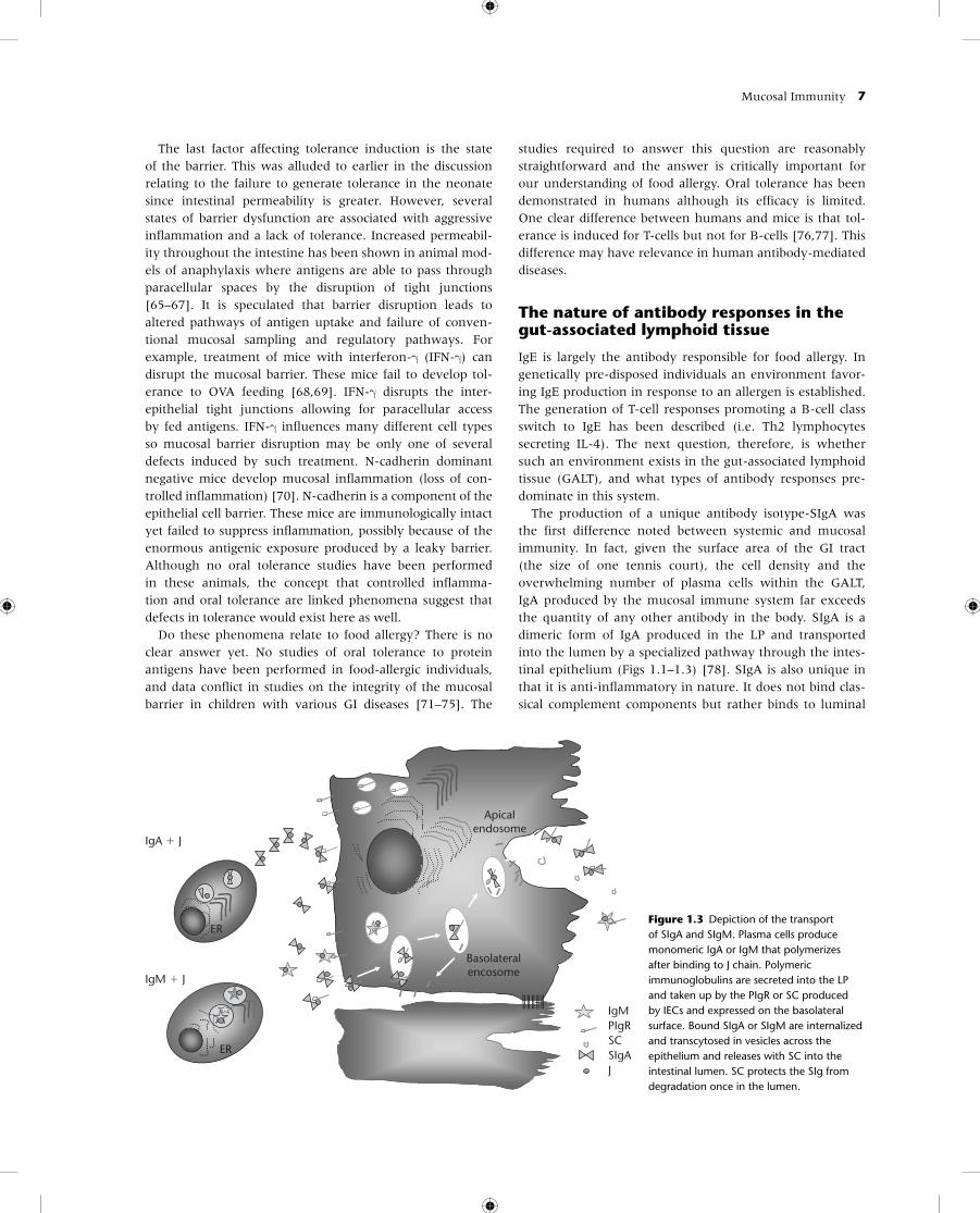
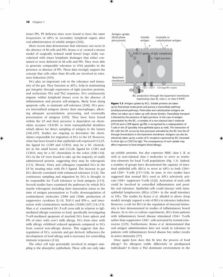
The production of a unique antibody isotype-SIgA was the first difference noted between systemic and mucosal immunity. In fact, given the surface area of the GI tract (the size of one tennis court), the cell density and the overwhelming number of plasma cells within the GALT, IgA produced by the mucosal immune system far exceeds the quantity of any other antibody in the body. SIgA is a dimeric form of IgA produced in the LP and transported into the lumen by a specialized pathway through the intes-tinal epithelium (Figs 1.1–1.3) [78]. SIgA is also unique in that it is anti-inflammatory in nature. It does not bind clas-sical complement components but rather binds to luminal
IgA � J
IgM � J
Apicalendosome
Basolateralencosome
ER
ER
IgMPIgRSCSIgAJ
Figure 1.3 Depiction of the transport of SIgA and SIgM. Plasma cells produce monomeric IgA or IgM that polymerizes after binding to J chain. Polymeric immunoglobulins are secreted into the LP and taken up by the PIgR or SC produced by IECs and expressed on the basolateral surface. Bound SIgA or SIgM are internalized and transcytosed in vesicles across the epithelium and releases with SC into the intestinal lumen. SC protects the SIg from degradation once in the lumen.
8 Chapter 1
antigens, preventing their attachment to the epithelium or promoting agglutination and subsequent removal of the antigen in the mucus layer overlying the epithelium. These latter two events reflect “immune exclusion,” as opposed to the non-specific mechanisms of exclusion alluded to earlier (the epithelium, the mucus barrier, proteolytic digestion, etc.). SIgA has one additional unique aspect – its ability to bind to an epithelial cell-derived glycoprotein called secre-tory component (SC), the receptor for polymeric Ig recep-tor (pIgR) [79–82]. SC serves two functions: it promotes the transcytosis of SIgA from the LP through the epithelium into the lumen, and, once in the lumen, it protects the anti-body against proteolytic degradation. This role is critically important, because the enzymes used for protein diges-tion are equally effective at degrading antibody molecules. For example, pepsin and papain in the stomach digest IgG into F(ab)’2 and Fab fragments. Further protection against trypsin and chymotrypsin in the lumen allows SIgA to exist in a rather hostile environment.
IgM is another antibody capable of binding SC (pIgR). Like IgA, IgM uses J chain produced by plasma cells to form poly-mers; in the case of IgM, a pentamer. SC binds to the Fc portions of the antibody formed by the polymerization. The ability of IgM to bind SC may be important in patients with IgA deficiency. Although not directly proven, secretory IgM (SIgM) may compensate for the absence of IgA in the lumen.
What about other Ig isotypes? The focus for years in mucosal immunity was SIgA. It was estimated that upward of 95% of antibody produced at mucosal surfaces was IgA. Initial reports ignored the fact that IgG was present not only in the LP, but also in secretions [83,84]. These latter observations were attributed to leakage across the barrier from plasma IgG. However, recent attention has focused on the potential role of the neonatal Fc receptor, FcRN, which might serve as a bidirectional transporter of IgG [85,86]. The FcRN is expressed early on, possibly as a mechanism to take up maternal IgG in breast milk. Its expression was thought to be downregulated after weaning, but recent studies suggest that it may still be expressed in adult lung, kidney, and possibly gut epithelium. As suggested above, there are new data indicating that it might serve to trans-port IgG both to and from the lumen. In a series of inflam-matory diseases of the bowel, marked increases in IgG in the LP and lumen have been observed [87].
We are left then with IgE. Given the modest amounts present in the serum, it has been even more difficult to detect IgE in mucosal tissues or secretions. However, there have not been many studies attempting to do so. Mucosal mast cells are well described in the gut tissue. The IgE Fc receptor, FcεRI, is present and mast cell degranulation is reported (although not necessarily IgE related). FcεRI is not expressed by the intestinal epithelium so it is unlikely that this molecule would serve a transport function. CD23 (FcεRII), however, has been described on gut epithelial cells,
and one model has suggested that it may play a role in facili-tated antigen uptake and consequent mast cell degranula-tion [88,89]. In this setting, degranulation is associated with fluid and electrolyte loss into the luminal side of the epithe-lium, an event clearly associated with an allergic reaction in the lung and gut. Thus, the initial concept that IgA was thebe-all and end-all in the gut may be shortsighted and roles for other isotypes in health and disease require further study.
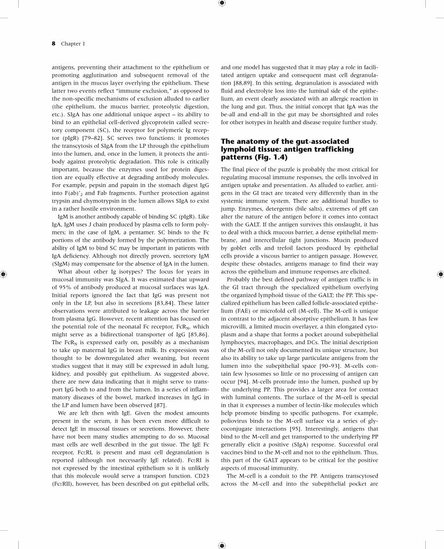
The anatomy of the gut-associated lymphoid tissue: antigen trafficking patterns (Fig. 1.4)
The final piece of the puzzle is probably the most critical for regulating mucosal immune responses, the cells involved in antigen uptake and presentation. As alluded to earlier, anti-gens in the GI tract are treated very differently than in the systemic immune system. There are additional hurdles to jump. Enzymes, detergents (bile salts), extremes of pH can alter the nature of the antigen before it comes into contact with the GALT. If the antigen survives this onslaught, it has to deal with a thick mucous barrier, a dense epithelial mem-brane, and intercellular tight junctions. Mucin produced by goblet cells and trefoil factors produced by epithelial cells provide a viscous barrier to antigen passage. However, despite these obstacles, antigens manage to find their way across the epithelium and immune responses are elicited.
Probably the best defined pathway of antigen traffic is in the GI tract through the specialized epithelium overlying the organized lymphoid tissue of the GALT; the PP. This spe-cialized epithelium has been called follicle-associated epithe-lium (FAE) or microfold cell (M-cell). The M-cell is unique in contrast to the adjacent absorptive epithelium. It has few microvilli, a limited mucin overlayer, a thin elongated cyto-plasm and a shape that forms a pocket around subepithelial lymphocytes, macrophages, and DCs. The initial description of the M-cell not only documented its unique structure, but also its ability to take up large particulate antigens from the lumen into the subepithelial space [90–93]. M-cells con-tain few lysosomes so little or no processing of antigen can occur [94]. M-cells protrude into the lumen, pushed up by the underlying PP. This provides a larger area for contact with luminal contents. The surface of the M-cell is special in that it expresses a number of lectin-like molecules which help promote binding to specific pathogens. For example, poliovirus binds to the M-cell surface via a series of gly-coconjugate interactions [95]. Interestingly, antigens that bind to the M-cell and get transported to the underlying PP generally elicit a positive (SIgA) response. Successful oral vaccines bind to the M-cell and not to the epithelium. Thus, this part of the GALT appears to be critical for the positive aspects of mucosal immunity.
The M-cell is a conduit to the PP. Antigens transcytosed across the M-cell and into the subepithelial pocket are
Mucosal Immunity 9
taken up by macrophages/DCs and carried into the PP. Once in the patch, TGF-β-secreting T-cells promote B-cell isotype switching to IgA [45]. These cells leave the patch and migrate to the mesenteric lymph node and eventually to other mucosal sites where they undergo terminal matu-ration to dimeric IgA producing plasma cells. In relation to food allergy and tolerance mechanisms, Frossard et al. com-pared antigen-specific IgA-secreting cells in PP from mice sensitized to β-lactoglobulin resulting in anaphylaxis ver-sus tolerant mice. Tolerant mice were found to have higher numbers of β-lactoglobulin-specific IgA-secreting cells in PPs in addition to higher fecal β-lactoglobulin-specific IgA titers compared to anaphylactic mice. The increase in antigen-specific SIgA is induced by IL-10 and TGF-β production by T-cells from PPs [96].
Several groups have suggested that M-cells are involved in tolerance induction as well. The same TGF-β producing cells activated in the PP that promote IgA switching also suppress IgG and IgM production and T-cell proliferation. These are the Th3 cells described initially by Weiner’s group [39]. There are some problems with this scenario however. First, M-cells are more limited in their distribution, so that
antigen sampling by these cells may be modest in the con-text of the whole gut. Second, M-cells are rather inefficient at taking up soluble proteins. As stated earlier, soluble pro-teins are the best tolerogens. These two factors together suggest that sites other than PPs are important for toler-ance induction. Recent studies have attempted to clearly define the role of M-cells and the PP in tolerance induction. Work initially performed by Kerneis et al. documented the requirement of PP for M-cell development [97]. The induc-tion of M-cell differentiation was dependent on direct con-tact between the epithelium and PP lymphocytes (B-cells).
In the absence of PP there are no M-cells. In B-cell defi-cient animals (where there are no PP), M-cells have not been identified [98]. Several groups looked at tolerance induction in manipulated animals to assess the need for M-cells in this process. In most cases, there appeared to be a direct correlation between the presence of PP and tolerance; however, each manipulation (LTβ–/–, LTβR–/–, treatment with LTβ-Fc fusion protein in utero) [99–101] is associated with abnormalities in systemic immunity as well (e.g. no spleen, altered mesenteric LNs, etc.) so interpretation of these data is clouded. Furthermore, compared to mice with
Lamina propria
Epithelial cells
Dendritic cell
Particulate antigen andreceptor mediated
uptake
M-cell
Peyer’spatch
CD8� T-cell activation
Th3 T-cellsCD4� T-cells
Soluble protein uptake
Figure 1.4 Sites of antigen uptake in the gut. Antigen taken up by M-cells travel to the underlying PP where Th3 (TGF-β secreting) T-cells are activated and isotype switching to IgA occurs (B-cells). This pathway favors particulate or aggregated antigen. Antigen taken up by intestinal epithelial cells (IECs) may activate CD8� T-cells which suppress local (and possibly systemic – tolerance) responses. This pathway favors soluble antigen.
10 Chapter 1
intact PPs, PP deficient mice were found to have the same frequencies of APCs in secondary lymphoid organs after oral administration of soluble antigen [102].
More recent data demonstrate that tolerance can occur in the absence of M-cells and PPs. Kraus et al. created a mouse model of surgically isolated small bowel loops (fully vas-cularized with intact lymphatic drainage) that either con-tained or were deficient in M-cells and PPs. They were able to generate comparable tolerance to OVA peptides in the presence or absence of PPs. These data strongly support the concept that cells other than M-cells are involved in toler-ance induction [103].
DCs play an important role in the tolerance and immu-nity of the gut. They function as APCs, help in maintaining gut integrity through expression of tight junction proteins, and orchestrate Th1 and Th2 responses. DCs continuously migrate within lymphoid tissues even in the absence of inflammation and present self-antigens, likely from dying apoptotic cells, to maintain self-tolerance [104]. DCs proc-ess internalized antigens slower than macrophages, allow-ing adequate accumulation, processing, and eventually presentation of antigens [105]. They have been found within the LP and their presence is dependent on chem-okine receptor CX3CR1 to form transepithelial dendrites which allows for direct sampling of antigen in the lumen [106,107]. Studies are ongoing to determine the chem-okines responsible for migration of DCs to the LP. However, what has been found is that epithelial cell-expressed CCL25, the ligand for CCR9 and CCR10, may be a DC chemok-ine in the small bowel, and CCL28, ligand for CCR3 and CCR10, may be a DC chemokine in the colon [108–110]. DCs in the LP were found to take up the majority of orally administered protein, suggesting they may be tolerogenic [111]. Mowat, Viney and colleagues expanded DCs in the LP by treating mice with Flt-3 ligand. The increase in gut DCs directly correlated with enhanced tolerance [112]. The continuous sampling and migration by DCs is thought to be responsible for T-cell tolerance to food antigens [113]. Several studies have examined the pathways by which DCs maybe tolerogenic including their maturation status at the time of antigen presentation to T-cells; downregulation of costimulatory molecules CD80 and CD86, production of suppressive cytokines IL-10, TGF-β and IFN-α, and inter-action with costimulatory molecules CD200 [107,114,115]. Man et al. examined DC–T-cell cross-talk in relation to IgE-mediated allergic reactions to food, specifically investigating T-cell-mediated apoptosis of myeloid DCs from spleen and PPs of mice with cow’s milk allergy. DCs from mice with milk allergy exhibited reduced apoptosis compared to DCs from control non-allergic donors. This suggests that dys-regulation of DCs, systemic and gut derived, influences the development of food allergy and is necessary for controlling immune responses [116].
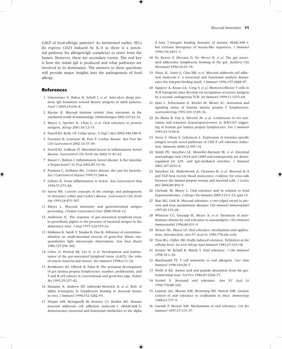
The other cell type potentially involved in antigen sam-pling is the absorptive epithelium. These cells not only take
up soluble proteins, but also expresses MHC class I, II, as well as non-classical class I molecules to serve as restric-tion elements for local T-cell populations (Fig. 1.5). Indeed, a number of groups have documented the capacity of intes-tinal epithelial cells (IECs) to serve as APCs to both CD4� and CD8� T-cells [117–124]. In man, in vitro studies have suggested that normal IECs used as APCs selectively acti-vate CD8� suppressor T-cells [122]. Activation of such cells could be involved in controlled inflammation and possi-bly oral tolerance. Epithelial cells could interact with intra- epithelial lymphocytes (IELs) (CD8� in the small intestine) or LPLs. The studies by Kraus et al. alluded to above (loop model) strongly support a role of IECs in tolerance induction. However, a role for IECs in the regulation of mucosal immu-nity is best demonstrated in studies of inflammatory bowel disease. In in vitro co-culture experiments, IECs from patients with inflammatory bowel disease stimulated CD4� T-cells rather than suppressive CD8� cells activated by normal ente-rocytes [125]. Furthermore, Kraus et al. demonstrated that oral antigen administration does not result in tolerance in patients with inflammatory bowel disease but rather results in active immunity [77].
Once again how does this fit into the process of food allergy? Do allergens traffic differently in predisposed individuals? Is there a Th2 dominant environment in the
Figure 1.5 Antigen uptake by IECs. Soluble proteins are taken up by fluid phase endocytosis and pursue a transcellular pathway (endolysosomal pathway). Particulate and carbohydrate antigens are either not taken up or taken up with slower kinetics. Paracellular transport is blocked by the presence of tight junctions. In the case of antigen presentation by the IEC, a complex of a non-classical class I molecule (CD1d) and a CD8 ligand, gp180, is recognized by a subpopulation of T-cells in the LP (possibly intra-epithelial space as well). The interaction of IEC with the LPL occurs by foot processes extruded by the IEC into the LP through fenestrations in the basement membrane. Antigens can also be selectively taken up by a series of Fc receptors expressed by IEC (neonatal FcεR for IgG or CD23 for IgE). The consequences of such uptake may affect responses to food antigens (food allergy).
Antigen uptake(fluid phasepinocytosis)
Basementmembrane
IEC projection through the basement membraneexpressing class Ib, class I, or class II MHC
Solubleantigen
Tight junction
Insoluble orcarbohydrate antigen
Class I
FcRn (? uptake of IgG complexes)
CD1d/gp180 complexClass II
Mucosal Immunity 11
GALT of food-allergic patients? As mentioned earlier, IECs do express CD23 induced by IL-4 so there is a poten-tial pathway for allergen/IgE complexes to enter from the lumen. However, these are secondary events. The real key is how the initial IgE is produced and what pathways are involved in its dominance. The answers to these questions will provide major insights into the pathogenesis of food allergy.
References
1 Untersmayr E, Bakos N, Scholl I, et al. Anti-ulcer drugs pro-
mote IgE formation toward dietary antigens in adult patients.
Faseb J 2005;19:656–8.
2 Kiyono H. Mucosal immune system: close encounter in the
uncharted world of immunology. Ophthalmologica 2001;215:22–32.
3 Mayer L, Sperber K, Chan L, et al. Oral tolerance to protein
antigens. Allergy 2001;56:12–15.
4 Farrell RJ, Kelly CP. Celiac sprue. N Engl J Med 2002;346:180–8.
5 Freeman H, Lemoyne M, Pare P. Coeliac disease. Best Pract Res
Clin Gastroenterol 2002;16:37–49.
6 Farrell RJ, LaMont JT. Microbial factors in inflammatory bowel
disease. Gastroenterol Clin North Am 2002;31:41–62.
7 Basset C, Holton J. Inflammatory bowel disease: Is the intestine
a Trojan horse? Sci Prog 2002;85:33–56.
8 Prantera C, Scribano ML. Crohn’s disease: the case for bacteria.
Ital J Gastroenterol Hepatol 1999;31:244–6.
9 Geboes K. From inflammation to lesion. Acta Gastroenterol Belg
1994;57:273–84.
10 Sartor RB. Current concepts of the etiology and pathogenesis
of ulcerative colitis and Crohn’s disease. Gastroenterol Clin North
Am 1995;24:475–507.
11 Mayer L. Mucosal immunity and gastrointestinal antigen
processing. J Pediatr Gastroenterol Nutr 2000;30:S4–12.
12 Anderson JC. The response of gut-associated lymphoid tissue
in gnotobiotic piglets to the presence of bacterial antigen in the
alimentary tract. J Anat 1977;124:555–62.
13 Ishikawa K, Satoh Y, Tanaka H, Ono K. Influence of convention-
alization on small-intestinal mucosa of germ-free Wistar rats:
quantitative light microscopic observations. Acta Anat (Basel)
1986;127:296–302.
14 Cebra JJ, Periwal SB, Lee G, et al. Development and mainte-
nance of the gut-associated lymphoid tissue (GALT): the roles
of enteric bacteria and viruses. Dev Immunol 1998;6:13–18.
15 Rothkotter HJ, Ulbrich H, Pabst R. The postnatal development
of gut lamina propria lymphocytes: number, proliferation, and
T and B cell subsets in conventional and germ-free pigs. Pediatr
Res 1991;29:237–42.
16 Hamann A, Andrew DP, Jablonski-Westrich D, et al. Role of
alpha 4-integrins in lymphocyte homing to mucosal tissues
in vivo. J Immunol 1994;152:3282–93.
17 Shyjan AM, Bertagnolli M, Kenney CJ, Briskin MJ. Human
mucosal addressin cell adhesion molecule-1 (MAdCAM-1)
demonstrates structural and functional similarities to the alpha
4 beta 7-integrin binding domains of murine MAdCAM-1,
but extreme divergence of mucin-like sequences. J Immunol
1996;156:2851–7.
18 De Keyser F, Elewaut D, De Wever N, et al. The gut associ-
ated addressins: lymphocyte homing in the gut. Baillieres Clin
Rheumatol 1996;10:25–39.
19 Viney JL, Jones S, Chiu HH, et al. Mucosal addressin cell adhe-
sion molecule-1: a structural and functional analysis demar-
cates the integrin binding motif. J Immunol 1996;157:2488–97.
20 Saparov A, Kraus LA, Cong Y, et al. Memory/effector T cells in
TCR transgenic mice develop via recognition of enteric antigens
by a second, endogenous TCR. Int Immunol 1999;11:1253–64.
21 Qiao L, Schurmann G, Betzler M, Meuer SC. Activation and
signaling status of human lamina propria T lymphocytes.
Gastroenterology 1991;101:1529–36.
22 De Maria R, Fais S, Silvestri M, et al. Continuous in vivo acti-
vation and transient hyporesponsiveness to TcR/CD3 trigger-
ing of human gut lamina propria lymphocytes. Eur J Immunol
1993;23:3104–8.
23 Vezys V, Olson S, Lefrancois L. Expression of intestine-specific
antigen reveals novel pathways of CD8 T cell tolerance induc-
tion. Immunity 2000;12:505–14.
24 Smith PD, Smythies LE, Mosteller-Barnum M, et al. Intestinal
macrophages lack CD14 and CD89 and consequently are down-
regulated for LPS- and IgA-mediated activities. J Immunol
2001;167:2651–6.
25 Smythies LE, Maheshwari A, Clements R, et al. Mucosal IL-8
and TGF-beta recruit blood monocytes: evidence for cross-talk
between the lamina propria stroma and myeloid cells. J Leukoc
Biol 2006;80:492–9.
26 Chehade M, Mayer L. Oral tolerance and its relation to food
hypersensitivities. J Allergy Clin Immunol 2005;115:3–12; quiz 13.
27 Xiao BG, Link H. Mucosal tolerance: a two-edged sword to pre-
vent and treat autoimmune diseases. Clin Immunol Immunopathol
1997;85:119–28.
28 Whitacre CC, Gienapp IE, Meyer A, et al. Treatment of auto-
immune disease by oral tolerance to autoantigens. Clin Immunol
Immunopathol 1996;80:S31–9.
29 Weiner HL, Mayer LF. Oral tolerance: mechanisms and applica-
tions. Introduction. Ann NY Acad Sci 1996;778:xiii–xviii.
30 Titus RG, Chiller JM. Orally induced tolerance. Definition at the
cellular level. Int Arch Allergy Appl Immunol 1981;65:323–38.
31 Strober W, Kelsall B, Marth T. Oral tolerance. J Clin Immunol
1998;18:1–30.
32 MacDonald TT. T cell immunity to oral allergens. Curr Opin
Immunol 1998;10:620–7.
33 Webb Jr KE. Amino acid and peptide absorption from the gas-
trointestinal tract. Fed Proc 1986;45:2268–71.
34 Strobel S. Neonatal oral tolerance. Ann NY Acad Sci
1996;778:88–102.
35 Lamont AG, Mowat AM, Browning MJ, Parrott DM. Genetic
control of oral tolerance to ovalbumin in mice. Immunology
1988;63:737–9.
36 Garside P, Mowat AM. Mechanisms of oral tolerance. Crit Rev
Immunol 1997;17:119–37.
12 Chapter 1
37 Lack G, Fox D, Northstone K, Golding J. Factors associated
with the development of peanut allergy in childhood. N Engl J
Med 2003;348:977–85.
38 Hsieh KY, Tsai CC, Wu CH, Lin RH. Epicutaneous exposure to pro-
tein antigen and food allergy. Clin Exp Allergy 2003;33:1067–75.
39 Friedman A, Weiner HL. Induction of anergy or active suppres-
sion following oral tolerance is determined by antigen dosage.
Proc Natl Acad Sci USA 1994;91:6688–92.
40 Hafler DA, Kent SC, Pietrusewicz MJ, et al. Oral administration
of myelin induces antigen-specific TGF-beta 1 secreting T cells in
patients with multiple sclerosis. Ann NY Acad Sci 1997;835:120–31.
41 Fukaura H, Kent SC, Pietrusewicz MJ, et al. Induction of cir-
culating myelin basic protein and proteolipid protein- specific
transforming growth factor-beta1-secreting Th3 T cells by oral
administration of myelin in multiple sclerosis patients. J Clin
Invest 1996;98:70–7.
42 Inobe J, Slavin AJ, Komagata Y, et al. IL-4 is a differentiation fac-
tor for transforming growth factor-beta secreting Th3 cells and
oral administration of IL-4 enhances oral tolerance in experimen-
tal allergic encephalomyelitis. Eur J Immunol 1998;28:2780–90.
43 Kunimoto DY, Ritzel M, Tsang M. The roles of IL-4, TGF-beta
and LPS in IgA switching. Eur Cytokine Netw 1992;3:407–15.
44 Kim PH, Kagnoff MF. Transforming growth factor beta 1
increases IgA isotype switching at the clonal level. J Immunol
1990;145:3773–8.
45 Coffman RL, Lebman DA, Shrader B. Transforming growth
factor beta specifically enhances IgA production by lipopoly-
saccharide-stimulated murine B lymphocytes. J Exp Med
1989;170:1039–44.
46 Borsutzky S, Cazac BB, Roes J, Guzman CA. TGF-beta recep-
tor signaling is critical for mucosal IgA responses. J Immunol
2004;173:3305–9.
47 Groux H, O’Garra A, Bigler M, et al. A CD4� T-cell subset
inhibits antigen-specific T-cell responses and prevents colitis.
Nature 1997;389:737–42.
48 Levings MK, Roncarolo MG. T-regulatory 1 cells: a novel subset
of CD4 T cells with immunoregulatory properties. J Allergy Clin
Immunol 2000;106:S109–12.
49 Roncarolo MG, Bacchetta R, Bordignon C, et al. Type 1 T regu-
latory cells. Immunol Rev 2001;182:68–79.
50 Frossard CP, Tropia L, Hauser C, Eigenmann PA. Lymphocytes
in Peyer patches regulate clinical tolerance in a murine model
of food allergy. J Allergy Clin Immunol 2004;113:958–64.
51 Sakaguchi S, Toda M, Asano M, et al. T cell-mediated main-
tenance of natural self-tolerance: its breakdown as a pos-
sible cause of various autoimmune diseases. J Autoimmun
1996;9:211–20.
52 Sakaguchi S, Sakaguchi N, Shimizu J, et al. Immunologic tol-
erance maintained by CD25� CD4� regulatory T cells: their
common role in controlling autoimmunity, tumor immunity,
and transplantation tolerance. Immunol Rev 2001;182:18–32.
53 Shevach EM, Thornton A, Suri-Payer E. T lymphocyte-
mediated control of autoimmunity. Novartis Found Symp
1998;215:200–11; discussion 211–30.
54 Nakamura K, Kitani A, Strober W. Cell contact-dependent
immunosuppression by CD4(�)CD25(�) regulatory T cells
is mediated by cell surface-bound transforming growth factor
beta. J Exp Med 2001;194:629–44.
55 Zhang X, Izikson L, Liu L, Weiner HL. Activation of
CD25(�)CD4(�) regulatory T cells by oral antigen administra-
tion. J Immunol 2001;167:4245–53.
56 Chung Y, Lee SH, Kim DH, Kang CY. Complementary role of
CD4�CD25� regulatory T cells and TGF-beta in oral tolerance.
J Leukoc Biol 2005;77:906–13.
57 Hori S, Nomura T, Sakaguchi S. Control of regulatory T cell
development by the transcription factor Foxp3. Science 2003;
299:1057–61.
58 McHugh RS, Shevach EM. The role of suppressor T cells
in regulation of immune responses. J Allergy Clin Immunol
2002;110:693–702.
59 Mowat AM, Lamont AG, Strobel S, Mackenzie S. The role
of antigen processing and suppressor T cells in immune responses
to dietary proteins in mice. Adv Exp Med Biol 1987:709–20.
60 Mowat AM, Lamont AG, Parrott DM. Suppressor T cells, antigen-
presenting cells and the role of I-J restriction in oral tolerance to
ovalbumin. Immunology 1988;64:141–5.
61 Mowat AM. Depletion of suppressor T cells by 2’-deoxygua-
nosine abrogates tolerance in mice fed ovalbumin and per-
mits the induction of intestinal delayed-type hypersensitivity.
Immunology 1986;58:179–84.
62 Mowat AM. The role of antigen recognition and suppres-
sor cells in mice with oral tolerance to ovalbumin. Immunology
1985;56:253–60.
63 Whitacre CC, Gienapp IE, Orosz CG, Bitar DM. Oral tolerance
in experimental autoimmune encephalomyelitis. III. Evidence
for clonal anergy. J Immunol 1991;147:2155–63.
64 Chen Y, Inobe J, Marks R, et al. Peripheral deletion of antigen-
reactive T cells in oral tolerance. Nature 1995;376:177–80.
65 Brandt EB, Strait RT, Hershko D, et al. Mast cells are required
for experimental oral allergen-induced diarrhea. J Clin Invest
2003;112:1666–77.
66 Li XM, Schofield BH, Huang CK, et al. A murine model of IgE-
mediated cow’s milk hypersensitivity. J Allergy Clin Immunol
1999;103:206–14.
67 Berin MC, Kiliaan AJ, Yang PC, et al. The influence of mast cells
on pathways of transepithelial antigen transport in rat intes-
tine. J Immunol 1998;161:2561–6.
68 Madara JL, Stafford J. Interferon-gamma directly affects bar-
rier function of cultured intestinal epithelial monolayers. J Clin
Invest 1989;83:724–7.
69 Zhang ZY, Michael JG. Orally inducible immune unrespon-
siveness is abrogated by IFN-gamma treatment. J Immunol
1990;144:4163–5.
70 Hermiston ML, Gordon JI. Inflammatory bowel disease and
adenomas in mice expressing a dominant negative N-cadherin.
Science 1995;270:1203–7.
71 Jakobsson I. Intestinal permeability in children of different
ages and with different gastrointestinal diseases. Pediatr Allergy
Immunol 1993;4:33–9.
Mucosal Immunity 13
72 Troncone R, Caputo N, Florio G, Finelli E. Increased intestinal
sugar permeability after challenge in children with cow’s milk
allergy or intolerance. Allergy 1994;49:142–6.
73 Laudat A, Arnaud P, Napoly A, Brion F. The intestinal permea-
bility test applied to the diagnosis of food allergy in paediatrics.
West Indian Med J 1994;43:87–8.
74 Ahmed T, Fuchs GJ. Gastrointestinal allergy to food: a review.
J Diarrhoeal Dis Res 1997;15:211–23.
75 Kalach N, Rocchiccioli F, de Boissieu D, et al. Intestinal perme-
ability in children: variation with age and reliability in the diag-
nosis of cow’s milk allergy. Acta Paediatr 2001;90:499–504.
76 Husby S, Mestecky J, Moldoveanu Z, et al. Oral tolerance in
humans. T cell but not B cell tolerance after antigen feeding.
J Immunol 1994;152:4663–70.
77 Kraus TA, Toy L, Chan L, et al. Failure to induce oral tolerance
to a soluble protein in patients with inflammatory bowel dis-
ease. Gastroenterology 2004;126:1771–8.
78 Kagnoff MF. Immunology of the intestinal tract. Gastroenterology
1993;105:1275–80.
79 Brandtzaeg P, Valnes K, Scott H, et al. The human gastrointes-
tinal secretory immune system in health and disease. Scand
J Gastroenterol Suppl 1985;114:17–38.
80 Mostov KE, Kraehenbuhl JP, Blobel G. Receptor-mediated
transcellular transport of immunoglobulin: synthesis of secre-
tory component as multiple and larger transmembrane forms.
Proc Natl Acad Sci USA 1980;77:7257–61.
81 Mostov KE, Blobel G. Transcellular transport of polymeric
immunoglobulin by secretory component: a model system
for studying intracellular protein sorting. Ann NY Acad Sci
1983;409:441–51.
82 Mostov KE, Friedlander M, Blobel G. Structure and function of
the receptor for polymeric immunoglobulins. Biochem Soc Symp
1986;51:113–15.
83 Raux M, Finkielsztejn L, Salmon-Ceron D, et al. IgG subclass
distribution in serum and various mucosal fluids of HIV type
1-infected subjects. AIDS Res Hum Retroviruses 2000;16:583–94.
84 Schneider T, Zippel T, Schmidt W, et al. Increased immunoglob-
ulin G production by short term cultured duodenal biopsy sam-
ples from HIV infected patients. Gut 1998;42:357–61.
85 Israel EJ, Taylor S, Wu Z, et al. Expression of the neonatal Fc
receptor, FcRn, on human intestinal epithelial cells. Immunology
1997;92:69–74.
86 Dickinson BL, Badizadegan K, Wu Z, et al. Bidirectional FcRn-
dependent IgG transport in a polarized human intestinal epi-
thelial cell line. J Clin Invest 1999;104:903–11.
87 MacDermott RP, Nash GS, Bertovich MJ, et al. Alterations of
IgM, IgG, and IgA synthesis and secretion by peripheral blood
and intestinal mononuclear cells from patients with ulcerative
colitis and Crohn’s disease. Gastroenterology 1981;81:844–52.
88 Berin MC, Kiliaan AJ, Yang PC, et al. Rapid transepithelial anti-
gen transport in rat jejunum: impact of sensitization and the
hypersensitivity reaction. Gastroenterology 1997;113:856–64.
89 Berin MC, Yang PC, Ciok L, et al. Role for IL-4 in macro-
molecular transport across human intestinal epithelium. Am J
Physiol 1999;276:C1046–52.
90 Bhalla DK, Owen RL. Migration of B and T lymphocytes to
M cells in Peyer’s patch follicle epithelium: an autoradio-
graphic and immunocytochemical study in mice. Cell Immunol
1983;81:105–17.
91 Kraehenbuhl JP, Pringault E, Neutra MR. Review article:
intestinal epithelia and barrier functions. Aliment Pharmacol
Ther 1997;11:3–8; discussion 8–9.
92 Neutra MR. Current concepts in mucosal immunity. V. Role of
M cells in transepithelial transport of antigens and pathogens to
the mucosal immune system. Am J Physiol 1998;274:G785–91.
93 Owen RL. Sequential uptake of horseradish peroxidase by
lymphoid follicle epithelium of Peyer’s patches in the nor-
mal unobstructed mouse intestine: an ultrastructural study.
Gastroenterology 1977;72:440–51.
94 Allan CH, Mendrick DL, Trier JS. Rat intestinal M cells contain
acidic endosomal–lysosomal compartments and express class II
major histocompatibility complex determinants. Gastroenterology
1993;104:698–708.
95 Sicinski P, Rowinski J, Warchol JB, et al. Poliovirus type 1 enters
the human host through intestinal M cells. Gastroenterology
1990;98:56–8.
96 Frossard CP, Hauser C, Eigenmann PA. Antigen-specific secre-
tory IgA antibodies in the gut are decreased in a mouse model
of food allergy. J Allergy Clin Immunol 2004;114:377–82.
97 Kerneis S, Bogdanova A, Kraehenbuhl JP, Pringault E.
Conversion by Peyer’s patch lymphocytes of human enterocytes
into M cells that transport bacteria. Science 1997;277:949–52.
98 Gonnella PA, Waldner HP, Weiner HL. B cell-deficient (mu
MT) mice have alterations in the cytokine microenviron-
ment of the gut-associated lymphoid tissue (GALT) and a
defect in the low dose mechanism of oral tolerance. J Immunol
2001;166:4456–64.
99 Fujihashi K, Dohi T, Rennert PD, et al. Peyer’s patches are
required for oral tolerance to proteins. Proc Natl Acad Sci USA
2001;98:3310–15.
100 Spahn TW, Fontana A, Faria AM, et al. Induction of oral toler-
ance to cellular immune responses in the absence of Peyer’s
patches. Eur J Immunol 2001;31:1278–87.
101 Spahn TW, Weiner HL, Rennert PD, et al. Mesenteric lymph
nodes are critical for the induction of high-dose oral tolerance in
the absence of Peyer’s patches. Eur J Immunol 2002;32:1109–13.
102 Kunkel D, Kirchhoff D, Nishikawa S, et al. Visualization of
peptide presentation following oral application of antigen
in normal and Peyer’s patches-deficient mice. Eur J Immunol
2003;33:1292–301.
103 Kraus TA, Brimnes J, Muong C, et al. Induction of mucosal
tolerance in Peyer’s patch-deficient, ligated small bowel loops.
J Clin Invest 2005;115:2234–43.
104 Steinman RM, Hawiger D, Nussenzweig MC. Tolerogenic den-
dritic cells. Annu Rev Immunol 2003;21:685–711.
105 Delamarre L, Pack M, Chang H, et al. Differential lysosomal
proteolysis in antigen-presenting cells determines antigen fate.
Science 2005;307:1630–4.
106 Niess JH, Brand S, Gu X, et al. CX3CR1-mediated dendritic cell
access to the intestinal lumen and bacterial clearance. Science
2005;307:254–8.
14 Chapter 1
107 Niess JH, Reinecker HC. Lamina propria dendritic cells in the
physiology and pathology of the gastrointestinal tract. Curr
Opin Gastroenterol 2005;21:687–91.
108 Kunkel EJ, Campbell DJ, Butcher EC. Chemokines in lym-
phocyte trafficking and intestinal immunity. Microcirculation
2003;10:313–23.
109 Zhao X, Sato A, Dela Cruz CS, et al. CCL9 is secreted by the
follicle-associated epithelium and recruits dome region Peyer’s
patch CD11b� dendritic cells. J Immunol 2003;171:2797–803.
110 Caux C, Vanbervliet B, Massacrier C, et al. Regulation of dendritic
cell recruitment by chemokines. Transplantation 2002;73:S7–11.
111 Chirdo FG, Millington OR, Beacock-Sharp H, Mowat AM.
Immunomodulatory dendritic cells in intestinal lamina pro-
pria. Eur J Immunol 2005;35:1831–40.
112 Viney JL, Mowat AM, O’Malley JM, et al. Expanding dendritic
cells in vivo enhances the induction of oral tolerance. J Immunol
1998;160:5815–25.
113 Mowat AM, Donachie AM, Parker LA, et al. The role of dendritic
cells in regulating mucosal immunity and tolerance. Novartis
Found Symp 2003;252:291–302; discussion 302–5.
114 Gorczynski RM, Lee L, Boudakov I. Augmented induction of
CD4�CD25� Treg using monoclonal antibodies to CD200R.
Transplantation 2005;79:488–91.
115 Yamagiwa S, Gray JD, Hashimoto S, Horwitz DA. A role for
TGF-beta in the generation and expansion of CD4�CD25�
regulatory T cells from human peripheral blood. J Immunol
2001;166:7282–9.
116 Man AL, Bertelli E, Regoli M, et al. Antigen-specific T cell-
mediated apoptosis of dendritic cells is impaired in a mouse
model of food allergy. J Allergy Clin Immunol 2004;113:965–72.
117 Bland PW. Antigen presentation by gut epithelial cells: secre-
tion by rat enterocytes of a factor with IL-1-like activity. Adv
Exp Med Biol 1987:219–25.
118 Bland PW, Kambarage DM. Antigen handling by the epithe-
lium and lamina propria macrophages. Gastroenterol Clin North
Am 1991;20:577–96.
119 Hershberg RM, Framson PE, Cho DH, et al. Intestinal epithe-
lial cells use two distinct pathways for HLA class II antigen
processing. J Clin Invest 1997;100:204–15.
120 Hershberg RM, Cho DH, Youakim A, et al. Highly polarized
HLA class II antigen processing and presentation by human
intestinal epithelial cells. J Clin Invest 1998;102:792–803.
121 Hershberg RM, Mayer LF. Antigen processing and presenta-
tion by intestinal epithelial cells – polarity and complexity.
Immunol Today 2000;21:123–8.
122 Mayer L, Shlien R. Evidence for function of Ia molecules on
gut epithelial cells in man. J Exp Med 1987;166:1471–83.
123 Mayer L. The role of the epithelium in mucosal immunity. Res
Immunol 1997;148:498–504.
124 Kaiserlian D, Vidal K, Revillard JP. Murine enterocytes can
present soluble antigen to specific class II-restricted CD4�
T cells. Eur J Immunol 1989;19:1513–16.
125 Mayer L, Eisenhardt D. Lack of induction of suppressor T cells
by intestinal epithelial cells from patients with inflammatory
bowel disease. J Clin Invest 1990;86:1255–60.