Embed Size (px)
Citation preview
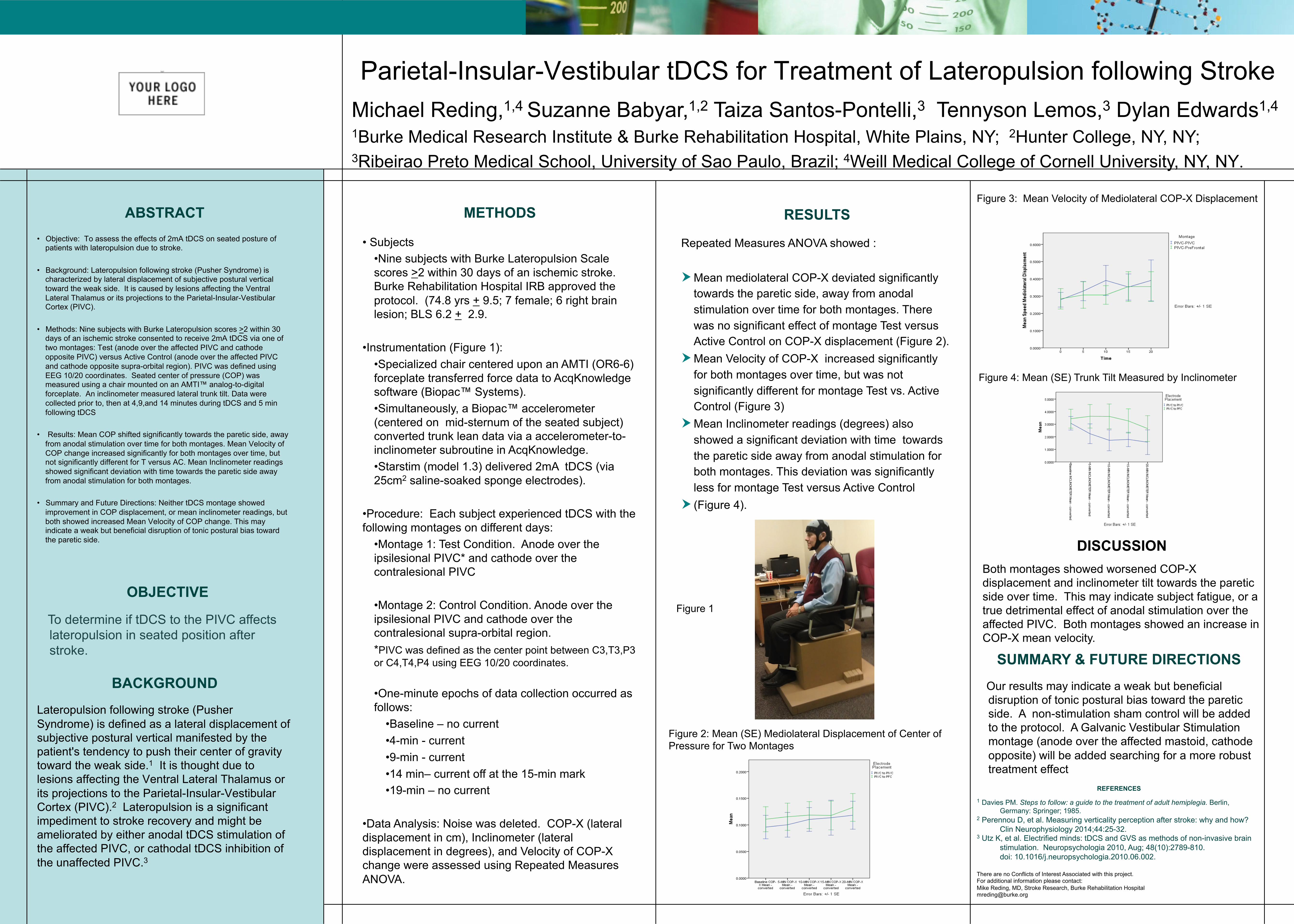
Parietal-Insular-Vestibular tDCS for Treatment of Lateropulsion following Stroke
ABSTRACT • Objective: To assess the effects of 2mA tDCS on seated posture of
patients with lateropulsion due to stroke. • Background: Lateropulsion following stroke (Pusher Syndrome) is
characterized by lateral displacement of subjective postural vertical toward the weak side. It is caused by lesions affecting the Ventral Lateral Thalamus or its projections to the Parietal-Insular-Vestibular Cortex (PIVC).
• Methods: Nine subjects with Burke Lateropulsion scores >2 within 30
days of an ischemic stroke consented to receive 2mA tDCS via one of two montages: Test (anode over the affected PIVC and cathode opposite PIVC) versus Active Control (anode over the affected PIVC and cathode opposite supra-orbital region). PIVC was defined using EEG 10/20 coordinates. Seated center of pressure (COP) was measured using a chair mounted on an AMTI™ analog-to-digital forceplate. An inclinometer measured lateral trunk tilt. Data were collected prior to, then at 4,9,and 14 minutes during tDCS and 5 min following tDCS
• Results: Mean COP shifted significantly towards the paretic side, away
from anodal stimulation over time for both montages. Mean Velocity of COP change increased significantly for both montages over time, but not significantly different for T versus AC. Mean Inclinometer readings showed significant deviation with time towards the paretic side away from anodal stimulation for both montages.
• Summary and Future Directions: Neither tDCS montage showed
improvement in COP displacement, or mean inclinometer readings, but both showed increased Mean Velocity of COP change. This may indicate a weak but beneficial disruption of tonic postural bias toward the paretic side.
BACKGROUND
Lateropulsion following stroke (Pusher Syndrome) is defined as a lateral displacement of subjective postural vertical manifested by the patient's tendency to push their center of gravity toward the weak side.1 It is thought due to lesions affecting the Ventral Lateral Thalamus or its projections to the Parietal-Insular-Vestibular Cortex (PIVC).2 Lateropulsion is a significant impediment to stroke recovery and might be ameliorated by either anodal tDCS stimulation of the affected PIVC, or cathodal tDCS inhibition of the unaffected PIVC.3
Michael Reding,1,4 Suzanne Babyar,1,2 Taiza Santos-Pontelli,3 Tennyson Lemos,3 Dylan Edwards1,4 1Burke Medical Research Institute & Burke Rehabilitation Hospital, White Plains, NY; 2Hunter College, NY, NY; 3Ribeirao Preto Medical School, University of Sao Paulo, Brazil; 4Weill Medical College of Cornell University, NY, NY.
OBJECTIVE
To determine if tDCS to the PIVC affects lateropulsion in seated position after stroke.
METHODS
• Subjects • Nine subjects with Burke Lateropulsion Scale scores >2 within 30 days of an ischemic stroke. Burke Rehabilitation Hospital IRB approved the protocol. (74.8 yrs + 9.5; 7 female; 6 right brain lesion; BLS 6.2 + 2.9.
• Instrumentation (Figure 1): • Specialized chair centered upon an AMTI (OR6-6) forceplate transferred force data to AcqKnowledge software (Biopac™ Systems). • Simultaneously, a Biopac™ accelerometer (centered on mid-sternum of the seated subject) converted trunk lean data via a accelerometer-to-inclinometer subroutine in AcqKnowledge. • Starstim (model 1.3) delivered 2mA tDCS (via 25cm2 saline-soaked sponge electrodes).
• Procedure: Each subject experienced tDCS with the following montages on different days:
• Montage 1: Test Condition. Anode over the ipsilesional PIVC* and cathode over the contralesional PIVC
• Montage 2: Control Condition. Anode over the ipsilesional PIVC and cathode over the contralesional supra-orbital region. *PIVC was defined as the center point between C3,T3,P3 or C4,T4,P4 using EEG 10/20 coordinates. • One-minute epochs of data collection occurred as follows:
• Baseline – no current • 4-min - current • 9-min - current • 14 min– current off at the 15-min mark • 19-min – no current
• Data Analysis: Noise was deleted. COP-X (lateral displacement in cm), Inclinometer (lateral displacement in degrees), and Velocity of COP-X change were assessed using Repeated Measures ANOVA.
RESULTS
Repeated Measures ANOVA showed :
� Mean mediolateral COP-X deviated significantly towards the paretic side, away from anodal stimulation over time for both montages. There was no significant effect of montage Test versus Active Control on COP-X displacement (Figure 2).
� Mean Velocity of COP-X increased significantly for both montages over time, but was not significantly different for montage Test vs. Active Control (Figure 3)
� Mean Inclinometer readings (degrees) also showed a significant deviation with time towards the paretic side away from anodal stimulation for both montages. This deviation was significantly less for montage Test versus Active Control
� (Figure 4).
SUMMARY & FUTURE DIRECTIONS
Our results may indicate a weak but beneficial disruption of tonic postural bias toward the paretic side. A non-stimulation sham control will be added to the protocol. A Galvanic Vestibular Stimulation montage (anode over the affected mastoid, cathode opposite) will be added searching for a more robust treatment effect
REFERENCES
1 Davies PM. Steps to follow: a guide to the treatment of adult hemiplegia. Berlin, Germany: Springer; 1985.
2 Perennou D, et al. Measuring verticality perception after stroke: why and how? Clin Neurophysiology 2014;44:25-32.
3 Utz K, et al. Electrified minds: tDCS and GVS as methods of non-invasive brain stimulation. Neuropsychologia 2010, Aug; 48(10):2789-810.
doi: 10.1016/j.neuropsychologia.2010.06.002. There are no Conflicts of Interest Associated with this project. For additional information please contact: Mike Reding, MD, Stroke Research, Burke Rehabilitation Hospital [email protected]

Both montages showed worsened COP-X displacement and inclinometer tilt towards the paretic side over time. This may indicate subject fatigue, or a true detrimental effect of anodal stimulation over the affected PIVC. Both montages showed an increase in COP-X mean velocity.
Figure 2: Mean (SE) Mediolateral Displacement of Center of Pressure for Two Montages
Figure 3: Mean Velocity of Mediolateral COP-X Displacement
DISCUSSION
Figure 1
Figure 4: Mean (SE) Trunk Tilt Measured by Inclinometer