Embed Size (px)
Citation preview

25
NOAA Technical Report NMFS 25
Parasitology and Pathologyof Marine Organisms ofthe World Ocean
William J. Hargis, Jr. (Editor)
March 1985
U.s. DEPARTMENT OF COMMERCENational Oceanic and Atmospheric AdministrationNational Marine Fisheries Service

NOAA TECHNICAL REPORTS NMFS
The rna po ililies of the Nalional Marine Fisheries Service (NMFS) are to monitor and assess the abundance andgeographic distribution of fishery resources, to understand and predict fluctuations in the quantity and distribution of these
resources, and to establish levels for oplimum use of the resources. NMFS is also charged with the development and implemen
tation of policies for managing national fishing grounds, development and enforcement of domestic fisheries regulations,
surveillance of foreign fishing off United States coastal waters, and the development and enforcement of international fisheryagreements and policies. NMFS also assists the fishing industry through markeling service and economic analysis programs,
and mortgage insurance and vessel construction subsidies. It collects. analyzes. and publishes statistics on various phases of the
industry.
The NOAA Technical Report NMFS series was established in 1983 to replace two subcategories of lhe Technical Reports
series: "Special Scientific Report-Fisheries" and "Circular." The series contains the following types of reports: Scientific
investigalions that document long-term continuing programs of NMFS, intensive scientific reports on studies of restricted
scope, papers on applied fishery problems, technical reports of general inleresl intended to aid conservation and management,
reports that review in considerable detail and at a high technical level certain broad areas of research, and technical papers
originating in economics studies and from management investigations.
Copies of NOAA Technical Report NMFS are available free in limited numbers to governmental agencies, both Federal
and State. They are also available in exchange for other scientific and technical publications in the marine sciences. Individual
copies may be obtained from: U.S. Department of Commerce, National Technical Informalion Service, 5285 Port Royal Road,
Springfield, VA 22161.
NOAA Technical Report NMFS 25
Parasitology and Pathologyof Marine Organisms ofthe World Ocean
William J. Hargis, Jr. (Editor)
March 1985
u.s. DEPARTMENT OF COMMERCEMalcolm Baldrige, Secretary
National Oceanic and Atmospheric AdministrationJohn V. Byrne, Administrator
National Marine Fisheries ServiceWilliam G. Gordon, Assistant Administrator for Fisheries

FOREWORD
The Symposium in which the communications, as they were called during the meeting, comprisingthis volume were presented was held at the Zoological Institute of the Academy of Sciences of theU.S.S.R. in Leningrad during 13 to 16 October 1981. Conducted as part of the cooperative program ofthe U.S.A.-U.S.S.R. Working Group on Biological Productivity and Biochemistry of the World Ocean,the Leningrad meeting was sponsored by the Academy of Sciences of the U.S.S.R. (the Zoological Institute) and the Ministry of Fisheries of the U.S.S.R. (The Scientific Council on Fish Diseases of theIchthyological Commission). It was an extremely interesting and successful Symposium, offering allparticipants the opportunity to describe the results of their studies and reviews during the course of theformal presentations and direct interchange between scientists during breaks in the program and theorganized and casual social activities. The facilities provided by the Zoological Institute were quite adequate and the assistance offered by its Director, O. A. Scarlato and his staff in organization,logistics, andtranslation was excellent. Several of our Soviet colleagues presided over the proceedings, as did I. Allwere businesslike and efficient, yet graceful and accommodating. To O. N. Bauer Jell the brunt of programmatic detail and follow-up. He bore his burdens well and, with Director Scarlato and his staff, including A. V. Gussev and others of the professional and technical staffs of the Zoological Institute,helped make our stay pleasant and the Symposium productive. These organizations and individualsdeserve much credit and praise as well as the thanks of their American and British colleagues.
William J. Hargis, Jr.
ii

CONTENTS·
I. INTRODUCTION .II. COMMUNICATIONS TREATING GENERAL OR HISTORICAL ASPECTS
BAUER, O. N., and Yu. I. POLIANSKI. Present state and perspectives of Soviet investigations on marine parasitology . 5SINDERMANN, C. J. Recent studies on marine fish parasites and diseases 7KUROCHKIN, Yu. V. Applied and scientific aspects of marine parasitology. . . . . . . . . . . . . . . . . . . . . . . . . . . . . . . . . . . 15
Ill. COMMUNICATIONS TREATING PARASITOLOGY AND/OR PATHOLOGY OF FISHES1. REPORTS INCLUDING A MIXTURE OF MAJOR PARASITE TAXA
ZUBCHENKO, A. V. Use of parasitological data in studies of the local groupings of rock grenadier, Coryphaenoidesrupestris Gunner . . . . . . . . . . . . . . . . . . . . . . . . . . . . . . . . . . . . . . . . . . . . . . . . . . . . . . . . . . . . . . . . . . . . . . . . . . . . . . . . . 19
GAEVSKAYA, A. V., A. A. KOVALIOVA, and G. N. RODJUK. Parasitofauna of the fishes of the Falkland-Patago-nian region 25
ALIOSHKINA, L. D., A. V. GAEVSKAYA, and A. A. KOVALIOVA. Parasitofauna of fishes of the Whale Ridge. .. .. 29RODJUK, G. N. Parasitic fauna of the fishes of the Atlantic part of the Antarctic (South Georgia Island and South
Shetland Isles) . . . . . . . . . . . . . . . . . . . . . . . . . . . . . . . . . . . . . . . . . . . . . . . . . . . . . . . . . . . . . . . . . . . . . . . . . . . . . . . . . . . 31DUBINA, V. R. On the parasitofauna of Xiphioidea of the northwest area of the Indian Ocean 33KONOVALOV, S. M., and T. E. BUTORINA. Parasites as indicators of specific features offish ecology. . . . . . . . . . . . . 35POZDNYAKOV, S. E. The taxonomic composition and origin of fish helminths in the epipelagic zone of the World
Ocean , .. . . .. . .. .. . . 39LYADOV, V. N. Zoogeographical characteristics of the helminths of fishes from the Antarctic zone of the World
Ocean... .. .. . . .. .. .. .. ... ..... ... . . .. . . .. .. .. . .. .. ... . .... . .. . . . . . . .. ... . . . .. . . . . . . .. . . . . . .. . . 41TKACH UK, L. P. Special features of the helminth fauna of Helicolenus maculatus (Cuvier) . . . . . . . . . . . . . . . . . . . . . . . 45ZHUKOV, E. V. The flatworm fauna of fishes of the Gulf of Mexico and its genetic relations. . . . . . . . . . . . . . . . . . . . . . 47SHCHEPKINA, A. M. The influence of helminths on the tissue lipid content of Black Sea anchovy, Engraulis encrasi-
colus ponticus, and bullhead, Neogobius melanostomus, during the annual cycle 492. REPORTS INVOLVING INDIVIDUAL MAJOR PARASITE TAXA
STEIN, G. A. Certain results of the study of ciliates of the family Trichodinidae (Peritrichida) inhabiting fishes of theseas of the U.S.S.R. . . . . . . . . . . . . . . . . . . . . . . . . . . . . . . . . . . . . . . . . . . . . . . . . . . . . . . . . . . . . . . . . . . . . . . . . . . . . . . . 53
KOVALIOVA, A. A., and S. S. SCHULMAN. Special features of the myxosporidian fauna from sea and ocean fishes .. 55KRASIN, V. K. Myxosporidia of fishes of the North Pacific " , . .. .. .. 59USPENSKAYA, A. V. Investigations of the ultrastructure and cytochemical peculiarities of Kudoa quadratum (Thelo-
han, 1895) (Myxosporidia, Multivalvulea) , , , . . .. . . .. . . 61KOROTAEVA, V. D. Trematodes [digeneids2 ] of commercial fish of the Pacific of practical importance . . . . . . . . . . . . 63IVANCHENKO, O. F., and T. A. GROZDILOV A. Infestation rate of the young of White Sea herring, reared under
experimental conditions and caught in the sea, by trematodes [digeneids] and their pathogenic effect .. . . . . . . . . . . . 65NIKOLAEVA, V. M. Trematodes-Didymozoidae fauna, distribution and biology. . . . . . . . . . . . . . . . . . . . . . . . . . . . . . 67GHICHENOK, L. A. Comparative analyses of monogenean faunas and populations from several Beloniformes fishes. . 73EGOROVA, T. P. New data on the capsalid fauna of the World Ocean and questions of its specificity. . . . . . . . . . . . . . . 75LEBEDEV, B. Iv. On the taxonomic position of the monogenean Pseudaxine mexicana Meserve, 1938 77AVDEEV, G. V. Plerocercoids of some Cestoda as bioindicators of the population structure of Podonema longipes . . . . . 79SOLONCHENKO, A. I. Development of larval stages of Bothriocephalus scorpii . . . . . . . . . . . . . . . . . . . . . . . . . . . . . . . . 83KAZACHENKO, V. N., and V. M. TITAR. Special features of the geographical distribution and practical significance
of the parasitic copepods of fishes of the Pacific. . . . . . . . . . . . . . . . . . . . . . . . . . . . . . . . . . . . . . . . . . . . . . . . . . . . . . . . 85AVDEEV, V. V. Specific features of the distribution of marine parasitic isopod crustaceans of the family Cymothoidae
(Isopoda, Flabellifera). . . . . . . . . . . . . . . . . . . . . . . . . . . . . . . . . . . . . . . . . . . . . . . . . . . . . . . . . . . . . . . . . . . . . . . . . . . . . 893. REPORTS DEALING WITH DIVERSE TOPICS OR MIXED HOSTS (MISCELLANEOUS)
WOLKE, R. E., C. J. GEORGE, and V. S. BLAZER. Pigmented macrophage accumulations (MMC; PMB): Possiblemonitors of fish health .. . . . . . . . . . . . . . . . . . . . . . . . . . . . . . . . . . . . . . . . . . . . . . . . . . . . . . . . . . . . . . . . . . . . . . . . . . . 93
POTIEVSKI, E. G., L. A. TSAREVA, and V. V. BURLIN. Infectious diseases of fish involved in marine aquaculture inthe Soviet Far East 99
IV. COMMUNICATIONS TREATING PARASITOLOGY AND/OR PATHOLOGY OF MOLLUSKS AND CRUSTACEANSHARGIS, W. J., Jr. Recent studies in the United States on parasites and pathogens of marine mollusks, with emphasis
on diseases of the American oyster, Crassostrea virginica GmeHn 101MATSHKEVSKI, V. K. Some aspects of the biology of the trematode, Proctoeces maculatus, in connection with the
development of mussel farms on the Black Sea. . . . . . . . . . . . . . . . . . . . . . . . . . . . . . . . . . . . . . . . . . . . . . . . . . . . . . . . . 109GALAKTIONOV, K. V. Special features of the infection of the mollusk, Littorina rudis (Maton, 1797), with parthen-
itae of Macrophallus pygmalus (Levinson, 1881) nec Odhner, 1905 and M. piriformes (Odhner, 1905) GaJaktionov,1980 (Trematoda: Microphallidae) from the White Sea. . . . . . . . . . . . . . . . . . . . . . . . . . . . . . . . . . . . . . . . . . . . . . . . . . III
IOrganized into groupings according to types or classes of papers into which several communications can be placed, albeit somewhat arbitrarily.'As employed in these communications, the words trematode or Irematodes means Digenea or digeneids, respectively. Mosl Russian parasitologists accord Class slalus to
Trematoda or Digenea as well as to Monogenea or Monogenoidea.
iii
NAIDENOYA, N. N., C. M. NIGMATULLIN, and A. Y. GAEYSKAYA. The helminth fauna and host-parasite relations of squids (Sthenoteuthis oualaniensis) (Lesson) (Cephalopoda, Ommastrephidae) in the Indian Ocean and theRedSea 113
OYERSTREET, R. M. Some parasitological aspects of shrimp culture in the United States. . . . . . . . . . . . . . . . . . . . . . . . II?NAIDENOYA, N. N., and T. N. MORDYINOYA. The helminths and commensals of crustaceans of the Black Sea. . . . 123
Y. COMMUNICATIONS TREATING PARASITOLOGY AND/OR PATHOLOGY OF MARINE MAMMALSDELAMURE, S. L., and A. S. SKRIABIN. Achievements of Soviet scientists in investigations of helminthofauna of
marine mammals of the World Ocean.... . .. .. .. . 129
The National Marine Fisheries Service (NMFS) does not approve, recommend or endorse any propriety product or proprietary material mentionedin this publication. No reference shall be made to NMFS, or to this publication furnished by NMFS, in any advertising or sales promotion whichwould indicate or imply that NMFS approves, recommends or endorses anyproprietary product or proprietary material mentioned herein, or which hasas its purpose iln intent to cause directly or indirectly the advertised product to be used or purchased because of this NMFS publication.
iv

Parasitology and Pathology of Marine Organisms ofthe World Ocean
Proceedings of the U.S.A.-U.S.S.R. Symposium conducted within the program"Biological Productivity and Biochemistry of the World Ocean" held at
the Zoological Institute of the Academy of Sciences of the U.S.S.R.,Leningrad, October 13-16, 1981 1
WILLIAM J. HARGIS, JR. (Editor?
INTRODUCTION
Background of the Symposium
In 1973 the President of the United States of America and thePremier of the Union of Soviet Socialist Republics signed anagreement establishing the common interest of the two countriesin the phenomena and problems of the World Ocean and callingfor cooperative studies. The resulting joint activities, which involved programs in a number of scientific areas, were placed inthe hands of a combined U.S.A.-U.S.S.R. Committee to providegeneral guidance and oversight and maintain high-level communications. The different disciplinary or operational segments of theseveral programs were assigned to the control of several jointU.S.A.-U.S.S.R. subcommittees called Joint Working Groups.That portion of the programmatic activity dealing with nonfisheries biological phenomena and processes was placed under theJoint Working Group on Biological Productivity andBiochemistry of the World Ocean. Early on, the Chairmen andmembers of the Joint Working Groups met to arrange mutuallysuitable activities, which included exchanges of scientists, jointlysponsored symposia, conferences, or workshops as well as cooperative publications, expeditions, and other projects of mutual interest. These programs and projects were solemnized in formalworking agreements or protocols which included lists of the activities planned for the period in question. The overall program, dueto expire in 1981, has been extended for three more years, until1984.
The last formal protocol of the Working Group on BiologicalProductivity and Biochemistry, developed and signed in Tallin(Estonia) U.S.S.R. in 1979, called for two companion symposia onparasites and pathogens of marine organisms of the World Ocean.One was to be held in each nation to affort maximum opportunityfor participation by scientists of each and for significant interchange of information.
For various reasons the first symposium did not take place untilfall of 1981 in Leningrad, U.S.S.R. We hope that the second orreciprocal meeting in the United States will be convened within ayear or two.
'Contribution No. 1170 from the Virginia Institute of Marine Science, College ofWilliam and Mary, Gloucester Point, VA 23062.
'Professor of Marine Science, The School of Marine Science and The VirginiaInstitute of Marine Science, The College of William and Mary in Virginia; and U.S.Co-Chairman of the U.S.A.-U.S.S.R. Working Group on Biological Productivity andBiochemistry of the World Ocean.
The 1981 meeting, hosted by the Zoological Institute of theAcademy of Sciences of the U.S.S.R., involved over 45 Sovietscientists along with 4 from the United States and 1 from GreatBritain. Pretranslated or instantaneously translated reports werepresented by most participants. It was agreed that acceptablepapers would be published in both languages in the respectivecountries.
The Symposium was extremely productive. The program involved about 50 oral communications or papers on a wide varietyof subjects related to the general topic-marine parasitology andpathology. In accordance with the publication plan, the Sovietshave already or are now presenting the reports in their nationallanguage through the normal publication channels open to them.Many were published in a single appropriately titled volumeprinted and released in Leningrad in 1981 (Bauer et al. 1981).Communications not included in that volume are being publishedin appropriate journals in the Soviet Union.
The volume at hand, designed to provide English-reading audiences with the results of the Symposium, includes all of thecommunications or papers whose authors have agreed to theirpublication and made them available. It is hoped that it will serveto indicate the wide variety of topics discussed and demonstratethe extent and productivity of Soviet workers in this importantfield of biological oceanography or marine ecology. Only fivenon-Russian scientists were able to participate, for variousreasons. Their contributions, though noteworthy and valuable,were overshadowed by those of the numerous Soviet speakers, aswould be expected.3
But the Soviet contributions were impressive in more thannumber or bulk. They reflected the emphasis that Soviet marinescience places upon parasites, pathogens, and diseases of sea andmarine organisms. They also ind'cated the magnitude of theSoviet biological and fishery research efforts in the World Ocean.Personnel of their numerous fishery reconnaissance and fisheryresearch vessels, biological research ships, and the many elementsof their world-ranging fishing fleet have assisted in this work bymaking parasitological collections themselves or by providingcollecting and research platforms for many of the specialistsinvolved.
'Several Soviet participants and our Welsh colleague (Jack Llewellyn) chose not
to have their communications presented here, otherwise the number of paperspresented in this volume would be greater. Also some who apparently did want theirpapers included did not or could not answer my plea for translations, abstracts, andreferences. In such cases the papers arc presented as they were available, with appropriate editing or as we were able to translate and fill them out ourselves.

Without disparaging the significant and valuable efforts ofother countries, including our own, the magnitude of Sovietresearch activity in the parasites, pathogens, and diseases ofmarine OIganisms, especially those of direct or indirect economicimportance, is impressive. Russian scientists have made majorcontributions in fundamental and applied parasitology and pathology of the World Ocean as well as of their own coastal waters andint~rior seas. Thousands of individual hosts were involved inmany of their faunistic and zoogeographic studies-some projectsused many thousands apiece. Hundreds of host species have beenstudied and many are reported in this volume. Our Russian colleagues deserve much credit for their great interest, activity, andproductivity in this vital field.
In preparing the papers for this volume every effort has beenmade to remain faithful to the first translations provided or to theoriginals where translations have been made locally. In many instances extensive editing was required to make them readily intelligible to English readers. In these presentations the editor's interpretations of the meaning of obscure or unusual words or phrases(to American readers) has been included in brackets after thewords or phrases in question in order that the author's words couldbe retained. I regret and apologize to the original authors ortranslators, where their versions are significantly different fromthose presented herein (and I hope there are few), for any divergences from original meaning which may have been introduced inthe extensive editing process.
It is hoped that this volume will convey to the reader the scopeand detail of the communications contributed by Soviet colleaguesas well as our own and will prove a useful addition to the worldliterature of parasitology and pathology.
Organization of the Volume
Developing a suitable organization for this volume was difficult. The papers presented during the symposium were varied intopic, scope or range, emphasis, and extent. Some were historical,dealing witb past and current activities in research on parasitesand pathogens. Some were reviews. Some presented findings ingeneral fashion. Others were quite specific in treating results ofrecent research. A few dealt with tbe practical aspects of fisheryparasitology and disease studies such as the effects on perishability (shelf-life), attractiveness, and marketability of fishery prod.ucts and even their possible effects on the health of humans.Several presented discussions of the effects of diseases andparasites on fish and invertebrates under culture and techniques ofdealing with them. A larger number dealt with parasites as bioindicators, useful in studies of paleogeography, zoogeography, andphylogeny of their bosts. Zoogeographical topics are significant ina number of them. A few treated quite different aspects such as theinfluence of parasites on the physiology of their hosts, and onedealt with the possible use of pigmented macrophage accumulations as indicators of fish health or of stress from anthropogenicpollution. Certain presentations focused on single major taxonomic groups (mostly classes) of parasites and pathogens, whileothers dealt with a number of different classes or even phyla in asingle paper. The largest number of presentations (communications) treated fish as hosts, with those dealing with mollusk andcrustacean hosts ranking as distant seconds and thirds, respectively. One paper, which had to be translated from the Russian to beincluded here, dealt with parasites of marine mammals.
With such a diversity of presentations as were made during theSymposium, any selection of organizational arrangement for this
2
publication could only be somewhat arbitrary. As shown in theContents, general or historical papers are presented first (Itemsunder II); those dealing with parasites and pathogens of fish aresecond (Items under III); and those treating invertebrate hosts arethird (Items under IV). One of the papers (Infectious Diseases ofFish Involved in Marine Aquaculture in the Soviet Far East, byPotievski et al.) treated both fish and mollusks now under culturein northeastern Russia and is interposed as a "bridge" betweenthose offerings dealing with fish hosts on the one hand and invertebrate hosts on the other. The single paper on marine mammals isincluded under V.
There are two major categories-Parasites and Pathogens ofFish, and Parasites and Pathogens of Invertebrates. Within thefirst category, which is the larger, the organization into subcategories is as follows: 1) Mixed major parasitic taxa, 2) individual major taxa (i.e., protozoans, digeneids, monogeneids,cestodes, acanthocephalans, nematodes, and crustaceans, whichmay, along with viruses and bacteria, all be included in the "mixed major parasitic taxa" subcategory mentioned above), and 3)miscellaneous. The miscellaneous parasite topic or subcategoryincludes the pigmented macrophage accumulations offering ofWolke et al. as well as the Russian offering dealing with pathogensof salmon and mollusks in the "aquatoria" of the Soviet far east(Potievski et aI., cited above).
ACKNOWLEDGMENTS
A large number of individuals are due acknowledgments andthanks for their assistance in the preparation of this volume. Imust especially mention O. N. Bauer of the Zoological Institute ofthe Academy of Sciences of the U.S.S.R. who contacted andstimulated our Russian colleagues to provide their communications or papers. Those who responded to his urgings should bethanked also. Since their names are included with their offerings,they will not be listed here.
A number of my associates here at the Virginia Institute ofMarine Science of the College of William and Mary must be mentioned with appreciation. Included are: Alice Lee Tillage, ShirleyO. Sterling, and Regina A. Marsh for assistance with typing theseveral early drafts of the original manuscript and Annette C.Stubbs, Dorothy M. E. Smith, and Ruth A. Edwards of the VIMSWord Processing Unit for later drafts.
My wife, Marcia Jean McK. Hargis, assisted in the timeconsuming editing process by reading from the first translationdrafts to assure that the final draft did not stray. To her my gratefulthanks.
Preparation of this volume was made possible by ContractNA82 FAC 0071 from the Northeast Fisheries Center of the National Marine Fisheries Service, National Oceanic and Atmospheric Administration, U.S. Department of Commerce.
Robert L. Edwards, immediate past Director of the NortheastFisheries Center and my able associate in the workings ofthe U.S.half of the Joint Working Group on Biological Productivity andBiochemistry of the World Ocean, encouraged the entire programand provided support from the Center. Helen F. Mustafa hasassisted in her service as NMFS Project Manager. O. A. Scarlato,Director of the Zoological Institute in Leningrad, provided thefacilities and staff assistance which made the Symposium possible. To all of these individuals and to Carl J. Sindermann of theNational Marine Fisheries Service, into whose competenteditorial hands the manuscript was delivered, my appreciation andthanks.

LITERATURE CITED
BAUER, O. N., P. I. GERASEV, A. V. GUSSEV, E. V. ZHUKOV, V. G. KULACHKOVA, Yu. I. POLIANSKI, and T. A. TIMOFEEVA.
1981. Symposium on Parasitology and Pathology of Marine Organisms. Abslracts of Communications Publishing House (Nauke), Leningrad, U.S.S.R.,115 p.
3


Present State and Perspectives of SovietInvestigations on Marine Parasitology
O. N. BAUER! and Yu. I. POLIANSKP
ABSTRACT
Soviet investigations in marine parasitology are reviewed. During the first half of this century the parasitesof fishes living in inland rivers, lakes, and ponds, and, to a lesser extent, those of fishes of the inland (Aral andCaspian Seas) and adjacent seas were studied. At the end ofthe 1950's,long-term studies of marine organisms indifferent parts of tbe World Ocean were undertaken. Less work has been done on parasites and diseases ofcultured sea organisms because maricultnre Is as yet poorly developed In the U.S.S.R.
Investigations of parasites and diseases of sea fishes in theSoviet Union were begun in the early 1930's. This happened afterthe organization of the first department on fish diseases by ourprominent zoologist and parasitologist, Professor Valentin Dogiel.The first expeditions of this department were to the Aral Sea in1930 and to the Caspian Sea in 1931-32. The materials gatheredwere not only faunistic. Data on the biology of the parasite faunaand on the influence of the environment on the fish contributed tothe development of ecological parasitology. The influence ofsalinity on parasitic infection of fishes was discussed thoroughlyin the publications by Dogiel and Bychovski (1934, 1938) basedupon these data. It has been shown that the parasite fauna of fishesof the Aral Sea is truly freshwater in nature. But in the CaspianSea brackish and sea (marine) elements have been found. The useof parasites as indicators of local fish stocks was discussed for thefirst time.
During the second half of the 1930's, parasitological data weregathered from Black Sea fishes. The paper of Osmanov (1940)dealing with these data is especially noteworthy.
Investigations of fish parasites from the Pacific region had beenundertaken earlier by Layman (1930). In 1937 an expedition ofLeningrad University, headed by V. Dogiel, began investigationsof the parasites of fishes and invertebrates of the Sea of Japan. Yu.I. Polianski, M. M. Isakova-Keo, and A. V. Gussev participated.Materials dealing with parasitic Protozoa of fishes (Dogiel 1948),Mollusca and Echinodermata (Polianski 1951a, b), and parasiticCopepoda (Gussev 195 I) from this expedition were published inpostwar years. Interesting data on the parasite fauna of Leuciscusbrandti, a migratory cyprinid, were also published by Isakova-Keoin 1952.
In postwar years investigations of marine parasites increased. Along-term survey of parasites of fishes and shellfishes of theBarents Sea was accomplished by Polianski (1955) and his pupils,Uspenskaya (1960) and Chubrik (1966). The fish parasites of theWhite Sea were thoroughly investigated by Schulman andSchulman-Albova (1953). Helminths of fishes of Pacific seas[bights, bays or basins-coastal waters] were studied by Zhukov(1960), Strelkov (1960), and Oshmarin et al. (1960). A summaryof all these materials was made by V. A. Dogiel in 1954. Many
'Zoological Institute of the Academy of Sciences of the U.S.S.R., Leningrad,U.S.S.R.
'Leningrad State University, Leningrad, U.S.S.R.
s
papers on Monogenea of Pacific fishes were published by B. E.Bychovski.
As can be seen, until the 1950's and 1960's, the chief work inmarine parasitology had been done in the continental and adjacentseas of the Soviet Union.
At the end of the 1950's our country began a long-term program to increase catches of fish and other sea products in the openparts of the World Ocean. These catches increased up to 20million tons in 1975. At the beginning of the program it wasdiscovered that fish stocks of several species in some parts of theWorld Ocean are heavily infected by parasites and could not beused for consumption. Since then the problems of marine parasitology have been recognized to be of economic importance, andthe need for special departments has been emphasized. TheDepartment of Marine Parasitology of the Institute of Biology ofSouthern Seas in Sevastopol headed by V. M. Nikolaeva becamethe first such department. It began its investigations in 1958.Later, similar departments were organized in the Research Institute of the Ministry of Fisheries. In 1966 [two were established],one in the Pacific Institute (TlNRO)-headed by Yu. V. Kurochkin-and the other in the Atlantic Institute (AtlantNIRO)headed by A. V. Gaevskaya, and others. At the beginning of thisprogram, work on helminths dominated. Since then attention hasbeen given to other groups of parasites-Protozoa (especiallyMyxosporidia) and Crustacea (both Copepoda and Isopoda). During the last 20 yr an enormous amount of materal has beengathered. Summaries of much of this work will be presented in thecontributions to this Symposium.
We now have trained and experienced scientists with expertisein different groups of parasites who can solve problems oftheoretical and economic importance. Some examples of the last:Of great economical value are the investigations of the TlNROparasitologists on how to trim carcasses of teragra, Theragrachalcogramma, that are heavily infected by tetrarhynchids. Scientists of the Zoological Institute and AtlantNIRO have worked outrecommendations connected with pelagic fishes infected byMyxosporidia-Multivalvulea of the genus Kudoa. This parasitecauses muscle lysis in stored fish.
Less research has been done on parasites of shellfishes but during the last several years it has increased. Several papers of thisSymposium will be devoted to this problem. An example is theresearch in the Barents and White Seas on ecology, histology, andphysiology of some trematodes heavily infecting fish and seabirds.

The parthenogenetic stages of these species infecting molluskshave been thoroughly studied using cytochemical and electronmicroscopical (ultramicroscopical) methods. Such methods helpus to understand the host-parasite relations of the system. Theresults of this research, done by A. A. Dobrovolski, K. V. Galaktinov, and 1. A. Tichomirov, have shown that recent ecologicalparasitology, marine parasitology included, must be done not onlyon the organismal but also on cellular and molecular levels.
Until now little has been done in our country in the culture offish and shellfish in coastal waters. But during recent years moreattention has been given to this area. Experience of other countries, especially the United States, Japan, and England, has shownthat such culture is often accompanied by mass diseases, thecausative pathogens of which are not only parasites of animalnature, especially those lacking intermediate hosts (i.e., Protozoa,Monogenea, and parasitic crustaceans), but of others in differentsystematic positions such as bacteria, viruses, and fungi. Duringthe last decade a large number of such diseases of marine andanadromous fish have been described. Many are of greateconomic importance in mariculture.
In our country the earliest experience in such diseases was obtained when culture of salmonids was begun in the coastal watersof the Baltic Sea. Vibrio anguillarum was the first pathogen ofeconomic importance studied. BaltNIRKH and its branch inTallin [Estonia] have studied this disease and employed methodsof control, including immunoprophylaxy. Vaccines from the German Democratic Republic and the United States have been examined with good results. However, an increase of such research inmariculture is greatly needed.
It is also noteworthy that the experience of Soviet fish parasitologists in seafish culture has shown that even widely distributedparasites, which are described as harmless, can act as pathogens.Scientists of the Zoological Institute have described mortalities ofyoung White Sea herring infected by only a few specimens oftrematodes such as members of the well-known Lecithaster andBrachyphallus genera. Fish parasitologists from other countrieshave also described such cases. The same will be said when wediscuss the culture of shellfish.
By the end of the 1960's, the urgency of coordinating researchin parasitology and pathology of sea organisms was clearly recognized. In 1970, the first All Union Symposium on Parasitologysponsored by the Council on Fish Diseases of the IchthyologicalCommission was held in Sevastopol with great success. The second took place in Kaliningrad in 1976. Now we are gathered forthe third time, but this time with our colleagues from the UnitedStates and one from Great Britain. During the first two Symposia
6
little attention was paid to pathology of marine organisms,especially cultured ones. We hope that this problem will bethoroughly discussed at this Symposium.
LITERATURE CITED3
CHUBRIK, G. K.1966. Fauna and ecology of Trematoda larvae in Ihe molluscs of Barents and
White seas. [In Russ.) Tr. Murm. MOrsL BioL Stanzii. 10(14):78-166.DOGIEL, V. A.
1948. Parasitic Protozoa of fish of Peter the Great Bay. [In Russ.] lzv. Vses.Nauchno··lsled. lnst. Ozem. Rechn. Ryhn. Kboz. 27:17-66.
1955. The general character of the parasitc faona of animals inhabiting fareastern seas. [In Russ.) Tr. Zool. lost. 21:.53-59.
DOGIEL, V. A., and B. E. BYCHOVSKI.
1934. The fish parasite fauna of the AraI Sea. [In RIISS-, EogL somm.) Para·zitol. Sb. 4:241-346.
1938. Parasites of Caspian fishes. [In ROllS.., EogI. summ.) Publishing houseof the USSR Acad. Sci., Leningrad. 151 p.
GUSSEV, A. V.1951. Parasitic copepods from some sea fishes. [In RUSSo] Parazitol. Sb. 13:
394·463.ISAKOVA-KEO, M. M.
1952. Parasite fauna of uuciscus bnuulti aDd its peculiarities. (In Russ.]Uch. Zap. Leningr. Gos. Univ. 141:230-237.
LAYMAN, E. M.1930. Parasitic worms of fisbes of Peter the Great Bay. (In Russ., Eng!.
summ.) Izv. Tikhookeanskoy nauchoo-pmmyslovoy staozii. 3:1-102.OSMANOV, S. O.
1940. Studies on the parasitology ofBIac1I: Sea fishes. [In Russ.. EogL summ.]Uch. Zap. Leningr. Gos. Pedagog. lost. 30:187-265.
POUANSKI, Yu. I.1951a. On some parasitic ciliates from sea molluscs and Hololhuria. [In
Russ.] Parazito!. Sb. 13:355-370.1951 b. Cmates of the intestine of the sea an:hins. [In Ross.) Parazitol. Sb.
13:371·393.1955. Materials on fish parasitology of the USSR Northern seas. Parasites of
fishes of the Barents Sea. [In Russ.) Tr. Zoollost. 19:.5-170.SCHULMAN, S. S., and R. E. SCHULMAN-ALBOVA.
1953. Parasites of fishes of the White Sea. [In R......] Publishing house ofthe USSR Acad. Sci., Leningrad., 198 p.
STRELKOV, Yu. A.1960. Endoparasitic worms of the sea fisbcs of castero Kamchatka. [In Russ.)
Tr. Zoo!. Inst. 28:147·196.USPENSKAYA, A. V.
1960. Parasitofaune des crustaces benthiques de Ia mer de Barents (Exposepreliminaire). An. de Parasitologic Hum. et Comp. 35(3):221-242.
ZHUKOV, E. v.1960. Endoparasitic worms of fishes of the Japanese Sea. [In Russ.) Tr.
Zoo!. Inst. 28:3-146.
'Not all citations mentioned in tbe text are included here.

Recent Studies on Marine Fish Parasites and Diseases
CARL J. SINDERMANNI
ABSTRACT
Progress in research on marine flSb diseases and parasites in tbe United States can be discussed in tbreecategories: Diseases and parasites in natural populations, disease in degraded babitats, and disease in marineaquaculture. Principal interest is in tbe effects of diseases on wild or cultured populations of economic importance, but tbere are subsidiary interests in diseases and parasites as indicators of environmental cbanges, inpublic bealtb aspects of fisb diseases and parasites, and in effects of parasites on market quality of flSb products.
Studies of natural populations of fisb bave concentrated on patbogens and parasites wbicb occur atepizootic levels. Important bere are viral diseases, particularly viral erytbrocytic necrosis, some bacterialdiseases, sucb as tbat caused by Pasteurella piscicida, and a systemic fungus disease caused by lchthyophonushoferi. Attempts to quantify tbe effects of disease on fisb abundance bave increased, and tbere is growing acceptance of tbe conclusion tbat disease-related mortality is a significant environmental factor in tbe sea.
Recent emphasis has been placed on diseases which may be associated with estuarine/coastal pollution in thevicinity of buman population centers. Several diseases and disease signs-notably integumental lesions sucb asulcers, fin erosion, papillomas, and Iymphocystis-bave been associated (some only circumstantially) with environmental degradation. Chromosomal anomalies and developmental abnormalities (skeletal malformations)have been studied also, from tbe viewpoint of increased prevalences in polluted zones. An important role forfacultative microorganisms-particularly bacteria of tbe Vibrio, Pseudomonas, Aeromonas group-bas emerged.Evidence is accumulating for localized effects of pollutants on fish populations, and for the utility of certaindisease signs as indicators of environmental stress.
The slow hut continuous development of marine aquaculture in tbe United States has been accompanied byincreasing attention to diseases and disease control. Sea cage rearing of Pacific salmon (genus Oncorhynchus) onboth coasts has been possible only by control of vibriosis (principally Vibrio anguillarum). Effective polyvalentvaccines now limit the problem, but other diseases, such as bacterial kidney disease (BKD) and furunculosis(caused by Aeromonas salmonicida) persist. Additionally, certain salmonid virus diseases seem capable of transmission and of causing mortality of salmon in seawater.
Parasites which reduce market quality of marine fish include the bistozoic myxosporidan Protozoa andlarval belmintbs, particularly larval nematodes. Few of tbese are of public health concern, but many result inrejection of infected fish as food.
Witb tbe impetus provided by declining marine fish stocks, increasing coastal pollution, and developingaquaculture, fisb disease researcb has expanded in recent decades. There is still a need for quantification ofdisease effects on population abundance.
INTRODUCTION
Many parasilological and pathological problems concerningmarine fish are being addressed by scientists in a number of countries. Interest is focused logically on those which affect survival,either in natural or cultivated populations. Of the many topicswhich might be considered, I have selected the following four fordiscussion in this paper: 1) Vibriosis, 2) the emerging role ofviruses in marine fish-especially lPN-like viruses, 3) blood Protozoa, and 4) fungus (/chthyophonus) epizootics.
In discussing these categories, I will try to emphasize resultsobtained since 1970, recognizing that a number of the diseaseentities have been known and examined well before that date.
After this consideration of examples of progress in diseaseresearch in natural and cultivated fish populations, I would thenlike to consider pollution-associated diseases, and our presentunderstanding of the relationship of estuarine/coastal pollutionand fish diseases.
I Northeast Fisheries Center Sandy Hook Laboratory, National Marine FisheriesService, NOAA, Highlands, NJ 07732, U.S.A.
7
VIBRIOSIS OF FISH
Vibriosis of marine fish has had a long history of attention byfish pathologists-beginning over half a century ago with "reddisease" of eels caused by Vibrio anguillarum (Bergman 1909).Vibriosis has occurred at epizootic levels in wild populations aswell as cultivated ones. Juvenile saithe, Gadus virens, have experienced repeated mortalities on the Norwegian coast due to V.anguillarum outbreaks, the most recent being 1967 and 1974(Anonymous 1975). Vibrio anguillarum has also been identified asa pathogen of winter flounder, Pseudopleuronectes americanus,from the western North Atlantic (Levin et aJ. 1972).
With the continued development of marine aquaculture, thegenus Vibrio has emerged as probably the most importantpathogen group for fish and invertebrates (Sindermann 1977).Vibrio infections and Vibrio-caused mortalities have been reportedfor virtually every species which has been cultivated extensivelyin saltwater. Vibriosis has been characterized as a haemorrhagicsepticemia, with lesions attributable to both exotoxin and endotoxin (Bullock and Conroy 1971; Umbreit and Ordal 1972;Harbell et aJ. 1979).
Probably nowhere have Vibrio infections been of greater consequence than in saltwater culture of salmonids. At times it seemedas if the continued existence of seawater cultivation of salmon

depended on successful solution of the vibriosis problem, andsome aspects of the problem still exist today, even though remarkable advances have been made in disease control, particularlythrough immunization.
Sea pen culture of Pacific salmon of several species, but particularly of coho salmon, Oncorhynchus kisu/ch, began on the westcoast of the United States in the early 1970's, and mortalities dueto vibriosis were noted almost immediately (Harrell et al. 1975).Initial approaches to control emphasized antibiotic treatment, butoral and other types of immunization developed rapidly. Immunization by injection of juveniles with killed pathogens was effective but labor-intensive. This method has now been replaced byhyperosmotic spray and bath immunization, augmented whennecessary by antibiotic treatment or use of furanace (where permitted) (Egidius and Andersen 1979). Oral immunization has alsobeen demonstrated to be effective (Fryer et al. 1978). Polyvalentbacterins have been produced, and vibriosis, while still a problem,is now considered a controllable factor in salmon cultivation inthe Pacific Northwest and in European waters. Vaccination is nowpart of the standard protocol for cultivation of Pacific salmon insea cages in the northwestern United States.
Experience with vibriosis in cage culture of Atlantic salmon,Salmo solar, in the northeastern United States and in northernEurope has profited from advances made in the Pacific. Thedisease, for example, has been a threat to Norwegian cage culture,particularly in coastal areas where vibriosis epizootics have alsooccurred in wild populations of saithe.
An emerging phenomenon, both in North America and inEurope, is the appearance of new strains or serotypes of V. anguillarum, which must be incorporated into the immunization protocol (Sawyer and Towle 1976; Egidius and Andersen 1978).Bacterins of west coast origin have been augmented with fractionsreactive with additional strains of Atlantic vibrios to controldisease in U.S. east coast stocks. The probability of a large numberof serotypes complicates bacterin production, and requires constant vigilance to detect new forms of the disease. Some crossprotection is provided by various vaccines, but not enough and notconsistently.
Another emerging phenomenon is the appearance of some ofthe weD-known salmonid diseases, such as furunculosis (causedby Aeromonas salmonicida), bacterial kidney disease (caused byRenibacterium salmoninarum), and Viral Hemorrhagic Septicemia(VHS)--which have been and continue to be serious hatcheryproblems.--and causes of mortality in seawater as well (Frantsi etaI. 1971; Castric and deKinkelin 1980; Paterson et al. 1981).These diseases may be carried in a latent state from fresh to saltwater, but furunculosis and VHS have been transmitted from fishto fisb in seawater (Scott 1968; Novotny 1975; Castric anddelGnkelin 1980). Novotny, for example, cited instances of mortalities in sea cages of chinook salmon, Oncorhynchus tshawy/scha,of up to 80% in 5 mo, caused by simultaneous epizootics of furunculosis and vibriosis. The furunculosis organism in this studyproved to be salt-tolerant, as well as sulfa- and oxytetracyclineresistant..
Other halophilic Vibrio species (V. parahemolyticus, V. alginolytials. and V. cholerae) have also been involved, along with V.anguillarum, in diseases of cultivated shrimps, crabs, and mollusksin several countries (DiSalvo et a1. 1978; Liebovitz 1978; Brownand Losee 1978). Vibriosis affects larval and juvenile stages inparticular.. Additionally, vibriosis was also responsible for highmortalities in juvenile turbot (Scophthalmus maximus) being farmed in the United Kingdom (Horne et al. 1977).
8
VIRUSES IN MARINE FISH
lPN-Like Viruses
Knowledge of viral diseases of marine fish has increasedremarkably in the past decade. Much recent attention has beengiven to the so-called lPN viruses. A disease of salmonids knownas Infectious Pancreatic Necrosis (IPN) was first described byMcGonigle (1941) and its viral nature demonstrated by Wolf et al.(1960). Resembling a reovirus, the pathogen has since beendescribed from a wide range of vertebrate and invertebrate hosts(including several species of mollusks) (Hill 1976; Underwood etaI. 1977). Probably the most interesting recent finding in theUnited States is that a highly lethal and common disease of Atlantic menhaden, Brevoonia tyrannus, long known as "spinningdisease," has been associated with infection of the central nervoussystem by an lPN-like virus (Newman 1980; Stephens, Hetrick,and Newman 1980; Stephens, Newman, Zachary, and Hetrick1980).
The reports represent a satisfying conclusion to decades ofuncertainty about the cause of annual mortalities of menhadenwhich bave occurred at various locations on the U.S. east coastand for wbich records of mass deaths extend back to the 18th century. The disease was transmitted by intraperitoneal injection;typical spinning signs were produced, and the virus was reisolatedfrom experimental fish. The virus was antigenically similar toIPN. Subsequent exposures of menhaden and American shad,Alosa sapidissima, to water seeded with virus also produced infections. It is relevant that some gross signs of IPN infection aresimilar in menhaden and salmonids. The spinning behavior,exophthalmia, and heightened pigmentation are similar, butmenhaden do not usually exhibit visceral hemorrhages.
Additionally, severe outbreaks of lPN-like virus have beenreported in eels (Anguilla japonica and A. anguilla) from Japan(Sano 1976; Pilcher and Fryer 1980). The virus, antigenicallyrelated to lPN, and called EVE, has produced extensive annualmortalities in eel culture ponds. It has been transmitted directlyand by inoculation in juvenile eels, producing mortalities of 55 to60% of experimental fish. The virus was reisolated from moribund fish, and seems distinct from other viruses isolated from theblood of European eels with stomatopapilloma (cauliflowerdisease).
From these and other studies, it seems that the former narrowconcept of IPN virus of salmonids must be broadened to includean entire array of lPN-like viruses from fish and" invertebrates,and from freshwater, anadromous, catadromous, and marinefishes.
Viral Erythrocytic Necrosis
Among the blood parasites of fishes, much attention has beenpaid during the past decade to a disease known as "Piscine Erythrocytic Necrosis" (PEN). Viral etiology has been indicated insome studies, so the disease has been redescribed as "Viral Erythrocytic Necrosis" (YEN). Cod, Gadus morhua, have been foundto be infected on both sides of the Atlantic (Laird and Bullock1969; Reno and Nicbolson 1980; Smail and Egglestone 1980).Other Atlantic species of commercial importance-herring(ClIlpea harengus), alewife, Alosa pseudoharengus, American eel,Anguilla rostrata, and rainbow smelt, Osmerus mordax-havebeen found to be infected (Doyle 1970; Sherburne 1973, 1977).Additionally, the disease has been recognized in Pacific salmon.Bell and Margolis (1916) reported a VEN-Iike agent associated

with an epizootic of cultured pink salmon, Oncorhynchus gorbuscha, in sea pens at Nanaimo, B.C., transmissible by injection tochum salmon, O. keta, and to other pink salmon. Subsequently thedisease was found in several other species of salmonids (Evelynand Traxler 1978) and in Pacific herring, Clupea harengus pallasi,in prevalences up to 59% (MacMillan and Mulcahy 1979). Heavily infected fish had surface hemorrhages.
The usual VEN disease signs-intracytoplasmic inclusions andnuclear changes, often with accompanying anemia-may occur inhigh prevalences. For example, up to 50% of the blood cells of codwere found to be infected in one study (Smail and Egglestone1980). VEN has not been definitely associated thus far with massmortalities, but infected chum salmon were found to be moresusceptible to vibriosis, less resistant to oxygen depletion, andwith decreased osmoregulatory ability (MacMillan et aI. 1980). Itmay well be that the disease has a greater effect on fish populations than was formerly believed.
BLOOD PROTOZOA
In addition to recent attention to the blood disease called ViralErythrocytic Necrosis (VEN), other blood parasites of fish havebeen reexamined from the viewpoint of mortality. The hemoflagellate Trypanoplasma bullocki, found in a number of marinefishes from the u.S. east coast, has been implicated in extensivemortalities of young-of-the-year summer flounder, Paralichthysdentatus (Strout 1965; Daily 1978; Newman 1978; Burreson andZwerner 1982). Field and experimental evidence suggest thatinfections are lethal during the colder months of the year. Thefinding of 100% infection in trawled samples from ChesapeakeBay led to the possibility that the pathogen could have a severe effect on survival of entire year classes of juvenile summer flounder(Burreson and Zwerner 1982).
In other recent studies of hematozoa of fishes, a trypanosome.vas found in high prevalences (up to 94%) .in cod, Gadus morhua,from the Canadian Atlantic coast (Kahn et aI. 1980). Infestationvaried markedly among the stocks of cod sampled.
Another group of blood-inhabiting Protozoa, the haemogregarines, are among the most common of blood parasites of fish.Long considered as benign and widely distributed, their potentially pathogenic role in aquaculture was recently demonstrated(Kirmse 1980). Cultured turbot, Scophthalmus maximus, raised atelevated water temperature, were found to be infected withHaemogregarina sachai, which caused gross tumorous lesions ofthe musculature and viscera, in addition to severe blood parasitemias with marked anemia. The lesions were found in up to 6% ofcultured populations (Kirmse 1978; Kirmse and Ferguson 1976).This seems to be an excellent example of the potentially pathogenic role which may be assumed in aquaculture by parasites considered benign in wild populations-in which mortalities, debilitation, decreased resistance to other pathogens, and reducedmarket value may result.
ICHTHYOPHONUS EPIZOOTICS
A pathogen (or pathogen complex) with a remarkable worldwide distribution, in fresh and saltwater, is the systemic fungusparasite Ichthyophonus hoferi. It is of interest to us here because ofrepeated and extensive epizootics in a number of marine speciesin several parts of the world. New information about the pathogenhas been developed in North America and elsewhere. Originallydescribed as a parasite of rainbow trout in Europe (Plehn and
9
Mulsow 1911), it has subsequently been reported in outbreak proportions in mackerel in coastal waters of the British Isles(Sproston 1944); in Atlantic herring on the east coast of NorthAmerica (Sindermann 1970) and at present in haddock and plaicein waters north of Scotland (McVicar 1980). Furthermore, it hasbeen reported in high prevalence in yellowtail flounder, Limandaferruginea, from the offshore fishing banks of Canada (Powles etaI. 1968; Ruggieri et aI. 1970), from farmed rainbow trout inAustralia (Munday 1976), and from rainbow trout in Japan(Miyazaki and Kubota 1977). Epizootics in cultivated salmonidshave been traced repeatedly to use of raw marine fish in diets(Rucker and Gustafson 1953; Munday 1978).
One of the most interesting aspects of this disease is its apparent periodicity-with six recorded epizootics in herring of thewestern North Atlantic (Fig. I). The most recent outbreak inherring and mackerel of the Gulf of St. Lawrence occurred in1954-56, with effects on population abundance that extended wellinto the 1960's. In its acute form the disease is rapidly fatal, and itwas estimated that almost half the spring-spawned herring of theGulf were destroyed by the epizootic-an estimate that wassupported by statistics from the fishery. For the 5-yr periodpreceding the most recent outbreak, average landings in the southern Gulf of St. Lawrence were 10,800 t; for the 5-yr period immediately following they were only 6,700 t-a decrease of almost40%, with no apparent change in fishing effort (Fig. 2).
Growth rate increased immediately following the mortalities,and larval abundance declined drastically. Some recovery wasnoted beginning in 1960-61 (Tibbo and Graham 1963), andlonger term effects on herring stocks have been described recentlyin the Canadian literature (Parsons and Hodder 1975). Reductionin competition and larval predation seemed to provide conditionsfavorable for production of good year classes-and the 1958 and1959 year classes were very strong. Abundance of these two yearclasses as juveniles from 1959-62 and as adults in 1963-68 mayhave contributed to poor survival of young for almost a decade.
In this example of environmental stress from epizootic diseasecan be seen a mechanism which tends to increase the amplitude ofpopulation fluctuations-positively as well as negatively-andeven to provide some clues about stock recruitment. Man's inter-
.~ :. '. ::. '..
Reported fungus eplzootlc8 InSaint Lawrenee herring
~DL..,-J---L--J:1940 1960 1960
Reported fungus epizootic! In
Gull 01 ,Dhll~_rr_,n...9_L-...J
1920 1940 1960 1960
Figure I.-Reported epizootics oflchthyophonus holeri in herring of the westernNorth Atlantic. The 1940 outbreak is included on the basis or newspaperaccounts only.

Figure 2.-Herring landings in the southern Gulf of St. Lawrence.
addition to mortality, include spoilage and decay of flesh of heavily infected haddock, and often extreme emaciation and jellylikeflesh of infected plaice (McVicar 1980).
POLLUTION-ASSOCIATED DISEASES
The important and timely matter of pollution-associateddiseases of marine fish can be discussed in fi ve categories: I)Disease caused by facultative pathogens: Fin erosion and ulcers;2) stress-provoked latent infections: Shrimp baculovirus, oysterherpes virus, herpes-virus disease of turbot, and Iymphocystis; 3)environmentally induced abnormalities: Neoplasms and skeletalanomalies; 4) genetic abnormalities: Egg and larval geneticdamage; and 5) experimentally induced lesions. Each of thesecategories can be examined here very briefly, with examplesdrawn in part from our ocean pollution research in the westernNorth Atlantic.
605852 54 56
YEAR1948 50
CJ)z~ 12
o~~w~
LL 6o(f)cz0((JJ:::>o 0 ..::I:~
I'tpn 3.-Recent trends In berrinl akbes III tile westera Nortll Atlatlc.
800
Our best candidate in this category is fin erosion or "fin rot" offish, which is probably the best known but least understooddisease syndrome of fish from polluted waters. It has been foundin the New York Bight, the California coast, Puget Sound, Biscayne Bay, Escambia Bay, Irish Sea, Tokyo Bay, and the Frenchcoast. It occurs in two types: 1) Site-specific-especially indemersal fish; probably from contact with contaminated sediments; and 2) generalized-found especially in pelagic fish; withinvolvement of all fins but especially the caudal; occasionalbacterial infections are found.
In the New York Bight, fin erosion has been found in 22 speciesof fish, and has been demonstrated in flounders to be statisticallymore abundant in that highly polluted wne than in other comparable coastal areas. It has a statistical association with high coliform and high heavy metal levels in sediments.
In another U.S. coastal area, southern California, fin rot hasbeen found in many species, and is clearly associated with proximity to ocean outfalls of sewage systems. Wherever studied, finrot disease signs include: Epidermal hyperplasia, dermal fibrosis,hyperemia, occasional hemorrhage, no consistent bacterial infection, and no pronounced inflammatory response.
The possible role of environmental chemical contamination inthe etiology of fin erosion emerges more clearly as additionalstudies are reported. Fish from the New York Bight, reported instudies by Mahoney et a!. (1973), Murchelano (1975), andZiskowski and Murchelano (1975), exist in a highly contaminatedarea, with chemicals such as heavy metals and petroleum residuesin sediments far above background levels. McDermott and Sherwood (1975) in California found DDT to be significantly higherin fish with fin erosion, and PCB levels slightly higher in such fishthan in normal individuals. Wellings et a!. (1976) found abnormally high concentrations of PCB in English sole and starryflounders with fin rot from the Duwamish River in Washington.
Several authors have postulated that fID erosion in flatfish maybe initiated by direct contact of tissues with contaminated sediments. Mearns and Sherwood (1974), for example, suggested thattoxic substances (sulfides, heavy metals, chlorinated hydrocarbons, etc.) could remove or modify the protective mucus coat andexpose epithelial tissues to the chemicals. Sherwood and Bendele(1975) reported that Dover sole from California with fin erosionproduced much less mucus than normal fish.
It seems quite likely that the "fin erosion" syndrome in fish includes chemical stress, probably acting on mucus and epithelium;
Disease Caused by Facultative Pathogens
197019601940 1950YEAR
1930O.l,----.----..,-----~-___,.---___,.--------'
1920
Northwest Atlantic Herring Catch
vention here, if any, may have been a positive one-to reducepopulation size by overfishing beginning in the !Jlid-1960's (Fig.3), thereby reducing density-dependent factors which increasevulnerability of the population to future epizootics. It is possiblethat outbreaks may actually be delayed by this mechanism.
The most recent lchthyophonus outbreak-in haddock, Melanogrammus aeglefinus, and plaice (Pleuronectes platessa)-occurredand still persists in waters north of Scotland. Presence of thefungus was noted as early as 197 1; prevalences in haddock from1976 to 1980 reached 85% in some samples. Prevalences werelower in plaice, not exceeding 25%, but the disease was considered terminal in that species because of the acute nature of theinfection and inefficient host responses. Effects of infection, in
20P
LLarno~ 400rn::laIf-
rnz~()
a: 600f-W~
10

stress resulting from marginal dissolved oxygen concentrations,possibly enhanced by a sulfide-rich environment; and, secondarybacterial invasion in at least some instances.
A second disease possibly related to pollution and effects offacultative pathogens is ulcer disease-which is, next to fin erosion, probably the commonest abnormality in fish from pollutedwaters. It appears to be usually of bacterial etiology. Systemic infections with V. anguillarum have been associated with ulcers inNew England and Europe. Last year, at a special meeting ondiseases of marine fish and shellfish held in Copenhagen, ulcerdisease of cod was described from Denmark, the Irish Sea, and thenorthern coast of France-associated with areas of severe pollution. Vibrio anguillarum was the most common isolate, but Danishworkers have also identified two viruses in ulcerated fish.
Beginning in 1979, pathologists at our laboratory in New Jerseyhave recognized an ulcer disease of red hake, Urophycis chuss. Thedisease appears soon after the late winter inshore migration of thatspecies (Murchelano and Ziskowski 1979).
So, for this first category, diseases caused by facultative pathogens, there is much room available for study at the boundary between infectious and noninfectious diseases. This is the areawhere environmental stress and facultative microorganisms areimportant; where high bacterial populations in eutrophic watersinteract with exposed or injured or chemically modified surfacemembranes; and where nonspecific pathologies such as fin rot andulcers can occur in epizootic proportions.
Stress-Provoked Latent Infections
It is generally recognized that certain salmonid diseases infreshwater-such as kidney disease and furunculosis in latent orcarrier states-may be provoked into patency by environmentalstress. Recent work suggests that latent infections in marinespecies may be provoked into patency by pollutant stress. Wehave found, for example, that Iymphocystis disease of striped bass,Morone americanus-normally a rare disease in natural populations-can occur in high prevalences (up to 25%) in fish overwintering in heated effluents of electric generating stations(Sindermann 1979). Also, a disease caused by a herpes-type viruswas reported from turbot in Scotland in 1978. The flatfish werebeing raised in a fish farm using warmed seawater from a nuclearpower plant. One-year-old fish died in large numbers from thedisease. Viral arrays were seen in wild fish, but not mortalitiessuggesting that the infections were enzootic and latent in the wildturbot population.
Additionally, another herpes-type virus has been found to causemortalities in oysters on the New England coast, and a baculovirusproduced experimental mortalities in penaeid shrimp of the Gulfof Mexico when exposed to PCB's. The conclusion must be thatpollutant stress can give us very important clues about the role ofcarrier states and later infections in the epizootiology of marinediseases.
Environmentally Induced Abnormalities-Neoplasmsand Skeletal Anomalies
We have at least 40 yr of circumstantial evidence-beginningwith Schlumberger and Lucke's (1948) description of catfish epitheliomas in grossly polluted rivers, through the spread of cauliflower disease (Blumenkohlkrankheit) of eels in northern Europesince the 1950's, and the extensive work with flatfish of thePacific coast of North America (Stich et al. 1977)-for a possible
11
relationship of fish neoplasms and environmental pollution. Anextensive literature has been developed in the United States andelsewhere, but a direct causal relationship has not beendemonstrated.
Skeletal anomalies, particularly those of the spinal column offishes, are the subject of an extensive literature. Included arespinal flexures and compressions, vertebral fusions, and head andfin anomalies. Recent studies in California have reported increased prevalences of skeletal deformities, considered to be pollutionrelated. Valentine (1975) presented the most convincing evidencefor environmental influences on induction of abnormalities inthree species of fish from the sOllthern California and BajaCalifornia coasts. He found significantly higher prevalences ofskeletal anomalies, especially gill raker deformities, from LosAngeles and San Diego than from Baja. He suggested a relationship with high heavy metal and chlorinated hydrocarbon levels inCalifornia, but stopped short of stating a direct causal relationship.
Genetic Abnormalities
The mutagenic properties of a number of chemical contaminants, including pesticides and heavy metals, have beendemonstrated in experimental studies with terrestrial animals. Recent work suggests high percentages of chromosomal anomaliesand high prevalence of dead fish eggs in polluted areas of the NewYork Bight (Longwell 1976). All degrees of chromosomal damagehave been found, and higher percentages of anomalies seemassociated statistically with the degree of environmental degradation. It may well be that a new and significant mortalityfactor- increased genetic damage-may have been introducedwith increasing chemical pollution. These genetic disturbancesfall clearly within the definition of disease.
Experimentally Induced Lesions
There is a vast and almost unmanageable literature aboUlinduced lesions in fish after experimental exposure to chemicalcontaminants. Fortunately, a number of good reviews are available on pathological effects of heavy metals, pesticides, and petroleum. Some generalizations that may be drawn are:
Increasing dosages, beyond a threshold, produce increasinglysevere tissue abnormalities;
particular contaminants often exert effects on specific targettissues, but specific lesions cannot usually be described as characteristic of any group or class of chemicals;
much histopathology is nonspecific response to stress or infection;
effects that may be of chemical origin may be obscured bystress-provoked infections with facultative pathogens; and
principal target tissues and organs seem to be gills, liver (orinvertebrate hepatopancreas), and neurosensory cells.
Much experimental evidence exists, but it has numerous flaws:
Dosage levels are often beyond maximum observed environmental levels;
usually single chemicals are tested, ignoring synergisms andantagonisms;
often the tests are static acute and not chronic flow-through;and
experimental animals are often under stress from the mere act

of confinement-they may be injured, sick, or dying at the beginning of the experiment.
These and other flaws reduce some of the value of the experimental evidence, but a great pool of such information exists and isof value because of its volume.
It. is, of course, necessary to be conservative in conclusionsabout associations of pollution and disease, but evidence for suchassociations is increasing.
SOME EMERGING CONCEPTS IN MARINEFISH PATHOLOGY
Disease is a significant factor in survival of fish and shellfish.Information about the role of pathogens in marine populations isaccumulating at an accelerating rate. Among the emerging concepts in marine pathology are the following:
I) The effects of parasites and diseases on marine fishes arequantifiable. In those few instances where quantitative studies ofdisease-caused mortalities have been made (such as the examination of Ichthyophonus epizootics) significant reductions in population size have been observed or inferred.
2) Some of the diseases of salmonids, such as furunculosis andbacterial kidney disease, long considered as problems in freshwater hatcheries, are emerging as problems and causes of mortality during marine phases of host life cycles. Conversely, vibriosis,characteristically a marine problem, can be transported to freshwater by species such as eels.
3) Virus diseases, in latent or patent form, have been identifiedin marine fish, particularly during the past decade. Some of them,such as Iymphocystis and eel stomatopapilloma seem relativelybenign, while others, such as the lPN-like virus of menhaden, arelethal to the host.
4) Studies of diseases in marine aquaculture have shown thatsome parasites, such as the haemogregarines, long consideredbenign, must be reassessed as potential pathogens and causes ofmortalities.
5) In considering pollution-associated diseases of fish thefollowing conclusions seem warranted:
a) The significance of environmental stress from pollutants isemerging as a possible determining factor in a number of fishdiseases. This may take the form of direct chemical-physicaldamage to cell membranes or tissues, modification of biochemicalreactions, and buildup of facultative microbial pathogens.
b) Some circumstantial evidence for the role of environmentalcarcinogens in inducing neoplasms of fish and shellfish is accumulating, and is suggestive, but definitive conclusions are notjustified at present.
c) The presence of marginal or degraded estuarine/coastal environments may be signalled by the appearance of fin erosion,ulcers, Iymphocystis, and skeletal anomalies in teleost fishes. Aclear cause and effect relationship has not yet been demonstratedin every instance, but some statistical associations have beenmade.
d) A number of viruses have been found in fish, crustaceans,and mollusks within the past decade, and the pathogenic role of anumber of them has been demonstrated by increasing environmental stress. It may well be that other latent virus infections willbe identified by similar experimental methods.
12
LITERATURE CITED
ANONYMOUS.
1975. Vibriosis hits Norwegian fish. Mar. Fish. Rev. 37(2):42-43.BELL, G. R., and L. MARGOLIS.
1976. The fish health program and the occurrence of fish diseases in thePacific region of Canada. Fish PathoI. 10:115-122.
BERGMAN, A. M.
1909. Die rote Beulenkrankheit des Aals. Ber. K. Bayer. BioI. Verso Stn.,Miinchen 2:10-54.
BROWN, c., and E. LOSEE.
1978. Observations on natural and induced epizootics of vibriosis in Crassos/rea virginico larvae. J. Invertebr. Pathol. 31:41-47.
BULLOCK, G. L., and D. A. CONROY.
1971. Vibrio diseases. In S. F. Snieszko and H. R. Axelrod (editors), Diseases
of fishes, 2A. Bacterial diseases of fishes, p. 42-50. TFH Pubis., Jersey City,N.J.
BURRESON, E. M., and D. E. ZWERNER.
1982. The role of host biology, vector biology, and temperature in the distri
bution of Trypanop/asma bullocki infections in the lower Chesapeake Bay.J. Parasitol. 68:306-313.
CASTRIC, J., and P. De KINKELIN.
1980. Occurrence of viral haemorrhagic septicaemia in rainbow trout Sa/mo
gairdneri Richardson reared in sea-water. J. Fish Dis. 3:21-27.DAILY, D. D.
1978. Marine fish hematozoa from Maine. J. Parasitol. 64:361-362.DiSALVO, L. H., J. BLECKA, and R. ZEBAL.
1978. Vibrio anguil/arum and larval mortality in a California coastal shellfishhatchery. Appl. Environ. Microbiol. 35:219-221.
DOYLE, W. L.
1970. Occurrence of a virus in erythrocytes of the eel, Anguilla. Bull. Ml.Desert Island BioI. Lab. 10: II.
EGIDIUS, E. c., and K. ANDERSEN.
1978. Host-specific pathogenicity of strains of Vibrio anguil/arum isolatedfrom rainbow trout Salma gairdneri Richardson and saithe Pollachius virens
(L.). J. Fish Dis. 1:45-50.
1979. Bath-immunization-a practical non-stressing method of vaccinatingsea farmed rainbow trout Salrna gairdneri Richardson against vibriosis. J.Fish Dis. 2:405-410.
EVELYN, T. P. T., and G. S. TRAXLER.
1978. Viral erythrocytic necrosis: Natural occurence in Pacific salmon andexperimental transmission. J. Fish. Res. Board Can. 35:903-907.
FRANTSI, c., J. A. RITTER, and P. F. ELSON.
1977. Effect of corynebacterial kidney disease on ocean survi val and return ofAtlantic salmon (Salmo salar). ICES CM 1977/M:29.
FRYER, J. L., J. S. ROHOVEC, and R. L. GARRISON.1978. Immunization of salmonids for control of vibriosis. Mar. Fish. Rev.
40(3):20-23.
HARBELL, S. c., H. O. HODGINS, and M. H. SCHIEWE.
1979. Studies on the pathogenesis of vibriosis in coho salmon Oncorhynchuskisulch (Walbaum). J. Fish Dis. 2:391-404.
HARRELL, L. W., H. M. ETLINGER, and H. O. HODGINS.1975. Humoral factors important in resistance of salmonid fish to bacterial
disease. I. Serum antibody protection of rainbow trout (Sa/mo liairdneri)
against vibriosis. Aquaculture 6:211-219.HILL, B. J.
1976. Isolation of lPN-like viruses from bivalve molluscs. FAO Aquacult.Bull. 8(1):13.
HORNE, M. T., R. H. RICHARDS, R. J. ROBERTS, and P. C. SMITH.
1971. Peracute vibriosis in juvenile turbot Scophthalmus maximus. J. Fish
BioI. 11:355-361.
KAHN, R. A., J. MURPHY, and D. TAYLOR.1980. Prevalence of a trypanosome in Atlantic cod (Gadus morhua) especially
in relation to stocks in the Newfoundland area. Can. J. Fish. Aqua!. Sci. 37:1467-1475.
KIRMSE, P. D.1978. Haemogregarina sachai n. sp. from cultured turbot Scophlhalmus maxi
mus (L.) in Scotland. I. Fish Dis. 1:337-342.1980. Observations on the pathogenicity of Haemorgregarina sachai Kirmse,
1978, in farmed turbot Scophlhalmus maximus (L.). J. Fish Dis. 3:101-114.
KIRMSE, P., and H. FERGUSON.
1976. Toxoplasma-like organisms as the possible causative agents of a proliferative condition in farmed turbot (Scophlhalmus maximus). In L. A. Page
(editor), Wildlife diseases, p. 561-564. Plenum Press, N.Y.

LAIRD, M., and W. BULLOCK.1969. Marine fish haematozoa from New Brunswick and New England. J.
Fish. Res. Board Can. 26:1075·1101.LEVIN, M. A., R. E. WOLKE, and V. J. CABELLI.
1972. Vibrio anguillarum as a cause of disease in winter flounder (Pseudopleu
ronectes american us). Can. 1. Microbiol. 18:1585-1592.
LIEBOVITZ, L.1978. A study of vibriosis at a Long Island shellfish hatchery. ICES, CM
1978/F:17.8 p.LONGWELL, A. C.
1976. Chromosome mutagenesis in developing mackerel eggs sampled fromthe New York Bight. Am. Soc. Limnol. Oceanogr., Spec. Symp. 2:337-339.
MacMILLAN, J. R., and D. MULCAHY.1979. Artificial transmission to and susceptibility of Puget Sound fish to viral
erythrocytic necrosis (VEN). J. Fish. Res. Board Can. 36:1097-1101.MacMILLAN, J. R., D. MULCAHY, and M. LANDOLT.
1980. Viral erythrocytic necrosis: some physiological consequences of infection in chum salmon (Oncorhynchus ketal. Can. 1. Fish. Aquat. Sci. 37:
799-804.MAHONEY, J. B., F. H. MIDLIGE, and D. G. DEUEL.
1973. A fin rot disease of marine and euryhaline fishes in the New YorkBight. Trans. Am. Fish. Soc. 102:596-605.
McDERMOTT, D. J., and M. J. SHERWOOD.1975. DDT and PCB in diseased Dover sole. Annu. rep., Soutbern Calif.
Coastal Water Res. Proj., EI Segundo, p. 33-35.
McGONIGLE, R. H.1941. Acute catarrhal enteritis of salmonid fingerlings. Trans. Am. Fish.
Soc. 70:297-303.
McVICAR, A. H.1980. The effects of /chthyophonus infection in the haddock Melanogrammus
aegle[inus and plaice Pleuronectes platesso in Scottish waters. ICES, Spec.
Meet. Diseases of Commercially Important Marine Fish and Sbellfish. Doc.No. 16.8 p.
MEARNS, A. J., and M. SHERWOOD.1974. Environmental aspects of fin erosion and tumors in southern California
Dover sole. Trans. Am. Fish. Soc. 103:799-810.MIYAZAKI, T., and S. S. KUBOTA.
1977. Studies of Ichthyophanus disease of fishes. Bull. Fac. Fish. Mie Univ.
4:45-80.
MUNDAY, B. L.1976. Disease of Ichthyosporidiosis in trout fed raw saltwater fish. Aust.
Vet. Pract. 6:46.1978. Some diseases of farmed trout. In Proc. No. 36, Course for Veterinar
ians, Fauna Part B, p. 191-195. Univ. Sidney, Post graduate commirtee inveterinary science.
MURCHELANO, R. A.1975. The histopathology of fin rot disease in winter flounder from the New
York Bight. J. Wildl. Dis. 11:263-268.MURCHELANO, R. A., and J. ZISKOWSKI.
1979. Some observations on an ulcer disease of red hake, Urophycis chuss,from the New York Bight. ICES, CM 1979/E:23, 5 p.
NEWMAN, M. W.
1978. Pathology associated with Cryprobia infection in a summer flounder(Paralichrhys den/atus). J. Wildl. Dis. 14:299-304.
1980. IPN virus disease of clupeid fishes. ICES, Spec. Meet. Diseases ofCommercially Important Marine Fish and Shellfish. Doc. No. 24, 5 p.
NOVOTNY, A. J.
1975. Net-pen culture of Pacific salmon in marine waters. Mar. Fish. Rev.37(1):36-47.
PARSONS, L. S., and V. M. HODDER.1975. Biological characteristics of southwest Newfoundland herring, 1965
71. ICNAF Res. Bull. II: 145-160.PATERSON, W. D., S. P. LALL, and D. DESAUTELS.
1981. Studies on bacterial kidney disease in Atlantic salmon (Salmo salar) inCanada. Fish PathoI. 15:283-292.
PILCHER, K. S., and J. L. FRYER.
1980. The viral diseases of fish: a review through 1978. Part I. Diseases ofproven viral etiology. CRC Crit. Rev. Microbiol. 7(4):287-363.
PLEHN, M., and K. MULSOW.
1911. Der Erreger der "Taumelkrankheit" der Salmoniden. Centr. Bakt.Parasitenk. 59:63-68.
POWLES, P. M., D. G. GARNETT, G. D. RUGGIERI, and R. F. NIGRELLI.
1968. Ichthyophonus infection in yellowtail flounder (Limanda [erraginea)
off Nova Scotia. J. Fish. Res. Board Can. 25:597-598.RENO, P. W., and B. L. NICHOLSON.
1980. Viral erythrocytic necrosis (VEN) in Atlantic cod (Gadus morhua): in
13
vitro studies. Can. J. Aquat. Sci. 37:2276-2281.RUCKER, R. R., and P. V. GUSTAFSON.
1953. An epizootic among rainbow trout. Prog. Fish-Cult. 15: 179-181.RUGGIERI, G. D, R. F. NIGRELLI, P. M. POWLES, and D. G. GARNETT.
1970. Epizootics in yellowtail flounder, Limanda [erraginea Storer, in thewestern North Atlantic caused by hhrhyophanus, an ubiquitous parasiticfungus. Zoologica (N.Y.) 55:57-62.
SANO, T.1976. Viral diseases of cultured fishes in Japan. Fish Pathol. 10:221-226.
SAWYER, E. S., and F. G. TOWLE.1976. Commercial salmon culture on the Maine coast utilizing heating waler
from a power plant. Sea Grant Final Report SGO-4-5- I58-50.
SCHLUMBERGER, H. G., and B. LUCKE.1948. Tumors of fishes, amphibians, and reptiles. Cancer Res. 8:657-754.
SCOTT, M.1968. The pathogenicity of Aeromonas salmonicida (Griffin) in sea and brack
ish waters. J. Gen. Microbiol. 50:321-327.
SHERBURNE, S. W.1973. Erythrocyte degeneration in the Atlantic herring, Clupea harengus
harengus L. Fish. Bull., U.S. 71:125-134.1977. Occurrence of piscine erythrocyte necrosis (PEN) in the blood of the
anadromous alewife, Atosa pseudoharengus. from Maine coastal streams. J.Fish. Res. Board Can. 34:28 I -286.
SHERWOOD, M. J., and R. A. BENDELE.1975. Mucous production in Dover sole. Annu. rep., Southern Calif. Coastal
Water Res. Proj., EI Segundo, p. 5 I -53.
SINDERMANN, C. J.1970. Principal diseases of marine fish and shellfish. Acad. Press, N.Y.,
369 p.1979. Pollution-associated diseases and abnormalities of fish and shellfish: A
review. Fish. Bull., U.S. 76:717-749.
SINDERMANN, C. J. (editor).1977. Disease diagnosis and control in North American marine aquaculture.
Elsevier, Amsterdam, 329 p.
SMAIL, D. A., and S. I. EGGLESTONE.1980. Virus infections of marine fish erythrocytes: prevalence of piscine ery
throcytic necrosis in cod Gadus morhua L. and blenny Blennius phalis L. incoastal and offshore waters of the United Kingdom. J. Fish Dis. 3:41-46.
SPROSTON, N. G.
1944. Ichthyosporidium ho[eri (Plehn & Mulsow, 19 I I), an internal fungoidparasite of the mackerel. I. Mar. BioI. Assoc. U.K. 26:72-98.
STEPHENS, E. B., F. M. HETRICK, and M. W. NEWMAN.
1980. A viral etiology for spinning disease in the Atlantic menhaden. ICES,
Spec. Meet. Diseases of Commercially Important Marine Fish and Shellfish.Doc. No. 33, 4 p.
STEPHENS, E. B., M. W. NEWMAN, A. L. ZACHARY, and F. M. HETRICK.1980. A viral aetiology for the annual spring epizootics of Atlantic menhaden
Brevoortia tyrannus (Latrobe) in Chesapeake Bay. J. Fish Dis. 3:387-398.
STICH, H. F., A. B. ACTON, K. OISHI, F. YAMAZAKI, T. HARADA, T. HIBINO,and H. G. MOSER.
t977. Systematic collaborative studies on neoplasms in marine animals as
related to the environment. Ann. N.Y. Acad. Sci. 298:374-388.
STROUT, R. G.t 965. A new hemoflagellate (Genus Cryprobia) from marine fishes of north
ern New England. J. Parasitol. 51 :654-659.TIBBO, S. N., and T. R. GRAHAM.
1963. Biological changes in herring stocks following an epizootic. J. Fish.Res. Board Can. 20:435-449.
UMBREIT, W. W., and E. J. ORDAL.1972. Infection of goldfish with Vibrio anguillarum. Am. Soc. Microbiol.
News 32:93-96.UNDERWOOD, B. 0., C. J. SMALE, F. BROWN, and B. J. HILL.
1977. Relationship of a virus from Tellina tennuis to infectious pancreaticnecrosis virus. J. Gen. Virol. 36:93-109.
VALENTINE, D. W.
1975. Skeletal anomalies in marine teleosts. In W. E. Ribelin and G. Migaki
(editors), The pathology of fishes, p. 695-718. Univ. Wis. Press, Madison.WELLINGS, S. R., C. E. ALPERS, B. B. McCAIN, alld B. S. MILLER.
1976. Fin erosion disease of starry flounder (Platichthys stellarus) and English
sole (Parophrys vetu/us) in the estuary of the Duwamish River, Seattle, Washington. J. Fish. Res. Board Can. 33:2577-2586.
WOLF, K., S. F. SNIESZKO, C. E. DUNBAR, and E. PYLE.1960. Virus nature of infectious pancreatic necrosis in trout. Proc. Soc. Exp.
BioI. Med.104:105-108.Z1SKOWSKI, J., and R. MURCHELANO.
1975. Fin erosion in winter flounder. Mar. Pollut. Bull. 6:26-28.


Applied and Scientific Aspects of Marine Parasitology
Yu. V. KUROCHKINI
'Pacific Research Institute of Fisheries and Oceanography (TlNRO), Vladivostok,U.S.S.R.
Table I.-Approximate number of species in some main groups ofparasites of marine animals. I
'Calculations were made by Manter (1966) 'on trematodes [digeneans),Lebedev (in litl.) on monogeneans, and Kazachenko (in lilt.) on parasiticcopepods of marine fishes.
Parasitism is widely distributed among marine animals. Atpresent about 15,000 species of parasitic organisms are reportedfrom marine ecosystems. But even the most understated calculation shows that on the average not more than 25% of livingmarine parasite species have been described. PracticalIy alIspecies of free-living marine animals are also involved inparasitism, serving as hosts of the appropriate stages of parasitesfrom different systematic groups.
The parasite fauna of marine fishes and invertebrates isextremely rich and diverse and has the largest total biomass. Thisis primarily due to the numerous helminth larvae (which usualIyhave wide specificity) and the peculiar and very rich fauna ofparasitic copepods and isopods (Table I). Many dozens of speciesof parasites are usualIy reported from those fish species whichhave been extensively studied by parasitologists. Numbers ofparasites per host individual in fishes, and especialIy in marinemammals, may reach hundreds of thousands or even milIions.Many parasites are highly pathogenic to their hosts. They cancause diseases and affect the population sizes of marine animals.The role of parasites in natural marine ecosystems is undoubtedlyvery important. Besides, as is generalIy known, some marineparasites can be dangerous for human health. Ultimately, theimmensity of the seas and oceans; the great diversity of theecological conditions due to the wide range of abiotic and bioticfactors (such as temperature, depth, movement of the watermasses etc., the abundance of marine vertebrates and invertebrates and their parasites, and the great biocenotic diversity); thenumerous food species of marine invertebrates and vertebrates,often with very abundant stocks; the highly developed coastal andoffshore fisheries; and, hence, the huge economic losses [whichcan be] caused by parasites-alI these features of applied marineparasitology provide a boundless field for diverse trends of appliedand fundamental investigations.
The principal trends of the development of marine parasitology(as in any other branch of the science) are determined, on the onehand, by the volume and level of knowledge accumulated in thefield and prospects based upon it. On the other hand they dependon the development of research techniques, the general development of adjacent scientific fields and, especialIy, on today's urgentneeds of industry. In marine parasitology such practical requirements arise from the experience and prospects of the marinefisheries.
Taking into account available data on the above-mentionedconditions, I shall try to review the most important and promisingdirections of applied marine parasitological investigations andsome of the fundamental scientific aspects involved.
One must bear in mind that the volume and content of appliedbranches of aquatic parasitology are very different dependingupon the aquacultural, natural freshwater, and natural seawaterconditions. Each of these three branches has its own problems, itsown strictly definite possibilities, and its own methodology(Kurochkin 1979a). In this paper I shalI discuss the applied andscientific aspects of marine parasitology itself, gi ving no attentionto the problems of parasitology in mariculture.
Marine aquacultural parasitology, of course, is a very importantbranch of marine parasitology and it needs special consideration.However, marine aquaculture in our country has not yet reachedthe necessary level of development, and problems of control ofdiseases of cultured marine animals are not so acute for us as compared with some other countries. It is a future problem for us(though we are working on it now). Today's most important applied and, accordingly, scientific problems of marine parasitologyare not in the field of parasitology of the few marine fish andinvertebrate species under culture. Rather they are in the subjectof parasitology of the mass of commercial and the trophicallyimportant noncommercial species of animals of the naturalmarine ecosystems connected with them.
I must add here that in natural marine ecosystems, contrary tomarine aquaculture, it is not the objects of microbiology (i.e.,viruses, bacteria, and fungi) but the multicellular (helminths,parasitic crustaceans, and some protowan parasites) that are ofthe greatest practical importance. The direct economic losses inthese cases are not caused by the diseases themselves (as a factorreducing the abundance of commercial animals) but by theparasites which damage the food and its technical qualities andthereby reduce the marketability of commercial marine fishes andinvertebrates. PracticalIy speaking there is no possibility (at leastfor the present) of employing therapeutic or prophylactic methodsto control diseases an.! parasites in natural marine ecosystems (incontrast to marine aquaculture). Here we must use quite differentmethods to reduce economic losses.
Based upon our experiences, I shalI consider below the mostsignificant principal directions [problem areas] of applied marineparasitology and the fundamental aspects directly connected withthem. Six principal directions [problem areas] are enumeratedbelow:
15,000-30,0009,900-13,0002,000- 2,5001,500- 2,000
300- 5002,500-28,0002,000- 3,000
Probable number ofliving species
3,0002,200
800500200
2,500900
Number of speciesdescribed to date
Systematic groupof parasites
TrematodesMonogeneansCestodesNematodesAcanthocephalansParasitic copepodsParasitic isopods
15

1. The correct detennination of the taxonomic (specific) position of parasites.
Unfortunately, the importance or significance of this thesissometimes still needs to be proved (Willmott 1976), but it is quiteimpossible to carry out any applied or fundamental investigationsor to accomplish any practical measures without an exact identification of the parasites involved.
In marine parasitology, identification of the parasites isespecially important and often is connected with certain specificdifficulties. Here, unlike medical and veterinary parasitology (andthe parasitology of freshwater aquaculture), only a relatively smallportion of living parasite species (probably from 10 to 60% ingeneral systematic groups) are known at this point. The number ofspecies of marine parasites, as already stated, is extremely great,but their systematics is still imperfect. In treating parasitologicalcollections one often has to describe new species of parasites orcreate new taxa of higher rank. At times economically importantparasites appear to be new species. An example is the copepodPennella hawaiiensis Kazachenko and Kurochkin, 1974, affectingthe commercial fish Pentaceros richardsoni. (Some infonnation onthis case will be given below.) Another example is the trematodeGonocerca oshoro Shimazu, 1970, which was originally describedby Shimazu (1970) from a few specimens of trematodes from asingle fish. Parasitologists of TINRO soon discovered that thesetrematodes are often found in the ovaries of their fish hosts (Coryphaenoides pectoralis) in great numbers. As a result the hard roe ofparasitized fish becomes inedible and has to be discarded. Obviously, this trematode, Gonocerca oshoro, may also have adverseeffects on the reproduction of the host.
Often it becomes necessary to identify not only mature parasites but also their larval stages, of which only their remains oreven traces may persist. We (Kurochkin 1969; Kazachenko andKurochkin 1974) have described a case where the marketing ofcommercial catches of the fish Pentaceros richardsoni was temporarily prohibited until the origin of [obvious] dark fonnationsin the muscles of the fishes was identified. First, the marketing of36,000 tons of this fish was delayed, then as many as 133,000tons. It is clear that the economic losses due to the delay of themarketing of such a large amount of fish (even if one takes intoconsideration only the cost of the electrical energy spent for thefreezer storage of this quantity of fish) are rather high. The banwas removed only after some months, after our parasitologists hadfound and proved that the above-mentioned dark fonnations infish muscles are the remains and traces of a new copepod species(Pennella hawaiiensis) which is not dangerous to man.
Correct detenninations of the taxonomic position of parasitesand, in particular, the elaboration of specific methods of identifying them, as is necessary for the fisheries, are impossible withoutfurther development of a certain complex of fundamental trendsof marine parasitology. Primarily, I mean the systematics and taxonomy (with nomenclature) based on morphology (where studying the individual, age, hostal and geographical variability ofparasites is especially important) and then corresponding aspectsof the ecology, biology, physiology, and biochemistry of parasitesand some aspects of their phylogeny. Preparation and publicationof systematic catalogues and monographs and keys for the determination of different groups of parasites are also of great practicalimportance.
Z. Studies of the spatial distribution of the infection of marinecommercial animals by important species of parasites, withdue regard for the age, seasonal and long-tenn quantitativeand quali tative changes.
16
This complex of investigations provides the parasitologicalcharacteristics of the commercial marine animals and fishingareas. These characteristics, if sufficiently complete and detailed,allow, in many cases, the planning beforehand of the proper kindof processing of the catches which otherwise could not be utilizedproperly because of high infestation by certain parasites. Sometimes it becomes possible for us to avoid significant economiclosses by not sending the fishing vessels to those areas wherecatches could not be utilized properly because of high infestationby certain parasites. Of course, parasitological investigationsconcerning new commercial species or new fishing areas must beaccomplished ahead of the development of a commercial fishery.But the accumulation of parasitological data in well-exploitedfishing areas over many years has to be accomplished also, bymeans of necropsies of larg.:: numbers of fishes obtained fromcommercial fishing vessels.
It takes parasitologists a long time and much trouble to collectthe materials and data necessary for the above-mentioned purposes, and it is almost impossible to do so without special researchvessels. For example, the Laboratory of Parasitology of SeaAnimals of the Pacific Research Institute of Fisheries and Oceanography (TINRO), Valdivostok, U.S.S.R., (as well as other laboratories of this Institute) has a large research fleet at its disposal.During the 15 yr of the existence of this laboratory (1966 to 198 I)its parasitologists have employed the research vessels of the Institute heavily. The duration of these expeditions totals up to morethan 36 yr! More than 70,000 fishes, 500,000 invertebrates, and6,000 marine mammals were dissected by parasitologists, whoalso carried out some experimental investigations. Besides, a largenumber of fresh and frozen marine fishes and invertebrates werestudied by our parasitologists during numerous coastal parasitological expeditions and in the laboratory in Vladivostok. Ourinvestigations in the direction [problem area] under discussionhave often yielded good practical results. For example, the utilization of our proposals based on the above-mentioned investigations, regarding only the Alaska pollock, Theragra chalcogramma,in 1977 allowed our fishing industry to produce an additional200,000 tons of food (Kirillova et al. 1979). As a result of ourwork during the last 5 yr, our country has received over 1.5million tons extra of food production from Alaska pollock.
To develop this, one of the most important directions [problemareas] of applied marine parasitology, it is useful and necessary todevelop a certain complex of fundamental investigations with themain purpose of understanding the general mechanisms and factors governing the spatial distribution of infestation of marineanimals by parasites. It is very important to have knowledge of thepennissible limits of extrapolation of some parasitological data inthis field. It is also necessary for the purpose of developing theability to forecast future changes in the distribution of infestationand in the quantitative and qualitative indices of invasion. Therefore, the relevant aspects of the parasitofaunistic, ecologicoparasitological, parasitocoenological, and zoogeographical studies aswell as investigations in the fields of population biology andpopulation ecology of parasites are of great practical importance.
3. Studies of the distribution of the important parasite specieswithin the bodies of marine fishes and other commercialmarine animals.
Here I do not mean the generally accepted registrations [recordings] of the localization [location] of parasites (gills, stomach andintestine, etc.). These data are not very useful for most practicalpurposes. Usually relatively few species of parasites, mainly frommuscles and other edible parts of the host's body, are of practical

interest. In some cases very detailed and reliable statistical information about the topospecificity of these parasites are needed.Obtaining such information demands much labor, especially if wemust consider the possibilities of age and seasonal changes in thedistribution of parasites.
Data thus obtained allow us to advise on methods of utilizationof catches of some fish species or the most suitable kinds ofprocessing for these fishes. Also these data can be used as the basisfor working out the rational geometry of dressing-out (cutting)fishes or other commercial animals in order to remove the highestpossible number of parasites, or their remains. Sometimes we,ourselves, are able to propose new types of industrial processingbased on the data obtained. More often we use our information tocheck new types of processing proposed by workers of the fishingindustry.
To illustrate the practical importance of investigations of thisdirection [problem area], I shall give some figures of the economicallosses which we hope to eliminate using our proposals. Usingthe rejection of large amounts of tinned saury because of invasionand infections by bright red or orange acanthocephalans of thegenus Rhadinorhynchus as an example-economic losses due tothe discarding of some part of the production at only one of ourfish canneries reached the sum of more than a million roubles in1980.
Of the fundamental investigations immediately connected withthe above-mentioned applied direction [problem area] of marineparasitology one can notice, especially, research on parasite topospecificity, inyestigations of mechanisms of orientation andmigration of parasites in the host's body, and certain aspects ofparasitocoenology, physiology, and population biology ofparasites.
4. Studies of the survival and behavior of parasites in the fishhost after death, during storage, and under the influence ofvarious physical and chemical factors.
The main aims of this experimental direction [problem area] ofapplied marine parasitology are to develop methods and conditions of processing raw fish which guarantee the reliable killing ofparasites dangerous to man (e.g., anisakid larvae); to findmeasures to prevent the wandering of some parasites (e.g.,Nybelinia larvae) in the fish before it is frozen or processed; and toelaborate measures preventing the spoilage of the fish flesh bycertain myxosporidian parasites (e.g., of the genus Kudoa) duringthe storage and after the fish had been frozen and defrosted.
From this point of view it is very important to test new as wellas routine methods of fish processing and to check the effects ofsuch factors as temperature, time, salt (NaCl) content, etc.
Results of research by numerous investigators, both in theU.S.S.R. and abroad, have demonstrated the great pract;.cal significance of this applied area of marine parasitology.
The fundamental trends of investigations related to theseapplied works are the experimental ecology of the relevant stagesof development of parasites and certain aspects of the physiologyof parasites.
5. Studies of the influence of parasites on the quality of raw andprocessed marine fish and invertebrates.
Parasites can cause different local and general disorders in thehost organism. These are manifested in undesirable changes ofappearance, structure [texture], flavoring, and other technicalqualities of flesh or other tissues of marine commercial animals.These changes are usually registered [detected and recorded] with
17
the help of the organoleptic and laboratory biochemical indices.Studies of the rate of such changes, with due regard for thechemical composition of parasites themselves, constitute animportant practical problem. The results of these investigationsallow us to decide what kinds of uses are best for certain batchesof fish, to develop quantitative criteria for permissible contents ofparasites and injuries [parasite damage] in marine food stuffs, andto introduce necessary corrections into the procedures and technology of processing of marine fishes or invertebrates.
Many studies of this direction [problem area] have been carriedout in the U.S.S.R. and in other countries, but the area as a wholecannot yet be considered to be well developed.
This applied direction is based on relevant [fundamental]aspects of biochemistry, pathophysiology, pathoanatomy, histopathology, and toxicology.
6. Development and improvement of methods and standards forthe parasitological inspection of marine fishes and invertebrates.
The main aim of this practical direction [problem area] of workis prevention of the marketing of marine foodstuffs of poor qualityor dangerous for human health (due to parasites) on the one handand on the other, prevention of unjustified rejection of marinefood production which is essentially of good quality. It is ratherdifficult to achieve both of these mutually exclusive requirements,but, unfortunately, unjustified and erroneous rejections of foodfish are not rare, and they cause significant economic losses.
I shall enumerate the most important things that can be done tohelp avoid or reduce economic losses:
1) Elaboration of standard procedures for the parasitologicalinspection of fish and other commercial marine animals(including establishment of sample sizes, etc.).
2) Elaboration of fast and reliable methods of detection andcalculation of the most important parasites in raw materialand in foodstuffs made of marine fishes or invertebrates.
3) Elaboration of express methods for identification of important species and groups of parasites.
4) Elaboration of quantitative criteria and norms for the permissible (or inadmissible) contents of parasites, their remains, orinjuries [damage] in different kinds of raw and processedmarine fishes and invertebrates.
5) Receiving approval of the appropriate official organizationsfor the new regulations and standards that have been workedout and introducing them into the State or departmental standards and technological instructions; preparing and publishing various methodological textbooks and manuals, includingthe handbooks for parasitological inspection of marine fishesand invertebrates, and, finally, performance of the parasitological inspections themselves.
The experience of the Laboratory of Parasitology of SeaAnimals of TINRO concerning all of these questions shows thatvery often it is possible to succeed in reversing (in proper time)[where time allows] erroneous decisions to discard certain batchesof marine fish because of the presence of parasites. Such batchesoften contain several hundred tons of fish, and unjustified rejections cause great economic losses which could be avoided. Forexample, about a thousand tons of frozen Alaska pollack werediscarded without good reasons (because of presence of parasitesin the intestine and in the gills, etc.) in 1978. But in many caseswe have time to step in and save large amounts of fish of goodquality.

The practical importance of reference books and other researchpublications is illustrated by the fact that two editions of"Methodical handbook for parasitological inspections of marinefishes" (Kurochkin 1980a, b) were published and sold out (basedon orders from the fishing industry) within a year.
It is clear that this important direction [problem area] ofapplied marine parasitology is connected with a large complex ofdifferent fundamental studies which were mentioned above whenI considered the other applied directions.
The enumerated six main directions [problem areas] of appliedmarine parasitology are based on the present immediate needs andtasks of our fishing industry. Results of investigations in theseareas are not merely of conditional or of indirect practical importance but of real effectiveness, which usually is very high and maybe easily estimated in terms of weight or money.
It would be extremely useful for the future development ofapplied and, therefore, fundamental investigations in marineparasitology if both the general and detailed information on casesand on the scale of economic losses were always estimated andpublished. Economical losses in this field and the practical(economical) possibilities of applied marine parasitology aremuch more significant than generally realized. The main purposeof this report is to invite attention to the possibility of avoidingvery large economic losses by development of the applied aspectsof marine parasitology.
Of course, it will not be possible to forestall [obviate] all manifestations of parasites' ravages. For example, justifiable rejectionsof highly infested fish are inevitable. Even so, parasitologicalinspection of such fish performed as soon as possible (not afterlong-term storage and long transportation) will diminisheconomic losses.
There are other trends of investigations in marine parasitologywhich, with good reason, may be considered as applied, thoughthey usually are not of direct applicability. They are:
I) Studies of the medical aspects of the parasitofauna of marineanimals (diagnostics, pathogenesis, clinical course ofdiseases, epidemiology, prophylaxis, etc.).
2) Studies of the diseases caused by parasites and their influenceon the populations of marine animals (levels or degrees ofmortality, reduction of reproduction, estimation of losses ofweight or number of fishes, estimations of reductions in thevolume of catches, clinical signs of diseases, etc.).
3) Studies of the infectious (viral, bacterial, and fungal) diseasesof marine animals and their role in the natural ecosystems.
4)Studies of the interaction between infectious and invasivediseases of marine animals.
5) Studies of the possibility of using parasites as tags of population structure, migration routes, and some other aspects of theecology and biology of marine free-living animals.
It is obvious that at least three of the above trends of investigations are of direct practical importance for aquaculture in themarine environment. I shall not dwell upon the subject.
18
Some other important trends of applied and fundamental investigations in marine parasitology are outlined by Sindermann(1970), Rohde (1976), Llewellyn and Owen (1960), Llewellyn(1972), and others. Very interesting reviews on the problems offish parasites and of parasite coenoses in fish (with many questions concerning marine parasitology) were published by Bauer(1978,1980). There are other papers containing considerations ofthe subject.
Undoubtedly, all aspects of marine parasitology have to be welldeveloped. Among them special attention has to be paid to thoseapplied aspects I have mentioned. Doing so will further the subsequent development of both applied and fundamental investigations in the field.
LITERATURE CITED
BAUER, O. N.1978. Parasite coenoses in fish. In !togi i perspektivy issledovaniy po para
zitozenologii v SSSR, p. 51-56. [In Russ.) Moscow, Nauka.1980. Population of fish parasites, recent state and perspectives. [In Russ.)
Parazito!. Sb. 29:24-34.KAZACHENKO, V. N., and Yu. V. KUROCHKIN.
1974. On a new species of parasitic copepod, Pennella hawaiiensis sp. nov.,from the boarfish, Pen/aceros richardsoni. [In Russ.) Izv. T1NRO 88:4253.
KIRILLOVA, L. P., P. S. SHEENKO, and G. G. SCHEVCHENKO.1979. Contribution to the problem of infestation of muscles of Alaska pollack
by Nybelinia larvae. VII Vsesoyuznoye soveshcha niye po parazitam i boleznyam ryb. [In Russ.) Tez. DoH Leningrad, Nauka, p. 53-54.
KUROCHKIN, Yu. V.1969. Contributions to the problem of the economic imponance of parasites
of marine fishes. In Problemy parazitologii. Pt. 2, p. 245-248. [In Russ.)Kiev.
1979. Specific characterislics and content of applied marine ichthyoparasitology. VII Vsesoyuznoye soveshcha niye po parazitam i bolenznyam ryb.[In Russ.) Tez. DoH Leningrad, Nauka, p. 63-64.
1980a. Methodical handbook for parasitological inspecting of marine fish.[In Russ.) Vladivostok, p. 1-84.
1980b. Methodical handbook for parasitological inspecting of marine fish.2d ed. [In Russ.1 Vladivostok.
LLEWELLYN, J.1972. Behavior of monogeneans.ln E. G. Canning and C. A. Wright (editors),
Bahavioural aspects of parasite transmission, p. 19-30. Zoo!. J. Linn. Soc.(Supp!. I) 5\.
LLEWELLYN, J., and 1. L. OWEN.1960. The attachment of the monogenean Discocoty/e sagilla/a Leuckan to the
gills of Sa/rno /rulla L. Parasitology 50:51-59.MANTER, W. H.
1966. Parasites of fishes as biological indicators of recent and ancient conditions. /n J. E. McCauley (editor), Host-parasite relationships, p. 59-7\.Oregon State Univ. Press, Corvallis.
ROHDE, K.1976. Marine parasitology in Australia. Search 7(11-12):477-482.
SHIMAZU, T.1970. Gonocerca oshom sp.n. (Trematoda: Hemiuridae) from the ovary of the
rat tail, Nerna/onurus pectoralis. from the Gulf of Alaska. Jpn. J. Parasitol.19(3):271-28\.
SINDERMANN, C. J.1970. Principal diseases of marine fish and shellfish. Acad. Press, N.Y.,
369 p.WILLMOTT, S.
1976. The role of the taxonomist in biological parasitology. Wiad. Parazyto!. 33(4-5):391-394.

Use of Parasitological Data in Studies of theLocal Groupings of Rock Grenadier,
Coryphaenoides rupestris Gunner
A. V. ZUBCHENKO'
ABSTRACT
The local groupings of rock grenadier in the northwestern, northeastern, and open Atlantic are identified onthe basis of parasitological data. It is shown that these are temporary groupings mixing with each other. Theproblem of interrelation of the fishes under study in different parts of the area is discussed. The pattern of thelife cycle of the rock grenadier involves the migration of Immature fishes from the southern areas of the northwest Atlantic (Flemish Cap and northern Newfoundland Banks) to the reproductive areas located in the easternregions of the North Atlantic.
Studying the population structure of certain sea fishes by usingparasitological data is not a new subject. It is sufficient to mentionthe papers of Sindermann (1961), Yanulov (1960,1962),Templeman (1967), Kabata (1959, 1963, 1967), Schulman(1950,1956), Konovalov (1971), and others. These papers are notequal in value but they proved the possibility of using parasites tostudy fishes. True, in all these cases freshwater, diadromous, shelfdwelling fish and also fish dwelling in the upper pelagic layers,served as the subjects of study. Deepwater fish have not beenstudied in this respect.
The rock grenadier is a typical bathyal dweller. This species iswidespread, being distributed near the coast of North America,from Baffin Island to Cape Hatteras; in the waters around Greenland and Iceland; in the central part of the North Atlantic alongthe Mid-Atlantic Ridge up to lat. 40oN; on the continental slopeof Europe; and near the Scandinavian coast up to the SkagerrakStrait. The biology of this species has been studied only poorly.This species is of especial interest because near the coast of NorthAmerica only immature fish were found, whereas in the centraland eastern parts of the North Atlantic fish of all stages of maturity were captured. It is not clear yet whether there is an interrelation between these groups, though there is a supposition that suchrelations exist (Zakharov and Mokanu 1970). Such supposition isbased on indirect data. Also, a preliminary conclusion based onparasitological data was made on the existence of a common rockgrenadier population in the western and central parts of the NorthAtlantic (Zubchenko 1976). There are data which confirm thissupposition (Alekseev et al. 1979), and there also are contraryopinions (Marshall 1965; Galaktionova 1978; Dushchenko 1979).
The first information on parasites of rock grenadier appeared in1911 when Odhner (1911), using fish which were caught offshoreof Norway, described the trematode Proctophantastes(=Steganoderma) abyssorum. Later, Brinkmann (according toSproston 1946) described the monogeneid Dactylicotyle(=Diclidophora) macruri in fish from the Skagerrak Strait, andBerland (1961) found the nematode Contracaecum aduncum ingrenadier from the Norwegian Sea. During the last decade study ofthe parasite fauna of rock grenadier was rather intensive. Myx-
1Arctic Research Institute of Fisheries and Oceanography (PINRO) Murmansk,U.S.S.R.
19
idium sp., Contracaecum aduncum, and the cestode, related to theDibothriocephalidae family, were found in fish from theNorwegian Sea (Nobel et al. 1972). The rock grenadier of theNorth Atlantic were found to contain Myxidium coryphaenoidium(Yoshino and Noble 1973; Moser et al. 1976); Hemiurus (=
Glomericirrus) macrouri and Gonocerca macrouri (Gaevskaya1975, 1979); Myxidium melanostigmum, M. melanocetum, M.(noblen), Myxidium sp., Zschokkella hildae, Auerbachia pulchra,Diclidophora macruri, Bothriocephalus sp., Scolex pleuronectis I.,Aporocotyle simplex, Hemiurus macrouri, Aponurus argentini,Derogenes varicus, Gonocerca macrouri, Sclerodistomum sp., Contracaecum aduncum, Anisakis sp. 1. and Copepoda gen. sp. (Zubchenko 1975, 1976; Zubchenko and Krasin 1980); andDolichoenterum sp., Gonocerca crassa (Szuks 1975), and Paraccacladium jamiesoni (Bray and Gibson 1977). Considering onlythose whose diagnoses have been verified, 21 species of parasitesare known for the rock grenadier.
Collections of parasites in rock grenadier which were made in1973-80 on different areas of the North American continentalslope, on the Mid-Atlantic Ridge, and on European deepwaterbanks. such as Bill Bailey's Bank, Lousy Bank, George BlighBank, Hatton Bank, and Porcupine Bank, provide the material forthe present paper.
Twenty-five species of parasites were found in 1,126 fishesinvestigated by the method of complete parasitological dissectionand examination. (Among those listed above only Steganodermaabyssorum, Dolichoenterum sp., and Gonocerca crassa were notfound.) Nine of them (Myxidium melanostigmum, M. melanocetum,M. (noblen), Auerbachia pulchra, Diclidophora macruri,Philiobytos atlanticus, Glomericirrus macrouri, Gonocercamacrouri, and Paraccacladium jamiesoni) are typical for theparasitic fauna of the rock grenadier. It is not ascertained yet towhich species such parasites as Myxidium sp. and Nematoda gen.sp. belong. The other parasites (Zschokkella hildae, Grillotiaerinaceus, Scolex pleuronectis 1., Pseudophyllidea gen. sp.,Aporocotyle simplex, Lecithophylium antheroporum, Derogenesvaricus, Gonocerca macroformis, Contracaecum aduncum, Anisakissp. 1., Echinorhynchus sp., Chondracanthus radiatus, Lateracanthus quadripedis, and Ciavella aduncum) are widespread and havea wide circle of hosts. Invasion [infection] of fish by them ismainly light and has an accidental character. In this group of

parasites only the copepod Lateracanthus quadripedis (previouslyobserved in the Pacific Ocean), is of a proper interest (the veryfact of its finding is historically unique). The same is so with themicrosporidian Zschokkella hildae and the trematodes [digeneids]Gonocerca macrofonnis and AporocotyLe simpLex. The first twospecies are typical for Gadidae and the third for plaices. Findingthese parasites in the rock grenadier bears witness to thephylogenetic and ecological closeness between fish of the ordersMacruriformes, Gadiformes, and Pleuronectiformes.
In any case, the most interesting are the parasites from the firstgroup which are widespread and which, as a consequence, characterize the parasitic fauna of the rock grenadier. In the fish studiedoffshore of North America-where, as was already mentioned,.only immature fish were observed-l 9 species of parasites werefound, 8 of which were related to the first group. Only the trematode [digeneid] Paraccacladium jamiesoni was not found. In NorthAtlantic waters we can identify three main groups of fish basedupon differences in the degree of infection of fish by parasites:The northern group--dwelling in the area between the GreenlandCanadian Threshold and the southern edge of Baffin Island; thecentral group-dwelling in the Labrador area; and the southerngroup-dweIling in the area of the Grand Newfoundland Bankand the Flemish Cap Bank. (The names of groups are provisional)
Fish from the northern grouping were not infected by Myxidiummelanocetum or Glomericirrus macrouri, but they were severelyinfected by Auerbachia pulchra (85.3%); moderately by Myxidiummelanostigmum (38.6%); and weakly by Gonocerca macrouri(14.6%) and other parasites. Fish of the southern grouping wereinfected to a lesser level by Auerbachia pulchra (60.0%) but theywere more severely infected by Myxidium melanostigmum thanthose in the north (46.6%) and also by Gonocerca macrouri(33.3%). In fish oftbis grouping, spores ofMyxidium melanocetumwere found (16.6%) and also the trematode [digeneid] Glomericirrus macrouri (13.3%). Due to its degree of infection the centralgrouping occupies an intermediate position, so to speak. Just as infish from the northern region, Glomerieirrus maerouri were notfound in the fish studied from this area. Infection by Auerbachiapulehra in these fish appears like that in fish from the southernarea, and the infection by Myxidium meLanostigmum (33.3%) appears like that in the northern area fish. Spores of Myxidiummelanocetum were also found in a small number (4.4%), just as inthe south.
While investigating rock grenadier from these areas I learnedthat infection by Auerbaehia puLehra is the most severe in thenorthern areas (from 13.3 up to 100%) and especially in fishhaving lengths of from 42 to 70 cm, which are heavily invaded.To the south where the fish infected by Auerbaehia pulehra had anaverage length of 65-85 cm, the invasion was less severe. Mainlysporadic [occasional] spores were found in this case while tensand hundreds of spores were observed under the microscope veryrarely.
In fish invaded by the trematode [digeneid] Gonocercamacrouri, and the myxosporidian Myxidium meLanostigmum, thepicture was just the contrary. Here the infection of fish increasedin the direction from north to south. In this case big fish (from 65cm and longer) were infected most severely. As was mentionedabove, fish in the southern areas were invaded by Myxidiummelanocetum and Glomericirrus macrouri which were not found infish of the northern grouping.
At first sight, all these differences have an age character whichrelates first and foremost to the parasite's developing with thechange of intermediate hosts. But having taken duplicate samples,
20
first after 1 to 1.5 mo and then in a year, there were no changes inthe degree of infection of fish of the northern grouping by theprincipal parasites, except that they were infected by the cestodePhillobytos atlantieus to a lesser degree (from 40 to 13.3%).Similar changes were observed in fish from the central and southern groupings. Besides, the fish had a decreased incidence of attacks by Microsporidae. For example, invasion by Auerbaehiapulchra decreased from 60 to 40% and their invasion by Phillobytos atlanticus increased from 13.3 to 33.3%. These facts testifythat the differences observed in the distribution of parasites arenot related only to age. The changes observed in the quantitativeindices of invasion of fish by a group of parasites during shortperiods of time are connected, in my opinion, with the migrationof the fish (as they grow) from the northern areas to the southernones. This supposition is confirmed indirectly by the fact that fishfrom the southeni grouping are, in general, larger (mode 69-74em) than those from the northern groupings (mode 63-65 cm).The great number of parasites common to these three areas (75%)also indicates connections between these groups.
Studies of fish from the open part of the North Atlantic disclosed 17 species of parasites, among which are all of the 9species characteristic for rock grenadier. According to the differences in infection of the fish by those nine species of parasites(just as offshore of North America), we may identify three maingroups of fish: The northern--dweIling between Iceland and themiddle part of the Reykjanes Ridge; the central--dwelling close tothe southern part of this ridge; and the southern--dwelling in thenorthern waters of the North Atlantic Ridge (all the names of thegroups are conditional), but these differences are expressed ratherslightly.
The trematode [digeneid] Glomericirrus macrouri was not foundin the rock grenadiers of the northern group, but they were invaded [infected] slightly by the microsporidians Myxidium meLanostigmllm (24.2%) and Auerbachia pulchra (29.3%). The fish wereinfected more severely by the trematode [digeneid] Paraecacladillm jamiesoni (37.4%). Fish of the central group (like thoseof the northern group) were not infected by GLomericirrusmacrouri, also. But fish of the central group were infected to alesser degree by Myxidium melanostigmum (18.9%), Auerbaehiapulchra (14.9%), and Paraccacladium jamiesoni (12.3%) thanthose in the north. In the southern areas, specimens of GLomericirrus macrouri were found only rarely. But the fish there were infected by Myxidium meLanostigmum (30.2%) and, more severely,by Allerbachia pulchra (34.8%). Fish of the southern group wereinfected by Paraccacladium jamiesoni more heavily than those ofthe central group.
Analysis of my data disclosed that the degree of fish infected bythe principal parasites varied significantly during the differentperiods covered by the investigation. Thus, during the winterperiod fish of the northern group were infected severely by Paraecacladium jamiesoni (33.3-53.3%) and slightly by Auerbaehiapulchra (6.6-13.3%). In fish of the central and southern groups.Paraccacladium jamiesoni was not found at all and the degree oftheir invasion by Auerbachia pulchra was very low (6.6-20.0%and 6.6-13.3%, respectively). In April the infection of the northern rock grenadier group by Paraccacladium jamiesoni decreasedto 13.3% and then gradually increased to 25.0% in May and50.0-58.3% in June-August. In April the degree of infection offish of the central and southern groups by this trematode increasedabruptly (38.5% and 50.0%, respectively). Afterwards, in the central area it decreased to 12.5-13.0% (May to July) and again increased in August to September up to 20.0-33.3%. Invasion of

fish of the northern grouping by Auerbachia pulchra during theperiod April to August was very low (13.3-23.6%) and it increased only in September (33.3%).
The picture in the two other groups of fish infected by thismicrosporidian was quite different. Invasion of fish from the central grouping increased sharply in April (46.6%). It decreased inthe period May to August (20.0-12.5%) and again increased inSeptember (46.6%). During almost all this period, fish from thesouthern grouping were severely invaded by Auerbachia pulchraand only in September did the degree of infection decrease to33.3%. Other quantitative and qualitative changes were observedduring our investigations. So, in April and May the microsporidian Myxidium melanocetum disappeared from the northerngrouping of fish and the cestode Phillobytos atlanticus, not observed in winter, appeared (3.6%). Incidence of infection by themonogenean Diclidophora macruri decreased (l0.8%). But inJune, fish of this grouping became infected by Myxidium melanocetum (8.3%), while the percentage of fish invaded byDiclidophora macruri increased (33.2%) and in July to August theinitial picture of the infection was restored approximately, exceptfor an increased infection by Phillobytos at/anticus (25%). Noticeable quantitative and qualitative changes took place also in thetwo other groupings. These changes coincided with the maximalspawning activity of the rock grenadier also.
The facts presented testify that an interconnection exists between the groupings of rock grenadier dwelling on the MidAtlantic Ridge, and the nature of the changes in infection of fishindicates that fish from the northern areas migrate south to thespawning grounds where all the groupings mix with each other.After spawning, the fish migrate back to the feeding areas, whichis confirmed by the restoration of the initial picture of infection.
On the deepwater banks of the European continental slopewhere 19 species of parasites, including all the main species, werefound in rock grenadiers, quantitative and qualitative changes alsowere observed which allow delineation of three groupings of fish.The first grouping dwells in the area of the Bill Bailey's and LousyBanks. The microsporidian Myxidium (noblei?) was not found infish of that grouping, nevertheless they were heavily infected bythe microsporidian Auerbachia pulchra (46.6%) and the monogenean Diclidophora macruri (46.6%), and slightly infected by themicrosporidians Myxidium melanostigmum (26.6%) and Myxidiummelanocetum (22.2%). Infection by other parasites was very lowand did not exceed 13.3%. The trematode [digeneid] Paraccacladium jamiesoni was not found in fish of the second grouping,dwelling on the northern part of Hatton Bank and on George BlighBank. Though fish of that grouping were quite heavily infected byMyxidium melanostigmum (50.0%) and Auerbachia pulchra(40.0%), invasion by Myxidium melanocetum was less severe(33.3%). Invasion by other species of parasites was slight. Thethird grouping of fish is distinguished by the absence of thecestode Phillobytos atlanticus. Nevertheless, it is heavily infectedby Auerbachia pulchra and less infected by Myxidium melanostigmum (33.3%) and Myxidium melanocetum (33.3%). This groupingdwells on the southern part of Hatton Bank and on PorcupineBank. This particular grouping had the most permanent characteristics in winter when duplicated investigations did not expose[disclose] any substantial qualitative and quantitative changes ofthe parasitic fauna. Sucfl\changes were marked in summer whenthe percentage of fish of the first grouping infected by Paraccacladium jamiesoni increased abruptly (53.3%). Invasion of thefish by other parasites was almost unchanged.
In fish of the second grouping, infection by Myxidium melanostigmum and M. melanocetum increased still more (73.3 and
21
66.6%, respectively). Infection by Auerbachia pulchra decreased(20.0%), and the trematode [digeneid] Paraccacladium jamiesoni-not observed earlier-appeared.
In fish of the third grouping, infection by Myxidium melanostigmum and M. melanocetum also increased up to 93.3% and53.3%, respectively, and the cestode Phillobytos atlanticus also appeared. In my opinion, the changes in the parasitic fauna of therock grenadier, which were identified in the summer period, indicate an interconnection of the fishes from different groupings.
The intermingling of fish from neighboring groupings as well asa considerable increase in the percentage of fish infected by Myxidium melanostigmum, M. melanocetum, and Paraccacladiumjamiesoni, and the appearance of Phillobytos atlanticus testify thatfish from the central part of the North Atlantic, where a highdegree of infection by these parasites was observed, migrate to theEuropean coast.
But this is not the only fact indicating the existence of an interconnection between the rock grenadier groupings which dwell onthe European continental slope, on the Mid-Atlantic Ridge, andalong the coast of North America. While studying the parasiticfauna of fish collected in these areas ! learned that of the 25species of parasites discovered, 15 species (60%) may be found inall of the above mentioned areas. Among these parasites eight ofnine (88.9%) are common and characteristic for rock grenadier.For example, if we compare the parasite faunas of the northerngrenadier, Macrurus berglax, studied in the same areas, only 4 of26 species (15.4%) were common-3 of them are widespread andonly I species (A uerbachia pulchra) is characteristic forMacrouridae.
Infection of the rock grenadier by this species is very peculiarand interesting. In the Mid-Atlantic Ridge area and offshoreEurope, fish of larger size (63-104 em) are infected by Auerbachiapulchra more severely than those offshore of North America(55-73 cm). Additionally, the most severe infections in the firsttwo areas occur in fish from 70 to 95 cm long. About 80.0% offish infected by that microsporidian are distributed in thesouthern areas, i.e., those areas where fish of different groupingswere mixed (as was mentioned above). Offshore of NorthAmerica, fish of the northern grouping are severely infected bythis parasite (85.3%). The invasion [infection] decreases to thesouth but, nevertheless, remains high enough (48.8-60%). Exactlythe same picture may be observed in fish dwelling on the continental slope of Europe. But, on the contrary, infection by Auerbachia pulchra in fish of the Mid-Atlantic Ridge decreases fromsouth to north. Additionally, it is peculiar that in this area thepercentage of infected fish does not exceed 6.6 to 20.0% duringthe winter period, but in summer the number of infected fish increases and reaches 57.1% in August. Minimal invasion (13.320.0%) by Auerbachia pulchra in fish dwelling on the continentalslope of Europe is observed in summer, and in winter the percentage of infected fish substantially increases and reaches 53.3% inthe northern part of the area. We relate the fluctuations in rockgrenadier infection by Auerbachia pulchra to the fact that rockgrenadier from offshore of North America, where the level ofinfection by this microsporidian is constant and high, migrate tothe southern areas of the Mid-Atlantic Ridge and also to thenorthern areas of the continental slope of Europe.
The observed regularities in rock grenadier invasion [infection]by the cestode Phillobytos atlanticus support this supposition. Inthe summer of 1973, 14.9% of dissected and examined fish fromthe Mid-Atlantic Ridge were invaded by this parasite. Worms offrom 4 to 10 cm long, with well-developed sexual complexes,were found in the fish. In a year, Phillobytos atlanticus were not

found in fish of this area but were found in rock grenadier (19.8%)caught in the North Newfoundland Bank area. The length of theworms was 2-3 cm and their sexual complexes were poorly developed. In the winter of 1974-75, Phillobytos at/anticus were foundin 15.3% of fish captured offshore of North America. These werevery young worms, having lengths of from 2 mm to I cm. At thesame time the percentage of fish infected by this cestode decreased to 6.6% in the area of the North Newfoundland Bank, where itusually fluctuated between 19.8 and 33.3%. Finally, during thesummer of 1975, Phillobytos at/anticus were found in fish dwelling on the Mid-Atlantic Ridge. A quite similar picture was observed in 1976-77, in 1978-79, and in 1979-80 in the southern areasof Hatton Bank. Disappearance and appearance of Phillobytosat/anticus in rock grenadier from the eastern areas of the NorthAtlantic are evidently cyclic in nature. It should be added thatcyclic recurrence is observed also in infections of fish by Auerbachia pu/chra.
From these findings we may suppose that rock grenadier migrations from the coast of North America have a 2-yr cycle,apparently. The fact that during some years fish of the easternareas were not infected by Phillobytos at/anticus indicates that themain breeding ground of invasion by this cestode is in the NorthAtlantic and the parasite is brought to the Mid-Atlantic Ridge bymigrating fish. The same thing relates also to the microsporidianMyxidium (nob/ei?) infection which decreases from west to east.According to the data of Gaevskaya (1979), exactly the same picture is observed in fish infected by G/omericirrus macrouri.
Thus, the results obtained allowed us to discover that in theareas investigated the rock grenadier forms local groupings [populations] which mix with each other during certain periods. Thepeculiarities of rock grenadier distribution and differences in theirinvasion by Auerbachia pu/chra and Phillobytos at/anticus, and bysome other parasites, show the interrelation between fish dwellingnear the coast of North America, on the Mid-Atlantic Ridge, andon the continental slope of Europe. Considering these data, thescheme of rock grenadier migrations proposed earlier (Zubchenko1976) should be clarified or modified as follows: Rock grenadierlarvae are carried by the North Atlantic Current to the northernareas of the Ridge where some of the larvae settle. The rest reachDavis Strait with the western branch of the Current and therebecome distributed along the Canadian coast. As the rock grenadier [individuals] grow, they move to the Grand NewfoundlandBank and Flemish Cap Bank whence they migrate to the MidAtlantic Ridge and Hatton Bank areas where their main spawninggrounds are situated. Two spawning areas may be identified on theMid-Atlantic Ridge, the southern area, where fish from the Canadian coast spawn, and the northern one, where the main part ofthe grouping consists of repeat spawners. Also there are reasons tosuppose that fish from the northern areas of the Mid-AtlanticRidge migrate to the European banks areas. Spawned-out fish donot return to the Canadian coast, but remain in the eastern areas ofthe North Atlantic.
LITERATURE CITED
ALEKSEEV, F. E., E. I. ALKSEEVA, and N. V. TITOVA.
1979. A polymorphic system of muscular esterases, ecologic structure of
settling and investigation of species structure of rock grenadier (Macrurus
rupestris Gunn.). In Biochimicheskaya i populyatsionnaya genetika ryb, p.
58-63. [In Russ.1BERLAND, B.
1961. Nematodes from some Norwegian marine fishes. Sarsia 2:1-50.
BRAY, R. A., and D. I. GIBSON.
1977. The Accacoeliidae (Digenea) of fishes from the North-East Atlantic.
Bull. Br. Mus. (Nat. Hist.) ZooL 31 :51-99.
22
DUSHCHENKO, V. V.
1979. Quick esterases phenotypes frequencies of the Hatton Bank rock grena
dier (Macrurus rupestris). In Biochimicheskaya i populyatsionnaya genetikaryb, p. 54-57. [In Russ.]
GAEVSKA YA, A. V.
1975. Two new species of trematodes, Gonocera macrouri sp. n, and Hemiurus
macrouri sp. n. from Macrurus rupestris of the North-East Atlantic. [In Russ.,EngL summ.] Parasitology 9(5):457-459.
1979. On the taxonomic position of Hemiurus macrouri Gaevskaya, 1975
(Trematoda: Hemiuridae). [In Russ., EngI. summ.) Parasitology 13(3):269-270.
GALAKTIONOVA, A. I.
1978. Intraspecific structure of rock grenadier (Coryphaenoides rupestris G.)
from the northern part of the Mid-Atlantic Ridge. [In Russ.] Synopsis ofthesis for a candidate's degree. Sevastopol, p. 1-24.
KABATA, Z.
1959. Investigations of the subdivisions of North Sea whiting population. II.
Some observations on gall-bladder Protozoa in North Sea whiting. ICES,Near Northern Seas Committee, p. 36.
1963. Parasites as biological tags. ICNAF, Spec. PubL 4(6):31-37.
1967. Whiting stocks and their gall-bladder parasites in British waters.Mar. Res. 2:5-11.
KONOV ALOV, S. M.
1971. Differentiation of sockeye salmon local stocks. [In Russ.] Izdatelstovo"Nauka," M., p. 1-229.
MARSHALL, N. B.
1965. Systematic and biological studies of the macrourid fishes (Anacanthini
Teleostii). Deep-Sea Res. 12:299-322.MOSER, M., E. R. NOBLE, and R. S. LEE.
1976. The genus Myxidium (Protozoa: Myxosporida) in macrourid fishes. J.ParasitoL 62:685-689.
NOBLE, E. R., J. D. ORIAS, and T. D. RODELLA.
1972. Parasitic fauna of the deepsea fish, Macrourus rupestris (Gunnerus)
from Korsfjorden, Norway. Sarsia 50:47-50.
ODHNER, T.
1911. Zum naturlichen System der digenen Trematoden. II. ZooL Anz. 37:237-253.
SCHULMAN, S. S.
1950. Fish parasites of the Latvian water basins. [In Russ.] Tr. Gel'mintoL
Lab. A.S. USSR 4:278-281.1956. Parasitic fauna of the White Sea herring, smelt and navaga. [In Russ.]
Tr. Karelo-finsk. FiL A.S. USSR, 4, Ser. ParasitoL, p. 50-67.
SINDERMANN, C. J.
1961. Parasitological tags for redfish of the western North Atlantic. Rapp.
P.-V. Reun. Cons. Perm. Int. Explor. Mer 150:111-117.
SPROSTON. N. G.1946. A synopsis of the monogenetic trematodes. Trans. Zoo!. Soc. Lond.
25: I 85-600.SZUKS, H.
1975. Zum Befall von Macrourus rupestris (Gunnerus) aus dem Bereich vonLabrador mit digenen Trematoden. Wiss. Z. Pad. Hochsch. Liselotte
Hermann Gustrow. Math.-naturwiss. Fa. 2:225-231.
TEMPLEMAN, W.
1967. Adult redfish, Sebastes mentella, pelagic over oceanic depths in the
Labrador Sea. J. Fish. Res. Board Can. 24:1275-1290.YANULOV, K. P.
1960. On locality of redfish stocks in the Newfoundland and Labrador areas.
[In Russ.] Sovet. Rybokhoz. Issled. v Moryah Evropeysk. Severa. M., p.
299-307.1962. Parasites as indicators of redfish stocks. [In Russ.] Sovet. Rybokhoz.
Issled. v Moryah Evropeysk. Severa. M., p. 273-283.
YOSHINO, T. P., and E. R. NOBLE.
1973. Myxosporida in macrourid fishes of the North Atlantic. Can. J. Zoo!.
51:745-752.
ZAKHAROV, G. P., and I. D. MOKANU.
1970. Distribution and biological characteristics of rock grenadier (Macrurusrupestris) from the Davis Strait in August-September, 1969.ln Sovremennoe
sostoyanie biologicheskoy produktivnosti i syryievych biologicheskih
resursov Mirovogo okeana i perspecktivy ih ispolzovaniya, p. 146-154. [InRuss.] Kaliningrad.
ZUBCHENKO, A. V.
1975. On the parasitic fauna of the North Atlantic Macrourids. [In Russ.]
Tr. Pol. N.-iss!. in-ta Morsk. Rybn. Khoz. i Oceanogr. 35:234-238.
1976. On the existence of a single population of rock grenadier (Macrouris

rupestris Gunner.) in the Northwest Atlantic in the light of parasitologic data.[In Russ.] Kr. Theses Dokl. " Vsesoyuzn. Sympos. po Parasitam i Bolezn.Ryb i Morsk. Zhivotnyh. Kaliningrad, p. 29-30.
23
ZUBCHENKO, A. V., and V. K. KRASIN.1980. Myxosporidia of the genus Myxidium in some macrurids from the
Northern Atlantic and Pacific. [In Russ.] Parasitology 14(2):168-176.


Parasitofauna of the Fishes of theFalkland-Patagonian Region
A. V. GAEVSKAYA, A. A. KOVALIOVA, and G. N. RODJUKI
ABSTRACT
Parasitological research of fish from the Falkland-Patagonian region was carried out during the period1972-80. As a result, the number of known parasite species [from the area) has been increased from 129 to 206and the number of fish species studied parasitologicaDy increased from S3 to 64. The degree of study of eachgroup of parasites is shown. Tbe inadequacy of research on the elUates, myxosporldians, microsporldians,monogeneans, and parasitic copepods is noted. Some 30% of tbe fish species in the region have not yet beenexamined for parasites.
INTRODUCTION
The Falkland-Patagonian Shelf region is a part of the SouthAmerican notal area and covers the shelf and continental slope ofthe southern extremity of South America. The region is subject tothe influence of two currents: A cold Falkland Current runningfrom the south, northward, and manifesting itself especially overthe shelf, on the slope, and at great depths; and a warm, southwardBrazil Current over parts of the shelf from 50 to 300 m deep. Mixing of the cold and warm waters of these currents, which occursapproximately at lat. 45 aS, results in the formation of highly productive zones there.
The ichthyofauna is rather scanty on the shelf in terms ofspecies composition. According to Hart (1946), the shelf is usually inhabited by 95 fish species. The northern part, with warmerwater, produces a subtropical fauna similar to that in the Mediterranean, while the southern waters are dominated by representatives of cold-loving fauna-notothenoids, gadoids, etc. Due to theproximity of the shelf to Atlantic waters, many fish species thereare common to those occurring in the Antarctic region.
With regard to the above-mentioned faunistic, ecological, andhydrographical pecularities of the Falkland-Patagonian region, aswell as to the poor knowledge of the fish parasitofauna there, weconducted parasitological studies of the most abundant fishspecies of the region in 1972-75.
This study increased the number of fish species examined forall parasites from 53 to 64, and the number of parasite speciesreported from these fishes was 206 compared with the previousrecord of 129.
Since no analytical report on the extent of parasitism on thefishes of the Falkland-Patagonian region has been available untilnow, an attempt will be made in this presentation to analyze theparasitofauna of the fish of the region under study based on personal and literary data.
First of all, the absence of any evidence of the availability[presence] of the protozoan parasites such as haemogregarines andtrypanosomes should be noted. Coccidia were represented by asingle species, Eimeria clupearum, reported from the mackerel,Scomber colias (Evdokimova 1973). This species is a commonly
I Atlantic Scientific Research Institute of Marine Fisheries and Oceanography(AtlantNIRO). Kaliningrad. U.S.S.R.
25
reported coccidian from clupeoids of the Atlantic and PacificOceans.
Thirty-one species of 14 genera of Myxosporidia were found.Five species (Zschokkella meglitschi, Z. globulosa, Davisianewfoundlandia, Parvicapsula asymmetrica, and Auerbachiapulchra) were new locality records, and nine representatives of thegenera Zschokkella, Myxoproteus, Myxidium, Sphaeromyxa,Ceratomyxa, and Kudoa turned out to be new to science. Ourdiscovery of so many new species (30% of the total) indicates thatthe study of this group of parasites in the southwestern Atlantichad been given little attention.
Based upon their distribution pattern, all Myxosporidia can beclassified as follows: I) Those associated exclusively with theFalkland-Patagonian region, and 2) those occurring in other areasof the World Ocean. The first group is rather numerous and involves 24 species, or 77% of the total number. This is partly due tothe endemism of the fish harboring these parasites and to theirrather limited host-specificity, and to a certain extent, to thepaucity of information on oceanic Myxosporidia.
A second group, consisting of seven species, is basically associated with deepwater fishes and usually occurs with its hosts allover the region. So, Z. meglitschi, Z. globulosa, D. newfoundlandia,and M. coryphuenvidium found on the shelf have been reportedfrom Macrouridae in many areas of the World Ocean. RegardingZ. globulosa, this species is likely to possess a rather highecological valency, because it has been found in both the urinarybladders and the gallbladders of many sea fishes belonging to different taxonomic groups (i.e., Macrouridae, Cottidae, Tetraodontidae, and Heteropneustidae) (Kovaliova and Gaevskaya in litt.).Auerbachia pulchra, described from Macrouridae of the NorthAtlantic and California waters, was also reported from the shelf.
The presence of P. asymmetrica in Macrourus holotrachys of thesouthwest Atlantic is noteworthy, because the only host everknown to harbor this parasite was the lumpfish, Cyclopterus lumpus, in the White Sea (Schulman 1953). There has been no indication of P. asymmetrica infecting fish of other Atlantic regions;therefore, its migration routes to southern waters are not clear yet.This group of widely spread species probably also includesChloromyxum ovatum reported from the blue shark, Prionaceglauca, (Kuznetzova 1977) and certain other Pacific sharks(Jameson 1929, 1931).
Microsporidia were represented by a single species, Nosemaovoideum, found in the Iiver of Patagonian hake, Merluccius hubb-

si (Reimer and Jessen 1974; personal data). The paucity of thespecies composition of the Microsporidia fauna suggests that thestudy of parasites of this group has not been given due attention.
The Infusoria fauna includes three species of the genus Trichodina, two of which were found only on the shelf. The third, T.oviducti, reported from four species of skates, also on the shelf(Evdokimova et al. 1969), was first described from Barents Seaskates (Polianski 1955).
Trematoda [Digenea] is the most numerous group of parasitesof fish of the Falkland-Patagonian region, numbering 50 speciesof 96 genera of 16 families. The families Hemiuridae, Derogenidae, Lepocreadiidae, and Fellodistomidae have the highest variety[largest number] of species. The life cycle of trematodes of thefirst two families is closely related to that of planktophagic fishes,which show a rather large extent of invasion [infection] (about 50species per fish). Lepocreadiids and fellodistomids generally usebenthic invertebrates as second intermediate hosts and infectfishes inhabiting the near-bottom layer which feed on thoseinvertebrates. The same trematode species can be found in fishbelonging to different taxonomic groups, but using similar itemsof diet. For example, Elytrophalloides oatesi (family Hemiuridae)were present in Macrouridae, Gadoidae,Notothenia, Gobiidae, etc.
The availability of numerous endemic forms (30% of the totalnumber of species) is a characteristic feature of the trematodefauna of the region, which is evidently related to the distributionof the first intermediate hosts, the mollusks, which have a limitedhost-specificity for trematodes.
Additionally, the hydrographical peculiarities of the shelf mayprevent the settling of the host mollusks in new oceanic areas.
The species composition of the trematode [digeneid] faunaincludes Stephanostomum lineatum, Lepocreadium trullaforme,Brachyphallus parvus, Dissosaccus gravidus, and Aponurus laguncula occurring in the coastal Atlantic waters off of Europe andNorth America, and such species as Monascusfiliformis, Opechonaorientalis, Ectenurus virgula, Derogenes varicus, Gonocerca spp.,and Otodistomum spp. are widely distributed in many areas of theWorld Ocean.
The explanation for the presence of elements of Antarcticorigin on the shelf appears to be based on the proximity of theshelf and the Antarctic. These are: Elytrophalloides oatesi,Lecithaster australis, Lepidapedon antarcticus, and Opechonamagnatestis.
It is highly possible that more extensive penetration of the Antarctic species onto the shelf is prevented by the regions [distanceand hydrographic features] separating the shelf and the Antarctic.
The trematode [digeneid] fauna of deepwater macrourids, M.
holotrachys and Coelorhynchus fasciatus, and the "deepwaterdory," Allocyttus verrucosus, studied in 1975, is very peculiar. Thetrematode species compositon on these fishes appears to be entirely different from that on other fishes of the shelf and simultaneously very similar to the trematode fauna of some bathyal fishesof the West African continental slope.
Only two species of aspidogastrids, Lobatostoma ringens andTaeniocotyle elegans, were reported respectively from Oncopterusdarvini from the northern part of the shelf (Szidat 1961) and fromthe chimaera (Kuznetzova 1974). The first species is characteristic of the subtropical and tropical waters of the western Atlantic,and the second occurs along both coasts of the Atlantic.
Monogenean trematodes [Class Monogenea] amount to 26species of 2 genera. Of those, 12 species parasitize Chondrichthyes fishes, of which 9 species were recorded only in thesouthwest Atlantic. Teleosts of the shelf yielded 14 monogenean
26
trematodes from 10 families. Some of them, Grubea cochlear,Anthocotyle merluccii, Heterobothrium affinis, Kuhnia scombri, andDiclidophora minor, are very common in Atlantic waters and theirarea of distribution is closely associated with that of their hosts.So, G. cochlear and K. scombri have been reported only from thenorthern shelf, where their hosts (mackerels) occur, and A. merluccii and D. minor migrate considerably further to the south withtheir hosts-hakes and poutassou.
At present, Neogrubea soni, Tribuliforus salilotae, and Megalocotyloides patagonicus are recorded only on the shelf and,evidently, are endemic species (Gaevskaya and Kovaliova 1975).
Pseudobenedenia nothothenia and Neopavlovskioides georgianusare two species of the Antarctic fauna that have reached as far asthe shelf. The banks of the South Antilles Ridge is the most likelypath of migration of such species as toothfish, Dissostichuseleginoides, which are the host of the above parasites. Toothfishare caught at depths to 200 m, and the depths of the banks varybetween 300 and 1,200 m. Although the shelf is separated fromthe subantarctic zone of the Atlantic Ocean by considerabledepths and the powerful Antarctic Current, the Antarctic Countercurrent, passing at great depths and overlapping the southern extremity of the Falkland-Patagonian shelf, may promote the migration of deepwater fishes to a certain extent. This might have beenone of the routes taken by Macruricotyle claviceps to appear in thisregion from the Indian Ocean (Gaevskaya and Kovaliova 1977).
Fishes of the southwest Atlantic harbor few hyrocotilids, onlythree species having been reported from a number of Chondrichthyes fishes at different times (Kuznetzova 1974; MacDonagh1927; Mendivil-Herrera 1946). Usually, these parasites are notstrictly associated with a certain locality but occur all over thedistribution area of their hosts.
Cestodes form one of the most numerous groups of parasites ofFalkland-Patagonian shelf fishes-32 species of 22 genera. Theoverwhelming majority of mature forms, 17 species, was found inChondrichthyes fishes with just 4 species in teleosts. The latteralso yielded seven more larval cestodes whose mature stages havenot yet been observed on the shelf although developing in cartilaginous fishes.
The predominance of cestodes in cartilaginous fishes is notcasual [or accidental] considering the phylogenetic correlationexisting between the development of this class of parasites andthat of Chondrichthyes. A long-standing "parasite-host" systemwith these helminths has resulted in their rather limited hostspecificity, which explains the great variety of the species composition of the cestode fauna of cartilaginous fishes to a certainextent. This factor is also responsible for the relative homogeneityof the cestode distribution among the cartilaginous fishes of theWorld Ocean. Anthobothrium cornucopia, for example, has beenreported from sharks of the southwest, northwest, and northeastAtlantic, and Platybothrium auriculatum from sharks of the Atlantic and Pacific coasts of South America, etc.
There is an immediate dependence of the species compositionof the cestode fauna in some species of cartilaginous fishes andtheir feeding patterns: The wider the spectrum of the fish feeding,the higher the variety of species composition of their cestodefauna; and, the higher the feeding rate the higher the invasion intensity [intensity of infection] (Kuznetsova 1975).
Infestation of teleosts with larval cestodes of Chondrichthyesshows wide fluctuations, which most likely depend on the placeoccupied by these fish in the life cycles of their parasites. Consequently, according to our data, larval Grillotia erinaceus werereported from 5.1% of poutassou, Micromesistius australis, (at an
invasion intensity of 2 to 42 parasites per fish), 15% of toothfish(I 10 12 parasites per fish), 6% of hakes (I to 2 parasites per fish),etc.
Out of seven acanthocephalan species recorded in the southwestAtlantic, five species infecting the fish were at mature and two atlarval stages. Echinorhynchus gadi (in six out of nine SaLiLotaaustraLis dissected) and Corynosoma humanni in Notothenia ramsai(in I 1.5% of the fish) were found in this area for the first time.The first parasite is very common in Atlantic and Pacific Oceans,and the second occurs in antarctic and subantarctic waters. Twoacanthocephalan species, Aspersentis johni and Hypoechinorhynchus mageLLanicus, are endemics in these regions.
The nematode fauna was represented by 15 species of IIgenera, Ascarophis nototheniae and Contracaecum oscuLatum beingreported from the shelf for the first time. The first species ischaracteristic of antarctic and subantarctic waters (Johnston andMawson 1945; Gaevskaya and Kovaliova 1975) and the larvae ofthe second species were found in the fishes of the White, Barents,Norwegian, and North Seas (Walter 1979). When describing larval Contracaecum from antarctic and subantarctic fishes theinvestigators (Johnston and Mawson 1945; Kagei and Watanuki1975; Leiper and Atkinson 1915) noted a quite definite similaritybetween some of these larvae and C. oscuLatum. Careful comparison between morphometric characters of the larvae found by theauthors and larval C. oscuLatum (Walter 1979) indicated a complete identity. We found these nematodes in poutassou (12% offish), hakes (7.8%), macrourids, M. hoLotrachys (1.7%), andtoothfish (2.7%).
Among nematodes reported from the southwest Atlantic, 12species-or 80% of the total number-are characterized by a widegeographical distribution and occur in many regions of the WorldOcean. This circumstance, however, is also indicative of a poorknowledge of the nematode fauna of the given region.
In the available literature we have not found any data concerning the leeches infecting Falkland-Patagonian fishes. The onlyleech, found on a toothfish, was considered to be a representativeof the genus IchthyobdeLLa.
Parasitic copepods are rather numerous, numbering 32 speciesof 19 genera. Twenty species were reported from cartilaginousfishes and 13 species from teleosts. (CaLigus eLongatus was presentin both types of hosts.) Most copepods are not strictly specific totheir hosts, their dispersion being exclusively stipulated [controlled] by ecological factors. However, the range of some speciessuch as Trifur tortuosus and Chondracanthus paLpifer is limited tothe region of distribution of their hosts, and they do not occur inother areas nor on other hosts. The data on the presence of theabove-mentioned copepods on the Chilean hake, M. gayi, (Brian1944) in the Buenos Aires region are incorrect. The mistake wasmost likely introduced during identification of the host. It isknown that M. gayi does not occur in that region (Hart 1946).Evidence of the occurrence of parasitic crustaceans, BrachieLLaLageniformis, Ch. paLpifer, and Ch. merLuccii in hakes (M. hubbsi)of the southwest Atlantic was used to study the evolution of thisspecies (Gaevskaya et aI. 1980).
Parasitic isopods of southwest Atlantic fish are few-threeNerociLa and one Aega species (Szidat 1955, 1961; Trilles 1975).
SUMMARY
As a result of the study conducted by the authors of this paper, atotal of 206 species of parasites were harvested [collected] fromFalkland-Patagonian fish, which exceeds the number of species
27
known at the beginning of the study by 40%. It has become clearthat the representatives of many parasitic groups, in particular Infusoria, Microsporidia, Myxosporidia, Monogenea, and Copepoda, require much more careful investigation. Besides, over 30%of the fish [residing in the area] have never been analyzed forparasitological infestation, and over 20% of the fish have beenstudied only poorly. Therefore, it can be suggested that as a resultof further parasitological studies of the fish of this region, thenumber of parasite species will increase to 350-400. For comparison, it can be mentioned that fishes of the northwest Atlantichave yielded over 600 species of parasites.
LITERATURE CITED
BRIAN, A.1944. Copepodos parasitos de peces y Cetaceos del Museo Argentino de
Ciencias naturales. Ann. Mus. Arg. Cienc. Nat. 41: 193-230.EVDOK[MOVA. E. B.
1973. Parasitofauna of the food fishes (Pisces: Teleostei) from the Patagonianshelf. [In Russ.) Kaliningrad-Leningrad.
EVDOK[MOV A, E. B., I. G. KUZNETOZOV A, and G. A. STE[N.1969. Parasitic ciliates of the family Urceloariidae (Peritricha, Mobilia) from
some fishes of the southwest Atlantic. [In Russ., Eng!. summ.) Zoo!. Zh. 48:1451-1454.
GAEVSKAYA, A. V., and A. A. KOVALIOVA.1975. Diseases of Atlantic food fishes. [In Russ.) Kaliningrad, p. 1-[24.1977. Special features of Monogenea fauna from the southwest Atlantic. [In
Russ.) [ssled. Faun. Monogenea in SSSR, p. 120-124.GAEVSKAYA, A. V., A. A. KOVALIOVA, and V. M. TITAR.
1980. Study of parasitic copepods to solve some problems of phylogeny andzoogeography of Atlantic hakes (Pisces: Merluccidae). [In Russ.) Tr. Nauch.Konf. Parazit. URSS 1:134-135.
HART, T. i.1946. Report on trawling surveys on the Patagonian Continental Shelf. Dis
covery Rep. 23:223-408.JAMESON, A. P.
1929. Myxosporidia from California fishes. J. Parasito!. 16:59-68.1931. Notes on Californian Myxosporidia. J. Parasito!. 18:59-69.
JOHNSTON, T. H., and P. M. MAWSON.1945. Parasitic nematodes. B.A.NZ. Antarct. Res. Exped. Rep. 1929-31,
Ser. B, 5:73-159.KAGE[, N., and T. WATANUKI.
1975. On the parasites of fishes from the Antarctic Ocean. Antarct. Rec. 54:84-93.
KUZNETOZOVA, I. G.1974. Parasitofauna of elasmobranch fishes from the Patagonian shelf. [In
Russ.) Kaliningrad-Leningrad.1975. Special features of the cestodofauna of sharks from the Patagonian
shelf and Northwest Atlantic.[In Russ.) Problemy Parazit. Kiev 1:282-283.1977. Myxosporidians of Chondrostei from the Patagonian shelf. (In Russ.]
Parazitologiya (Leningr.) 11:84-87.LE[PER, R., and S. ATK[NSON.
1915. Parasitic worms with a note on a free-living nematode. Br. Mus. (Nat.Hist.) Zoo!. 2(3):19-60.
MacDONAGH, E. J.1927. Parasitos de peces comestibles. III. Dos cestodarios: Gyrocoryle rugosa
del "Pez gallo" y Gyrocoryle maxima n. sp. del "Gatuso." La SemanaMedica, Buenos Aires 34:1232-1235.
MENDIV[L-HERRERA, J.
1946. Gyrocoryle meandrica n. sp., del intestino espiral de pez gallo, Callorhynchus callorhynchus (L.). Communicac. Zoolog. del Museo de Historiade Montevideo 2(36):1-12.
POLIANSK[, Yu. I.
1955. Parasites of fish of the Barents Sea. [In Russ.] Tr. Zoo!. Inst. Leningr.19:5-[70.
REIMER, L. W., and O. JESSEN.
1974. Ein Beitrag zur Parasitenfauna von Merluccius hubbsi Marini. Wiss.Z. Pad. Hochsch. "Liseloune-Hermann" Gustrow. Math.-naturwiss. Fak. 2:53-64.
SCHULMAN, S. S.
1953. New and linle known Myxosporidia of the While Sea. [In Russ., Eng!.summ.) Zoo!. Zh. 32(3):384-393.

SZIDAT, L.1955. La fauna de parasitos de "Merluccius hubbsi." Cienc. Zool. 3(1): I-54.1961. Versuch einer Zoogeographie des Sud-Atlantic mil Hilfe von Leitpara·
siten der Meeresfiche. Parasitol. Schriftenr. 13:1-98.TRILLES, J. P.
1975. Les Cymothoidae (lsopoda, Flabellifera) des collections du MuseumNational d'Histoire Naturelle de Paris. II. Les Anilocridae Schioedte et
28
Meinert, 1881. Gemes Ani/oera Leach, 1818, et Nuoei/a Leach, 1818.Bull Mus. NatI. Hist. Nat. 290:303-345.
WALTER. E. D.1979. Occunence of larval eontracaecum osculotum (Nematoda, Ascaridata)
in !be White Sea fishes. [In Russ.) Naucbn. DoH Vyssh. Shk. BioI. NaukiU:52-58.
Parasitofauna of Fishes of the Whale Ridge
L. D. ALIOSHKINA, A. V. GAEVSKAYA, andA. A. KOVALIaVAI
ABSTRACT
Forty-six species of parasites were found in 13 fish species from the Wbale Ridge area. Tbe myxosporidlanfauna was represented by 13 species of 6 genera found in 6 fisb species. Monogeneans numbered four species offour genera in tbree fish species. Tbe cestode fauna was represented by larval Gymnorhynch~s gigas. Tbetrematode [dlgeneid) fauna numbered eigbt species of six genera and was reported from eigbt host species.Nematodes of eigbt species of six genera were reported from eigbt flsb species. Two genera of acantbocepbalanswere found in tbree bost species. The parasitic copepod fauna was represented by six species of four generafound in tbree fisb species.
Four hundred and sixty-seven specimens of 13 species of fishwere examined for parasite infection from 1976 to 1978 in theWhale Ridge area. Simultaneously, the method of incompleteparasitological dissection was used to examine 768 specimens of 6species of fish. In all, 46 parasite species were recorded.
Myxosporidia were represented by 13 ~pecies of 6 genera (Coeeomyxa, Ceratomyxa, Alataspora, Parvieapsula, Myxidium, andSphaeromyxa) found in 6 fish species. The majority of speciesturned out to be new to science, which indicates that the knowledge of these parasites from southern latitudes is poor. The genusCeratomyxa is notable for a wide specific variety (5 species), therepresentatives of which are generally large forms, 0.5 mm thick(Gaevskaya and Kovaliova 1980). Myxosporidia of other generaare characterized by small sizes, nevertheless, all are well-adaptedto parasitism in bathypelagic fish species.
Most Myxosporidia species (five) were recorded in Beryx with acertain regularity in their distribution over the ridge. Therefore, itis suggested that these parasites be used as "tags" in the study ofBeryx populations (Kovaliova and Gaevskaya 1980).
Monogeneans were represented by four species of four genera(Neodipleetanotrema, Winkenthughesia, Microeotyle, and Protomieroeotyle) and recorded in three fish species with rather high indices of infection. For example, the infection of frostfish with W.australis is as high as 93%. The absence of W. australis from theNamibian Shelf can be noted, which along with other evidenceindicates that there is no correlation [contact] between the frostfish in these two areas.
The absence of mature cestode forms from the fish examined istypical of the cestode fauna of the area. However, larval cestodes(Gymnorhynehus gigas) were reported from the muscles of frost-
1Atlantic Scientific Research Institute of Marine Fisheries and Oceanography(AtlantNIRO), Kaliningrad, U.S.S.R.
29
fish and boarfish, and frostfish over 100 cm in length practicallycannot be used for food.
The trematode [Digenea] fauna numbers eight species of eightgenera recovered from six fish species. Some of the trematodesdiscovered (i.e., Paraeeacladium sp., Lampritrema mieseheri, andMonilieaeeum sp.) use these fish as additional hosts, and others(representatives of the genera Opeehona, Lepidapedon, Podocotyle,and Leeithophyllum) as final hosts. The species composition of thetrematode fauna is especially wide in large fish (i.e., four species)which, perhaps, can be related to a rich food spectrum of thelatter.
Eight species of nematodes of six genera (Ascarophis, Spin i
teetus, Piscillania, Cucullanus,Contraeaeeum, and Anisakis) werereported from eight fish species. In terms of frequency of occurrence, larval nematodes of the genus Anisakis, represented by threedifferent types, predominated.
The acanthocephalan fauna was very poor (two species of thegenera Bolbosoma and Rhadinorhynehus) were reported from threefish species at a low infection level only.
Parasitic copepods were also recorded in three fish species andwere represented by six species of four genera: Sareotaces,Periplexis, Lophoura, and Braehiella. Noteworthy is the infestation[infection] of the muscles of macrourids by large crustaceans, Sarcotaees, of 3 cm in length, resulting in specific swellings of thefish's body surface.
Undoubtedly, the results we obtained are preliminary and indicate that further parasitological studies are needed in this area.
LITERATURE CITED
GAEVSKAYA, A. V., and A. A. KOVALIOVA.
1980. New species of giant myxosporidians from the Atlantic bathypelagicfishes. [In Russ., Eng!. summ.] Zoo!. Zh. 57:942-944.
KOVALIOVA, A. A., and A. V. GAEVSKAYA.1980. Special features of the parasitofauna of the deep-water fishes of the
Atlantic Ocean, using Alfonsino (Beryx splendens) as an example. [In Russ.]Tr. Nauch. Konf. Parail., "Naukova dumka." Kiev, p. 132-134.


Parasitic Fauna of the Fishes of the Atlantic Part ofthe Antarctic (South Georgia Island and
South Shetland Isles)
G. N. RODJUK'
ABSTRACT
Five hundred and thirty-eight specimens of 21 fish species were examined for parasites off South GeorgiaIsland and Elephant Island (South Shetland Islands) during the period 1972 to 1979. The .parasitofauna of thefishes from these areas is compared. Twenty-two species of parasites appeared to be common. The greatestnumber of species of parasites (19) was reported from the toothfish, Dissaslichus eleginaides, which is a largepredator, capable of accumulating the parasites of other fishes.
During I972 to I 975 and I 978-79, 538 specimens of 21 fishspecies were examined for parasites off South Georgia Island andElephant Island of a group of South Shetland Islands situated inthe Atlantic Ocean south of the Antarctic convergence line. Fiftyspecies of parasites were found, among them 30 species off SouthGeorgia and 34 off Elephant Island. Nine species were reportedfrom this area for the first time.
Myxosporidia were represented by three species, two of thesebeing new to science (Ceratomyxa sp. and Neoparvicapsula Sp.2).Kudoa alliaria, usually harbored by Southern poutassou of theFalkland-Patagonian Shelf, were reported from the same host offof the South Shetland Islands. It is worth noting that the speciescomposition of myxosporidia in the Atlantic part of the Antarcticis much more scanty than on the Shelf, the ratio being 3 to 31species.
Trematodes [Digenea] were most numerous. A total of 14species was found, 10 off South Georgia and 11 off ElephantIsland. The families Opecoelidae and Lepocreadiidae (Plagioporuspennelli, Neolebouria georgianus, and Lepidapedon anlarcticus)were most common. Postmonorchis variabile was first reportedfrom the South Georgia area (from the yellow Notothenia-2.6%,I sp.), and Glomericirrus macrouri (from Chionodraco kathleena1.6%, I sp.) and Lepidapedon sp. (from Notothenia kempi-6.6%,10 spp.) from the Elephant Island area. It may be of interest thatElytrophalloides oatesi, the most commonly reported parasite fromthe fishes of the Falkland-Patagonian Shelf, South Georgia, andShag Rocks (Kovaliova and Gaevskaya 1978; Zdzitowiecki 1978;and the author's personal data), as well as of King George Island(South Shetland Islands), were less numerous (Zdzitowiecki 1978)and even absent from the Elephant Island area. Elytrophalloidesoatesi, therefore, is associated with the sub-Antarctic waters anddoes not penetrate far to the south beyond the Antarctic convergence line.
Unlike trematodes [digeneans], monogeneans have a less representative variety of species (4), which, evidently, can be related to
I Atlantic Scientific Research Institute of Marine Fisheries and Oceanography(AtiantNIRO), Kaliningrad, U.S.S.R.
'The species mentioned here have probably been described since the manuscriptwas submitted in OClober 1981. The same is true of the genus Neoparvicapsu/a, alsomentioned.
31
all parasites with a direct life cycle recovered from the fishes ofthis region. Neopavlovskioides georgianus, reported from the SouthGeorgia area from the Patagonian toothfish (Kovaliova and Gaevskaya 1977), was found in the Antarctic toothfish off the SouthShetland Islands. Pseudobenedenoides antarcticus, recovered fromNotothenia kempi in the South Georgia area and from the largeeyed Notothenia off Derwood Bank, evidently does not penetrateinto more southern latitudes. The notothenias of both SouthGeorgia and the South Shetland Islands were equally infected withPseudobenedenia nototheniae. Diclidophora minor, which is widelydistributed among the fishes of the genus Micromesistius of theAtlantic, was recovered from southern poutassou off ElephantIsland.
The cestode fauna numbered seven species, without any trace ofadult forms. Larval Phyllobothrium sp., Lacistorhynchus tenuis, andScolex pleuronectis, occurring in both investigated areas, weremost common. Dyphillobothrium sp. individuals were found exclusively in the South Shetland Islands area in the body cavity andstomach walls of seven fish species.
Eight acanthocephalan species were reported. Larval formswere represented by three species of the genus Corynosoma. Corynosoma bullosum was predominant, heavily infecting 10 fishspecies in all regions. Corynosoma hamanni and C. singularis werefound individually. Mature Aspersenthis megarhynchus werereported from the marbled Notothenia of both the South Georgiaand Elephant Island areas, as were Echinorhynchus sp. from SouthGeorgian toothfish and eelcods.
Nematodes were represented by four mature and five larvalforms. Among mature nematodes, Ascarophis lIoto/henia andCucullanellus fraseri were most common, the latter being reportedfrom eight fish species only from the South Georgia region. LarvalTerranova decipiens were recovered from 11 fish species all overthe region, and larval Contracaecum osculatum from 9 fish speciesoff Elephant Island.
The fauna of parasitic leeches numbered two species of thegenus Cryobdella found on six fish species from South Georgia andthe Elephant Island area.
Parasitic crustaceans were represented by two species of thegenus Brachiella reported from both regions.
On the whole, the parasitic fauna of the fishes of South Georgiaand the South Shetland Islands was very diverse in terms ofspecies composition as well as zoogeographically. Comparison of

the parasitic fauna showed that 22 parasite species were commonfor South Georgia and Elephant Island fishes. The most diversespecies composition of parasites was reported from the toothfish(D. eleginoides-19 species), which is a large predator capable ofaccumulating parasites of other fishes. The degree of resemblanceof the parasitic fauna (particularly of the species composition ofnematodes and cestodes) was considerably higher in the fishes ofthe Elephant Island region than in the same hosts of the SouthGeorgia region, which can be attributed to an increasing proportion of krill in the diet of the fishes of the former region (Tarverdieva and Pinskaya 1980).
32
LITERATURE CITED
KOVALIOVA, A. A., and A. V. GAEVSKAYA.1977. Two new species of monogeneans, parasites of AnlarCtic fishes. [In
Russ., Eng!. summ.] Zoo!. Zh. 56:783-786.1978. Ecological-geographical peculiarities of the tn:mlUodofauna of the
fishes from the southwest Atlantic. [In Russ.) Abstracts of the 1st AIIUnion Meeting of Parasitocaeno!., Pollava, pt. 3:61-62.
TARVERDIEVA, M. I., and I. A. PINSKAYA.1980. The feeding of fishes of the families Nototheniidac and Chaenich·
thyidae on the shelves of the Antarctic Peninsula and the South Shetlands.[In Russ.) J. Iehthyo!. 20:50-60.
ZDZITOWIECKI, K.1978. Digenetic trematodes in alimentary tracts of fisbcs in South Georgia
and South Shetlands (Antarctica). Acta Ieh. Piscatoria 9:15-31.
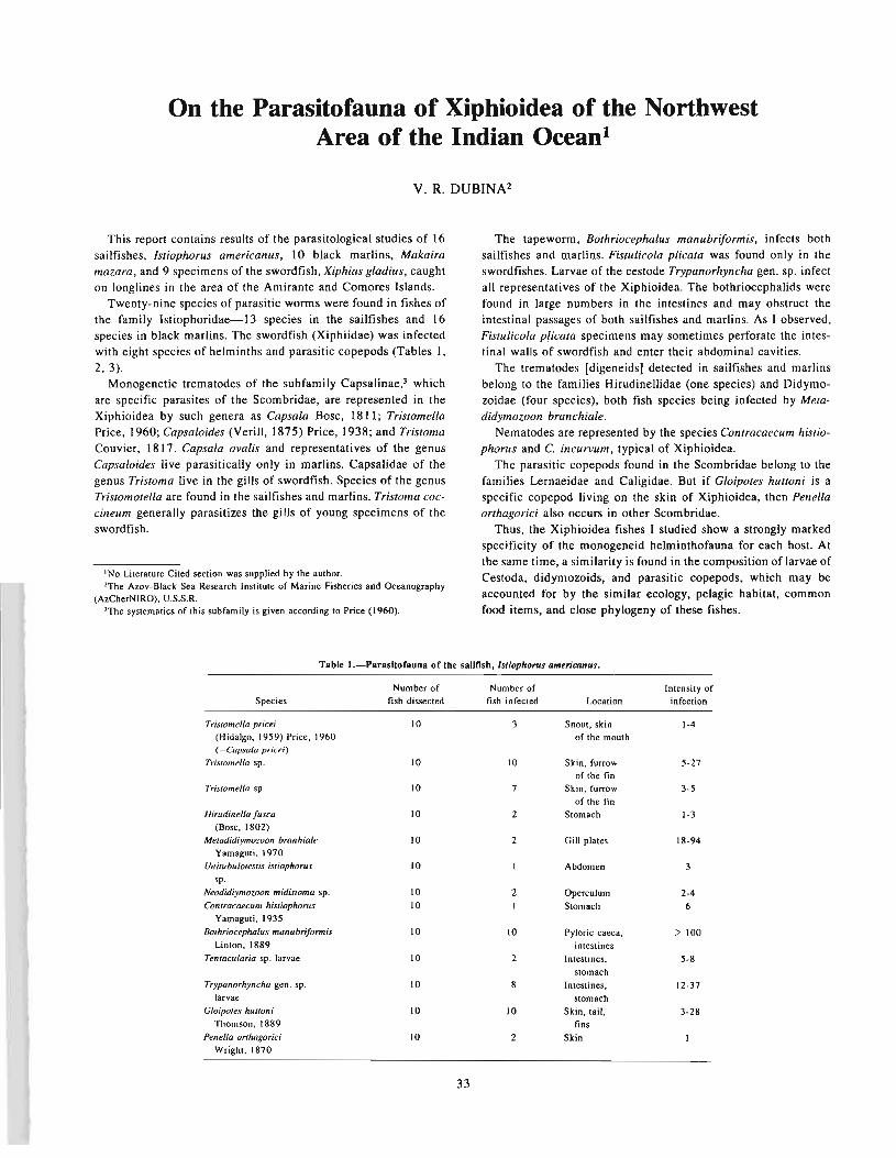
On the Parasitofauna of Xiphioidea of the NorthwestArea of the Indian Ocean!
V. R. DUBINA2
This report contains results of the parasitological studies of 16sailfishes, Istiophorus americanus, 10 black marlins, Makairamazara, and 9 specimens of the swordfish, Xiphias gladius, caughton longlines in the area of the Amirante and Comores Islands.
Twenty-nine species of parasitic wonns were found in fishes ofthe family Istiophoridae-13 species in the sailfishes and 16species in black marlins. The swordfish (Xiphiidae) was infectedwith eight species of helminths and parasitic copepods (Tables I,2,3).
Monogenetic trematodes of the subfamily Capsalinae,3 whichare specific parasites of the Scombridae, are represented in theXiphioidea by such genera as Capsala Bosc, 1811; TristomellaPrice, 1960; Capsaloides (Veri II, 1875) Price, 1938; and TristomaCouvier, 1817. Capsala ovalis and representatives of the genusCapsaloides live parasitically only in marlins. Capsalidae of thegenus Tristoma live in the gills of swordfish. Species of the genusTristomotella are found in the sailfishes and marlins. Tristoma coccineum generally parasitizes the gills of young specimens of theswordfish.
1 No Literature Cited section was supplied by the author.'The Amv-Black Sea Research Institute of Marine Fisheries and Oceanography
(AzCherNIRO), U.S.S.R.'The systematics of this subfamily is given according to Price (1960).
The tapewonn, Bothriocephalus manubriformis, infects bothsailfishes and marlins. Fistulicola plicata was found only in theswordfishes. Larvae of the cestode Trypanorhyncha gen. sp. infectall representatives of the Xiphioidea. The bothriocephalids werefound in large numbers in the intestines and may obstruct theintestinal passages of both sailfishes and marlins. As I observed,Fistulicola plicata specimens may sometimes perforate the intestinal walls of swordfish and enter their abdominal cavities.
The trematodes [digeneids] detected in sailfishes and marlinsbelong to the families Hirudinellidae (one species) and Didymozoidae (four species), both fish species being infected by Metadidymozoon branchiale.
Nematodes are represented by the species Contracaecum histiophorus and C. incurvum, typical of Xiphioidea.
The parasitic copepods found in the Scombridae belong to thefamilies Lernaeidae and Caligidae. But if Gloipotes hut/oni is aspecific copepod living on the skin of Xiphioidea, then Penellaorthagorici also occurs in other Scombridae.
Thus, the Xiphioidea fishes I studied show a strongly markedspecificity of the monogeneid helminthofauna for each host. Atthe same time, a similarity is found in the composition of larvae ofCestoda, didymozoids, and parasitic copepods, which may beaccounted for by the similar ecology, pelagic habitat, commonfood items, and close phylogeny of these fishes.
Table l.-Parasltofauna of the sailfish, /sliophoTUS americanus.
Species
Trislomella price;
(Hidalgo, 1959) Price, 1960(=Capsala pricei)
Trislomella sp.
Tristomella sp.
Hirudinel/a [usca(Bose, 1802)
Metadidiymozoon branhia/eYamaguti, 1970
Unitubulotestis iSliophorus
sp.Neodidiymozoon midistoma sp.Contracaecum histiophorus
Yamaguti, 1935Bothriocephalus manubrifonnis
Linton, 1889Tentacularia sp. larvae
Trypanorhyncha gen. sp.larvae
G/oipotes hUlloniThomson, 1889
Penel/a or/hagoriciWright, 1870
Number offish dissected
10
10
10
10
10
10
1010
10
10
10
10
10
33
Number offish infected
3
10
7
2
2
21
10
2
8
10
2
Localion
Snout, skinof the mouth
Skin, furrowof the fin
Skin. furrowof the fin
Stomach
Gill plates
Abdomen
OperculumStomach
Pyloric caeca,intestines
Intestines,
stomachIntestines,
stomachSkin, tail,
finsSkin
Intensity ofinfection
1-4
5-27
3-5
1-3
18-94
2-46
> 100
5-8
12-37
3-28

Table 2.-Parasitofauna of the black marlin, Makaira mazara.
Number of Number of Intensity ofSpecies fish dissected fish infected Location infection
Capsala avalis (Goto, (894) [6 16 Gill arches 2-54Yamaguti,I968
Tristomella pricei (Hidalgo, (959) 16 2 Snout, skin of 1-2Price, [960 (=Capsala pricei) mouth
Tristomella sp. 16 3 Skin [-[4
Capsaloides nairogi Yamaguti, 1968 16 2 Gill arches [-2
Capsaloides sp. 16 6 Gills, between [ [-137
gill archesHirudinella fusca (Bose, 1802) 16 4 Stomach [-3
Unitubulotestis malcairi [6 3 Body cavity 2Nikolaeva and Dubina, [978
Wedlia submaxillaris (Yamaguti, 1970) [6 2 Gill filament 4-8Niko[aeva. [978
Neodidymozoon macrostoma [6 3 Dursal fin, 4-6Yamaguti, 1970 gill arches
Metadidymozoon branhiale [6 3 Gill plates 12-23
Yamaguti, 1970Cantracaecum incurvum (Rud., 1819) 16 10 Stomach, intestine 2-97
Bay[is and Daubney, [922Contracaecum sp. larvae 16 12 Serose coverings 1-24
of the internalsBothriocephalus manubriformis 16 12 Intestine 14->100
Linton. 1889Trypanorhyncha gen. sp. [arvae 16 7 Serose of body 12-29
cavityGliopotes hunoni Thomson, 18·89 16 14 Skin 14~76
Penella onhagorici"Wright, 1870 16 I Skin 2
Table 3.-ParasltofaUlla of the swordflsb, .XipftillS gilltlills.
Number of Number of Degree ofSpecies fish dissected fish infected Location infection
Tristoma coccineum Cuvier. 1817 9 2 Gills 8-34Tristoma adintegrum Yamaguti, 1963 9 3 Gills 1-2Fistulicola plicala Rudolfi. 1819 9 9 Intestine 14-32Nybelinio sp. [arv..e 9 5 Stomach 3-11Tryponorhyncha gen. sp. larvae 9 9 Stomach. intestine 7-35Contracaecum incurvum (Roo., 1819) 9 I Stomach 12
Baylis and Daubney. 1922Gloipotes huttoni Thomson, 1889 9 6 Skin 3-15Pene/la onhagorici Wright, 1870 9 2 Skin I
34

Parasites as Indicators of Specific Featuresof Fish Ecology
S. M. KONOVALOVI and T. E. BUTORINA2
ABSTRACT
An analysis of tbe results of a number of investigations involving tbe use of parasite.indicators in studies offisb ecology was conducted. Using sawon species as an example we concluded tbat tbis approacb is very promis·ing, not only for comparisons of ecology of closely related species, but also of interspecific forms and for studiesof tbe biology of populations (Isolates) and subpopulations (subisolates), as weD as of groups being formedwitbin subisolates.
Use of parasites to study the ecology of their hosts (fish) has arather long history. Use of this method is possible only in caseswhere the life history of a parasite is well studied and the approximate circle of its intermediate hosts or place of infection (forparasites with direct cycles of development) is known. Even thefirst works of the Soviet parasitology school under V. A. Dogielaccomplished on sturgeons (Dogiel and Bykhovski 1939) and thenon other marine and freshwater fishes (Shulman et al. 1959)showed the possibilities of such an approach to studies of hostecology. As a result of using parasitological data and by tagging,Margolis and his' colleagues managed (Margolis et al. 1966) to explain the marine distribution and migration of sockeye salmon ofthe Asian and American stocks at the level of Bristol (all theisolates are of the Bering Sea coast of Alaska) and Kamchatkaisolate complexes (all the isolates are of Kamchatka Peninsula)(Figs. 1,2). Investigations made by English and Canadian parasitologists (Kabata 1963; Margolis 1965; Margolis et al. 1966)which disclosed fundamental solutions were employed byichthyologists for practical conclusions.
The studies made by the authors indicated above on anadromous salmons provided significant data on the marine and fresh-
1 Pacific Research Institute of Fisheries and Oceanography (TINRO), Vladivostok,U.S.S.R.
'Institute of Marine Biology and Far East Science Center, AS, U.S.S.R.
water periods of their lives. As did the previously mentionedscientists, S. M. Konovalov used parasites as indicators of localsockeye salmon stocks in 1971. It was found that by employingnot only the qualitative features (i.e., selected parasite species) butalso the quantitative ones (extensiveness and intensiveness of invasion), we were able to differentiate not only the complexes ofisolates but some local stocks (isolates). In particular, we managedto identify fish from Kurilskoye Lake (Kamchatka), but only atth~ first year of life, before they began mixing with Americanstocks, and during their spawning migration. Combined withspecial features of scale structure, [these techniques] permitted usto study the entire marine period of life of the local stock and tounderstand the rules of distribution of the fish of Asian andAmerican origin as well as of the different stocks in the high seasduring feeding and wintering. It was revealed that local sockeyestocks, which are highly isolated during spawning, became mixedwith each other during the feeding and wintering periods of theirmarine [high seas] phase of life. S. M. Konovalov in cooperationwith I. B. Birman discovered the feeding and wintering sites aswell as the feeding, wintering, and spawning migrations of different age groups of Kurilskoye Lake sockeye salmon (Fig. 3).
A number of proposals of practical importance for the regulation [management] of sockeye fishing in the North Pacific wasmade on the basis of these studies. Moreover, these works servedto encourage studies of the sea [oceanic] period of other salmon[species] using ichthyological data as a basis. To date, much infor-
-- 2 ----3~OO ---1
f50° 160· f70· fBO· 170· f60· 150· f40·
--1 ---2 -'-3 ----4 ~S-_----'--.__--'--__-L_-,---!.I__--I l-_--l
150· f~O· 170· 180· 170· f60· 150· 11(0 0
Figure I.-Distribution of immature Alaskan and Kamcbatka sockeye sawon intbe Nortb Pacific (according to Margolis et al. 1%6). 1 = boundary of tbe areasof western Alaska sockeye; 2 = boundary of tbe areas of Kamcbatka sockeye; 3= supposed boundary of tbe areas of Kamcbatka sockeye; 4 = mixed areas.
35
Figure 2.-Dlstribulion of mature Alaskan and Kamcbatka sockeye salmon intbe Nortb Pacific (according to Margolis et al. 1%6). 1 = boundary of tbe areasof western Alaska sockeye; 2 = supposed boundary of tbe areas of westernAlaska sockeye; 3 = boundary of tbe areas of Kamcbatka sockeye; 4 = supposedboundary of tbe areas of Kamcbatka sockeye; 5 = mixed areas.
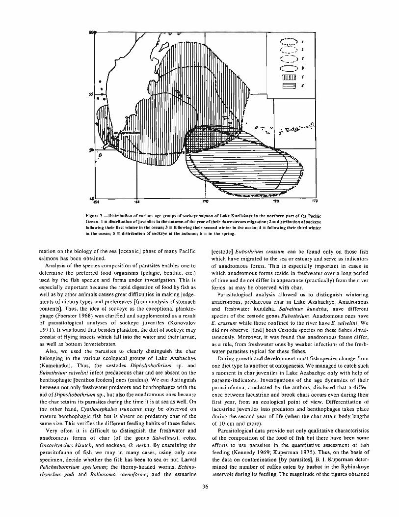
170.It11'4
~w....
<.".M....~ ,.~ ........
,... --- ..., 2... ___ 6'
."-'-:> J... -_.-c::> ..rnmm s
•
r~~ E3 I•s.s~
• •
I"
• ~d~~'~j)"_ 0
~ljfJft
~~.. ~
",.()
"
46.--......~...
Figure 3.-Distribution of various age groups of sockeye salmon of Lake Kurilskoye in the nortbern part of the PacificOcean. 1 = distribution of juveniles in the autumn of the year of their downstream migration; 2 = distribution of sockeyefollowing their first winter in the ocean; 3 =following their second winter in the ocean; 4 =following their third winterin the ocean; 5 = distribution of sockeye in the autumn; 6 = in the spring.
mation on the biology of the sea [oceanic] phase of many Pacificsalmons has been obtained.
Analysis of the species composition of parasites enables one todetermine the preferred food organisms (pelagic, benthic, etc.)used by the fish species and forms under investigation. This isespecially important because the rapid digestion of food by fish aswell as by other animals causes great difficulties in making judgements of dietary types and preferences [from analysis of stomachcontents]. Thus, the idea of sockeye as the exceptional planktophage (Foerster 1968) was clarified and supplemented as a resultof parasitological analyses of sockeye juveniles (Konovalov1971). It was found that besides plankton, the diet of sockeye mayconsist of flying insects which fall into the water and their larvae,as well as bottom invertebrates.
Also, we used the parasites to clearly distinguish the charbelonging to the various ecological groups of Lake Azabachye(Kamchatka). Thus, the cestodes Diphyllobothrium sp. andEubothrium salvelini infect predaceous char and are absent on thebenthophagic [benthos feeders] ones (malma). We can distinguishbetween not only freshwater predators and benthophages with theaid of Diphyllobothrium sp., but also the anadromous ones becausethe char retains its parasites during the time it is at sea as well. Onthe other hand, Cyathocephalus truncatus may be observed onmature benthophagic fish but is absent on predatory char of thesame size. This verifies the different feeding habits of these fishes.
Very often it is difficult to distinguish the freshwater andanadromous forms of char (of the genus Salvelinus), coho,Oncorhynchus kisutch, and sockeye, O. nerka. By examining theparasitofauna of fish we may in many cases, using only onespecimen, decide whether the fish has been to sea or not. LarvalPelichnibothrium speciosum; the thorny-headed worms, Echinorhynchus gadi and Bolbosoma caenoforme; and the estuarine
[cestode] Eubothrium crassum can be found only on those fishwhich have migrated to the sea or estuary and serve as indicatorsof anadromous forms. This is especially important in cases inwhich anadromous forms reside in freshwater over a long periodof time and do not differ in appearance (practically) from the riverforms, as may be observed with char.
Parasitological analysis allowed us to distinguish winteringanadromous, predaceous char in Lake Azabachye. Anadromousand freshwater kundzha, Salvelinus kundzha, have differentspecies of the cestode genus Eubothrium. Anadromous ones haveE. crassum while those confined to the river have E. saLvelini. Wedid not observe [find] both Cestoda species on these fishes simultaneously. Moreover, it was found that anadromous forms differ,a5 a rule, from freshwater ones by weaker infections of the freshwater parasites typical for these fishes.
During growth and development most fish species change fromone diet type to another at ontogenesis. We managed to catch sucha moment in char juveniles in Lake Azabachye only with help ofparasite-indicators. Investigations of the age dynamics of theirparasitofauna, conducted by the authors, disclosed that a difference between lacustrine and brook chars occurs even during theirfirst year, from an ecological point of view. Differentiation oflacustrine juveniles into predators and benthophages takes placeduring the second year of life (when the char attain body lengthsof 10 cm and more).
Parasitological data provide not only qualitative characteristicsof the composition of the food of fish but there have been someefforts to use parasites in the quantitative assessment of fishfeeding (Kennedy 1969; Kuperman 1975). Thus, on the basis ofthe data on contamination [by parasites], B. I. Kuperman determined the number of ruffes eaten by burbot in the Rybinskoyereservoir during its feeding. The magnitude of the figures obtained
36

by him proved to be very close to the data secured by ichthyologists for the same period.
During the last decade we collected vast material which disclosed that parasites might be used to study the ecology of elementary populations (subisolates). Analyzing the levels of infection byDiphyllobothrium sp. of juvenile and spawning age sockeye inLake Azabachye, we managed to learn that fish with differentscale types, as well as of different sex but with a similar scale pattern, have different levels of infection of this parasite. Maturetapeworms of Diphyllobothrium sp. used for the model were foundin bears, dogs, gulls, and terns in Lake Azabachye (Butorina andKuperman 1981). The first intermediate host of these cestodes arecopepods of the species Cyclops scutifer (Kuperman 1978).Sockeye salmon are parasitized by plerocercoids, which are concentrated on the external side [peritoneal side] of the stomach andnever die away [disappear] when the fish passes from freshwaterinto saltwater and back even though the life span of fish at seareaches 4 yr.
Examination of samples of about 25 individuals of each separate subisolate every year revealed the existence of a complicatedtrophic structure of sockeye subisolates. It was found thatjuveniles of one [each] year-class of every subisolate may be subdivided into groups which differ in preference for copepod consumption. We also uncovered different intensities and extensities[prevalences] of invasion [infection] of mature fish by Diphyllobothrium sp. We distinguished conventional planktophages(whose preferable diet is copepods) and benthophages (whosepreferable diet is flying insects and benthic organisms). Also, ifjuveniles remain in freshwater for a second year, four groups willappear, planktophage (1 st year) x planktophage (2nd year);planktophage (l st year) x benthophage (2nd year); benthophage(lst year) x planktophage (2nd year); and benthophage (1st year)x benthophage (2nd year). Hence, sockeye juveniles migrate fromLake Azabachye at the ages of 1 and 2 yr and have six trophicstrata and the same number of groups in every subisolate.
Comparison of the mean intensity of invasion [infection] ofsockeye groupings by Diphyllobothrium sp. with one or anotherscale type in different subisolates over many years revealed ratherobvious differences in both spring and summer races. Males orfemales of one subisolate differ by several times from males orfemales of the other in mean infection levels. Thus, the relation ofmean intensities of infection of males with the scale of "b" type
of subisolates 16 and 6 comprised 4.6 times in 1975. The sameparameter [measurement or comparison] for males with "b"3scale type for subisolates 13 and 2 was 2.6 times. This relation forsubisolates 4 and 6 with the males with scale of "bb" type was 2.9in 1975. In 1970, females with the same scale type showed adifference between the mean subisolate intensities of 24 and 27,2.7 times [that in 1975). The difference in the quantity ofplankton consumed for the same freshwater period comprisedfrom 2 to 7 times. Thus, there are fish groups with a differentgrowth rate within the subisolate. This is affirmed by the peculiarities of scale structure, which are well known for expressingselectivity of the food available to the sockeye while it is in freshwater.
Simultaneously, over II yr we collected a large amount of dataon the invasion by Diphyllobothrium sp. of fish of one generationof the same subisolate of similar freshwater age and scale type, butof different sex. It was found that in most cases (95% or more)there were significant differences between males and females inextensity [prevalence] and intensity of invasion [infection] by thisparasite, as it was observed with trophic groupings. Males, as arule, were characterized by a higher level of infection and, hence,by more active consumption of copepods [or vice versa). Femaleswere notable for their mixed diet. The relation of mean intensityof invasion of males to the corresponding female index was dominant, i.e., 1.5, with the maximum figure being up to 13.9 timeshigher (Fig. 4A). Thus, we distinguished another trophic levelwithin a subisolate. Each subisolate may total [contain] up to 12trophic groups simultaneously, six for males and six for females(Fig. 4).
In 1974, S. M. Konovalov conducted investigations of sockeyesalmon which were heavily infected by Diphyllobothrium sp. inLake Iliamna (Alaska) (Fig. 4B). In six of seven cases, males had ahigher intensity of invasion (two or more times) than females.Therefore, we may conclude that trophic divergence of sockeyesalmon may be observed [may occur] everywhere, since they arefound within isolates of both American and Asian continents.This phenomenon is also observed in other Pacific salmons. Thus,we found trophic groups of feeding juvenile silver salmon in Lake
'Original manuscript used "h" scale type here. It appeared to have been struckover until it looked like a "b." Since it is totally out of "sync" with the rest of themanuscript. it has been replaced with a "b."
1
(5
fO
5
"030
2IJ
10
(,5 ~.O Z.5 3.0
X.?/XJ,
Figure 4.-Dlstributlon of sockeye spawners of tbe same age· and scale·type from tbe different sublsolates by level ofdifferences in invasion by parasite-indicator Diphyllobothrium sp. pI. depending upon the se~: A : Azabacbye Lake(number of studied cases Is 284); B . Iliamna Lake (number of studied cases is 7). Note: relation XO' I X9 sbows the levelof invasion of mates relating to females.
37

Azabachye which prefer insects, benthos, or fish fry in their dietand, consequently, had different levels of parasite infection(Konovalov 1974; Butorina and Kuperman 1981).
An interesting investigation of the biology of kokanee of LakeKronotskoye (Kamchatka) was accomplished by S. I. Kurenkov(1979). Because of strong waterfalls this lake is isolated from thespawning migrations of anadromous sockeye and other Pacificsalmons and is inhabited by freshwater sockeye which spend theirentire lives within the lake. Studies of the parasitofauna ofkokanee from different spawning grounds (i.e., of differentsubisolates) helped to reveal the existence of two trophic forms:Benthophages and planktophages (Fig. 5). However, in Azabachye Lake and Iliamna Lake anadromous sockeye form trophicgroups within subisolates, and in Lake Kronotskoye formation ofthese groups is observed at the subisolate level. In other words, S.I. Kurenkov distinguished a great number of subisolates in whichplanktonic organisms were the preferred diet of all the individuals, as well as the subisolates-consisting of those fish preferringto feed on benthic organisms and flying insects (Kutenkov 1979).Parasites are such sensitive indicators of the feeding habits of thegroups of individuals within elementary populations that nowthere are no other [or no more] reliable methods of recording thelevel of consumption of any type of diet for a long period. This iswhy the use of parasite-indicators is still the only method ofdistinguishing trophic groups.
Population biology discloses a number of interspecific adaptations which lead to a reduction of competition between theindividuals of a population. For example, different growth ratescause [affect] diet by [feeding on] different-sized organisms [?].4The trophic adaptation discovered shows again the difficulty ofpopulation adaptation to the reduction of competition betweenindividuals in critical periods of their life history.
Thus, analysis of the investigations of many years dealing withstudies of fish ecology with the help of parasite-indicators allowsus to conclude that the above approach is very promising, not onlyfor comparison of the ecology of closely related species but also ofinterspecific forms and for studying the biology of populations(isolates) and subisolates (subpopulations) as well as of the groupsbeing formed [established] within subisolates. At that, in some
4This sentence is presented as it was in the original typescript. Words in bracketsare the editor's effort to clarity. It is still confusing. The authors probahly meant toindicate that competition for food, even within the same population of fish, wasreduced or affected in that individuals which grow faster eat larger prey, leaving thesmaller prey for smaller, slower growing individuals. Thus, competition for food isaffected. Obviously fish with greater access to suitable food (below a threshold level)will grow faster than those which are food-deprived.
ISOLATE
cases (e.g., trophic groups of anadromous forms of the genusSalvelinus) it is difficult to imagine other approaches and methodswhich would permit solution of the problems stated. On the basisof the above, we may consider it advisable to broaden [increase]the exploitation of parasitological data for the study of fishbiology.
LITERATURE CITEDs
BUTORINA. T. E.1978. Ecological analysis of the parasitofauna of char (g. Sa/velinus) of Kam
chatka and Okhota rivers. [In Russ.] Autoessay, Master's degree, Baku,24 p.
BUTORINA, T. E., and B. I. KUPERMAN.
198/. Ecological analysis of the· Cestoda invasion of fish of Kamchatka freshwaters. In Biology and systematics of animals helminths in the Soviet FarEast, p. 86-100. [In Russ.1 Vladivostok.
DOGIEL, V. A., and B. E. BYKHOVSKI.1939. Fish parasites of the Caspian Sea. [In Russ.) Tr. Kompl. komissii po
izucheniyu Kaspijskogo morya, No.7, 150 p.FOERSTER, R. E.
1968. The sockeye salmon, Oncorhynchus nerka. Fish. Res. Board Can.,Bull. 162, 422 p.
KABATA, Z.1963. Parasites as biological tags. ICNAF, Spec. Publ. 4(6):31-37.
KENNEDY, C. R.1969. Tubificid oligochaetes as coarse fish food. Proc. 4 British Coarse
Fish. Con f., Liverpool Univ., p. 5-7.KONOVALOV, S. M.
1971. Differentiation of local schools of sockeye stocks. [In Russ.) M.-L.Nauka, 227 p.
1977. Subisolate as a relatively inflexible system. The subisolate structure. [InRuss.) Zh. Obshc. Bioi. 35(6):819-837.
KUPERMAN, B. I.1975. Experience of using parasites for the quantitative assessment of fish
feeding. [In Russ.] Vopr. Ichthyol. 15(5):93 I -934.1978. Peculiarities of the life cycle and biology of Cestodes of salmons of
Kamchatka reservoirs. [In Russ.] Biologiya Morya 3:135-145.KURENKOV, S. I.
1979. Population structure of kokanee of Lake Kronotskoye. [In Russ.]Autoessay, Master's degree, M., 22 p.
MARGOLIS, L.1965. Parasites as an auxiliary source of information about the biology of
Pacific salmons (genus Oncorhynchus). J. Fish. Res. Board Can. 22:13871395.
MARGOLIS, L.. F. C. CLEAVER, Y. FUKUDA. and H. GODFREY.1966. Salmon of the North Pacific Ocean. Part VI. Sockeye salmon in off
shore waters. INPFC Bull. 20, 70 p.SHULMAN, S. S., Yu. N. BERENIUS, and E. A. ZAKHAROVA.
1959. Parasitofauna of local stocks of some fishes of Syamozero. [In Russ.]Tr. Karel. Fil. Akad. Nauk SSSR Vopr. Parasitol. Karelii, Vyp. 14:47-71.
SNot all citations mentioned in the text are included here. Also, some LiteratureCited do not appear in the text.
Figure S.-Spatlal structure of the kokanee population of Lake Kronotskoye.
38

The Taxonomic Composition and Origin of FishHelminths in the Epipelagic Zone of the World Ocean
S. E. POZDNYAKOV t
The epipelagic zone is a peculiar and very interesting biotopeparasitologically. At present, 738 species of helminths areregistered [recorded] from about 260 to 300 fish species inhabiting open waters (Parin 1968; Cohen 1970; Nikolski 1980). Themost abundant of them are trematodes [digeneids], of which 370species (from more than 2,500 recorded sea fishes) are related to[are associated with] fish of the epipelagic zone. Trematodes ofthe families Hirudinellidae, Syncoeliidae, and Accacoeliidae andmany subfamilies of the family Didymozoidae (Le., Adenodidymocystinae, Colocyntotrematinae, Glomeritrematinae, Koellikeriinae, Metadidymozoinae, Neodiplotrematinae, Nephrodidymotrematinae, and Reniforminae) form [constitute] a true oceanicgroup. The genera of the Didymozoidae subfamilies Annulocystiinae, Didymocodiinae, Didymozoinae, Nematobothriinae,Opepherotrematinae, Ossoodidymocodiinae,2 Phacelotrematinae,Pseudocolocyntotrematinae, Sicuotrematinae, Skrjabinozoinae,and separate [certain or different] genera of the abundant suborderHemiurata compose a neritic-oceanic group. Trematodes of thefamilies Bucephalidae, Allocreadiidae, Opecoelidae, Lepocreadiidae, and Fellodistomidae are not common for the pelagic zone,but are found there occasionally.
Of the more than 2,200 known species, only 139 species ofmonogenetic flukes [Monogenea] are found in epipelagic fish.These oceanic species belong to the genera Capsala, Caballerocotyla, Capsaloides, and Tristoma (family Capsalidae); Erpocotyle(family Hexobothriidae); Hexostoma and Neohexostoma (familyHexostomatidae); and Dionchus and Loimosina (family Dionchidae). Many genera of the families Monocotylidae, Gastrocotylidae, and Axinidae are far-neritic [distant neritic] and neriticones.
One hundred and thirty-six species of Cestoda from more than800 marine fishes occur in the epipelagic zone. Truly oceanicCestoda are representative of a portion of the family Litobothridae, i.e., the genera Hepatoxylon and Tentacularia (order Trypano-
'Pacific Research Institute of Fisheries and Oceanography (TINRO), Vladivostok,U.S.S.R.
'The spelling of this subfamily is unclear due to strike-overs in the original typescript. It could have been Osteodidymocodiinae.
39
rhyncha) and Fistulicola (family Ancistrocephalidae), as well as anumber of species of the Nybelinia genus (family Tentaculariidae).
Among 42 species of thorny-headed worms from fish of theoceanic epipelagic zone, none can be related to a really oceanicgroup. The family Rhadinorhynchidae is nerito-oceanic. Speciesof the other families occur occasionally in the epipelagic zone.
Only certain Nematoda species of the families Anisakidae,Philometridae, Cucullanidae, Camallanidae, and Rhabdochonidaeare found in the oceanic fish, as was observed in the case of theprevious group.
Analysis of the taxonomic composition of fish helminths in theepipelagic zone shows the close relationship between the oceanichelminthofauna complex and the neritic-pelagic one. Separationof the oceanic group from the neritic took place probably in themid-Tertiary period, i.e., during the formation of the epipelagicichthyocoenosis. There are some oceanic helminthofaunistic[helminthofaunal] groups in the epipelagic zone of the WorldOcean. The most abundant among them are tropical and relatedsubtropical groups. They form in the equatorial tropical and central water masses. The nuclei of tropical and subtropical groupsare in the range of the productive waters of the North and Southtropical divergences and at the periphery of the North and Southsubtropical and tropical convergences, as well as the area of eddycausing geostrophic currents. Independent oceanic helminthofaunistic [helminthofaunal] groups are absent from the boreal andnotal waters. Subtropical and tropical species of helminthspenetrate those waters along with their hosts as the latter forage inthe boreal and notal waters, and the boreal and notal species arecarried to the high seas by fishes able to leave the coastal andneritic waters at times in their lives. The ocean helminthofaunisticgroups are lacking in arctic and antarctic waters.
LITERATURE CITED
COHEN.D.M.1970. How many recent fishes are there? Proc. Calif. Acad. Sci. Fourth Ser.
38:341-346.NIKOLSKI. G. V.
1980. Struktura vida i zakonomernosti izmenchivosti ryb, Moscow. [In Russ.]Pischepromizdat, 180 p.
PARIN, N. V.
1968. The ichthyofauna of the ocean epipelagic zone. lIn Russ.) Moscow,Nauke, 185 p.


Zoogeographical Characteristics of the Helminths ofFishes From the Antarctic Zone of the World Ocean!
Vo No LYADOV2
The zoogeography of parasites of sea fishes has been discussedin a considerable number of Soviet and foreign papers (Dogiel andBykhovski 1939; Schulman and Schulman-Albova 1953;Schulman 1959; Polianski 1955; Strelkov 1956; Zhukov 1960;Lebedev 1967; Parukhin 1972, 1976; Konovalov 1971; ManterI (j47, 1955; Yamaguti 1963; and others) in which the distributionof helminths is analyzed almost throughout the entire WorldOcean-except for the vast areas of the subantarctic and antarcticzones. This may be accounted for by the fact that the parasitefauna of fishes of these areas is little known because sufficientdata are not available in the literature on the species compositionof fish parasites to permit a detailed zoogeographical analysis ofparasitic worms of antarctic fishes.
The most thorough in vestigations of fish parasites of the antarctic zone of the World Ocean were made by Gussev (1967) whodescribed about 20 species of fish helminths and, in general, about50 species from all species of animals. In their work, Gaevskayaand Kovaliova (1976) point to the scarcity of the trematode faunaand provide data on nine species of trematodes found in the fishesof the families Nototheniidae, Chaenichthyidae, and Bathydraconidae
Of foreign works, those of Prudhoe and Bray (1973), in whichdata on the distribution of the trematode and monogenean faunain the antarctic zone of the Atlantic Ocean are presented, shouldbe noted. Here I employ data from my own research and from theliterature in an attempt to analyze the distribution of parasiticworms in the antarctic zone and adjacent waters of the Pacific,Indian, and Atlantic Oceans, ignoring leeches and parasitic crustaceans.
The most abundant group of helminths is represented by thetrematodes [digeneids]. Thirty-five species of these flatworms areregistered for this region of the World Ocean (Table 1). Analysisof the trematode fauna of the Kerguelen and Glass [Glacial] subregions, which are separated by vast water areas with abyssaldepths, revealed a considerable specific affinity of trematodes inthese regions. Thus, from the total number of this group ofparasites 13 species of trematodes [digeneids] are common toboth subregions of the antarctic zone. These are Gonocercaphycidis, Derogenes varicus, Lecithaster australis, Plagioporus antarcticus, Opechona magnatestis, Lepocreadium trullaforme, Neolepidapedon antarcticus, Lecithophyllum anteroporum,Elytrophalloides oatesi, Glomericirrus macrouri, Genolinae bowersi,Postmonorchis variabilis, and Descoveritrema markowskii.However, in spite of the affinity of the trematode [digeneid] faunain these regions, endemism is rather strongly marked among them.In the Kerguelen subregion seven species of trematodes
1No Literature Cited section was supplied by the author. Most citations arepresented in Literature Cited sections of other papers, however.
'The Azov-Black Sea Research Institute of Marine Fisheries and Oceanography(AzCherNIRO), U.S.S.R.
41
[digeneids] characteristic of this area are registered [recorded]:Gonocerca muraenolepisi, G. medius,3 Plagioporus sp., Stenakronkergeulense, Lomosoma kergeleni, Benthotrema melanostigmi, andSteganodermatoides kergeleni. In the Glacial subregion 11 speciesof trematodes [digeneids] are recorded: Plagioporus pennelli,Lepidapedon sp., L. garrardi, L. antarcticus, Neolepidapedontrematomi, N. dubius, Lecithocladium orustatum, Stenakron spo,Otodistomum cestoides, Otodistomum sp., and Neoleburia georgiensis. Despite the closeness of the regions of the Kerguelen subregionand South Australia, their trematode [digeneid] fauna relationswere not marked. There were only two common species oftrematodes: Gonocera phycidis and Elytrophalloides oatesi, whilethe relationships of the trematode [digeneid] fauna of the Patagonian shelf were expressed more strikingly, there were five common species: Gonocerca phycidis, Derogenes varicus, Plagioporusantarcticus, Lepocreadium trullaforme, and Elytrophalloides oatesi.
Comparing the helminthofauna of the Kerguelen subregionwith that of the tropical Indian-West Pacific and South Moderateregions, it should be noted that, according to the data of Parukhin(1976), 6 families of trematodes [digeneids] (Monorchidae,Opecoelidae, Lecithasteridae, Hemiuridae, Halipegidae, andLepocreadidae) of 17 families and 3 species were common for theIndian Ocean and some other regions of the World Ocean.
Of the 10 known species of monogenetic trematodes of the antarctic region of the World Ocean,Pseudobenedenia notothenia wascommon for all regions. Pseudobenedenoides shorti, P. antarcticus,Neogrubea stromateae, Pavlovskioides prudhoei, and Neopavlovskioides georgianus were specific species of monogeneids of theGlacial subregion, and Neopavlovskioides dissosthychi, Macruricotyle clavipes, Rajoncocotyle sp., and Acanthocotyle sp. werecharacteristic monogeneids for the Kerguelen subregion.
The cestode fauna of this region has been investigated onlypoorly. Because of this we did not find sufficient data in the Sovietand foreign literature to permit an analysis of cestode distributionin this region of the World Ocean. Thus, we can list only sevenspecies of cestodes: Neobothriocephalus spo, Bothriocephalus sp.,Hepatoxylon sp. larvae, Phyllobothrium sp., Acanthobothrium spo,Scolex pleuronectis, and Tetraphyllidea sp. larvae, found in thefishes of the Kerguelen subregion. Parallel with this, it should benoted that combined species of the larvae of the cestodes Scolexpleuronectis and Tetraphyllidea sp. were widely distributed, notonly in antarctic waters, but also in other regions of the WorldOcean.
The acanthocephalan fauna in antarctic fishes was representedby five species: Aspersentis megarhynchus, Hypoechinorhynchusmagellanicus, Echinorhynchus debenhami, E. campbelli, and Corynosoma hamanni. Of the fauna of thorny-headed worms of theGlacial subregion, E. campbelli should be considered as endemic,
'Though given os a new species in the original typescript, this species probablywas first described elsewhere. It obviously is not described in this paper.
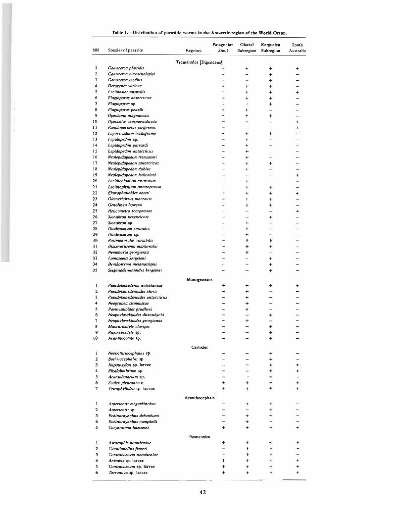
Table I.-Distribution of parasitic worms in the Antarctic region of the World Ocean.
Palagonian Glacial Kerguelen SouthNN Species of parasite Regions: Shelf Subregion Subregion Australia
Trematodes [Digeneans]I Gonocerca phycidis + + + +2 Gonocerca muraenolepisi +3 Gonocerca medius +4 Derogenes varicus + + +5 Lecithaster australis + + +6 Plagibporvs antarcticus + + +7 Plagioporvs sp. +8 Plagibporvs ~neUi + +9 O~clrona magnatestis + +
10 O~coelus scorpaenidicola +II Pseudo~coeluspiriformis +12 Lepocreadium trullaforme + + +13 LepidlJ~n sp. +14 LepidlJ~don garrartfi +15 LepidlJ~don antarcticus +16 Neokp~don trematomi +17 Neokpido~don antarcticus + +18 NeolepidlJ~don dubius +19 Neokpioo~don heUcoleni +20 Lecithocladium crustatum +21 Lecillrophyllum anteroporum + +22 Elyrrophalloides oatesi + + + +23 Glomericirrus macrour; + +24 Genolinea bowers; + +25 Helicometra scorpaenae +26 StenaJrron Jcerguelense +27 StenaJrron sp. +28 Otodistomum cestoides +29 Otadistomum sp. +30 Posrmonorcltis variabilis + +31 Discoveritrema marlcowskii + +32 Neokburia ~orgknsis +33 LomOSOfM Jcergekni +34 Benllrolrema melanosligmi +35 Sleganodermaloirks Jcergekn i +
Monogcneans
I PJeIuIobenetknia nOlOtheniae + + + +2 PseudobenetknoUks sharti +3 PseudobenedenoUks antarcticus +4 Neogrubea stromaleae +5 PavlovslcioUks prudhoei +6 Neopavlovs/ciQides dissoslhychi +7 Neopavlovslcibides georgianus +8 Macruricotyk clavi~s +9 Rajoncocotyle sp. +
10 Acanthocotyle sp. +
CestodesI NeobothriocephaJus sp. +2 Bothriocephalus sp. +3 HepalOX)1on sp. larvae + +4 Phyllobolhrium sp. + +5 Acantobolhrium sp. +6 &:olex pkuronectis + + + +7 Tetraphyllidea sp. larvae + + + +
Acanthocephala
I As~rsenris megarhinchus + +2 As~rsentis sp. +3 Echinorhynchus debenhomi + +4 Echinorhynchus campbelli +5 CorynOSiJf/lD homanni + + + +
Nematodes
I Ascoropltis notothen iae + + + +2 Cucullanellus fraseri + +3 Contracaecum norothenioe + +4 AnisaJris sp. larvae + + + +5 Contracaecum sp. larvae + + + +6 Terranova sp. larvae + + + +
42

while the other four species were characteristic for all antarcticregions.
The nematode fauna of fish of Antarctica was represented bysix species: Ascarophis nototheniae, Cucullanellus fraseri, Contracaecum nototheniae, Anisakis sp. larvae, Contracaecum sp. larvae,and Terranova sp. larvae, which were widely distributed in watersof this region of the World Ocean and were antarctic circumpolarspecies.
The existence of such an important similarity of the helminthofauna of fish of the Kerguelen and Glacial subregions could be explained first of all by the origin and development of the Antarcticfish fauna. The ichthyofauna of Antarctica is interesting in itsantiquity and originality, which has been determined by the presence of endemic genera and species of the group Nototheniiformis. This fish taxon, characteristic for the antarctic and subantarctic regions and almost completely limited by their boundaries,has developed a great quantity of forms which differ by structure,appearance, and mode of life. These facts allow me to assume thatthe Antarctic has been isolated and washed by a cold sea for avery long time, probably over the entire Tertiary period (Andriyashev 1964).
Therefore, the representatives of the Antarctic ichthyofaunasettled into very different ecological niches in the process ofecological development, and one branch of the subfamily Notothenieidae acquired white blood, a characteristic exceptional forfishes, which appeared in the Tertiary period (Andriyashev 1964).This feature evolved and formed several genera of the familyChaenchthyidae [Chaenichthyidae?] (Andriyashev and Tokarev1958; Andriyashev 1964; Martsinkevich 1961; Regan 1914).
J. E. Permitin (1977) provided a detailed analysis of theichthyofauna of the bottom fishes around the isolated oceanicislands of South Georgia and Kerguelen. He found that, in spite ofthe considerable endemism of each, the connection of the fauna ofthe bottom fishes remains substantial. Many authors (Andriyashev1964,1972; Permitin 1977; and others) consider that the [separation] of the populations of fishes and aquatic invertebrates tookplace in the past and probably continues nowadays because of theflow of the water mass of the Circumpolar Current. Since fluctua-
43
tions in this water mass probably occurred in the past and takeplace now, there are links in the trophic connection of the fishesof both the Glacial and Kerguelen subregions. In the first placethis must involve mollusks, which are the intermediate hosts.Generally, many groups of helminths show a great preference forthem. In all probability the similarity of the helminthofauna of thefishes of this area must be explained by these features.
In respect to the endemism of the fauna of the parasitic worms,which is determined by the endemism of the ichthyofauna of boththe Glacial and Kerguelen subregions, this phenomenon is alsorelated to the Antarctic Circumpolar Current-which leads to theintensification of these circumpolar processes around the islandand the submarine elevations. Local cyclonic systems are formedin these areas. Also, upwelling and the [consequent] enrichment ofthe shelf waters occur here. They cause a high biological productivity and playa great part in the formation of the local populations of the several species of fishes and of the endemic species ofparasitic worms.
Interesting results were obtained by Gayevskaya and Kovaliova(1976) and Gibson (1976), who compared the trematode[digeneid) fauna on the Falkland-Patagonian shelf with that of thearea around South Georgia and the South Orkney Islands wherethey reported 12 trematode species.
Representatives of the family Opecoelidae, which are absentfrom the shelf ichthyofauna, dominate in the basic fauna. Speciesof antarctic origin, such as Elytrophalloides oatesi, Gonocercatrematomi, and Opechona magnatestis, are detected in thetrematode [digeneid) fauna of the Falkland-Patagonian shelf. Thisphenomenon is explained by the closeness of the Antarctic.
Thus, analysis of the helminthofauna of the fishes of the antarctic area has allowed us to establish a substantial uniformity in itscomposition and to reveal several features of the endemism inboth the Kerguelen and Glacial areas and to establish the veryinsignificant relationship between the helminthofaunas of areas ofthe shelves of South Australia and Patagonia, adjacent to the Antarctic. In all probability these neighboring areas played nodecisive role in its [the helminthofauna of fishes of the Antarcticregion) formation.


Special Features of the Helminth Fauna ofHelicolenus maculatus (euvier)
L. P, TKACHUKI
ABSTRACT
This paper presents special features of the hebolnth fauna of the 6sh Helicolenus maculatus inhabiting thearea of Agulhas Bank and Walters Bank in the southeastern region of the Indian Ocean,
(%)
100
Iii!!!!!! !I!!!' KOEOGENOIDEA t I ACA JlTHOCEPHALA
~ TRmfATODA ~ mlATODA1111111111111111 CESTOIDEA I I CRUSTACEA
of the food of H. maculatus on the Agulhas Bank areEuphausiidae. Young individuals of deep-sea shrimps and fish aretaken in lesser numbers. While in the area of Walters Bank, thebulk of food was young deep-sea shrimps and hermit crabs. Thenamed species of [These] animals serve as bridging [intermediate]hosts of trematodes, cestodes, and nematodes, which is why theirquantitative correlations in the food of the fish hosts defines thequantitative levels of infection by the different groups of helminths. Agulhas Bank is characterized by increased biologicalproductivity of invertebrates, which brings about high intensitiesof invasion [infection] by such helminths as cestodes andnematodes.
The observed special features of the helminthofauna of H.
maculatus (differences of the composition of helminths and oftheir quantitative levels of infection) show that we investigated[discovered] the different groups [populations] of H. maculatusinhabiting the areas around the Agulhas Bank and Walters Bankin the southeastern region of the Indian Ocean.
Walters BankAgulhas Bank
40
60
20
80
Helicolenus maculatus (Scorpaeniformes) is an industriallyimportant species among purely sea [marine] fish, However, theliterature contains little data about the helminth fauna of thisspecies. A. M. Parukhin (1967) investigated H. maculatus in threeregions of the Indian Ocean. In the area of Durban (SoutheasternAfrica) he noted 12 species of helminths. According to my data,the composition of the parasite fauna of H. maculatus in theAgulhas Bank area (Southern Africa) includes eight species. Onlytwo of them, Microcotyle caudata and Lecithochirum rufoviride,are represented by pubertal helminths. More often the larvae ofGrillotia sp. and Anisakis sp. inhabit the fish. They also exhibit thehighest intensity of infection. Such a peculiar composition ofparasite fauna is characteristic of deep-sea fish, to which H.
maculatus is related. According to the literature, deep-sea fish areinfected primarily by larval nematodes and cestodes, trematodes(Hemiuridae), and crustaceans. Adult forms of helminths are encountered very rarely. Noble (1973) arrived at this conclusionwhile investigating parasites of Macrouriformes fish from theeastern part of the Pacific Ocean. Earlier (Tkachuk 1980) it wasnoted that the greatest part of the helminthofauna of Zeiformes iscomposed of adults. This seeming disparity with the above-notedspecial features of the helminthofauna of H. maculatus can onlybe explained by the different depths inhabited by these species offish. Thus, in the area of the Agulhas Bank, H. maculatus is caughtin depths of from 200 to 400 m, and Zeiformes at depths of from700 to 1,000 m. Another unusual feature of the helminth fauna ofthis area is a comparatively low infection by certain parasites, i.e.,[the monogeneid] Microcotyle caudata (see Fig. I).
For H. maculatus inhabiting the Walters Bank area, the characteristic feature is a smaller number of parasite species-seven, sixof which are common. The larva of Contracaecum sp. (II), recorded only in this area, is the single exception. Except for [In additionto] the marked specific features indicated, there are considerablylarger infections by such groups of parasites as Monogenea,trematodes [Digene.l], nematodes, and crustaceans. However, theinten8ity of infection by them is considerably lower than thataround the Agulhas Bank (Fig. I).
Such essential differences of the parasite fauna of H. maculatusfrom the two areas being compared are connected with thepeculiar features of its ecology. On Agulhas Bank the fish understudy were caught from depths of 200-400 m, and around WaltersBank from depths of 700-800 m. Above all, this is based on thecomposition of their food. According to my data the main portion
1The Institute of Biology of the Southern Seas, Academy of Sciences of theU.S.S.R., Sevastopol, U.S.S.R. Figure I.-Infection of Helicolenus maculatus (Cuvier) by groups of parasites.
45

LITERATURE CITED2
NOBLE, E. R.1973. Parasites and fishes in a deep-sea environment. Adv. Mar. BioI. 11:
2Not all citations mentioned in the text are included here.
46
121-195.
TKACH UK, L. P.1980. Die Eigenanen der He1mintenfauna der ozeanischen Zeiformes. IV.
Wessenschaftliche Konfarenz zu Fragen der Physiologie, Biologie und Parazitologie von Nutzfishen. Rostok.

The Flatworm Fauna of Fishes of the Gulf of Mexicoand its Genetic Relations!
E. V. ZHUKOV2
ABSTRACT
Host relations in "parasite.host" systems are considered with regard to lower monogenean and trematode[digeneanI infection of fishes in the southern Gulf of Mexico. The prevalence of monohostal relations is estahUshed. The genetic relations of the fauna of the subject worms have been investigated and their Indo·West Pacificorigin established. Lower monogeDeans appeared to have a considerably higher evolutionary rate in comparisonwith trematodes [digeneans). On the basis of fishes of the family Lutianidae, infected with monogeneans of thegenus HaliotN!ma, the impoverishment of the ancient complex of these parasites in the process of evolution oftheir hosts is traced. The role of faunistic barriers in trematode distributions is discussed.
The fauna of the Gulf of Mexico has a complex genesis, interoceanic relations in the region of Central America having played adefinitive role in its formation. Until the mid-Miocene they [thehydrographical features] permitted penetration of elements of theIndo- West Pacific tropical fauna into the newly formed basin.The coastal fish fauna of the Gulf of Mexico originated during theCretaceous and became transformed considerably in isolation.Study of its parasites is of great interest.
I collected the flatworm material at my disposal in CampecheBay and in the region of Havana during 1966 to 1968 and in1974. To date, work on the lower monogeneans and trematodes[digeneans] from 391 specimens of 94 species of fishes has beencompleted. The cestodes, not yet worked up, are represented bylarval forms.
Monogeneans of the subclass Polyonchoinea have been observed in 183 individuals of fishes belonging to 45 species. Thepercentage of infection of fishes by them in the open sea and inthe coastal zone appeared to be similar (about 63%). The lowermonogeneans are represented by 69 species, of which 57 (83%)are new. Worms of the family Dactylogyridae are predominantamong them. It includes 48 species, 42 of which (87%) are new.The family Diplectanidae includes 19 species; 15 (79%) newones. Two members of the family Capsalidae are noted. Thesedata reflect the high degree of endemism in the lower monogeneanfauna in the regions studied (Zhukov 1976, 1978, 1980).
The infection rate of fishes with trematodes [digeneans] wassomewhat lower in the open sea than in the coastal zone (92% and94%, accordingly). The fauna of this group is different from thatof monogeneans. It is represented by 149 species (and 4 forms notidentified to species) of which only 5 species (3.3%) were new.Out of 144 previously known species, only 85 (59%) are confinedto the region of Central American seas, while the other 59 (41 %)
are known beyond this body of water.A predominance of monohostal relations in the "parasite-host"
system is typical of both groups of parasites. Among the lowermonogeneans similar relations have been noted in 41 species(64%): Among digeneans of fishes of the coastal zone in 70species (58%) and in the open sea in 42 (66%).
I In this paper the word trematode means Digenea-an arrangement accepled increasingly by many Russian helrriinthologists and olhers.
2Zoological Institute, Academy of Sciences of the U.S.S.R., Leningrad, U.S.S.R.
47
This is primarily a result of the prolonged existence of theabove systems. For digeneans they are also a result of a narrowfood specialization of the hosts under conditions of high intensityof biotic relations among members of the tropical ichthyofauna.
Analysis of digeneans occurring in fishes of the families Serranidae, Lutianidae, and Pomadasyidae, which provided the mostabundant material for comparison, permitted me to distinguishspecies predominating in them. It has also shown that change indepth leads to redistribution of these forms and to a change in thespecies composition of trematodes. The presence of leading[predominating] species in the digeneid fauna of the above hostgroups suggests food specialization which leads to a reduction ofcompetition between related members in every group. Cases inwhich similar species were found in the predominant forms ofdigeneans in different hosts have shown that such specializationdoes not permit them to avoid food competition completely.
Among the species discovered, Metadena globosa appeared tobe predominant both for fishes of the family Lutianidae (Lutianusapodus, L. synagris, and Ocyurus chrysurus) and for a member ofthe family Pomadasyidae (Haemulon plumieri). For a number ofdigeneans (Stephanostomum sentum, Leurodera decora, andBrachadena pyriformis), obviously associated with fishes of thefamily Pomadasyidae, specimens occurring beyond this family areknown. Thus, one may observe certain dynamics in relation to thehistorical groups of parasites along with their relations to certainsystematic groups of hosts.
Analysis of the genetic relations of the above-mentioned flatworms has revealed their Indo-West Pacific origin and that thefauna of lower monogeneans beyond the barrier of the Isthmus ofPanama appeared to have changed considerably more than thedigeneid fauna, members of which have a complex developmentalcycle. Of 69 species of lower monogeneans only 2 or 3% werecommon with the fauna of the Indo-West Pacific region(Haliotrema similis and Diplectanum diplobulbus) which allows meto regard them as the most ancient elements of the Gulf of Mexico.The Pacific host of the former parasite, Priacanthus cruentatus, remained in Atlantic waters where it existed in a stable conditionand where the parasite transferred to the younger [evolutionarilynewer or more recent] host, P. arenatus, endemic to the AtlanticOcean. Another species, Diplectanum diplobulbus, parasitizingKyphosus cinerascens in the Pacific Ocean, was found beyond theIsthmus of Panama in a phylogenetically younger host, K. sectatrix, also endemic to the Atlantic Ocean. These are examples in

which components of the "parasite-host" system belong to different age periods, the host being younger in the evolutionaryrespect than the parasite.
Similar data have been obtained in the analysis of the distribution of monogeneans of the Indo-We~t Pacific genus, Haliotrema,among fishes of the family Lutianidae. Fish species in this family(of Indo- West Pacific origin) are endemics of the Atlantic Oceanand belong to the genera Lutianus, Ocyurus, and Rhomboplites, thelatter two being monotypical ones. Since members of the genusLutianus occur in the Pacific, I regard it as the most ancient. Thegenus Ocyms has an amphi-Atlantic distribution. The genusRhomboplites belongs among the endemics of the seas of CentralAmerica. The highest number of species of the genus Haliotremawas found in the most thermophilic members of the genus Lutianus-L. machogoni and L. synagris (six and seven species,respectively). Three species were found in Ocyrus chrysurus, andone in Rhomboplites aurorubens. No specific monogenean specieswere found in these phylogenetically younger hosts. Thus, wehave an example of impoverishment of the ancient complex ofgillparasites due to evolutionary changes of their hosts within a family. An analogous picture was observed in the monogenean speciesparasitizing fishes of the genus Lutianus.
The genetic relations of the digenean fauna of the Gulf of Mexico appeared to be wider. Among the 59 digenean species knownbeyond the limits of the seas of Central America and present inmy material, 54 (91 %) were found in the Pacific Ocean. Mostsignificant are these connections with the fauna of the CentralAmerican Pacific coast where 45 (76%) of these 59 species occur(i.e., 31 % of the total number of species of this group of parasitesin my collection) and where 23 (51 %) species among them areamphi-American endemics. Three factors are responsible for thehigh degree of these connections: I) The relationship of hosts onthe level of families and genera (but not species!), 2) interoceanicconnections in the region of the Isthmus of Panama providing forexchange between faunas of the above bodies of water up to thePliocene (2 to 5 million years ago), and 3) the conservatism ofevolutionary processes in digeneans as compared with hostspecies. One can derive an idea of the latter by comparing thefaunas of both of the groups presently occurring on both sides ofthe Isthmus of Panama. Out of the 1,000 species of coastal fishesknown from the basins being compared, only 12 (1.2%) have anamphi-American distribution (Briggs 1974). On the basis of datain the literature, I note 550 species of digeneans on them, ofwhich 57 (10.3%) belong to the category of amphi-Americanspecies. Thus, the coefficient of divergence in the evolutionaryrate of hosts and that of their parasites is equal to lOin this case.Further study can only increase this index.
As concerns other regions, the digenean fauna of the Gulf ofMexico is most closely related to that of southeastern Asia where
48
18 common species are known (Le., 30.5% of the group, or 12.5%of the total number of species analyzed).
Of particular interest are the connections with the digeneanfauna of the coastal waters of western Africa. In my material, 13species (9%) have an amphi-Atlantic distribution. Judging fromthe literature, 27 (5.4%) of the 499 digeneid species occurringboth in Central American seas and off .the coast of western Africabelong to the above category. Since the free-living period of larvaldigenean stages is very short, it appears impossible to concludethat their larvae are transported by extant currents from oneregion to another through the Middle Atlantic barrier. Thisphenomenon is of a deeply historical character and is associatedwith the existence in the far past (Upper Cretaceous) of a continuous range of the tropical fauna in the shelf zone which existeduntil the Brazil continental platform [plate] was separated fromthat of the African plate.
The data obtained from parasitological material have thusshown the error of the method for estimating efficiency offaunistic barriers proposed by the ichthyologist Briggs (1974). Ashas been calculated by the above author, the efficiency of theMiddle Atlantic barrier is 91 %. This value was obtained by comparing the total number of fish species of the tropical Atlanticshores (1,334) with the number of amphi-Atlantic ones (120, or9%). The above method suggests trans-Atlantic (trans-Pacific)transfer of pelagic iarvae from one region to another. Evenichthyological data contradict this view, as is shown by the fishfauna of Ascension Island, which consists of western elements to ahigh degree (30%) even though the Island is influenced by easterncurrents from the coast of Africa.
Thus, parasitological data permit a new approach to understanding the amphi-Atlantic nature of the distribution of coastalfishes in the tropical Atlantic. Analysis of the studied digeneidfauna at the generic level has shown a still closer connection of theGulf of Mexico with the above regions, since taxa higher thanspecies are more significant in reflecting the pathways of faunalformation because they reflect, or demonstrate, more ancientrelations.
LITERATURE CITED3
BRIGGS, J. C.1974. Marine zoogeography. McGraw-Hili Co., N.Y. 475 p.
ZHUKOV, E. V.1976. New monogenean species of the genus Haliotrema Johnson et Tiegs,
1922 from fishes of the family Lutianidae of the Gulf of Mexico. [In Russ.]Proc. Ins!. Bioi. Pedol. Far-East Sci. Centre (n.s.) 35(138):33-47.
1980. Monogenea of the genus Haliotrema Johnson et Tiegs, 1922 from thegills of fishes of the family Holocentridae and the family Acanthuridae of theGulf of Mexico. [In Russ.) Parasitol. Sbornic 29:41-52.
'Not all citations mentioned in the text are included here.

The Influence of Helminths on the Tissue LipidContent of Black Sea Anchovy, Engraulis encrasicholus
ponticus, and Bullhead, Neogobius melanostomus,During the Annual Cycle
A. M. SHCHEPKINAI
ABSTRACT
This paper presents dala on tbe lipid content in tissues of Black Sea anchovy and buUhead at different infec·lion intensity [levels of infectionI by helminths during an annual cycle. A decrease In the lipid content of musclesand liver was noticed in heavily infected anchovy and bullhead. Helminths had the most influence on the trigly.ceride fractions.
Infection by helminths is an important factor influencing thestatus of fish populations in different seasons of the year. Thestruggle against helminths has become especially important due tothe intensive development of freshwater fish-breeding and ofmarine and oceanic fisheries, as well as mariculture.
I planned to study the peculiarities of the lipid content of fisheswith differing levels of.invasion [infection] of helminths throughout an annual cycle. The research organisms chosen were the anchovy, Engraulis encrasicholus ponticus Aleksandrov, infected bylarvae of the nematode, Contracaecum aduncum (3 to 1,200 individuals) and the bullhead, Neogobius melanostomus Pallas, infected by metacercariae of the trematode Cryptocotyle concavum (3 to580 individuals). The qualitative lipid content was determined bythin-layer chromatography. The research was conducted on thelivers and the white and red muscles of the anchovy and bullhead.
'Institute of Biology of the Southern Seas. Academy of Sciences of the U.S.S.R..Sevastopol. U.S.S.R.
20000
The results obtained demonstrate impressive differences in thelipid content of heavily and lightly infected fishes during theannual cycle. These differences relate to the concentration valuesof separate lipid fractions and/or their relative content. Triglyceride concentration showed the greatest differences in all tissuesof heavily and lightly infected anchovies and bullheads (Figs. I,2). The triglyceride concentration in white muscles of heavilyinfected E. encrasiocholus ponticus is lower by 33 to 71 %; in redmuscles by 37 to 57%, as compared with lightly infected E.
encrasciocholus ponticus. Owing to the decrease in the triglycerideconcentration, the total lipid concentration decreases in tissues ofthe heavily infected fishes as well (Figs. 3, 4). In the whitemuscles of the anchovy the difference is from 22 to 50%; in liverby 27 to 60%; and in red muscle by 9 to 34%. In liver of heavilyinfected bullhead the total lipid concentration is lower by 29 to55%; in white muscles by 23 to 41 % as compared with lightly infected fishes. Thus, helminths impact the reserve lipids primarily.
The degree of impact of helminths on the reserve lipids (triglycerides) of E. encrasiocholus ponticus and N. melanostomus is not
rm
16000
12000
8000
4000
WINTER START SffiING END SffiING SUJOIER AUTUMN
Figure I.-Influence of Con/raca.cum aduncum infection on triglyceride concentration in anchovy tissues (mg%, wet
mass): I = liver; rm = red muscles; wm = white muscles; open bars = weakly infected; hatched bars = heavily infected.
49
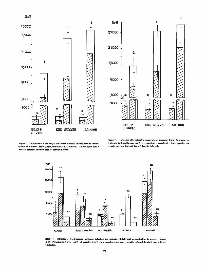
1
AUTUlINSTARTSUDER
1000
9000
3000
1
21000
27000
15000
1
AUTUMN
1
1
STARTSUMMER
3000
1000
9000
30000
27000
Figure 2.-lnnuence of Cryptocotyle cOllca.um infection on triglyceride concen.tratIon In bulIllead tissues (mg'Jl>, wet mass): m =muscles; I =liver; open bars =weakly hIIeded; ua:w ...... = beaYily Iafected.
Figure 4.-lnnuence of Cryptocotyu cOIlCO.um on summary [total) lipid concentration In bullbead tissues (m&,!,. wet mass): m =muscles; I =liver; open bars =weakly infected; batched bars = beavily Infected.
I'll
8000
4000
START SPIUBG DD BHmIG
Figure 3.-lnnueoce of COlltracaecum adullcum Infection on summary (total) lipid concentration In anchovy tissues(mg'Jl>, wet mass): I =Uver; rm =red muscles; wm =white muscles; open bars =weakly Infected; batcbed bars =beavlIy Infected.
50

identical for the different periods of the annual cycle. In contrastto triglycerides, the second fraction (by value) of phospholipids isnot diminished in heavily infected fishes. And what is more, during the fattening period the phospholipid concentration in whitemuscles of the heavily infected anchovy is higher by 64%, in redmuscles by 70%, and in liver by 42% (P<0.01). The livers ofheavily infected bullhead demonstrated the same phenomenonduring the spawning period. In other periods the phospholipidconcentration is not distinctly different.
By reducing the triglyceride supply in anchovy and bullheadtissues, higher levels of helminth infection may cause an important effect on the physiological state of individual fishes in apopulation, on the state of the population and on the dynamics oftheir number as a whole. Helminth infection reduces the state ofreadiness of the anchovy for migration and wintering, influencesthe quantitative and qualitative characteristics for sexual maturation, reduces brood survival, and so on. Regarding the bullhead,except [in addition to] the foregoing effects, its condition for nestprotection is reduced during the spawning period.
The data obtained allow me to assess the damage regarding thelipid supply. Thus, infection by helminths may [can] causedamage to the anchovy and bullhead populations. Such damagefor the anchovy totals 25% and for the bullhead 20%.
Thus, the results show the important role of helminths in thesubstance/energy balance of large fishery populations in the BlackSea and, consequently, their important role in the ecosystems ofthat body of water. Engraulis encrasiocholus ponticus and N.melanostomus are the examples for this.
51
REFERENCES
BROEK, W. L. F. VAN Den.1978. The effects of Lemaeocera branchialis on the Mer/angus merlangus
population in the Medway Estuary. J. Fish Bioi. 13:709-715.DAVIDOV, O. N.
1978. The effect of Bothriocephalus gowkongensis (Jan. 1955) on carp'smorphophysiological indices. [In Russ.] J. Hydrobiol. 14(6):59-64.
PROHOROVA, M. N., and Z. N. TUPIKOVA.1965. The great practical work on carbohydratical and lipid metabolism. L.
Press. Y.Y.U., p. 219.READER, T. A. J.
1971. Histochemical observations on carbohydrates, lipid and enzymes indigenean parasites and host tissues of Bithynia tentaculata. Parasitology 63:125-136.
SHABANOVA, 1. A.1967. Quantitative assay of phospholipids in the rats heart muscle by means
of thin-layer chromatography in silica gel. [In Russ., Eng!. summ.] Biokhimiia 32: 1155-1160.
SHEPELEV, V. M.1973. The colometrical [colorometric) determination of etheric-connected
fatty acid. Lab. Delo, No. 12:3-5.SCHULMAN, G. E.
1972. The physiological biochemical peculiarity of fish year cycle. [In Russ.)M. Pishchevaya prom., p. 368.
SINDERMANN, C. J.1966. Diseases of marine fishes. Adv. Mar. BioI. 4, 89 p.
STADNICHENKO, A. P.1972. The pathogenetic effect of trematode larvae on Viviparus viviparus (L.
1758) (Gastropoda, Prosobranchia). [In Russ.) Parasitology 6(2):154-161.

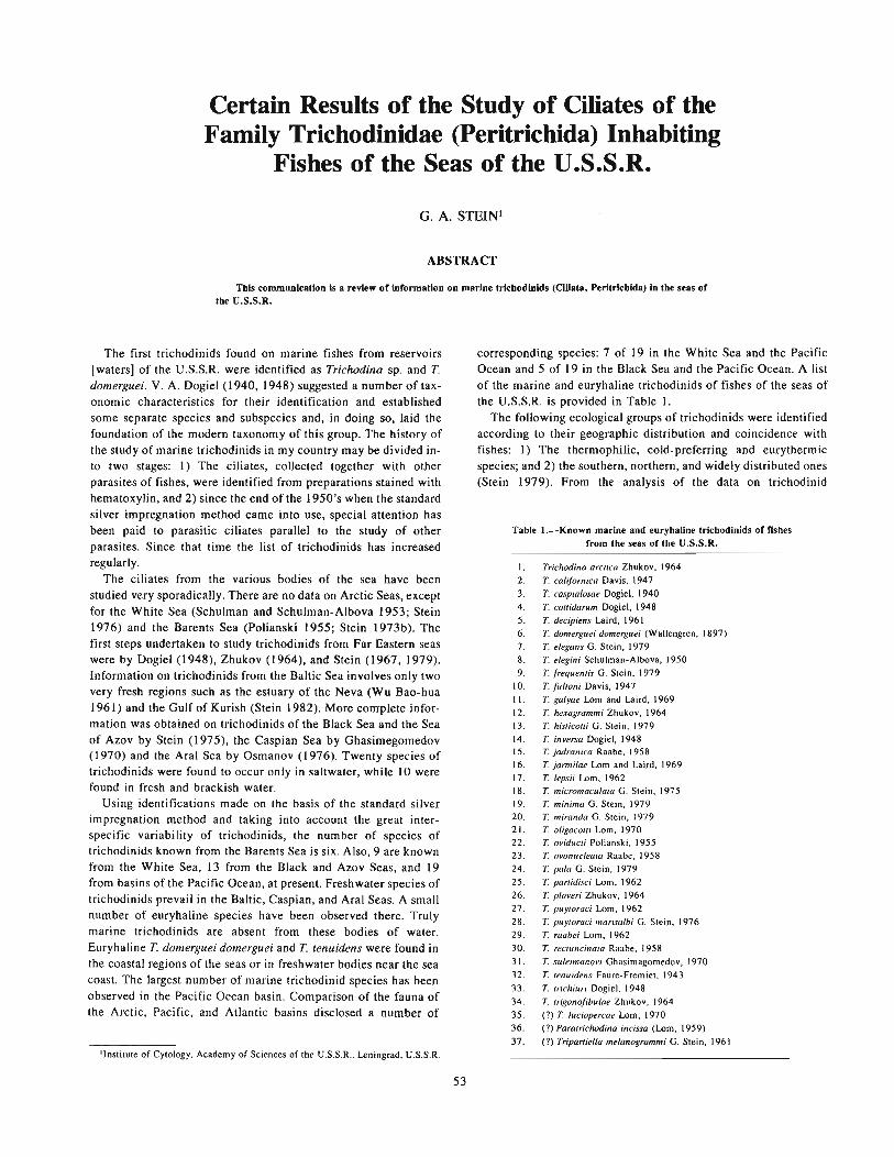
Certain Results of the Study of Ciliates of theFamily Trichodinidae (Peritrichida) Inhabiting
Fishes of the Seas of the U.S.S.R.
G. A. STEIN!
ABSTRACT
This communication Is a review of information on marine tricbodinids (Ciliata, Peritrlchlda) In the seas ofthe U.S.S.R.
The first trichodinids found on marine fishes from reservoirs[waters] of the U.S.S.R. were identified as Trichodina sp. and T.domerguei. V. A. Dogiel (1940, 1948) suggested a number of taxonomic characteristics for their identification and establishedsome separate species and subspecies and, in doing so, laid thefoundation of the modern taxonomy of this group. The history ofthe study of marine trichodinids in my country may be divided into two stages: I) The ciliates, collected together with otherparasites of fishes, were identified from preparations stained withhematoxylin, and 2) since the end of the 1950's when the standardsilver impregnation method came into use, special attention hasbeen paid to parasitic ciliates parallel to the study of otherparasites. Since that time the list of trichodinids has increasedregularly.
The ciliates from the various bodies of the sea have beenstudied very sporadically. There are no data on Arctic Seas, exceptfor the White Sea (Schulman and Schulman-Albova 1953; Stein1976) and the Barents Sea (Polianski 1955; Stein 1973b). Thefirst steps undertaken to study trichodinids from Far Eastern seaswere by Dogiel (1948), Zhukov (1964), and Stein (1967,1979).Information on trichodinids from the Baltic Sea involves only twovery fresh regions such as the estuary of the Neva (Wu Bao-hua1961) and the Gulf of Kurish (Stein 1982). More complete information was obtained on trichodinids of the Black Sea and the Seaof Azov by Stein (1975), the Caspian Sea by Ghasimegomedov(1970) and the Aral Sea by Osmanov (1976). Twenty species oftrichodinids were found to occur only in saltwater, while 10 werefound in fresh and brackish water.
Using identifications made on the basis of the standard silverimpregnation method and taking into account the great interspecific variability of trichodinids, the number of species oftrichodinids known from the Barents Sea is six. Also, 9 are knownfrom the White Sea, 13 from the Black and Azov Seas, and 19from basins of the Pacific Ocean, at present. Freshwater species oftrichodinids prevail in the Baltic, Caspian, and Aral Seas. A smallnumber of euryhaline species have been observed there. Trulymarine trichodinids are absent from these bodies of water.Euryhaline T. domerguei domerguei and T. tenuidens were found inthe coastal regions of the seas or in freshwater bodies near the seacoast. The largest number of marine trichodinid species has beenobserved in the Pacific Ocean basin. Comparison of the fauna ofthe Arctic, Pacific, and Atlantic basins disclosed a number of
'Institute of Cytology. Academy of Sciences of the U.S.S.R., Leningrad, U.S.S.R.
53
corresponding species: 7 of 19 in the White Sea and the PacificOcean and 5 of 19 in the Black Sea and the Pacific Ocean. A listof the marine and euryhaline trichodinids of fishes of the seas ofthe U.S.S.R. is provided in Table 1.
The following ecological groups of trichodinids were identifiedaccording to their geographic distribution and coincidence withfishes: 1) The thermophilic, cold-preferring and eurythermicspecies; and 2) the southern, northern, and widely distributed ones(Stein 1979). From the analysis of the data on trichodinid
Table I.-Known marine and euryhaline trichodinids of fishesfrom the seas of the U.S.S.R.
1. Trichodina arctica Zhukov. f9642. T. cali/ornica Davis, 19473. T. caspialosae Dogiel, 19404. T. cottidarum Dogiel, 19485. T. decipiens Laird, 19616. T. domerguei domerguei (Wallengren, 1897)7. T. elegans G. Stein, 19798. T. elegini Schulman-Albova, 19509. T. frequentis G. Stein, 1979
10. T. fultoni Davis, 1947I 1. T. galyae Lorn and Laird, 196912. T. hexagrammi Zhukov, 196413. T. histicotti G. Stein, 197914. T. inversa Dogiel, 194815. T. jadranica Raabe, 195816. T. jarmilae Lorn and Laird, 196917. T. lepsii Lorn, 196218. T. micromaculata G. Stein, 197519. T. minima G. Stein, 197920. T. miranda G. Stein, 197921. T. oligocotti Lorn, 197022. T. oviducti Polianski, 195523. T. ovonucleata Raabe. 195824. T. pala G. Stein, 197925. T. partidisci Lorn, 196226. T. ploveri Zhukov, 196427. T. puytoraci Lorn, 196228. T. puytoraci marisalbi G. Stein, 197629. T. raabei Lorn, 196230. T. rectuncinata Raabe, 195831. T. suleimanovi Ghasimagomedov, 197032. T. tenuidens Faure-Fremiet, 1943
33. T. trichiuri Dogiel, 194834. T. trigonofibulae Zhukov, 196435. (?) T. luciopercae Lorn, 197036. (?) Paratrichodina incissa (Lorn, 1959)
37. (?) Tripaniella melanogrammi G. Stein, 1961

coincidence with different hosts it might be concluded that thereis no narrow host-specificity in the majority of marine trichodinids. Fishes of the Order Perciformes prevail among the hosts oftrichodinids. From these facts I suppose [or conclude] (Stein1973a, 1979) that the trichodinids parasitized Perciformes fishesfirst and only later passed over to certain fishes of other orders.
Undoubtedly, further study of the ciliates of the family Trichodinidae from the seas of the U.S.S.R. will increase the faunisticlists of trichodinids and provide more exact ideas of their biologyand ecology.
LITERATURE CITED'
DOGIEL, V. A.1940. On the classification of the genus Trichodina. [In Russ.) Tr. Len.
Obsh. Yestiestvoispitatieley 68(4):8-31.1948. Parasitic protowans of fishes of Peter the Great Bay. III. The Ciliate
genus Trichodina. [In Russ.) Izvestia VNIORCH 27:44-66.GHASIMAGOMEDOV, A. A.
1970. New species of ciliates of the white-eyed bream (Abramis sapa). In
Voprosy physiologii, biochimii, zoologii i parasitologii, 4, p. 70-71. [InRuss.) Dagestansky Univ., Makhach-Kala.
OSMANOV, S. O. (editor).1976. Questions on the parasitology of the Aral Sea. [In Russ.) Izd. Fan,
Tashkent.
2Not all citations mentioned in the text are included here.
54
POLIANSKI, G. I.1955. Information on parasites of fishes of the northern seas of the USSR.
Parasites of fishes of the Barents Sea. [In Russ.) Tr. Zool. Inst. AN SSSR19:5-170.
SCHULMAN, S. S., and R. E. SCHULMAN-ALBOVA.1953. Parasites of fishes of the White Sea. [In Russ.) Izd. AN SSSR.
Moscow-Leningrad.STEIN, G. A.
1967. Parasitic ciliates (!'eritricha, Urceolariidae) of fishes of the Japan Sea.[In Russ.) Zool. Zh. 46, 5:657-668.
1973a. On the host specificity of marine parasitic ciliates of the familyUrceolariidae (Peritricha. Mobilia). In I'r<J8ress in protozoology, p. 399.Abstracts of papers read at the 4th Int. Congr. on Protozool. ClermontFerrand.
1973b. Parasitic ciliates (Peritricha. Urceolariidae) from some fishes of theBarents Sea. [In Russ.] Parasitologiya 1(6):489-496.
1975. Key to the ciliates of the family Urceolariidae F. Stein, 1867. In Keyto the parasites of the vertebrates from the Black and Awv Seas, p. 54-68.Izd. Naukova dumb. Kiev.
1976. Parasitic ciliates (peritricha, Urceolariidae) of fishes of the White Sea.[In Russ.• Engl. summ.] Acta Protozool 15(4):447-468.
1979. New data on parasitic ciliates (peritricha. Urceolariidae) of fishes ofthe basins of Pacific Ocean. [In Russ~ Engl. summ.) Acta Protozool. 18:531-552.
1982. Parasitic ciliates (Peritrichida, Trichodinidae) of some fishes of theGulf of KUrish. [In Russ.] Parasitologiya 16(1 ):24-29.
ZHUKOV, E. V.1964. On the parasitic fauna of fishes of the Chukotsk Peninsula and adjoin
ing Seas. Ill. The parasitic protozoa of marine and freshwater fishes. [InRuss.] Parasitologitchesky Sbomik Zool Inst. AN SSSR 22:224-262.

Special Features of the Myxosporidian Fauna FromSea and Ocean Fishes
A. A. KOVALIOVA and S. S. SCHULMANI
ABSTRACT
Regular research on Myxosporidia from open seas and oceans bas been conducted by investigators of tbefollowing researcb institutions: Tbe Zoological Institute of tbe Academy of Sciences of U.S.S.R., tbe AtlanticResearcb Institute, tbe Polar Researcb Institute, and tbe Pacinc Researcb Institute.
As a result of tbls researcb a new myxosporldlan fauna bas been discovered. Tbis provides a more completeidea of tbe order Multivalvulea and permits more precise definition of the marine Blvalvulea. Seven new generaand five new families have heen establisbed in the order Bivalvulea. The number of species has increased considerably.
The following problems are touched upon: The ways of adaptation of myxosporldian spores to living in different layers of the water body; the connection of different ichthyocoenosis in certain habitats of the ocean totheir myxosporidian fauna; and the ways [paths) of migration of some Multlvalvulea species from their initialdwelling place, the Indo-West Pacific, to tbe Atlantic, More evidence is presented on the evolution and phylogenyof the Myxosporidia.
Foci of infections and their agents are found in the Atlantic and, partly, in the Pacific Oceans. The species offishes most infected witb the genus Kudoa are revealed. Measures to control these Infections are given.
Research on sea and ocean Myxosporidia is of great theoreticaland practical importance. Analysis of the number of Myxosporidia species, their ecology and distribution among their fish hostsand in water aquatorias [water or ocean basins] has shown thatthese parasites originated in the seas. That is the reason speciesand genera with the most primitively constructed spores and mostprimitive vegetative forms occur only in the seas. Also, an exceptional diversity of myxosporidian forms is observed in the seas.This is mostly encouraged by the abundance of ecological niches.
Unfortunately marine Myxosporidia have been investigatedpoorly as compared with those in freshwater. Until 1941 only afew researchers studied Myxosporidia in the U.S.S.R. (Averinzew1907,1909,1911,1913; Dogiel and Bychovski 1939; Petruschevski 1932; Bazikalova 1932). Their works dealt with only afew myxosporidian species.
After 1945, new works on Myxosporidia appeared (Dogiel1948; Schulman and Schulman-Albova 1953; Polianski 1955).Many were dedicated to the study of the entire fish-parasite faunaof the Soviet seas. This was of especial importance for the worksof that period. Until 1970 only a few, but very interesting, workshad appeared in the U.S.S.R. (Zhukov (1964?]; Naidenova 1968;Naidenova and Zaika 1969a, b; Zaika 1966, 1968). [A numberhas been published since; see Literature Cited section.]
Regular research on Myxosporidia from open seas and oceanshas begun only in the last 10 yr based upon the three laboratoriesof Parasitology at three research institutes, i.e., the AtlanticResearch Institute, the Polar Research Institute, and the PacificResearch Institute. This paper is dedicated to the results of theseinvestigations, which is only the beginning of our efforts. First, wemust provide the names of the researchers. They are: A. V. Gaevskaya, A. N. ZUbchenko, A. A. Kovaliova, V. K. Krasin, and S. S.Schulman [see Literature Cited].
Investigations were conducted in the vast spaces of the AtlanticOcean from Baffins Land and Greenland to the Atlantic waters, inthe northern Pacific Ocean and in the open waters of its southeastern part. Unfortunately, the vast Indian Ocean has been verypoorly investigated thus far. During this research more than
'Zoological Institute, Academy of Sciences of the U.S.S.R., Leningrad, U.S.S.R.
55
50,000 specimens of more than 350 species of fishes have beenstudied. This has permitted discovery of a new myxosporidianfauna.
When the order Multivalvulea was established by S. S. Schulman in 1959 it consisted of only 3 families, 3 genera, and 12species. At present this order, owing to the works of Soviet protozoologists, numbers 4 families, 5 genera, and 40 species. Thus, wenow have a more complete understanding of the order.
Studies of the more numerous [larger] order, Bivalvulea, haveyielded data no less interesting.
Earlier certain researchers had reported on the interesting, aberrant, sea Myxosporidia, e.g., Auerbachia, Parvicapsula, Unicapsula, etc. Their systematic position and their importance in theMyxosporidia fauna were obscure. It has been found since thatthese species are members of fairly high taxa at the family levelwhich show a particular trend in the evolution of Myxosporidia.For example, the genus Auerbachia is a member of the familySphaeromyxidae which has the most primitive spores. This familyconsists of the initial genus Spaeromyxa, the genus Auerbachia,which originated from Sphaeromyxa (and has lost one polar capsule), and the genus Meglitchia, which was extracted from thegenus Ceratomyxa and has not been completely described.
The origin of the genus Parvicapsula is not clear. The discoveryof some new species belonging to this genus allowed us to tracethe pathway of its evolution from the genus Ceratomyxa. Soon anew genus Neoparvicapsula was discovered and described. Thus,the most peculiar family Parvicapsulidae, consisting of two genera(Parvicapsula and Neoparvicapsula), was formed. The extremelyinteresting genus Myxoproteus stands apart from all other taxa,because of the unique structure of its spores. It has been shownthat the two new genera Bipteria and Schulmania originated fromthis genus. The spores of Bipteria have two thin winglike processes; those of Schulmania, ventral and lateral keels. These threegenera form the single family Myxoproteidae.
Moreover, two new families-Alatosporidae and Paliatidaewere established. The members of the family Alatosporidae, withtheir small polar capsules, are very much like those of the genusCeratomyxa. But the spores of Ceratomyxa have a half-moonshape; while the spores of Alatosporidae are triangular and havetwo-winged appendices like Ceratomyxa. In the representatives of

the second genus of this family, Pseudoalatospora, these appendices are twofold and are united at their anterior ends. It is quiteevident that these structures promote [enhance] the ability of thespores to float in the water body [water column].
The monotypical family Paliatidae, whose hosts are deep-seafishes of the family Alepocephalidae, is also very interesting. Itsmembers have large spores with membTanelike structures resembling parachute domes.
It must be noted that the new families and genera consist, as arule, of 3 to 10 species. This provides evidence that the newfamilies form a considerable part of the myxosporidian fauna.
The number of species of many genera described earlier haveincreased considerably. Only in the genera Coccomyxa and Auerbachia has the number of species remained static. These generahave few species and their representatives occur very rarely.Moreover, analysis of the three species of the genus Auerbachiahas shown that they belong to a single, very variable, and widelyspread species.
Though our studies on Myxosporidia are far from being completed. we can report more than 100 new species of sea Myxosporidia even now. New findings and revisions of previouslydescribed species allow us to add considerable detail, to reorder,and, in some cases, reevaluate our view on taxa described longago. Thus, in such old and widely spread genera as Myxidium andCeratomyxG, species were found with gigantic spores. In Myxidiumthey reach up to 100 J.lm, whereas their size does not exceed 10-20lAm usually. In Ceratomyxa they reach 740 J.lm (Le., about 1 mm).
As previously mentioned, the spores of many sea [marine]Myxosporidia species develop various adaptive structures forfloating in different layers of the water body [water column]. Theyare winglike and threadlike processes, membranes, keels,manthes, etc. The spores of some species of Leptotheca, Sphaeromyxa, and Chloromyxum have threadlike appendages and thespores of some Davisia species have large hornlike prolongationsthat can change into threadlike devices at their posterior ends.
The decrease of the size of the spores in the genera Simuolinea,Chloromyxum, and Sphaerospora also promotes slow-sinking ofthe spores. On the other hand, the size of the spore increases insome representatives of Ceratomyxa. This is due to the largehollow cavities within the mass of the spore, which decrease thespecific weight of the spores, increasing their ability to float andpromoting slower sinking.
At the same time the large size of the spores, as well as that oftheir polar capsules, increase the weight of the spores. That is whytheir occurrence in one or the other layer of the water body [watercolumn] results from a range of oppositely directed factors. Allthese adaptations are of great importance for Myxosporidia livingin seas and oceans, as their distribution is very extensive horizontally (area) as well as vertically (depth).
The spores of Myxosporidia move passively. Therefore, to beeaten [ingested] by their hosts, they must occur in the same part ofthe same water body [as those hosts]. Ceratomyxa maxima canserve as an example illustrating the influence of depth on thespore's size. Its spores, ranging from 470 to 740 J.lm, occur in Berb:[Beryx?], which lives at depths of 800 to 900 m, while the sporesof the some [same?] species ranging from 230 to 480 J.lm occur inthe same fish host that lives at a depth of 240 to 400 m.
Thus, the enormous diversity of the shape and arrangement ofsea myxosporidian spores promotes their adaptation to certainecological conditions.
The distribution of fishes in the World Ocean is connected withtheir history as well as with their present life conditions. This hasits reflection in the distribution of the Myxosporidia. Ocean fishes
56
form icthyocoenoses which are related to certain dwelling areas;the main ones are the shelf zone, the continental slope, and theopen ocean waters. Each has a very complicated structure.
The shelf zone includes ecologically d. fferent fish groups thatare connected with the bottom, the pela~ic zone, and estuaries.Neritopelagic forms can also dwell here. As a rule the most abundant fauna of Myxosporidia and the fishes most infected by themoccur in these areas. The most numerous species, even thoserecently described, belong to genera described long ago. This isapparently because these sea [marine] areas have been the mostcompletely studied.
The genera Bipteria and Schulmania are quite new and arefound also in shelf waters. Their recency of discovery is due totheir occurrence in areas never before studied (Baffins Land,Greenland, the Bering Sea, and the Sea of Okhotsk).
Adaptation of the spores of Myxosporidia of the shelf zone (Le.,whose waters are shallow relative to those of the deep sea) forfloating occurs, but is not so striking. It is worthwhile mentioningthat the members of the genus Kudoa and other representatives ofthe order Multivalvulea occur only in the shelf zone (i.e., in thezone of vertical raising of waters) [vertical movement or upwelling]. These vertical movements upward [upwelling] prevent thespores of the Multivalvulea, which are poorly adapted to floating,from sinking to the bottom. Unfortunately, many fish complexesof the shelf zone, particularly estuarine fishes, have not yet beenstudied.
The shelf zone passes directly into the continental slope zonewhere bottom, meso- and bathypelagic, nerito-oceanic, andepipelagic fishes live. The meso- and bathypelagic fishes are ofparticular interest. Thus, a new genus, Paliatus was found amongrepresentatives of this zone (family Alepocephalidae). Also,gigantic spores of Myxosporidia were found first in fishes of thiszone.
Studies of the myxosporidian fauna of the open ocean werebegun recently. Myxosporidia from fishes dwelling in submarinedepths were studied. The myxosporidian fauna of ancient deepsea fishes, particularly the family Macrouridae, is of the most interest. The majority of their Myxosporidia are dispersed throughout the World Ocean; some, e.g., Auerbachia menatrosa, Myxidiumcoryphaenoides, etc., are nearly cosmopolitan.
The myxosporidian fauna of pelagic fishes is not yet fullyinvestigated. The first studies in the southeast Pacific havedisclosed a small numerical strength of Myxosporidia species inthis zone of the ocean.
Thus, 3 species of fishes [at least one belonging to the genusScomber; the other(s) to other genera2 ] of the 14 well-studied oneswere infected with 6 species of Myxosporidia belonging to 4genera. The incidence of infection in these fishes was also verylow. Some species of Myxosporidia have certain adaptations forfloating. The other species are typical parasites of bathypelagicfishes and usually have adaptations for floating only in thishorizon [or level]. Their occurrence in typically pelagic fishes isdue to powerful upsurges [upwellings] of the water.
The theoretical importance of our research is not exhausted bythe examples mentioned above. The following investigations[reviews or explanations] will be devoted to the genesis of waterbasins and their fauna. They will provide a clue to the solution ofzoogeographical and biological problems. We now have more
>only the Dame of the genus of one of the three species of fish was given in theoriginal typeocript. Its specific trivial name and the names of the other two species...,.., not included.

precise data on the phylogeny of the Myxosporidia and the way[manner] of distribution of some taxa.
Thus, analyses of the distribution of the Multivalvulea haveshown that they originated in the Indo-West Pacific. The mostprimitive species of this order, Trilospora californica (which livesin the gallbladder, the initial habitat of the Myxosporidia), isfound only in this region, as are all known families and genera ofthe Multivalvulea. Moreover, the number of species is muchgreater here than in the Atlantic, in spite of a lower rate of studiesin the Pacific. Lastly, the most numerous species of Kudoa havebeen found only in Pacific deep-sea fishes.
The order Multivalvulea is comparatively young. It arose afterthe formation of the present fish fauna of the Atlantic. Representatives of these Myxosporidia have a very typical local distribution. Hence, under t~ese circumstances the penetration of numerous species of Multivalvulea into the Atlantic Ocean has beenprevented.
Only 14 species of the genus Kudoa, I species of Trilospora,and I species of Unicauda were found in Atlantic fishes, and thiswas in spite of a much higher level of research on myxosporidiansin this ocean.
Because macrourid fishes (and other ancient deep-sea fishes)have limited migration ability, only one species of Trilospora haspenetrated into the Atlantic Ocean. Analysis of the number ofMultivalvulea species and their distribution by fish hosts, indicates that these parasites could have arrived in the Atlantic byseveral different routes. The most probable one was from thePacific Ocean around South Africa and its Atlantic shore. At present Kudoa thyrsitis occurs in the southeastern Atlantic. It occursalso in both the Pacific and Indian Oceans.
Kudoa clupeidae and K. funduli from [waters around] northwestern Africa are morphologically very similar to K. nova fromthe region of north Africa and the Mediterranean.3 It is possiblethat K. nova has penetrated to the North American shelf by usingsuch active migrators as Thunnus and Xiphidae. Here, this speciesmay have given rise to K. clupeidae and K. funduli.
The Kudoa species occurring on the Falkland-Patagonian shelf,penetrated into the Atlantic via South America. Hydrographicalbarriers have prevented their migration to the north.
The Kudoa species found in the Gulf of Mexico do not occur inany other regions of the Atlantic. These species may havepenetrated into the Gulf directly from the Pacific Ocean, beforethe junction of North and South America.
Finally, Trilospora from Coryphaenoides members of the midAtlantic ridge, apparently has penetrated directly with its host.Unfortunately, the pathways of invasion of this fish are unknown.
Investigations of Myxosporidia are of great importance forfisheries also. The Multivalvulea are not pathogenic to their fishhosts, but the fishery suffers great losses, some because certainspecies of this order reduce the quality of the flesh of infected fish.Others provoke lysis of the fish host's tissues even after prolongedfreezing. This makes the fish unsuitable for food.
As a result of our studies in the Atlantic and partly in thePacific, foci of infections and their agents were found; species offishes infected with Kudoa were revealed; and the seasonal distribution of these parasites in some regions of the ocean wasdiscovered. An instruction on how to utilize fishes infected withthe genus Kudoa was provided on the basis of this research.
As the studies of recent years have shown, members of theBivalvulea can be pathogenic also. Thus, Henneguya sp. can be
)The author may have meant". . from the northwestern Atlantic arc morphologically very similar.
57
pathogenic to fishes in aquacultural conditions.Recently we have obtained from Dr. Hoffman4 information on
a new species of Parvicapsula, parasitizing the kidneys of penreared coho salmon, O. kisutch, which causes mortality of up to30% of its hosts.
To search for measures to control these infections it isnecessary to investigate not only the number of species, but alsothe biology of the agents.
LITERATURE CITED
AVERINZEW, S.1907. Vber Myxosporidien aus der Gallenblase der Fische. Zoo/. Anz. 31:
831-834.1909. Studien iiber parasitische Protozoon. L Die Sporenbildung bei Ceraro
myxa drepanopsel/ae. Arch. Protistenkd. 14:74-112.191 I. Studien iiber parasitische Prolozoen. VII Vber Sporenbildung bei Myx
idium sp. aus der Gallenblase von Cottus sc"rpius. Arch. Protistenkd. 23:199-204.
1913. Ergenbnisse der Untersuchungen iiber parasitische Protozoen der tropischen Region Afrikas, II. Zoo/. Anz. 42:151-156.
BAZIKALOVA, A.1932. Contribution to the parasites of fishes from Murman. [In Russ.] Snab
tekhizdet - Sbornik nauchnopromyslovykh rabot na Murmane, p. 136-153.DOGIEL. V. A.
1948. Protozoa infecting fishes in St. Peter Bay. [In Russ.] Izvestiya Vsesoyunznogo nauchno-issledovatelskogo Instituta ozernogo i rechnogo rybnogo khozayistva 27:17-660.
DOGlEL, V. A., and B. E. BYCHOVSKI.1939. The parasites of fishes in the Caspian Sea. [In Russ.] Tr. kompleksnoi
komissii po izucheniyu Kaspiiskogo morya 7:1-149.EVIDOKIMOV A, E. B.
1973. A new myxosporidia species, Auerbachia sphaerica sp. n. and the systematic position of the genus Auerbachia meglitsch, 1968. [In Russ.] Parasitologiya 7(1):91-92.
1975. Some aspects of the ecology and the origin of Myxosporidia from teleost fishes. In Problemy parasitologii, p. 171-173. [In Russ.] Kiev.
1977. Myxosporidia from teleost fishes of the Patagonian Shelf. [In Russ.]Parasitologiya 11(2):166-168.
GAEVSKAYA, A. V., and A. A. KOVALIOVA.1979. Two new species of Myxosporidia, Davisia donecae sp. n. and
Ceratomyxa australes sp. n. from the horse mackerel in the southeasternAtlantic. [In Russ.] Biologiya Morya 3:8-81.
KOVALIOVA, A. A., and A. V. GAEVSKAYA.1979. Two new species of Myxoproteus (Myxosporidia, Bivalvulea) from
fishes in the Celtic Sea. [In Russ.] Parasitologiya 13(5):771-773.198 I. On new findings of Myxosporidia of the genus Parvicapsula in Atlantic
fishes. [In Russ.] Zoo/. Zh. 60:771-773.KOVALiOVA, A. A., and S. S. SCHULMAN.
1978. Multivalulea of fishes. In Fauna i sistematika odnokletochnykh zhyvolnykh, p. 16-30. [In Russ., Eng/. summ.] Tr. Zool. Inst. AN SSSR. t. 78.
KOVALIOVA, A. A., S. S. SCHULMAN, and V. N. JAKOVLEV.1979. Myxosporidia of the genus Kudoa (Myxosporidia, Multivalvulea) from
the Atlantic Ocean. In Sistemalika i ekologiya sporovikov i knidosporidii, p.42-64. [In Russ., Eng/. summ.] Tr. Zoo/. Inst. AN SSSR, t. 78.
KUZNETZOVA, I. G.1977. Myxosporidia from Chondroslei fishes in the Patagonian Shelf (the
coasl of Argentina). [In Russ.] Parasilologiya 11(1):74-77.NAIDENOVA, N. N.
1968. Ortholinea gobiusi sp. n. (Myxosporidia, Bivalvulea) from Gobius
ophiocephalus in the Black Sea. [In Russ.] Biologiya Morya 14: 15-18.1974. The parasites of Gobiidae fishes in the Black Sea and the Sea of Azov.
[In Russ.] Kiev, 182 p.NAIDENOVA, N. N., and V. E. ZAIKA.
1969a. Marine myxosporidian of original morphology and some aspects ofthe myxosporidian evolution. Progr. ProloZOO/., p. 379.
1969b. Two new species of Protozoa, parasites of fishes in the Black Sea. [InRuss.] Parasitologiya 3(1):97-101.
1970. Three new genera of myxosporidia-fish parasites from the IndianOcean. [In Russ., Eng/. summ.] Zoo/. Zh. 49:451-454.
4Believed by editor to be Dr. Glenn L. Hoffman of Fish Farming ExperimentalStation, U.S. Fish and Wildlife Service, P.O. Box 860, Stuttgart, AR 72160, U.S.A.
PETRUSHEVSKI, G. K.1932. Zur Systematik und Cytologie der Myxosporidia aus einigen Fischen
des Weihen Meeres. Arch. Protistenkd. 78:543-556.POLiANSKI, Yu. I.
1955. Parasites of fishes in the Barents Sea. [In Russ.) Tr. Zoo!. Inst. ANSSSR 19:5-170.
SCHULMAN, S.S.1953. New and poorly studied Myxosporidia (Myxosporidia, Bivalvulea)
from fishes in the White Sea. [In Russ.) Zoo!. Zh. 32:384-393.1956. The parasite fauna of herring, smelt and navaga in the White Sea. (1n
Russ.) Trudy Karelo-finskogo Filiala AN SSSR, v. 4. Seriya parasitologicheskaya, p. 50-67.
1966. The myxosporidian fauna of the USSR. [In Russ.] Izdatelstvo ANSSSR, M. 1.., 498 p.
SCHULMAN, S. S., A. A. KOVALIOVA, and V. R. DUBINA.1979. New myxosporidians from fishes in the Atlantic coast of Africa. [In
Russ.) Parasitologiya 13(1):71-78.
58
SCHULMAN, S. S., and R. E. SCHULMAN-ALBOVA.1953. The parasites of fishes in the White Sea. [In Russ.) Izdatelstvo AN
SSSR, M. 1.., 198 p.
ZAIKA, V. E.1966. Contribution to the protozoan fauna of the Black Sea. In Gelminto
fauna ahyvotnykh Yuzhnykh morei, p. 13-31. [In Russ.) Kiev.1968. Parasitic Protozoa from the Black Sea. [In Russ.) Biologiya Morya
14:41-46.
ZHUKOV, E. V.1964. Parasites from the fishes of the river Chukolka. II. The Protozoa from
!be marine and freshwater fishes. [In Russ.) Parasitologicheskii sbomikZoologicheskogo Instituta AN SSSR, 22, 22:224-263.
ZUBCHENKO, A. V., and V. K. KRASIN.1980. The genus Myxidium (Myxosporidia, Bivalvulea) from macrourid fishes
in !be North Atlantic and the Pacific Oceans. [In Russ.] Parasitologiya 14(2):168-176.

Myxosporidia of Fishes of the North Pacific
V. K. KRASINI
Myxosporidians of fishes of the northern Pacific have beenstudied only recently. The first information about them was notpublished until the 1920's (Fujita 1923; Davis 1924).
By now a large number of works dedicated to Myxosporidia offishes of that area have been published by foreign and Sovietscientists (Jameson 1929, 1931; Nobel 1938a, b, 1941, 1966;Moser 1976; Moser and Noble 1977; Dogiel 1948; Polianski1958; Zhukov 1964; Schulman 1966; and others).
As the result of these studies new species of Myxosporidia havebeen registered [recorded) and the number of hosts for previouslyknown species has increased. In addition, information on taxonomy, problems of ecology and development, and the relationships between parasites and hosts have been covered. Many interesting works deal with the spatial and seasonal distribution ofMyxosporidia in the ocean and their dependence on biotic andabiotic conditions as well as the influence of Myxosporidia on thefish hosts, themselves.
Since 1972 the TINRO Laboratory of Parasitology of MarineAnimals has conducted planned investigations of Myxosporidia offish of the northern Pacific.
Thus far, 1,809 fish belonging to 43 families, 81 genera, and132 species have been studied. In our collections 41 species ofMyxosporidia were new and 65 were found for the first time innew hosts, while 12 were not identifiable as to species because ofan insufficient number of spores.
In all, a total of 190 Myxosporidia have been registered[recorded) from the North Pacific, according to our data and thatfrom the literature. One hundred and twelve species of Myxosporidia were found off the Asian coast-96 of them belonging tothe order Bivalvulea and 16 to the order Multivalvulea. Myxosporidia of the genera Ceratomyxa (26 species), Myxidium (19species), Kudoa (14 species), and Chloromyxum (II species) weremost abundant.
Eighty-seven species were registered in the northeasternPacific, 75 of them from Bivalvulea and 12 from Multivalvulea.The genera Ceratomyxa (18 species), Myxidium and Kudoa (10species, and Leptotheca (9 species) were predominant as well as inthe northwestern Pacific.
In both areas 18 genera from 9 Myxosporidia families wereregistered [recorded), but myxosporidians of the genera Auerbachia and Unicapsula were not found in the northwestern Pacificwhile those of the genera Sphaerospora and Hexacapsula were notfound in the northeastern Pacific.
The faunal similarity of Myxosporidia of the shelf fish of theseareas at the species level is insignificant. It is interesting to notethat there are no common species of the order Multivalvuleaparasitizing mainly [primarily) shelf fish in the Pacific waters offthe North American and Asian coasts.
The Myxosporidia fauna of bathyal fishes has been studiedmuch less than that of shelf fishes. Nevertheless, available data on
I Pacific Research Institute of Fisheries and Oceanography (TINRO), Vladivostok,U.S.S.R.
59
the infection of Coryphaenoides spp., Laemonema longipes, andAnoplopoma fimbria allow us to state a somewhat closer speciessimilarity of Myxosporidia in the given areas.
Even my incomplete data (materials on bathyal fishes receivedin 1980 are still being processed) indicate that 10 species of Myxosporidia have been found to be common in these areas (i.e., Myxidium coryphaenoidium, M. melanocetum, M. pearcyi, M. melanostigmum, Ceratomyxa anoplopoma, C. obesa, C. asymmetrica, C.coryphaenoida, Davisia anoplopoma, and D. coryphaenoida).
Myxosporidia of the other Multivalvulea, which cause considerable economic losses to the fish industry due to impairment ofthe market value of the fish (even rendering the fish unfit forhuman consumption in some cases), have drawn the greatestattention.
Three years ago more than 40 species of multivalvular Myxosporidae were known. They were divided into four families, i.e.,Trilosporidae, Kudoidae, Pentacapsulidae, and Hexacapsulidae(Kovaliova and Schulman 1978). According to my data, 27species of Multivalvulea of three families (Trilosporidae,Kudoidae, and Hexacapsulidae) are registered [recorded) from theNorth Pacific.
By now a total of 50 species of multivalvular Myxosporidaehave been registered [recorded]. Two genera have been placed inthe family Trilosporidae: Unicapsula (one species) and Trilospora(two species) were found, Trilospora calijornica in the gallbladderand Trilospora [sp.?] in the muscles. The family Tetracapsulidae isrepresented by the 23 species of the genus Kudoa. Kudoa perecardialis was found in the pericardial cavity of Seriola quinqeradiata.The other species were located in the muscles of the fish. Thefamily Hexacapsulidae contains one monotypic genus, Hexacapsula, found in the muscles of Neothunnus macropterus. Therepresentatives of the other above-mentioned parasites have beenfound in 25 species offish. The muscles of the following fishes aremost greatly infected by Myxosporidae of the genus Kudoa:Merluccius productus (two species), 124 specimens of 151 examined were infected (85.2%); Engraulis mordax, 159 from 225 fishspecimens (70.6%); Coryphaenoides pectoralis, 34 infected fishspecimens of 66 investigated (51.5%); Atheresthes evermanni, 15infected fish specimens of 56 examined (26.8%); Hippoglossoideselassodon, 6 infected fish specimens of 33 studied (18.2%); andCoryphaenoides cinereus, 5 infected fish specimens of 30 examined (16.6%).
LITERATURE CITED
DAVIS, H. S.1924. A new myxosporidian parasite, the cause of "wormy" halibut. Rep.
U.S. Comm. Fish. for fiscal year 1923, Append. 8(Doc. 957), 5 p.DOGIEL, V. A.
1948. Parasitic Protozoa of fishes of lhe Peter the Great Bay. [In Russ.]Izvestiya Vsesoyuzn. Nauchno-issled. Institula Ozemogo i Rechnogo Rybnogo Khozyaystva. 27:17-66.
FUJITA, T.1923. Studies on Myxosporidia of Japan. J. Coil. Agric. Hokkaido Univ. 10:
191-248.

JAMESON, A. P.1929. Myxosporidia from Californian fishes. J. Parasito!. 16:59-68.1931. Notes on Californian Myxosporidia. J. Parasitot. 18:59-69.
KOVAUOVA, A. A., and S. S. SCHULMAN.1978. Multivalvulean myxosporidians of fishes. Tr. Zoot. Inst. AN SSSR
78:16-29.
MOSER,M.1976. Inter-specific associations of Myxosporidia (Protozoa) in macrourid
fishes. J. Parasitenk. 51:133-135.
MOSER, M., and E. R. NOBLE.1977. Myxosporidia genera Auerbachia, Sphaerospora, DaYisia and Chlor
omyxum in macrourid fisbes and the sablefish, Anoplaoma fimbria. J.Parasitenk.51(2):159-163.
NOBLE, E. R.1938a. Two new Myxosporidia from tide pool fishes of California. J.
ParasitoL 24:441-444.
60
1938b. Myxosporidia from tide pool fishes of California. J. Parasito!' 24(Suppt.):31.
1941. On distribution relationships between California tide pool fisbes andtheir Myxosporidian (Protozoan) parasites. J. Parasitot. 27:409-415.
1966. Myxosporidia in deepwater fisbes. J. Parasitot. 52:685-690.
POUANSKI, Yu. I.1958. Parasitofauna and environment. Some ecological questions of marine
fish parasites. In Osnovnye problemy parasitologii ryb. [In Russ.]
SCHULMAN, S. S.
1966. Myxosporidia of tbe fauna of tbe USSR. [In Russ.] M. L. Nauka,
505 p.
ZHUKOV, E. V.
1964. Parasitofauna of the fishes of the Cbnkotta Peninsula. 111. Protozoans(Protozoa) of marine and freshwater fishes. General conclusions. [In Russ.]
Parasito!. Heskiy Sbornik 22:224-263.

Investigations of the Ultrastructure and CytochemicalPeculiarities of Kudoa quadratum (Thelohan, 1895),
(Myxosporidia, Multivalvulea)
A. V. USPENSKAYA'
ABSTRACT
The ultrastructure, mode of nutrition, and host-parasite relations in Kudoa quadratum (Myxozoa) frommuscle fibers of MyoIocephalus scorpius are discussed.
The ultrastructure and cytochemical peculiarities of Kudoaquadratum, a parasite of muscle cells of Myoxocephalus scorpiusfrom the White Sea, were studied (Figs. 1,2,3). Kudoa quadratumis an intracellular parasite. Vegetative stages of K. quadratum andthe so-called "cysts" have nothing in common with the resting orreproductive cysts of other Protozoa. Kudoa quadratum has arather large (up to 2 x 0.3 mm) fusiform plasmodium bounded bya unit membrane. Usually one can see remnants of protofibrilsbetween the membrane of the plasmodium and the muscle cellmembrane. Plasmodia utilize the protofibrils by phagocytosis orby pinocytosis after lysis of the latter. Alkaline phosphatase wasdiscovered at the surface of plasmodia. The cytoplasm of thetrophozoites is rich in glycogen. There is no succinat degidrogenase [succinate dehydrogenase?] activity in trophozoites. It seemsthat trophozoites are anaerobic organisms and that glycogen is aprincipal source of energy for them. The development of thetrophozoites and sporogenesis take place inside one and the same
I Institute of Cytology, Academy of Sciences of the U.S.S.R., Leningrad, U.S.S.R.
Figure I.-The spore of Kudoa quadratum with four valves and four polar capsules.
61
muscle cell. I did not observe ruptures of muscle cell membranesas a result of infection. Only one plasmodium can be observed inside each infected muscle cell. The muscle fibrils adjoining the infected one seem to be undamaged if the trophozoite is not toolarge.
REFERENCE
USPENSKAYA, A. V.1982. Ultrastructure of so-called cysts of Myxosporidia. [10 Russ.] Parasit
ologiya 14(1):13-17.
Figure 2.-Trophozoite of Kudoa quadratum (t) inside muscle fiber. mmf =membrane of muscle fiber, mt = trophozoite membrane, Mmf = mitochrondriaof muscle fiber.

Figure 3.-U1trastructure of infected (imf) ..... noninfected (.-I) masele fiber.
62

Trematodes of Commercial Fish of the Pacific ofPractical Importance!
V. D. KOROTAEVN
Trematodes [digeneids] belong to the most numerous group offlatworms, with a considerable number being parasites of marinefishes. The total number of known trematode species invadingmarine species at different stages is about 3,000 species belongingto approximately 700 genera (Kurochkin in litL). Many of themare of great importance to marine ecosystems, though this aspectof marine parasitology requires further study. My attention isprimarily attracted by those trematode species of greatest practical significance.
The adult or larval forms of such trematode species are highlypathogenic for fish and may affect their number, reduce the quality of raw fish and fish products, or be harmful to a human being[and a few may infect humans]. [Hence, trematodes may be harmful (economically and medically) to humans.]
In this paper I shall review the most important trematodespecies of Pacific marine fishes encountered by commercialfishery specialists. These materials have been obtained whileprocessing trematodes of commercial marine fish collected byspecialists of the Laboratory of Parasitology of Marine Animals ofTINRO from the different parts of the Pacific Ocean. Data concerning the level of infection of fish by the above species and theforms of trematodes are presented as well.
Commercial fish whose muscles are infected by trematodes areobserved in almost all areas of the Pacific Ocean. Metacercariaeof trematodes of the family Acanthocolpidae Luhe 1909 are frequently recorded from marine fishes. Sometimes they invade themuscles under the skin or the fins in great numbers, thus makingsome fish catches unmarketable, which results in considerableeconomical losses.
Metacercariae of Stephanostomum spp. have been observed in29.5% of the fish examined (of 12 fish species) in the northwestPacific. They were found on 6.9% of fish (three fish species) inthe northeast Pacific; 54.5% (four fish species) in the Hawaiianarea; 10.8% (two fish species) in the Philippine Sea area; and51.6% (six fish species) in the Australia-New Zealand area.
The highest level of muscle infection has been observed byTINRO specialists on [several] flatfishes of the northwest Pacific,i.e., Lepidopsetta biliniata (39% at an intensity of up to 101individuals), Limanda aspera (58.5% at an intensity of up to 25individuals), and Hippoglossoides elassodon (20.4% at an intensityof up to 38 individuals).
Such high levels of infection off the Asian coast may be attributed primarily to the greater availability there of continentalshelves and slopes where fish have more chances of becominginfected from benthic invertebrates-the first intermediate hostsof these trematodes.
I As used here the word trematode means Digenea, as is now widely accepted byRussian scientists.
'Pacific Research Institute of Fisheries and Oceanography (T1NRO), Vladivostok,U.S.S.R.
63
On the basis of data in the literature, I recorded six larvae of thegenus Stephanostomum, which were identified to the species level,on various fishes of the World Ocean. Considering the still imperfect specific criteria of trematodes of this genus and the greatnumber of species described to date (more than 70), it is natural toadmit significant difficulties in their determination [identification]. I am of the opinion thaI it is impossible to refer [utilize] thenomenclatural and systematic changes offered by Dollfus (1972)for the genus Stephanostomum, since it would increase the confusion. I (Korotaeva undated) described some larval forms of metacercariae of the family Acanthocolpidae from the muscles of certain marine Pacific commercial fishes.
Larvae of trematodes of the family Bucephalidae Poche. 1907are of no less importance from a practical point of view. LarvalProsorhynchus crucibulus were observed in the northwest Pacificin the muscles and under the skin of 13.9% of fish of seven speciesincluding Myoxocephalus jack (66.7% of fish at an intensity of upto 7 specimens), Sebastes alutus (56% at an intensity of up to 142specimens), and Melletes papillio (50% at an intensity of up to 30specimens) revealed the highest level of infection. The highest intensity of infection, up to 1,000 specimens, was observed in Hexagrammos lagocephalus. According to the literature, 17 Pacificfish species are known to have their muscles infected by larvae ofthis species. Additionally, metacercariae of seven species oftrematodes of the family Bucephalidae were registered on variousbenthic fishes of the World Ocean.
Larvae of trematodes of the family Didymozoidae Poche, 1907are widely distributed among oceanic fish. They are observed indifferent organs and tissues. Their location in the muscles is quitecommon for fishes of the tropical and subtropical Pacific. AroundHawaii they are found in 75% of Epinephelus guarnus at an intensity of from one to two specimens. Fourteen percent of Ocycriusiaponica, at an intensity of one specimen are observed to be infected in the Philippine Sea. Forty percent of Saurida undosquamis at an intensity of up to 10 specimens, 33.3% of S. tumbelat an intensity of up to 23 specimens, 40% of Pentapodus senosusat an intensity of up to 10 specimens, and 75% of Pristipomoidesrypus at an intensity of from one to five specimens were found infected off northern and northwest Australia. In the New Zealandarea, 5 specimens of the trematode were observed in 1 of 35 individual Nemadactylus macropterus.
The helminthologicalliterature presents data concerning localization of some adult didymozoids in the muscles of the largepelagic fish. Large envelopes with cyst formations containing eggsof already degenerated adult didymozoid species are often observed in their muscles. Mature trematodes, Nematobothrioidesaustraliensis, were described by us (Nikolaeva and Korotaeva1970) from Scomber australasicus from the Great Australian Bay.They are localized in the gonads of females, where their [periodof) intensive maturing coincides with the spawning period [of thehostl.

Trematodes of the species Gonocerca ashoro (family Derogenidae) have a similar microhabitat, locating in the ovaries offemales of Nematonurus pectoralis. These trematodes reach 3 cmin length. They are of bright brown color and easily visible amongthe eggs, and may hinder marketing of this valuable product. Theywere registered [recorded] by TINRO specialists in the area oftheEmperor Range on 13.3% of fish with an average invasion [infection] of up to four specimens.
In the North Pacific we found an interesting phenomenon oflocalization of the trematode Aporocotyle theragrae in the musclesof 48 of the 1,937 Theragra chalcogramma studied (2.5%), at anintensity of up to 76 specimens. Such abnormal location of thesetrematodes may be explained by the fact that some larvae begingrowing after penetration into the small blood vessels of the hostand are not able to get into the larger blood vessels-their normallocation. Their size may reach that of adult trematodes, but sexualmaturity never occurs in this location and they perish. degenerate,and become calcified. In addition this fish, primarily its muscles.are infected by two species of metacercariae of trematodes of thefamily Cathaemasidae whose definitive hosts are birds.
Metacercarial Liliatrema skrjabini were observed in eight fishspecies and L. sobolevi in one fish species.
Among trematodes recorded as being in the muscles of marinefish, I know of only one species. Nanophyetus salmincola. which isof definite medical importance. It usually infects salmonids infreshwater. but the metacercariae are able to survive in themuscles of anadromous fish while they are in the ocean. When inthe human intestine, the live metacercariae leave the cysts andgrow to be mature trematodes, thus causing a disease-naoophyetosis, which is not very dangerous but is unpleasant. As indicated.N. salmincola is a parasite of freshwater origin. No marine
64
trematode species found in fish muscles are known to bepathogenic to humans.
Metacercariae of trematodes located in fish muscles are usuallyfound in specific hosts, though not so regularly as is the case withmaritae. Therefore, infection by them is not observed on allichthyofaunal components within any area of the sea but may befound only in particular commercial fish species. From the practical point of view this allows us to determine the commercial fishspecies whose muscles are always free from invasion, as well asfish which require constant or episodical [specific?] parasitological control for human consumption purposes.
I must indicate that I consider that nearly all known incidentsof rejection of marine fish catches during the last 15 yr because oftrematode infec;tions were groundless. In all these cases theparasites infecting the fish were not harmful for human beings andthe level of infection, except in some rare instances, was not sufficiently high to cause loss of marketing quality. The skill of thepersons conducting the parasitological inspections was inadequate.
LITERATURE CITED
DOLLFUS, R. P. H.1972. Parasilologia maurilanica. Digenetiques de Teleosleens Ire note. Di
genre Stephanochasmus A. Looss, 1900 (Trematoda Digenea). Bull. Inst.Fondam. Afr. Noire, 34, Ser. A (4):809-827.
KOROTAEVA, V. D.Undated. On infection of marine fish muscles with metacercariae of trema
todes of the family Acanthocolpidae Luhe, 1909. [In Russ.] IzvestiaTINRO 101:120-125.
NIKOLAEVA, V. M., and V. D. KOROTAEVA.1970. Nematobothrioides australiensis sp.n.-a new trematode of Scomber
australiasicus. [In Russ., Engl. summ.] Parasitologiya 4(4):330-333.

Infestation Rate of the Young of White Sea Herring,Reared Under Experimental Conditions and
Caught in the Sea, by Trematodes,and Their Pathogenic Effect 1,2
O. F. IVANCHENKO and T. A. GROZDILOVA3
While rearing White Sea herring we observed deaths of larvaeinfected by trematodes of the family Hemiuridae, Brachiphalluscrenatus (Rudolphi 1802) and Lecithaster gibbosus (Rudolphi1802), located in the digestive tract. Presence of B. crenatus
resulted in injury of the mucuous membranes hindering thepassage of food, causing embolism of the intestine and inhibitionof the growth of larvae and fry (lvanchenko and Grozdilova1971). Lecithaster gibbosus, actively moving in the digestive tractof the young by means of their suckers, injured the mucuous membrane of the intestine. As a result, the larvae and fry ceasedfeeding and died (Ivanchenko and Grozdilova 1971). These twotrematode species, pathogenic for herring larvae and fry, wereprimarily responsible for the deaths of the young in the aquarium[experimental containers]. Their pathogenic influence is apparently the same in nature. Therefore, we have begun a study intothe infestation rate of herring at early stages of ontogenesis underexperimental conditions and in nature. In 1979 work was performed on herring inhabiting the Chupa Inlet of Kandalaksha Bayin the White Sea. Under experimental conditions, hatching of thelarvae occurred on 5 June. Larvae were fed with plankton caughtin the sea. Four hundred and thirty larvae and fry of herringstudied during the period of rearing contained two species oftrematodes, B. crenatus and L. gibbosus. The first parasites were
1As used here the word trematodes means Digenea.'No Literature Cited section was supplied by the authors.'The Zoological Institute, Academy of Sciences of the U.S.S.R., Leningrad,
U.S.S.R.
65
noted on 2 July. The number of parasites was one to two per larva,the percentage of infestation being 4%. The percentage of infection by both trematode species increased to 10% by 11 July, to30% by 23 July, to 52% by 30 July, to 58% by 7 August, and, atthe end of the experiment on 13 August, it was 68% with the intensity of infestation being one to three parasites per single youngfish.
Abundant hatching of larvae in nature occurred at approximately the same time as in the experiment. The first larvae withparasites (B. crenatus and L. gibbosus) were discovered on 9 July.The percentage of infestation [infection] attained 8.2%; on 17July it became 57%; on 26 July it was 76%. The latest [for theyear] collecting in the sea was performed on 3 August. Thepercentage of infection was 67%, with an intensity of infection inthe digestive tract of one to five specimens per individual juvenile.The results suggest that infestations of White Sea herring at earlystages of ontogenesis under experimental conditions and in natureare synchronous and fluctuate within approximately the samelimits.
Apparently, the dynamics of infestation of the young reared inaquaria can reflect infestation of larvae and juveniles in the sea tosome degree. Experimental conditions make it possible toestimate relatively precisely the percentage of herring killed bythe above parasites. Such calculations would be impossible innature. Thus, artificial rearing permits a more detailed study of theinfluence of trematodes occurring on larvae and fry, and providesan idea of the degree of pathogenicity of these parasites forherring young in nature.


Trematodes-Didymozoidae Fauna, Distributionand Biology!
V. M. NIKOLAEVN
ABSTRACT
Questions on tbe specific structure [organization) of the family Didymozoidae, represented by 212 species,are considered. More tban 100 species of fisbes have been registered [recorded) as bosts of didymozoid marilae.
Tbe geograpbical distribution of didymozoids is analyzed and their connection witb tropical and subtropical
zones of tbe World Ocean is disclosed. An analysis of didymozoid development cycles is made according to the
scbeme of tbe tropbic relations of tbe fisb involved.
The maritae of trematodes of the family Didymozoidae (Monticelli, 1888), Poche, 1907 parasitize marine fishes mainly, rarelyfreshwater ones. Metacercariae are recorded from fishes, squids,crustaceans, and other invertebrates. The study of Didymozoidaebegan in 1819 when C. Rudolphi described the first species. Thehistory of the study of the didymozoids has been well described byK. 1. Skriabin (1955) and supplemented by me (Nikolaeva 1975).The greatest number of didymozoid species has been described byS. Yamaguchi-4 species in 1934,16 species in 1938, and 68 in1970-in all 115 species of Didymozoidae.
Descriptions of 200 species have been published to date. Studyof the numerous didymozoid collections at our disposal (collections of IBSS, TlNRO, and others) disclosed many new speciesfrom all regions of the World Ocean. They represent different subfamilies. I studied and described 40 didymozoid species. Twentyseven (or 67.5%) new ones were referred to eight subfamilies. Thedescriptions of 12 new species have not been published yet. 3 Ihave adopted the didymozoid system of Yamaguchi (1971). Thus,at present, the family numbers 212 species placed in 81 generaand 23 subfamilies (Table I). The systematics of the Didymozoidae is in a chaotic state. A number of species, genera, and subfamilies differ [only] by very insignificant characters. At the sametime, species with quite different morphology are sometimesunited into one genus. Analysis of the subfamily Koellikerinae(Nikolaeva 1978) allowed me to reestablish the competence andvalidity of the genus Wedlia Cobbold, 1860, and reduce the genusTricharrhen Poche, 1925 to synonymy. Further work on thecollection of trematodes of this subfamily confirmed the correctness of my point of view.
New representatives of this subfamily have been described. Thediscovery of a new species of the genus Koellikeria is especiallyinteresting (Fig. I). The type species, K. okeni, was described in1860 and for 120 yr it has been the only representative of thisgenus and a new species of the genus [Koellikeroides sp.] appearsto be unusual. Together with specimens having the usual sex ratio(1:1) [ratio of sexes], I found specimens with two or three malesinside a single female. In this case the males are situated, not in a
'As used here the word trematodes means Digenea.'Institute of Biology of the Southern Seas, Academy of Sciences of the U.S.S.R.,
Sevastopol, U.S.S.R.
]At the time the textual manuscript was provided to the editor in late raU or winterof 1981.
67
central cavity groove, but penetrate deep into the female's body[Figs. 2, 3].
Since the systematics of Didymozoidae is only now beingworked out, Mayr's expression (1971 :83) is quite correct [and applies]: "In groups where an inventory is still in full swing andwhen exceptions are too-poor local populations, it is difficult toapply the methods of population system" [systematics].
Anaiysis of the geographic distribution of didymozoida is difficult. The difficulty lies primarily in the fact that the fish to beexamined often have been bought at a market and the site wherethey were captured is not recorded or known. Second, in someregions of the World Ocean fish infections (by didymozoids) are
Table I.-Systematics of Didymozoidae.
Number
N/N Subfamilies Genera Species
1 Didyrnoloinae 21 79 (3)'2 Adenoididymocystiinae 1 I
3 Annulocystiinae 2 24 Colocyntotrematinae 1 25 Didymocodiinae 2 26 Glomeritrematinae I I7 Gonapodasmiinae 3 148 Koellikeriinae 4 19 (2)
9 Metadidymozoinae 1 110 Nematobothriinae 23 52 (6)II Neodidymoloinae 4 4 (I)12 Neodiplotrematinae I 113 Nephrodidymotrematinae I I14 Opepherocystiinae I I15 Opepherotrematinae 4 416 Osteodidymocodiinae 1 117 PatellokoeUi keri inae 1 118 Phacelotrematinae 1 119 Philopinninae 2 320 Pseudocolocyntotrematinae 2 321 Reniforminae 1 122 Sicuotrematinae I 123 Skrjabinozoinae 2 2
Genera not placed in sub·
families
81 200 (12)
212
'The parentheses contain data on new species as yet unpublished.[At the time the textual manuscript was submitted to the editor inthe fall or winter of 1981.]
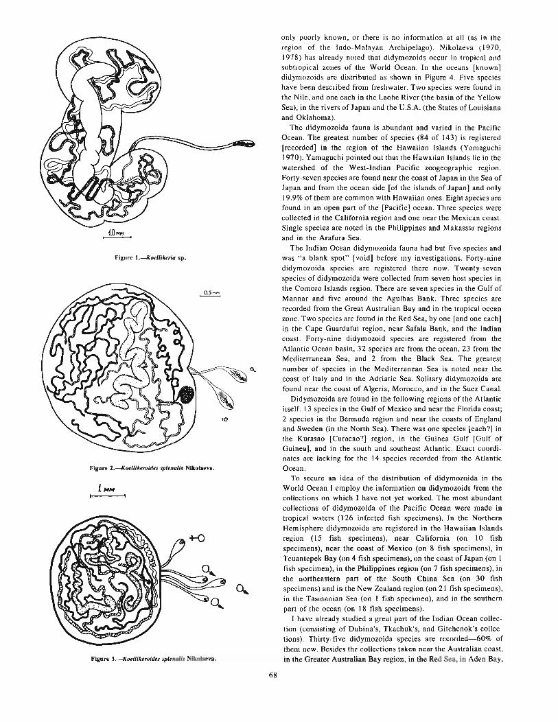
· Figure l.--Koellikeria sp.
Figure 2.--Koellikeroides splenalis Nikolaeva.
Figure 3.--KMllikeroitles splenalis Nikolaeva.
68
only poorly known, or there is no information at all (as in theregion of the Indo-Malayan Archipelago). Nikolaeva (1970,1978) has already noted that didymozoids occur in tropical andsubtropical zones of the World Ocean. In the oceans [known]didymozoids are distributed as shown in Figure 4. Five specieshave been described from freshwater. Two species were found inthe Nile, and one each in the Laohe River (the basin of the YellowSea), in the rivers of Japan and the U.S.A. (the States of Louisianaand Oklahoma).
The didymozoida fauna is abundant and varied in the PacificOcean. The greatest number of species (84 of 143) is registered[recorded] in the region of the Hawaiian Islands (Yamaguchi1970). Yamaguchi pointed out that the Hawaiian Islands lie in thewatershed of the West-Indian Pacific zoogeographic region.Forty-seven species are found near the coast of Japan in the Sea ofJapan and from the ocean side [of thc islands of Japan] and only19.9% of them are common with Hawaiian ones. Eight species arefound in an open part of the [Pacific] ocean. Three species werecollected in the California region and one near the Mexican coast.Single species are noted in the Philippines and Makassar regionsand in the Arafura Sea.
The Indian Ocean didymozoida fauna had but five species andwas "a blank spot" [void] before my investigations. Forty-ninedidymozoida species are registered there now. Twenty-sevenspecics of didymozoida were collectcd from seven host species inthe Comoro Islands region. There are seven species in the Gulf ofMannar and five around the Agulhas Bank. Three species arerecorded from the Great Australian Bay and in the tropical oceanzonc. Two species are found in the Red Sea, by one [and one each]in the Cape Guardafui region, near Safala Bank, and the Indiancoast. Forty-nine didymozoid species are registcred from theAtlantic Ocean basin, 32 species are from the ocean, 23 from theMcditerranean Sea, and 2 from the Black Sea. The greatestnumber of species in the Mediterranean Sea is noted near thecoast of Italy and in the Adriatic Sea. Solitary didymozoida arefound near the coast of Algeria, Morocco, and in the Suez Canal.
Didymozoida are found in the following regions of the Atlanticitself: 13 species in the Gulf of Mexico and near the Florida coast;2 specics in the Bermuda region and near the coasts of Englandand Sweden (in the North Sea). There was one species [each?] inthe Kurasao [Curacao?] region, in the Guinea Gulf [Gulf ofGuinea], and in the south and southeast Atlantic. Exact coordinates are lacking for the 14 species recorded from the AtlanticOcean.
To secure an idea of the distribution of didymozoida in theWorld Ocean I employ the information 00 didymozoids from thecollections on which I have not yet worked. The most abundantcollections of didymozoida of the Pacific Ocean were made intropical waters (126 infected fish specimens). In the NorthernHemisphere didymozoida are registered in the Hawaiian Islandsregion (15 fish specimens), near California (on 10 fishspecimens), near the coast of Mexico (on 8 fish specimens), inTeuantepek Bay (on 4 fish specimens), on the coast of Japan (on Ifish specimen), in the Philippines region (on 7 fish specimens), inthe northeastern part of the South China Sea (on 30 fishspecimens) and in the New Zealand region (on 21 fish specimens),in the Tasmanian Sea (on I fish specimen), and in the southernpart of the ocean (on 18 fish specimens).
I have already studied a great part of the Indian Ocean collection (consisting of Dubina's, Tkachuk's, and Gitchenok's collections). Thirty-five didymozoida species are recorded-60% ofthem new. Besides the collections taken near the Australian coast,in the Greater Australian Bay region, in the Red Sea, in Aden Bay,
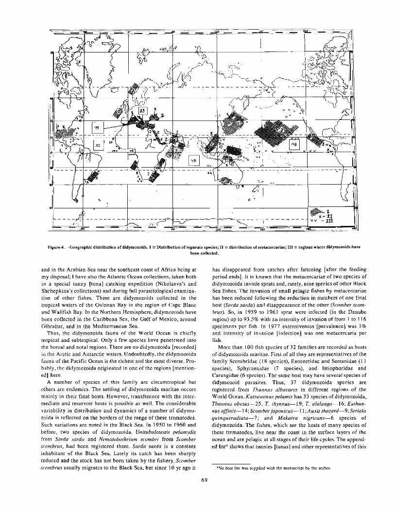
(J •
~--~=-I-=-=---.- - - - -~l
~-:'U ~vv - III- -'---=_':'-.:" .-_.:.---=--:.-_.
Figure 4.-Geographic distribution of didymozoids. I = Distribution of separate species; II = distribution of metacercariae; III = regions where didymozoids havebeen colIected.
and in the Arabian Sea near the southeast coast of Africa being atmy disposal; I have also the Atlantic Ocean collections, taken bothin a special tunny [tuna] catching expedition (Nikolaeva's andShchepkina's collections) and during full parasitological examination of other fishes. There are didymozoids collected in thetropical waters of the Guinean Bay in the region of Cape Blancand Wallfish Bay. In the Northern Hemisphere, didymozoids havebeen collected in the Caribbean Sea, the Gulf of Mexico, aroundGibraltar, and in the Mediterranean Sea.
Thus, the didymozoida fauna of the World Ocean is chieflytropical and subtropical. Only a few species have penetrated intothe boreal and notal regions. There are no didymozoida [recorded]in the Arctic and Antarctic waters. Undoubtedly, the didymozoidafauna of the Pacific Ocean is the richest and the most di verse. Probably, the didymozoida originated in one of the regions [mentioned] here.
A number of species of this family are circumtropical butothers are endemics. The settling of didymozoida maritae occursmainly in their final hosts. However, transference with the intermediate and reservoir hosts is possible as well. The considerablevariability in distribution and dynamics of a number of didymozoida is reflected on the borders of the range of these trematodes.Such variations are noted in the Black Sea. In 1950 to 1960 andbefore, two species of didymozoida, Unitubulotestis pelamydisfrom Sarda sarda and Nematobothrium scombri from Scomberscombrus, had been registered there. Sarda sarda is a constantinhabitant of the Black Sea. Lately its catch has been sharplyreduced and the stock has not been taken by the fishery. Scomberscombrus usually migrates to the Black Sea, but since 10 yr ago it
69
has disappeared from catches after fattening [after the feedingperiod ends]. It is known that the metacercariae of two species ofdidymozoida invade sprats and, rarely, nine species of other BlackSea fishes. The invasion of small pelagic fishes by metacercariaehas been reduced following the reduction in numbers of one finalhost (Sarda sarda) and disappearance of the other (Scomber scombrus). So, in 1959 to 1961 sprat were infected (in the Danuberegion) up to 93.5% with an intensity of invasion of from I to 116specimens per fish. In 1977 extensiveness [prevalence] was 1%and intensity of invasion [infection] was one metacercaria perfish.
More than 100 fish species of 32 families are recorded as hostsof didymozoida maritae. First of all they are representatives of thefamily Scombridae (18 species), Exocoetidae and Serranidae (IIspecies), Sphyraenidae (7 species), and Istiophoridae andCarangidae (6 species). The same host may have several species ofdidymozoid parasites. Thus, 37 didymozoida species areregistered from Thunnus albacares in different regions of theWorld Ocean. Katsuwonus pelamis has 33 species of didymozoida,Thunnus obesus-25, T. thynnus-19; T. alalunga-16; Euthunnus affinis-14; Scomber japonicus-ll; Auxis thazard-9; Seriolaquinqueradiata-7; and Makaira nigricans-6 species ofdidymozoida. The fishes, which are the hosts of many species ofthese trematodes, live near the coast in the surface layers of theocean and are pelagic at all stages of their life cycles. The appended list4 shows that tunnies [tunas] and other representatives of this
'No host list was supplied with lhe manuscript by the author.

family are the preferred hosts. Seven of 15 genera of the familyScombridae are hosts of didymozoida. One hundred and thirty-sixspecies of didymozoida (or 64.7%) are recorded from Scombridae.
A number of the didymozoid parasites of various species of tunny are of the same genera and species. Such taxonomic proximityof didymozoida species of tunnies of the World Ocean may be explained, except for the genetic relations, by area combination[overlapping ranges], the presence of cosmopolitan species, andby the high speed and long migrations of these fishes. All of thesefactors create conditions for invasion by the same helminths.
The common nature of the hosts' ecology and the same foodspectrum confirmed this resemblance [commonality or close correlation]. The species composition and number of didymozoidaspecies in each species of tunny have their peculiarities. They differ in the extensiveness [prevalence] and intensity of invasion [infection). As a rule the tunnies, marlins, and scombrids are invaded[infected] by varous didymozoid species. Specificity of thistrematode group is expressed at the genus level. Representatives ofthe genera Colocyntotrema, Metadidymozoon, Neodidymoloon,Makairatrema, and Glomeritrema are characteristic for marlins.These genera are not recorded from tunnies but many others are.
A great number of host species (64) have only one didymozoidspecies recorded. Fifteen species have two didymozoid speciesand four host species have four species each.
The utilization of a number of host species by didymozoida isvery high. Thunnus albacares in the Gulf of Mexico is 100% infected. The invasion [infection] intensiveness is from 8 to 1,167specimens (Nikolaeva 1968). In the Gulf of Guinea and in thecentral Atlantic the same host is generally infected and the intensity of infection is up to 148 specimens (Shchepkina 1976).Essentially, the figures on the intensity of infection should bedoubled, since a great number of didymozoida is encysted andevery cyst includes two species [author probably meant specimens] of didymozoida. It is the same for T. albacares in the Indianand Pacific Oceans.
There are practically no tunas invaded by didymozoids. Therefore the borders of didymozoida and the tuna's distribution coincide.s
Compared with the morphogenesis of the didymozoid maritaphase, the didymozoida metacercariae are very primitive.Kurochkin and Nikolaeva (1978) suggested a provisional systemfor didymozoida metacercariae. Using the presence or absence ofan acetabulum sucker, a pharnyx, a stomach, and glandular cells inthe region of bifurcation as generic characters, II genera weredescribed in all (Fig. 5). Dimensions of metacercariae, theirsuckers, the distance between suckers, shape of the intestinebranches and chambers, the ratio of body length to its width, andother characters were assigned species importance.
Unfortunately, the complete life cycle of didymozoids is notknown. The scheme for the cycle as proposed by me (Nikolaeva1965) has been mostly accepted by Yamaguchi (1975) and otherinvestigators. The first stages of the cycle are the least known.There is no new information about the parthenogenetic periods ofthe development of didymozoids, but my knowledge of the hostsof the metacercariae has been widened considerably. Themetacercariae which invade small pelagic fishes have been knownfor some time. The list of these fishes has been considerablyexpanded.
'The meaning of this paragraph is somewhat obscure. It is presented as il was inthe original manuscript. It would make better sense if the first seoteoce of theparagraph was eliminated.
70
Metacercariae are capable of numerous passages and acclimatization to unusual animals. This was confirmed when metacercariae were found even in the dogshark, Squalus acanthias.Recently, a considerable didymozoida metacercaria invasion wasrecorded from squids (a number of the works of Gaevskaya,Naidenova, and Nigmatullin) and on crustaceans (Reimer 1971).They have even been found free in plankton tests [samples](Slankis and Schevchenko 1974).
The cycles of didymozoida development have been analyzedaccording to the scheme of the trophic relationships of the pelagicfishes in tropical waters of the World Ocean (Parin 1968). Thecycles of didymozoida development proved to be complicated andvaried [Fig. 6). Didymozoida of flyingfishes of the genusGonapodasmius have the shortest life cycles. Three hosts(molluscs, copepods, and fishes) take part in their life cycle and apostcyclic host is possible also. It may be a tunny [tuna] or otherpredatory fish. Postcyclic hosts for the genus Gonapodasmiuscould be human as well. The didymozoida parasites of the moonfish possess this type of development also. Incidentally, they arethe longest worms of all the trematodes. Nematobibothrioideshistodii reaches 12 m in length.
The development [life] cycles of didymozoids invading tunniesacutally include reservoir hosts. Such species of didymozoids[with reservoir hosts] are numerous. The role of reservoir hosts isborne by lanternfish, sprats, flyingfishes, and other small pelagicfishes and squids. Similar are the life cycles of didymozoids frommarlins. Postcyclic hosts may take part in these cycles also. Theymay be sharks. It should be emphasized that the presence of postcyclic hosts is of greater importance than is believed.
As is known, all attempts to extract live miracidia from eggshave failed. First of all it is connected with the fact that mostdidymozoids (there are viviparous species) have an unformedmiracidium in their eggs. For its further development it must getinto a postcyclic host.
The people of tropical and subtropical countries customarily eatraw fish with spices. Such dishes are prepared from tunnies,marlins, flying fishes, etc. Along with other helminths, didymozoids may be gotten [ingested] by people. In cases where maritaeare ingested, people display transit eggs of didymozoids. Thissituation is known for the eggs of Gonapodasmius from flyingfishes in Japan (Kamegai 1971). However, these fishes may havedidymozoid metacercariae which may be acclimatized to humanbowels and begin the migrations peculiar to these larvae. It maybe dangerous for humans.
In conclusion, it should be emphasized that experiments on thedevelopment [life] cycle of didymozoids (or its separate phases)are necessary for solution of many questions of the biology, morphology, and systematics of this trematode group. Investigation ofthis interesting group provides new aspects of information abouttheir hosts, which are valuable [economically and ecologically]fishes.
LITERATURE CITED6
KAMEGAI, S.1971. The determination of a generic name of flying fishes' muscle parasite.
a didymoroid. whose ova have occasionally been found in human feces inJapan. lpn. l. Parasito!. 20:170-176.
KUROCHKIN, Yu. V.• and V. M. NIKOLAEVA.1978. The basis of didymozoid metacercaria systematics. In Theses of reports
of lbe All-Union Congress of Parasitocenologists, p. 82-84. [In Russ.]
6UletalUre Cited supplied by the author did Dot cover all citations in the text.

Anacatabul_ K. and No
Pa..amonlllca.cum It. and No
Postacerabullcaecum K••nd No
Neomonilicaecum K. and No
TONicaecum Yamagutl
Anesophagus K. and N.
Pseudoltlonilkaecum K. and No
Ne<>torticaecu. K. and No
Pseudolr..",r"'dI: L .....
Figure 5.--Scbeme or strDctlaft or uterior parts or didymozold metacercaria <see Karocbkln aDd NIkoI..... 1978).(Those genera accompaaled by tbe aotelioD K. aDd N. "ere described In 1978 by Kurocbkln aad Nikol.eva.]
71

fS
Figure 6.-Comparison 01 the cycle 01 dldymozoid development to the scheme 01 trophic relationships 01 pelagic fishes. Intermediate hosts (cross-hatched) = EuphausUda (2), Copepoda (3), shrimps (4); reservoir hosts (hatched) = nylngflshes (6),lantemflsh (8), squids (tl), CoryphtUllo (13); deOnltlve hosts (solld) = nylngflshes (6), sunnshes (9), COryphtUlIO (13), tuna (14),marUn (16); postcyc"" hosts (dotted) = middle size shark (17), large shark (18); while not registered In cycle developmeutdldymozold [those elements not recorded as taking part In the developmental cycle 01 dldymozolds) (open) = fishes "of migratedlayers" (5), hyperllda (7), smaD deep-water Ichtbyopbages (10), gyvatloa mackerel (11), aleplsaurans (15); phytoplankton (I) donot take part.
NIKOLAEVA, V. M.1965. On the development cycle of trematodes of the family Didymozoidae
(Monticelli, 1888) Poche, 1907. [In Russ.) J. Zool. 44(9):1317-1327.1968. The investigation of the helminth fauna of yellow-finned tuna and
marlin from the Gulf of Me~ico. In Collection of the invc:stigations of CenlealAmerican Seas 2, p. 150-157. [In Russ.) Kiev, Naukova dumb.
1975. Current situation and future tasks in investigation of trematodes of thefamily Didymozoidae. In Parasites and parasitosis of animals and people, p.180-185. [In Russ.) Kiev, Naukova dumb.
1978. On revision of suhfamily Koellikerinae (Trematoda, Didymowidae).In Sea biology, Vol. 45, p. 65-7 I. [In Russ.)
PARIN, N. V.1968. Ichthyofauna of the ocean epipelagic. [In Russ.) Moscow, Science,
72
p.186.SHCHEPKINA, A. M.
1976. Invasion by parasitic worms of tunnies and marlins of the centralAtlantic, In Thesis of reports of the second All Union Symposium on Parasites and Diseases of Sea Animals, p. 75-76. [In Russ.) Kaliningrad.
SKRIABIN, K. I.1955. Trematodes of animals and people. 11:5-254. [In Russ.)
YAMAGUCHI, S.1970. Digenetic trematodes of Hawaiian fishes. Keigaku Publ. Co., Tokyo,
436 p.197 I. Synopsis of digenetic trematodes of vertebrates. Keigaku Publ. Co.,
Tokyo, Vol. I, 1074 p.; Vol. II, 349 p.

Comparative Analysis of Monogenean Faunas andPopulations From Several Beloniformes Fishes!
L. A. GHICHENOK2
The material on which this paper is based, consists of a collection of monogeneans from 1,520 specimens of fishes belonging to6 species of the garfish order, i.e., Exocoetus volitans and E.monocirrhus (Exocoetidae); Euleptorhamphus viridis, Oxyporhamphus c. convexus and O. m. micropterus (Hemirhamphidae); andCololabis saira (Scomberesocidae). The majority of these fishesare component parts of the community of the oceanic epipelagicdomain. Four of them (E. volitans, E. monocirrhus, O. m.micropterus, and C. saira) belong to the oceanic group proper, being permanent dwellers of the epipelagic domain. Though it canbe found far away from the coast, E. viridis is a pseudo-oceanicspecies since it spawns in the neritic zone (Parin 1968). Only O. c.convexus, strongly related to coastal waters, belongs to the neriticgroup.
In all, 15 species and forms of monogeneans were recorded during this study, and all, except Axine belones from E. volitans, arerecorded from the investigated fishes for the first time. Fivemonogenean species proved to be new, three representing newgenera. No monogeneans were found in the saury.
A comparative analysis was conducted of both fauna andpopulation of monogeneans from the fish species under study using quantitative measures of similarity. Following Beklemishev(1970), I describe as a fauna of some unit [of a particular portion]of the environment a complete list of animal species living there,while a population is a real comity of animals to be found there,which depends not only on the list of species to be found there, butalso on the numbers of single-species populations.
I No Literature Cited section was supplied by the author.2Moscow State University, Moscow, U.S.S.R.
73
The similarity of the monogenean fauna of single fish specieswas estimated by the well-known index of Jaccard (1912), the
Lasimilarity of population, by the index b+c-La' 100% where a is
the least (of the two) index of abundance of each species commonto both of the hosts being compared, while band c are indices ofabundance of all the species in or on hosts I and II, respectively.
An analysis of indices allows me to conclude that the similarityof monogenean faunas of individual fish species depends first ofall on the amount of affinity [genetic or systematic] between thehosts. The closer the affinity, the greater the similarity of thespecies of one genus (0.56 and 0.57), the lowest, in fishes ofdifferent families (0.20).
The similarity of monogenean faunas seems to depend also onthe ecological similarity of the hosts, but to a lesser degree. As arule, fishes of the same ecological group have higher indices ofsimilarity (0.30, 0.56, and 0.50). Nevertheless the index ofsimilarity between the most ecologically different oceanic andneritic groups is far from being the lowest (0.33).
According to my data, the most closely related fish species havethe lowest indices of similarity of monogenean populations(18.3% and 5.4%); different genera of one family, 32%; anddifferent fish families, 23%. Fishes of one ecological group haverelatively high indices of similiarity of monogenean populations(14.7,18.3, and 73%). The lowest index (7.6%) was recorded forthe most ecologically different oceanic and neritic groups of fish.
This allows me to conclude that the similarity of the monogenean populations, in contrast to the similarity of species composition [faunas], does not show any clear dependence on the[systematic] affinities of the hosts, but chiefly depends on theirecological similarity.


New Data on the Capsalid Fauna of the World Oceanand Questions of its Specificity!
T. P. EGOROVA2
Extensive materials on capsalids of the different regions of thePacific, Indian, and South Atlantic Oceans and the Sea of Japanhave been studied for the past 10 yr. Eighteen species of capsalidswere recorded as a result of my study of the monogenean collections from 25 fish species.
I. Capsala gotoi Yamaguti, 1968 (from Thunnus obesus)2. C. martinierei Bose, 1811 (from Mola mola)3. C. neothunni Yamaguti, 1968 (from Thunnus albacares)4. C. nozawae (Goto, 1894) (from Thunnus obesus)5. C. ovalis (Goto, 1894) (from lstiophorus orientalis and
Makaira ampla)6. Capsaloides cristatus Yamaguti, 1968 (from Tetrapturus
angustirostris)7. C. magnaspinosus Price, 1939 (from Tetrapturus audax)8. Tristoma coccineum (Cuvier, 1817) (from Xiphias gladius)9. T. integrum (Diesing, 1850)
10. Nasicola klawei (Stunkard, 1962) (from Thunnus albacares)11. Benedenia derzhavini (Layman, 1930) (from Sebastodes3
glaucus, S. steindachheri, S. iracundus, Sebastodes introniger3 ,
S. alutus, Helicolenus papillosus, and Scorpaenidae g. et sp.)12. Entobdella hippoglossi (Muller, 1776) (from Reinchardtius
hippoglossoides matsuurae)13. E. squamula (Heath, 1902) (from Reinchardtius matsuurae
and Sebastes glaucus)14. Pseudobenedenia nototheniae Johnston, 1931 (from
Notothenia rossi and N. macrocephala)15. P. ovalis Yamaguti, 1968 (from Priacanthus boops and
Sebastodes proriger)16. Pseudoentobdella pugetensis Robinson, 1961 (from Atheres
thes stomias)17. Allosprostonia tauvinae Lawler and Hargis, 1968 (from
Epinephelus orientalis and Decapterus punctatissimus)18. Trochopus australis Robinson, 1961 (from Helicolenus
papillosus)
1No Literature Cited section was supplied by the author.'Institute of Biology and Pedology, Far East Science Centre, Academy of Sciences
of the U.S.S.R., Vladivostok, U.S.S.R.'Though the author presented the generic name Sebastodes twice, the first one pro
bably should have been Sebastes. Thus the first three species would belong in thisgenus. The citation of Sebaste.< gLaucus as a host of Entobdella squamuLa tends to confirm this.
75
The family Capsalidae Baird, 1853 is presently divided into177 species, 33 genera, and 7 subfamilies (according to Yamaguti1963-113,21, and 5, respectively).
My observations of the occurrence of genera on hosts revealedthat of 33 known genera, 19 are restricted to a single fish family.Fifteen of them are monotypical. The other four genera, containing more than one species (Ancyrocotyle-2, Dioncopseudobenedenia-2, Nitzschia-3, and Capsaloides-8) are closely specificfor fish of one specific family. Thus, Monogenea of the genus Capsaloides are only observed on the genera Tetrapturus, Makaira, andlstiophorus of the family Istiophoridae.
Monogenea of six genera (Benedeniella, Pseudoentobdella,Metabenedeniella, Allobenedenia, Pseudallobenedenia, and Mediavaginia) parasitize fish of two families. For example, the genusBenedeniella is recorded on members of the families Rhinopteridae (order Rajiformes) and Carangidae (order Perciformes), andthe genus Pseudoentobdella is recorded on Aetobatidae (orderRajiformes) and Hippoglossidae (order Pleuronectiformes), i.e., onfamilies not closely related, but the contrary is the case with theother four genera.
Members of the genera Trochopus and Tristoma are observed oneight families. The first occurs only on Perciformes, but the dataof the second are questionable (according to Bychovski 1957).The genera Entobdella, Megalocotyle, and Neobenedenia arerecorded on nine fish families. Entobdella parasitizes Pleuronectiformes and Rajiformes. Perciformes are typical for Megalocotyleand most species of Neobenedenia. Representatives of the genusCapsala, observed on II families, are also typical for fish of theorder Perciformes. The genus Encotyllabe parasitizes 16 familiesof Perciformes (and possibly on Salmonidae).
Thirty-four fish families, mostly Perciformes (23 families innature and 11 under artificial conditions), are recorded as hostsfor Monogenea of the genus Benedenia.
Thus, capsalids parasitize Perciformes mainly (with the exception of the genus Entobdella, the species of which are mostlyrecorded on Pleuronectiformes and Rajiformes). These basicregularities are also reflected in our data. Of 10 fish familiesobserved to have capsalids, 8 are related to the order Perciformes.Perciformes are confirmed as the primary hosts for capsalidmonogeneans.


On the Taxonomic Position of the Monogenean,Pseudaxine mexicana Meserve, 1938
B. Iv. LEBEDEVI
In 1938 F. G. Meserve published a large list of fish monogeneans from the waters of the Galapagos Archipelago based onstudies of the Allan Hancock Pacific Expedition collections.Pseudaxine mexicana Meserve, 1938 from the [Spanish] mackerel,Scomberomorus maculatus (Mitchill), of Tangola-Tangola (Mexico) was described in that paper along with other species.
In 1955, K. Koratha described a new species, Pseudaxine texana, from the same Spanish mackerel host species of the Caribbean Sea (Port Aransas) [Gulf of Mexico]. In 1956, W. J. Hargis,Jr. opined that these monogeneans are identical, and that the nameP. texana Koratha, 1955 is a junior synonym of the name P. mexicana Meserve, 1938. Judging from the works of Hargis (1956),Yamaguti (1963), and some other authors, they are certainlyrelated to the genus Pseudaxine Parona and Perugia, 1890.
My last revision of the genera Pseudaxine and PseudaxinoidesLebedev, 1968 (see Lebedev 1977) led me to doubt the genericidentification of the monogenean species described by F. Meserveand later mentioned by the other authors.
First, it is known (Yamaguti 1963; Dillon and Hargis 1965;Lebedev 1972, 1977; and others) that parasitism of carangidfishes (family Carangidae) is typical for species of the genusPseudaxine; whereas P. mexicana is recorded only from mackerelsof the family Cybiidae. Pseudaxine indicana Chauhan, 1945,described from sparid fishes (?)-family Sparidae, and not recorded since by anybody-is an exception. However, this one is aspecies dubia (Lebedev 1977).
Second, while analyzing the figure presented in the originaldescription (Meserve 1938, fig. 9/60-63) I discovered that theclamps of P. mexicana have nothing in common with those of theother Pseudaxine spp. The external appearances of P. mexicanaclamps are very similar to the clamps of the genus PriceaChauhan, 1945 and less [similar] to those of the genus Neothoracocotyle Hargis, 1956. The clamps of P. mexicana have similarrib like thickenings in the lateral fields of the capsule and the same"barrel-like" shape of the anterior part of the mediobasal (main)spring. Additionally, other structures observed in the figure show
'Institute of Biology and Pedology, Far East Science Centre, Academy of Sciencesof the V.S.S.R.. Vladivostok, V.S.S.R.
77
that the clamps of P. mexicana resemble those of Pricea spp. (orNeothoracocotyla spp.) closely.
Third, and last, the type and form of the haptor, described andfigured by F. Meserve, is perhaps [probably] typical for Pricea.
There is no doubt that Pseudaxine mexicana Meserve, 1938 hasto be removed from the genus Pseudaxine. The question of itsgeneric position (Pricea Or another genus) can be decided onlyafter exacting and careful reinvestigation of the type materials.Evidently, this mistake, having existed for more than 40 yr, mustbe corrected.
LITERATURE CITED2
DILLON. W. A., and W. J. HARGIS, Jr.1965. Monogenetic trematodes from the southern Pacific Ocean. 2. Polyopis
thocotyleids from New Zealand fishes: the families Discocotylidae, Microcotylidae, Axinidae, and Gastrocotylidae. In Biology of the Antarctic SeasII, p. 251-280. Antarctic Res. Ser. 5, Am. Geophys. Vnion.
HARGIS, W. J., Jr.1956. Monogenetic trematodes of Gulf of Mexico fishes. Part Xl. The family
Microcotylidae Taschenberg, 1879. Proc. Helminthol. Soc. Wash. 23:153162.
KORATHA, K. J.1955. Studies on the monogenetic trematodes of the Texas coast. 11. Descrip
tions of species from marine fishes of Port Aransas. Publ. Inst. Mar. Sci.,Vniv. Tex. 4:251-278.
LEBEDEV,B.1.1972. The taxonomy of monogeneans of the suborder Gaslrocotylinea. Proc.
Inst. BioI. Pedol., Far East Centre, V.S.S.R. Acad. Sci., new ser., 11(114):121145.
1977. Two new species of monogeneans from fishes of Indo-Pacific waterswilh noles concerning the genera Pseudaxine and Pseudaxinoides. In Excertaparasitologica en memoria del Dr. Eduardo Caballero y Cabellero. Inst. deBioI. Mexico, Publ. Espec. No.4, p. 69-78.
MESERVE, F. G.1938. Some monogenetic trematodes from the Galapagos Islands and the
neighboring Pacific. Rep. Allan Hancock Pac. Exped. (1932-1937) 2(5):31-89.
YAMAGVTI, S.1963. Systema helminthum. Vol. IV. Monogeneo and Aspidocotylea. John
Wiley and Sons, Inc., N. Y., 699 p.
'No Literature Cited was supplied by the author. Dennis A. Thoney prepared thisto match the author's citations.


Plerocercoids of Some Cestoda as Bioindicators of thePopulation Structure of Podonema longipes
G. V. AVDEEVI
ABSTRACT
Podonema longipes is an important commercial fish inhabiting the northwestern Pacific. During parasitological investigations of P. longipes I attempted to use some parasites as bioindicators to uncover certain featuresof its biology. The most suitable species were two plerocercoids (Nybelinia sp. and a diphyllobothriid larva)which have vividly expressed differences in invasion [infection) in comparison with other discovered [knownlparasites. They are able to accumulate in a host during its entire life. A total of 267 individual fisb of ages from 6to 26 yr, caugbt off the Japanese and northern Kuril Islands in 1976-80, were studied. The level of infection byboth species in the areas investigated is different. Infection by Nybelinia is bigher off Japan. On the contrary,infection by dipbyllobothriid larvae is bigher in the area off tbe northern Kurll Islands.
Invasion of P. longipes by botb species in the areas under comparison begins in different age groups.Around Japan infection by Nybelinia begins at ages from 9 to Il yr and diphyllobothriid larvae at 12 to 14 yr;around tbe northern Kurils at 15 to 17 and 9 to II yr, respectively. Invasion by both species increases as the fishbost ages.
Observed differences in infection of females and males by Nybelinia are the result of the different verticaldistribution of older age groups of male and female P. longipes (i.e., increase of the relative number of malestoward the upper limit of the habitat of P. longipes and, conversely, an increase of the relative number of femalesto the lower limit). The probability of infection by Nybelinia decreases as tbe habitat depth increases. This is confirmed by the higher infection by this parasite of Alaska pollock inhabiting the study area at lesser [shallowerldeptbs.
Differences in the invasion by Nybelinia of males and females off the northern Kurlls occur simultaneouslywith the beginning of infection by this parasite. In comparison in tbe area off Japan, differences in the infectionof females and males are seen from the age groups of from 12 to 14 yr, while the beginning of infection takesplace at 9 to Il yr.
In the northern Kurlls sex differentiation by depths of P. longipes begins in age groups of IS to 17 yr, and offJapan 12 to 14 yr. Thus, the formation of differences in the infection of females and males by Nybelinia coincideswith the beginning of sex differentiation by depth in each area.
My data reveal differences in the growth rates of males and females in older age groups. Besides, in the areaof the northern Kurlls, females begin to grow faster than males at tbe age of 18 and those off Japan, at the age of12. As we see, in each of the areas mentioned, the age at which differences in growth rate between males andfemales begins coincides with the formation of differences In the infection of males and females by Nybelinia and[of) differential sex distribution by depth.
Tbus, it may be assumed that in eacb of the areas investigated an independent P. longipes population, havingits specific parasitological and biological characteristics, exists.
In recent years the practice of using parasitological data tostudy the ecology of commercial species has become of greatimportance. The use of parasites as biological tags has permittedthe study of population structure and migrations of certain species(Baudin-Laurencin 1971; Margolis 1963; Kabata 1963; Kovaliova 1965; Konovalov 1971; Kabata and Forrester 1974; Platt1976; and others).
During parasitological studies of P. longipes I succeeded inusing certain parasites as bioindicators in order to reveal somefeatures of the biology of this fish. The most suitable [parasites forthis purpose] turned out to be the plerocercoids of two species: ANybelinia sp. and a diphyllobothriid larva. In comparison withother parasites found on P. longipes, their invasion level [infectionlevel2 ] has most vividly demonstrated differences related to anumber of factors (i.e., fishing ground, depth of residence, age,and sex of the host). Further, both plerocercoids can be accumulated in P. longipes during its entire life.
I Laboratory of Parasitology of Marine Animals, Pacific Research Institute ofFisheries and Oceanography (T1NRO). Vladivostok, U.S.S.R.
'In this as well as other papers in this volume, the word invasion means infection,usually, and can be used in its place.
79
Altogether 2,671 fishes of ages from 6 to 26 yr caught off Japanand around the northern Kuril Isles during 1976-80 were studied.
Analysis of the invasion level of P. longipes by these twoparasite species permitted discovery of the following specificfeatures:
I) The invasion [infection] level by the plerocercoids of bothspecies in the [geographical] areas under investigation is different.Extensity [prevalence] and intensity of invasion [infection] byNybelinia larvae is higher around Japan. Conversely, invasion bydiphyllobothriid larvae is much higher in the waters around thenorthern Kuril Islands (see Table I).
2) The extensity [prevalence] and mean intensity of invasion[infection] by Nybelinia larvae increase as the fish age. In olderage groups (Le., more than 20 yr of age), the rate of increase of invasion in the area of the northern Kuril Isles lowers [is reduced];in the area of Japan, invasion [infection] actually decreases (Fig. IA, B). Invasion by diphyllobothriid larvae in both areas becomeshigher with advancing age of the fish (Fig. I C, D).
3) The level of invasion of host males by Nybelinia larvae ishigher than of females in both areas (Fig. 2). As for diphyllobothriid larvae, male hosts have a higher level of invasion [infection]

7Table I.-Mean intensities of parasite infestation with location.
Number Nybelinia sunnenicola Diphyllobothriidae g. sp.
Depth of fish mean mean(m) studied % intensity % intensity
Northern Kuril Isles200-570 401 42.3 2.4 50.0 2.5580-710 656 36.7 1.7 51.7 2.7730-1,120 501 16.9 0.6 34.1 1.3
200-1,120 1,558 31.8 1.5 45.9 2.2
Japan210-540 397 70.3 8.7 24.4 0.6550-700 383 68.9 6.2 32.3 1.0710-930 333 81.7 4.1 54.6 1.9
210-930 1,113 75.2 6.5 35.7 l.l
i: 50~
:tl-e:!>Z 40l.Il...J
10
A.- d"
en20~
'"et:
10 ~a.,;
Z6 7 8 9 10 t1
Figure 2.-Averqe intensIty of infestation [infection) wltb NybeUnlae and tbeaverqe body Ienglb of males and females of PodollellUl IOllgipe. depending onftsb qe: A =Nortb Kurllislands waters; B =Japan Islands waters.
lobothriidae in the area of the northern Kuril Isles. At that, ahigher concentration of crustaceans (the intermediate hosts) in thetotal plankton biomass in this area is possible, as well as a higheroccurrence of procercoids on the intermediate hosts, themselves,as well as the high abundance of mackerel sharks, which are thefinal hosts of Nybelinia in this area according to the data of ichthyologists. I should note that invasion by Nybelinia from north to
in the area of the northern Kuril Isles, with the females being invaded more heavily around Japan (Fig. 3).
4) In both areas a decrease of invasion by Nybelinia larvae takesplace as the depth at which the host lives or was captured increases (see Table I). Invasion by diphyllobothriid larvae lowers[decreases] as the depth lowers [increases] in the area of thenorthern Kuril Isles [generally speaking3 ] and increases aroundJapan.
Below I shall attempt to explain the above-mentioned specificfeatures of the invasion of P. longipes by Cestoda larvae (Table I).
The unequal invasion of P. longipes by larvae of both Nybeliniaand diphyllobothriidae in the areas being compared is caused bythe different possibility of [opportunities for] invasion of the hostsby these parasites in each area. Obviously this occurs because ofthe greater abundance of the procercoids of Nybelinia in theplanktonic crustaceans constituting the diet of P. longipes aroundJapan and the reduced abundance of the procercoids of diphyl-
}The phrase "generally speaking" in brackets is employed because in Table 1 thepercentage of fish invaded and the intensity of invasion by Diphyllobothrium areactually slightly higher at mid-depths in the northern Kurils than at the surface.However, at greater depths both parameters are much lower!
60B
~ 50:I:l-e:!>Z40
'"...J30
___ cI'
30:tenLL.
20 ....CI)....t:'"10 ~et:a.o
Z
NORT~ I<URILJAPAN
JAPAN- NORTH I<URIL
,----~;,,,,,,,",r,,
6-8 9 "11 12-1~ 15-11 18-20 2\-:» 2+-2G
AgE-GROUP (YEARS)
o
6-8 9-11 12-~ t5-tl18-:lJD21-n "-2C
AGE -GROUP/YEARS)
50
50
%100 B
%100
I!2lllI NORTH KUR ILc::J JAPAN
Illll!lI NORTH I<URILc=I JAPAN
A
6-8 9 -11 12-''1 '5-17 t8-20 2t-23 2'1-26AO(-GROUP /V(ARSI
'5
10
:::J:~ 5..........Cf)l.&I~ 6-8"l4(a:~ C0..'0
0z
5
Figure I.-Percent occurrence and aver. intensity of InfestatIon [infection) of PodollellUJ ",,,gipe. wltb cestode plerocercolds depending on ftsb age: A, B = NybelillUl SIUrIIell;col,,; C, D = D1pbyllobotbrlldae.
80

Figure 3.-Average Intensity of Infestation [InfectionI with dlphyllobothrlld larvae of males and females ofPodonema longipes depending on fish age: A = NorthKurll Islands waters; B= Japan Islands waters.
south has been observed on Alaska pollock as well (Mamaev et al.1965). This obviously involves other fish species also.
The increase of invasion [infection] as P. longipes grows older iscaused by the ability of the larvae to live in the body of the hostduring the latter's entire life. I have never observed dead plerocercoids in P. longipes.
I already know from the literature (Mamaev et al. 1965) theopinion of those authors that, in the Bering Sea and the Gulf ofAlaska, the greater the depth of residence of Alaska pollock thelower the invasion [infection] by Nybelinia larvae. In the case ofAlaska pollock, as well as of P. longipes, the possibility of invasionby Nybelinia larvae apparently is reduced as the depth of theirresidence increases. The differing invasion [infection] of P.
longipes and Alaska pollock in the area under investigation confirms this. As I know, P. longipes and Alaska pollock are interchangeable [comparable or overlapping?] species in their distribution. Alaska pollock resides at lower [shallower] depths and, incomparison with P. longipes, is more heavily invaded [infected]by Nybelinia larvae.
In order to understand the reasons for the differing levels ofinvasion of male and female P. longipes by plerocercoids, I analyzed the sex ratio of P. longipes according to the depth of capture. Itwas found that the vertical distribution of male and femalejuveniles and older fish differs significantly. A relative increase offemale abundance is observed in fish older than 14 yr in the areaof the northern Kuril Isles and in fish older than 11 yr aroundJapan in the direction of the lower boundary of P. longipesresidence [in respect to depth], and, on the contrary, the numberof males increases as the depth lowers or decreases. The youngerfish show no change in sex ratio with an increase in depth.Females residing in mainly higher [greater] depths are invaded byNybelinia to a lower [lesser] extent and total invasion of P.longipes decreases with an increase in depth.
The fact that in each area the age at which a different verticaldistribution of P. longipes sexes begins to appear, coincides withthe age at which the difference in invasion [infection] by
Nybelinia between males and females appears, proves the correctness of the above-stated explanation concerning the availability[causes] of the differences in invasion of P. longipes. The difference in invasion by Nybelinia larvae between the sexes of P.longipes in the area of the northern Kuril Isles appears simultaneously with the beginning of invasion (15 to 17 yr), and aroundJapan from the moment of the sharp increase in invasion (12 to14 yr). However, the beginning of invasion by Nybelinia occurs atthe age group of 9 to 11 yr.
As for the diphyllobothriid larvae of the northern Kuril Islesarea, here I may discern a pattern close to that of Nybelinia larvae:Invasion decreases with increase of depth and males have a higherlevel of invasion than do females. Apparently, the possibility ofinvasion by diphyllobothriid larvae diminishes as the depth increases in this area.
In contrast, around Japan, invasion by diphyllobothriid larvaeincreases with depth and females have the higher level of invasion. This pattern of invasion is possible only in the case wherethe possibility of invasion by this parasite increases as the depthincreases. Apparently this is the case for this area.
In the northern Kuril Isles, the age at which the differences inthe invasion [infection] of males and females by diphyllobothriidlarvae appear coincides with the sharp increase in invasion (15 to17 yr), i.e., with the beginning of the change in vertical distribution of P. longipes. Podonema longipes individuals begin to beinvaded in this area at the age of 9 to 11 yr. A similar distinctrelationship was not observed around Japan.
Females of P. longipes older than 20 yr dominate at all depthranges of their residence. Evidently males have a higher mortalitylevel than females. As already mentioned, females are invaded byNybelinia to a lesser extent, which is why the total invasion [infection] of older fishes decreases. A similar decrease in invasion bydiphyllobothriid larvae does not occur, so a difference in invasion[infection] of males and females by this parasite is not so evidentas is the case with Nybelinia larvae.
For a difference in invasion of males and females of P. longipesto reach the values observed, a prolonged time of existence of thedifferent vertical distribution within the yearly cycle of P. longipesis necessary. Figure 4 (sexual distribution of P. longipes) showsthat the pattern described above continues during the differentseasons. I suppose that this vertical distribution of males andfemales is interrupted only during spawning, but have no data onthis point. However, the spawning period is not very long, usually,and the intensity of feeding by the fish diminishes sharply at thisperiod or ceases altogether.
By analyzing the data on age of P. longipes obtained duringparasitological dissections of fishes, I managed to detect differences in the rate of growth of males and females in the mature agegroups (Fig. 2). At that, females of the northern Kuril Isles beginto overtake males in growth at about 17 to 18 yr of age, whilearound Japan they do so at 11 to 12 yr of age.
As may be seen, in every area studied the age at which thedifference in growth rate between males and females appearscoincides with the inception of sexual differentiation by depth, asdoes the beginning of the differences in invasion of male andfemale hosts by Nybelinia. Around the northern Kuril Isles thesefeatures of P. longipes appear in the age group of 15 to 17 yr;around Japan, at the age of 12 to 14 yr. Apparently the simultaneous beginning of these phenomenona in P. longipes is connected with its attainment of sexual maturity, which begins infishes of younger age in Japanese waters than in the fish of thenorthern Kuril Isles.
2lj-266-8
10 A
5:xenu..--...~ 6-8LJJ~
U'>~10 Ba::c:ra.d
5z
81

MAY-JUNE
JANUARY - HBRUARY02.0" B
'::liLIui1tlill1."O~90-9~M
>- 5.oZUI::>olJl
~IOO
A
50
~':~zUl
5 JULY - AUGUST
'"~IOO~'O-StOM.... 50Z
"'U
~ 100J ~ 5~-8I:'"M _ ~ nQ.50~
100 100 -9tO MFigure 4.-Sex Interrelations of Podoll,.. IollgifH' from differentdepths: A = Nortb Kurllislands ..aters; B = Japan Islands ..aters.
On the basis of the above, I may postulate that independentpopulations of P. longipes exist in every area of investigation andthat each has its own parasitological and biological characteristics.
LITERATURE CITED
BAUDIN-LAURENCIN, F. G.1971. Crustaces et Helminthes parasites de I'Albacore (Thunnus albacares) du
Golfe de Guinee - Note preliminarie. Doc. Sci. Centre Rech. Oceanogr.Abidjan 2(1-2):11-30.
KABATA,Z.1963. Parasites as biological tags. Spec. Publ. Int. Comm. Northwest Atl.
Fish. 4:31-37.KABATA, Z., and C. R. FORRESTER.
1974. Antheresthes stomias (Jordan and Gilbert, 1880) (Pisces: Pleuronectiformes) and its eye parasite, Phrixocephalus cincinnatus Wilson 1908 (Cope-
poda: Lernaeoceridae) in Canadian Pacific waters. J. Fish. Res. Board Can.31:1589-1595.
KONOVALOV, S. M.1971. Differentiation of local stocks of sockeye salmon, Oncorhynchus nerka
(Walbaum). [In Russ.] L., p. 1-227. (An English translation exists.)KOVAUOVA, A. A.
1965. Helminthofauna of local stocks of Black Sea scad. [In Russ.] Mater.Conf. VOG, 2, M., p. 121-126.
MAMAEV, Y. L., O. M. BAYEVA, and V. D. LAKTIONOVA.1965. On invasion of Alaska pollock of the Bering Sea and North Pacific by
Nebelinia. [In Russ.] Rybnoye Hozyajstvo 6:66-68.MARGOUS. L.
1963. Parasites as indicators of the geographical origin of sockeye salmon,Oncorhynchus nerka (Wa1baum), occurring in the North Pacific Ocean andadjacent seas. INPFC Bull. II: 101-156.
PLATT, N. E.
1976. Codworm - a possible biological indicator of the degree of mixing ofGreenland and Iceland cod stocks. J. Cons. Int. Explor. Mer 37:41-45.
82

Development of Larval Stages ofBothriocephalus scorpii
A. I. SOLONCHENKOI
ABSTRACT
I detennlned tbat tbe cycle of development of Bothriocephalus scorpii may proceed witb eitber one intermediate bost (Copepoda) or two (Copepoda and fisb).
Bothriocephalus scorpii was found to infect Scophthalmusmaeoticus torosus and Sc. m. maeoticus up to 100%, with an intensity of invasion [infection] of from 2 to 90 specimens. Even youngindividuals (3 cm in length) which had just been transformed werefound by Bulgurkov (1963) to be infected by B. scorpii. Sinceplaice is the object of aquaculture, B. scorpii may represent anessential danger for their larvae and young specimens, causingbothriocephalus disease. To investigate this possibility was theobjective of my studies of the life cycle of this cestode. Accordingto the literature, development of B. scorpii involves the participation of one intermediate copepod host (Essex 1928) or the participation of two intermediate hosts, a copepod and a fish (Markovski1935).
Markovski (1935) infected the copepod Eurytemora hirrundowith coracidia and examined them only on the 4th and the 17thdays of development. The occurrence of calcifications, of cercomers, and the timing of their appearance as plerocercoids werenot explained [discovered] by him due to the brevity of theexperiment. He found the second intermediate host during autopsyof Gobius minutus. Feeding their intestines containing theplerocercoids he had found to gloss, he obtained a fully grownstage of the adult helminth.
I found plerocercoids of B. scorpii in Trachurus mediterraneusponticus and Odontogadus merlangus in the Azov Sea andNaidenova (1974) found that Gobius had them too.
I determined the period of development and the emergence ofthe coracidium from the eggs as well as its viability under different temperature regimes (Solonchenko 1979). To discover thesecond intermediate host, I used the bottom fishes Gobius minutusand Scorpaena porcus. Like plaice, S. porcus is also a definitivehost of B. scorpii. Five Acartia clausi containing procercoids wereintroduced by dropper [syringe] into the ventricles of two Gobiusminutus and Scorpaena porcus. One Scorpaena porcus wasdissected on the 6th day of development of B. scorpii and a contractile compressed body of rounded shape, with a size of 0.602 x0.392 mm was found. The cuticula of the body was found to besmooth, containing many calciferous bodies of different shapesand sizes in the parenchyma. No bothrium was found at this time.Upon penetration into the body cavity of Gobius minutus, the procercoids lose the long appendage-bearing hook which is connected with the main part of the body by a narrow isthmus. On the18th day of development, the bodies of the procercoids increasedin size up to 0.938 x 0.431 mm (Fig. I). The body of the procer-
IInstitute of Biology of the Southern Seas, Academy of Sciences of the U.S.S.R.,Sevastopol, U.S.S.R.
83
Figure l.-Plerocercold from Gobiusminutus on tbe 18th day of development.
coids in Scorpaena porcus had attained an elongated shape with asize of 0.602 x 0.126 mm by the 18th day of development. Theanterior portion was expanded and one could clearly distinguishthe formation of the scolex. The remainder of the body was of thesame width, 0.098 mm. On both sides [of the body] there areslight transverse furrows, out of which the limbs [appendages?]are formed. The cuticula of the body is smooth and there arecalcifications of different shapes and sizes in the parenchyma(Fig. 2).
Unfortunately I could not follow the development of B. scorpiifurther. However, it has been clearly shown experimentally thatplerocercoids can develop in both Gobius minutus and Scorpaenaporcus. Development proceeds primarily in a different fashion inGobius minutus, the second intermediate host in which only thescolex develops; while in Scorpaena porcus, the definitive host ofB. scorpii, the parasite begins to develop the scolex and thestrobilae simultaneously.
Thus, I have established that the cycle of development of B.
scorpii may proceed with the participation of one intermediatehost or with two. Plaice may be infected in two ways: I) Throughthe copepod, when they feed on the plankton (the larval stage), or2) through the fish (when they are eaten).
LITERATURE CITED2
BULGURKOV, K.1963. On the biology of the tapeworm, Borhriocephalus scorpii (Muller) in
turbot. Bull. de l'Institut Central de Recherche Scient. de Pisciculture et dePecherie. Varna 111:263-264.
2Not all citations mentioned in the text are included here.

Figure 2.-Plerocercold from Scorpaena POTCUS on the 18th day of development.
84
ESSEX, H. E.1928. On the life history of Bothriocephalus cuspidatus Cooper, 1917, a tape
worm of the wall-eyed pike. Trans. Am. Microsc. Soc. 47:348-355.MARKOVSKI, S.
1935. Uber den Entwicklungszyclus von Bothriocephalus scorpii (Muller,1776). Bull. Int. Acad. Polon. Sci. Lett. Cl. Sc. el Nat. S. B. Sc. Nat. II(1-2):1-17.
SOLONCHENKO, A. I.1979. The cycle of development of the cestode Bothriocephalus scorpii
(Muller, 1776). [In Russ.] All-Union Conference on Parasites and FishDiseases. Abstracts of reports. Leningrad.

Special Features of the Geographical Distribution andPractical Significance of the Parasitic Copepods of
Fishes of the Pacific!
V. N. KAZACHENK02 and V. M. TITAR3
ZOOGEOGRAPHICAL ASPECTS
At present, 1,700 species of copepods parasitizing fish,representing 256 genera, are known. About 1,450 species of 232genera, i.e., 85.3% are sea [marine] species. Approximately 700species of 173 genera of parasitic copepods of sea [marine] fisheswere recorded in the Pacific Ocean. Thus, some 48% of all knownsea [marine] parasitic copepods were found in the Pacific Ocean.
Parasitic copepods are distributed as are the hosts of thoseparasitic copepods (Kazachenko and Titar 1980). Second, as wasfound (Titar in litt.) in the case of Pumiliopes capitulatus, thedistribution of parasitic copepods depends mainly on the distribution of water masses (after Dobrovolski 1961). And last, historicalfactors, primarily the geological history of the Pacific Ocean,paleoclimatological changes, etc., are of great importance,enabling us to solve such problems as amphiboreal distribution,bipolarity, etc.
The peculiarities [special features] of bottom geomorphology,vertical structure of water masses, and the presence of vast waterspaces in the Pacific Ocean make possible the existence of severalmain complexes of parasitic copepods there, i.e., coastal (shelf),epipelagic, and deep water ones. On the whole these complexescorrespond to those defined for sea fish (Parin 1968). As a rule,the special features of the geographical distribution of copepodsfrom the various complexes are different. So, we shall considerthem separately, beginning with the shelf fauna.
In all, 554 species of 125 genera of 25 families of parasiticcopepods are recorded from shelf fishes of the Pacific Ocean.Analysis of this material has shown that all of the copepodfamilies have many more tropical representatives (400) in theircomposition than boreal (97) or notal (48) ones. The representatives of the families Euryphoridae, Kroyeridae, Pseudocychidae,and Tegobomolochidae were recorded only from the tropical(together with the subtropical) part of the Pacific Ocean on shelffishes. Many genera are also confined primarily to the tropics indistribution. For example, from the family Bomolochidae therewere recorded Nothobomolochus, Orbitacolax, Pumiliopes,Unicolax, and Pumilopsis; from Caligidae, the genera Anuretes,Caligulus, Dentigryps , Heniochophilus, Mappates, Parapetalus,Pseudanuretes, Pseudocaligus, Pseudopetalus, Pupulina, Sciaenophilus, and Synestius; from the family Chondracanthidae, thegenera Brachiochondria, Ceratochondria, Humphreysia, Immanthe,Juanettia, Neobrachiochondria, Praecidochondria, Prochondracanthopsis, Prochondracanthus, Protochondria, Pseudacanthocanthopsis, Pseudoblias, Pseudodiocus, and Scheherazade; from the family
'No Abstract or Literature Cited section was supplied by the authors.'Institute of Hydrobiology, Kiev, U.S.S.R.'Pacific Research Institute of Fisheries and Oceanography (TINRO), Vladivostok,
U.S.S.R.
85
Eudactylinidae, the genera Bariaka and Nemesis; from the familyLernaeoceridae, the genera Impexus and Peniculisa; from the family Lernaeopodidae, the genera Anaclavella , Clavellisa, Clavellopsis,Epiclavella, Isobranchia, and Thysanote; from the family Lernanthropidae, the genera Norion and Sagum; from the family Pandaridae, the genera Dinemoleus and Pseudopandarus; from thefamily Philichthyidae, the genus lchthyotaces; and from the familyTaeniacanthidae, the genera Anchistrotos, Metataeniacanthus,Parataeniacanthus, Pseudotaeniacanthus, Taeniacanthus, andTaeniastrotos.
Most of the parasitic copepods of shelf fish of the tropicalPacific are distributed circumtropically. Representatives of theabove genera (as well as circumtropical species) are rarely foundbeyond the tropical regions. Their appearance, for example, inPeter the Great Bay (the Sea of Japan) is caused by the heating ofthe subsurface layer in summer, forming water masses whosetemperature characteristics are very close to those of the surfacewater masses of tropical seas.
The largest quantity of tropical parasitic copepod species ofshelf fish was registered [recorded] in the western Pacific, whichis part of the Indo-West Pacific region. For example, in thewestern Pacific, 334 tropical species were registered [recorded],but in the eastern Pacific only 60 were reported. It is rather difficult to understand the reasons for the extraordinary abundanceof [species in] the Indo-West Pacific fauna. On the one hand, wemay assume that a larger quantity of ecological niches (hosts) exists in the western Pacific and as a result of that, a larger quantityof parasitic copepod species. On the other hand, Ekman (1953),who studied the entire fauna, preferred to explain this by the factthat the present Indo- West Pacific fauna is in some ways a relictof the Tethys fauna; and the greater number of Tethys relictsbelongs to the Indo-Malaysian archipelago. The assumption ofEkman is probably true with regard to the tropical fauna ofparasitic copepods.
As was mentioned, the boreal and notal parasitic copepodspecies in the Pacific Ocean are fewer in quantity than tropicalones. [However] boreal and notal waters are inhabited by manycopepods: i.e., from the family Caligidae, the genus Pseudolepeophtheirus; from the family Chondracanthidae, the generaDiocus and Markevitchielinus; from the family Lernaeoceridae, thegenera Haemobaphes and Trifur; from the family Lernaeopodidae,the genera Charopinus, Dendrapta, Lernaeopodina, and Nectobrachia; from the family Lernanthropidae the genera Aethon andParaleranthropus; and from the family Pandaridae the genusDemoleus.
The genera consisting of a large number of species such asAcanthochondria, Chrondracanthus, Lepeophtheirus, Brachiella,and others, have representatives in both tropical and boreal (notal)waters. Such distribution can be explained by the wide, adaptiveradiation of these species, which extend also to the hosts of non-

tropical areas of the Pacific Ocean and even of the otherecological zones (epipelagic, bathyal, etc.).
The complex of parasitic copepods second in number of speciesis the epipelagic. There were 119 species of 49 genera and 16families registered in this complex. We have studied the distribution of parasitic copepods by ichthyogeographical areas as determined by Parin (1968) for the epipelagic zone. The southernboreal area is inhabited by 9 species of 6 genera; the northern subtropical area, by 77 species of 38 genera; the tropical area, by 76species of 36 genera; and the southern subtropical, by 33 speciesof 21 genera. It is evident that the majority of species of theepipelagic complex inhabits tropical (subtropical) waters as is thecase with the shelf fauna. It is noteworthy that the northern borealarea lacks a specific epipelagical ichthyofauna almost completely.This fauna has been ecologically substituted [replaced] in this areaby fishes of the salmon genus, Oncharhynchus. The copepodLepeophtheirus salmonis, being often found on these salmon, canbe considered as almost the only representative of this group inthe epipelagical zone of the northern boreal area.
The approximately equal numbers of species and .genera ofparasitic copepods recorded in northern subtropical and tropicalareas is due tc the better knowledge of the fonner area, i.e., thearea of Japan. Approximately half the parasitic copepods wereregistered [recorded] in the southern subtropical area rather thanin the tropical area, which is quite natural. The numbers ofparasitic copepods in the southern subtropical (33) and notal (30)areas are also approximately equal. Many tropical species wereregistered in the latter area. Most were found near New Zealand.This type of distribution of parasitic copepod species suggests thatthe boundary between the northern subtropical and notal areas offthe coast of New Zealand must extend further to the south. But thegeographical distribution of parasitic copepods of epipelagic fishrequires additional investigation.
The deep-water complex of parasitic copepods has been investigated even more poorly. The Pacific Ocean complex consists of44 species of 26 genera of 9 families. The species of the deepwater complex generally belong to the same families as thosespecies inhabiting the shelf (i.e., Lernaeoceridae, Lernaeopodidae,Sphyriidae, Chondracanthidae, ctc.). The only exception is thefamily Hyponeoidae, consisting of a single species, Hyponeoaustralis, and found only in deep water.
Different groups of parasitic copepods must have moved intothe ocean depths quite independently and at different times. Thisis confinned by the diversity of taxonomic isolation of parasiticcopepods of deep-water fish in comparison with those found onshelf fish: From various species of the same genus to the selfsustained family.4 Therefore, deep-water species of parasiticcopepods are divided into two groups-an ancient [or primarily]deep-water group and a secondary one (Titar 1979). Each of thesegroups has its own special features of geographical distribution.The ancient deep-water group is characterized, as a rule, by vast(most often panoceanic) areas of residence. For example, the areaof residence of Sarcotretes eristaliformis covers the depths of thePacific, Indian, and Atlantic Oceans. The stable environmentalconditions of the vast deep-water masses probably contribute tosuch a wide distribution. On the contrary, the secondary deepwater species (the establishment of which took place quite recently in tenns of geological time) have much more limited areas ofresidence. Many of them occur only in the northwestern portionof the Pacific Ocean.
'This phrase is presented as it was in the original manuscript.
86
It should be noted that some deep-water parasitic copepods(i.e., Hyponeo australis and Periplexis antarcticensis) might serveas an example of false bipolarity. The well-known phenomenon ofisothennic submergence accounts satisfactorily for the distribution of such copepods (Ekman 1953).
PRACTICAL ASPECTS
We now proceed to a discussion of the practical significance ofparasitic copepods. Since there are no monographs, except thebook by Kabata (1976), on the practical significance of parasiticcopepods, it is worthwhile to provide a general assessment of theirsignificance according to the following aspects: 1) Influence ofparasitic copepods on the hosts; 2) the role of parasitic copepodsin ecosystems; 3) the significance of parasitic copepods for commercial fisheries; and 4) utilization of parasitic copepods for solving scientific and practical problems.
1) The influence of parasitic copepods on their hosts isestablished by their influence on organs, tissues, and on theorganism of the host as a whole.
The influence of copepods is most often demonstrated byvarious changes in the quantitative characteristics of fish (weight,body length, and body proportions). Numerous works show thatparasitic copepods reduce the weight of fish (Broek 1978; Grabda1957; Nataraian and Balakrishnan 1977; and others). Losses infish weight range from 3 to 50% (Abrosov et al. 1963; Dieuzeideand Roland 1956; Kabata 1958; Lechler 1935; Mann 1964) andmuscles suffer the heaviest losses in comparison with other partsof the fish (Kabata 1970). The loss of weight is closely correlatedwith such characteristics as the coefficient of body condition,which decreases for infected fish (Abrosov et al. 1963; Pronin etal. 1975; Harding and Wheeler 1958; Hotta 1962; ReichenbackKlinke et al. 1968; Schaperclaus 1954). The proportions of thefish body also change; the heads of infected fish are relativelylarger and the bodies are thinner than those of uninfected fish. Thepercentage of water content changes as well, increasing by 8% incontaminated [infected] fish (Debrosses 1948; Mann 1960;Kabata 1970). Total weight decrease is related to a reduction infatness. Fat content is reduced two to four times in fish infected bycopepods (Abrosov et al. 1963; Kabata 1958).
Infected areas in fish muscles [to be used for human food] mustbe removed because of copepods. For instance, Panella hawaiiensis infects from 2 to 5 cm3 of the muscle tissue of boarfish, Pentaceros richardsoni, that is an average of 3% and a maximum of16% of the muscles of the infected boarfish (Kazachenko andKurochkin 1974). This Penella usually accumulates in the host'sbody throughout its entire life span. The same is true for thecopepod Sphyrion lumpi infecting the redfish, Sebastes mentellaand Sebastes marinus. Sphyrions can infect up to 90% of fish ofcommercial size at numbers up to seven parasites each (Berland1969; Herrington et al. 1940; Kelly and Barker 1965; Nigrelli andFirth 1939; Perlmutter 1951; Templeman and Squires 1960;Williams 1963).
As is quoted in many works (Abrosov et al. 1963; Denisov1977; Gjl1lsaeter 1971; Kelly and Barker 1965; Lechler 1935;Nataraian and Balakrishnan 1977), copepods affect the length[entirety] of the host's body. Gills are most often infected. Withinthe branchial system filaments, gill rakers, and gill arches aregenerally infected. Parasitic copepods cause hypertrophy of gillfilaments and gill rakers (Kazachenko and Valova 1979) andmucus fonnation at the surface of gill filaments. Also fusion of thegill filaments takes place with the hypertrophy of gill epithelium

(Kabata and Cousens 1977; Khalifa and Post 1976). The branchial system is most infected by the genera Haemobaphes and Lernaeocera, although their invasion intensity is usually not high(Froiland 1974; Grabda 1975, 1977; Kabata 1958; Rojo 1959).
Parasitic copepods are able to damage fish skin while feedingon mucus, epidermis, and even blood of the host, forming skinsores and causing hyperemia, pigmentation, and necrosis(Baudouin 1917; Boxshall 1977; Dzidziul 1973; Grabda 1972;Shotter 1971). Copepods make fish skin thinner and may destroyit completely (Hotta 1962; Khalifa and Post 1976; Luling 1953;White 1940).
The parasites Phrixocephalus cincinnatus and Colobomatuskyphosus infect the sense organs of the fish. The first infectshalibut's (Atherestes stomias) eyes causing complete blindness ofthe eyes and hematoma in the eyeball. The color of the eyebecomes red and eye volume increases. Some 82.8% of halibut isinfected by Ph. cincinnatus. Parasites are more often attached tothe right eye (66%) than to the left one (5.7%); 28.3% of fish hadboth eyes infected, with the number of copepods larger in the righteye than the left (Kabata 1967, 1969; Kabata and Forrester 1974).Fish eyes are often affected by copepods of the genus Lemaeenicus with a resultant loss of sight (Carvalho 1953; Joubin 1888;Rauck 1976). The second parasite is located in the sensor channels of the fishes head, namely in the cavities of the frontal bonesof fishes of the genus Sebastes. Here a hematoma is also formedaround the parasite.
Parasitic copepods affect the internal organs of the hosts.Representatives of Lernaeoceridae often penetrate into theabdominal cavity where cysts form around them, which press oninternal organs impairing their normal activity. The head ofHaemobaphes diceraus reaches the aortic bulb or ventricle of theheart of Alaska pollack, Theragra chalcogramma, with the neck inthe abdominal aorta or in one of the gill arteries and emerges inthe gill cavity, forming an inflamed wound (Kazachenko 1969;Grabda 1975). Copepods, Cardiodectes meduseus, [can also]penetrate the heart cavity of the fish directly (Moser and Taylor1978). The aortic bulb changes shape. Under the influence of thecopepod Lemaeocera branchialis, the caliber of the bore is reduced and sometimes thrombosis of the blood vessels is observed(Grabda 1977).
Copepods affect the liver, kidney, spleen, intestines, hypophysis, and blood vessels, penetrating into these organs, reducingthem, and causing hemorrhages (Bauer et al. 1977; Goreglyad1955; Musselius 1973; Dzidziul 1973; Grabda 1975; Joubin1888; Khalifa and Post 1976; Nataraian and Balakrishnan 1973).
Parasitic copepods are able to injure the skeletons of their hostsas well. Those which can do so are Lemaeolophus sultanus andVanbenedenia kroyeri; but usually they are the representatives ofthe Philichthyidae family which can cause proliferation of thebony tissue and thrombosis of the sensory canals simultaneously(Grabda 1972; Kabata 1970; Quignard 1968).
Parasitic copepods alter the blood composition of fish (Denisov1977; Pronin et al. 1975; Einszporn-Oreska 1973; Kabata 1970;Mann 1964; Nataraian and Balakrishnan 1977; Srinivasachar andKatre 1975). The data on this question, however, are contradictory and further investigations are needed.
Parasitic copepods do not always cause significant damage totheir host, as is the case with Pennella hawaiiensis where the bodycondition of boarfish infected by this parasite did not decrease(Kazachenko and Kurochkin 1974). This phenomenon wasobserved repeatedly and fish infected by immature copepods hadgreater weight. When the copepods matured, the fish's weight
87
decreased. A similar picture was observed in other features as well(percentage of total fat, percentage of fat in the kidney, andhemoglobin content) (Dzieuzeide and Roland 1956; Kabata 1958;Mann 1964; Nataraian and Balakrishnan 1977).
2) The role of parasitic copepods in ecosystems.According to Bauer, the role of parasitic copepods in aquatic
ecosystems is determined by I) their role in elimination of hydrobionts; 2) their influence on the growth rate and conditions ofhydrobionts; 3) their influence on the reproductive systems ofhydrobionts; 4) their influence on the productivity of reservoirs;and 5) their role in the trophic chains of the ecosystem. However,in relation to parasitic copepods, not all the above-mentionedaspects of their roles in the ecosystem have been adequatelystudied. We have already discussed some of them.
The role of parasitic copepods in eliminating their hosts is mostclearly revealed as a result of heavy infections of hosts (Khalifaand Post 1976). The majority of such cases are known from artificial fish rearing [aquacultural situations] (Stinn 1970; Urawa etal. 1979; Matumoto 1980). It is difficult to detect and record fishdeaths caused by parasites in the seas and oceans, but some indirect data show this feature (Kabata 1970; Gjlllsaeter 1971;Moser and Taylor 1978). For example, Kabata found that rockfishes of smaller sizes are more [heavily] infected by the endoparasitic copepod Sarcotaces arcticus. Therefore, he assumed thatin [natural] fish populations a large percentage of fry die due tothe pathogenic influence of parasites. Similar results were obtained in Antimora roslrata infected by Sarcotaces komai (Avdeev1975) and in Alaska pollack infected by Haemobaphes diceraus(authors' data).
Fish mortality may increase during secondary infection whenprotozoa and fungi appear in wounds inflicted by copepods(Musselius 1973; Khalifa and Post 1976; Nigrelli and Firth 1939;Schaperclaus 1954).
Castration of hosts, decrease of the numbers of spawning fish,or retardation of spawning time are among the mechanisms bywhich parasitic copepods may influence the populations of theirhosts (Giard 1888; Gjlllsaeter 1971; Moser and Taylor 1978). Theweight of gonads of infected fish is less than that of noninfectedones (Nataraian and Balakrishnan 1977; Kabata 1958).
Parasites may cause changes in fish behavior (Cheng 1973;Limbauch 1961;5 Losey 1971). Very often infected fish preferwater with low oxygen content and choose places where there aremany fish cleaners which collect parasites from infected fish.Infected fish display unnatural behavior, jumping out of water,etc. Sometimes fish of a species may clean parasitic crustaceafrom infected individuals of the same species (Hotta 1961; Kabata1970; Kollatsch 1959).
In commenting on the role of parasitic copepods in ecosystems,one should not forget that they form a certain production, themselves, which contributes to the total production process [productivity] of the reservoir. Copepods produce a great number of eggsand free-living larvae. [Only] a small percentage of this biomassenters its host fishes, eventually supporting the continuation of thespecies. The remaining portion either enriches the reservoir bybiogenes or is used as food by other hydrobionts. For example,parasitic copepods compose a constant part of the food of somefish, Le., remoras (Cressay and Lachner 1970).
'Probably Conrad Limbaugh (1961) who wrote on "cleaners" during this period.

3) The significance of parasitic: copepods for the fisheries.This aspect is a constituent part of the questions discussed
above. We have already cited examples of the negative influenceof parasitic copepods on some organs and tissues, as well as on theentire fish organism and their populations. Ultimately all of thisinfluences the fish productivity of reservoirs [all types of bodies ofwater). Kabata (1955) provided the following figures: If everycopepod causes a decrease in the weight of the whiting of I oz(28.3 g), the yield of the Scottish fishery would be reduced by1,660 tons of fish. In the Hawaiian boarfish fishery, it was foundthat the muscles of a great number of fish contained large, dark,consolidated inclusions which were found to be the parasiticcopepod Pennella hawaiiensis. In this connection the marketing ofboarfish has been halted in order to clarify the etiology of infection. Caligus macarovi, parasitizing saury, is one of the economically important parasites. Saury products are sometimes rejectedas defective because of the presence of this copepod.
We have already mentioned the role of parasitic copepods inartificial fish rearing. For example, such potentially dangerouscopepods in the Pacific Ocean for salmons are Lepeophtheirussalmonis and species of the genus Salmincola; for floundersAcanthochondria cornuta, Lepeophtheirus hospitalis,L. parvicruris,Pseudolepeophtheirus longicauda, Nectobrachia indivisa; and forthe genus Sebastes-Brachiella robusta, Chondracanthus pinguis,Clavella parva, Colobomatus kyphosus, and Naobranchia occidentalis. Caligus orientalis may cause deaths of cultured fish as well.
88
4) Application of parasitic copepods to solving scientific practical problems.
In a sense we mean the use of parasitic copepods as biologicaltags. Local fish stocks have been investigated with the help ofsuch biological tags (Dogiel 1962; Dogiel and Bykhovski 1939;Kovaliova 1965; Konovalov 1967, 1971; Kulachkova 1977;Mamaev et aI. 1959; Nikolaeva 1963; Petrushevski 1940; Polianski 1958; Popov et aI. 1980; Pronin 1965; Schulman 1963, 1968;Schulman et aI. 1959; Gibson 1972; Kabata 1959, 1963; Mackenzie 1974; Margolis 1963; Sindermann 1957a, b, 1961; andothers).
Parasitic copepods have been repeatedly used as biological tagsboth individually and together with other parasites. Lernaeocerabranchialis was found to be an indicator of cod stocks. Fish areinfected by this parasite mainly off the coasts [in coastal waters].Fish inhabiting deep waters are either not infected or infectedonly weakly in comparison with coastal fish (Broek 1978, 1979;Sherman and Wise 1961; Sproston and Hartley 1941; Templemanand Fleming 1963; Templeman et aI. 1976). Another species ofparasitic copepod, Phrixocephalus cincinnatus, also provides thepossibility of differentiating the stocks of halibut, Atherestesstomias (Kabata and Forrester 1974).
Also, differences in the rate of infection of fish enable us toassume that there are differences between hosts at the populationlevel (Shotter 1971). Different saury stocks in the Pacific Oceanhave different levels of infection of the same copepod species(Sokolovski 1969).

Specific Features of the Distribution of MarineParasitic Isopod Crustaceans of the
Family Cymothoidae (Isopoda, Flabellifera)
V. V. AVDEEVI
Nearly a century has passed since publication of the last of fourmonographs by Schioedte and Meinert (1884) concerning parasitic isopods of the suborder Flabellifera. In that summary workdescriptions and redescriptions of 142 isopod species werepresented along with data concerning the places of theirdiscovery. During this century, a significant number of works onthe fauna of the isopod groups of different areas of the WorldOcean under consideration have been published, as were descriptions of more than 90 new species. However, until now there havebeen no works attempting to synthesize the data available aboutthe places of discovery of parasitic isopods of the suborder Flabellifera in order to learn the rules of [rules governing] theirgeographical distribution.
Processing of the large amount of data on isopods available atmy disposal (involving analysis of 55,917 specimens of more than600 marine fish species collected during 85 cruises, as well as theanalysis of data in the literature on this parasite group), allowedme to consider their distribution.
The objects of my investigations were isopods of the familyCymothoidae, which parasitize the gill-mouth cavity of fish orform zoocecidium [zooecidium or zooecidia] in the body wall ofthei r hosts.
Examination of the geographical distribution of Cymothoidaein both meridianal [longitudinal] and latitudinal directionsdisclosed that the marine fauna of these isopods is subdivided intotwo ecological-geographical groupings, unequal in size. The firstgrouping, which includes a vast number of cymothoid species, isnoteworthy for its distribution within the coastal zones of continents and insular archipelagos. The characteristic features of thesecond grouping (represented only by the genus Glossobius) is itsdistribution not only in the coastal zone but in the open sea aswell.
The reason for such a division of marine Cymothoidae lies inthe historically established [evolutionarily established] two trendsof formation of the host circle [their circle of hosts]. As my investigations showed, the first (and main) trend is characterized byspecialization of these isopods for parasitizing coastal fishes. Thesecond trend is the result of the separation of anotherCymothoidae group (small in number of species) from the maintrend toward specialization for parasitizing coastal-oceanic andprimarily, oceanic fishes. The reason for this phenomenon is the[relatively] recent transition of the ancestors of the flyingfishes(representatives of the genus Glossobius), which are the principalhosts, to residence in the epipelagic zone of the open ocean.
In most cases the association of Cymothoidae with tropicalfishes turned out to be the factor determining the distribution of
I Laboratory of Parasitology of Marine Animals, Pacific Research Institute ofFisheries and Oceanography (TINRO). Vladivostok, U.S.S.R.
89
the predominant number (80%) of isopod species in the tropicalregion of the World Ocean. Some of these isopods, having certaineurythermal features and flexibility in selection of the hosts,penetrated into subtropical parts of the boreal and antiborealregions.
Such penetration became possible as a result of the inclusion ofsubtropical and moderate-warm water fish species within the circle of hosts. Further penetration of isopods into boreal (7 species)and anti-boreal (4 species) regions may be explained, on onehand, by the inclusion of fish residing in moderate and moderatecold waters among their number of hosts; and on the other hand,by the availability of regularly occurring translocation of warmwaters (currents) in the meridianal [or longitudinal] direction.
The examples are: I) Penetration of the Cymothoidae, Lironecavulgaris, into the boreal region where it began parasitizing fish ofthe families Merlucciidae and Hexagrammidae, and 2) penetration of the isopod L. raynaudii, which employs representatives ofthe family Nototheniidae, common in Antarctic and sub-Antarcticwaters as hosts, into anti-boreal regions.
For example, I may note that in the boreal region of the westernAtlantic, the distribution of Cymothoidae coincides with the zoneof the warm current-the Gulf Stream, which is a component ofthe northern subtropical anticyclonic circulation. Originating inthe tropical region, this current flows along the coast of NorthAmerica up to about Cape Cod. This favors the northward penetration of the tropical hosts of both ecological and geographicalgroupings of Cymothoidae. Further, when the current turns eastward into the open ocean, only one of the two representatives ofthe second ecological and geographical grouping, Glossobius impressus, penetrates there.
Cymothoidae are totally absent from the Arctic and Antarcticregions.
The reasons for the latitudinal distribution [extent] of the areain which isopods of the family Cymothoidae occur in the following four coastal areas (where a high level of endemism or isolationis observed) are: I) The coastal character of the distribution of themain number of marine Cymothoidae species; 2) existence of theoverwhelming number of their hosts in warm waters (or inmoderate-warm waters); as well as 3) the meridianal [or longitudinal] orientation of continents (as the limiting factor), (see Fig.I).
I. The area [the Indo-West Pacific] from the Cape of GoodHope, including the Indian Ocean coasts of Africa and Asia, thePacific Ocean coasts of southern and western Asia (up to Ayan inthe Sea of Okhotsk), the Indo-Malayan Archipelago, the islands ofHawaii and Samoa, and the coasts of Australia and New Zealand.
II. The area of the Mediterranean Sea and the Atlantic Oceancoasts of Europe and Africa, approximately from lat. 48 ON southward to Angola.
III. The area of the Atlantic Ocean coasts of North and SouthAmerica from Cape Cod southward to Rio de Janeiro.

~-1
B-2-3
+ - ~... - 5
.......
........
Figure I.-The present dlstrlbntlon of marine isopods of tbe family Cymotboidae. 1 = places of tbe most abundantdistribution of endemic species and genera; 2 =places of individual distribution of endemic species and genera; 3 =distribution of widely observed endemic species; 4 = places of observation of endemic Lironeca raynarulii In sub-Arcticwaters; and 5 = distribution of tbe genus Glossobi..s in epipelaglc zones of tbe open ocean.
IV. The area of the Pacific Ocean coasts of North and SouthAmerica from lat. 48°N southward to Valparaiso (Chile).
As I noted earlier (Avdeev 1979), these special features of thegeographical distribution of isopods of the family Cymothoidae inthe latitudinal direction [i.e., north-south] developed as a result ofthe fact that the natural barrier to the hosts' penetration (alongwith the orientation of the coasts of the continents meridianally)was water temperature. The depths also interrupted the distribution latitudinally even where favorable temperature conditionsexist.
Comparison of the Cymothoidae distribution list disclosed avery weak community [commonality] of Cymothoidae fauna atthe species level between the above-mentioned areas. Thus, in thefirst area, 71 of75 species are endemic; in the second, 13 of 15; inthe third and fourth, II of 14 and 7 of 9, respectively. Five Cymothoidae species were recorded as occurring simultaneously in twoareas. Two of them belong to the genus Glossobius (the parasitesof flyingfishes, which accomplish large migrations). There was nospecies common to all four areas!
Existence of a great number of endemic species of isopods inthe distributional areas indicated for the Cymothoidae (i.e., theareas with essentially isolated faunas) and of endemic genera intwo of them demonstrates the existence of the four independentcenters of the formation of species of parasitic isopods. In actuallyfixing the location of these centers, the nature of the distributionof the endemic species and genera was taken into account in orderto discover the places of their greatest concentration.
Of the 71 known endemic species of the first area (i.e., theentire Indo-West Pacific), 37 are distributed in the Indo-MalayanArchipelago while 18 species reside in the Red Sea, South andsoutheastern Africa, southern and southwestern Australia, Japan,the area of Ayan in the Okhotsk Sea, Hawaii, New Caledonia, andSamoa, places distant from the location of the main Cymothoidaeconcentration. The other species endemic for this area are widelydistributed throughout the area. The distribution of the endemicgenera shows (as in the case with the endemic species) that the
Indo-Malayan Archipelago is the place of their highest concentration. Eleven of 12 Cymothoidae genera are registered [recorded] from this region.
The place of residence of the largest number recorded in thesecond area (i.e., the Mediterranean Sea and the Atlantic coast ofEurope and Africa, including the area from the English Channelsouthward to Angola) is the Mediterranean Sea, where 13 of 15species are registered.
The third area, encompassing the Atlantic coast of Americafrom Cape Cod down to Rio de Janeiro, has the Caribbean Sea asthe place [center] of distribution of 8 of the II endemic species.The other species are recorded from the coast of Brazil.
Five of seven endemic Cymothoidae species of the fourth area,encompassing the Pacific coasts of North and South America fromlat. 48 ON to Valparaiso, are distributed from the boundary between Ecuador-Peru northward to about Los Angeles. The distribution of the other two species endemic to this area along thecoasts of Panama and Ecuador allows me to consider the tropicalcoasts of Central and South America as the place of the greatestconcentration of endemic Cymothoidae fauna.
Thus, the following centers of Cymothoidae species formationare revealed: Indo-Malayan, Mediterranean, Caribbean, andtropical eastern Pacific (Fig. 2).
Speaking about the pathways of Cymothoidae distribution inthe World Ocean, it is necessary to point out that the IndoMalayan center has historically established conditions differingfrom those in other areas. Cymothoidae in the other areas wereable to expand in a meridianal [longitudinal] direction from theircenters of formation only along the coasts of Africa, Europe, andNorth and South America (because of the depth barrier). In theIndo-West Pacific the coastal hosts penetrate significant regionsin both meridianal and latitudinal directions. This occurs becauseof the location [and orientation] of the Indo-Malayan Archipelagoat the boundary between the Pacific and Indian Oceans and theformation by the continents and insular archipelagos of a continuous zone of coastal shoals in all four directions, together with
90
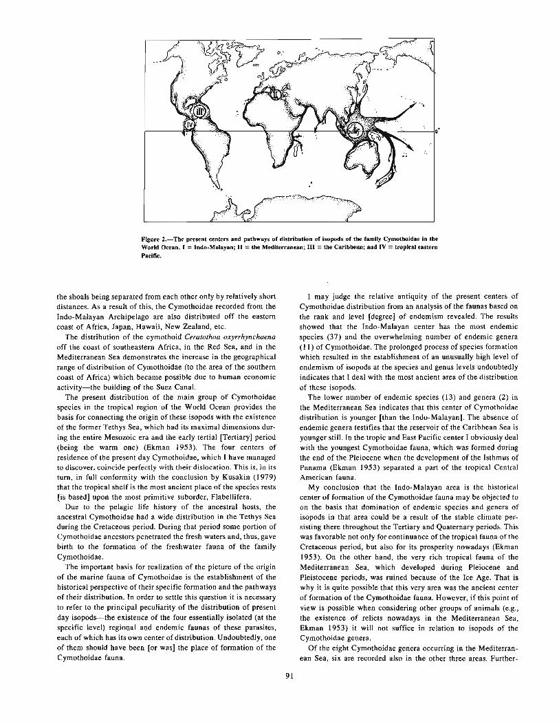
Figure 2.-The present centers and pathways of distribution of isopods of the family Cymothoidae in theWorld Ocean. I =Indo-Malayan; II =the Mediterranean; III =the Caribbean; and IV =tropical easternPacific.
the shoals being separated from each other only by relatively shortdistances. As a result of this, the Cymothoidae recorded from theIndo-Malayan Archipelago are also distributed off the easterncoast of Africa, Japan, Hawaii, New Zealand, etc.
The distribution of the cymothoid Ceratothoa oxyrrhynchaenaoff the coast of southeastern Africa, in the Red Sea, and in theMediterranean Sea demonstrates the increase in the geographicalrange of distribution of Cymothoidae (to the area of the southerncoast of Africa) which became possible due to human economicactivity-the building of the Suez Canal.
The present distribution of the main group of Cymothoidaespecies in the tropical region of the World Ocean provides thebasis for connecting the origin of these isopods with the existenceof the former Tethys Sea, which had its maximal dimensions during the entire Mesozoic era and the early tertia1 [Tertiary] period(being the warm one) (Ekman 1953). The four centers ofresidence of the present day Cymothoidae, which I have managedto discover, coincide perfectly with their dislocation. This is, in itstum, in full conformity with the conclusion by Kusakin (1979)that the tropical shelf is the most ancient place of the species rests[is based] upon the most primitive SUborder, Flabellifera.
Due to the pelagic life history of the ancestral hosts, theancestral Cymothoidae had a wide distribution in the Tethys Seaduring the Cretaceous period. During that period some portion ofCymothoidae ancestors penetrated the fresh waters and, thUS, gavebirth to the formation of the freshwater fauna of the familyCymothoidae.
The important basis for realization of the picture of the originof the marine fauna of Cymothoidae is the establishment of thehistorical perspective of their specific formation and the pathwaysof their distribution. In order to settle this question it is necessaryto refer to the principal peculiarity of the distribution of presentday isopods-the existence of the four essentially isolated (at thespecific level) regional and endemic faunas of these parasites,each of which has its own center of distribution. Undoubtedly, oneof them should have been [or was] the place of formation of theCymothoidae fauna.
91
I may judge the relative antiquity of the present centers ofCymothoidae distribution from an analysis of the faunas based onthe rank and level [degree] of endemism revealed. The resultsshowed that the Indo-Malayan center has the most endemicspecies (37) and the overwhelming number of endemic genera(11) of Cymothoidae. The prolonged process of species formationwhich resulted in the establishment of an unusually high level ofendemism of isopods at the species and genus levels undoubtedlyindicates that I deal with the most ancient area of the distributionof these isopods.
The lower number of endemic species (13) and genera (2) inthe Mediterranean Sea indicates that this center of Cymothoidaedistribution is younger [than the Indo-Malayan]. The absence ofendemic genera testifies that the reservoir of the Caribbean Sea isyounger still. In the tropic and East Pacific center I obviously dealwith the youngest Cymothoidae fauna, which was formed duringthe end of the Pleiocene when the development of the Isthmus ofPanama (Ekman 1953) separated a part of the tropical CentralAmerican fauna.
My conclusion that the Indo-Malayan area is the historicalcenter of formation of the Cymothoidae fauna may be objected toon the basis that domination of endemic species and genera ofisopods in that area could be a result of the stable climate persisting there throughout the Tertiary and Quaternary periods. Thiswas favorable not only for continuance of the tropical fauna of theCretaceous period, but also for its prosperity nowadays (Ekman1953). On the other hand, the very rich tropical fauna of theMediterranean Sea, which developed during Pleiocene andPleistocene periods, was ruined because of the Ice Age. That iswhy it is quite possible that this very area was the ancient centerof formation of the Cymothoidae fauna. However, if this point ofview is possible when considering other groups of animals (e.g.,the existence of relicts nowadays in the Mediterranean Sea,Ekman 1953) it will not suffice in relation to isopods of theCymothoidae gen"era.
Of the eight Cymothoidae genera occurring in the Mediterranean Sea, six are recorded also in the other three areas. Further-

more, their composition includes the three most abundant andancient specific genera-Lironeca, Cymothoa, and Ceratothoa.The representatives of two other rnonotypical genera, Mothociaand Emetha, are morphologically close to isopods of the generaLironeca and Ceratothoa, respectively.
Thus, specific features of the geographical distribution ofCymothoidae argue in favor of the Indo-Malayan Archipelago asthe center of faunal formation of this group of parasitic isopods.
The area is possibly [probably] the historical place of formationof the entire fauna of all the isopod species. I may conclude this onthe basis of the features of the present distribution of the representatives of the primitive families, Cirolanidae and Sphaeromatidae.As Kusakin (1979) pointed out, these isopods are totally absent inthe Arctic and their level of penetration into boreal waters is verylow. Even so, in the Southern Hemisphere they are represented ingreat numbers not only in boreal but also in antarctic waters.
Such an asymmetric pattern of distribution of primitive isopodswould have been formed only under similar earlier conditions. Tomy mind one of them should have been the location of the mostancient center of formation of fauna of isopods in the tropicsdirectly close to the Southern Hemisphere. Of the four existingcenters of Cymothoidae distribution, only the Indo-Malayanmeets these requirements. Moreover, if I retain the scheme byLevites (1961) concerning the distribution of the four continents
92
in the Carboniferous (the time of the most ancient finds of fossilisopods), then the above-mentioned center of the distribution ofparasitic isopods lay in the area adjacent to the eastern part ofGondwana. All the conditions for free penetration of ancestralisopods in the southern direction existed there, while the opportunity for their distribution from other centers was provided later,after the disintegration of the megacontinent-Gondwana-andformation of the Indian and Atlantic Oceans.
LITERATURE CITED
AVDEEV, V. V.1979. On the character of the geographic distribution of isopods-the para
sites of marine fishes. [In Russ.) Tez. Dokl. VII Vsesoyuzn. Sov. Parazit. iBo1eznyan Ryb. L. Nauka, p. 3-4.
EKMAN, S.1953. Zoogeography of the sea. Sidgwick and Jackson, Lond., 417 p.
KUSAKIN, O. G.1979. Marine and brackish-water isopods (Isopoda) of cold and moderate
waten; of the northern hemisphere. Sub-order Flabellifera. lsopods. [In Russ.)I. L. Nauka, 471 p.
LEVlTES, Y. M.1961. Historical geology witb the fundamentals of paleontology and geology
of the USSR. [In Russ.) M., 225 p.SCHIODTE, J. c., and F. MEINERT.
1884. Symbolae ad Cymothoaunn Crustacearum lsopodum Familiae.Naturhist Tiddskr. 3(14):221-454.

Pigmented Macrophage Accumulations (MMC; PMB):Possible Monitors of Fish Healthl
R. E. WOLKE,2 C. J. GEORGE,3 and V. S. BLAZER2
ABSTRACT
Anthropogenic pollution of the World Ocean is a growing problem. Areas most heavily contaminated areoften sites where many important fishes grow and reproduce. It is not easy to relate levels of pollution to diseasesand fish mortalities in nature and, though it has been done in but few cases, circumstantial evidence in manyothers is very strong.
Environmental pollution has direct (acute) and indirect (chronic) effects, i.e., causing deaths quickly as wellas increasing diseases, causing other debifities, and reducing reproduction, but evaluation of these effects is difficult. Histopathological examination of representatives of stressed populations, the most useful technique forestablishing or monitoring the disease-related effects of pollution, is difficult, costly, and time consuming. Moreefficient health-monitoring systems or indicators are needed.
The characteristics and capabilities of a suitable system are discussed. After examination of the reticuloendothelial system of higher fishes, the visceral macrophage response systems (i.e., the melanin-macropbagecenters-MMe, and the pigmented mictic bodies-PMB) of these fishes appear to have those qualities neededfor a relatively efficient health monitoring system. Investigations into applicability of these systems as indicatorsof fish health are in order since it appears that such an approach would be more productive than classical bloodcbemistry determinations.
INTRODUCTION
Anthropogenic pollution of the World Ocean is a growing problem. It is attributable to increasing industrialization but is relatedas well to the outdated concept that the oceans have an unendingability to accept waste. The effects of this pollution are manifold,but certainly nowhere more apparent than on the productivity ofanimal life. It is ironical that the points of heaviest pollutionshould also be sites for reproduction and growth of many of themost important commercial fishes. Pollution, however, is not confined to such "nurseries" of fin and shellfish. Deep ocean dumping of wastes also raises the levels of various pollutants to whichfinfish are exposed.
Toxicity of most pollutants, which include such substances aspetroleum-derived hydrocarbons, heavy metals, insecticides,herbicides, and sewage, have been examined in the laboratory andtheir LC50 and/or chronic effects for fish and shellfish determined (National Science Foundation 1974; Environmental ProtectionAgency 1979). Correlation of these data with decreasing populations of fish on a worldwide scale is much more difficult. Somemass mortalities and individual decreases in numbers of fish havebeen observed and documented, but determining the generalhealth of fish over broad geographic areas is not easily achievedand not well documented (Brown et al. 1970; van Loon andBeamish 1977; McFarlane and Franzin 1978; Gray 1979).
'This work was supported in part by a grant from the National Science Foundationand the Woods Hole Oceanographic Institution, Mass. Rhode Island Agricultural Experiment Station No. 2066.
'Department of Aquaculture Science & Pathology, University of Rhode Island,Kingston, RI 02881. U.S.A.
'Department of Biological Sciences, Union College, Schenectedy, NY 12308,U.S.A.
93
A healthy organism is an organism in physiological balancewith its environment. Disease, on the other hand, is an ongoingprocess which results in upsets to the physiological balance, upsetsin homeostasis, and upsets in the relationship of the organism andits environment. Initially these imbalances are at the biochemicallevel, eventually they become functional and morphological. It isthis whole concept of pathogenesis and its resulting chemical andanatomical changes that are of interest to the pathologist.
The disease process brought about by the toxicant may manifestitself on any of the aforementioned levels, or, in fact, it may berelated with a natural disease which masks the effects of the toxicant per se. At best, the toxicant results in pathognomonic lesionsand the condition is easily identified as to cause and readily classified as an "environmental" disease (Wolke et al. 1975, Sindermann 1979). Unfortunately it appears that diseases of this sort arerelatively few and that toxicant lesions are often quite nonspecific.Coupled with this nonspecificity is the fact that the levels of thetoxicant to which the fish is exposed in the marine environmentare far less than those studied in the laboratory. There is mountingevidence, however, that these chronic subclinical levels, while notreadily lending themselves to detection in terms of host response,increase disease susceptibility resulting in increased prevalence ofdisease processes and parasite burdens in exposed populations(Ziskowski and Murchelano 1975; Mearns and Sherwood 1977;Overstreet and Howse 1977; Welling et al. 1977). This introducesthe concept of an "environmentally associated" disease in whichthe noxious agent has an indirect rather than a direct effect-amore realistic concept [than others] which is based upon themultitude of information from necropsy examinations which havebeen conducted on marine fish from "polluted" and"nonpolluted" sites.
Evaluation of these subtle effects on the health of fish, considering in turn their genetic variability and the variability of theirenvironment, appears difficult. Classical methods used to determine the health of higher vertebrates, such as blood chemistry

determinations, might be of assistance if sufficient base data couldbe collected. Unfortunately, tests of this nature have showntremendous variability among individuals of the same species, requiring large numbers of determinations to achieve statisticalsignificance. When this observation is coupled with the variability among families and geographical locations, the approachseems impractical. Given time, much base data could be collected,but a problem of such international concern does not allow theluxury of time.
Histopathological examination of group representative individuals is more informative as a monitor of health, but is logisticallyimpractical and may not reveal even those nonspecific lesions oftoxicants previously discussed. Histopathology as a tcchnique,however, may be of value, especiaUy if it is used to examine aspecific system sensitive to a wide range of insults.
Availability of a more effecient system to monitor the health offishes in relation to the environment is important. Such a healthmonitoring system must have certain qualities. First, it must be asystem which is common to all fishes, regardless of their source.Second, it must be basic, and by basic is not meant unsophisticated but rather a system which is first in the chain of eventsfollowing an insult. In this sense then, the system should be rapidly responsive in reflecting upsets in homeostasis and the presenceof foreign antigens. Also, the proposed system should reflect orpresage early tissue damage. The third quality is accessability.This is the ease with which the system is· examined by the investigator and the speed with which data can be generated.
The purpose of this paper is to suggest a system which might beused for the monitoring of fish health. While studies have shownthe proposed system to have the necessary qualities it has not yetbeen used as a monitor, thus the feasibility of the hypothesis proposed here remains to be tested.
RETICULO-ENDOTHELIAL SYSTEM
The reticulo-endothelial system (RES) of the fish is sophisticated and highly functional (Ellis 1974). It is similar, but not identical, to Aschoffs RES in homeotherms (Aschoff 1924). Thesystems differ in that the fish has phagocytically active cells liningthe atrial endocardium and serving as a supportive endothelium(pillar cells) in the gill (Ferguson 1975; Chilmonczyk and Monge1980). In addition, accumulations of pigmented macrophages arerandomly distributed in the hepatic, renal, and splenic parenchyma. Kupffer cells are absent or, if present, are only weaklyfunctional.
The relationship of the fish RES to the homeotherm Mononuclear Phagocyte System (MPS) of van Furth is uncertain atpresent (van Furth et al. 1972). The MPS does not include facultatively phagocytic cells, i.e., reticular cells, fibroblasts, and, mostimportantly, endothelial cells. Light microscopic examination offish atrial endocardium reveals cells morphologically identical toclassical endothelial cells. These cells have the capacity to hypertrophy, acquire a rounded profile, and phagocytize blood-borneparticulate matter. This phenomenon may be elicited by simplyinoculating carbon particles intraperitoneally in higher teleosts.The mechanisms by which these cells are excited and theirfacultative nature are unknown. Further, there are other criteriaused to classify phagocytic cells for inclusion in the MPS. Suchcells must contain receptor sites for immunoglobulins and complement on their cell surfaces. A small percentage of macrophagesin plaice spleen and kidney have surface immunoglobulin (Ellis1974). The presence of these receptors on other fish phagocytesneeds to be determined.
94
PIGMENTED MACROPHAGEACCUMULATIONS
Groups of pigmented macrophages, present primarily in theliver, spleen, and kidney, are an important component of thefishes' RES. These structures were probably first described byJolly in 1923 and were later considered by Roberts (1975) whocoined the term melanin-macrophage centers (MMC). Still later,Agius (1979b, 1980) carefully described morphological speciesdifferences and pigment content of the accumulations. Otherworkers who have described these structures, which occur inamphibians and reptiles as well, include Yoffey (1929),Mackmull and Michels (1932), Ellis et al. (1976), and Ferguson(1976).
In the higher Teleostei the accumulations vary from a few to alarge number of cells forming rounded islands 40 to 1,000 /lm indiameter. The edges of the islands are usually regular and in someinstances the accumulation may have a degree of encapsulation.These fibers have been described as argyophilic (Roberts 1975). Athin rim of lymphocytes may be found surrounding the accumulations (Fig. 1).
Cells within the aggregates are primarily macrophages withirregular, poorly defined outlines and vesicular nuclei. Theircytoplasm contains black to yellowish pigmented material and/orphagocytized organisms. The cells vary in size from 10 to 30 /lm.There are also present a few true melanogenic cells (melanocytes).These are smaller (7 to 15 /lm) and their cytoplasm is heavilyladen with brown to black granules.
Ultrastructurally the macrophages are not morphologicallyunique. The nucleus is often kidney shaped or indented andchromatin frequently lies just inside the nuclear membrane. Theusual complement of organelles lie close to and surround thenucleus. Golgi complexes are common. Much of the cytoplasm isoccupied by vacuoles and lysosomes. Pigment, when present, isfound within membrane-bound vacuoles (melanosomes, ceroid).
The general color of the accumulations in higher fish tends tobe yellow brown. Their distribution is random (perhaps with sometendency to accumulate nearer blood vessels) in the parenchymaof liver, spleen, and kidney (anterior and posterior). However, incertain pathological processes their distribution is far more wide-
Figure t.-MlICropbage accumulation (MMC; PMB) in spleen of a higher teleost(upomis mtu:rocIJirus). Note round shape, sUght fibrous encapsulation, and thinrim of lymphocytes. Hemotol<yUn & eosin )( 121.

spread, including the gastrointestinal tract, peritoneum, andconnective tissue.
The general microscopic appearance of macrophage accumulations may differ in certain fish and early investigations seem to indicate a relationship to evolutionary development (Agius 1980).In the salmonids the accumulations are smaller, more irregular inoutline, and much darker (Fig. 2). As in higher fishes bothmacrophages and melanocytes are present, however the lattercomprise a far larger proportion of the cells than in the centers ofhigher fishes.
Figure 2.-Macrophage accumulations (MMC; PMB) in the spleen of a salmonid(Salmo gaird".ri). Note irregular shape and dense melanization. Hemotoxylln &
eosin x 308.
PIGMENTS
The pigments contained in the macrophage component areidentifiable by means of light microscope histochemistry andelectron microscopy. The pigments can be identified as hemosiderin, melanin, and ceroid.
Hemosiderin
Hemosiderin is an endogenous hemoglobin-derived storageform of iron. It is, therefore, a normal and important constituentof the body and of hemoglobin metabolism. When red blood cellsare destroyed, hemoglobin is degraded to iron, biliverdin, andglobin. The iron, as ferritin or bound to transferrin within themacrophage, is stored in the spleen or moved to sites of red bloodcell formation. It is known as hemosiderin and is seen as brown toblack intracytoplasmic granules. Its presence in the spleen is normal, both in fish and homeotherms. Special staining techniquesmay be used to identify and better outline the deposits. The Prussian blue method (potassium ferrocyganide) stains the granules abright blue (Thompson 1966).
Excessive accumulations of the pigment or accumulations inother organs is known as hemosiderosis and reflects a pathologicalprocess. The condition may be metabolic in origin, but in the vastmajority of the cases it is due to a sudden breakdown inhemoglobin secondary to a hemolytic anemia.
95
Melanin
Melanin is a complex polymer of uncertain structure whichmay be formed from the amino acid tyrosine or the catecholamineepinephrine. It is a common pigment in the integument of mostanimals and results in a brown to black coloration (Riley 1980).Cells which contain melanin in the fish include the melanocyte,melanophore, and macrophage. It is not certain whether all threecells are melanogenic, the concensus being that the first two are,while the macrophage acquires melanosomes via an, at present,unknown transfer mechanism. This probably occurs when macrophages come in contact with dendritic melanophores andphagocytize portions of the cell or their extruded melanosomes.
It is interesting to note that relative amounts of melanin appeargreater in the centers of cold-water fish or in fish kept at coldertemperatures. This observation may relate to the suggested functions of the pigment melanin. The pigment appears to have twoprimary functions. First, protective and second, destructive. In theprotective sense it is similar to alpha-tocopherol, an electrondonor. This relates to its resonation with its semiquinone freeradical precursor (Riley 1980). It can absorb free radicals or inhibit the propagation of the free radical chain reaction and willprotect the cell from the reactive substances produced during lipidoxidation (Edelstein 1971). Its presence in a cell responsible forcollecting effete lipids is self-evident.
Its second function, that of destruction, comes about by its ability to oxidize nicotinamide-adenine dinucleotide (NADH) andproduce hydrogen peroxide, which, in conjunction with peroxidase and iodide, can be bacteriocidal, perhaps via halogenation ofthe bacterial cell wall. It can therefore function in destruction ofinvading microorganisms which have been ingested by themacrophage in a manner essentially similar to the generation ofhydrogen peroxide by the probable NADH oxidase and halogenation in homeotherms. However, there is also the possibility thatquinone precursors of melanin may serve a bacteriocidal functionas well since quinones are strong oxidizing agents in their ownright. Because the mechanisms by which melanin functions arenonenzymatic it is interesting to speculate that melanin plays animportant role in the defense mechanisms of poikilotherms whereenzymatic activity is so temperature dependent.
Ceroid/Lipofuscin
The third pigment commonly associated with visceral macrophage accumulations is ceroid or lipofuscin. This is a yellowishgreen, autofluorescent age pigment. It is a breakdown product oflipid peroxidation and tissue destruction. Massive increases in thispigment are seen in vitamin E deficiency and/or rancid fat problems of fish (Poston et al. 1976; Watanabe and Takashima 1977;Roald et al. 1981).
FUNCTION
Information on the function of piscine visceral macrophageaccumulations may be collected from studies of the fate of injected particulate matter and the types and functions of pigmentpresent in the centers. Roberts in 1975 alluded to such a functional systhesis and recent investigations have added credence to thissynthesis.
When particulate matter (carbon, latex beads) is inoculatedinterperitoneally or intervascularly the material is rapidly ingestedby the various phagocytic cells of the RES (Mackmull andMichels 1932; Ferguson 1976; Ellis et al. 1976; Chiimonczyk and

Monge 1980). After ingestion, the laden macrophages migrate tovisceral accumulations or begin new accumulations in the parenchyma of spleen, liver, and kidney. The speed with which this isaccomplished appears related to the route of injection and totemperature. When the stimulated macrophages reach the cententhey are not simply added to the surface of the accumulation butare found to mix with the other cells so that one can appreciate theaccumulations as a dynamic moving body. This has led Georgeand Agius to suggest that these accumulations be referred to as"pigmented mictic (Gr = mixing) bodies" or PMB.
This movement of macrophages to a point of accumulation suggests that their function is to centralize foreign material for anumber of purposes. First, to present such antigens to cellscapable of producing humoral antibody, second, to remove toxicsubstances and to centralize their detoxification, and third, to storereuseable substances.
Further evidence for this hypothesis is found in the pigmentspresent and their previously discussed significance. Ceroid andlipofuscins are peroxidation products, potentially capable of producing free radicals as well as reflecting tissue breakdown (Hartroft and Porta 1965). Ceroid is commonly stored in PMB.Melanin is capable of accepting dangerous free radicals and indestroying microorganisms. Melanin is found in macrophageIysosomes in relation to bacteria and ceroid. Finally, hemosiderinreflects red blood cell destruction, storage of reuseable iron, and,in hemosiderosis, hemolytic processes. Anemia is a common signof disease in fish and often reflects the activity of endotoxins aswell as some water-borne toxins.
METHODS
The visceral macrophage accumulations (MMC; PMB) ofhigher fish appear to have those qualities needed for a healthmonitoring system. They are common to all fishes and they arebasic in that their macrophage component is first to come in contact with foreign antigens or remove endogenous and exogenoustoxic substances to a centralized area. They collect pigmentswhich reflect pathological processes and tissue destruction. Finally, they are easily accessible, requiring only routine histopathological methods for examination.
The literature is replete with studies that indicate increases innumbers of PMB in relation to age, starvation, disease, and dietaryimbalances. There is a body of evidence that these structuresreflect upsets in homeostasis and early disease processes (Roberts1975; Agius 1979a; Agius and Roberts 1981).
It is suggested therefore that some effort be placed on investigating the use of these structures as indicators of fish health. Itappears that such an approach would be more productive thanclassical blood chemistry determinations.
These structures will certainly lend themselves to mass determinations either by the point-counting planimetric method ofHennig (1958) following the reasoning of Chalkey (1943) or bymeans of colorometric assessment of the fluorescing ceroid withinthe centers. The latter method, however, essentially equates ceroidwith mass and may prove to be specious.
Subjective evaluation of hemosiderin and melanin content mayalso be made, especially with reference to the organ in which thehemosiderin is found. It is suggested thai all sections be routinelystained using the Prussian Blue reaction (Perls method for iron)and counter-stained with nuclear fast reds. The macrophages areapparent as pale yellow accumulations, melanin is black to brown,and hemosiderin bright blue. All three pigments are thereforerapidly and easily visualized. Objective evaluation of these
96
pigments in individual centers may also lend themselves to colorometric determinations.
The authors recognize that measurement of the suggestedsystem, because of obvious variables, may in fact be difficult.Investigators should recognize that the mass of PMB will vary asto age so that base data must, as in other systems, be collected.These data can be taken from only one species at a time and froma species inhabiting an environment as pristine as possible.Statistical questions must be answered regarding numbers of sections, numbers of organs, and surface area to be examined. Thesedo not seem, however, to be insurmountable problems and it is feltthat investigation of the use of PMB as monitors would be worthwhile.
If figures could be generated indicating mass and pigment content/age class/species these data could then be compared with datataken from smaller numbers of the same species in other geographical areas. The hypothesis could be tested by examiningpopulations from known polluted sites. If statistically significantdifferences were present then relatively rapid surveys of important geographic areas could be conducted to determine if heretofore unsuspected stresses were being applied to indigenouspopulations.
LITERATURE CITED
AGIUS, C.1979a. The role of melano-macrophage centres in iron storage in normal and
diseased fish. J. Fisb Dis. 2:337-343.1979b. Aspects of the melana-macrophage centres in fish. Ph.D. Thesis,
Univ. Stirling, Scotland, U.K.1980. Phylogenetic development of melano-macropbage centres in fisb. J.
Zool. Lond. 191:11-31.AGIUS, C., and R. J. ROBERTS.
1981. Effects of starvation on the melana-macropbage centres of fish. J.Fisb BioI. 19:161-169.
ASCHOFF, K.1924. Das reticulo-endotheliale system. Ergeb. Inn. Med. u. Kinderh. 26: 1.
BROWN, V. M., D. G. SHURBEN, and D. SHAW.1970. Studies on water quality and the absence of fish from polluted English
rivers. Water Res. 4:363-382.CHALKEY.H.W.
1943. Method for the quantitative analysis of tissues. J. Natl. Cancer Inst.4:47-53.
CHILMONCZYK, S., and D. MONGE.1980. Rainbow trout gill pillar cells: demonstration of inen panicle phago
cytosis and involvement in viral infection. J. Reticuloendothelial Soc. 28:237-332.
EDELSTEIN, L. M.1971. Melanin: a unique biopolymer. Pathology Annual 1:309-324.
ElliS,A.E.1974. Aspects of the Iympboid and reticula-endothelial systems in the plaice,
Pkuront!C/es platessa L. Pb.D. Thesis, Univ. Aberdeen, Scotland, U.K.ElliS, A. E., A. L. S. MUNRO, and R. J. ROBERTS.
1976. Defense mechanisms in fish. I. A study of the phagocytic system andthe fate of intraperitoneaUy injected paniculate material in the plaice(Pkuronectes platessa) L. J. Fish BioI. 8:67-78.
ENVIRONMENTAL PROTECTION AGENCY.1919. Toxicity bandbook, mammalian and aquatic data. Book I: Toxicity
data. B. A. Scbneider (editor). U.S. Environ. Prot. Agency, Beltsville, Md.,563 p.
FERGUSON, H. W.1975. Pbagocytosis by tbe endocardial lining cells of the atrium of plaice
(Pkuronectes platessa). J. Compo Pathol. 85:561-569.1976. The relationship between ellipsoids and melano-macrophage centres in
tbe spleen of turbot (Scophlhaimus maximus). J. Compo Pathol. 86:377-380.GRAY, J. S.
t 979. Pollution-induced cbanges in populations. Philos. Trans. R. Soc.
Lond. B 286:545-561.HARTROFI', W. S., and E. A. PORTA.
1965. Ceroid. Am. J. Med. Sci. 250:324.

HENNIG, A.1958. Kritische Betrachtungen zur Volumen und Oberfliischenmessung in der
Mikroskope. Zeiss Werkzeitschrift 30:78-86.JOLLY, J.
1923. Traite techniques alhemotologie. A. Moloine et fils, Paris.MACKMULL, G., and N. A. MICHELS.
1932. Absorption of colloidal carbon from the peritoneal cavity in the teleost,Taalaga/abras adspersas. Am. J. Anat. 51 :3-47.
McFARLANE, G. A., and W. G. FRANZIN.1978. Elevated heavy metals: a stress on a population of white suckers, Cala
slamas cammersani, in Hamell Lake, Saskatchewan. J. Fish. Res. BoardCan. 35:963-970.
MEARNS, A. J., and M. J. SHERWOOD.1977. Distribution of neoplasms and other diseases in marine fishes relative
to the discharge of waste water. In H. F. Kraybill, C. J. Dawe, J. C. Harshbarger, and R. G. Tardiff (editors), Aquatic pollutants and biological effects withemphasis on neoplasia, p. 210-224. Ann. N.Y. Acad. Sci. 298.
NATIONAL SCIENCE FOUNDATION.1974. Effects of pollutants on marine organisms. International Decade of
Ocean Exploration Effects of Pollutants on Marine Organism Study, August1-14,1974,46 p.
OVERSTREET, R. M., and H. D. HOWSE.1977. Some parasites and diseases of estuarine fishes in polluted habitats of
Mississippi. In H. F. Kraybill, C. J. Dawe, J. C. Harshbarger, and R. G. Tardiff (editors), Aquatic pollutants and biological effects with emphasis onneoplasia, p. 427-462. Ann. N.Y. Acad. Sci. 298.
POSTON, H. A., G. F. COMBS, Jr., and L. LEIBOVITZ.1976. Vitamin E and selenium interrelations in the diet of Atlantic salmon
(Sa/rna sa/or): gross, histological and biochemical deficiency signs. J. Nutr.106:892-904.
RJLEY, P. A.1980. Melanins and melanogenesis. Pathobiol. Annu. 10:223-251.
ROALD, S. 0., D. ARMSTRONG, and T. LANDSVERK.1981. Histochemical, fluorescent and electron microscopical appearance of
hepatocellUlar ceroidosis in the Atlantic salmon Sa/rna solar L. J. Fish Dis.4:1-14.
97
ROBERTS, R. J.1975. Melanin-containing cells of the teleost fishes and their relation to
disease. In W. E. Ribelin and G. Migaki (editors), The pathology of fishes, p.399-428. Univ. Wisconsin Press, Madison.
SINDERMANN, C. J.1979. Pollution-associated diseases and abnormalities of fish and shellfish:
A review. Fish. Bull., U.S. 76:717-749.THOMPSON, S. W.
1966. Selected histochemical and his.topathological methods. C. ThomasPubl., Springfield, 111., 1639 p.
van FURTH, R., A. Z. COHN, J. G. HIRSH, J. H. HUMPHREY, W. G. SPECTOR,and H. J. LANGEVORRT.
1972. The mononuclear phagocyte system: a new classification of macrophages, monocytes and their precursor cells. Bull. WHO 46(6):845-852.
van LOON, J. c., and R. J. BEAMISH.1977. Heavy-metal contamination by atmospheric fallout of several Flinn
Flon area lakes and the relation to fish populations. J. Fish. Res. Board Can.34:899-906.
WATANABE, T., and F.· TAKASHIMA.1977. Effect of alpha-tocopherol deficiency on carp. VI. Deficiency symp
toms and changes of fatty acid and triglyceride distribution in adult carp.Bull. Jpn. Soc. Sci. Fish. 43:819-830.
WELLING, S. R., C. E. ALPERS, B. B. McCAIN, and M. S. MEYERS.1977 .Fish disease in the Bering Sea. In H. F. Kraybill, C. J. Dawe, J. C.
Harshbarger, and R. G. Tardiff (editors), Aquatic pollutants and biologicaleffect with emphasis on neoplasia, p. 290-304. Ann. N.Y. Acad. Sci. 298.
WOLKE, R. E., G. R. BOUCK, and R. K. STROUD.1975. Gas-bubble disease: a review in relation to modern energy production.
In S. B. Saila (editor), Fisheries and energy production: A symposium, p. 300.Lexington Books, D. C. Heath Co., Lexington, Mass.
YOFFEY, J. M.1929. A contribution to the study of the comparative histology and physiol
ogy of the spleen, with reference chiefly to its cellular constituents. J. Ana!.Lond. 63:314-344.
ZISKOWSKI, J., and R. MURCHELANO.1975. Fin erosion in winter flounder. Mar. Poilu!. Bull. 6:26-29.


Infectious Diseases of Fish Involved in MarineAquaculture in the Soviet Far East
E. G. POTIEVSKI, L. A. TSAREVA, and V. V. BURLINI
Intensive development of aquaculture in the Soviet Far Eastresults from the complex of factors favorable for the progress ofthis prospective branch of the national economy. The culture ofsalmon, mollusks, algae, etc., is not only possible but highlyprofitable.
Among the problems of the development of intensive aquaculture, that involving the protection of cultured fish from infectiousdiseases which can cause great losses, is of major importance.
Prophylactic treatment of fish against infectious diseases inaquaculture is one of the important conditions for successfuleconomic management and its profitability. Rich aquacultureexperience accumulated in the Pacific basin countries (Canada,U.S.A., Japan and others) clearly demonstrates the necessity ofstudying the infectious diseases of maricultural organisms and ofdeveloping methods for diagnosis and preventive treatment ofdisease.
In this connection the Laboratory of the Infectious Diseases ofMariculture Objects (TINRO) began to investigate infectiousdiseases of salmons and bivalves, as the most important organismsof the Soviet Far East. Preliminary data acquired while examininghealthy and infected fish and mollusks are presented in this paper.
During April and May of 1980 an epizootic disease of the fry ofcultured coho salmon, Oncorhynchus kisutch, was observed at theTINRO geothermal station. Daily mortality during a single monthwas I to 2%, with increases in some days. Infected and dead fishhad pale, swollen gills covered with mucus. Spots of yellow oryellow-orange color were easily seen in the mucus. Visiblechanges in the internal organs were not seen during autopsy. Usingphase-contrast microscopy of unstained slides of the gills, a greatnumber of motile bacteria, as well as the destruction of gillfilaments, were discovered.
Cultures of the motile, gram-negative bacteria, which developed successfully on cytophaga-agar, forming yellow pigment,were found on the gills and in the kidneys of dead and infected
I Pacific Research Institute of Fisheries and Oceanography (TINRO), Vladivostok,U.S.S.R.
99
fish. The type of growth of the colonies is prostrate. The culturesgrew well at a temperature of +5°C. The bacteria were gramnegative, motile, oxidase-positive bacilli able to grow without air.A typical feature of these cultures was bacterial mobility on thesurface of the agar at the margin of the colony.
An epizootic disease with similar clinical and bacteriologicalfeatures was also recorded among chum salmon fry cultured at theexperimental TINRO station..
On the basis of these disease study data, we can conclude that inboth cases it was the cold-water salmon disease, caused by flexibacteria.
Along with these investigations, the microflorae of scallops(Patinopeeten yess) and oysters (Crassostrea gigas) were alsostudied. Upon studying the microflora of the scallop it was foundthat the infection level of its internal organs is heterogenous:Minimum microflora was on the muscle while the mantle andIiver were more heavily infected. Qualitative variety of the microflora was most evident in summer, i.e., at high seawater temperatures. When water temperature decreased, the gram-negativemicroflora was displaced by a gram-positive one. A relationshipbetween the environmental microflora and the microflora of thescallop organism was observed.
In August of 1980, oyster beds were investigated in Posyet Bay(South Primorye), where a considerable oyster mortality wasobserved. A great number of dead and injured oysters had alsobeen discovered there on the collectors. Spots of black or brownblack color were seen on the internal surfaces of the shells, mostlyin the zone of adductor muscle junction. No significant musclechanges were discovered. Bacteria of the genus Vibrio were mainly found in mantle, muscle, and liver. Some of them were luminescent. Other microflora was rather poor. Probably, the oyster mortality was caused by an intensive infection of the oysters withVibrio spp., due to the over-density of mollusks on the collectorsand high seawater temperatures.
The facts mentioned above suggest the necessity of further,more intensive studies of diseases of aquaculture organisms in theSoviet Far East.


Recent Studies in the United States on Parasites andPathogens of Marine Mollusks With Emphasis on
Diseases of the American Oyster,Crassostrea virginica Gmelin1
WILLIAM J. HARGIS, Jr.2
ABSTRACT
Morphological, systematic, faunal, and life cycle studies predominated research on marine parasites andpathogens in the United States before World War II. Much was primarily basic or academic in nature. Sincethen it has grown and diversified under pressure of efforts to: 1) Increase yields of invertebrate-based fisheries,in nature and under controlled conditions; and 2) understand, protect, and improve the resources, estuarine andmarine environments, and human health and welfare. Over the last 30 yr pathobiological investigations ofeconomically and ecologically Important marine Invertebrates have broadened into submlcroscopical anatomy(TEM and SEM techniques), physiology, immunology, genetics, host-parasite ecology, interactions betweenenvironmental pollution and disease, and prophylaxis and treatment of their diseases.
Importation of foreign oysters (and other shellfish species) and their transfer and transplantation betweenthe coastal regions, provinces, and states of North America have resulted in growing disease problems and acorresponding interest in the parasitology and pathology of the mollusks involved. It has also spawned efforts tocontrol introductions and transfers.
Two major diseases have been found to interfere with production of native Atlantic oysters along the Gulfandlor Atlantic coasts of the United States. These are the "Dermo" or "fungus" disease, caused by the apicomplexan protistan Perkinsus marinus (both coasts) and "MSX" or Delaware Bay disease, caused by the sporozoanHaplosporidium nelsoni (the Atlantic coast-prlncipaUy in Chesapeake and Delaware Bays). Knowledge of theseimportant epizootic-producing diseases is reviewed and discussed, along with that of other parasites andpathogens of molluskan sheUfish in North America, and an extensive References section of the results of recentresearch on moUuskan parasites and diseases is presented.
INTRODUCTION
Most of the research on parasites and pathogens of marineinvertebrates has taken place in this century. Faunal, morphological, systematic, and life cycle studies predominated theliterature before World War II. Much early work on parasites ofinvertebrates was primarily basic or academic in nature or onanimals used in experimentation, but even then the necessity toexplain unusual or catastrophic events, such as fish kills, majordeclines in fishery catches, impacts of plankton blooms (such asred tides and mahogany waters), the impacts of pollution onfishery populations, and other practical questions predominated.
Since World War II, research has increased markedly. Morescientists and institutions are involved in pathobiology of marinevertebrates and invertebrates than ever before. Academic interestcontinues to be a force but the need to produce more shellfish(Mollusca and Crustacea) on a sustained basis from coastal andocean waters and to protect the environment and human healthhave been primary factors prompting government support forthese studies. The growth of commercial display aquaria and oflaboratory experimentation with marine fishes and invertebrateshas also encouraged interest in parasites and diseases.
New knowledge, techniques, and equipment have enabledpathobiological investigations to broaden into submicroscopic
'Contribution No. 1158 from the Virginia Institute of Marine Science. Russianversion published in 1983 in Parasitologiya 17(4):256-260.
2School of Marine Science and Virginia Institute of Marine Science, College ofWilliam and Mary, Gloucester Point, VA 23062, U.S.A.
101
anatomy (i.e., studies of ultrastructure using scanning and transmission electron microscopes), immunobiology, immunochemistry, genetics, host-parasite ecology, microecology, physiologicalecology, and research on techniques for prophylaxis and treatmentof disease.
The development of marine aquaculture has increased the needto understand the impacts of parasites and pathogens on themarine invertebrates being used. Mollusks, being among theeasiest of marine invertebrates to "farm" or bring under cultureare of special interest. Most research activity on parasites andpathogens of this group centers upon bivalves of the genera Ostreaand Crassostrea of the family Ostreidae because of their economicimportance, but numerous other shellfish are involved also.
The increase in importation of mollusks from other countriessuch as Crassostrea gigas Thunberg 1793, the Pacific oyster, andOstrea edulus Linnaeus 1758, the European flat oyster, has'resulted in introduction of new parasites and diseases into theUnited States. Further, transfer of these and endemic species ofmollusks around the coast of North America has caused the spreadof their parasites (and predators as well). The problem of importation of foreign parasites and pathogens (from other countries orregions) has become of sufficient importance to cause development of specific control procedures such as the establishment ofcontrolled or prohibited areas and species, examination and certification, and quarantine arrangements. Of concern to science, industry, and government are the oysters mentioned above as well asthe mussels Mytilus edulus, Linnaeus, 1758; Brachidontes recurvisRafinesque, 1820; and others. Interest in the parasites and diseasesof other commercially important mollusks such as Mercenaria

mercenaria (Linnaeus, 1758), the hard clam or quahog; Myaarenaria (Linnaeus, 1758), the soft clam; Spisula solidissima(Dillwyn, 1817), the surf clam; Placopecten magellanicus (Gmelin,1791), the ocean scallop; Aequipecten irradians (Lamarck, 1819),the bay scallop; and Haliotis rufescens Swainson, 1822, the redabalone, has grown also in the last decade due to increased interestin improved management or culture of these organisms. Consequently, most of the research is concentrated in the molluskproducing regions of the northwestern United States (Oregon andWashington) and adjacent British Columbia, the Gulf Coast(Texas, Louisiana, Mississippi, Alabama, and Florida), the MidAtlantic (Virginia, Maryland, Delaware, New Jersey, and NewYork), the northeast (Connecticut, Rhode Island, Massachusetts,and Maine), and the nearby maritime provinces of Canada (Fig.1). Research activity related to abalone occurs mostly in California. (A number of relevant publications are provided in theReferences section.)
The literature mentions a large number of symbionts (employedhere in the broad sense) which infect oysters (Ostrea spp. andCrassostrea spp.) and other bivalves and cause diseases or aresuspected of doing so (Appendix I). In his very useful recentpublication, Sindermann (1977) listed 10 diseases of culturedoysters (Crassostrea virginica Gmelin, Crassostrea gigas Thunberg,and Ostrea edulus Linnaeus) in the United States and Canada andmentioned at least 6 others and there are more. The 10 primarydiseases cause significant mortalities. Some of those in the "mentioned" category are merely suspected of causing disease, havingbeen found in host tissues during surveys, but some are known tocause deaths also.
Undisturbed or relatively undisturbed natural populations ofmost (if not all) animals and plants bear parasites and pathogens.
..O'r-----;:...'-------T-------r----T---~...._-7T___,
o°I------+-----"""7i---=if--------+------+---1
.... IT[O STATES
.i-----II--<---~~:..<::j==---_._____+-----__+_I
""---\-\---+-------+-------+------+--i"'"
,,0
Figure l.-Eut rout or tlte United States.
102
Undoubtedly, disease-related mortalities occur in such populations. When man intervenes, either by altering the environment,by moving animals and plants to new habitats or by bringing themunder culture, losses due to disease increase-sometimes so drastically that epidemics or epiwotics involving numerous deathsoccur. At times the extensive mortalities result in severe diminishment of production and ensuing commercial losses. Animals underculture or in experimental or display aquaria, which usually arecrowded, are particularly troublesome in this regard.
In North America, oysters have been widely transported andtransplanted and under crude culture since the end of the last century. In recent years oyster culture techniques have become moresophisticated, though most commercial producers continue toutilize procedures developed and refined well before World WarII, which involve transplantation of young oysters, called "seedoysters" from good setting beds to "grow-out" or fattening bedswhence they are harvested when they or the market are ready. Aswould be expected, "oyster-kills" (severe mortalities) have occurred with some frequency on all coasts of the United States andCanada where oysters are under such culture. Two such diseaseevents are the subject of the remainder of this paper.
Mortalities of Crassostrea virginica Gmelin populations alongthe gulf and east coasts of the United States and in Canada (Fig. 1)in the late 1940's and the 1950's and later in the late 1950's andearly 1960's caused interest in the parasites and pathogens of Cvirginica and related oysters to intensify. Federal and state governments and industry became especially concerned. Since my institition, the Virginia Institute of Marine Science, was involveddeeply in both efforts, they will be described as examples.
"DERMO" DISEASE OR "FUNGUS"DISEASE OF CRASSOSTREA VIRGINICA
When the oil industry of the Gulf of Mexico was accused byoystermen of causing deaths in natural and cultivated beds ofoysters (C virginica) along the U.S. coast of the Gulf of Mexico(the gulf coast) in the late 1940's, research was begun to find thepossible cause or causes.
Scientists found the apicomplexan protistan Perkinsus marinus[Phylum Apicomplexa, Class Perkinsia, Order Perkinsida: firstnamed Dermocystidium marinum and then Labrynthomyxa marina(which was then considered to be a fungus-hence the confusionin the common name of the disease)] to be responsible (seeAppendix II). Investigations made elsewhere for the same reasonsshowed the organism to be present in natural and cultivatedpopulations of oysters (C virginica Gmelin) along the entire eastcoast of North America from Florida to Massachusetts as well ason the gulf coast (Fig. 1). From Chesapeake Bay southward andinto the Gulf of Mexico, it causes mortalities of epizootic proportions; elsewhere it is mostly enwotic [except in Delaware Bay(Figs. 1, 2) in the mid-1950's when mass mortalities, apparentlydue to importation of infected oysters from Virginia, werereported].
The pathogen can be identified easily in prepared cultures or insquashes or sections of tissue taken from live or moribund hosts.Crude experimental infections can be brought about in the laboratory but the etiological agent has not been obtained in pureculture.
Infections can be reduced or mitigated chemically underlaboratory conditions, but not eliminated. Chemical control innature is not now possible and may never be feasible due to thedilutive and dispersive nature of the "universal" solvent and/or

"MSX" DISEASE OR " THE DELAWAREBAY" DISEASE OF CRASSOSTREA VIRGINICA
Figure 3.-Distribution of MSX in oyster-producing regions in lower Chesapeake Bay. Areas are classified according to levels of infestation by Haplosporidium nelsoni (which causes MSX disease) under conditions of average salinitydistribution. Type I (very dark) areas are heavily infested and oysters plantedthere experience significant mortalities. Oyster culture in this area must be carried out on a very cautious basis. Areas with lower infestation levels (Types II,III, and IV) are normaily more productive and less troublesome, experiencingprogressively fewer deaths and requiring fewer accommodations and precautions for successful oyster (C. virginica) culture. During prolonged drought inthe upper watersheds salinity moves upstream as does MSX disease. Duringsevere wet periods in the upper watersheds they move downstream. Prolongeddrought periods cause serious mortality problems and economic losses forindustry. They must be anticipated and responded to. (From Haven et al. 1978.)
"..
TYPE
TYPE
TYPE
TYPE
In 1957, mortalities began to occur in cultivated populations ofoysters (c. virginica) in Delaware Bay. Because of the proximity,even contiguity of Chesapeake Bay through the connectingChesapeake and Delaware Canal and via the intervening Atlanticcoastal lagoons and embayments and the associated intracoastalwaterway, and because of frequent transfer of hosts between thesebodies of water by fishermen, scientists anticipated infestation ofthe Chesapeake system (Fig. 2). Industry and government becameconcerned, and considerable scientific effort was mounted in thestates of New Jersey (Rutgers University), Virginia (VirginiaInstitute of Marine Science-VIMS), and Maryland (ChesapeakeBiological Laboratory at Solomons Island, Maryland-now a partof the University of Maryland Center for Environmental andEstuarine Studies).
The disease, called "MSX-disease" (for Multinucleated SporeX-a name coined before the causative agent was discovered anddescribed), or "Delaware Bay disease," was found by epidemiological techniques to be caused by • "sporozoan" (PhylumBalanosporida, Class Stellatosporea, Order Balanosporida)parasite Haplosporidium nelsoni (=Minchinia nelsoni). SeeAppendix II for taxonomic placement. The pathogen was found inpopulations of C. virginica from Massachusetts to North Carolinabut was most concentrated in populations in Delaware Bay andChesapeake Bay, where it caused mortalities of from 50 to 60%annually until the commercial plantings in infested areas werekilled by the pathogen or harvested by industry. In Delaware Bay(Fig. 2), oyster production dropped from about 2 million bu toabout 10,000 bu annually in about 2 yr. In Virginia's ChesapeakeBay (Figs. 2, 3), it dropped from 4 million to 2 million bu/yr in a
carrier-water. Under farming conditions, losses can be reducedby avoiding planting seed oysters in infested high salinity areas.This salinity relationship has been demonstrated in the laboratoryand in nature. As recently as 1982 the pest moved into watersfrom which it was previously excluded, when drought in 1980 and1981 caused salinities in Chesapeake Bay to increase and "move"upstream. The parasite does not do well in salinities below 15 ppt.Losses can also be reduced by timing the planting and harvestingof the host. The parasite is also temperature-dependent and causesmost mortalities in summer and early fall when water temperatures are between 25° and 33°C. Intensity of infection and mortalities can be reduced by controlling the density of planting, sinceit has been found to be density-dependent as well.
Because the pathogen, P. marinus, has not been isolated in pureculture, Koch's postulates have not been satisfied. Additionally,the life cycle is not completely understood and it is not knownwhether reservoir or intermediate hosts are required or involvedbut it may not require them because of its mode of sporulation andthe fact that in some observations infections seem to be spread toother hosts by material from disintegrating neighbors. It formssporangia which release biflagellate zoospores. That some relatedorganisms have direct life cycles adds strength to the arguments ofJ. D. Andrews of the Virginia Institute of Marine Science andothers who contend that no alternate hosts are involved. It (orclosely related species) has (have) been found in many otherbivalves (Appendix I).
Figure 2.-Chesapeake and Delaware Bays and intervening coastal waters.
103
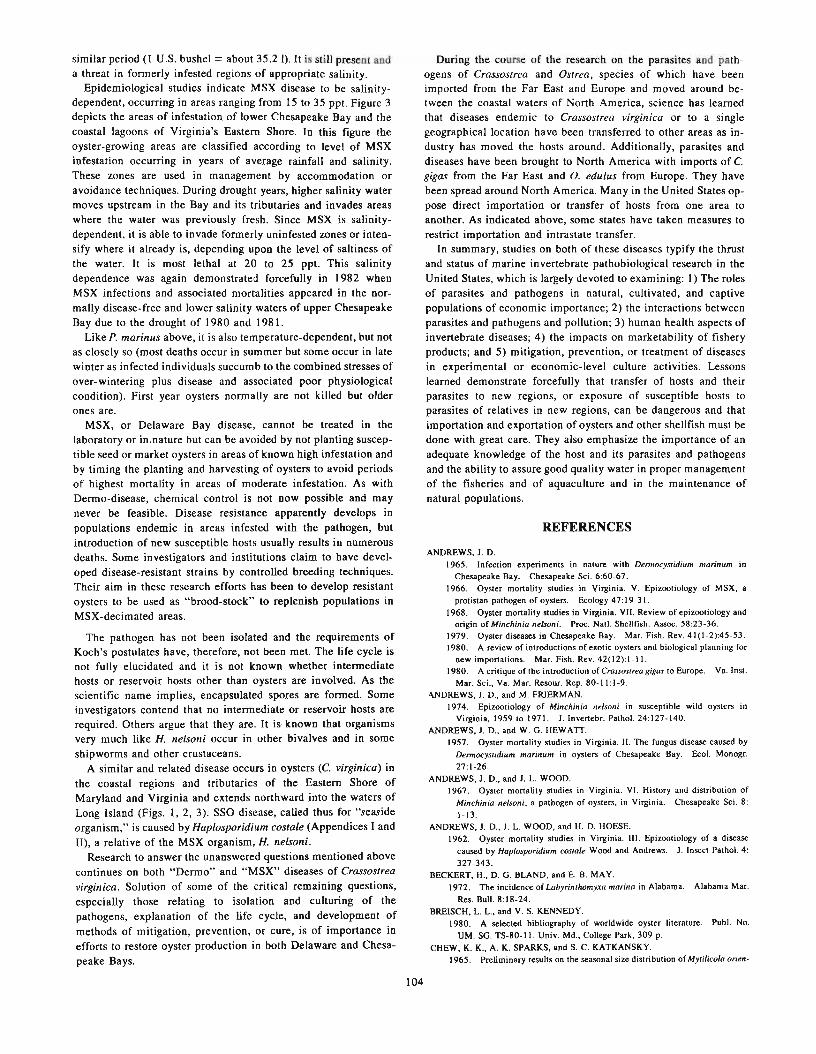
similar period (I U.S. bushel = about 35.2 I). It is still present anda threat in formerly infested regions of appropriate salinity.
Epidemiological studies indicate MSX disease to be salinitydependent, occurring in areas ranging from 15 to 35 ppt. Figure 3depicts the areas of infestation of lower Chesapeake Bay and thecoastal lagoons of Virginia's Eastern Shore. In this figure theoyster-growing areas are classified according to level of MSXinfestation occurring in years of average rainfall and salinity.These zones are used in management by accommodation oravoidance techniques. During drought years, higher salinity watermoves upstream in the Bay and its tributaries and invades areaswhere the water was previously fresh. Since MSX is salinitydependent, it is able to invade formerly uninfested zones or intensify where it already is, depending upon the level of saltiness ofthe water. It is most lethal at 20 to 25 ppt. This salinitydependence was again demonstrated forcefully in 1982 whenMSX infections and associated mortalities appeared in the normally disease-free and lower salinity waters of upper ChesapeakeBay due to the drought of 1980 and 1981.
Like P. marinus above, it is also temperature-dependent, but notas closely so (most deaths occur in summer but some occur in latewinter as infected individuals succumb to the combined stresses ofover-wintering plus disease and associated poor physiologicalcondition). First year oysters normally are not killed but olderones are.
MSX, or Delaware Bay disease, cannot be treated in thelaboratory or in.nature but can be avoided by not planting susceptible seed or market oysters in areas of known high infestation andby timing the planting and harvesting of oysters to avoid periodsof highest mortality in areas of moderate infestation. As withDermo-disease, chemical control is not now possible and maynever be feasible. Disease resistance apparently develops inpopulations endemic in areas infested with the pathogen, butintroduction of new susceptible hosts usually results in numerousdeaths. Some investigators and institutions claim to have developed disease-resistant strains by controlled breeding techniques.Their aim in these research efforts has been to develop resistantoysters to be used as "brood-stock" to replenish populations inMSX-decimated areas.
The pathogen has not been isolated and the requirements ofKoch's postulates have, therefore, not been met. The life cycle isnot fully elucidated and it is not known whether intermediatehosts or reservoir hosts other than oysters are involved. As thescientific name implies, encapsulated spores are formed. Someinvestigators contend that no intermediate or reservoir hosts arerequired. Others argue that they are. It is· known that organismsvery much like H. nelsoni occur in other bivalves and in someshipworms and other crustaceans.
A similar and related disease occurs in oysters (c. yirginica) inthe coastal regions and tributaries of the Eastern Shore ofMaryland and Virginia and extends northward into the waters ofLong Island (Figs. I, 2, 3). SSO disease, called thus for "seasideorganism," is caused by Haplosporidium costale (Appendices I andII), a relative of the MSX organism, H. nelson;'
Research to answer the unanswered questions mentioned abovecontinues on both "Dermo" and "MSX" diseases of CrassostreaYirginica. Solution of some of the critical remaining questions,especially those relating to isolation and culturing of thepathogens, explanation of the life cycle, and development ofmethods of mitigation, prevention, or cure, is of importance inefforts to restore oyster production in both Delaware and Chesapeake Bays.
During the course of the research on the parasites and pathogens of Crassostrea and Ostrea, species of which have beenimported from the Far East and Europe and moved around between the coastal waters of North America, science has learnedthat diseases endemic to Crassostrea yirginica or to a singlegeographical location have been transferred to other areas as industry has moved the hosts around. Additionally, parasites anddiseases have been brought to North America with imports of C.gigas from the Far East and O. edulus from Europe. They havebeen spread around North America. Many in the United States oppose direct importation or transfer of hosts from one area toanother. As indicated above, some states have taken measures torestrict importation and intrastate transfer.
In summary, studies on both of these diseases typify the thrustand status of marine invertebrate pathobiological research in theUnited States, which is largely devoted to examining: 1) The rolesof parasites and pathogens in natural, cultivated, and captivepopulations of economic importance; 2) the interactions betweenparasites and pathogens and pollution; 3) human health aspects ofinvertebrate diseases; 4) the impacts on marketability of fisheryproducts; and 5) mitigation, prevention, or treatment of diseasesin experimental or economic-level culture activities. Lessonslearned demonstrate forcefully that transfer of hosts and theirparasites to new regions, or exposure of susceptible hosts toparasites of relatives in new regions, can be dangerous and thatimportation and exportation of oysters and other shellfish must bedone with great care. They also emphasize the importance of anadequate knowledge of the host and its parasites and pathogensand the ability to assure good quality water in proper managementof the fisheries and of aquaculture and in the maintenance ofnatural populations.
REFERENCES
ANDREWS, J. D.1965. Infection experiments in nature with Dermocystidium marinum in
Chesapeake Bay. Chesapeake Sci. 6:60-67.
1966. Oyster mortality studies in Virginia. V. Epizootiology of MSX, a
protistan pathogen of oysters. Ecology 47:19-31.
1968. Oyster mortality studies in Virginia. VII. Review of epizootiology andorigin of Minchinia nelsoni. Proc. Natl. Shellfish. Assoc. 58:23-36.
1979. Oyster diseases in Chesapeake Bay. Mar. Fish. Rev. 41 (1-2):45-53.1980. A review of introductions of exotic oysters and biological planning for
new importations. Mar. Fish. Rev. 42(12):1-11.1980. A critique of the introduction of erassos/rea gigas to Europe. Va. Inst.
Mar. Sci., Va. Mar. Resour. Rep. 80-11:1-9.
'\NDREWS, J. D., and M. FRJERMAN.1974. Epizootiology of Minchinia nelson; in susceptible wild oysters in
Virginia, 1959 to 1971. J. Invertebr. Pathol. 24:127-140.
ANDREWS, J. D., and w. G. HEWATI.1957. Oyster mortality studies in Virginia. II. The fungus disease caused by
Dermocyslidium marinum in oysters of Chesapeake Bay. Ecol. Monogr.
27:1-26.ANDREWS, J. D., and J. L. WOOD.
1967. Oyster mortality studies in Virginia. VI. History and distribution of
Minchinia nelsoni, a pathogen of oysters, in Virginia. Chesapeake Sci. 8:
1-13.ANDREWS, J. D., J. L. WOOD, and H. D. HOESE.
1962. Oyster mortality studies in Virginia. III. Epizootiology of a disease
caused by Haplosporidium costaIe Wood and Andrews. J. Insect Pathol. 4:
327-343.BECKERT, H., D. G. BLAND, and E. B. MAY.
1972. The incidence of Labyrinlhomyxa marina in Alabama. Alabama Mar.
Res. Bull. 8:18-24.BREISCH, L. L., and V. S. KENNEDY.
1980. A selected bibliography of worldwide oyster literature. Publ. No.
UM. SG. TS-80-11. Univ. Md., College Park, 309 p.
CHEW, K. K., A. K. SPARKS, and S. C. KATKANSKY.1965. Preliminary results on the seasonal size distribution of My/ilieola orien-
104

talis and the effect of this parasite on the condition of the Pacific oyster,Crassostrea gigas. J. Fish. Res. Board Can. 22:1099-1101.
COUCH, J. A.1967. Concurrent haplosporidian infections of the oyster, Crassostrea virgin
iea (Gmelin). J. Parasitol. 53:248-253.COUCH, J. A., C. A. FARLEY, and A. ROSENFIELD.
1966. Sporulation of Minehinia nelsoni (Haplosporidia, Haplosporidiidae) inCrassostrea virginica (Gmelin). Science (Wash., D.C.) 153:1529-1531.
COUCH, J. A., and A. ROSENFIELD.1968. Epizootiology of Minehinia costalis and Minchinia nelsoni in oysters
introduced into Chincoteague Bay, Virginia. Proc. Natl. Shellfish. Assoc.58:51-59.
DAVIS, H. c., V. L. LOOSANOFF, W. H. WESTON, and C. MARTIN.1954. A fungus disease in clam and oyster larvae. Science (Wash., D.C.)
120:36-38.DRJNNAN, R. E., and L. A. ENGLAND.
1965. Further progress in rehabilitating oyster stocks. Fish. Res. BoardCan., Gen. Ser. Circ., BioI. Stn. St. Andrews, N.B. 48: 1-4.
FARLEY, C. A.1967. A proposed life cycle of Minchinia nelsoni (Haplosporida, Haplospori
diidae) in the American oyster, Crassostrea virginiea. J. Protozool. 14:616625.
1968. Minchinia nelsoni (Haplosporida) disease syndrome in the Americanoyster Crassostrea virginica. J. Protozool. 15:585-599.
1973. Virus diseases of oysters. 1973 Wildlife Disease Conf., MeetingAbstract, p. 19.
1975. Electron microscopy of virus infections in oysters (Crassostrea virginica). Society for Invertebr. Pathol., Abstracts of Papers, 8th Annu. Meet.Corvallis, Oreg., p. 28.
1975. Epizootic and enzootic aspects of Minchinia nelsoni (Haplosporida)disease in Maryland oysters. J. Protozool. 22:418-427.
1981. Phylogenetic relationships between viruses, marine invertebrates andneoplasia. In C. J. Dawe et al. (editors), Phyletic approaches to cancer, p.75-87. Japan Sci. Soc. Press.
FORD, S. E., and H. H. HASKIN.1982. History and epizootiology of Haplosporidium nelsoni (MSX), an oyster
pathogen in Delaware Bay, 1957-1980. J.lnvertebr. Pathol. 40:118-141.GALTSOFF, P. S.
1964. The American oyster Crassostrea virginica Gmelin. U.S. Fish Wildl.Serv., Fish. Bull. 64, 480 p.
HASKIN, H. H., and S. E. FORD.1982. Haplosporidium nelsoni (MSX) on Delaware Bay seed oyster beds: A
host-parasite relationship along a salinity gradient. J. Invertebr. Pathol. 40:388-405.
HASKIN, H. H., L. A. STAUBER, and J. B. MACKIN.1966. Minchinia nelsoni n. sp. (Haplosporida, Haplosporidiidae): causative
agent of the Delaware Bay oyster epizootic. Science (Wash., D.C.) 153:1414-1416.
HAVEN, D. S., W. J. HARGIS, Jr., and P. C. KENDALL.1978. The Virginia oyster industry: its status, problems and promise. A
comprehensive study of the oyster industry in Virginia. Special Papers inMarine Science, No.4, Va. Inst. Mar. Sci., 1024 p.
HELM, M. M., and F. M. SMITH.1971. Observations on a bacterial disease in laboratory-cultured larvae of the
European nat oyster Ostrea edulis L. Int. Counc. Explor. Sea, Shellfish andBenthos Committee, Rep. No. CM1971! K:IO, 9 p.
KATKANSKY, S. c., and R. W. WARNER.1970. Sporulation of a haplosporidan in a Pacific oyster (Crassostrea gigas) in
Humboldt Bay, California. 1. Fish. Res. Board Can. 27:1320-1321.KEARN, F. G.
1976. Minchinia nelsoni (MSX) disease of the American oyster. Mar. Fish.Rev. 38(10):22-24.
1976. Sporulation of Minchinia sp. (Haplosporida, HaplospOlidiidae) in thePacific oyster Crassostrea gigas (Thunberg) from the Republic of Korea. J.Protozool. 23:498-500.
KERN, F. G., L. C. SULLIVAN, and M. TAKATA.
1973. Labyrinthomyxa-Iike organisms associated with mass mortalities ofoysters, Crassostrea virginica, from Hawaii. Proc. Natl. Shellfish. Assoc. 63:43-46.
KRANTZ, G. E., L. R. BUCHANAN, C. A. FARLEY, and H. A. CARR.1972. Minchinia nelsoni in oysters from Massachusetts waters. Proc. Natl.
Shellfish. Assoc. 62:83-85.LOGIE, R. R.
1956. Oyster mortalities, old and new, in the Maritimes. Fish. Res. BoardCan., Progr. Rep. Atl. Coast Stn. 65:3-11.
105
LOGIE, R. R., R. E. DRINNAN, and E. B. HENDERSON.1960. Rehabilitation of disease-depleted oyster populations in eastern
Canada. Proc. Gulf Caribb. Fish. Inst. 13th Annu. Sess., p. 109-113.MACKIN, J. G.
1952. Oyster disease caused by Dermocystidium marinum and other microorganisms in Lousiana. Publ. Inst. Mar. Sci., Univ. Tex. 7:132-229.
1960. Status of researches on oyster diseases in North America. Proc. GulfCaribb. Fish. Inst. 1960:98-113.
1961. Mortalities of oysters. Proc. Nat!. Shellfish. Assoc. 50:21-40.1962. Oyster disease caused by Dermocystidium marinum and other micro
organisms in Louisiana. Publ. Inst. Mar. Sci., Univ. Tex. 7:132-229.1970. Oyster culture and disease. Proc. First Annu. Workshop, World Mari
cult. Soc. 1:35-38.MACKIN, J. G., H. M. OWEN, and A. COLLIER.
1950. Preliminary note on the occurrence of a new protistan parasite, Dermoeystidium marinum n. sp. in Crassostrea virginica (Gmelin). Science (Wash.,
D.C.) 111:328-329.MACKIN, J. G., and S. M. RAY.
1966. The taxonomic relationship of Dermocystidium marinum, Mackin,Owen, and Collier. J. Invertebr. Pathol. 8:544-545.
MANDELLI, E. F.1975. The effect of desalination brines on Crassostrea virginiea. Water Res.
9:287-296.MIX, M. c., and V. SPRAGUE.
1974. Occurrence of a haplosporidan in nalive oysters (Ostrea lurida) fromYaquina Bay and Alsea Bay, Oregon. J. Invertebr. Pathol. 23:252-254.
NEEDLER, A. W. H., and R. R. LOGIE.1947. Serious mortalities in Prince Edward Island oysters caused by a con
tagious disease. Trans. R. Soc. Can. 3(V), 41:73-89.NUMACHI, K., J. OIZUMI, S. SATO, and T. IMAI.
1965. Studies on the mass mortality of the oyster in Matsushima Bay. III. Thepathological changes of the oyster caused by gram-positive bacteria and thefrequency of their infection. [In Jpn., Engl. summ.) Bull. Tohoku Reg. Fish.Res. Lab. 25:39-48.
ODLAUG, T. O.1946. The effect of the copepod, Mytilicola orientalis, upon the Olympia
oyster, Ostrea lurida. Trans. Am. Microsc. Soc. 65:311-317.PERKINS, F. O.
1969. Ultrastructure of vegetative stages in Labrynthomyxa marina (=Dermocystidium marinum), a commercially significant oyster pathogen. J.Invertebr. Pathol. 13: 199-222.
1969. Electron microscope studies of sporulation in the oyster pathogen,Minchinia costalis (Sporozoa: Haplosporida). J. Parasitol. 55:897-920.
1971. Sporulation in the trematode hyperparasite Urosporidium crescens DeTurk, 1940 (Haplosporida: Haplosporidiidae)--an electron microscopestudy. J. Parasitol. 57:9-23.
1974. Phylogenetic considerations of the problematic thraustochytriaceouslabrynthulid Dermocystidium complex based on observations of fine structure.Pub1. Inst. Oceanogr. Res. Bremerhaven, Suppl. 5, p. 45-63.
1975. Fine structure of Minchinia sp. (Haplosporida) sporulation in the mudcrab, Panopeus herbstii. Mar. Fish. Rev. 37(5-6):46-60.
1976. Ultrastructure of sporulation in the European nat oyster pathogen,Maneilia refringens-taxonomic implications. J. Protozool. 23:64-74.
1979. Cell structure of the shellfish pathogens and hyperparasites in thegenera Minchinia, Urosporidium. Haplosporidium, and Marteilia-taxonomicimplications. Mar. Fish. Rev. 41(1-2):25-37.
PERKINS, F. 0., and R. W. MENZEL.1966. Morphological and cultural studies on a motile stage in the life cycle of
Dermocystidium marinum. Proc. Natl. Shellfish. Assoc. 56:23-30.1967. Ultrastructure of sporulation in the oyster pathogen Dermocystidium
marinum. J. Invertebr. Pathol. 9:205-229.PERKINS, F. 0., and P. H. WOLF.
1976. Fine structure of Maneilia sydneyi sp. n.-Haplosporidian pathogen ofAustralian oysters. J. Parasitol. 62:528-538.
QUAYLE, D. B.1961. Denman Island disease. Fish. Res. Board Can., Manuscr. Rep. Ser.
(BioI.) 713, 9 p.
QUICK, J. A., Jr.1971. Pathological and parasitological effects of elevated temperatures on the
oyster Crassostrea virginica with emphasis on the pathogen Labyrinthomyxamarina. Fla. Dep. Nat. Resour., Prof. Pap. Ser., Mar. Res. Lab. No. 15, p.105-176.
1972. Fluid thioglycollate medium assay of Labryinthomyxa parasites inoysters. Fla. Dep. Nat. Resour., Leaf!. Ser. Vol. 6, Pt. 4, No.3_ 12 p.

APPENDIX I
A PARTIAL LIST OF IDENTIFIED DISEASESOF BIVALVE MOLLUSKS IN
NORTH AMERICAN MARINE WATERS·
QUICK, J. A., Jr., and J. G. MACKIN.1971. Oyster parasitism by Labryinthomyxa marina in Florida. Fla. Dep.
Nat. Resour.. Prof. Pap. Ser. No. 13.55 p.RAY, S. M.
1954. Biological studies of Dermocystidium marinum, a fungus parasite ofoysters. Rice Inst. Pamphlet Monogr. in Biology, Spec. Issue, 114 p.
1966. A review of the culture method for detecting Dermocystidium marinum,with suggested modifications and precautions. Proc. Nail. Shellfish. Assoc.54:55-69.
1966. Cycloheximide: inhibition of Dermocystidium marinum in laboratorystocks of oysters. Proc. Natl. Shellfish. Assoc. 56:31-36.
RAY, S. M., and A. C. CHANDLER.
1955. Dermocystidium marinum, a parasite of oysters. Exp. Parasitol. 4:172·200.
ROSENFIELD, A.1969. Oyster diseases in North America and some methods for their control.
In K. S. Price and D. L. Maurer (editors), Proceedings of the conference onartificial propagation of commercially valuable shellfish, p. 67-78. Coil.
Mar. Stud.. Univ. Del.
SINDERMANN, C. J.
1968. Oyster mortalities, with particular reference to Chesapeake Bay and theAtlantic coast of North America. U.S. Fish Wildl. Serv., Spec. Sci. Rep.Fish. 569, 10 p.
1976. Oyster mortalities and their control. FAO Tech. Conf. on Aquaculture, Kyoto. Doc. FIR:AQI76/R34, 25 p.
1979. Pollution-associated diseases and abnormalities of fish and shellfish:A review. Fish. Bull., U.S. 76:717-749.
A. Viral1. Herpes-type virus diseases
a. Digestive gland virus disease-Crassostrea virgmlca,mortalities reported, Piscataqua River, Maine
b. Genital tissue or ovacystis disease-C. virginica(Papova virus?)
c. Digestive gland virus disease-C. virginica (C-typeoncorona virus)
d. Neoplasmic disease-Mya arenaria (uncharacterizedfilterable agent)
e. Lukemic disease-Mya arenaria (probable virus infection)
B. ChlamydiaChlamydial disease-Mercenaria mercenaria, the hardclam (digestive diverticulum-Chesapeake Bay)
C. BacteriaI. Bacillary necrosis--C. vlrgmlca larvae (Vibrio anguil
larum, V. alginolyticus, Vibrio spp., also possibly aeromonads and pseudomonads, U.S. east coast)
2. Bacillary necrosis-Mercenaria mercenaria larvae (sameetiological agents as above, U.S. east coast)
3. Focal necrosis-C. gigas (unidentified gram-positivebacterium-Willapa Bay, Wash.)
D. Fungi1. Dermo-disease-C. virginica (Perkinsus marinus-severe
mortalities in waters of gulf coast and east coast, United
IAdapted from various sources, including Sindermann (1977) and Farley (1981).
106
SINDERMANN, C. J. (editor).1977. Disease diagnosis and control in North American marine aquaculture.
Developments in Aquaculture and Fisheries Science, No.6, 330 p. ElsevierSci. Publ. Co., N.Y.
SPARKS, A. K.1962. Metaplasia of the gut of the oyster Crassostrea gigas (Thunberg) caused
by infection with the copepod Mytilicola orientalis Mori. J. Insect PathoI.4:57-62.
SPRAGUE, V.197 I. Diseases of oysters. Annu. Rev. Microbiol. 25:211-230.
SPRAGUE, V., E. A. DUNNINGTON, Jr., and E. DROBECK.1969. Decrease in incidence of Minchinia nelsoni in oysters accompanying
reduction of salinity in the laboratory. Proc. Natl. Shellfish. Assoc. 59:2326.
TUBIASH, H. S., P. E. CHANLEY, and E. LEIFSON.1965. Bacillary necrosis, a disease of larval and juvenile bivalve mollusks. I.
Etiology and epizootiology. J. Bacteriol. 90:1036- I044.TUBIASH, H. S., R. R. COLWELL, and R. SAKAZAKI.
1970. Marine vibrios associated with bacillary necrosis, a disease of larvaland juvenile bivalve mollusks. J. Bacteriol. 103:271-272.
VISHNIAC, H. S.1955. The morphology and nutrition of a new species of Sirolpidium. My
cologia 47:633-645.
WOOD, J. L., and J. D. ANDREWS.1962. Haplosporidium costaIe (Sporozoa) associated with a disease of Virginia
oysters. Science (Wash., D.C.) 136:710-71 I.
States). Also found in Ostrea equestris, O. frans, Mercenaria mercenaria, Mya arenaria, Argopecten irradians,Brachiodontus recurvis, Martesia spp., and others.
2. Larval mycosis of oysters-C. virginica (Sirolpidiumzoophthorum-U.S. east coast)
3. Larval mycosis of clams-Mercenaria mercenaria (Si1'Olpidium zoopthorum-U .S. east coast)
E. Protozoan1. MSX disease or Delaware Bay disease--C. virginica
(Haplosporidium nelsoni-U.S. east coast)2. Seaside disease-C. virginica (Haplosporidium costalis
high salinity coastal waters extending from ChesapeakeBay mouth to Long Island Sound)
3. Hexamitiasis--C. virginica [Hexamita infiata (a flagellate)-Prince Edward Island, Canada]
4. Nematopsiasis-C. virginica [Nematopsis ostrearum (agregarine)-Chesapeake Bay to Louisiana]
F. Metazoans1. Mytilicola or red worm disease-C. gigas and Ostrea
lurida-the Olympic oyster [Mytilicola orientalis (parasitic copepod)-U.S. west coast]. Also reported frommussels.
2. Trematodiasis-C. virgmlca [Bucephalus haimeanus(Digenea)-brackish waters U.S. east coast and elsewhere]
G. Diseases of unknown etiology1. Malpeque Bay disease-C. virgmlca (causative agent
unknown-Gulf of St. Lawrence, Canada)2. Denman Island disease--C. gigas (agent unknown
British Columbia, Canada)

APPENDIX II
CURRENT TAXONOMIC ARRANGEMENTS OF THE PROTISTAN OYSTER PARASITES DISCUSSEDI
Subkingdom Protozoa
Phylum Apicomplexa Levine, 1970Class Perkinsea Levine, 1978
Order Perkinsida Levine, 1978Genus Perkinsus (= Dermocystidium, partim)
Perkinsus marinus (Mackin, Owen, and Collier, 1950)(= Dermocystidium marinum Mackin, Owen, and Collier, 1950)
Phylym Ascetospora Sprague, 1978Class Stellatosporea Sprague, 1978
Order Balanosporida Sprague, 1978Genus Haplosporidium (= Minchinia, partim)
Haplosporidium nelsoni (Haskin, Stauber, and Mackin, 1966)(= Minchinia nelsoni Haskin, Stauber, and Mackin, 1966)
Haplosporidium costalis (Wood and Andrews, 1962)(= Minchinia costalis Wood and Andrews, 1962)
Order also includes the Genera Urosporidium and Minchinia
'Levine, N. D., J. O. Corliss, F. E. G. Cox, G. Deroux, J. Grain, B. M. Honigberg, G. F. Leedale, A. R. Loeblich III, J. Lorn, D. Lynn, E. G. Merinfeld, F. G. Page, G. Poljansky, V.Sprague, J. Vavra, and F. G. WaUace. 1980. A newly revised classification of the Protozoa. J. Protozool. 27:35-58.
107


Some Aspects of the Biology of the Trematode,Proctoeces maculatus, in Connection With the
Development of Mussel Farms on the Black Seal
V. K. MATSHKEVSKP
ABSTRACT
An e"tensive infection of mussels, My/ilus gallaprovincialis Lam., by parthenitae of the trematode Proctoecesmaculatus Odhner, 1911 (family Fellodistomatidae Nicoli, 1913) has heen recognized within the mussel farm inthe southwestern region of the Black Sea (Yegorlltsky Gulf).
The life history of the above trematodes has heen studied. Mussels are the first intermediate host for thesehelminths. Other invertebrates are supplementary hosts for P. maculatus. Fishes proved to he the definitive hostsfor P. maculatus.
In studies of the biocoenotic bonds of P. maculatus it has heen found that invasive cercariae discharged intothe ambient environment are eliminated in large quantities by animals common to coastal bioceonoses.
An extensive infection of mussels (Mytilus gallaprovincialisLam.) by trematode parthenitae (Proctoeces maculatus Odhner,1911; family Fellodistomatidae Nicoll, 1913) was recognizedwithin the mussel farm in the southwestern region of the BlackSea (Yegorlitsky Gulf).
The life history of this trematode has been studied and is thesubject of this report. Mussels have been found to be the first intermediate host for these helminths. Development of the series ofparthenogenetic sporocyst generations takes place in these hosts.Supplementary hosts for P. maculatus are invertebrates such as thegastropods Rissoa splendida and R. membranacea as well as thepolychaete Nereis succinea. The cercariae develop into metacercariae within both the gastropods and the polychaetes. In somecases single large metacercariae equipped with a fully developedgenital system were revealed in mussels, yet I tend to believe thisto be an abnormal phenomenon. The definitive hosts for P.
maculatus proved to be the fishes Grenilabrus ocellatus and Gr.griseus. The above-mentioned gastropods and polychaetes comprise a substantial part of the diets of these fishes.
It should be noted that R. membranacea and N. succinea wererecorded as supplementary hosts for P. maculatus for the firsttime. Experimental evidence points to the fact that supplementaryhosts surely participate in the life history.
When feeding the definitive hosts of these trematodes infectiveor invasive cercariae, I obtained evidence that they are digestedwithin the digestive system of fishes.
In considering the potential destructiveness of P. maculatus inmussel farms it should be noted that among the hosts the worstinfection by any particular form of P. maculatus is recorded inmussels (28,000 sporocysts per individual). Parthenitae infect thepallium, gonads, hepatopancreas, and other organs, comprisingone-fifth (20%) of the biomass of the soft tissues in the mussels insome cases.
Infected mussels have a depressed appearance, their valvesclose loosely [weakly], their attachment to the substratum
I As employed here. trematode signifies Digenea.'Institute of Biology of the South Seas. Ukranian Academy· of Sciences. Sevas
topol. U.S.S.R.
109
becomes weak, and finally they break away from the collectors.When mussels are kept in seawater deficient in oxygen thoseinfected by the parasites perish first. Histological examination ofinfected and uninfected tissues shows that the damage to the tissuestructures by the parthenitae results in the complete or partialcastration of the host. The negative effect of sporocysts upon boththe carbohydrate and lipid metabolism in Bivalvia is established.
A characteristic feature of the distribution of P. maculatuswithin the mussel population is its wide occurrence (45%) amongthe 2- to 3-yr-old mussels (Fig. I) comprising the bulk of thepopulation in the Black Sea (Vorobiev 1938; Ivanov 1968). The[deleterious] effect of the parasite upon the reproductive potentialof the host is beyond doubt [question]. Experiments showed thatwhen artificially stimulated to breed by elevated temperature,mussels are able to partially divest themselves of their parasites.Sporocysts and cercariae are ejected from the acinus of the gonadsand discharged into the ambient water where they could be seenby the unaided eye. This is one of the methods of recognizing infected mussels.
In studies of the biocoenotic bonds of P. maculatus, it has beenfound that invasive cercariae discharged into the surrounding environment are ingested in large quantities by animals common tocoastal biocoenoses such as: the Crustacea-Palaemon elegans, P.adspersus, Gammarus olivii, Pachygrapsus marmoratus, andMyzidacea gen. sp.; the polychaetae Nereis succinea; themolluscs-Bittium reticulatum, Nana donovani, Rissoa splendida,and R. membranacea; and the fishes--Grenilabrus ocellatus, Gr.griseus, Neogobius melanostomum, N. niger, and Pomatoshistusmicrops Leopardinus. Some of these animals participate in the lifehistory of P. maculatus as intermediate or definitive hosts.
Proceeding from my own data, as well as those in the literature,I regard the trematode P. maculatus as a characteristic componentof the coastal biocoenoses in the Black Sea where the fishes andinvertebrates representing their intermediate or definitive hostsare present constantly.
Continued development of mussel farms in the Black Sea requires further studies into the biology of P. maculatus in order toprovide information required for effective management of thosesea farms.

-z0j:0
!iOwlL
~ 16....z0 41II)
<~>
~
lL 1Il0
iii 2i0zw 2D...J<> 15wa:IL .,....>-!:: 5II)
zw 0l-Xw#
--- EXTENSITY(PREVALANCE)OF INVASION(lNFECTlON)
- - -INTENSITY OF INVASION(lNFECTlON)
_...J--/,.,-/--
.26-30 31-.~5 36-40 41-45 46-50 51-55 56-60 61-66 66-1\:) 71-"15 78-60
SIZE GROUPS OF MUSSELS
Figure I.-Extensity Bnd intensity of Infection by size groups.
LITERATURE CITED
....00 ....Z
<lOllO 01-.
36lIJ0IIIII.
3200z...Z
2IlOO 0II)
<2«10 >z2000 lL
0BIl >-
I-
1200II)
zw
eoo I-Z
400z<w
0::;
IVANOV,A. I.1968. Mussels (Myrilus gallaprovincialis Lam.) of the Black Sea, perspective
of their utilization. [In Russ.) Avtoref. Diss., Odessky Univ., p. 1-28.
110
VOROBIEV, V. P.1938. Mussels of the Black Sea. [10 Russ.) Pub!. Sci. lost. Fish. Oceaoogr.
11:3-30.
Special Features of the Infection of the Mollusk,Littorina rudis (Maton, 1797), With Parthenitae of
Microphallus pygmaeus (Levinsen, 1881) nec Odhner,1905 and M. piri/ormes (Odhner, 1905) Galaktionov,
1980 (Trematoda: Microphallidae) From the White Seal
K. v. GALAKTIONOV2
Microphallus pygmaeus and M. piriformes are the most commonparasites in the littoral mollusk, Littorina rudis, in the Barents andWhite Seas (Galaktionov 1980). Their life cycles are characterized by the absence of free-swimming cercariae and second intermediate hosts. The common eider, Somateria mollissima, is thefinal host.
The material on which this study was based was collected fromthe Tchupa Bay of the Kandalaksha Gulf during the summers andautumns of 1978, 1979, and 1980. Altogether, 5,748 samples ofL. rudis were examined. Microphallid daughter sporocysts couldbe subdivided into four age groups: 1) Sporocysts containing embryo balls; 2) sporocysts containing cercariae embryos; 3)sporocysts with developing metacercariae, and 4) maturesporocysts with metacercariae ready for invasion.
Infection of L. rudis by mature sporocysts of both M. pygmaeusand M. piriformes is relatively constant during the entire summer.The high infection extensivity [prevalence] seen in June is probably related to migration of healthy mollusks into the sublittoralzone. The development of the parasites within littorines infectedin autumn of the preceding year is fully completed by July orAugust. The common eider appears in the region of the Kandalaksha Gulf in May and begins occupying the Islands [offthe coast ofthe continental land mass] for nesting even when they are stillcovered with snow. The littoral zone is also still ice-coated, whichmakes a spring invasion of mollusks practically impossible.Owing to this fact the percentage of littorines infected with youngparthenitae in summer is very low. Mass invasion of the WhiteSea L. rudis with both M. pygmaeus and M. piriformes occurs inJuly when the first broods of eider are encountered. Since youngbroods remain together near the nesting places, mollusks infectedwith sporocysts containing embryo balls are found only on the
1No Literature Cited section was supplied by the author.2Murmansk Marine Biological Institute, Murmaosk, U.S.S.R.
111
islands in the open sea in September. In November, littorinesinfected with young parthenitae are found everywhere. In autumn,when weather conditions are worsening, the mortality of mollusksincreases. Those with severe infections of the inner organs diefirst. Because of this, a decrease in extensivity [prevalence] of L.rudis invasion [infection] with mature microphallid partheniteswas observed in autumn.
The extensivity [prevalence] of infection of L. rudis with parthenitae of M. piriformes increases from the coastlines of the opensea islands towards the emergent continental masses. I observed areverse tendency in the process of littorine infection with sporocysts of M. pygmaeus.
It seems obvious that the greater number of mollusks infectedwith mature parthenitae of M. piriformes successfully survive coldmonths due to favorable littoral ice-regime conditions in the continental bays. The effect produced by this type of parasite on theinner organs of the littorine is relatively insignificant. The principalloss occurs in littorines infected by mature parthenitae of M.pygmaeus, which cause complete liver and gonad destruction inthe mollusk.
Because of the instability of the ice cover, living conditionsaround the off-lying islands are extremely inclement during coldmonths. The selective advantages that are characteristic ofmollusks infected with M. piriformes compared with those infected by M. pygmaeus apparently seem not to be realized [in thewaters around the islands], and the great majority of littorinescontaining both types of mature parthenitae is lost during winter.Thus, infection with M. piriformes among the littoral population ofL. rudis from the continental bays is accumulated from year toyear. Indeed, the percentage of littorines infected with thisparasite reaches values as high as 80 to 90%. Moreover,specimens of older age groups ~re heavily infected. And sincemicrophallid parthenitae cause parasitic castration of mollusks,the maintenance of L. rudis populations here depends exclusivelyon females breeding for the first time and upon migration of littorines from neighboring populations.


The Helminth Fauna and Host-Parasite Relations ofSquids Sthenoteuthis oualaniensis (Lesson)(Cephalopoda, Ommastrephidae) in the
Indian Ocean and the Red Sea
N. N. NAIDENOVA, C. M. NIGMATULLIN, and A. V. GAEVSKAYAI
ABSTRACT
Nine species of helminths, including two species of trematodes, three ofcestodes, two of nematodes, and onespecies of acanthocephalans, were found hi the squid, Sthenoteuthis oualaniensis, from the Indian Ocean and theRed Sea. According to the incidence of infection and the degree of density of the parasite·host relationships, aUof the helminths were distributed into three groups: Main, secondary, and accidental helminths [parasltesl. Theage and geographic changes of the helminth fauna ofS. oualaniensi. are presented. The significance ofS. oualan·iensis in the life cycle of helminths is discussed. The helminth fauna of the squids S. oualaniensis and S. pteropusfrom the Atlantic Ocean is compared. The principle resemblance of the helminth fauna of these species of squidsis revealed. The possible reasons for the reduced diversity of species of helminths of S. oualaniensis arediscussed.
INTRODUCTION
Many reports on the important role of squids in the life cyclesof different systematic groups of helminths have been publishedrecently (Gaevskaya and Nigmatullin 1975; Naidenova and Zuev1978; Hochberg 1975; Stunkard 1977; and others).
In the eupelagic of the tropic zone of the World Ocean twospecies of the genus Sthenoteuthis-Atlantic S. pteropus (Steenstrump, 1855) and the Indo-Pacific S. oualaniensis (Lesson,I 830)-dominate the squids in terms of mass (numbers) and size.The role of the first species in the parasite fauna of the eupelagicof the Atlantic Ocean has been studied relatively well, but wehave only fragmentary information about the helminth fauna ofS.oualaniensis.
In this report we describe the results of a quantitative andqualitative study of the helminth fauna of S. oualaniensis in thetropical parts of the Indian Ocean and (partially) of the Red Sea.
The distributional area of S. oualaniensis includes the IndoPacific region from the west coast of Central and South Americato the Cape of Good Hope and from Japan to northern Australia.Its numbers are dominant among the nektonic predators-consumers of the III to IV orders. The mantle length reaches 45 cm,but specimens larger than 25 cm are very rare. Males are smallerthan females; their maximum length is 24.5 cm.
Considering certain morphophysiological and ecological features there are two well-differentiated forms within the speciesa small early spawning form and a large late spawning one (the Aand B forms mentioned in foreign literature) (Clarke 1966 andNesis 1977).
According to our data the food links of these squids are variousand change essentially in ontogenesis. The main food of young, 8to 10 cm long, squids are large copepods, euphausiids, amphipods,young decapod crustaceans, chaetognaths, and fish larvae. The
I institute of Biology of the Southern Seas, Academy of Sciences of the U.S.S.R.,SeYastopol, U.S.S.R.
113
role of crustaceans in their food is reduced as the squids grow, andthose> 13 to 15 cm long eat pelagic, open-ocean fish almost solely (Myctophidae in 60 to 80% of the cases) as well as smallspecies and juvenile forms of squids-often of their own species.Sometimes they eat large crustaceans such as shrimps and blueswimming-crabs. At the same time mesoplanktonic organisms(copepods, amphipods, ostracods, chaetognaths, and others) areusual in the stomachs of even the largest squids. They aredevoured with the main food organisms-Myctophidae. The foodspectrums of the representatives of small and large forms aresimilar.
In their turn young squids serve as general food for many kindsof marine birds, dolphin fishes, snake mackerels, and lancetfishes.Middle-sized and large squids are eaten by yellowfin, longfin, andbluefin tunas; marlins; sailfishes; whitetip sharks; cachalots; anddolphins (Wormuth 1976; Young 1975; and our data). Thereby, S.oualaniensis is an important intermediate link between macroplankton and small fishes in the trophic structure of the eupelagiczone-planktophages on the one side and large predatory teleostfishes, sharks, marine mammals, and birds on the other side.
Our materials were taken in the tropical zone of the IndianOcean and the Red Sea between May and August of 1978. Threehundred and thirty-seven squids were examined on board the shipProfessor Vodyanitsky.
QUALITIES OF THE HELMINTH FAUNA
Examination of the squids disclosed nine species of helminths-trematodes [digeneids], cestodes, nematodes, and acanthocephalans.
About 2,000 metacercariae of Didymozoidae gen. and sp. werefound in oval cysts with thin, transparent envelopes. They arelocated mainly in the external coverings of the stomach and lessfrequently in its muscular layer or caecum covers. The most compact accumulations of cysts were found in the forepart of thestomach, usually near a general [principal] blood vessel. Fromthere they spread to the rest of its surface. Worms move in their

cysts. The fact that upon fixation some larvae have long narrowbodies and others have wide flattened ones demonstrates thisfeature.
Judging from the morphology of the metacercariae they are ofthe Monilicacecum type, with a large muscular stomach situatedabove the level of the ventral sucker. The oral sucker is small. Thepharynx is rather powerful and muscular. The acetabular sucker issmall. The larvae differ by size and number of chambers percaecum. It should be noted that only large forms are registered[recorded] as being in the internal muscular layer of the stomach.Judging from the sizes of the metacercariae, they grow inside thesquid. Upon reaching the maximum size, metacercariae live for ashort time and perish, after which the cover of the cyst dissolvescompletely and the larvae become macerated. The same phenomenon was observed in didymozoid metacercaria from S. pteropusof the Atlantic. It usually occurs when the squid's mantle reaches20 cm in length and the age is from about 6 to 8 mo. Since the beginning of mass infection occurs in 2- to 3-mo-old squids, the duration of metacercarial life in the squid does not exceed 4 to 6 mo.
Didymozoid metacercariae use a wide variety of hosts, such asdifferent plankton organisms (i.e., copepods, euphausiids, andchaetognaths) and small teleost fish (Madhavi 1968; Reimer1971; Overstreet and Hochberg 1975; Gaevskaya and Nigmatullin1977). High indices of infection in the squid reflect the high intensity of feeding. According to our data, the daily diet of youngsquid constitutes 10 to 15% of the body mass and 4 to 5% of thebody mass of adults.
At the same time, didymozoid cercariae are noted as independent components of the plankton. Carried by water currents intothe mantle cavity of squids, these cercariae can actively penetrateinto the squid through its coelomic coverings.
The enormous infection of squid by didymozoid metacercariaeindicates an important role of squids in the life cycle of thesetrematodes. Heavy infection of the squids is a cause of the highinfection of the final fish hosts, mainly tunas, scombroids, andxiphoids. In the Indian Ocean around 100% of wahoo and 50% ofthe tunas are infected by the adult stages of didymozoids, with anintensity of about 100 cysts/fish (Nikolaeva and Dubina 1978).
Immature stages of the trematode [digeneid] Hirundiitella ventricosa are encountered smgly in the coelomic cavities of squids.Large specimens, with the characteristic narrowing of the bodybehind the acetabular sucker, have pointed body extremities.Average dimensions of the trematodes are 6.5 x 1.5 mm. The oralsucker is 0.55 x 0.72 mm, the pharynx is 0.39 x 0.40 mm, andthe acetabular sucker is 1.5 mm in diameter. The morphologicalcharacteristics of these trematodes are similar to those occurringin Atlantic squids.
Upon attaining large size, cercariae of azygiat trematodes enterinto the plankton and can infect squid. They occur in the host'sdigestive organs with ingested food. The final hosts of H. ventricosa are predatory teleosts---dolphins and scombroids.
Larval Nybelinia lingua/is (Cuvier, 1817)-the plerocercoidslocalize in intestinal mesenteries, on the ovary, and on the external coverings of the stomach. They encyst also in the stomach. Thescolex length is 1.6 to 1.65 mm; the bothrium portion is 0.94 to1.1 mm long and 1.4 mm wide; the tentacular sheath is 0.52 to0.55 mm long. Dimensions of the tentacular bulbs are 0.286 to0.319 x 0.11 mm.
Nybelinia larvae are widely spread among teleost fishes of theWorld Ocean. Twelve species of cephalopods have them in theAtlantic and Pacific Oceans (Kurochkin 1972; Brown and Threlfall 1968; Dollfus 1964; and others). Dollfus (1967) discovered
114
free-living Nybelinia larvae to be an organic [a regular] part of theplankton.
Indices of infection of squid by Nybelinia exceed those of thefishes. Thus, the extensiveness of the infection of squid reaches90%. More than 200 plerocercoids may be found in one squid. Incontrast, infection of fish seldom exceeds 40% and the intensity ofinfection constitutes 1 to 30 plerocercoids as a rule.
Probably, the squids acquire these parasites while ingestingtheir intermediate hosts-copepods and euphausiids, etc. However, considering the magnitude of the alteration of infection thesecond intermediate hosts-small teleost fishes-become themain source of infection. The latter circumstance promotes a second accumulation of Nybelinia plerocercoids in squids.
The final hosts of Nybelinia are large active sharks of thefamilies Carcharhinidae, Isuridae, and others, which can ingestsquids both alive and dead (after the period of spawning). In thelife cycle of Nybelinia,. the squid S. oualaniensis is an insert host[and may be a reservoir host] between small teleost fishes andselachians.
Larval Tentacularia coryphaenae Bosc, 1802 are registered[recorded] for the first time as parasites of squids of the IndianOcean with 65% infection. Large (3.0 to 8.3 mm), mobile plerocercoids locate in the internal organs and the mantle cavity,especially in its posterior end. Plerocercoids can actively embed inthe mantle wall of the squids. They are widely dispersed onmarine teleosts and are recorded in four species of cephalopods ofthe Atlantic Ocean. The final hosts of these cestodes are sharks.The first intermediate hosts are planktonic crustaceans and thesecond are teleost fishes. Plerocercoids infect the squids wheningested by them.
Phyllobothrium sp. larvae, encountered rarely, usually locate inthe caecum, more often in the rectum. After the squid die they canactively migrate into different portions of the body. The averagelength of the plerocercoids reaches 5 mm, the maximum width1.2 mm, the apical sucker-0.143 to O. I76 mm in diameter. Thedimensions of the lateral suckers are 0.275 to 0.297 x 0.352 mm.The bothridia are heart-shaped and measure 0.55 x 0.72 mm.
Plerocercoids of the genus Phyllobothrium are rather widelydispersed in the squids of the Atlantic Ocean (Stunkard 1977;Gaevskaya 1976; and others). As the work of American scientists(Brown and Threlfall 1968) showed, infection of the squids byplerocercoids experiences monthly and yearly variation. It isapparently connected with the short (1 yr) life cycle of thesesquids.
In the life cycles of the phyllobothriums, S. oualaniensisassumes the role of insert host [another host which is inserted]between small teleost fishes and squids and the final hostssharks.
Two types of Porrocaecum sp. larvae are recorded in 24 to 57 %of squids with an intensiveness of infection of 1 to 100 specimens.They are situated in connective tissue capsules in the body cavity.Small larvae (3 to 5 mm) localize on the external wall of thestomach; large ones (20 to 25 mm) on the internal mantle wall.Adentic nematodes were found also in squids of the AtlanticOcean, but their infection was considerably higher (96%). It maybe supposed that both forms are successive stages of the development of a single species of nematode, since their morphology issimilar. However, the absence of transitional forms between thesmall and large larvae causes bewilderment.
The life cycle of Porrocaecum sp. is probably as follows: Thefirst intermediate hosts are planktonic crustaceans (euphausiids),the second are teleost fishes, the third are squids, and the final

hosts are mammals. It should be noted that Porrocaecum caballero,whose morphological features coincide with those of larvaeregistered [recorded] on the squids, has been described fromMakaira mitsukuri from Mexican waters.
Anisakis sp. larvae are encountered relatively rarely (0.9 to3.8%) with a low intensity of infection (one to two specimens).They locate in the ovary and coelomic membranes of the sexualcavity, rarely on the caecum walls. Ending their development inmarine mammals, the larvae show great activity at high temperatures and retain their vitality for a long time. The life cycles ofthese larvae and the position the squids occupy in them are similarto those of the Porrocaecum species.
The Acanthocephala gen. and sp. I are very long and threadlike.They are found in sections of the pericardial coelom and usuallyattached to the mantle wall or liver with their probosces. Liveworms measure from 10 to 20 cm. The morphometric charactersof these acarithocephalans conform to those described from thesquids of the Atlantic Ocean by Naidenova and Zuev (1978). Twomature females were found near Sokotra Island in 1964. Our mostrecent finds represent immature forms.
Acanthocephala gen. and sp. II were twice found in the stomach<:avity of the large squids. They measured 1.0 to 1.3 cm. Their.ystematic identity was not determined because of poor preservation.
QUANTITATIVE INDICES OF INFECTIONAND THEIR CHANGES
The total infection of the squids we examined was 98.4%.Eighty to 84% of the juveniles 2 to 8 cm long are infected withhelminths. The extensiveness [prevalence] of infection o~ thelarger squids is 100%.
According to the indices of extensiveness [prevalence] and intensity of infection, and the degree of narrowness of host-parasiterelations of the squid, we may divide the helminths discoveredinto three groups: 1) Principal helminths (occurring at an extensiveness [prevalence] of infection of from 30 to 95% and an intensity from a few specimens to tens of hundreds of specimens)metacercarial didymozoidae, Porrocaecum larvae, Nybelinia, andTentacularia. 2) Secondary helminths (2.5 to 3.5%, single specimens)-cavital Acanthocephala gen. and sp. I, Phyllobothrium larvae, Anisakis larvae, and H. ventricosa. 3) Causal helminths «1%)-small Acanthocephala gen. and sp. II from the stomach.
As the size of the squids increases, infection by various speciesof helminths changes unequally [disproportionately]. The poorestis the qualitative composition of helminths of the youngest squids2 to 4 cm long. Only metacercarial didymozoidae have been foundin them. Specimens of the next size group (4 to 12 cm) haveNybelinia larvae in addition. As squids attain a mantle size of 12 to14 cm in length they have all species of helminths. From then ononly an increase of infection by the principal species of helminthsoccurs. Three types of size-age dynamics of squid infection maybe identified: 1) The first type is characteristic of the largest massof parasites--the didymozoidae. The indices of extensiveness[prevalence] of in fection are large for the squids of all size groupsand are approximately at the same level (80.0 to 98.7). Only theintensity of infection changes noticeably. 2) In the second type,the change of infection by the other principal helminths-Porroceacum, Nybelinia, and Tentacularia larvae-occurs. Theseparasites appear in squids 8 to 10 cm long, and when they are 14to 18 cm long the indices of infection increase almost to the maximum. The secondary helminths-Anisakis and Phyllobothriumlarvae-join this type. But the sharp rise of infection by these
115
parasites takes place later, when the squids attain more than 20 to22 cm in length. 3) In contradistinction to the other helminths, H.ventricosa and cavital acanthocephalans begin to infect specimens10 to 12 cm long and maintain an essentially low level of infection index as the squids grow.
Infection by Didymozoidae occurs thanks to copepods, euphausiids, and chaetognaths-the principal food of the smallest squids.Further accumulation of these trematodes probably occurs whenthese invertebrates get into the stomachs of the squid. On thewhoie the formation of the helminth fauna coincides with thesquid's attainment of adulthood when they become 10 to 14 cmlong. This takes place when the adult squid begin to feed on adultspecimens of small fishes and small squids, which are the suppliers [carriers] of cestodes, nematodes, trematodes, H. ventricosa,and probably of small acanthocephalans, as their principal sourceof nourishment. To judge from the special features of the size-agemetamorphosis of the squid's infection, the majority of helminthspecies get into them with the representatives of different groupsof fishes and small squids or with different age stages of one oranother food object.
When comparing the qualities of helminth fauna and the indices of the early spawning and late spawning squids as a whole[all together], their main similarity is discovered. Their differenceconsists of a poorer species composition (five to nine) and low indices of infections of the early spawning forms by almost allhelminths, with the exception of didymozoidae and Nybelinia. Atthe same time, all species of helminths found in early maturingsquids are common for both forms, and the indices of infection ofrepresentatives of early and late maturing forms on the whole areconnected with the difference of their dimensions (the length ofearly maturing specimens in our collections did not exceed 12.7cm) and, in the end, of the length of life and food composition.
Within the late maturing forms the structure of the helminthfauna and the squid's infection may vary considerably dependingon the geographical region of the research. In this respect the RedSea region stands out for the paucity of its helminth fauna. It hasonly three species. Perhaps this is connected to an insufficientsample of squids examined from a single station. However, we donot exclude the possibility that the results obtained reflect the truesituation only partially and may be a consequence of a scarcity offinal hosts of the squid helminths. The high indices of infection ofRed Sea squids by Nybelinia and the low indices of infection bydidymozoidae attracted our attention.
DISCUSSION
We discovered that the chief factor leading to the infection ofsquids by helminths is food. As the ontogenesis of food links occurs, the helminth fauna and quantitative indices of infection alsochanges. The food factor promotes the transmission of thesehelminths to the final hosts. Xiphoid fishes, sharks, and [marine]mammals mainly use the most heavily infected medium- andlarge-sized squid. The latter become most accessible after spawning. Small and medium S. oualaniensis occur at the same time aslarge squids in the food of the tunas. Therefore, in this case wehave full accordance between trophic and parasitic links.
Except for acanthocephalans and juvenile H. ventricosa, allhelminths parasitizing squids represent larval forms. They havevery wide [broad] specificity and use various invertebrates, teleostfishes, and squids at the same stages of their life cycles. Therefore,the use of other squids for food is the cause of the secondaryaccumulation of didymozoidae and different species of nematodesand cestodes in the squid. With the increase of age and size and

transition to a higher trophic level, the number grows. In the lifecycles of didymozoidae, cestodes, and nematodes, there is aninsert host between the second intermediate hosts-planktonic invertebrates and micronekton (i.e., fish-planktophytes and smallsquids), and the final hosts-large nektonic predators, occupyingthe top of the food chains of the eupelagic zone. To speak of thesquid's position in the life cycle of acanthocephalans at presentwould be premature. We may only suppose that the squids aretheir final hosts.
Considering the high indices of infection, which are notencountered in other hosts of these helminths, the high quantity,wide dispersion, and the variety and stability of trophic links wemay consider S. oualaniensis to be one of the main obligatoryhosts for didymozoidae, Nybelinia, Tentacularia, and Porrocaecum. Stenotheuthis oualaniensis is the vehicle for their transmission to the final hosts. Wide [broad] specificity and the "fish"larvae "aspect" of the helminth fauna of S. oualaniensis shows itto have been "acquired" by the squids relatively recently. Theparasites have successfully acclimated and are incorporating thisspecies of host, which occupies a key position in the pathway ofcirculation of helminths among oceanic pelagic communities [into their circle of hosts]. At present we find different species ofhelminths in different stages of forming parasite-host relationships. They are strongly expressed in didymozoidae, Porrocaecum,Nybelinia, Phyllobothrium, and Tentacularia. Though the foodlinkages of young squids are very close to marine birds, larvae ofbird parasites are absent in squids.
The helminth fauna of S. oualaniensis represents (as a matter offact) an impoverished version of that in the Atlantic vicariate ofS.oualaniensis, i.e., 8 species as compared with 15. Moreover, thefirst species has no helminth which does not occur in the secondone. The impoverishment of the species of their parasite fauna occurs primarily because of the disappearance [or lack] of thesecondary and casual species of helminths.
At times the difference in the infection indices of the commonspecies of helminths tends toward the side of a larger quantity inS. pteropus. In our opinion the causes of the main differences arerelated to the difference in the number of individuals examined(303 specimens of S. oualaniensis as compared with 2,200specimens of S. pteropus) and in the dimension of the squids (S.pteropus are 2 to 50 cm in length with a mode of 20 to 30 cm). Itmiturally depends on the presence or absence of the secondaryand, especially, of the casual species of helminths and the degreeof accumulation of the principal helminths.
The main conclusion of this comparison is that both geographicvicariates have a similar position in respect to the parasite faunaof conformable [comparable?] associations of the tropicaleupelagic. This is more indirect evidence that their econiches arealmost identical, though that ofS. pteropus is somewhat wider, dueto its larger size.
The ancestor of S. oualaniensis penetrated into the Indo-Pacificfrom the Atlantic Ocean, rounding the southern extremity of
116
Africa during one of the warming Pleistocene periods. During theensuing drop in temperature and upon isolation two allopatricspecies were formed. Undoubtedly, the squids themselves couldnot have carried the helminths from the Atlantic to the IndoPacific, since they were only the intermediate hosts. Consideringthat these geographic vicariates occupy similar econiches inhomologous associations of the World Ocean and consideringtheir high ecological valency, the formation of the helminth faunaof both species evidently proceeded simultaneously.
LITERATURE CITED2
ASH MOLE, N. P., and M. J. ASHMOLE.1967. Comparative feeding ecology of sea birds of a tropical oceanic island.
Bull. Peabody Mus. Nat. Hist. 24:1-131.BROWN, E. L., and W. THRELFALL.
1968. A quantitative study of the helminth parasites of the Newfoundlandshort-finned squid, l/lex il/ecebrosus il/ecebrosus (leSueur) (Cephalopoda:Decapoda). Can. J. Zool. 46:1087-1093.
CLARKE, M. R.1966. A review of the systematics and ecology of oceanic squids. Adv. Mar.
BioI. 4:91-300.DOLLFUS, R.-P.
1964. Enumeration des cestodes du plancton et des invertebres marins (6e
contribution). Ann. Parasit. 39:329-379.1967. Enumeration des cestodes du plancton et des invertebres marins (7e
contribution). Ann. Parasit. 42:155-178.GAEVSKAYA, A. V.
1976. On the helminthofauna of Atlantic squids, Ommuslrephes bar/ramileSueur. [In Russ., Engl. summ.] Tr. AtlantNIRO 69:89-96.
1977. Helminthofauna of the Atlantic squid, Slhenoleulhis pleropus (Steenstrup). [In Russ.) Biologicheski Nauki 8(164):47-52.
KUROCHKJN, Yu. V.1972. On the parasitofauna of the Pacific squid, Todarodes pacificus Steen
strup. [In Russ.) Parasity vodnykh bespozvonochnykhzyvotnykh, Lvov, p.53-54.
NAIDENOVA, N. N.1978. Some data on helminth faunas of the intraspecies groups of the squid
(Ommuslrephes pleropus). [In Russ.) Abstracts of the I st All-Union Meeting of Parasitocaenolog., Po1tava, pI. 3:103-105.
NESIS, K. N.1977. Population structure in the squid, Slhenoleulhis oualaniensis (Lesson,
1830) (Ommastrephidae) in the western tropical Pacific. [In Russ., Engl.summ.) Tr. P. O. Shirshov Inst. Oceanol. 107:15-29.
NIKOLAEVA, V. M., and V. R. DUBINA.1978. New species of Didymozoidae from fishes of the Indian Ocean. [In
Russ., Engl. summ.) Biologia Morja, Kiev, 45:71-80.WORMUTH, J. H.
1976. The biogeography and numerical taxonomy of the oegopsid squidfamily Ommastrephidae in the Pacific Ocean. Bull. Scripps Inst. Oceanogr.Univ. Calif. 23, 90 p.
YOUNG, R. E.1975. A brief review of the biology of oceanic squid, Symplecloleulhis oualan
iensis (Lesson). Compo Biochem. Physiol. 52B:141-143.
'Not aU citations mentioned in the text are included here. Also, not all of the Literature Cited are mentioned in the text.

Some Parasitological Aspects of Shrimp Culturein the United States
ROBIN M. OVERSTREETl
ABSTRACT
Penaeid shrimps are presently being reared conimerciaUy in the United States and elsewhere. Parasites anddiseases, however, continue to be a problem in most rearing Cacilities. Viruses, algae, bacteria, Cungi, ciliates,mlcrosporidans, digeneans, cestodes, nematodes, and mlsceUaneous conditions include representatives that havean errect on aquaculture, innuence hosts in their natural environment, and produce potential public health problems.
INTRODUCTION
Commercial culture of marine crustaceans in the United Statesof America or by U.S. companies abroad is primarily confined topenaeid shrimps. Additionally, some facilities rear the blue crab,Callinectes sapidus, for the soft-shelled seafood industry;American lobster, Homarus american us, and "freshwater"shrimps (Macrobrachium spp.) for human consumption; andvarious crustaceans belonging to different orders for research purposes. This paper is concerned primarily with infections ofpenaeids.
Many of the problems of hatching and rearing penaeid specieshave been solved and some penaeid species can be profitablycultured (e.g., Penaeus stylirostris, P. vannamei, P. setiferus, P.aztecus, P. duorarum, and P. japonicus). Some of the successfulfacilities are in the U.S. and others are in Central and SouthAmerica as joint ventures with U.S. companies. As an example ofthe potential value of shrimp from ponds, Texas A&M University,jointly with Central Power and Light Company of Corpus Christi,Tex., has a hatchery that can produce 3 to 4 million larval shrimpper day or enough to stock 10 to 13 ha (I ha = 0.4047 acre) of"grow-out" ponds per day. A spokesman for that operation,basing the wholesale value of shrimp at $4.40/kg for entireshrimp, estimated a return of $1,620 worth of shrimp per hectarefor $325 to $400/ha invested in operational costs for a farm having 405 ha (Rosenberry 1980a, b). Ralston Purina has a largehatchery in Crystal River, Fla., as well as productive ponds inPanama. Productive ponds in Ecuador have been estimated asoccupying 40,500 to 77 ,000 ha, with applications received by theEcuadorian government for 58,300 ha more and one prediction of485,800 ha of ponds by 1985 (Rosenberry 1981 a). In 1980,9,435,000 kg of penaeids were farmed in Ecuador at a value of$66 million. Some farmers obtained about 500 to 700 kg/ha peryr (I kg/ha = 1.12 lb/acre) in two crops, whereas serious farmersobtained over 2,000 kg/ha per yr in three crops with initial investments of about $500 to $800/ha (Rosenberry 1981 b). Companies from the United States and elsewhere are becoming involved in these projects and in hatcheries providing young shrimp forthem. Facilities other than ponds are also extremely productive.Coca-Cola, F. H. Prince, the University of Arizona, and the
'Gulf Coasl Research Laboratory. Ocean Springs. MS 39564, U.S.A.
117
University of Sonora collectively developed a flow-throughraceway system in Mexico. A similar system is now producingshrimp in Hawaii, matching production of about 62,500 kg ofshrimp abdomens per ha (Lightner2).
Control of parasites and diseases in shrimps still constitutes asignificant obstacle to maintaining a continual, profitable production of these seafood products. In this report, I discuss somediseases affecting penaeids and briefly relate them to aquaculture,the natural environment, or potential public health problems.Usually, but not always, disease conditions and mortality arerelated to poor water quality in culture facilities.
VIRUSES
Each year additional undescribed viruses are discovered fromcrustaceans. A few have been shown to cause disease and death incultured hosts. One conspicuous species from penaeids, Baculovirus penaei, infects the nuclei of hepatopancreatic cells. Refractile polyhedral inclusion bodies (PIB's) containing rod-shaped,deoxyribonucleic acid-virions located within the enlarged hostnuclei make some infections detectable using a light microscope(Fig. I). Stress, caused both by crowding shrimp in culture facilities and by contaminating them with specific toxicants (e.g., thepolychlorinated biphenyl Aroclor 12543), has increased theprevalence of PIB's in shrimp. Examination of individuals from amassive mortality of larval and postlarval brown shrimp, however,indicated that an internal ciliate (Parauronema sp.) and flagellate(?Leptomonas sp.) concurrent with the virus may have contributedto the deaths which occurred (Couch 1978).
ALGAE
Few observations have associated algae with shrimp health, butblue-green algae can grow on gills (e.g., Schizothrix calcicolareported by Overstreet 1973) and apparently their toxins cancause disease. Recently, S. calcicola and another filamentous bluegreen alga (Spirulina subsalsa) have been implicated in hemocytic
'D. V. Lightner. Environmental Research Laboratory, University of Arizona,Tucson International Airport, Tucson, AZ 85706-6985, pers. commun. December1980.
'Reference to trade names does nOI imply endorsement by the National MarineFisheries Service, NOAA.

Figure I.-Refractile tetrahedral inclusion bodies containing Baculovirus penaei. In hepatopancreatic nuclei of penaeld shrimp, as well as free because of fresh squashtechnique (from Overstreet 1978, taken by J. A. Couch).
Figure 2.-Penaeus aztecus exhibiting chltlnoclastlc "sbell disease" In carapace (from Cook and Lofton 1973).Figure 3.-"Black nodule disease" of presumed bacterial etiology in a reared specimen of Penaeus setiferus (from Overstreet 1983).Figures 4 to 6.-Leucothrix mucor from Artemia salina. 4. Rosettes on seta. 5. Moderately Infested brine shrimp. 6. Slougbed mat of bacterium and associated debris (all
from Solangl et at. 1979).Figures 7 and 8.-Apostome ciliate in Penaeus aztecus. 7. "Black gill" syndrome resulting from melanin deposition. Tbe causative ciliate is only one of several organisms
or conditions that can cause the syndrome. 8. Close-up showing organism (both from Overstreet 1978).
118

®
Figure 9.-A small colony of the peritrich Zoolhamnium sp. on tip of penaeid gill filament (from Overstreet 1978; Foster et al. 1978).Figure IO.-The mlcrosporldan Agmasoma [Hnaei Infecting cells lining the circulatory system of Penaeus seliferus (from Overstreet 1978).Figure II.-Abdomens of Penaeus auecus. Top specimen Is Infected by the mlcrosporldan Ameson nelsoni and the bottom one Is unlnfected control. Spores surround and
eventually replace the muscle bundles, resulting In "cotton shrimp disease" (from Overstreet 1978).Figure 12.-Young shrimp with experimental Infection of Microphallus lurgidus metacercarlae (from Overstreet 1978).Figures 13 and 14.-Metacestode of tetrarhynchldean Prochristianella hispida In Penaeus seliferas. 13. Five individuals under capsular lining and embedded in the
hepatopanceas proper (from Overstreet 1983). 14. Individual showing four characteristic inverted, armed tentacles within scolex.Figure IS.-At least three developing Individuals of the second stage larva of Hysterothylacium relinquens In hemocoel of the harpactlcold Tigriopus californicus (from
Overstreet 1983). The third stage larva commonly Infects penaeids.Figure 16.-Larval Hyslerolhylacium type MB migrating toward muscularis mucosae In stomach of rhesus monkey loS h after being administered. Note numerous sur
rounding eosinophlls (from Overstreet and Meyer 1981).
119

enteritis (HE) in shrimp cultured with algae as a means of maintaining an adequate oxygen level and water quality as well as providing supplemental food and vitamins (Lightner 1978;Rosenberry 1981 b). Medicated food helped reduce mortalitiesfrom secondary bacterial infections, and Lightner plans to testmethods of immunizing postlarval or early juvenile shrimp to HEby feeding them killed cultures of the algae.
Other algae in shrimps, such as a diatom in the hemocoel(Overstreet and Safford 1980) and algae in muscle tissue, havebeen observed, but probably have little detrimental influence onentire stocks.
BACTERIA
Several bacteria have been implicated as causes of mortalitiesof shrimps, especially of juveniles. Major contributors to bacterialsepticemia in at least Penaeus aztecus, P. setiferus, and P.duorarum are Vibrio anguillarum, V. parahaemolyticus, and V.alginolyticus (the latter is also considered V. parahaemolyticusbiotype alginolyticus). Lightner and Lewis (1975) discussedvarious aspects of bacterial septicemia in penaeids. Moreover, inhumans, V. parahaemolyticus can cause gastroenteritis and V.alginolyticus can cause inner ear and wound infections. Otherbacteria of no public health import have been isolated fromdiseased shrimp tissue. This includes the cuticle which may beinfected by a variety of chitinoclastic bacteria and fungi. Cookand Lofton (1973), however, found that Beneckea type I only wascommon to different crustaceans exhibiting black-spot, or shelldisease (Fig. 2). This aesthetically displeasing disease is mostserious in crowded shrimp maintained at about 30°C and in somecases causes mortalities. Other conditions-including "blacknodule disease" that affects penaeids (Fig. 3), Macrobrachiumspp., and other shrimps-probably have a bacterial cause, eventhough Koch's postulates have not been satisfied for many ofthem.
Vaccines may prove useful for preventing bacterial diseases inpenaeids. Preliminary studies exposing juvenile penaeids to autoclaved V. anguillarum by Lewis (1981) suggest the shrimp's immune system has humoral components that provide an immediate,short-lived response to an agent and cellular components that playa role in long-term protection.
Larval penaeids and many other crustaceans are susceptible tofilamentous bacteria such as Leucothrix mucor. A strain of thisbacterium from the brine shrimp, Artemia salina, diagnoseddifferently than one from penaeids, can be treated with a dip orlong-term dose of 100 ppm Terramycin (Figs. 4-6). Treated brineshrimp slough the bacterium as a single entire mat (Solangi et aI.1979). This infestation typically results from high organic levelsin the water rather than from crowding.
When antibiotics are used extensively for a bacterial disease orfor any purpose, penaeids or other crustaceans may increase theirsusceptibility to other conditions. Extensive mortalities of larvalPenaeus vannamei and P. stylirostris from an unidentified ameobaappear to be a good example of what can happen when antibioticsare overused (Laramore and Barkate 1979).
FUNGI
Several species of fungi infect penaeids and other crustaceans,and some of them are seriously pathogenic to their hosts. Somelodge in the gill lamellae and cause "gill disease," a complex ofseveral diseases caused by numerous organisms and conditions inaddition to fungi. Gills can be destroyed or the host can suffocate
120
from mechanical blockage (Lightner 1975). One of two seriouscausative agents of fungal disease is Lagenidium sp. In P. aztecusand P. setiferus, it causes mortality, primarily in protozoeal stages.Whereas malachite green oxalate and Treflan can be used to treatinfections, separating healthy nauplii by using a bright lightsource seems to be more practical (Lightner footnote 2). Fusariumsolani can be severely pathogenic to certain shrimps. Sincevarious penaeids exhibit different degrees of susceptibility to thisimperfect fungus, selection of shrimp-species for culture can becritical to economic success. Resistance by P. californiensis toinfection is minimal compared with that by P. aztecus and P.setiferus. The two resistant shrimps quickly destroy low numbersof fungal spores in their gills, whereas the fungus kills most infected individuals of P. californiensis (see Solangi and Lightner1976). Macroconidia actively enter abrasions in shrimp maintained in cement tanks. In order to prevent those lesions and subsequent infection, sand can be introduced into such tanks in sufficient quantity to allow shrimp to bury themselves.
CILIATES
Ciliates circulating in the hemocoel of various crustaceans(Couch 1978; Overstreet and Whatley 1975) have been reported,but they probably reproduce most extensively in already-stressedhosts, rather than being the cause of the original stress. An unidentified apostome (Figs. 7, 8) is another cause of "gill disease"(Overstreet 1978). It probably could cause problems in cultureconditions, but appears to be benign in shrimp from their naturalenvironment. Also, the peritrich Zoothamnium sp. on the gills ofpenaeids (Fig. 9) does not harm its host in the wild (Foster et aI.1978), but in crowded culture conditions it can heavily infest thegills and compete with the host for oxygen, especially when thedissolved oxygen concentration of the system drops to a low level(Overstreet 1973). This association is in contrast to Epistylis sp. onscaled fishes in southeastern U.S. estuaries and freshwater. Thatperitrich appears to erode the epithelium from the scale, sometimes even resulting in exposed flesh and bone, rather than to attach to a layer secreted by the ciliate (Lorn 1973). Aeromonashydrophila, a bacterium capable of Liquifying host tissue withoutthe ciliate present, may secondarily infect Epistylis sp.-infestedfishes and cause serious disease and mortalities (Overstreet andHowse 1977). Zoothamnium sp. on shrimps can be controlledusing up to 250 ppm Formalin for a short dip or 25 ppm for anindefinite period in a pond (Johnson 1974).
MICROSPORIDANS
Several species of microsporidans, each infecting differentsites, make infected penaeid hosts unmarketable (Figs. 10, 11).They constitute serious threats to shrimp culture, especially inponds or enclosed natural bodies of water where the cycle canmore easily be completed. Preliminary studies in my laboratoryhave shown that different chemicals can be used to prevent infections in the related Ameson michaelis in the blue crab (Overstreetand Whatley 1975; Overstreet 1975).
DlGENEANS
Few metacercariae (opecoelids and microphallids) infectpenaeids and these do not seriously harm their hosts unless infected by unusually large numbers. Heavy infections seldom occurexcept in experimental conditions (Fig. 12). The potential for

human infections of some microphallids does exist (Overstreet inpress; Heard and Overstreet unpubl. data).
CESTODES
Several species of cestodes infect penaeids. These includetetrarhynchideans, tetraphyllideans, and at least one cyclophyllidean. Metacestode stages of tetrarhynchideans in the hepatopancreas attract the most attention because they often can be seenwithout a microscope through the host's carapace and consequently cause infected shrimp to be labeled "wormy" and unfit to eat.Penaeids infected with any of the several described tetrarhynchideans can be eaten raw without any worry of human infectionwith those cestodes. Other groups of parasites and disease agentsin shrimps, of course, can infect man when products are eatenraw.
The cestode most often reported in shrimps is Prochristianellahispida (Figs. 13, 14). No life cycle of this or any other tetrarhynchidean has been demonstrated completely. Tom Mattis, in mylaboratory, has nearly completed the cycle of P. hispida. Ratherthan having a single crustacean intermediate host as an addition tothe dasyatid ray definitive host assumed in the literature, an intermediate copepod host also appears necessary to infect thepenaeids and other shrimps. Because of the absence of elasmobranch hosts from or near most pond facilities, tetrarhynchideansand tetraphyllideans pose little threat to aquaculture. In theirnatural habitats, shrimps seldom are infected by cestodes or anyother helminths in high enough densities to have a detrimental effect on stocks.
NEMATODES
Although nematode larvae (Fig. 15) pose little threat to thehealth of shrimps, at least one species will invade the gastrointestinal tract of a variety of mammals (Fig. 16). This ascaridoid,Hysterothylacium type MB, penetrates the stomach and upperduodenum of the rhesus monkey within a few hours after beingadministered (Overstreet and Meyer 1981). It causes mucosalhemorrhage and attracts local tissue eosinophils. In Mississippi,three distinct forms of Hysterothylacium spp. infect penaeids(Deardorff and Overstreet 1981), but only one has been demonstrated to invade mammalian tissue (with tests using miceNorris and Overstreet 1976). The larval camallanid Spirocamallanus cricotus and other non-ascaridoid nematodes also infectvarious penaeids, but are typically uncommon in and probablyhave little effect on those shrimps.
MISCELLANEOUS STRESS CONDITIONS
When maintained densely in holding facilities, as is done bybait suppliers, penaeids and other shrimps often exhibit stressconditions. Two common conditions have been referred to as"spontaneous necrosis" and "cramps." In the former, abdominalmuscles of affected shrimp degenerate when the shrimp undergoesstress from low oxygen concentration or rapid environmentalchanges (Lakshmi et al. 1978). If oxygen can be supplied orconditions can be improved considerably before the terminal portion of the affected shrimp turns chalky white, the progressivecondition can reverse itself, and the shrimp may survive. A"cramped" shrimp develops a completely or partially flexed abdomen, usually in water conditions of low salinity and low temperature. The flexure becomes so rigid that it cannot be straightened.Affected shrimp can swim with this flexure until the imbalance of
121
their internal ions becomes severe enough that they die (Venkataramiah et al. 1977 and Venkataramiah4 ).
Cultured shrimps are also prone to excess dissolved nitrogen(Lightner et al. 1974) and oxygen (Supplee and Lightner 1976),especially in heated water. "Gas-bubble disease" should be takeninto account when designing culture facilities.
Numerous other parasites, conditions, and toxic substancesinfect or affect penaeids both in the United States and elsewhere.Some do and others will probably on occasion have a strong influence on individuals or stocks both in culture and in their naturalhabitats. Some of these are discussed or reviewed in papers byOverstreet (1973, 1978), Lightner (1975), Johnson (1974, 1978),Overstreet and Van Devender (1978), and Couch (1978,1979).
ACKNOWLEDGMENTS
This study was conducted in cooperation with the U.S. Department of Commerce, NOAA, NMFS, under PL 88-309, Project2-382-R. I gratefully acknowledge Joan Durfee and Rena Krol fortechnical assistance.
LITERATURE CITED
COOK, D. W., and S. R. LOFTON.1973. Chitinoclastic bacteria associated with shell disease in Penaeus shrimp
and the blue crab (Callinecles sapidus). J. Wildl. Dis. 9:154-159.COUCH, J. A.
1978. Diseases, parasites, and toxic responses of commercial penaeid shrimpsof the Gulf of Mexico and South Atlantic coasts of North America. Fish.Bull., U.S. 76:1-44.
1979. Shrimps (Arthropoda: Crustacea: Penaeidae). In Pollution ecology ofestuarine invertebrates, p. 235-258. Acad. Press, N.Y.
DEARDORFF, T. L., and R. M. OVERSTREET.1981. Larval Hysrerorhylacium (=Thynnascaris) (Nematoda: Anisakidae)
from fishes and invertebrates In the Gulf of Mexico. Proc. Helminthol. Soc.Wash. 48:113-126.
FOSTER, C. A., T. G. SARPHIE, and W. E. HAWKINS.1978. Fine structure of the peritrichous ectocommensal Zoolhamnium sp.
with emphasis on its mode of attachment to penaeid shrimp. 1. Fish Dis.1:321-335.
JOHNSON, S. K.1974. Toxicity of several management chemicals to penaeid shrimp. Tex.
A&M Fish Dis. Diag. Lab. Publ. FDDL-S3, 4 p.1978. Handbook of shrimp diseases (rev. ed.). Tex. A&M Univ. Sea Grant
Coil. TAMU-SG-75-603, 23 p.LAKSHMI, G. J., A. VENKATARAMIAH, and H. D. HOWSE.
1978. Effect of salinity and temperature changes on spontaneous musclenecrosis in Penaeus aztecus Ives. Aquaculture 13:35-43.
LARAMORE, C. R., and J. A. BARKATE.1979. Mortalities produced in the protozoae stages of penaeid shrimp by an
unspeciated amoeba. Tex. A&M Fish Dis. Oiag. Lab. Pub!. FODL-S 12, p.1-7.
LEWIS, D. H.1981. Serologic response of shrimp to Vibrio bacterins. Abstr. World Mari
cult. Soc. Tech. Sess., p. 47.LIGHTNER, O. V.
1975. Some potentially serious disease problems in the culture of penaeidshrimp in North America. In Proc. 3rd U.S.-Japan Meet. Aquacult., Tokyo,Japan, October 15-16, 1974, p. 75-97. Niigata: Fish. Agency, JapaneseGovernment and Jpn. Sea Reg. Fish. Res. Lab.
1978. Possible toxic effects of the marine blue-green alga, Spirulina subwlsa,
on the blue shrimp, Penaeus stylirostris. J. Invertebr. Patho!. 32:139-150.LIGHTNER, O. V., and O. H. LEWIS.
1975. A septicemic bacterial disease syndrome of penaeid shrimp. Mar.Fish. Rev. 37(5-6):25-28.
LIGHTNER, O. V., B. R. SALSER, and R. S. WHEELER.1974. Gas-bubble disease in the brown shrimp (Penaeus aztecus). Aquacul
ture 4:81-84.
4A. Venkataramiah, Physiology Section, Gulf Coast Research Laboratory, OceanSprings, MS 39564, pers. commun. June 1978.

LOM,J.1973. The mode of attachment and relation to the host in Apiosoma piscicoLa
Blanchard and EpisryLis Lwoffi Faure-Fremiet, ectocommensals of freshwater fish. Folia Parasitol. (Prague) 20: I05-112.
NORRIS, D. E., and R. M. OVERSTREET.1976. The public health implications of larval Thynnascaris nematodes from
shellfish. J. Milk Food Technol. 39:47-54.
OVERSTREET, R. M.1973. Parasites of some penaeid shrimps with emphasis on reared hosts.
Aquaculture 2:105-140.1975. Buquinolate as a preventive drug to control microsporidosis in the blue
crab. J. Invertebr. Pathol. 26:213-216.1978. Marine maladies? Worms, germs and other symbionts from the north
ern Gulf of Mexico. Mississippi-Alabama Sea Grant Consort. PubI.MASGP-78-02I, 140 p.
1983. Metazoan symbionts of crustaceans. In A. J. Provenzano (editor), Thebiology of Crustacea, p. 155-250. Acad. Press, N.Y.
OVERSTREET, R. M., and H. D. HOWSE.1977. Some parasites and diseases of estuarine fishes in polluted habitats of
Mississippi. Ann. N.Y. Acad. Sci. 298:427-462.OVERSTREET, R. M., and G. W. MEYER.
1981. Hemorrhagic lesions in stomach of rhesus monkey caused by a piscineascaridoid nematode. J. Parasitol. 67:226-235.
OVERSTREET, R. M., and S. SAFFORD.1980. Diatoms in the gills of the commercial white shrimp. Gulf Res. Rep.
6:421-422.
122
OVERSTREET, R. M., and T. VAN DEVENDER.1978. Implication of an environmentally induced hamartoma in commercial
shrimps. J. Invertebr. PathoI. 31:234-238.OVERSTREET, R. M., and E. C. WHATLEY, Jr.
1975. Prevention of microsporidosis in the blue crab, with notes on naturalinfections. Proc. 6th Annu. Meet. World Maricult. Soc. 6:335-345.
ROSENBERRY, R. (editor).1980a. Shrimp 5.8.95. Aquacult. Dig. 5(8):29-30.1980b. Shrimp 5.9.105. Aquacult. Dig. 5(9):32-33.1981a. Shrimp 6.10.79. Aquacult. Dig. 6(10):22-23.1981 b. Shrimp 6.11.75. Aquacull. Dig. 6(11):21.
SOLANGI, M. A., and D. V. LIGHTNER.1976. Cellular inflammatory response of Penaeus aztecus and P. setiferus to
the pathogenic fungus, Fusarium sp., isolated from the California brownshrimp, P. californiensis. J. Invertebr. Pathol. 27:77-86.
SOLANGI, M. A., R. M. OVERSTREET, and A. L. GANNAM.1979. A filamentous bacterium on the brine shrimp and its control. Gulf
Res. Rep. 6:275-281.SUPPLEE, V. c., and D. V. LIGHTNER.
1976. Gas-bubble disease due to oxygen super saturation in raceway-rearedCalifornia brown shrimp. Prog. Fish-Cull. 38:158-159.
VENKATARAMIAH, A., G. J. LAKSHMI, P. BIESIOT, J. D. VALLEAU, and G.GUNTER.
1977. Studies on the time course of salinity and temperature adaptation in thecommercial brown shrimp Penaeus aztecus Ives. Contract No. DACW 3973-C-0115, U.S. Army Engineer Waterways Experiment Station, Vicksburg,Mississippi, 370 p.

The Helminths and Commensals of Crustaceansof the Black Sea
N. N. NAIDENOVA and T. N. MORDVINOVAI
ABSTRACT
Twenty species of organisms, epibiontic on crustaceans, of the Black Sea, are typical parasites. They are larvae of trematodes, cestodes, nematodes, and acanthocepbalans. It is interesting that only one of nine of the larvaltrematodes completes its development in fishes. Tbe otber eigbt species complete their development in marinebirds.
The ciliate fauna associated with marine littoral crustaceans in the Black Sea numbers 3S species. Thespecies ofZoothamnium, Heliochona, and Cothurnia occurred in great numbers everywhere. The frequency ofinfeetion varied between 70 and 100%.
The pathogenicity of helminth and ciliate infections was not observed.
INTRODUCTION
Study of the parasite fauna of crustaceans of the Black Sea isrelatively recent. The roles of certain common species of mollusksin the life cycles of trematodes [digeneids] of the Black Sea havebeen determined already (Dolgikb 1965), but further clarificationof the role of invertebrates of this body of water as carriers ofparasitic organisms is of certain practical and theoretical interest.
The present paper describes the infection of certain littoralcrustacean species of the Black Sea coast. We examined about35,000 individual crustaceans of the following species and subspecies: Altlutha typica, Tisbe sp., Palaemon elegans, P. adspersus,Crangon crangon, Hippolyte longirostris, Diogenus pugilator,Clibanarius crythropus, Carcinus maenas, C. mediterraneus,Macropipus holsatus, Pilumnus hirtelus, Eriphia verricosa, Xanthaporessa, Pachygrapsus marmoratus, Tanais carolini, Sphaeromaserratum, Nassa bidentata, Idothea baltica, I. baltica basteri,Synisoma capito, Nalophilosa fucarum, Gammarus subtypicus, G.aequicauda, G. insensibilis, G. olivii, G. sp., Gammarelluscarinatus, Orchestia gammarella, O. bottae, O. mediterranea, O.montagui, Hyale pontica, Ploenexes gammaroides, Corphiumbonelli, Caprella danilevskyi, C. acanthifera, and Zigia italica.
The crustacean host material was collected at three localities: 1)
In the Black Sea along the west coast of Crimean Karadag; 2) inthe bays of Sevastopol (i.e., Kazatchaya, Streletskaya, Omega,Karantinnaya, Kamyshovaya, Martynova, and KonstantinovskayaBays); and 3) in the Karkinitsky creek and the Egorlitsky estuary.
The crustaceans examined represent only a small portion of thespecies of invertebrates of the Black Sea and their populations.Undoubtedly, the ciliate associates of decapods reported by us arebut a small fraction of those which actually occur. The pathogenicity of the helminth and ciliate infections was not observed.In spite of the infection mode of some ciliate species, theyevidently are harmless commensals.
'Institute of Biology of the Southern Seas, Academy of Sciences of the U.S.S.R.,Sevastopol, U.S.S.R.
123
PROTOZOANS
CLASS CILIOPHORA
Heliochona pontica Jankovski, 1970-Belongs to an order, allspecies of which are ectocommensals of Crustacea, presentlyfound mostly on the free edges of the gills of Gammarus olivii andG. insensibilis. It occurs by the thousands on the gills of the hostsin summer in three regions of Karadag and in several Sevastopolbays (i.e., Omega, Karantinnaya, and Martynova). Jankovski(1970) described this species from Gammarus sp. along the coastof Yalta.
Epistylus amphitae Naidenova, 1980-Numerous colonies consisting of four to six zooids at the base of the pleopods on theabdomens of Amphitae vallenti. This ciliate was found in Martynova Bay (Sevastopol) in summer.
Epistylus sevastopoli Naidenova, 1980-0n the mouth parts ofthe crab Pachygrapsus marmoratus. A few colonies, rarely of morethan six individuals, were found in Omega Bay in summer.
Myoschiston duplicatum Precht, 1935-Numerous low colonieson the abdomen of the isopod Idathea baltica were found inOmega and Pesotchanaya Bays along the coast of Karadag. Thisspecies was described by Precht (1935) who found it on isopodsand crabs from Kieler Bucht.
Intrastylum sp.-Single specimens were found on the abdomenof Gammarus insensibilis, which live along the coast near Sevastopol (Omega Bay, Kazatchaya Bay).
Carschesium gammari Precht, 1935-Heavily infecting theantennae and mouth parts of Gammarus insensibilis were found inOmega Bay. The maximum infection occurs at the end of summerand in autumn. This species was described by Precht (1935), whofound it on gammarids from the Baltic Sea.
Vorticella micra Naidenova, 1980-Single zooids wereobserved on the body cuticle of the crab Carcinus maenas of theSevastopol coast in summer.

Vorticella idothea Naidenova, 198Q--Rarely occurred on theabdomen of ldothea baltica in the warm time of year in theSevastopol and Karadag regions.
Vorticella companula Ehrenberg, 1831-Single specimensoccurring on the tests of crabs (Carcinus mediterraneus) andisopods (ldothea baltica) were found in spring near Sevastopol.This species was previously known from the Black Sea as a freeliving form.
Vorticella minima Naidenova, 1980-This ciliate was found onthe posterior segments of the abdomen of the isopod Sphaeromaserratum. Of rare occurrence near Sevastopol in spring.
Vorticella striata Dujardin Kent, 11141 [(Dujardin 1841) Kent?]-This ciliate lives in small numbers on the body surfaces of thecrab Macropipus holsatus and two species of isopods, Idotheabaltica and Nassa bidentata. It is found in all localities of Sevastopol bays and along the coast of Karadag. This species has beendescribed as free-living but was not found in the Black Sea earlier.Precht (1935) recorded it as an ectocommensal of many invertebrates of the Baltic Sea.
Vorticella sphaeroma Naidenova, 1980-This species occurredsingly on the body surface of Sphaeroma serratum in Sevastopolbays in the spring and autumn.
Vorticella nebulifera O. F. Miiller, 1773-0n the body surfaceof Carcinus mediterraneus and Idothea baltica near Sevastopol.This species was recorded as habitually free-living in the BlackSea, by Mereschkovski (1880) and Dagaeva (1928).
Vorticella convallaria (Linnaeus, 1767)-Occurred singly onthe carapace of the crab Carcinus mediterraneus along the coast ofSevastopol in summer. This species was found free-living byMereschkovski (1880) along the Crimean coast and by Buschinski (1897) in Odessa creeks.
Vorticella claparedi Andrusova, l886-Found in low numberson the carapace and telson of the crab Carcinus mediterraneusalong the coast of Sevastopol. This species was also described asfree-living in the Black Sea by Andrussova (1886).
Vorticella lima Kahl, 1933-Found on the legs of Sphaeromaserratum and N. bidentatum from localities on the Karadag andSevastopol coast.
Vorticella microstoma Ehrenberg, 1830-0ccurred rarely on thelegs of the crab Carcinus mediterraneus and the isopod Idotheabaltica in Omega Bay (Sevastopol) in summer. Gaevskaya (1924)and Dagaeva (1928) reported this species as free-living in theBlack Sea.
Vorticella patellina O. F. Miiller, 1776-This species wasobserved on the body surfaces and legs of the crabs Carcinusmediterraneus and Macropipus holsatus from localities in OmegaBay. It was reported by Precht (1935) as a commensal of invertebrates in the Black Sea.
Zoothamnium sevastopoli Naidenova, 1980-A great number ofcolonies, of as many as 200 individuals, were counted on thesingle side-plates and anterior peraeopods of Gammarus olivii andG. insensibilis from Sevastopol bays and Karadag all year round.
124
In spring and summer their number reaches some thousands ongammarids.
Zoothamnium ponticum Perejaslawzewa, 1886-Srnall colonies, usually consisting of only five to eight zooids, occurred onthe dorsal side of Gammarus insensibilis from Sevastopol inautumn. This ciliate has been described previously as free-livingfrom the Black Sea.
Zoothamnium alternans Clap. et Lachm., 1858-Tall, slendercolonies, consisting of up to 17 individuals, were attached to theabdomens of the isopod Sphaeroma serratum and on the legs of thecrab Carcinus mediterraneus. This ciliate was often found on crustaceans from Karadag waters, rarely in Sevastopol Bay.
Zoothamnium duplicatum Kahl, 1933-This species was mostlyfound on the pleopods of Gammarus sp. examined in Karadagregion. About 20 colonies consisting of two to eight individualswere observed. This species is recorded as an ectocommensal ongammarids from Scandianavian waters by Fenchel (1966) andPrecht (1935).
Zoothamnium laomedeae Precht, 1935-Small colonies, usuallyconsisting of only two individuals, were observed on the legs ofGammarus insensibilis from Sevastopol bays. This species wasrecorded by Perejaslawzewa (1886) as free-living in the BlackSea.
Zoothamnium nanum Kahl, 1933-Numerous colonies occurred on the body surface of Idothea baltica. This ciliate is regularlyfound on isopods from Sevastopol waters in summer and autumn.The species is known from the Black Sea as a free-living form.
Zoothamnium sphaeroma Naidenova, 1980.-Numerous colonies consisting of 2 to 20 zooids were always found on the gills ofSphaeroma serratum from two Sevastopol bays (Omega and Kruglaya) and along the coast of Karadag. This species is encounteredall year round.
Zoothamnium perejaslawzeva (Perejaslawzewa, 1886)-High,slender colonies consisting of about 200 zooids were often observed on the dorsal surface ofSphaeroma serratum from Omega Bayin early spring. This ciliate was described from the Black Sea asfree-living.
Cothurnia achtiari Naidenova, 1980-Always found on legs ofSphaeroma serratum and N. bidentata. Up to 20 individuals werecounted on a single isopod in Sevastopol waters. They areespecially numerous in spring.
Cothurnia sevastopoli Naidenova, 1980-The most numerousspecies of this genus, it occurs on the abdomen and leg bases ofGammarus olivii all year round. Maximum infection was recordedat the end of summer and in early autumn in several Sevastopolbays (Le., Martynova, Kazatchaya, and Omega).
Cothurnia gammari Precht, I 935-Found on the abdomen andpleopods of Gammarus insensibilis from Omega Bay. This is acharacteristic ciliate of gammarids from Scandinavian waters(Precht 1935; Fenchel 1966).
Delamurea loricata Naidenova, 1978-Each colony may consist of up to 32 zooids. Numerous colonies were found in the

pseudolorica on the abdomen of Sphaeroma serratum. It occurredat all localities in three Sevastopol bays (i.e., Martynova, Karantinnaya, and Pesotchnaya). In winter they are encountered rarelyas single findings. In spring and summer infection reaches somehundreds.
Delamurea maeatica Naidenova, 1978-0ccurred in coloniesusually consisting of only two to four zooids and never of morethan six. The pseudolorica are attacked at the junction betweenthe rami and the peduncle of the pleopods ofSphaeroma serratum.They are found in all localities in the Sevastopol bays all yearround.
Tanriella lomi Naidenova, 1980-Low colonies surroundedwith a "collar" occurred rarely on the gill bases ofSphaeroma serratum. They are encountered in summer along the coast ofKaradag and Balaklava. The colonies are small, consisting of up to12 zooids.
Gymnodinoides sp.-The phoronts always occurred as smallgroups of 5 to 15 individuals at the junction of each pleopod andon the abdomen where they could be seen rotating in their cysts.The maximum number of cysts was noted in spring. The phorontswere found in localities on all species of gammarids examined inseveral Sevastopol bays (i.e., Martynova, Omega, Streletskaya,and Kasatchaya). Fenchel (1966) noted the settlement of phorontsof the species Gymnodionoides irkystans on gammarids of theBaltic Sea.
Aceneta tuberosa Ehrenberg, 1833-This suctorian was numerous in autumn on the crabs C. mediterraneus, Macropipus holsatus,and Sphaeroma serratum. It could be found in various places onthe host's body. It was found in Sevastopol bays, along the coast ofKaradag. It has been recorded as free-living in the Baltic Sea.
Aceneta foetida Eichwald, 1847-This species is numerous inautumn on the legs of Gammarus olivii from all localities along thecoast of Sevastopol.
HELMINTHS
CLASS DIGENEA
Family Opecoelidae Ozaki, 1926.
Helicometra fasciata Rudolphi, 1819-Metacercariae werefound in muscles of the shrimps Palaemon elegans and P. adspersus. Intensity of infection was up to 22/host. The first species wasthe most heavily infected (30%). Metacercariae were found inseveral Sevastopol bays (Le., Omega, Kazatchaya, and Streletskaya), along the Balaklava coast. The final hosts of thesetrematodes are many species of littoral fishes (Gobiidae, Labridae,etc.) (Naidenova and Dolgikh 1969).
Family Microphallidae Travassos, 1920.
Microphallus papillorobustus (Rankin, 1940)-Metacercariausually were localized in the body cavity of Gammarus subtypicusand G. aequicauda. The sites of localization were KarkinitskyCreek, the Egorlitsky estuary, and the Sevastopol coast. Matureforms infect the marine birds Larus melanocephalus and Larusgenei, Limicolae (Smosorzhevskaya 1979). Intensity of infection
125
was up to 37. The maximum infection was registered in Karkinitsky Creek (87%) in summer.
Microphallus hoffmanni (Rebekq, 1964)-Localized in thebody cavities of Gammarus insensibilis, G. subtypicus, and G.aequicauda inhabiting the Egorlitsky esturary and the Sevastopolbays. Infection intensity was up to 174. Infection of up to 57% ofthe hosts was characteristic for these regions in late summer andin autumn. Mature specimens were found in the bird Numenius arquata (Iskova 1967).
Microphallus claviformis (Brandes, 1888)-Occurred in thebody cavity of ldothea baltica basteri from the Egorlitsky estuary.Infection intensiveness was from one to four. The infection wasnot high. The final hosts are the birds Tringa hypolencos, T.totanus, Charadrius alexandrinus, Recurvirostra ovosetta,Squatorola squatorola, Arenaria interpres, Vanellus vanellus, andAnus acuta (Belopolskaya 1966; Iskova 1968; Smogorzhevskaya1976).
Levinseniella bucephalae (Yamaguti, 1936)-Displayed onGammarus aequicauda, G. subtypicus, and G. insensibilis in twoSevastopol bays (Kazatchaya and Omega) and in the waters of theEgorlitsky estuary. Infection was low (up to 6%), and intensitywas up to 12 per individual host. Mature trematodes inhabit theintestine of the bird Tadorna tadorna (Iskova 1968).
Levinseniella sp.-Found singly in the body cavity of Gammarus insensibilis in Omega Bay.
Maritrema subdolum Jagerskiold, 1909-Found in the bodycavity of Gammarus insensibilis, G. subtypicus, and G. aequicaudawith an intensity of up to 35. They were found also on ldotheabaltica basteri with an intensity up to 300 specimens in the Sevastopol bays, Karkinitsky Creek, and the Egorlitsky estuary. InKarkinitsky Creek, infection is 100% of the last species. Gammarus infection is only 50 to 71 %. Mature trematodes are common for the birds of the groups Limicolae and Lari and rare forSteganopodes and Anseres (Leonov 1958; Smogorzhevskaya1976; Garkavi 1965).
Gynaecotyla longintestinata (Leonov, 1958)-Metacercariaewere found in kidneys of the crabs Carcinus mediterraneus andMacropipus holsatus, in the Sevastopol bays and along the coast ofKaradag. Intensity of infection was up to 180 specimens andinfection was 24.2%. The final hosts are herring gulls, Larusargentatus.
Microphallidae gen. sp.-Seldom found on gills of the shrimpsPalaemon elegans and P. adspersus in Sevastopol bays and alongthe coast of Evpatoria. Intensity of infection was not high(1-17%).
CLASS CESTODA
Family Tentaculariidae Poche, 1926.
Nybelinia sp.-Found as single larvae in the body cavity ofGammarus insensibilis in Omega Bay. In the Black Sea, Nybelinialingualis larvae are recorded from the intestines of pelamida, Sarda sarda (Reshetnikova 1955).
Family Lecistozhynchidae Guiart, 1927.

Christianella minuta-Larvae were noted in the liver of Pi/umnus hirtelus. Intensity was up to five in the Egorlitsky estuary.Infection reached 23.3%. Mature cestodes parasitize the fishesRaja clavata, Dasyatis pastinaca, and Squa/us acanthias (Karnoshin and Solonchenko 1978).
Family Phyllobothriidae Broun, 1900.
Scolex p/euronectis Muller, 1788-Single individuals werefound in the intestines of Carcinus mediterraneus and C/ibanariuserythropus in Pesotchanaya Bay (Sevastopol region). Larvae ofmore than 20 species are widely dispersed in many kinds of fishesof the Black Sea.
PHYLUM ACANTHOCEPHALA
Family Echinorhynchidae (Cobbold, 1879) Hamman, 1892.
Metechinorchynchus truttae (Schrank, 1788)-Found singly inmuscles of Gammarus subtypicus in the waters of the KarkinitskyCreek region. Mature Acanthocephala are registered for the fishesSa/mo truttae /abrax and S. truttae /abrax v. /ocustris in the watersof the Caucasus region (Kurashvili 1960, 1967).
Family Telosentidae, Petrotchenko, 1956.
Te/osentis exiguus Linstow, 1901-Single larvae were found inthe body cavity of Gammarus insensibi/is and G. aequicauda in thewaters of three Sevastopol bays (Kazatchaya, Streletskaya, andOmega). Mature forms are common parasites of many Black Seafishes.
Family Rhadinorhynchidae Travassos, 1923.
Go/vanacanthus prob/ematicus Mordinova and Parukhin, 1978-Found individually on Gammarus olivii in Omega Bay (Sevastopol). Adult forms are known only from the Mediterranean Sea.
CLASS NEMATODA Rudolphi, 1808
Family Proleptidae E. Skrjabina, 1968.
Proleptus sp.-Single larvae were found in the body cavity ofthe crab Carcinus mediterraneus in Streletskaya Bay. Only onespecies of the genus, Pro/eptus robustus, is known in the BlackSea. It parasitizes the intestines and stomach of Raja clavata(Osmanov 1943; Naidenova and Nikolaeva 1964).
Family Rhabdochonidae Skrjabin, 1946.
Ascarophis sp.-Displayed in the muscles and gills of the crabsPachygrapsus marmoratus and Xantha poressa and the shrimpsPa/aemon e/egans, Gammarus olivii, and Orchestia bottae, in twoSevastopol bays (Omega and Martynova). Infection is low. Intensity is up to 4%.
Family Anisakidae Skrjabin and Karokhin, 1945.
Contracaecum filiforme (Stossich, 1904)-Single specimenswere found on [in] the liver and stomach of the crab Carcinusmediterraneus in Kazatchaya Bay. Mature nematodes inhabit thefishes Uranoscopus scaber and Contracaecum aduncum (Rudolfi1802). Infection was 7.5%. Intensity was up to 15 specimens.
126
These nematode larvae infect many Black Sea fishes. Matureforms parasitize predatory fishes, such as Scomber scomber, Umbrina cirrosa, Be/one be/one euxini, and Odontogadus mer/anguseuxinus, etc. (Pogoreltseva 1952; Kovalaeva 1970; Naidenova1974).
Family Spiruridae Oerly, 1885.
Spiruridae gen. sp. I-Single larvae were found in the muscle ofPachygrapsus marmoratus in Martynova Bay.
SUMMARY
The present ciliate fauna associated with the marine littoralcrustaceans in the Black Sea numbers 35 species. The Vorticellaspecies habitually are found on the body surfaces of crabs,isopods, and gammarids. Almost 100% of all littoral crustaceanswere infected. They are encountered rarely and in low numbers,compared with free-living forms. The ciliates of the Black Seahave been studied inadequately, therefore it is difficult to determine whether they are commensals or accidentally found[accidentally occur on these host organisms). The species ofZoothamnium, Heliochona, and Cothurnia occur in great numberseverywhere. Infection frequency varied between 70 and 100%.
Investigation of the rate of reproduction of ciliate ectocommensals on marine crustacean species disclosed their considerable rolein the productivity of the benthic populations.
Thus, 20 species of organisms, epibionts of crustaceans of theBlack Sea, are typical parasites. They are intermediate stages oftrematodes [digeneids], cestodes, nematodes, and acanthocephalans. It is interesting that only one of nine species of larvae oftrematodes [digeneids] completes its development in fishes.Helicometra fasciata is a mass parasite of the intestines of littoralfishes. Shrimps make up a great part of their food. The majority ofthe invertebrates examined are inaccessible for [not included in]fishes' diet. This explains why they [fishes] are absent among thesecond intermediate hosts of trematodes. It is interesting as wei:that cestode and nematode larvae, connected with large predatorssuch as sharks and rays, occur in crabs. Larvae of nematodes andtrematodes [digeneids] are found in crabs which are included inthe food spectrum of predatory fishes-sharks, rays, and scombrids. They are the final hosts of helminths. The other eightspecies of trematode larvae complete their development in marinebirds. All of the metacercariae we found are connected with thesite inhabited by the proper [appropriate] hosts. The land banks ofKarkinitsky and Egorlitsky Creeks shelter great colonies of birds.Their food spectrum includes littoral invertebrates such as Gammarus, ldothea, and crabs. They are intermediate hosts oftrematodes. It is interesting that metacercaria trematodes of birds-ducks of Omega Bay-are found in Gammarus. The findings ofnematodes, cestodes, and acanthocephalans are single [?). Theycomplete their life cycles in fishes. All acanthocephala larvae arefound in small invertebrates. Their life cycle is connected withfishes inhabiting the littoral-thicket biocenosis [swamps orwetlands].
LITERATURE CITED2
ANDRUSS0y'A, J.1886. Uber die Infusorien der Bucht von Kertsch. Trav. de la Soc. des Nat.
(Zoot) St. Petersburg, 17.
'Not all citations mentioned in the texl are included here Also, not all of theLiterature Cited is mentioned in the text.

BELOPOLSKAYA,M.M.1966. Trematodes of the snipes of the Black Sea coast. [In Russ.) Materials
of the Scientific Conference of the AII-Union Socie~y of Helminthologists3:35-42.
BUSCHINSKI, P.1897. Die Protozoen-Fauna der Salzsee-Iimane hei Odessa. Zool. Anz. 20:
194-197.
DAGAEVA, V.1930. Infusorien des salzigen Sees der Bucht von Kruglaja in der Nahe von
Sevastopol. Trav. Stn. BioI. Sevastopol, 2.
FENCHEL, T.
1965. On the ciliata fauna associated with the marine species of the amphipod genus Gammarus J. G. Fabricius. Ophelia 2, 2:281-303.
GARKAVI, B. L.1965. The development cycle of Maritrema subdolum Jiigerskiiild (Trematoda,
Microphallidae) the parasite of domestic ducks. [In Russ.] Materials of theScientific Conference of the All-Union Society of Helminthologists 2:58-62.
ISKOVA, N.!.
1967. Trematodes of waterfowl and bog birds of the northwest of the BlackSea coast. [In Russ.) Report of the Bachelor of Biological Sciences, Kiev,p.23.
1968. Seasonal c~anges of trematodofauna of lamellibills and snipes of thenorthwest part of the Black Sea coast. The First Scientific Conference on the
127
Developmen~ of Hunting Economy of the Ukrainian SSR. [In Russ.) Thesisof reports, Kiev, 1:81-84.
JANKOVSKI, A. M.1970. Subclass Chonotricha. Infusoria. lIn Russ.) Fauna UdSSR 2:356.
LEONOV, V. A.1958. Helminthofauna of the Black Sea reservation and lhe contiguous ter
ritory of the Kherson region. [In Russ.) The Scientific Notes of the GorkyPedagogical Institute 20:266-296.
MERESCHKOVSKI, C.1880. Materialien zur infusorien fauna Schwarzen Meers. Trav. de la Soc.
des Nat. (Zool.) St. Petersburg, 11.NAIDENOVA, N. N., and A. V. DOLGlCH.
1969. Toward the revision of some species of gen. Helicometra. BiologicalSciences. lIn Russ.) Scientific Reports of High School 7:7-10.
OSMANOV, S. U.1940. Materials on the parasitofauna of the Black Sea fishes. [In Russ.] The
Scientific Notes of the Leningrad State Pedagogical Inslitute 30:187-264.PEREJASLAWZEW A, S.
1886. Protozoen des Schwarzen Meers. Arb. Natrforsch. des Odessa, 10.PRECHT, H.
1935. Epizoen der Kiler Buchl. Nova acta Leopold., 15.SMOGORZHEVSKAYA, L. A.
1976. Helminths of waterfowl and bog birds of the Ukrainian fauna. [InRuss.) Kiev, Naukova Dumka, p. 414.


Achievements of Soviet Scientists in Investigationsof the Helminthofauna of Marine Mammals
of the World Ocean 1,2
S. L. DELAMURE and A. S. SKRIABINJ
ABSTRACT
During the last 40 yr (1940 to 1980), Soviet helmlnthologlsts have examined about 9,680 specimens of marinemammals in the Northern and Southern Hemispheres, using uniform or standard methods In most cases. As aresult, 20 new species of trematodes [dlgeneldsl, 19 cestodes, 9 acanthocephalans, and 23 nematodes have beendescribed. Also several new supraspeclftc taxa have been establlsbed. Systematic revisions of Campulldae,Tetrabothrlldae, PseudalUdae, and Anlsakldae have been undertaken.
The morphology, adaptation of helminths to their environments and to the host's mode of life, infectionrates and characteristics, and the seasonal and age dynamics of the helmlnthofauna have been studied. Methodsof distinguishing host populations using quantitative and qualitative differences as well as the geographicvarlablUty of the helmlnthofauna have been developed. Consequently local stocks of mammals have been revealed, as has the pathogenicity of several helminth species.
All of these data have been summarized from ecological, zoogeographical, and phylogenetic points of view.Results of these studies are now employed by zoologists Interested In sea mammals.
In 1780 Fabricius published a scientific description of thecestode Pyramicocephalus phocarum (Fabricius, 1780), the first ofthe helminth species to become known to science as a parasite ofmarine mammals. Some time later Karl Rudolphi (Rudolphi 1802to 1819) described another eight species (trematodes [digeneids],cestodes, and acanthocephalans) parasitizing these animals. Without becoming too immersed in the history of the development ofknowledge of the parasites of marine mammals (for details seeDelamure 1955), we wish to indicate that over the last 200 yr(1780-1980) some 276 species of helminths infecting marinemammals of the World Ocean have been discovered by Soviet andforeign scientists.
Today the parasites of these animals are of interest, not only toparasitologists but also to mammalogists studying marine mammals. They are attracted by the possibilities of using helminthological data for the purposes of studying the ecology, phylogeny,dynamical statistics [population dynamics?], and the populationstructure of the [various] species of marine mammals, includingthose of commercial importance.
Helminths of marine mammals are not without significance tomedical and veterinary specialists, especially since it is knownthat there are pathogenic species common to pinnipeds, cetaceans,wild and domestic terrestrial mammals, and also to man: Opistorchis felineus (see Kurochkin 1960); Diphyllobothrium cordatum(see Yamaguti 1959); D. pacificus (see Atias and Catton 1976);Pyramicocephalus phocarum (see Rausch et al. 1967); Taeniasolium larvae (see Graff et al. 1980); Trichinella spiralis (see
I Virginia Institute of Marine Science Translation Series No. 30.'The text and Literature Cited sections of this paper were translated from the Rus
sian by w. J. Hargis, Jr., with assistance of fellow students in the 1982 Russianscientific reading course of the School of Marine Science of the College of Williamand Mary. Despite the assistance of colleagues, the translator-editor must assume fullresponsibility for its accuracy and faithfulness of style. The abstract portion of thepaper was translated by O. N. Bauer of the Zoological institute of the Academy ofSciences of the U.S.S.R.
'The M. V. Frunze Simferpol State University. Crimea, U.S.S.R.
129
Rausch et al. 1956; Fay 1960; Smirnov 1963; Kozlov 1966, 1971;Treshev and Serdukov 1968; Arvy 1970); larval Anisakidae(Kurochkin and Mamaev 1972; Shiraki 1974; and others).
The interest which has developed in studying helminths ofmarine mammals will be illustrated by examining the dynamics ofrecent publications by 10-yr periods.
Before 1940 Soviet scientists had published eight works in thisone area. Between 1941 and 1950 they published 15, from 1951to 1960-31, from 1961 to 1970-91, and from 1971 to1980-143 articles.
According to the summary of Scheffer and Rice (1963), theWorld Ocean is inhabited by 31 species of pinnipeds and 80species of cetaceans. Not all of them have been studied in helm inthological respects. Better known than others are the helminthofaunas of the walrus and long-eared seals of the Northern Hemisphere, the true seals and major commercial whales of temperateand cold waters of the Northern and Southern Hemispheres, and,likewise, certain species of small whales. This is due, in no smallmeasure, to Soviet scientists who for the first time investigated thehelminthofauna of the gray whale (Treshev 1966a); Bryde's whale(Skriabin 1970a); harbor seals inhabiting the Bering Sea(Yurakhno 1970); island seals (Nikolsky and Kazikanov 1970);small seals of the Baltic Sea and Lake Ladoga (Barysheva andBauer 1957; Delamure et al. 1980); Caspian seals (Schupakov1936; Kurochkin 1958; and others); and the Baikal seal (Sudarikov and Ryzhikov 1951; Delamure et al. 1979a; and others).
In this connection it should be indicated that in the last 40 yrSoviet helminthologists have examined 9,680 marine mammalsfrom various zones of the World Ocean (pinnipeds-6,222 andcetaceans-3,458). The large majority of those examinations(8,432) were carried out using the same accepted methodology(Delamure and Skriabin 1965b), by colleagues of the chairman ofZoology of the Crimean Pedogogical Institute (since 1972, theSimferopol State University), by participants in the 33 expeditions to the Black, Azov, Caspian, Baltic, Greenland, Barents,White, Eastern Siberian, Chukchi, Bering, and Okhotsk Seas;
Lakes Ladoga and ~aikal; and expanses of the Southern Hemisphere-including the waters of the Antarctic. Regarding ourwaters, we have not as yet been able to investigate the marinemammals of the Karskogo Sea and the Sea of Laptevykh.
Turning now to the most common results of the efforts of Sovietscientists in faunal areas, it is interesting to compare them withanalogous investigations performed abroad. Calculation hasshown that until 1900 only 16 species of trematodes, parasitic inpinnipeds and cetaceans, were known, but now there are 72. During the last 40 yr 27 species have been described (principally ofthe families Campulidae, Notocotylidae, and Microphallidae). Ofthat number, 20 or 74% have been by Russian scientists (Skriabin1944, 1947; Krotov and Delamure 1952; Gubanov V. Kh.: [in:]Delamure 1955;4 Delamure and Kleinenberg 1958; Kurochkin1958,1962; Skriabin 1959, 1969a, 1970a; Petrov 1963; Treshev1966a, 1968; Yurakhno 1967, 1968, 1969; Treshev et al. 1969;Kurochkin and Nikolsky 1972. New taxa have beenestablished-the genera Oschmarinella Skriabin 1947; LeucasiellaKrotov and Delamure 1952; Cetitrema Skirabin 1969a; and thefamily Campuloidea (A. Skriabin 1976).
Significant progress has been made in the studies of thecestodes parasitizing marine mammals. Known at present are 71species of cestodes (versus only 20 in 1900). These discoveries include interesting tetrabothriid species (of the genera Tetrabothrius,Priapocephalus, and Trigonocotyle) and diphyllobothriids (of thegenera Diphyllobothrium, Diplogonoporus, Tetragonoporus, Hexagonoporus, and Polygonoporus). Of the 32 cestode speciesdescribed in the last 40 yr, some 19 (or 59%) have been by Sovietscientists (Gubanov in Delamure 1955; Delamure 1955, 1971;Belopolskaya 1960; Skriabin 1961, 1967; Delamure et al. 1964;Delamure and Skriabin 1966; Delamure and Parukhin 1968;Muraeva and Treshev 1970; Yurakhno 1973). New genera havebeen established (Tetragonoporus Skriabin, 1961; HexagonoporusGubanov in Delamure 1955; Polygonoporus Gubanov in Delamure1955; and Polygonoporus A. Skriabin 1968); and a subfamily,Polygonoporinae Delamure and Skriabin 1968. Occupying aspecial place is the discovery by A. Skriabin of a 30 m longdiphyllobothriid, Polygonoporus giganticus A. Skriabin, 1967,parasitizing the cachalot, with strobilae about 5 cm wide, havingsome members [strobilae] with 14 gonads in the sexual complex.s
Diplo- and polygonadal diphyllobthriids have been the subject ofspecial investigations which attempt to clarify their systematicsand phylogeny (Delamure and Skriabin 1966, 1968). There havebeen studies of larval forms or stages of cestodes invading cetaceans (Delamure 1955; Skriabin 1965, 1970a, 1971).
Research has added significant information about acanthocephalans. At present, 35 species parasitizing pinnipeds and cetaceansare known (versus 8 in 1900). Since 1940, 19 species have beendescribed (of the genera Bolbosoma and Corynosoma). Of these,nine have been described by Soviet scientists (Krotov andDelamure 1952; Gubanov in Delamure 1955; Skriabin 1959;Treshev 1966a, 1970; Nikolsky 1974a).
By 1900, 23 species of nematodes were known from marinemammals; at present there are 98. In the last 40 yr, 46 specieshave been discovered (of the families Pseudaliidae, Folaroididae,Crassicaudidae, Anisakidae, and Capillariidae), of which 23(50%) were described by Russian scientists (Skriabin 1933, 1942;Delamure in Skriabin 1942; Delamure 1946, 1951a; Gubanov
'v. Kh. seems to translate best as "in the work" or "in the work of," which can bereduced to "in" as it has been throughout this translation.
'This phrase concerning the sexual complex was particularly difficult to translate.It is hoped that it is correct, but may not be.
130
1951; Mozgovoy 1951; Gubanov in Delamure 1955; Kurochkinand Zablotski 1958; Skriabin 1959, 1966, 1969a; Delamure andAlekseev 1966; Delamure and Kleinenberg 1968; Zablotski 1971;Yurakhno and Skriabin 1971; Nikolski 1974a). The new genera,Otophocaenurus Skriabin, 1942; Skrjbinalius Delamure inSkriabin, 1942, Placentonema Gubanov, 1951; and DelamurellaGubanov in Delamure 1955, were established. The discovery ofthe 9 m long nematode (Placenotrema gigantissima Gubanov,1951), which parasitizes the placenta of cachalots by H. Gubanovdeserves special attention.
At present 276 species of helminths parasitizing pinnipeds andcetaceans of the World Ocean are known. In addition to the Sovietscientists contributing significantly to knowledge of the helminthfauna during the last decade, one must acknowledge investigatorsfrom other countries, especially Baylis, Baer, Davey, Dailey,Dollfus, Dougherty, Margolis, Markowski, Rausch, Stunkard,Shultz, and Yamaguti.
The study of helminths of marine mammals has not beenlimited to discovery of new species, to redescription of old ones, tocreation and revision of higher taxa, to studies of morphology andsystematics, to development of better characteristics to clarifysynonymies and permit more exact definitions, or to the discoveryof principal hosts. In recent times there has been a change notonly toward consideration of the morphological variables of thehelminths of marine animals but also toward comparison of thefactors of development, host-parasite relationships, zoogeography,and variables of routes and intensity of infection. Manifestationsof the variability of different, separate indicators and also thecauses of that variability have important meanings for thesystematics of helminths and for the study of the population structure of their hosts-pinnipeds and cetaceans.
Study of the accumulated materials permitted revision of thetaxonomic characteristics of marine mammals according to thetaxa of helminths such as ampulids, tetrabothriids, pseudaliids,and anasakids (Skriabin 1942; A. Skriabin 1970a; Kontrimavichus et al. 1976; Temirova and Skriabin 1978; and others).
Helminths are not second in any respect to other animals in thenumber of their adaptations toward their environments and thelife histories of their different hosts; e.g., those of the pseudaliidinfecting the respiratory organs of dolphins (Delamure 1951 a,1955), as well as those of the crassicaudid nematode which infectsthe genito-urinary systems of whales (Skriabin 1969a). The originof their adaptations is closely connected with those changes whichthe whales underwent in the course of their evolution.
The adaptations of gigantic cestodes and nematodes, whichassure survival of the species over the wide expanses of the ocean,are very interesting (Delamure and Skriabin 1965a, 1968).Adaptation of the parasite to the anatomical and physiologicalpeculiarities of the principal hosts together with the damage itcauses have been studied by Skriabin (1960) using Placentonemagigantissima as a model or example. The importance of such investigations to the understanding of the host-parasite system is evident. Attempts have been made to study the life cycles of helminths of marine mammals. The studies of A. V. Uspenskaya(1960), who deciphered the life cycle of the nematode Terranovadecipiens, and T. I. Popova, A. A. Mosgovoy, and M. A. Dimitrienko (1964), who devoted attention to the same question, are relevant. A. A. Mozgovoy, T. I. Popova, and V. I. Shakhmatova(1963) studied the life cycle of Parafilaroides gymnurus and B. E.Sudarikov and K. M. Rhyzhikov (1951) attempted to comprehendthe cycle of Contracaecum osculatum baicalensis under the conditions found in Lake Baikal. These investigations supplement thefindings of H. M. Pronin et al. (1980). Incidentally, comparison of
the helminth fauna of the Baikal seal, Pusa siberica, with thehelminth fauna of other seals disclose their similarity to thespecies of the genus Phoca inhabiting the Asiatic sector of thenorthern Pacific and the Arctic and in no way supports theirsimilarity to the helminth fauna of the Caspian seal, Pusa caspica(Mozgovoy and Ryzhikov 1950; Delamure et al. 1978). Comparative studies of the helminth fauna of the Caspian seals over anextensive period of time confirm the thesis of V. A. Dogiel (1947)regarding the relict parasite fauna having adapted its patterns tothose of the host (Delamure et al. 1964, and others).
Massive examination of pinnipeds conducted over the last fewyears allows us to approach, for the first time, study of the intensity and extensiveness [prevalence] of invasion or infestation,seasonality, and growth dynamics of the helminthofauna of commercial marine animals inhabiting the waters of the Soviet Unionand adjacent waters. Walruses, polar bears, sea lions, greenlandseals, ringed seals, common seals (harbor seals), island seals, ribbon seals, bearded seals, and hooded seals were investigated fromthese points of view (Delamure and Treshev 1966; Treshev andPopov 1970, 1975; Delamure and Popov 1975; Kovalenko 1975;Popov 1975; Delamure et al. 1968, 1976).
It was established that the harbor seals inhabiting the BeringSea begin to be infected by helminths at about 1 to 1.5 m in sizewhen they cease feeding from their mothers and proceed to liveand acquire nourishment independently. By the age of 2 mo theseal is usually attacked by two to four species of helminths. Laterthe infections increase very quickly and by the age of 4 [yr] theseals are struck [infected] by up to 12 species of helminths. Inanimals older than 12 yr of age a certain decline can be detected(9 to 10 species) [down to 9 to 10 species], which can possibly beexplained by the changing ecology of the host. It is characteristicthat in several species, infections become more intensive and alsoextensive [prevalent] as the seals grow but they are especiallygreat or large in the young and also in the very old animals(Delamure and Alekseev 1962; Delamure et al. 1978; Treshev1969; Yurakhno 1970b).
The generalized methodology for differentiating the populations of marine mammals, based upon the study of the qualitativeand quantitative differences in their helminthofauna and also thegeographical variability of the helminths (Delamure et al. 1979a;Popov and Marichev 1979) by comparison of the helminthofaunas and the characteristics of infestation of the differentpopulations of marine mammals, demonstrated localized herds ofwalrus, Greenland seal, ringed seal, harbor seal, bearded seal, andthe striped whale of the northern Pacific. It has also unearthed ordisclosed the boundaries of the summer habitations [ranges,grounds, or habitats] of a certain population of whales of theSouthern Hemisphere (Skriabin 1958, 1970a, 1972, 1975, 1978;Shustov 1965; Treshev 1970, 1978; Yurakhno 1970a; Kosyginand Popov 1972; Delamure and Popov 1978, 1980; Delamure etal. 1978, 1979a). These investigations confirm the opinions ofmammalogists that existing stocks of pinnipeds and cetaceansmust be protected. A comparison of the character [or nature] ofinfection with the findings (or data) of mammalogists on the mortality of seals permitted the assumption that helminths are one ofthe causes of their deaths.
Combining these data with histopathological investigationsconfirms the fact that pinnipeds whose hearts are stricken by thenematode Dipetalonema spirocauda perish during the first years[first year or early years] of life (Delamure et al. 1965; Delamureand Treshev 1966; Treshev et al. 1967; and others). Intensiveinfections of Baikal seal by the nematode Contracaecumosculatum baiculensis lead to the formation of large ulcers and
131
fistulas in the walls of their stomachs and cause internal bleeding.Such a picture has been established from those animals whichwere killed during commercial harvesting and those which perished naturally and washed up on the shores of the lake by waves(Zhaltsanova 1980). Information on the damage which can bedone to the health of sea animals is found in a number of otherpublications (Vsevolodov 1948; Chupakhina 1969; Zablotski1971; Temirova and Usik 1971; Gurevich and Evdokimov 1972;Nikolski and Kandinski 1974; Popov 1975; Starostin 1975;Delamure et al. 1976; Kovalaeva et al. 1978; and others). The datawhich have been introduced unquestionably confirm that the helminths emerge as one of the primary factors in the mortality ofmarine mammals, a fact that must be taken into account when oneis establishing or studying their numerical or populationdynamics.
Having gathered and analyzed a large amount of material wenaturally arrived at the necessity of forming generalizations aboutit from the viewpoints of ecology, zoogeography, and phylogeny(Delamure 1952, 1955, 1956; Delamure and Skriabin 1958;Skriabin 1958). The results established the rules [regularities] ofthe patterns of the helminth faunas of the pinnipeds and cetaceansand likewise the patterns of distribution of helminths in theanimals of the World Ocean. This showed that among the speciesand genera of helminths there are some which are dispersed inbipolar, amphiboreal, and amphi-Pacific [fashion]. This wasaccomplished for the first time on the basis of helminthologicalmaterial and it was also explained in light of the history of thefree-living fauna of the World Ocean (Berg 1922, 1934; Andriashev 1939; Zenkovich 1947, 1951; and others).
Comparison between the helminthofauna and the large taxa oftheir hosts in light of their ecology and phylogeny is of great interest. Here it has been established that, though the helminthofaunaof pinnipeds shows characteristics of independence, it was developed principally from ancestors derived from the helminthofaunaof terrestrial animals. In contrast with that of pinnipeds, thehelminthofauna of cetaceans possesses clearly expressed traits ofindependence; it includes such helminth families as Pholeteridae,Brauninidae, Crassicaudidae, Pseudaliidae, and Campulidaealmost completely. Investigations of the composition of the helmithofauna clearly reflect fundamental differences in ecology andphylogeny of these unique animals (Delamure 1955).
Sharp differences were demonstrated also when comparing thehelminthofauna of toothed and baleen whales. Because of thesmall number of species (11), narrow specialization and feedingonly at the surface of the sea on the not very varied [on therelatively homogeneous] plankton organisms-sometimes onmollusks and sometimes on schooling fishes, and other peculiarities of baleen whales; they have developed in themselves ahelminthofauna which has no marked originality. It is totallydifferent with the toothed whales. The numerous species of thesecetaceans (69 species), which have settled widely in the WorldOcean-even in brackish and fresh waters, have won for themselves the most varied ecological niches and accustomed themselves to feeding at different depths on varied kinds of food. In theprocess of evolutionary development of the toothed whales, theyhad a much greater opportunity than the baleen whales to becomespecific hosts for numerous species and genera and even a fewfamilies of helminths. There are essential differences between thehelminthofauna of toothed and baleen whales which result fromthe long process of ecological selection, and it is possible to usethem to compare the phylogenetic differences between the suborders of whales and also to compare their relationships withother animals (Delamure 1970).
Comparing the helminths of dolphins and beaked whales withthe helminths of the Viverridae family [civets) in the historical[evolutionary) aspects, allows us to conclude [postulate) that thesegroups of hosts are close in phylogenetic respects (Delamure1960).
Helminthological workers have discovered many valuable andinteresting answers in their investigations of native mammals,which have involved their comprehensive studies of the parasitesand also the characteristics of pinnipeds and cetaceans indigenousto the waters of the U.S.S.R. and also [those from) beyond theirborders (Kleinenberg 1956; Tomilln 1957; Kleinenberg et al.1964; Berzin 1971; Yablokov et ai. 1972; Fedoseev 1975; Geptner et ai. 1976; and others). These publications utilize and alsoexemplify the scientific investigations of parasites of marinemammals. It is not possible to encompass the subject without theirnumerous works in this area.
We have presented the work of the last 10 decades by Soviethelminthologists before many All-Union scientific conferences aswell as those of several republics and now before the SovietAmerican conference, conducted within the framework of theagreement, in surroundings that provide immeasurable supportand assistance.
From this accounting it is evident that studies of the helminthsof pinnipeds and cetaceans are of notably practical importance. Itis possible to report with confidence that study of the helminths ofanimals, especially those of industrial importance living in thewaters of the Soviet Union, compare favorably with those of ourfriends, and that this is possible thanks to the support and assistance provided by the Laboratory of Helminthology of the U.S.S.R.Academy of Sciences, and the Ministry of Fisheries of theU.S.S.R., the Ichthyological Commission, VNIRO and the foundation related to it, the Zoological Institute of the U.S.S.R. Academyof Science, Vsyesoyunmogo Institute of Helmintho}ogy, theLimnologicallnstitute CO [SOJ Academy of Sciences of U.S.S.R.,the Astrakhan Zapovyednika, Ministry of Enlightment [orInstruction) of the U.S.S.R.., and the administration of theSimferopol University and the institutions of our associates.
LITERATURE CITED6
ANORIASHEV, A. P.1939. On tbe ampbiPacific (Japan to Oregon) distribution of tbe marine
fauna of tbe nortbern rcgiOllS of the Pacific Ocean. (In Russ.} Zool. Zb. 2:181-194.
ARVY,L.1970. Tricbinosis in cetaa:aDs. Invest. Cetacea. V. 10. Berne, p. 325-330.
BARYSHEVA, A. F., and O. N. BAUER..1957. Parasites of Iisbcs of Lake Ladoga. (In Russ.) Izv. Vniorkb. 42:175
226.BELOPOLSKAYA, M. M.
1960. The belminth fann. of the barbor seal (PhlJca vitulina largha Pall.). [InRuss.) Vcstnik. Lcningr. Gas. nn-ta. Ser. Viol. 15(3):113-121.
BERG, L. S.1934. On tbe ampbibon:al distribution of the marine fauna of tbe nortbern
bemispbere. (In Russ.) lzv. Gcogr. o.-va. 66~):69-78.1944. Climate and life. (In RlIIL) Gas. izd-vo Geograf. Literatury, 356 p.
BERZIN, A. A.1971. Cacbalot. (In Russ.) Izd. '1'isbcbycvaya promysb1ennosti M", 367 p.
CHUPAKHINA, T. 1.1969. Helminth fann. of fur scaIs and harbor seals. (10 Russ.) V. sb. chyet
vyertoye Vsycs. oovyabcbaniyc po izucb. morskikh mylepitayushchikh.
Tyez. Dokl. M., p. 166-168.
'Not all citations mentioned in the text arc included bere. Also, not all of theLiterature Cited arc mentioned in the ten.
132
OELAMURE, S. L.
1946. Three species of single host (?) nematodes of dolphins of the Black andAzov Seas. [In Russ.} Helminthol. Sborn. Posvyashch. Akad. K. 1. SkriabinIzd. A. N. SSSR, p. 104-114.
1950. Phylogenetic affinities of dolphins and terrestrial predators (Mustelidae) in the light of helminthological science. [In Russ.) Dokl. A. N. SSSR73:237-239.
1951 a. A new pseudaliid parasite of the lungs of the white-sided dolphin. [InRuss.) Tr. Gel'mintol. Lab. A. N. SSSR 5:93-97.
1951 b. Origin and adaptation of the pseudaliid parasitic in the respiratoryorgans of whales. [In Russ.) Tr. Gel'mintol. Lab. A. N. SSSR 5:98-104.
1952. Zoogeographical characteristics of the helminth fauna of pinnipeds andwhales. [In Russ.) Tr. Gel'mintol. Lab. A. N. SSSR 6:235-250.
1955. The helminth fauna of marine mammals of the world, their ecology andphylogeny. [In Russ.] Izd. A. N. SSSR, 517 p.
1956. On the amphiboreal and hipolar distribution of the helminths of marinemammals. [In Russ.) Dokl. A. N. SSSR 107:621-623.
1970. The helminth fauna of toothed and haleen whales. [In Russ.] VoprosyMorskoy Parasitol. Kiev. Naukova dumka, p. 18-21.
1971. Diphyllobothrium ponticum n. sp. a new diphyllohothriid of the bottlenose dolphin (Tursiops truncatus Montagu). [In Russ.) V. sb. Raboty poGel'mintol. Posvashch. 90-lyetiyu Akad. K. 1. Skriabin Izd. "Kolos", M., p.123-125.
DELAMURE, S. L., and E. V. ALEKSEEV.1963. Survey of the helminth fauna of White Sea herds of Greenland seal. [In
Russ.) V. sb. Nauchno-issl. Rabot Syev. PINRO, Arkangelysk, p. 73-100.1966. Parafilaroides arcticus n. sp., parasite of ringed seals of the Chukchi
Sea. [In Russ.] V. sb. Parazity, promezhutchnye khozyayeva i pyeryenoschiki. Kiev. Naukova dumka, p. 11-15.
DELAMURE, S. L., and S. E. KLEINENBERG.1958. New facts on the helminth fauna of the beluga. [In Russ.) Byullyeteny
Mask. ob-va Isp. Prirody. Old BioI. 63(3):25-32.DELAMURE, S. L., Yu. V. KUROCHKIN, and A. S. SKRIABIN.
1964. On the helminth fauna of the Caspian Seal (Phoca caspica Gm.). [InRuss.) Tr. Astrakansk. Zapovyednika. 9:105-118.
DELAMURE, S. L., E. C. MIKHALEV, V. N. POPOV, and V. D. PASTUKHOV.1978. Information on the belminth fauna of the Baikal seal. [In Russ.) Mor
skiye mlyekopitayushchiye. Tyez. Dokl. 7-go vsyes Sovyeshchonia, M., p.103-104.
DELAMURE, S. L., and A. M. PARUKHIN.1968. A new diphyllobothriid parasite of the southern ocean fur seal. [In
Russ.] V. sb. Biologiya Morya, 14, Parazity Morskikg Fivotnykh. Kiev. Izd.A. N. USSR, p. 25-33.
OELAMURE, S. L., and V. N. POPOV.1978. Helminthological facts on local seal herds living in the Okhotsk Sea.
[In Russ.) Tyez. Dokl. I-go sbyezda Parazitotsyenogov. Ch. 3, Kiev,Naukova dumka, p. 30-32.
1980. Helminthological facts on local populations of harbor seals of tbeOkhotsk Sea. [In Russ.) Dyevyataya Konf. Ukrainskogo Parazitol. ob-va.Kiev, Naukova dumka, p. 29-30.
OELAMURE, S. L., V. N. POPOV, and A. N. TRASHCHYENKOV.1980. On tbe knowledge of the helminth fauna of seals of the Baltic Sea and
Lake Ladoga. [In Russ.] Naukbn. Ook!. Vysshyey Shkoly BioI. 7:41-45.OELAMURE, S. L., and A. S. SKRIABIN.
1958. Principal laws governing tbe geographical distribution of the helmintbsof marine mammals. [In Russ.] Izv. Krymsk. otd. Geogr. ob-va SSSR,5:244-250.
1965a. Origin and morphological pecularities of the polygonadic diphyllobothriids. Tyez. Dok!. ryespublikanskoy Naukhn. Konf. po problyemye"Biologichyeskiye osnovy rationalynogo ispolyzovaiya i okhrany rastityelynogo i zbivotnogo mira." [In Russ.) Simfyeropoly, p. 210-213..
1965b. On the methods of making helminthological dissections of marinemammals. (Pecularities of collecting helminthological materials from pinnipeds and cetaceans). [In Russ.) V. sb. Morskiye mlyekopitayuschchiye. M.Nauka, p. 302-310.
1966. A new dipllyllobothriid - Diphyllobothrium pterocephalum sp. nova.parasitic in the hooded seal - Cystophora cristata. Helminthologia T. 7. [InRuss.) Academia Scientiarum Slavaca, Bratislava, p. 65-70.
1968. Origin and systematic position of diplogonadal and polygonadaldiphyllobothriids. [In Russ.) V. kh. Gyelyminty Chyelvyeka ZhivotnykhRastyeniy i Myery Boryby s Nimi Izd. Nauka, p. 159-166.
OELAMURE, S. L., and V. V. TRESHEV.1966. On the helmintb fauna of the hooded seal (Cystopbora cristata Erxle
ben), inhabiting the seas of Greenland. [In Russ.) Zool. Zh. 45:1867-1871.DELAMURE, S. L., M. V. YURAKHNO, and V. N. POPOV.
1976. On the helminth fauna of Bering Sea pinnipcds in Peter-the·Great Bay.[In Russ.) Zh. Parazitol. Nauka 10(4):325-332.

1979a. On perspectives obtained from morphological investigations of thehelminths of marine mammals toward knowledge of the internal structureof their hosts. [In Russ.] V. sb. Sostoyaniye i pyerspektivy razvitiya morfologii. Matyerialy k vses. sobyeshchanyu. M. Nauka, p. 154-155.
1979b. Composition of helminths as indicators of seal populations. [In Russ.]Zh. Priroda 5:111-112.
DELAMURE, S. L., D. D. ZAVALEEVA, and G. A. FEDOSEEV.1965. Materials on the fauna of parasitic worms of the ringed seal (Pusa
hispida krascheninikovi). [In Russ.] Tyez. Dokl. Itogovoy Nauk. Konf.Profyessoroko - Pryepodavatyelyskogo Sostava Krymsk, Goc. Ped. in-taSimfyeropoly, p. 251-253.
DOGIEL, V. A.1947. A course in general parasitology. [In Russ.] Uchpyedgiz, 371 p.
FABRICIUS, O. F.1780. Fauna Groenlandica, Systematice sistens Animalia Groenlandicae
Occidentalis Hactenus Indagata, etc. Hafnia and Lipsiae.FEDOSEEV, G. A.
1975. Structure and dynamics of pinniped populations as bases governingtheir numbers and availability. [In Russ.] V. sb. Morskiye mlyekopitayushchiye. Matyerialy 6-go Vses. sovyeshchaniya. Kiev, Naukova dumka 2:4 143.
GEPTNER, B. G., K. K. CHAPSKY, B. A. ARSENEV, and B. E. SOKOLOV.1976. Mammals of the Soviet Union. [In Russ.] T. Z. Izd. "Vysshaya
shkola", 717 p.GRAFF, A. S., P. D. SHAUGHNESSY, R. M. McCULLY, and A. VESTER.
1980. Occurrence of Taenia so/ium in a cape fur seal (Arc/ocephalus pusillus).
Onderstepoort J. Vel. Res. 47:119-120.GUBANOV, A. M.
1951. A gigantic nematode of the placenta of whales, PlacenlOnema gigan
/issima n.g. et n. sp. [In Russ.] Dokl., A. N. SSSR 77:1123-1125.GUREVICH, V. S., and V. N. EVDOKIMOV.
1972. The importance of x-irradiation in the control of lung pneumonia ofdOlphins. [In Russ.] Tyez. Dokl. 5-go vses. sovyeshchaniya po izuchyeniyumorskikh mlyekopitayushchikh Ch. 2. Makachkala, p. 68-70.
KLEINENBERG, S. E.1956. Mammals of the Black and Azov Seas. (Opyt biologo - promyslovogo
issledovanniya). [In Russ.] Izd. A. N. SSSR. 286 p.KLEINENBERG, S. E., A. V. YABLOKOV, V. M. BELKOVICH, aDd M. M.
TARASEVICH.1964. Belukha. [In Russ.] Opyt monograficheskogo isslyedoveniya vida.
Izd. Nauka. M., 455 p.KOVALENKO, L. M.
1975. On the helminth fauna of long-eared seals inhabiting the Kurile Islands.[In Russ.] V. sb. Morskiye mlyekopitayushchiye. Ch. I. Matyerialy 6-goVsyes. Sovyenshchaniya. Kiev, Naukova dumka, p. 137-140.
KOZLOV, D. P.1966. Trichinellids of walruses of the Soviet Arctic. [In Russ.] Matyerialy
Konf. vsyes. ob-va gel'mintol. M., p. 131-133.1971. Questions on the routes of infection of trichinellids of pinnipeds. V.
sb. Voprosy biologii fiziologii i biokhimii gyelmintov zhirotnykh i rastyeniy.Tr. Gel'mintol. Lab. A. N. SSSR 21:36-40.
KOLYEVATOVA, A. I., L. A. SAFRONOVA, E. Y. SEREBRANNIKOV, and V. V.FORMIN.
1978. Uncinarial-caused fatalities in the fur seals of the K-omandordskyIslands. [In Russ.] V. sb. morskiye mlyekopitayushchiye Tyez. Dokl. 7-govsyes. Sovyeshchaniya. M., p. 160-162.
KONTRlMAVICHUS, V. L., S. L. DELAMURE, and S. N. BOEV.1976. Metastrongyloids living in wild animals. [In Russ.] Izd. Nauka. M.,
237 p.KROTOV, A. I., and S. L. DELAMURE.
1952. Materials on the parasitic worm fauna of birds and mammals of theUSSR. [In Russ.] Tr. Gel'mintol. Lab. A. N. SSSR 6:278-292.
KOSYGIN, G. M., and V. N. POPOV.1972. Orr the local herds of bearded seals. [In Russ.] Tyez. Dokl. 5-go vsyes.
Sovyeshchaniya po izuchyeniyu morskikh mylekopitayushchikh. Ch. I.Makhachkala, p. 38-40.
KUROCHKIN, Yu. V.1958. The helminth fauna of Caspian seals during their reproductive period.
[In Russ.] V. sb. Raboty po gyelmintologii K. 8'0-lyetiya Akad. K. I.Skriabin. Izd. A. N. SSSR, p. 188-194.
1960. On the Caspian source of opisthorchosis. [In Russ.] Tyez. Dokl.Matyerialy Konf. po vopr. zoogyelografii sushi. Izd. A. N. KazSSR, Almaata, p. 80-81.
1962. Helminth fauna of Caspian seals during autumn breeding. [In Russ.]Gel'mintol. sb. (matyerialy 315-oy STE). Tr. Astrakhansk Zapovyednika.6:119-126.
133
KUROCHKIN, Yu. V., and Yu. L. MAMAEV.1972. Conclusions and perspectives on knowledge of the parasites and
diseases of fish of the Pacific Ocean. [In Russ.] V. sb. Parazity. M. Nauka.KUROCHKIN, Yu. V., and O. P. NIKOLSKY.
1972. On the trematode fauna of Antarctic seals. [In Russ.] Izv. Tikhookyeansk NIl ryb. Kh-va i Okyeanogr. 81 :266-269.
KUROCHKIN, Yu. V., and V. I. ZABLOTSKII.1958. On the helminth fauna of Caspian seals. [In Russ.] Tr. Astrakhansk.
Zapovyednika 4:337-343.MOZGOVOY, A. A., T. I. POPOVA, and V. I. SHAKHMATOVA.
1963. On the biology of Parafilaroides gymnurus (Railliet, 1889). [In Russ.]·V. Kn. Stroityelysttro gyelmintologichyeskoy nauki i praktiki v. SSSR. T. Z.,Izdat A. N. SSSR (tsit po K. I. Skriabin s soavl., 1963).
MOZGOVOY, A. A., and K. M. RYZHIKOV.1950. Questions on the origin of Baikal seals in the light of helminthological
science. [In Russ.] Dokl. A. N. SSSR 72:997-999.MURAEVA, S. I., and V. V. TRESHEV.
1970. A new cestode - Priapocephalus eschrich/ii sp. n. (Cestoda:Tetrabothriidae) - parasite of the gray whale of the Chukotsk (Chukchi) Sea. [InRuss.] Zh. Vystnik Zoologii, Naukova dumka, 1:84-86.
NIKOLSKI, O. P.1974a. The acanthocephalan fauna of pinnipeds of the Pacific Ocean section
of the Antarctic Sea. [In Russ.] Izv. T1NRO 88:101-106.I974b. Con/racaecum mirounga sp. n. (Nematoda: Anisakidae) - new nema
tode of the elephant seal of the Antarctic. [In Russ.] Izv. T1NRO 88:107109.
NIKOLSKI, O. P., and P. A. KANDINSKI.1974. Certain pathological modifications of the organism [organs?] of para
sitized seals with their etiology. [In Russ.] V. sb. Isslyed. po biologii ryb ipromysl, okyenogr., p. 122-128.
PETROV, A. M.1963. A new trematode - Zalopha/rema lubimori nov. sp. - of the livers of
seal lions. [In Russ.] 'fr. Vsyes. in-ta Gel'mintol. 10:26-29.
POPOV, V. N.1975. On the helminth fauna of Sakhalin populations of Phoca vi/u/ina
largha. [In Russ., Engl. summ.] Zool. Zh. 54:769-770.POPOV, V. N., and V. S. MARlCHEV.
1979. Variability of the morphological characteristics of Corynosoma s/rumosum from the ringed seal from various chastyey areas. [In Russ.] V. sb.Sostoyaniye k vsyes. covyestchaniyu razvitiya morfologii Materialy k vsyes.sovyeshchaniya. M. "Nauka", p. 368-369.
POPOVA, T. I., A. A. MOZGOVOY, and M. G. DMITRlYENKO.1964. On biological studies of ascarids of animals of the White Sea. [In Russ.]
V. sb. Ekspyerimyentalyoaya i ekologichyeskaya gyelyminologiya. Tr.Gel'mintol. Lab. A. N. SSSR 14:163-169.
PRONIN, N. M., D. ZHALTSANOVA, L. N. BRYKOVA, and A. P. GLADYSH.1980. On the ecology of Con/racaecum oscula/um baicalensis - parasite of
seals and fishes of Lake Baikal. [In Russ.] Gyelmintozy chyelovyeka,shivotnykh, rastyeniy i myery bory boryby s nimi. Tzez Dokl. Vsyes. ob-vaGyelmintol. M., p. 144-145.
RAUSCH, R. L., B. B. BABERO, V. R. RAUSCH, and E. L. SCHILLER.1956. Studies on the helminth fauna of Alaska. 27. The occurrence of larvae
of Trichinella spira/is in Alaskan mammals. J. Parasitol. 42:259-271.RAUSCH, R. L., E. M. SCOTT, and V. R. RAUSCH.
1967. Helminths in Eskimos in Western Alaska, with particular reference toDiphyllobo/hrium infection and anemia. Trans. R. Soc. Trop. Med. Hyg. 61:351-357.
RUDOLPH1, K. A.1819. Entozoorum synopsis cui accedunt mantissa duplex et indices locupli
tissimi. Berolini, 811 p.SCHEFFER, V. B., and D. W. RlCE.
1963. A list of the marine mammals of the world. U.S. Fish Wildl. Serv.,Spec. Sci. Rep. Fish. 431, 12 p.
SCHILLER, E. L.1954. Studies on the helminth fauna of Alaska. 27. Notes on the intermediate
stages of some helminth parasites of the sea otter. BioI. Bull. (Woods Hole)106:107-121.
SCHUPAKOV, I. G.1936. Parasite fauna of the Caspian harbor seal. On questions of relict
parasite faunas. [In Russ.] Uchyenyye zap. Lyeningr. Un-tao Syer. BioI.7(3):134-143.
SH1RAKI, T.1974. Larval nematodes of family Anisakidae (Nematoda) in the northern Sea
of Japan as a causative agent of eosinophilic phlegmone or granuloma in thehuman gastro-intestinal tracl. Acta Med. BioI. 22:57-98.

SHUSTOV, A. P.1965. Certain pecularities of the helminth fauna of ribbon seal of the Bering
Sea. [In Russ.) Izv. Tikhovkyeansk. n.-i in-ta Rybn. kh-va i Okyenogr.:Magadan, 59:193.
SKRIABIN, A. S.1958. Zoogeographical characteristics of the helminth fauna of marine
mammals of the boreoPacific subregion. [In Russ.] Izv. Krymsk. Gos. Pyed.in-ta Simfyeropoty, p. 5-45.
1959. New species ofhelmintbs of marine mammals of the Pacific Ocean andthe Far-eastern seas. [In Russ.) Izv. Krymsk. Gos. Ped. in-ta Simferopoly34:99-118.
1961. A new diphyllobothriid parasite of the cachalot - Terragonoporuscalyprocephalus n. sp. Helminth. 3:311-315.
1965. Larval forms of helminths of marine mammals of far-eastern seas. [InRuss.) V. sb. Morskiye mlyekopitayushchiye. Nauka, M., p. 3 I 1-3 16.
1966. A new crassicauda (Crassicauda delamureana n. sp.) - parasite of thesea whale. [In Russ.) V. sb. Krayevaya parasitologiya i prirodna ochagovosty transmissiica bolyexnyey. Kiev. Naukova dumka, p. 100:107.
1967. A gigantic diphyUobothriid, Polygonoporus gigan/icus n.g., n. sp. parasite of the cachalot. [In Russ.) Zh. Parazitol. 1(1):131-136.
1969a. A new species of trematodes of the genus Ogmogas/er (Notocotylidae)- parasite of whalebone whales. [In Russ.) Zoo!. Zh. 48:1882-1885.
1969b. A new nematode Crassicauda cos/ala sp. n. - parasite of the rightwhale. [In Russ.] Zh. Parazito!. 3,3, Izd. Nauka, p. 258-265.
1969c. Adaptations of the nematode genus, Crassicauda (Spirurida), for parasitizing the organs and tissues of cetaceans. [In Russ.) V. sb. ProblyemyParazito!. Kiev, Nauka dumb, p. 227-229.
1970a. On the helminth fauna of Bryde's whale (Baloenop/era edeni) of thenorthwestern sector of the Indian Ocean. [In Russ.) V. sb. Voprosy Morskoy Parazitol. Kiev, Naukova dumb, p. 112-113.
1970b. A new trematode, Ce/i/rema foliiformis g.n., sp. n. - parasite of theliver of Bryde's whale. Zh. ParasitoI4(1):34-38.
1970c. Original discoveries of the larval cestode, Monogryma delphini (Tetraphyliidea: Phyllobothriidae) of Antarctic whales. [In Russ.) Zool. Zh. 49(II): 1728-1799.
197 I. A contribution to the teratology of Phyllobo/hrium delphini larvae(Cestoda). [In Russ.) Zool. Zh. 50:582-584.
1972. On studies of the helminth fauna of Cachalots of the southern hemisphere. [In Russ.] Tyez. Dokl. 5-go Vsyes Sovyeshchaniya po IzuchyenyuMorskikh Mlyekopitayushehika. Ch. 2, Maknachkala, p. 225-229.
1975. On studies of the helminth fauna of sei whales of the southern hemisphere. [In Russ.) V. sb. Morskiye Mlyekopitayuchhiye. Ch. 2, Matyerialy6-go Vsyes Sovyeshchaniya. Kiev, Naukova dumka, p. 79-81.
1978. Remarks on the systematics of the genus Anisakis Dujardin, 1845 (orderAscaridida). [In Russ.) V. sb. Morskiye Mlyekopitayuyushch. Tyez Dokl.7-go Vsyes Sovyeshchaniya, M., p. 289-291.
SKRIABIN, K. I.1933. Ku/assicaulus n.g., nuveau represententant des Nematodes de la sous
famille des Dictyocaulinae Skrjabin 1933 [Ku/assicaulus n.g. new representative nematode of the sub-family Dictyocaulinae'). Ann. Parasitol. Hum.Compo 22:359-363.
1942. The ways of the phylogenetic evolution of nematodes of the familyPseudaliidoe - parasitic of the auditory apparatus, circulatory system andrespiratory organs of marine mammals. [In Russ.] Dokl. A. N. SSSR 37:41-46.
1944. On the trematode genus Odhneriella Skriabin, 1915 of marine mammals. [In Russ.) DoH A. N. SSSR 41(7):331-332.
1947. Oschmarinella sobolevi n.g., n. sp. - new trematode of the liver ofwhales. [In Russ.) DoH A. N. SSSR 57:857-859.
SMIRNOV, G. G.1963. Nature and symptoms of trichinellosis of the Arctic. [In Russ.) Zool.
Zh.42(3):338-344.STAROSTIN, N. M.
1972. Untsinarioz [studies?) of fur seals of the Bering Sea. [In Russ.) 8-yVsyes Konf. po prirodnoy ochanovosti bolyezney zhivotnykh i okhrange ikhchislyennosti z, Kirov.
SUDARIKOV, V. E., and K. M. RYZHIKOV.1951. On the biology of Con/racoecum oscula/um baicaknsis, nematode of
Baikal seals. [In Russ.) Tr. Germinto!. Lab. A. N. SSSR 5:59-66.TEMIROVA, P. V., and V. D. USfK.
1971. Pathomorphologicalinodification in the lungs of Azov and Black Seaseals (Phacoena phacoena L.) caused by the pseudaliid parasite, Halocercus
'Editor's translation.
134
ponticus Delamure, 1946. [In Russ.] V. sb. Matyerialy Nauchn. Konf.Vsyes. ob-va Germinto!. M., A. N. SSSR 22:256-260.
TEMIROVA, S. I., and A. S. SKRIABIN.1978. The suborder Tetrabothriata (Ariola, 1899) Skriabin, 1940. [In Russ.)
V. kn. Osnoby tsyestologii. 9, M. Nauka, p. 1-117.TOMILIN, A. G.
1957. Animals of the SSSR and adjacent regions. Cetaceans. Izd. A. N.SSSR, M., 756 p.
TRESHEV, V. V.
1966a. New species of the genus Ogmogas/er Jiigerskiold, 1891. (Ogmogasrerdelamurei n. sp.). [In Russ.) V. sb. Parazity promyezhutochnyye khozyayeva i pyeryenoschiki. Kiev, Naukova dumka, p. 22-25.
I966b. Species of Skriabina of whales of the Chkotskogo Sea. [In Russ.]Ryespublikanskiy myezhvyedomstvyennyy sbornikh. Syernya "Problyemyparazitologiyii". Kiev, Naukova dumka, p. 112-115.
1968. A new campulid, Orthasplanchnus albamarinus sp. n. (Trematoda:Campulidae) - parasite of the beluga. [In Russ.) Zool. Zh. 47(6):937-940.
1969. On studies of the helminth fauna of White Sea herds of the Greenlandseal (Phagophoca groenlandica Erxleben, 1777). [In Russ.] V. sb. Voprosymorskoy biologii, Tyez, 2-go Vses. cimpoziuma molodykh uchyenyx. Kiev,Naukova dumka, p. 129- I 30.
1970. On questions of localization of herds of the Greenland seal (Pagophoca
greenlandica Erxleben, 1777). [In Russ.] V. sb. Voprosy morskoy parazitologii. Kiev, Naukova dumka, p. 119-12 I.
1980. On studies of skriabinid parasites of marine mammals of the Arctic[In Russ.) Dyevyataya Konf. Ukrainskogo parazitol ob.-va. Kiev, Naukov.dumka,74:96-97.
TRESHEV, V. V., and V. N. POPOV.1970. On the helminth fauna of the ringed seal of the northern part of the Sea
of Okhotsk. [In Russ.) V. sb. Voprosy morsky parazitologii. Kiev, Naukovadumka, p. 121-124.
1975. Helminths of the ringed seal of the south-eastern section of the BarentsSea. [In Russ.) V. sb. Morskiye Mlyekopitavushchiye Ch. 2. Matyerialy6-go Vsyes. Sovyeshchanya. Kiev, Naukova dumka, p. 131-132.
TRESHEV, V. V., V. A. POTELOV, and D. D. ZAVALEEVA.1967. On knowledge of the helminth fauna of the ringed seal and the hooded
sea!. [In Russ.) Tr. Polyarogo Nauchno-Isslyedov. Vi Protektnogo in-taMorskogo Rybnogo-kh-va i Okyeanogr. 21: 194-197.
TRESHEV, V. V., and A. M. SERDUKOV.1968. On problems of trichinellids of the Arctic. [In Russ.) V. sb. Biologiya
morya, 14. Parazity morskikh zhivotnykh. Izd. A. N. SSSR, p. 35-37.TRESHEV, V. V., M. V. YURAKHNO, and V. V. ZIMUSHKO.
1969. On the helminth fauna of the grey whale of the Bering and ChukchiSeas. [In Russ.] V. sb. voprosy morzskoy biologii. Tyez 2-go. Vsyes simpoziuma molodykh uchyenykh. Kiev, Naukova dumka, p. 130-131.
USPENSKAYA, A. V.1960. Parasitofaune des crustaces benthiques de 13 mer du Barents. [Parasite
fauna of the benthic crustaceans of the Barents Sea). Ann. Parasito!. Hum.Comp.35:221-242.
YABLOKOV, A. V., V. M. BELKOVICH, and V. M. BORISOV.1972. Whales and dolphins. [In Russ.] Izd. "Nauka", 472 p.
YAMAGUTI, S.1959. Systema helminthum. V. 2. The cestodes of vertebrates. Interseience,
N.Y., 860 p.YURAKHNO, M. V.
1967. New species of the genus orthosplanchnus - Orthosplanchnus pig
moeus (Trematoda: Campuidae) - parasite of whales. [In Russ.) Zh.Vyestink Zool. Kiev, Naukova dumka, 3:79-82.
1968. A new trematoda, Microphallus orien/alus sp. n. (Trematoda: Microphallida) - parasite of Pacific Ocean walrus and bearded sea!. [In Russ.)Zool. Zh. 47(4):630-631.
1969. A new trematode, Orthosplanchnus oscula/us sp. n. (Trematoda: Campulidae) - parasite of Pacific Ocean walrus. [In Russ.) Zh. Vyestnikzoologii. Kiev, Naokova dumka, 4:29-31.
1970a. New information on the helminth fauna of Bering Sea harbor seals.[In Russ.) V. sb. Voprosy morskoy parazitol. Kiev, Naukova dumka, p.149-152.
1970b. Summary of certain knowledge of the helminth fauna of Bering Searibbon seal. [In Russ.) V. sb. Voprosy morskoy parazitol., Kiev, Naukovadumka, p. 152-155.
YURAKHNO, M. V., and A. S. SKRIABIN.197 I. The krascheninnikova parafilaroides (Parafilaroides krascheninnikove)
sp. n. - parasite of the ringed seal (Pusa hispida krascheninnikova Naumovet Smirnov). [In Russ.) Zh. Vyestnik Zool. Kiev, Naukova dumka 1:32-36.
ZABLOTSKI, B. I.1971. Capallaria delamurei sp. n. (Nematoda: Trichocephalata) of Caspian
Sea seals. Zh. Parazito!. 5(3):251-253.

ZENKEVICH, L. A.1947. Fauna and biological productivity of the sea. Izd. "Sovyetskaya
Nauka", T. 2., 588 p.
135
1951. Fauna and biological productivity of the sea. Izd. "SovyetskayaNauka", T. I., 505 p.