Embed Size (px)
Citation preview
CASE REPORT
Paraplegia: An Unusual Presentation of Follicular Lymphoma
Aniruddha Dayama • Jasmita Dass •
Manoranjan Mahapatra • Hara Prasad Pati
Received: 9 February 2013 / Accepted: 15 January 2014
� Indian Society of Haematology & Transfusion Medicine 2014
Abstract Paraplegia is a rare complication of Non-
Hodgkin Lymphoma and is mostly associated with high
grade B cell lymphomas. We are presenting a rare case of
follicular lymphoma, presenting with isolated paraspinal
mass leading to paraplegia without any lymphadenopathy
or hepatosplenomegaly.
Keywords Paraplegia � Follicular lymphoma �Neurological
Introduction
Follicular lymphoma (FL) is the commonest indolent Non-
Hodgkin’s lymphoma [1]. It usually presents with advanced
stage disease and bone marrow involvement is seen in around
40 % of patients [2]. Although the disease is widespread at
presentation, CNS involvement is very rare. We describe a
rare case of follicular lymphoma with paraplegia.
Case
A 66 year old male, a known diabetic on insulin therapy
presented to us with dull lower back pain for 15 days. He
also had history of progressive anemia and significant
weight loss for last 6 months. The patient had been trans-
fused four units of packed red cells in the past 2 months.
There was no history of bleeding manifestations and
jaundice. On admission, he was afebrile with mild pallor
but there was no lymphadenopathy or hepatosplenomeg-
aly. In the next 2 days, his back pain progressed further
and he suddenly developed complete paraplegia with
bladder involvement. Neurological examination revealed
complete paraplegia with a sensory level at L1 level.
Hemogram showed Hemoglobin of 10.2 gm/dl, total
leucocyte count of 5,500/mm3 with a differential showing
N70, L15, M12 and a platelet count of 601,000/mm3.
Liver and renal functions were within normal limits.
Direct Coomb’s test was negative. MRI spine showed an
extradural mass extending C6–T4 with compression at
T2–T3 (Fig. 1a).
A CT scan of the neck, chest and abdomen did not
reveal any lymphadenopathy or organomegaly but reported
a paraspinal mass from C7–T4. He underwent urgent
decompression surgery with resection of the extradural
mass and laminectomy. The biopsy specimen showed a
vague follicular pattern and the follicles were composed of
small cleaved centrocytic cells. Centroblasts formed a
minor component comprising \5/hpf (Fig. 1b). The neo-
plastic follicles were positive for CD20, CD10, bcl-2
(Fig. 1c) and negative for CD2, CD3, CD5 and cyclinD1
by immunohistochemistry. Hence, the tissue biopsy of the
mass was reported as grade 1 follicular lymphoma. The
bone marrow aspiration showed *30 % atypical lymphoid
cells and the bone marrow biopsy showed diffuse
involvement of a few intertrabecular spaces by atypical
lymphoid cells while normal marrow components were
also preserved in other spaces. There were several areas of
bone marrow necrosis and bone marrow fibrosis in the
biopsy. The diagnosis was consistent with stage IVB ex-
tranodal FL grade 1 with cord compression. The FLIPI
score was 2 and hence the patient was in intermediate risk
category. The patient was managed with chemotherapy
(CVP) with palliative radiotherapy (single fraction 8 Gy).
A. Dayama � J. Dass (&) � M. Mahapatra � H. P. Pati
Department of Hematology, All India Institute of Medical
Sciences, New Delhi 110029, India
e-mail: [email protected]
123
Indian J Hematol Blood Transfus
DOI 10.1007/s12288-014-0340-1
After four cycles of chemotherapy, the patient is currently
stable; however there was no neurological improvement.
Discussion
Paraplegia because of cord compression as a presenting
complaint has not been reported with FL. In our case,
paraplegia due to cord compression was the presenting
feature of FL without any lymphadenopathy or hepato-
splenomegaly. CNS involvement occurs in *3 % indolent
lymphomas [3]. In a series of 140 lymphomas with CNS
involvement, B symptoms, bone marrow involvement and
skin involvement were predictors of CNS disease [4] but
this is not true for FL as bone marrow involvement is seen
in *70 % cases at diagnosis [3]. Only a few cases of
follicular lymphoma with CNS involvement have been
reported and in most of these, the CNS disease occurred
few months to years following the diagnosis of the FL [3,
5]. Spectre et al. [3] reported CNS involvement four cases
of FL out of which two patients developed hemiparesis but
all cases developed CNS involvement later in the course of
disease. In a second series comprising 25 cases, working
formulation classification was used and there were three
cases of follicular architecture. However, the Bcl-2 and
CD10 status of these cases is not known [5]. Only one case
of primary FL of the dura was reported but this case was
also Bcl2 negative [6]. The unique feature of our case is the
unusual presentation of follicular lymphoma as the patient
presented with paraplegia secondary to isolated extranodal
involvement.
Conclusion
Low grade lymphomas may present with paraplegia.
References
1. Jaffe ES, Harris NL, Stein H, Vardiman JW (2001) Pathology and
genetics of tumours of haematopoietic and lymphoid tissues. In:
Kleihues P, Sobin LH (eds) World Health Organization Classifi-
cation of Tumours. International Agency for Research on Cancer
Press, Lyon
2. Armitage JO, Weisenburger DD (1998) New approach to classi-
fying non-Hodgkin’s lymphomas: clinical features of the major
histologic subtypes. Non-Hodgkin’s Lymphoma Classification
Project. J Clin Oncol 16:2780–2795
3. Spectre G, Gural A, Amir G et al (2005) Central nervous system
involvement in indolent lymphomas. Ann Oncol 16:450–454
4. Hollander A, Kvaloy S, Lote K et al (2000) Prognostic factors in
140 adult patients with non-Hodgkin’s lymphoma with systemic
central nervous system (CNS) involvement. A single centre
analysis. Eur J Cancer 36:1762–1768
5. McDonald AC, Nicoll JAR, Rampling RP (2000) Non-Hodgkin’s
lymphoma presenting with spinal cord compression; a clinico-
pathological review of 25 cases. Eur J Cancer 36:207–213
6. Beriwal S, Hou S, Miyamoto C et al (2003) Primary dural low
grade BCL-2 negative follicular lymphoma: a case report.
J Neurooncol 61:23–25
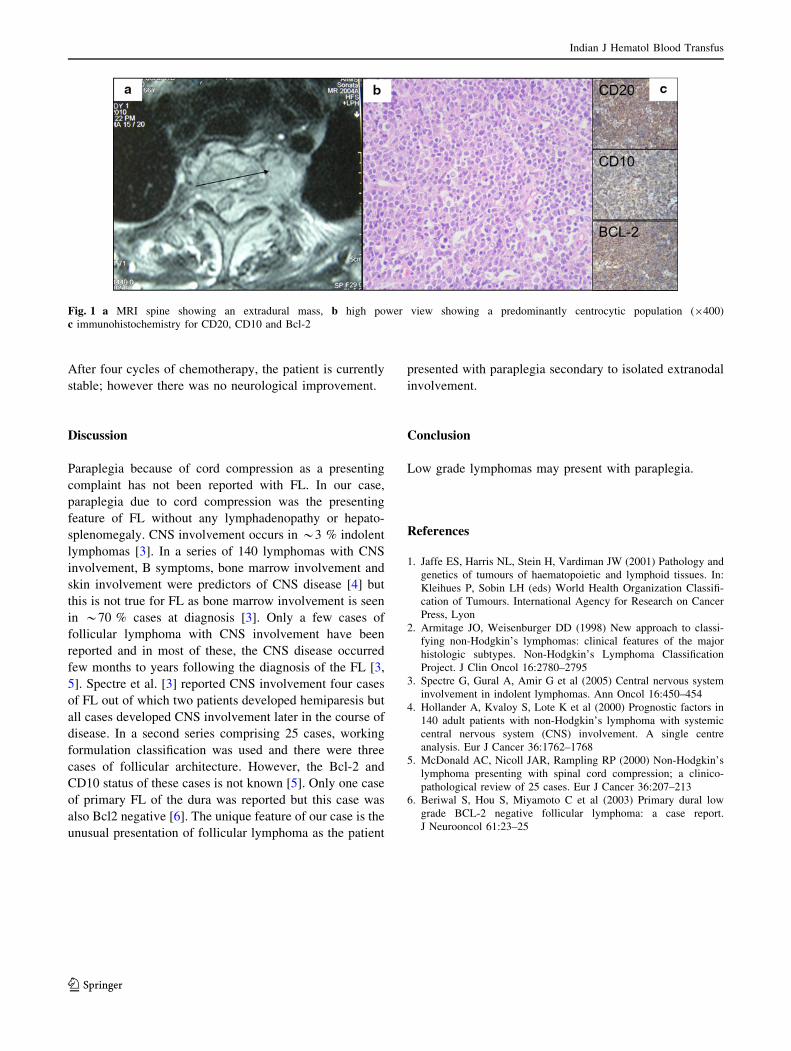
Fig. 1 a MRI spine showing an extradural mass, b high power view showing a predominantly centrocytic population (9400)
c immunohistochemistry for CD20, CD10 and Bcl-2
Indian J Hematol Blood Transfus
123