Embed Size (px)
DESCRIPTION
sinus paranasal
Citation preview
1
Paranasal Sinuses: Anatomy and Function
Glen T. Porter, MD
Francis B. Quinn, MD
UTMB Department of Otolaryngology
Galveston, TX
January 2002
2
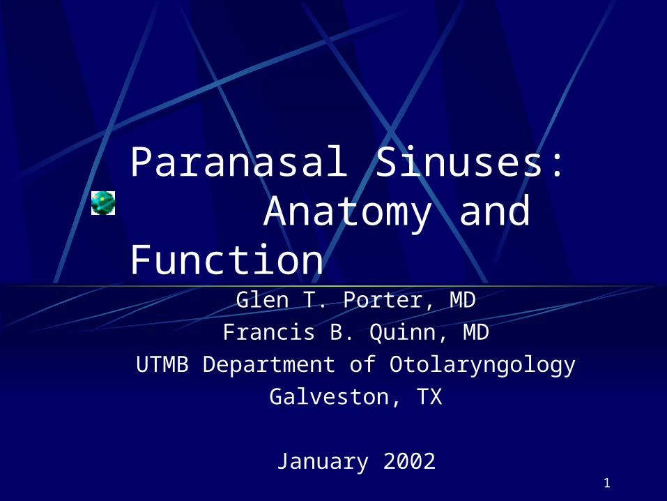
Case Report—1000B.C.
3
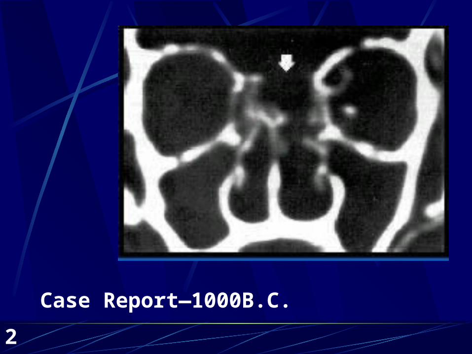
7 bones4 paired sinuses4 turbinates3 meatiDrainage systemNervous supplyVascular supplyRelated structures
Sinus Anatomy Overview
4
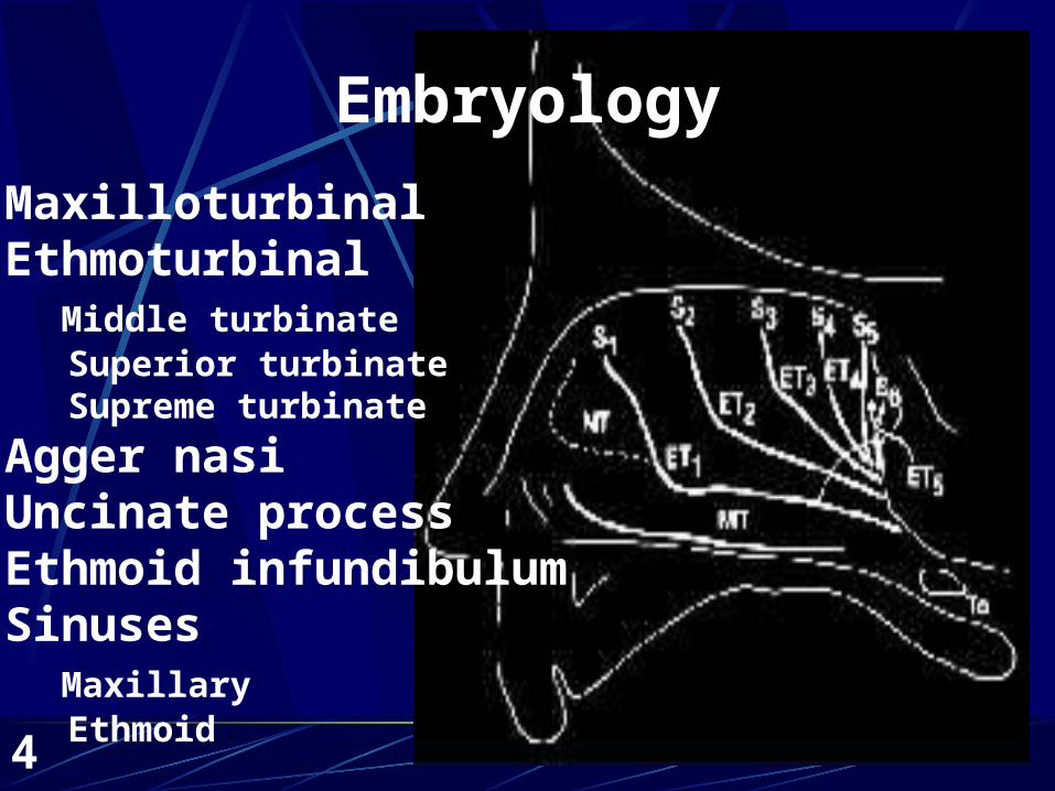
MaxilloturbinalEthmoturbinal Middle turbinate Superior turbinate Supreme turbinate
Agger nasiUncinate processEthmoid infundibulumSinuses Maxillary Ethmoid
Embryology
5
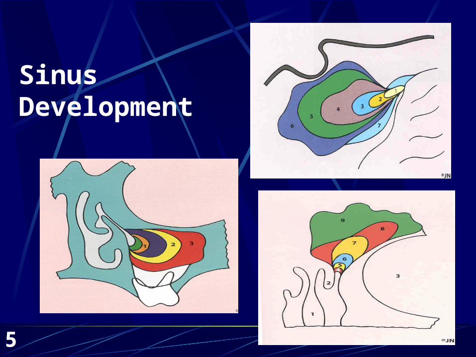
SinusDevelopment
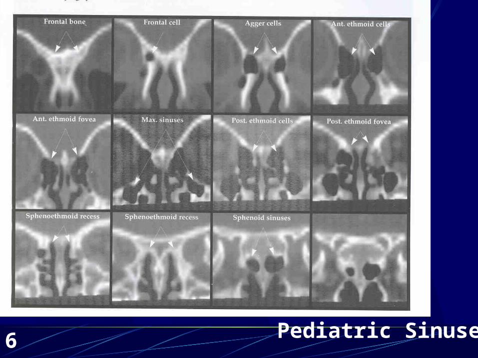
6Pediatric Sinuses
7
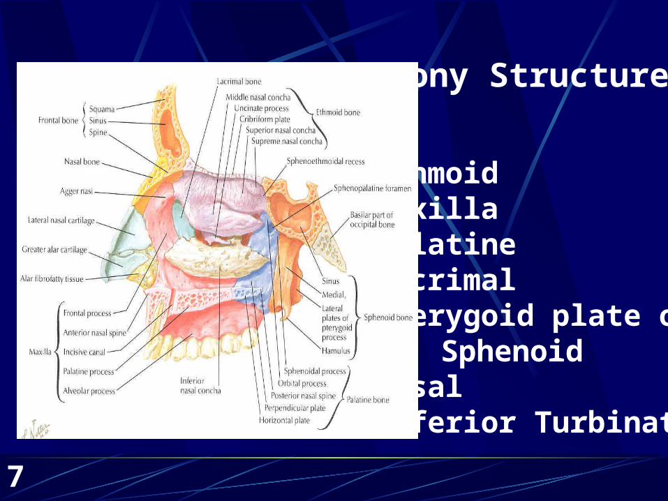
EthmoidMaxillaPalatineLacrimalPterygoid plate of
SphenoidNasal Inferior Turbinate
Bony Structure
8
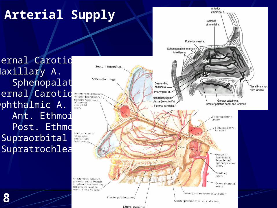
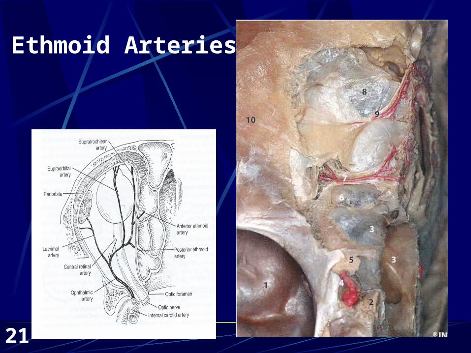
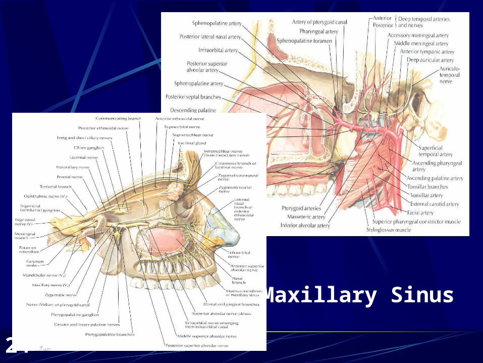
Arterial Supply
External Carotid Maxillary A. SphenopalatineInternal Carotid Ophthalmic A. Ant. Ethmoid Post. Ethmoid Supraorbital Supratrochlear
9
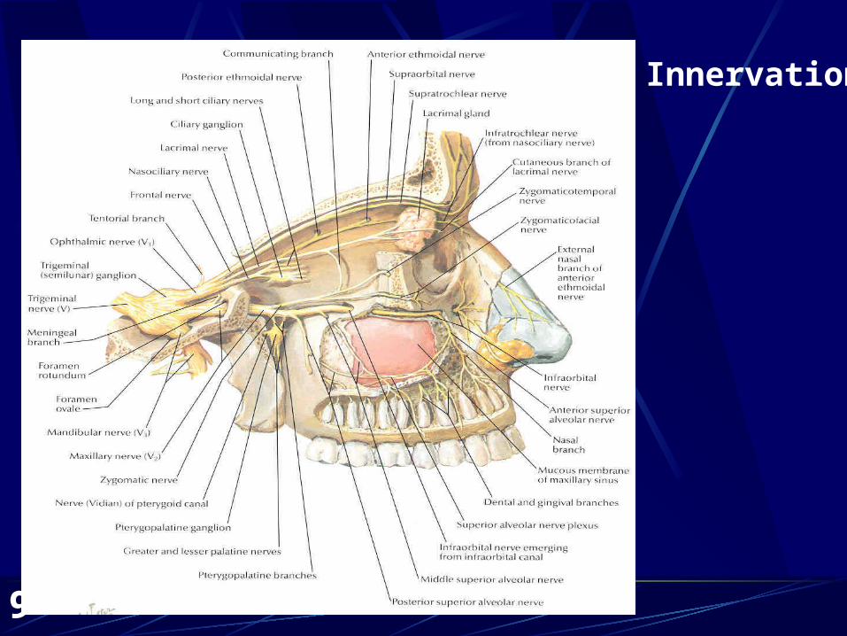
Innervation
10
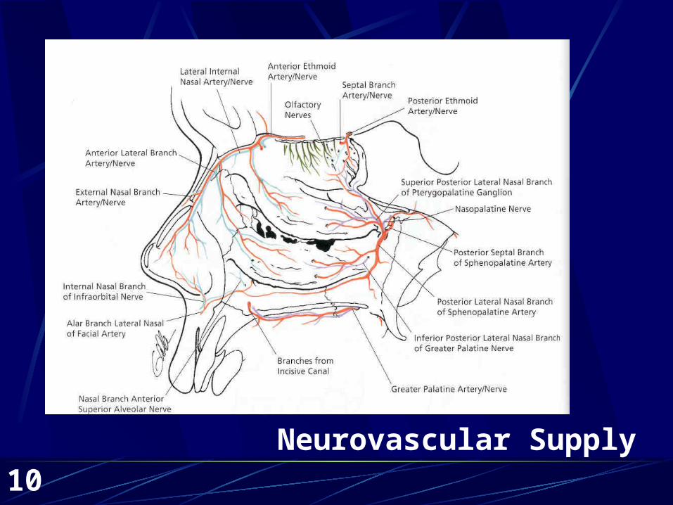
Neurovascular Supply
11
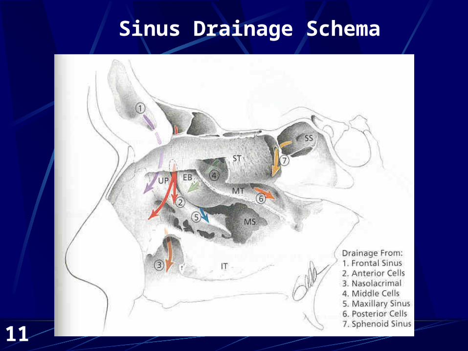
Sinus Drainage Schema
12
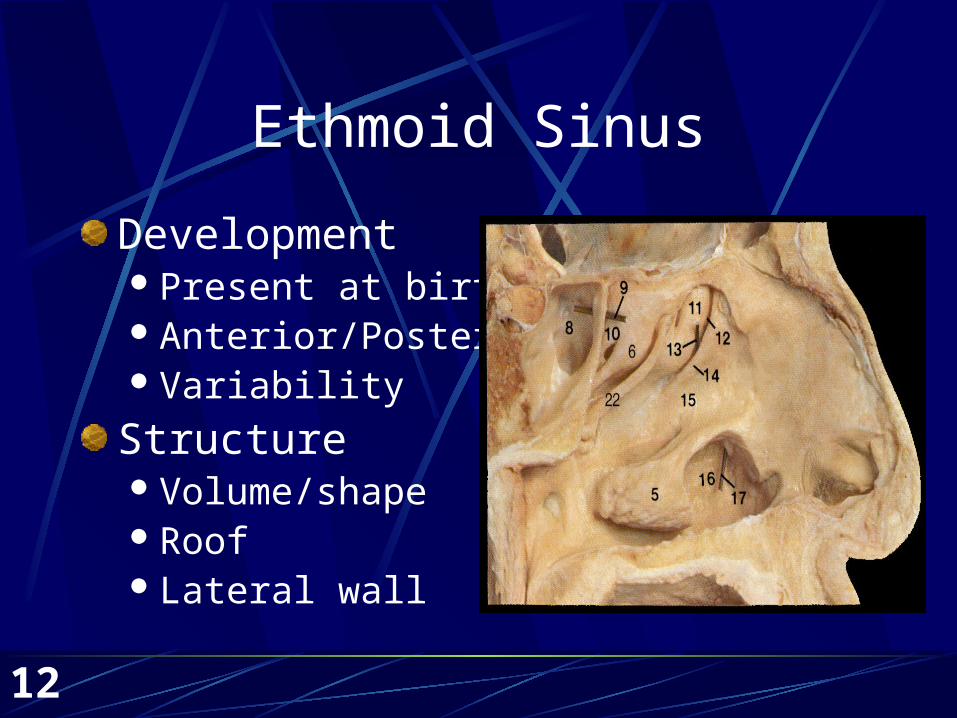
Ethmoid Sinus
DevelopmentPresent at birthAnterior/PosteriorVariability
StructureVolume/shapeRoofLateral wall
13
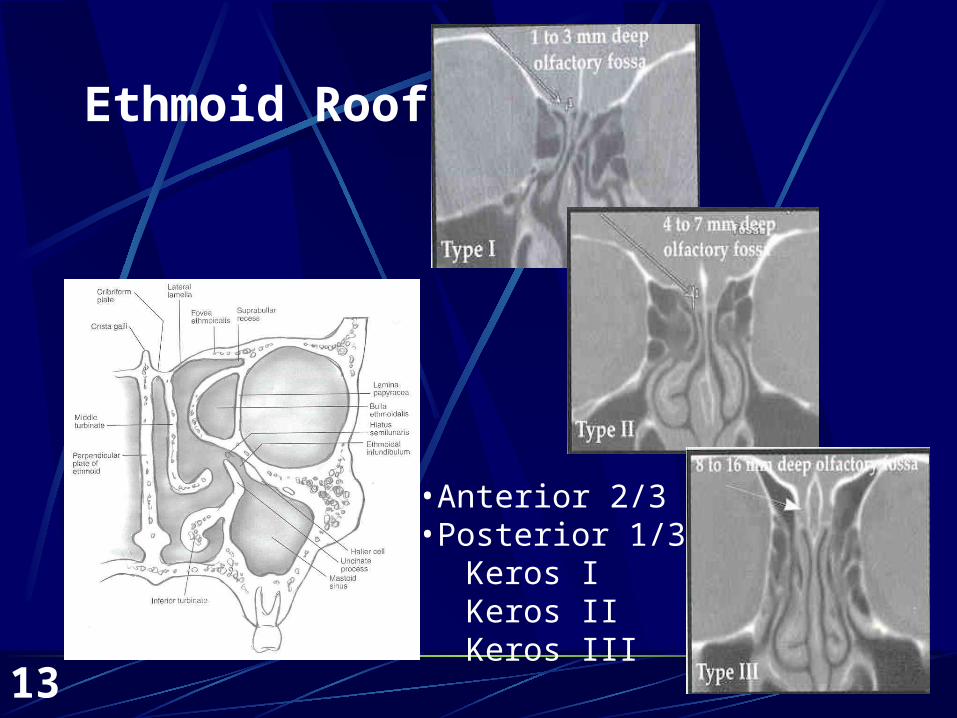
Ethmoid Roof
•Anterior 2/3•Posterior 1/3
Keros IKeros IIKeros III
14
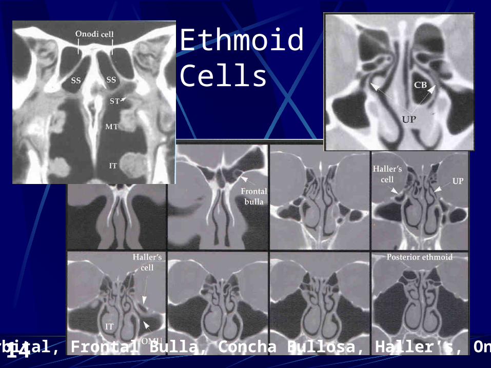
Ethmoid Cells
Supraorbital, Frontal Bulla, Concha Bullosa, Haller’s, Onodi Cells
15
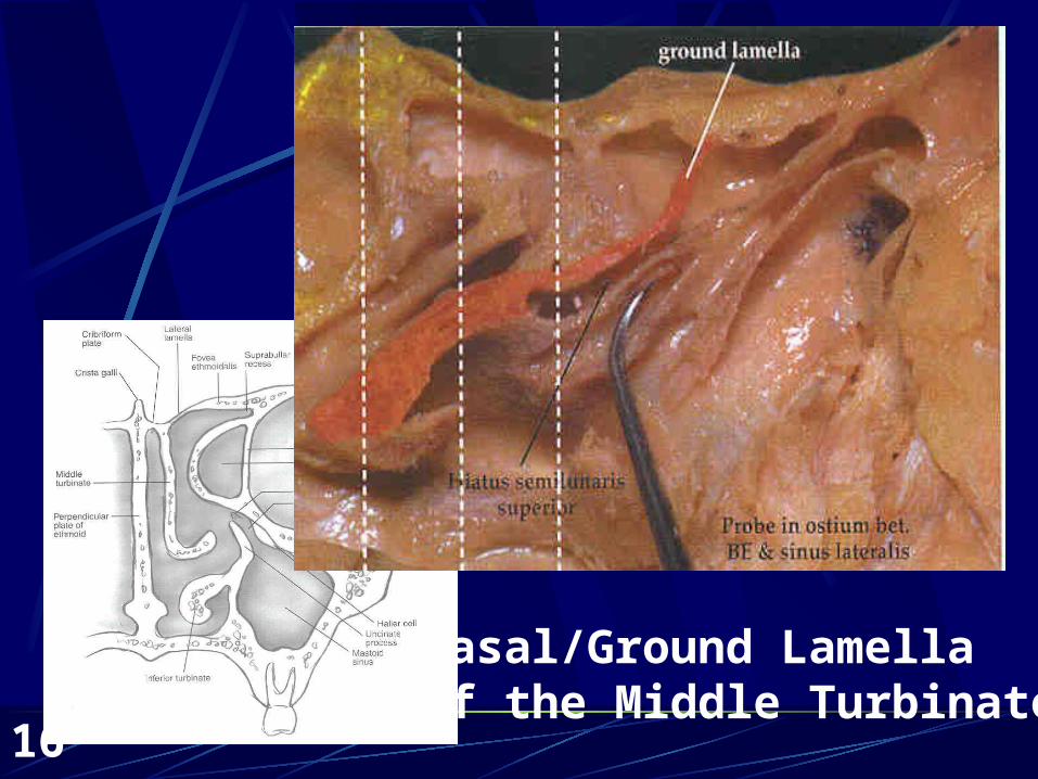
Ethmoid Sinus—Related Structures
Basal Lamella of the Middle Turbinate Three planes
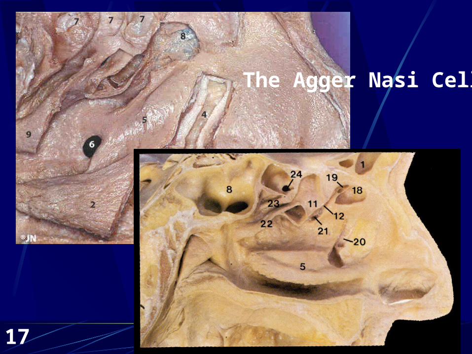
Agger nasi cell Childhood sinus
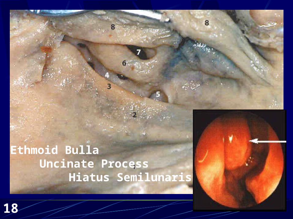
Ethmoid Bulla Hiatus Semiluninaris/Superior Hiatus Semilunaris Suprabullar/retrobullar recesses (Sinus Lateralis)
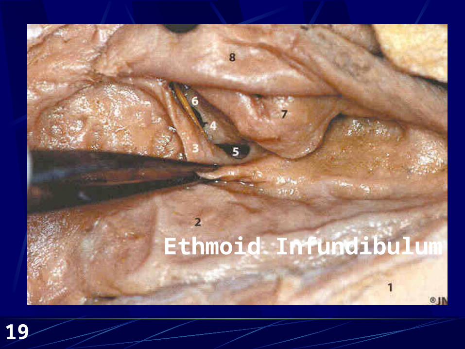
Ethmoid Infundibulum/Uncinate ProcessAnterior/Posterior Ethmoid ArteriesOsteomeatal complex
16
Basal/GroundLamella
Basal/Ground LamellaOf the Middle Turbinate
17
The Agger Nasi Cell
18
Ethmoid BullaUncinate Process
Hiatus Semilunaris
19
Ethmoid Infundibulum
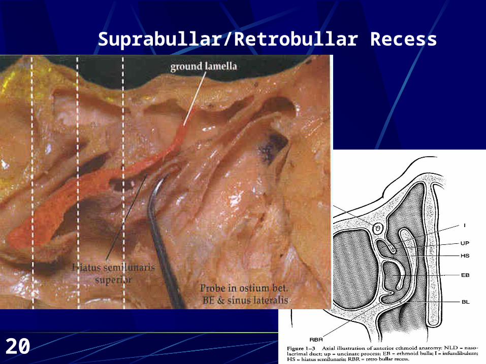
20
Suprabullar/Retrobullar Recess
21
Ethmoid Arteries
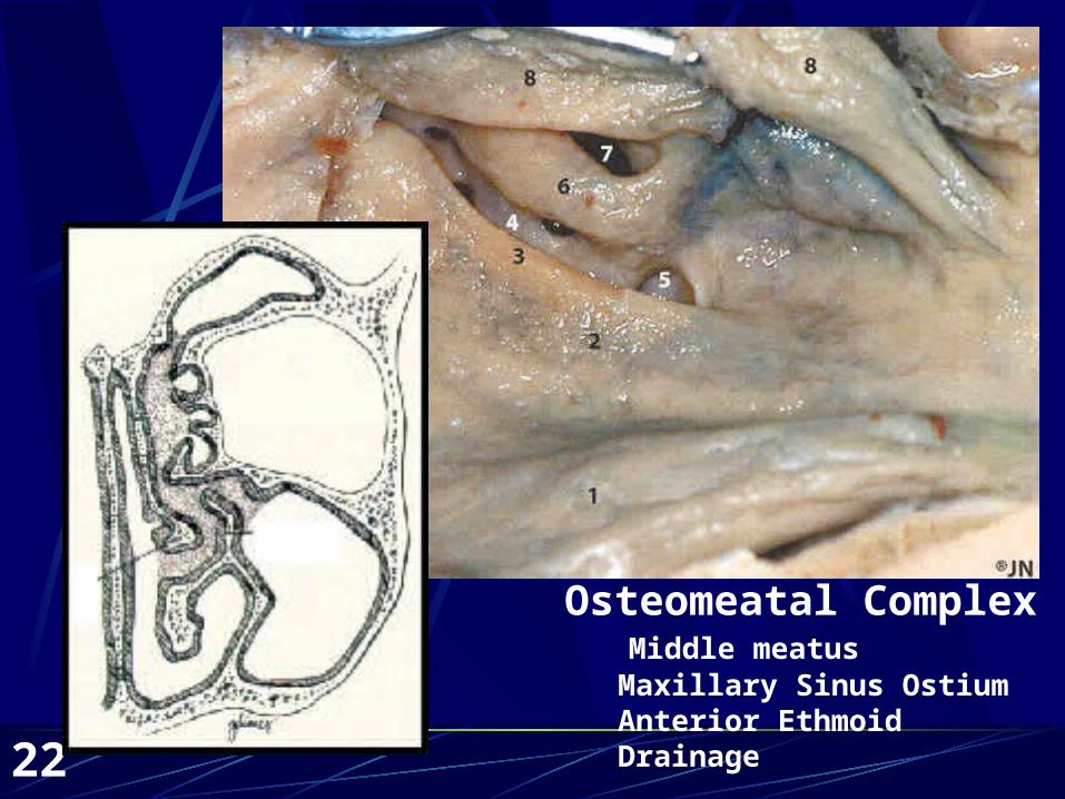
22
Osteomeatal Complex Middle meatus Maxillary Sinus Ostium Anterior Ethmoid Drainage
23
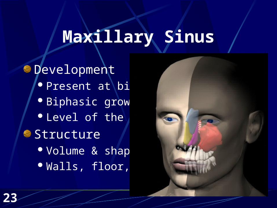
Maxillary Sinus
DevelopmentPresent at birthBiphasic growthLevel of the floor
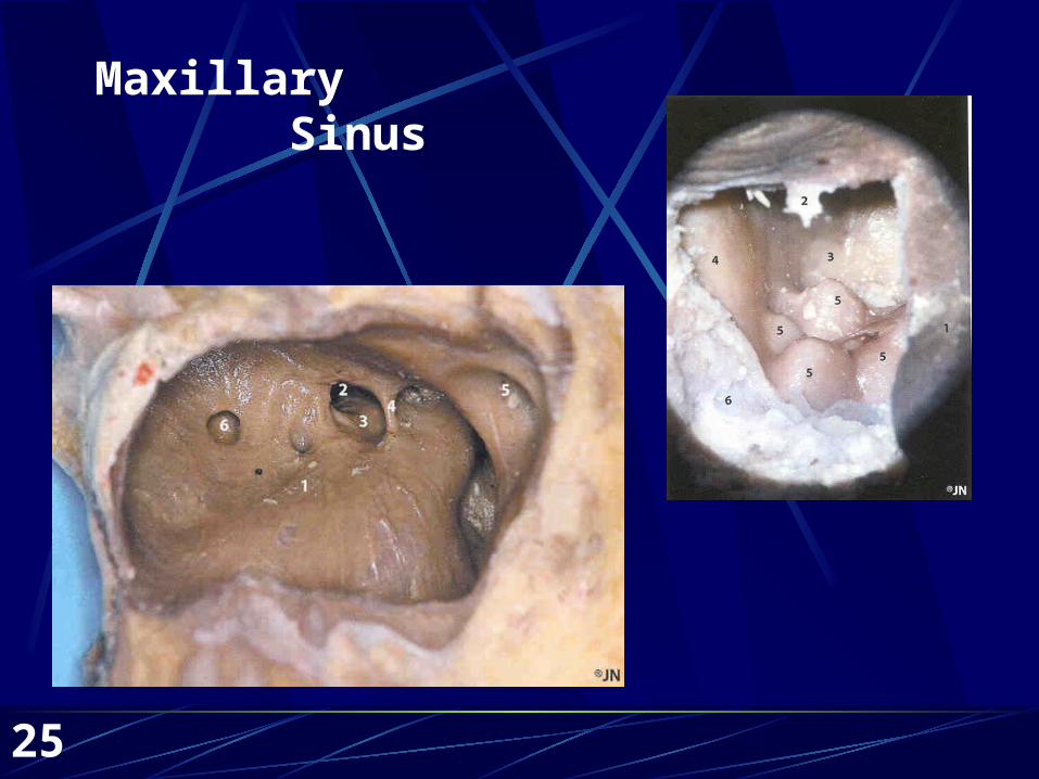
StructureVolume & shapeWalls, floor, roof
24
Maxillary Sinus
25
Maxillary Sinus
26
Maxillary Sinus
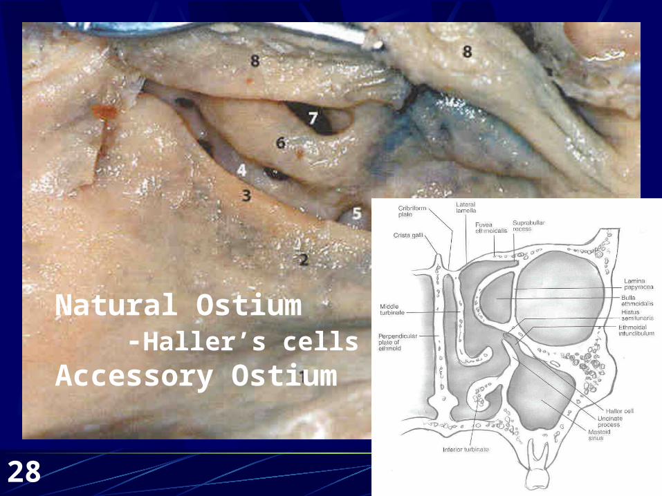
Related StructuresFontanellesNatural ostium
Haller’s Cells &SinusitisOsteomeatal complex
Accessory OstiumNasolacrimal duct
27
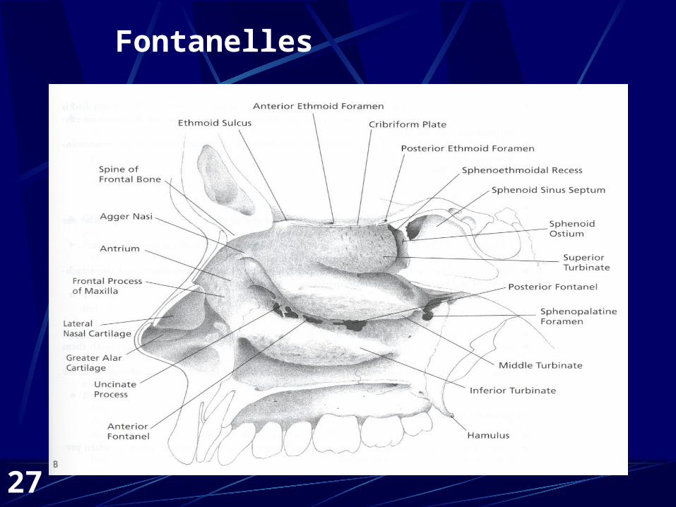
Fontanelles
28
Natural Ostium -Haller’s cellsAccessory Ostium
29
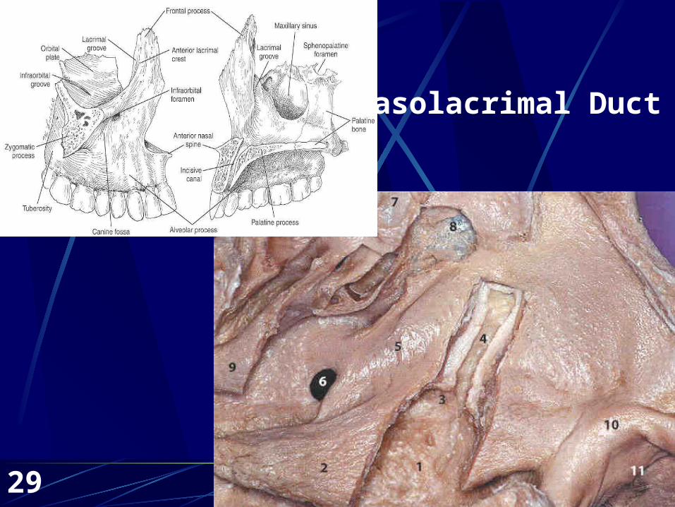
Nasolacrimal Duct
30
Frontal Sinus
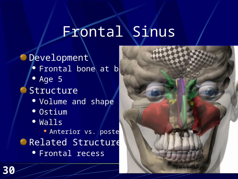
Development Frontal bone at birth Age 5
Structure Volume and shape Ostium Walls
Anterior vs. posterior
Related Structures Frontal recess
31
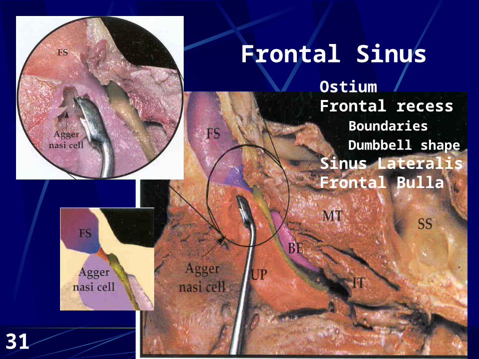
Frontal SinusOstiumFrontal recess Boundaries
Dumbbell shape
Sinus LateralisFrontal Bulla

32
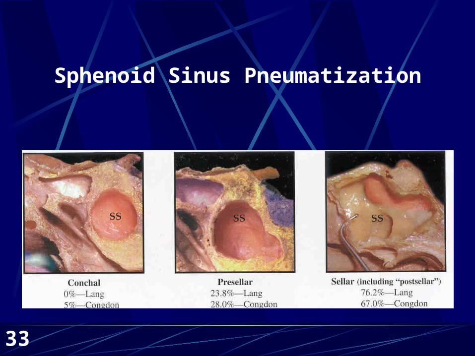
Sphenoidal Sinus
DevelopmentArise within the nasal capsule (no pouch)
Age 3 begins to pneumatize
Structure Volume/variable pneumatization Wall thickness Position within the sphenoid
Relation to sella turcica Sellar and postsellar relationships
33
Sphenoid Sinus Pneumatization
34
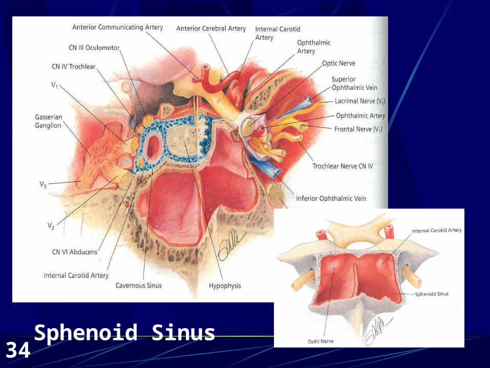
Sphenoid Sinus
Sphenoid Sinus
35
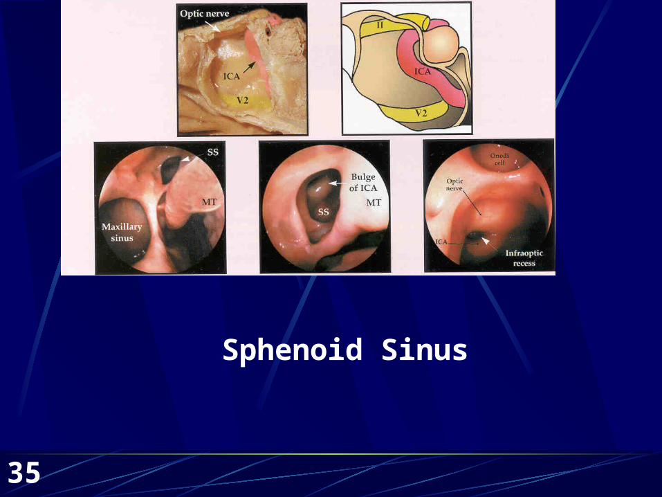
Sphenoid Sinus
36
Sphenoid Sinus
Ostium Size (.5-4mm) Location (sinus floor, anterior nasal floor, anterior
sinus wall, superior turbinate, cribiform plate) Bony dehiscence
Related Structures Sphenoethmoidal recess Sphenoid rostrum Onodi cell
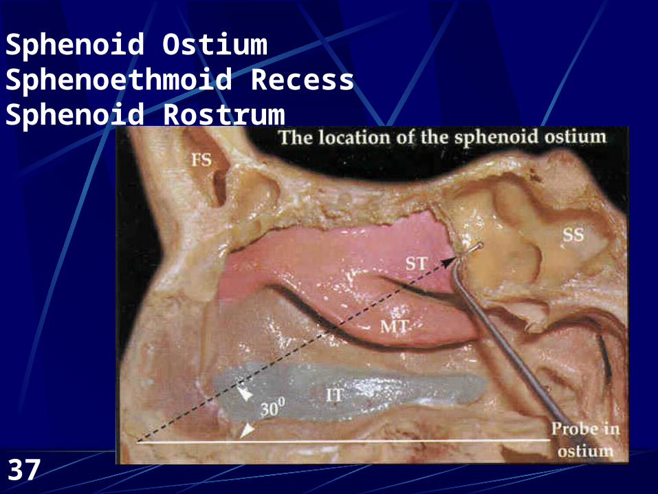
37
Sphenoid OstiumSphenoethmoid RecessSphenoid Rostrum
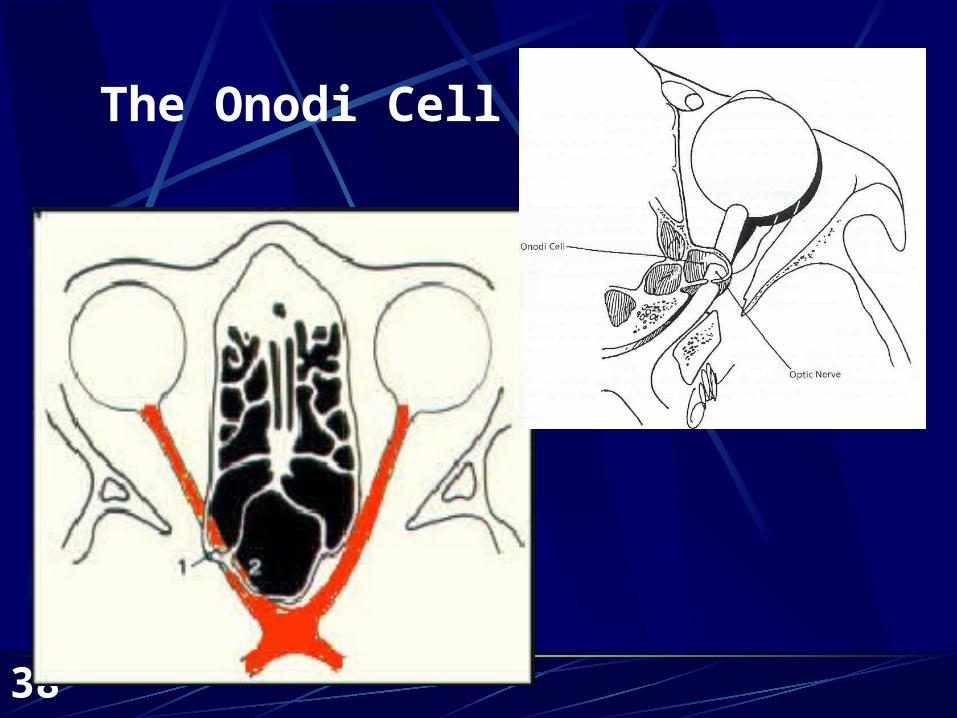
38
The Onodi Cell
39
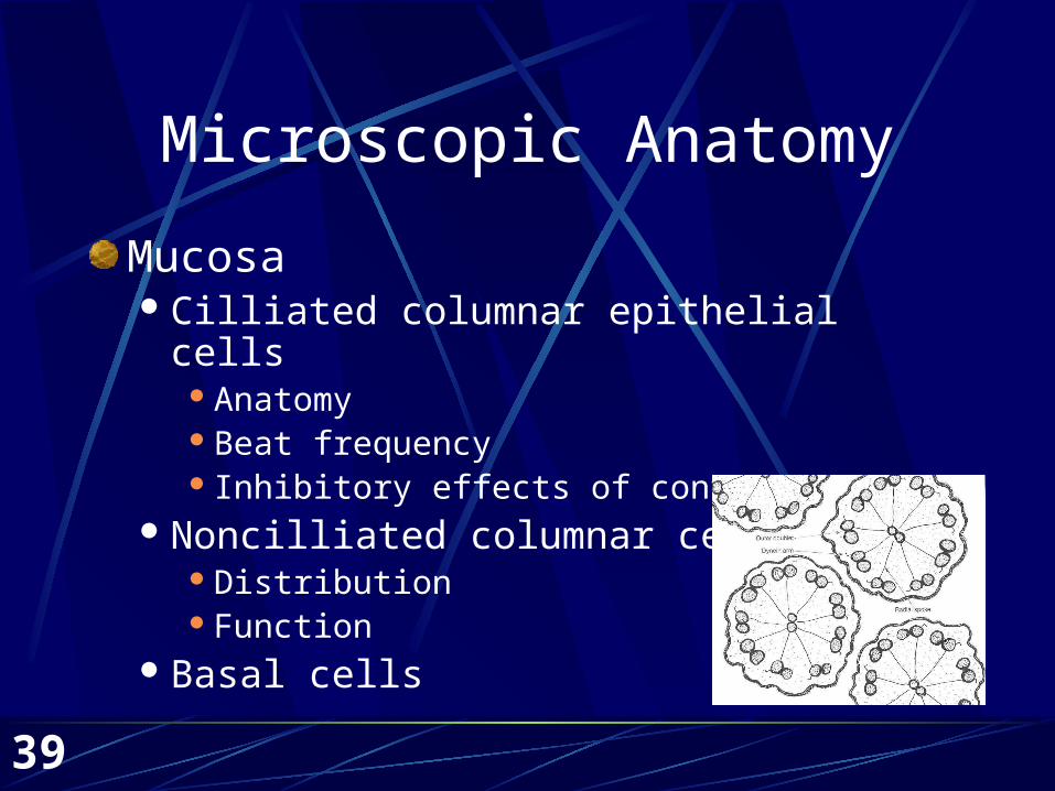
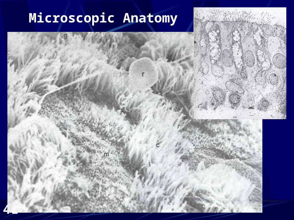
Microscopic Anatomy
MucosaCilliated columnar epithelial cells
AnatomyBeat frequency Inhibitory effects of contact
Noncilliated columnar cellsDistributionFunction
Basal cells
40
Microscopic Anatomy—Cont’d
Goblet CellsGlycoproteins—viscosity and elasticity Innervation (para=thick, symp=thin)
Basement membraneSubmucosal glands
Distribution
41
Microscopic Anatomy
42
Mucous Blanket
Two layersSuperficial layerSol layer
FunctionSuperficial layer traps bacteria and
particulate matter.Enzymes, antibodies, immune cells
43
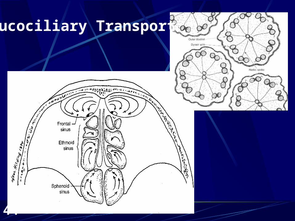
Mucociliary Transport
Directional Flow of MucousToward the choanae
Ostium drainage—a stubborn beastHilding, MD
Contact inhibitionHaller’s cellsSurgery
44
Mucociliary Transport
45
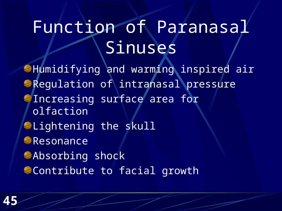
Function of Paranasal Sinuses
Humidifying and warming inspired air
Regulation of intranasal pressure
Increasing surface area for olfaction
Lightening the skull
Resonance
Absorbing shock
Contribute to facial growth
46
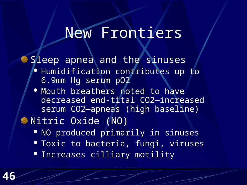
New Frontiers
Sleep apnea and the sinuses Humidification contributes up to 6.9mm Hg serum
pO2 Mouth breathers noted to have decreased end-tital
CO2—increased serum CO2—apneas (high baseline)
Nitric Oxide (NO) NO produced primarily in sinuses Toxic to bacteria, fungi, viruses Increases cilliary motility
47
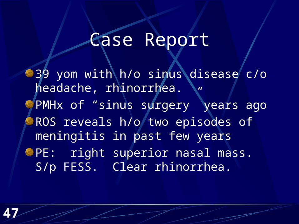
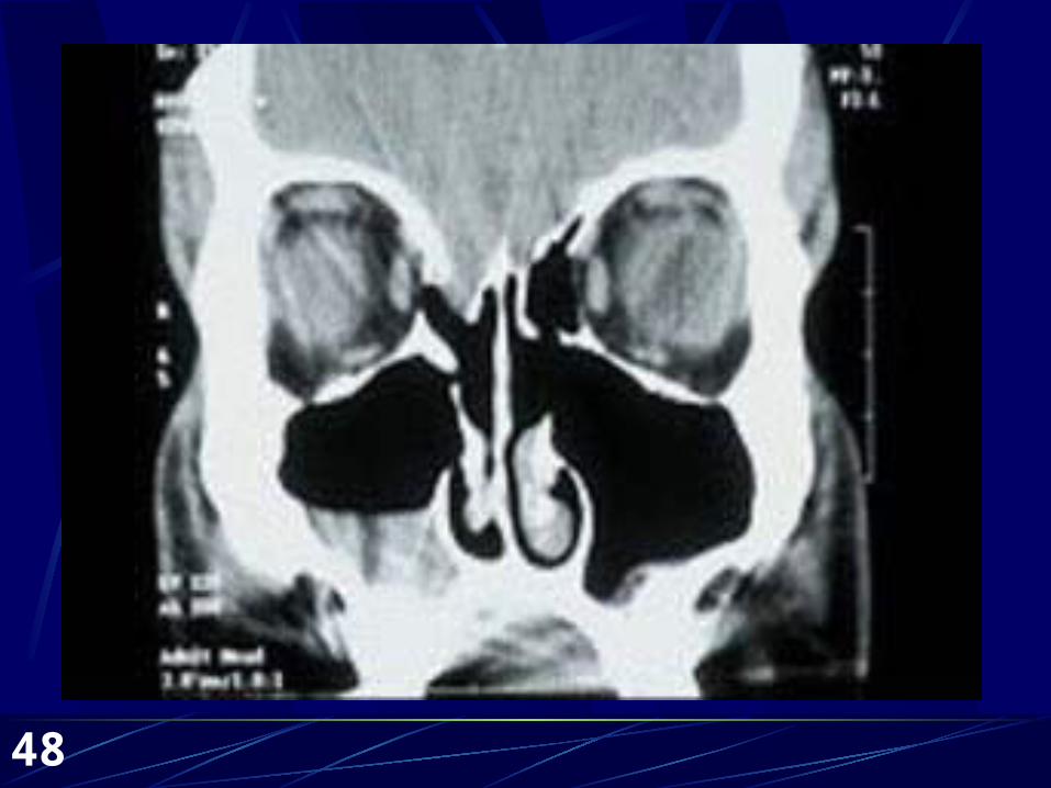
Case Report
39 yom with h/o sinus disease c/o headache, rhinorrhea.
PMHx of “sinus surgery” years ago
ROS reveals h/o two episodes of meningitis in past few years
PE: right superior nasal mass. S/p FESS. Clear rhinorrhea.
48
49
References
Anon, Jack B., et al, Anatomy of the Paranasal Sinuses, Theime, New York, c1996. Bhatt, Nikhil J., Endoscopic Sinus Surgery: New Horizons, Singular Publishing Group, Inc., San Diego, c1997. Bailey, Byron J., et al, Head & Neck Surgery -- Otolaryngology, Lippincott Williams & Wilkins, Philadelphia, c2001. Lundberg, J., Weitzberg, E. Nasal Nitric Oxide in Man. Thorax 1999; 54(10):947-952. McCaffrey, Thomas V., Rhinologic Diagnosis and Treatment, Thieme, New York, c1997. Marks, Steven C. Nasal and Sinus Surgery, W.B. Saunders Co., Philadelphia, c2000. Navarro, Joao A.C., The Nasal Cavity and Paranasal Sinuses, Springer, Berlin, c2001. Watelet, J.B., Cauwenberge P. Van, Applied Anatomy and Physiology of the Nose and Paranasal Sinuses. Allergy 1999; 54, Supp 57:14-25.