Embed Size (px)
Citation preview

22
IJOI 28 iAOI CASE REPORT
Introduction
Mini-implant anchorage has been proven to be an effective therapeutic strategy in treating various kinds of malocclusions.1-13 It can be applied in many clinical orthodontic conditions successfully, including maximal retraction in protrusion cases, Class I I correction, Class I I I correction, molar distalization in crowding cases, molar intrusion in molar elongation cases, deep bite correction, open bite correction, midline correction, and the correction of occlusal plane canting and posterior crossbite.1-13
Class III malocclusions are commonly found in Asian population and bimaxillary dentoalveolar protrusion is another common characteristic in oriental races. One of the critical considerations for Class III treatment is to determine whether patients’ profile will become protrusive after anterior crossbite correction. If the answer is yes, then maybe extraction is a better option for treatment plan. With the help of mini-implant anchorage, Class IIII malocclusion can be treated successfully with a nonextraction approach without subsequent perioral protrusion. This paradigm shift in treatment of Class III malocclusion greatly reduces treatment duration and achieves more pleasing profile change after anterior crossbite correction.
The most common position to place mini-implants is on the upper posterior area. It is not only because
Paradigm Shift in Class III Treatment with TADs
█ Fig. 2: Pretreatment intraoral photographs
█ Fig. 1: Pretreatment facial photographs
█ Fig. 3: Pretreatment study models
23
Paradigm Shift in Class III Treatment with TADs IJOI 28
of anchorage reinforcement required in the upper arch but also more favorable anatomical situations of attached gingiva and surrounding movable mucosa.14,15 The conventional use of mini-implant anchorage in the lower posterior area is either interdental minicrews or exo-dentitional miniplates. Interdental miniscrews are primarily for maximal anterior retraction and vertical control. Screw insertion in the attached gingival area is less skill-intensive, but not suitable for whole dentition distalization because of possible interference with root movement. Meanwhile, miniplates require two mini-screws to secure the position in the exo- dentitional bone and can be extended to the proper position for anchorage.16,17 Whole dentition movement is no longer restrained by the mini-implant itself. However, this procedure requires a flap surgery which often causes significant patient discomfort.
In this article the authors propose an alternative use of exo-dentitional miniscrews in the mandible to treat a Class III malocclusion by full lower dentition distalization with a nonextraction approach.
History And Etiology
A 24 year old female pat ient requested for orthodontic treatment with the chief complaint of mandibular prognathism and anterior crossbite.
Johnny JL Liaw, Director, Beauty Forever Dental Clinic (left)W. Eugene Robert, Consultant,
International Journal of Orthodontics & Implantology (right)
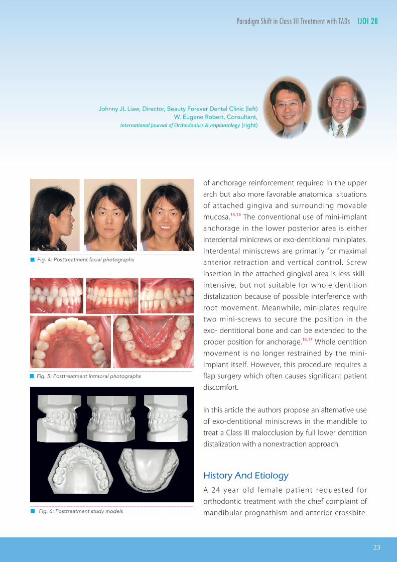
█ Fig. 4: Posttreatment facial photographs
█ Fig. 5: Posttreatment intraoral photographs
█ Fig. 6: Posttreatment study models

24
IJOI 28 iAOI CASE REPORT
█ Fig. 9:
Cephalometric superimpositions showed full dentition retraction in the lower arch and the mandible rotated backward sightly so the profile became more orthognathic. The upper incisors were mildly flared out in despite of severe crowding in the upper arch. During alignment and correction of the anterior crossbite, the upper incisors were quite flared out as seen in Fig. 20 and were retracted back to nearly the original positions by the upper TADs.
█ Fig. 8: Posttreatment pano and ceph radiographs █ Fig. 7: Pretreatment pano and ceph radiographs
25
Paradigm Shift in Class III Treatment with TADs IJOI 28
Diagnosis
Skeletal: • Skeletal Class III ( ANB: -2˚) • Average mandibular plane angle ( SN-MP: 32˚)
Dental: • Anterior crossbite • Deep overbite • Class III molar and canine relationships on the
right • Molar Class I and canine Class III on the left • Endodontically treated on #11, 15 • Horizontal impactions of bilateral lower wisdom
teeth
Facial: • Concave profile with lower lip everted.
The ABO Discrepancy Index (DI) was 26 as shown in the subsequent worksheet.
Specific Objectives Of Treatment
Maxilla ( all three planes): • A - P: Maintain • Vertical: Maintain • Transverse: Maintain
Mandible ( all three planes): • A - P: Maintain • Vertical: Slightly increase • Transverse: Maintain
Maxillary Dentition: • A - P: Slightly advance • Vertical: Slightly increase • Transverse: Maintain
Mandibular Dentition: • A - P: Retract lower dentition • Vertical: Maintain • Transverse: Maintain
Her extraoral frontal photograph showed no obvious asymmetry. Her vertical proportion was within normal limit. Upper anterior malalignment was obvious in the smiling view. The lateral view showed a concave profile because of mandibular prognathism (Fig. 1). Anterior crossbite and deep bite can be observed in the intraoral frontal photographs. Dental midline discrepancy was also noted. Arch length discrepancy in the upper arch was 6.5mm and 1.5mm in the lower arch. The upper right second molar was missing. Molar Class III and canine Class III relationships were noted on the right side. Molar Class I and canine Class III were noted in the left side (Fig. 2, 3). The panoramic X-ray showed horizontal impaction of the two lower wisdom teeth, and upper right central incisor and upper right second premolar were endodontically treated. Cephalometric X-ray revealed a skeletal Class III relationship (ANB: -2). Mandibular plane angle was within normal range (SN-MP: 32). Dental compensation for skeletal Class III was noted ( U1-SN:
113˚, L1-MP: 83˚) (Fig. 7).
The patient reported that some family members had a prognathic jaw but it was not a common characteristic. She first noted the anterior crossbite when her permanent incisors erupted at the age of six. It was concluded that the etiology of the malocclusion was a genetic predisposition to a skeletal class III malocclusion complicated by ectopic eruption of the maxillary incisors.
This patient was treated with a nonextraction approach in conjunction with TADs. The treatment results were documented in Figs. 4-6. The pre-treatment and post-treatment radiographs were shown in Figs. 7-8. The cephalometric tracings before and after treatment are superimposed in Fig. 9.
26
IJOI 28 iAOI CASE REPORT
Facial Esthetics: • Improve the facial profile by increasing the
upper lip support and lower lip retraction
Treatment Plan
Two options were proposed.
Option 1: Extraction of upper second premolars, lower f irst premolars and lower horizontally impacted wisdom teeth.
The treatment goal for upper second premolar extraction is to relieve upper anterior crowding while extraction of lower first premolar aims to correct anterior crossbite. The proposed extraction pattern was for anchorage consideration of molar Class III correction.
Option 2: Extraction of lower horizontally impacted wisdom teeth only.
Mini-implant anchorage will be used to retract the whole lower dentition. After correction of anterior crossbite, mini-implant anchorage will be used to retract upper and lower dentition simultaneously.
After thorough discussion and communication, option 2 was accepted and reevaluation would be made after the occlusion was corrected. If the profile was too protrusive, then four bicuspids extraction would be considered as the back-up treatment plan.
As to the missing upper right second molar, implant prosthesis was proposed and was to be decided later in the treatment.
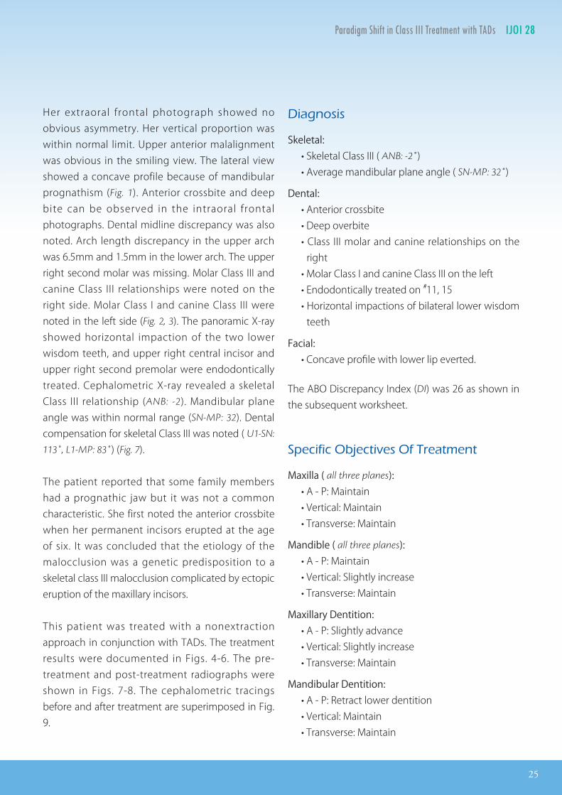
Appliances And Treatment Progress
The orthodontic treatment started after the removal of the lower wisdom teeth.
Start (5-14-04): After extraction of mandibular 3rd molars, the maxillary arch was bonded with Damon 2 brackets (Ormco Corp., Orange, CA) and the initial archwire was a .014” CuNiTi. A customized bite turbo was bonded on the lingual surface of lower left central incisor to avoid bracket interference during the initial maxillary anterior alignment (Fig. 10). Since there was no significant functional shift, it was necessary to open the bite for about 6mm at the incisors. The patient was instructed to pursue a soft diet until posterior occlusion was restored.
9 weeks (7-19-04): Mandibular arch was bonded with Damon 2 brackets and a .014” CuNiTi archwire was inserted. Two stainless steel miniscrews (OrthoBoneScrew, Newton’s A, Inc. 2x12mm) were inserted on the buccal shelves of the mandible bilaterally. Two NiTi coil springs of 150gm were attached from the head of the miniscrews to the brackets of mandibular canines (Fig. 11).
12 weeks (8-6-04): Both archwires were changed to . 016”x.025”CuNiTi.
16 weeks (9-4-04): The anterior crossbite was corrected to an edge-to-edge relationship. An elastic chain extended from mandibular canine to canine and bilateral NiTi coil springs were attached from the mandibular canines to the miniscrews to retract the entire mandibular dentition (Fig. 12).

27
Paradigm Shift in Class III Treatment with TADs IJOI 28
█ Fig. 10:
A bite turbo was bonded on the lingual surface of lower left central incisor to avoid occlusal interference with upper brackets on the first day of upper bonding.
█ Fig. 11:
Miniscrews were inserted on the buccal shelves of mandible for lower canine retraction.
█ Fig. 12:
An elastic chain extended from mandibular canine to canine and bilateral NiTi coil springs were attached from the mandibular canines to the miniscrews to retract the entire mandibular dentition.
0
169
28
IJOI 28 iAOI CASE REPORT
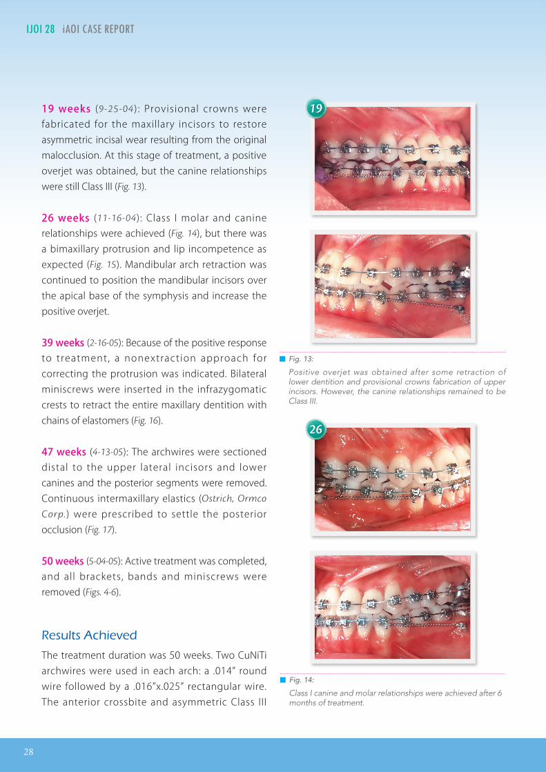
19 weeks (9-25-04 ) : Provisional crowns were fabricated for the maxillary incisors to restore asymmetric incisal wear resulting from the original malocclusion. At this stage of treatment, a positive overjet was obtained, but the canine relationships were still Class III (Fig. 13).
26 weeks (11-16-04): Class I molar and canine relationships were achieved (Fig. 14), but there was a bimaxillary protrusion and lip incompetence as expected (Fig. 15). Mandibular arch retraction was continued to position the mandibular incisors over the apical base of the symphysis and increase the positive overjet.
39 weeks (2-16-05): Because of the positive response to treatment, a nonextraction approach for correcting the protrusion was indicated. Bilateral miniscrews were inserted in the infrazygomatic crests to retract the entire maxillary dentition with chains of elastomers (Fig. 16).
47 weeks (4-13-05): The archwires were sectioned distal to the upper lateral incisors and lower canines and the posterior segments were removed. Continuous intermaxillary elastics (Ostrich, Ormco
Corp.) were prescribed to settle the posterior occlusion (Fig. 17).
50 weeks (5-04-05): Active treatment was completed, and all brackets, bands and miniscrews were removed (Figs. 4-6).
Results Achieved
The treatment duration was 50 weeks. Two CuNiTi archwires were used in each arch: a .014” round wire followed by a .016”x.025” rectangular wire. The anterior crossbite and asymmetric Class III
█ Fig. 13:
Positive overjet was obtained after some retraction of lower dentition and provisional crowns fabrication of upper incisors. However, the canine relationships remained to be Class III.
█ Fig. 14:
Class I canine and molar relationships were achieved after 6 months of treatment.
19
26

29
Paradigm Shift in Class III Treatment with TADs IJOI 28
█ Fig. 16:
Two miniscrews were installed on the upper posterior areas, in conjunction with the miniscrews on bilateral buccal shelves, to retract both arches simultaneously.
█ Fig. 17:
The archwires were cut distal to the upper lateral incisors and lower canines. The posterior segments were removed. Up-and -down finishing elastics were prescribed to settle the occlusion.
█ Fig. 15:
Bimaxillary protrusion was noted after the correction of anterior crossbite.
buccal relationships were corrected (Figs. 5-6), and a pleasing, more orthognathic profile was achieved (Fig. 4). The post-treatment panoramic radiograph documents normal root parallelism and good maintenance of supporting alveolar bone (Fig. 8). The mandibular right 2nd molar was excessively tipped distally as the dentition was retracted; additional treatment was not needed because extraction of the unopposed tooth was planned.
The post-treatment cephalogram documents an acceptable orthognathic profile, but the ANB angle improved to only -2°. The skeletal response was typical for a camouflage treatment of a Class III skeletal malocclusion: increased vertical dimension of occlusion, flaring of the maxillary incisors and decreased inclination of the mandibular incisors. Accordingly, the cephalometric analysis showed that the mandibular plane angle was increased ~10 (Tab.
1). Clockwise mandibular rotation was noted in the cephalometric superimposition (Fig. 9). Consistent with camouflage treatment, the axial inclination of the maxillary incisors increased and the mandibular incisors decreased (Tab. 1).
39
47

30
IJOI 28 iAOI CASE REPORT
█ Fig. 18:
Serial photographs during treatment:A. Profile before treatment.B. Profile in the 6th month of treatment when the anterior crossbite was just corrected.C. Profile in the 8th month of treatment after upper TADs were placed.D. Profile after treatment.
█ Fig. 19:
Corresponding serial radiographs.A. Pre-treatment cephalogram.B. Cephalogram in the 6th month when the anterior crossbite was just corrected.C. Cephalogram in the 8th month after upper TADs were inserted.D. Post-treatment cephalogram.
0 6 8 12
A B C D
0 6 8 12
A B C D

31
Paradigm Shift in Class III Treatment with TADs IJOI 28
Maxilla ( all three planes): • A - P: Maintained • Vertical: Maintained • Transverse: Maintained
Mandible ( all three planes): • A - P: Maintained • Vertical: Slightly increased • Transverse: Maintained
Maxillary Dentition: • A - P: Slightly advanced • Vertical: Slightly increased • Transverse: Maintained
Mandibular Dentition: • A - P: Lower dentition retracted • Vertical: Maintained • Transverse: Maintained
Facial Esthetics: • Improved by increasing upper lip support and
retracting lower lip
Retention
Upper and lower clear retainers were delivered, and the patient was instructed to wear the retainers
█ Fig. 20:
The superimpositions of the Pre-treatment and the 6th month cephalograms showed the upper incisors were significantly flared out after anterior crossbite was corrected. The profile became more protrusive after the correction of the anterior crossbite.
█ Fig. 21:
The superimpositions of the 8th month and post-treatment cephalograms showed both dentitions were retracted by TADs to correct the mid-treatment protrusion.
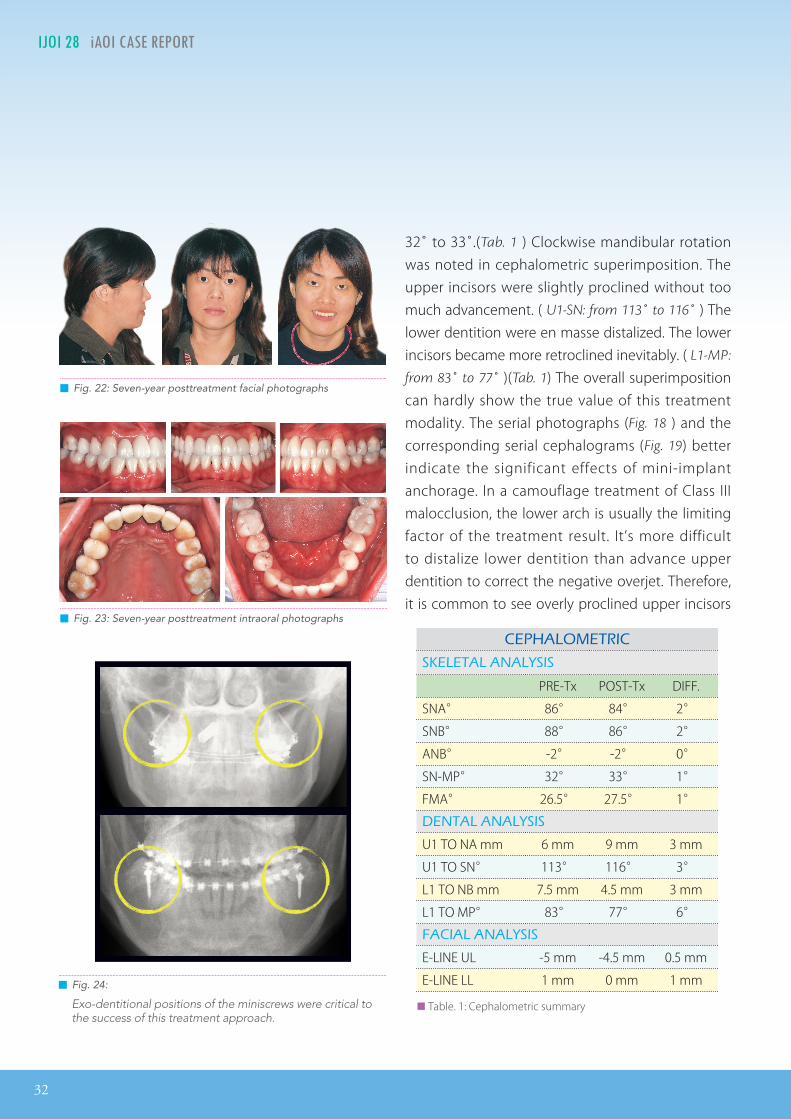
full time for first 6 months and night time only thereafter. The retainers were renewed after the permanent prosthesis were fabricated. Seven-year post-treatment records of the present patient show satisfactory stability (Figs 22, 23).
Final Evaluation Of Treatment
The ABO Cast-Radiograph Evaluation score was 11 points. The major discrepancies were in the left occlusal relationships, alignment/rotation, and marginal ridges. It was mainly contributed to the space regaining for block-out canine. More distal root movements were needed to upright the root angulations of upper left posterior teeth, which will
consequently improve the occlusal contacts at the distal parts of the dentitions.
Discussion
The post-treatment cephalogram looks more orthognathic profile, but the ANB angle is still -2˚. The mandibular plane angle was increased from
32
IJOI 28 iAOI CASE REPORT
CEPHALOMETRIC
SKELETAL ANALYSIS
PRE-Tx POST-Tx DIFF.
SNA° 86° 84° 2°
SNB° 88° 86° 2°
ANB° -2° -2° 0°
SN-MP° 32° 33° 1°
FMA° 26.5° 27.5° 1°
DENTAL ANALYSIS
U1 TO NA mm 6 mm 9 mm 3 mm
U1 TO SN° 113° 116° 3°
L1 TO NB mm 7.5 mm 4.5 mm 3 mm
L1 TO MP° 83° 77° 6°
FACIAL ANALYSIS
E-LINE UL -5 mm -4.5 mm 0.5 mm
E-LINE LL 1 mm 0 mm 1 mm
██ Table. 1: Cephalometric summary
█ Fig. 24:
Exo-dentitional positions of the miniscrews were critical to the success of this treatment approach.
32˚ to 33˚.(Tab. 1 ) Clockwise mandibular rotation was noted in cephalometric superimposition. The upper incisors were slightly proclined without too much advancement. ( U1-SN: from 113˚ to 116˚ ) The lower dentition were en masse distalized. The lower incisors became more retroclined inevitably. ( L1-MP:
from 83˚ to 77˚ )(Tab. 1) The overall superimposition can hardly show the true value of this treatment modality. The serial photographs (Fig. 18 ) and the corresponding serial cephalograms (Fig. 19) better indicate the significant effects of mini-implant anchorage. In a camouflage treatment of Class III malocclusion, the lower arch is usually the limiting factor of the treatment result. It’s more difficult to distalize lower dentition than advance upper dentition to correct the negative overjet. Therefore, it is common to see overly proclined upper incisors
█ Fig. 22: Seven-year posttreatment facial photographs
█ Fig. 23: Seven-year posttreatment intraoral photographs

33
Paradigm Shift in Class III Treatment with TADs IJOI 28
combined with excessively retroclined lower incisors after a Class III camouflage treatment. In addition, the posttreatment profile tends to seem protrusive. If whole distalization of lower dentition with the miniscrews can be achieved during treatment, the excessive advancement of upper incisors can be avoided and the posttreatment profile may appear less protrusive. In this case, even with lower miniscrews, upper incisors advancement was still more significant than lower incisors distalization (Fig.
20). The profile remained protrusive after anterior crossbite correction. Another two miniscrews were then placed in the infrazygomatic crest to distalize the whole upper dentition and finally, a more orthognathic posttreatment profile was achieved (Fig. 21).
Treatment result of this case can be obtained either by four bicuspids extraction or by nonextraction treatment supported with mini-implant anchorage.18 The treatment time is reduced to 12 months with the nonextraction approach and mini-implant anchorage. Nonextraction treatment of some Class III malocclusion with mini-implant anchorage is not only conservative but also efficient. The criteria of the cases election for this approach is basically the same with camouflage treatment of Class III treatment. That is
1) mild to moderate skeletal discrepancy 2) no or less dental compensation 3) acceptable profile except perioral imbalance4) the last but not the least, clearance for whole
dentition distalization. Wisdom teeth should be removed, the skeletal boundaries of dentition should be verified carefully by radiographs and
soft tissue boundaries of dentition should be observed clinically.
The positions of the miniscrews are exo-dentitional, not in the interdental area (Fig. 24). This is critical in this nonextraction approach so that the miniscrews will not come in contact with the moving dentitions. The insertion points of the miniscrews on the maxilla is the attached gingival area of upper molars ranging from the mesial to distal interdental area of upper first molars. Initially the miniscrews was inserted perpendicular to the bony surface. After initial engagement of the cortical bone, the miniscrews were redirected to about 60 degrees to the occlusal plane to avoid the roots and aimed at the infrazygomatic crest. The implant sites on the mandible are the buccal shelves between the lower first and second molars. The selection of insertion point depends on patients’ individual anatomy. The flatter platform of the buccal shelves and the more attached gingiva are the favorable factors to insert a miniscrew. The direction of miniscrew insertion on buccal shelves is perpendicular to the platform and insert all the way until proper amount of head exposure. The insertion technique of miniscrews is quite simple and safe. It needs only local infiltration of analgesia. No flap or pilot drilling is needed, even on the buccal shelves of the mandible. Screwdriver is the only tool needed to insert the miniscrews.
Bite turbo used at initial stage of this treatment can not only prevent the occlusal interference with the upper brackets, but also help intrude lower incisors and the extrude the upper posterior teeth, which subsequently rotated the mandible backward slightly.

34
IJOI 28 iAOI CASE REPORT
The “Lip bumper effect” claimed by the Damon bracket system was not obvious in preventing the incisors from flaring out. The success of this treatment relies more on the mini-implant anchorage than the chosen bracket system.
Conclusions
Treatment of anter ior crossbi te in C lass I I I malocclusions with a nonextraction approach often results in protrusive profiles and flared upper incisors. With the help of TADs, distalization of the entire lower dentition can be achieved to correct the Class III relationship without excessive procumbency of the upper incisors. Consequentially, the profile could be improved without premolar extraction. The exo-dentitional position of the miniscrews is critical for the success of this treatment approach in whole dentition distalization. In terms of treatment effects, miniscrews are as efficacious as miniplates in whole dentition distalization.
Acknowledgment
Thanks to Ms. Tzu Han Huang for proofreading this article.
References
1. CaranoA,VeloS,LeoneP,SicilianiG.ClinicalapplicationsoftheMiniscrewAnchorageSystem.JClinOrthod2005;39(1):9-24;29-30.
2. Park HS, Bae SM, Kyung HM, Sung JH. Micro-implantanchorage for treatment of skeletal Class I bialveolarprotrusion.JClinOrthod1997;35;763-7.
3. ChoiNC,ParkYC,LeeHA,LeeKJ.TreatmentofClass IIprotrusionwith severe crowdingusing indirectminiscrewanchorage.AngleOrthod2007;7(6):1109-18.
4. JeonJM,YuHS,BaikHS,LeeJS.En-massedistalizationwithminiscrewanchorage inClassIInonextractiontreatment. JClinOrthod2006;40(8):472-6.
5. KyungSH,ChoiJH,ParkYC.Miniscrewanchorageusedtoprotractlowersecondmolarsintofirstmolarextractionsites.JClinOrthod2003;37(10):575-9.
6. Lee KJ, Park YC, Hwang WS, Seong EH. Uprightingmandibularsecondmolarswithdirectminiscrewanchorage.JClinOrthod2007;41(10):627-35.
7. LinJC,LiouEJ,YehCL.Intrusionofovereruptedmaxillarymolarswithminiscrewanchorage.JClinOrthod2006;40(6):378-83;358.
8. LinJC,YehCL,LiouEJ,BowmanSJ.Treatmentofskeletal-origingummysmileswithminiscrewanchorage.JClinOrthod2008;42(5):285-96.
9. PaikCH,NagasakaS,HirashitaA.Class IIInonextractiontreatment with miniscrew anchorage . J Cl in Orthod2006;40(8):480-4.
10. KimTW,KimH,LeeSJ.Correctionofdeepoverbiteandgummy smile by using a mini-implant with a segmentedwire inagrowingClassIIDivision2patient.AmJOrthodDentofacialOrthop2006;130(5):676-85.
11. ErverdiN,KelesA,NandaR.Theuseofskeletalanchorageinopenbitetreatment:acephalometricevaluation.AngleOrthod2004;74(3):381-90.
12. Jeon YJ, Kim YH, Son WS, Hans MG. Correction of acanted occlusal plane with miniscrews in a patient withfacial asymmetry. Am J Orthod Dentofacial Orthop2006;130(2):244-52.
13. KookYA,KimSH:TreatmentofClassIIIrelapseduetolatemandibulargrowthusingminiscrewanchorage.JClinOrthod2008;42(7):400-11.
14. Yamada K, Kuroda S, Deguchi T, Takano-Yamamoto T,YamashiroT.Distalmovementofmaxillarymolarsusingminiscrewanchorageinthebuccalinterradicularregion.AngleOrthod2009;79(1):78-84.
15. PoggioPM,IncorvatiC,VeloS,CaranoA."Safezones":aguideforminiscrewpositioning inthemaxillaryandmandibulararch.AngleOrthod2006;76(2):191-7.
16. SugawaraJ,KanzakiR,TakahashiI,NagasakaH,NandaR.Distalmovementofmaxillarymolarsinnongrowingpatientswiththeskeletalanchoragesystem.AmJOrthodDentofacialOrthop.2006;129(6):723-33
17. SugawaraJ,DaimaruyaT,UmemoriM,NagasakaH,TakahashiI,KawamuraH,MitaniH.Distalmovementofmandibularmolars inadultpatientswiththeskeletalanchoragesystem.AmJOrthodDentofacialOrthop.2004;125(2):130-8.
18. KonstantonisD.The impactofextractionvsnonextractiontreatment on soft tissue changes in Class I borderlinemalocclusions.AngleOrthod.2012;82(2):209-17.

35
Paradigm Shift in Class III Treatment with TADs IJOI 28
DISCREPANCY INDEX WORKSHEET
(Rev. 9/22/08)
OVERJET
0 mm. (edge-to-edge) = 1 pt.1 Ð 3 mm. = 0 pts.3.1 Ð 5 mm. = 2 pts.5.1 Ð 7 mm. = 3 pts.7.1 Ð 9 mm. = 4 pts.> 9 mm. = 5 pts.
Negative OJ (x-bite) 1 pt. per mm. per tooth =
OVERBITE
0 Ð 3 mm. = 0 pts.3.1 Ð 5 mm. = 2 pts.5.1 Ð 7 mm. = 3 pts.Impinging (100%) = 5 pts.
ANTERIOR OPEN BITE
0 mm. (edge-to-edge), 1 pt. per tooth
then 1 pt. per additional full mm. per tooth
LATERAL OPEN BITE
2 pts. per mm. per tooth
CROWDING (only one arch)
1 Ð 3 mm. = 1 pt.3.1 Ð 5 mm. = 2 pts.5.1 Ð 7 mm. = 4 pts.> 7 mm. = 7 pts.
OCCLUSION
Class I to end on = 0 pts.End on Class II or III = 2 pts. per side pts.
Full Class II or III = 4 pts. per side pts.
Beyond Class II or III = 1 pt. per mm. pts. additional
LINGUAL POSTERIOR X-BITE
1 pt. per tooth Total = 0
BUCCAL POSTERIOR X-BITE
2 pts. per tooth Total = 2
CEPHALOMETRICS (See Instructions)
ANB ≥ 6¡ or ≤ -2¡ = 4 pts.
SN-MP
≥ 38¡ = 2 pts.
Each degree > 38¡ x 2 pts. =
≤ 26¡ = 1 pt.
Each degree < 26¡ 4 x 1 pt. = 4
1 to MP ≥ 99¡ = 1 pt.
Each degree > 99¡ 2 x 1 pt. = 2
OTHER (See Instructions)
Supernumerary teeth x 1 pt. =
Ankylosis of perm. teeth x 2 pts. =
Anomalous morphology x 2 pts. =
Impaction (except 3rd molars) x 2 pts. =
Midline discrepancy (≥3mm) @ 2 pts. =
Missing teeth (except 3rd molars) x 1 pts. =
Missing teeth, congenital x 2 pts. =
Spacing (4 or more, per arch) x 2 pts. = 2
Spacing (Mx cent. diastema ≥ 2mm) @ 2 pts. = 2
Tooth transposition x 2 pts. =
Skeletal asymmetry (nonsurgical tx) @ 3 pts. =
Addl. treatment complexities x 2 pts. =
Identify:
Total = 1
Total = 5
Total = 0
Total = 0
Total = 5
Total = 0
Each degree > 6¡ x 1 pt. =
Each degree < -2¡ x 1 pt. =
Total = 8
CASE # 1 PATIENT CHAO-YUEN CHIU PATIENT CHAO-YUEN CHIU PATIENT CHAO-YUEN CHIU
TOTAL D.I. SCORETOTAL D.I. SCORETOTAL D.I. SCORE 25
Total = 4
EXAM YEAR 2009
ABO ID# 96112
0
0
8
2
0
63
DISCREPANCY INDEX WORKSHEET
(Rev. 9/22/08)
OVERJET
0 mm. (edge-to-edge) = 1 pt.1 Ð 3 mm. = 0 pts.3.1 Ð 5 mm. = 2 pts.5.1 Ð 7 mm. = 3 pts.7.1 Ð 9 mm. = 4 pts.> 9 mm. = 5 pts.
Negative OJ (x-bite) 1 pt. per mm. per tooth =
OVERBITE
0 Ð 3 mm. = 0 pts.3.1 Ð 5 mm. = 2 pts.5.1 Ð 7 mm. = 3 pts.Impinging (100%) = 5 pts.
ANTERIOR OPEN BITE
0 mm. (edge-to-edge), 1 pt. per tooth
then 1 pt. per additional full mm. per tooth
LATERAL OPEN BITE
2 pts. per mm. per tooth
CROWDING (only one arch)
1 Ð 3 mm. = 1 pt.3.1 Ð 5 mm. = 2 pts.5.1 Ð 7 mm. = 4 pts.> 7 mm. = 7 pts.
OCCLUSION
Class I to end on = 0 pts.End on Class II or III = 2 pts. per side pts.
Full Class II or III = 4 pts. per side pts.
Beyond Class II or III = 1 pt. per mm. pts. additional
LINGUAL POSTERIOR X-BITE
1 pt. per tooth Total = 0
BUCCAL POSTERIOR X-BITE
2 pts. per tooth Total = 2
CEPHALOMETRICS (See Instructions)
ANB ≥ 6¡ or ≤ -2¡ = 4 pts.
SN-MP
≥ 38¡ = 2 pts.
Each degree > 38¡ x 2 pts. =
≤ 26¡ = 1 pt.
Each degree < 26¡ 4 x 1 pt. = 4
1 to MP ≥ 99¡ = 1 pt.
Each degree > 99¡ 2 x 1 pt. = 2
OTHER (See Instructions)
Supernumerary teeth x 1 pt. =
Ankylosis of perm. teeth x 2 pts. =
Anomalous morphology x 2 pts. =
Impaction (except 3rd molars) x 2 pts. =
Midline discrepancy (≥3mm) @ 2 pts. =
Missing teeth (except 3rd molars) x 1 pts. =
Missing teeth, congenital x 2 pts. =
Spacing (4 or more, per arch) x 2 pts. = 2
Spacing (Mx cent. diastema ≥ 2mm) @ 2 pts. = 2
Tooth transposition x 2 pts. =
Skeletal asymmetry (nonsurgical tx) @ 3 pts. =
Addl. treatment complexities x 2 pts. =
Identify:
Total = 1
Total = 5
Total = 0
Total = 0
Total = 5
Total = 0
Each degree > 6¡ x 1 pt. =
Each degree < -2¡ x 1 pt. =
Total = 8
CASE # 1 PATIENT CHAO-YUEN CHIU PATIENT CHAO-YUEN CHIU PATIENT CHAO-YUEN CHIU
25
Total = 4
EXAM YEAR 2009
ABO ID# 96112
Discrepancy Index Worksheet
26
9
2
0
0
7
2
0
0
4
2
26
2

36
IJOI 28 iAOI CASE REPORT
Total Score:
Case # Patient
4
1
11
1
11
20
0
1
2
1
0
1
1
Alignment/Rotations
Marginal Ridges
Buccolingual Inclination
Overjet
Occlusal Contacts
Occlusal Relationships
Interproximal Contacts
INSTRUCTIONS: Place score beside each deficient tooth and enter total score for each parameter in the white box. Mark extracted teeth with ÒXÓ. Second molars should be in occlusion.
11
Cast-Radiograph EvaluationTotal Score:
Case # Patient
1
1
����� Alignment/Rotations
Marginal Ridges
Buccolingual Inclination
Overjet
Occlusal Contacts
Occlusal Relationships
Interproximal Contacts
INSTRUCTIONS: Place score beside each deficient tooth and enter total score for each parameter in the white box. Mark extracted teeth with ÒXÓ. Second molars should be in occlusion.
IBOI Cast-Radiograph Evaluation
Root Angulation
2
2
1 1
1 1
![Orthopedic Correction of Class III Malocclusions …Macdonald et al. [3] recommended over correction of Class III malocclusion to compensate for post protraction growth deficiency](https://img.pdfslide.us/doc/110x75/5ed57e5e276f2405802692da/orthopedic-correction-of-class-iii-malocclusions-macdonald-et-al-3-recommended.jpg)