Embed Size (px)
Citation preview

PARACTIVATINGPEPTIDES

PAR-Activating Peptides
2
PAR-ACTIVATING PEPTIDES OFFERED BY BACHEM
Proteinase-activated receptors (PARs) are a class of G-protein-coupled receptors consisting of four known members (PAR-1 to PAR-4). PARs are coupled to multiple signal transduction pathways and are involved in various physiological and pathophysiological processes including platelet activation, arterial thrombosis, in-flammation and tumor progression. Receptor signaling is initiated by enzymatic cleavage at a specific site within the extracellular N-terminal domain of PARs by a serine protease. Proteolysis results in exposure of a tethered ligand domain that interacts with the receptor resulting in activation. Short synthetic peptide sequences corresponding to the tethered ligand motif of the proteolytically generated new N-terminal region can also bind to and activate PARs. In this monograph, Bachem offers a range of PAR-activating peptides and analogs useful for studying receptor function.
Proteinase-Activated ReceptorsProteinase-activated receptors (PARs) be-long to the family of seven transmembrane G-protein-coupled receptors. Activation of PARs is initiated by proteolytic cleavage at a specific site near the N-terminus result-ing in the unmasking of a particular peptide sequence, known as the tethered ligand sequence. The proteolytically generated new N-terminal domain interacts with the receptor within the extracellular loop-2 and triggers signal transduction (Fig. 1A). Pro-teolysis is mediated by a variety of serine proteases, among them enzymes involved in coagulation and inflammation. Short five- to six-amino acid peptides, based on
the proteolytically revealed N-terminal sequence, can also activate PARs without the unmasking of the tethered ligand motif (Fig. 1B). To date, four proteinase-activated receptors have been identified, designated as PAR-1 through PAR-4. They differ in their N-terminal cleavage site, tethered ligand sequence and pharmacological character-istics.
PAR-1Human, mouse and hamster PAR-1 (for-merly known as “thrombin receptor”) were cloned in the early 1990’s. PAR-1 contains a thrombin cleavage site at position 41 and 42 in the N-terminal extracellular region

3
followed by the tethered ligand sequence SFLLRN. C-terminal to this motif a cluster of negatively-charged amino acid residues is found. The sequence of this cluster shows homologies to a motif present in the leech anticoagulant protein hirudin and a number of other proteins that interact with the anion-binding exosite I of thrombin (Fig. 2).
PAR-2PAR-2, which was cloned in 1995, lacks the cluster of negatively-charged amino acid residues within the N-terminal extracellular domain. PAR-2 can be activated by trypsin, but, in contrast to the other members of the PAR family, does not respond to thrombin. The human and mouse homologs, which share 83% overall identity, show differences in their tethered ligand sequences. Cleav-age of human PAR-2 occurs between Arg34 and Ser35 and results in the unmasking of the N-terminal tethered sequence SLIGKV, whereas the mouse receptor is cleaved be-tween Arg38 and Ser39 revealing the tethered ligand motif SLIGRL. Chinese hamster ovary (CHO) cells trans-fected with human PAR-2 respond to both human PAR- (SLIGKV) and mouse PAR (SLIGRL)-activating peptides, and to the PAR-1-derived sequence SFLLRNP.
PAR-3PAR-3 represents an additional thrombin receptor. Similar to PAR-1, a negatively-charged motif (FEEFP) is located distal to the thrombin cleavage site at Lys38 and Thr39. Synthetic ligands mimicking the putative tethered ligand sequence (TFRGAP and TFRGAPPNS) or the PAR-1- and PAR-2-activating peptides, SFFLRN and SLIGRL, respectively, showed little or no activity at PAR-3. In contrast to the human recep-tor, the murine PAR-3 did not signal upon exposure to thrombin when overexpressed in COS-7 cells.
PAR-4The cloning of PAR-4 was based on the identification of PAR-like sequences in expressed sequence tag (EST) databases. Human PAR-4 consists of 385 amino acids and shares 33% sequence identity with PAR-1, PAR-2, and PAR-3. It contains a potential cleavage site for thrombin and trypsin between Arg47 and Gly48. The hexapeptide GYPGQV representing the tethered ligand sequence of this receptor was able to trigger signal transduction in COS-7 cells transiently transfected with human PAR-4.

PAR-Activating Peptides
4
Receptor ActivationThrombin activates PAR-1 and PAR-3 by first bind-ing via its anion-binding exosite to the negatively charged amino acid motifs (DKYEPF for PAR-1 and FEEFP for PAR-3) C-terminally to the cleavage site. Mutations within these sequences were shown to reduce the capacity of thrombin to activate PAR-1 and PAR-3. Furthermore, γ-thrombin, which lacks the anion-binding exosite, is about a 100 times less potent than α-thrombin in cleaving PAR-1 and PAR-3 at the activation site. PAR-4, which is devoid of a hirudin-like binding site for thrombin, can be activated equally well by α- and γ-thrombin. As ac-tivation by thrombin is shown to be approximately 50 times less than with PAR-1, PAR-4 is considered to be a low affinity receptor.
In certain cell types, various members of the PAR family are coexpressed. Human platelets, for example, express PAR-1 and PAR-4 which exhibit different potencies and kinetics of desensitization.
PAR-1 activation is transient and occurs rapidly in response to low concentrations of thrombin whereas PAR-4 responds to high concentrations of thrombin but shows a sustained action. Coexpres-sion of this dual receptor system may have impor-tant functions in platelet aggregation.Mouse platelets express PAR-3 and PAR-4, but not PAR-1. In this cell type, PAR-3 appears to act as a cofactor for PAR-4. Evidence for this relationship came from the observations that coexpression of PAR-3 in COS cells increased the thrombin signal-ing to PAR-4 6-15 times and that coexpression of the transmembrane domain of CD8 linked to the N-terminus of PAR-3 with its cluster of negatively charged amino acids facilitated thrombin signaling via PAR-4. Thrombin cleavage may be promoted by the ability of PAR-3 to concentrate thrombin at the cell surface in the vicinity of PAR-4.
Non-receptor proteins with high affinity binding sites for thrombin may equally act as cofactors for
Signaling Events
NH2
COOH
NH2
COOH
NH2
COOH
Tethered Ligand Sequence
Protease Cleavage
Signaling Events
A
B
Fig. 1.PAR Activation Mechanisms

5
KEQTIQVPGLNITTCHDVLNETLLEGYYA
GYPGQV
LKQEYYLVQPDITTCHDVHNTCESSSPFQL
QRQTFRLARSDRVLCHDALPLDAQASHWQPAFTC
VKQTIFIPALNITTCHDVLPEQLLVGDMFN
SFLLRN PN DKYEPF
SLIGKV
TFRGAP PNS FEEFP
PAR-1 (human)
PAR-2 (human)
PAR-3 (human)
PAR-4 (human)
425 aa
397 aa
374 aa
385 aa
Tethered Ligand
Hirudin-like Sequence
Extracellular Loop 2 Sequence
Fig. 2.Human Proteinase-Activated Receptors
PAR-Activating Peptides
6
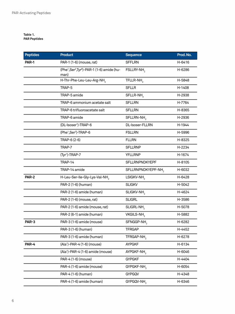
Peptides Product Sequence Prod. No.
PAR-1 PAR-1 (1-6) (mouse, rat) SFFLRN H-6416
(Phe1,Ser2,Tyr6)-PAR-1 (1-6) amide (hu-man)
FSLLRY-NH2 H-6286
H-Thr-Phe-Leu-Leu-Arg-NH2 TFLLR-NH2 H-5848
TRAP-5 SFLLR H-1408
TRAP-5 amide SFLLR-NH2 H-2938
TRAP-6 ammonium acetate salt SFLLRN H-7764
TRAP-6 trifluoroacetate salt SFLLRN H-8365
TRAP-6 amide SFLLRN-NH2 H-2936
(DL-Isoser1)-TRAP-6 DL-Isoser-FLLRN H-1944
(Phe1,Ser2)-TRAP-6 FSLLRN H-5996
TRAP-6 (2-6) FLLRN H-8325
TRAP-7 SFLLRNP H-2234
(Tyr1)-TRAP-7 YFLLRNP H-1674
TRAP-14 SFLLRNPNDKYEPF H-8105
TRAP-14 amide SFLLRNPNDKYEPF-NH2 H-6032
PAR-2 H-Leu-Ser-Ile-Gly-Lys-Val-NH2 LSIGKV-NH2 H-6428
PAR-2 (1-6) (human) SLIGKV H-5042
PAR-2 (1-6) amide (human) SLIGKV-NH2 H-4624
PAR-2 (1-6) (mouse, rat) SLIGRL H-3586
PAR-2 (1-6) amide (mouse, rat) SLIGRL-NH2 H-5078
PAR-2 (6-1) amide (human) VKGILS-NH2 H-5882
PAR-3 PAR-3 (1-6) amide (mouse) SFNGGP-NH2 H-6282
PAR-3 (1-6) (human) TFRGAP H-4452
PAR-3 (1-6) amide (human) TFRGAP-NH2 H-6278
PAR-4 (Ala1)-PAR-4 (1-6) (mouse) AYPGKF H-6134
(Ala1)-PAR-4 (1-6) amide (mouse) AYPGKF-NH2 H-6046
PAR-4 (1-6) (mouse) GYPGKF H-4404
PAR-4 (1-6) amide (mouse) GYPGKF-NH2 H-6054
PAR-4 (1-6) (human) GYPGQV H-4348
PAR-4 (1-6) amide (human) GYPGQV-NH2 H-6346
Table 1.PAR Peptides

7
PAR activation. This has been demonstrated for glycoprotein Ib (gp1b) on human plate-lets. Inhibition of thrombin binding to gp1b resulted in a decrease in PAR-1 hydrolysis. The underlying mechanism might be similar to the one suggested for PAR-3 and PAR-4 on mouse platelets, in that gp1b concen-trates thrombin at the cell surface and thereby facilitates PAR-1 hydrolysis.
Apart from thrombin and trypsin, additional proteases cleave and activate members of the PAR family. Among them are leu-kocyte proteases (such as cathepsin G and proteinase-3), the mast cell protease tryptase, in addition to enzyme complexes functioning in the coagulation cascade like TF-FVIIa-FXa and TF-FVIIa.
Some of the proteases may act as activating or inactivating enzymes depending on the targeted receptor. For example, cathepsin G is able to activate PAR-4 but disables PAR-1, PAR-2, and PAR-3 while proteinase-3 can activate PAR-2 but inactivates PAR-1.Interestingly, PARs can also be activated by non-mammalian enzymes. Activation by proteases from dust mites or Porphy-romonas gingivalis, a major mediator of periodontitis, has been associated with the development of allergies and cardiovascu-lar disorders, respectively.
Deactivation MechanismsPARs can couple to multiple signal trans-duction pathways and can therefore, regulate many cellular functions. Although cleavage results in receptors that are con-stitutively exposed to the tethered ligand sequences, signaling via PARs is transient. Considerable effort has been directed towards the investigation of signal termi-nation mechanisms with a primary focus on human PAR-1 and PAR-2. Apart from the possible deactivation by cell surface proteases that cleave PARs at sites relevant for activity, signaling via PARs might also be terminated by desensitization mechanisms. These mechanisms may involve rapid phos-phorylation of the receptors by G-protein receptor kinases (GRK) and/or second mes-senger kinases such as protein kinase C
(PKC). Subsequent binding of β-arrestins to the phosphorylated PARs results in disrup-tion of the association with heterotrimeric G-proteins and termination of the signal. In addition to receptor desensitization, responsiveness to agonists is also regu-lated by receptor endocytosis, which can be triggered by PAR activation. Internaliza-tion of both PAR-1 and PAR-2 proceed via a clathrin-mediated process. In contrast to PAR-1, endocytosis of PAR-2 requires β-arrestins, which act as adaptor proteins that link GRK-phosphorylated receptors to clathrin. Adaptor proteins, such as AP2, may participate in endocytosis of PAR-1.
Since activation of PARs is an irreversible mechanism, sustained signaling via these receptors requires either de novo synthe-sis or mobilization of intracellularly stored receptor molecules. Resensitization of re-sponses to PAR-1 activators in endothelial cells and fibroblasts are for example due to the repopulation of the cell membrane with receptors derived from prominent Golgi stores. In megakaryoblasts, on the other hand, resensitization is achieved by the synthesis of new receptors. Platelets, which only need to respond once to PAR activa-tors, lack both of these resensitization mechanisms.
Pathophysiological Functions
Cardiovascular diseaseThrombin and its receptors play a pivotal role in blood coagulation and thrombus formation. Inappropriate aggregation of platelets contributes to occlusive vascu-lar disorders such as stroke, angina, and myocardial infarction. For these reasons PAR-1 antagonists have gained interest as potential anti-thrombotic agents. In a cynomolgus monkey arterial injury model, the heterocycle-based peptide-mimetic PAR-1 antagonist RWJ-58259 was shown to exhibit considerable anti-thrombotic activ-ity despite the fact that the platelets from these animals also express PAR-4. A recent study demonstrated that PAR-1 and PAR-4 on human platelets formed heterodimers. A combination of bivalirudin (a specific,

PAR-Activating Peptides
8
reversible and direct thrombin inhibitor) and a novel PAR-4 cell-penetrating peptide (pepducin) antagonist effectively inhibited aggregation of human platelets to high concentrations of thrombin and offered superior protection against thrombosis in guinea pigs versus single-receptor inhibi-tion. These results suggest that targeting the PAR-1/PAR-4 complex on the outside and/or inside cell surface may represent a novel therapeutic opportunity to prevent arterial thrombosis. Since thrombin is also involved in the recurrent narrowing of blood vessels after vascular injury, PAR antago-nists may also be of interest in decreasing the risk of restenosis following cardiac/vas-cular surgery and angioplasty.
InflammationPARs play an important role in inflamma-tion and immune responses. Following tissue injuries, thrombin mediates several inflammatory-associated effects on the vascular endothelium via activation of PAR-1, among them the synthesis and release of various cytokines and growth fac-tors and the induction of adhesion molecule expression. PAR-2, which can be activated by trypsin and mast cell tryptase, has been implicated in pathological conditions involving inflammatory processes including arthritis, asthma, and colitis. In an animal monoarthritis model of chronic inflamma-tion, joint swelling was substantially inhib-ited in PAR-2-deficient mice compared with wild-type mice. Joint inflammation induced by intra-articular carrageenan/kaolin (C/K) injection could be attenuated by antagoniz-ing PAR-2 activation. These results pointed at PAR-2 as a novel target for the future treatment of arthritis.
In a mouse model of colitis, 2,4,6-trini-trobenzene sulfonic acid (TNBS)-induced colitis was dose-dependently reduced by the administration of the mouse PAR-2 tethered ligand SLIGRLamide, whereas the scrambled control peptide, LSIGRLamide, was ineffective. In another study, however, a pro-inflammatory role has been attributed to activation of PAR-2 in the mouse colon. A similar controversial situation concern-ing the pro- or anti-inflammatory effects of PAR-2 activation has been observed as well
within the airways. PAR-2 is also localized on sensory neurons where it plays a role in inflammatory hyperalgesia. Thermal hyper-algesia depends on PAR-2-induced sensi-tization of the transient receptor potential vanilloid receptor 1 (TRPV1) which is gated by capsaicin, protons and noxious heat. The underlying mechanism involves activation of protein kinase Cε (PKCε) and protein kinase A (PKA).
CancerIncreasing evidence suggests that PARs may play a role in invasive and metastatic processes of various cancers. Recent stud-ies have demonstrated that PAR-1 mRNA is expressed in human colon cancer cell lines but not in normal colonic epithelial cells. Ac-tivation of PAR-1 in the human colon cancer cell line HT-29 by thrombin or by tethered ligand sequences resulted in cell prolifera-tion. The signaling cascade downstream of PAR-1 involved a matrix metalloproteinase-dependent release of transforming growth factor-α (TGF-α), transactivation of the epidermal growth factor (EGF) receptor and subsequent activation of extracellular signal-related protein kinase 1/2 (ERK1/2).
PAR-1 was also found to be significantly overexpressed in atypical nevi and melano-mas in comparison with common melano-cytic nevi.
Another study demonstrated that prein-cubation of human melanoma cells with thrombin resulted in enhanced chemotactic migration. The process not only required the activation of PAR-1, but was also dependent on PAR-2 since desensitization with PAR-2 agonists abolished the effect. Con-sistent with this finding, a combination of PAR-2 and PAR-1 agonists (SLIGRL and TFLLRNPNDK) evoked enhanced cell motility whereas PAR-1 or PAR-2 agonists alone did not stimulate migration. Similarly, activation of PAR-1 and PAR-2 also enhanced chemo-kinesis in prostate cancer cells. Interest-ingly, in a panel of prostate cancer cell lines, increased PAR-1 expression was demon-strated in those cell lines derived from bone metastases.
PAR-1 is preferentially expressed in highly

9
metastatic human breast carcinoma cell lines and breast carcinoma biopsy speci-mens. Its expression correlated with tumor progression.
Recently, it was demonstrated that MDA-MB-231, a highly invasive breast cancer cell line, expressed very high levels of functional PAR-1, PAR-2 and PAR-4 whereas mini-mally invasive MCF7 cells had considerably lower levels of these receptors. Although PAR-2 and PAR-4 could act as chemotactic receptors in both MDA-MB-231 and MCF7 breast cancer cells, activation of PAR-1 with thrombin or a PAR-1 agonist unexpect-edly inhibited migration and invasiveness of MDA-MB-231 cells when applied as a concentration gradient in the direction of the cell movement.
In a different study PAR-1 was shown to be a matrix metalloprotease-1 (MMP-1) recep-tor that promotes invasion and tumorigen-esis of breast cancer cells.
ConclusionsPARs with their unique activation mecha-nism play a key role in hemostasis but are also involved in pathophysiological process-es such as arterial thrombosis, inflamma-tion, and tumor progression. The underlying mechanisms involving a variety of proteases and signaling components are only partly understood.
PAR agonists have proven valuable tools for specifically addressing the role of the individual receptors and have significantly improved our understanding of this recep-tor family. Future work, in particular the use of receptor knock-out animals, will be needed to gain a deeper insight into the complex network of PAR signaling.

PAR-Activating Peptides
10
W.F. Xu et al.Cloning and characterization of hu-man protease-activated receptor 4.Proc. Natl. Acad. Sci. USA 95, 6642-6646 (1998)M. Nakanishi-Matsui et al.PAR3 is a cofactor for PAR4 activa-tion by thrombin.Nature 404, 609-613 (2000)E. De Candia et al.Binding of thrombin to glycoprotein Ib accelerates the hydrolysis of Par-1 on intact platelets.J. Biol. Chem. 276, 4692-4698 (2001)S.R. Macfarlane et al.Proteinase-activated receptors.Pharmacol. Rev. 53, 245-282 (2001) ReviewL. Kamath et al.Signaling from protease-activated receptor-1 inhibits migration and invasion of breast cancer cells.Cancer Res. 61, 5933-5940 (2001)N. Cenac et al.Induction of intestinal inflammation in mouse by activationof proteinase-activated receptor-2.Am. J. Pathol. 161, 1903-1915 (2002)C.H. Chay et al.A functional thrombin receptor (PAR1) is expressed on bone-derived prostate cancer cell lines.Urology 60, 760-765 (2002)
M.D. Hollenberg and S.J. ComptonInternational Union of Pharmacol-ogy. XXVIII. Proteinase-activated receptors.Pharmacol. Rev. 54, 203-217 (2002) ReviewS.R. Coughlin and E. CamererPARticipation in inflammation.J. Clin. Invest. 111, 25-27 (2003) ReviewD. Darmoul et al.Aberrant expression and activation of the thrombin receptor protease-activated receptor-1 induces cell proliferation and motility in human colon cancer cells.Am. J. Pathol. 162, 1503-1513 (2003)W.R. Ferrell et al.Essential role for proteinase-acti-vated receptor-2 in arthritis.J. Clin. Invest. 111, 35-41 (2003)M.D. HollenbergProteinase-mediated signaling: proteinase-activated receptors (PARs) and much more.Life Sci. 74, 237-246 (2003) ReviewB.E. Maryanoff et al.Discovery of potent peptide-mimetic antagonists for the human thrombin receptor, protease-activated recep-tor-1 (PAR-1).Curr. Med. Chem. Cardiovasc. Hema-tol. Agents 1, 13-36 (2003) Review
U.B. Rasmussen et al.cDNA cloning and expression of a hamster alpha-thrombin receptor coupled to Ca2+ mobilization.FEBS Lett. 288,123-128 (1991)T.K. Vu et al.Molecular cloning of a functional thrombin receptor reveals a novel proteolytic mechanism of receptor activation.Cell 64, 1057-1068 (1991)S. Nystedt et al.The mouse proteinase-activated re-ceptor-2 cDNA and gene. Molecular cloning and functional expression.J. Biol. Chem. 270, 5950-5955 (1995)A.J. Connolly et al.Role of the thrombin receptor in development and evidence for a second receptor.Nature 381, 516-519 (1996)H. Ishihara et al.Protease-activated receptor 3 is a second thrombin receptor in humans.Nature 386, 502-506 (1997)S. Even-Ram et al.Thrombin receptor overexpression in malignant and physiological inva-sion processes.Nat. Med. 4, 909-914 (1998)M.L. Kahn et al.A dual thrombin receptor system for platelet activation.Nature 394, 690-694 (1998)
REFERENCES

11
S. Amadesi et al.Protease-activated receptor 2 sensitizes the capsaicin receptor transient receptor potential vanilloid receptor 1 to induce hyperalgesia.J. Neurosci. 24, 4300-4312 (2004)D. Darmoul et al.Protease-activated receptor 2 in colon cancer: trypsin-induced MAPK phosphorylation and cell prolif-eration are mediated by epidermal growth factor receptor transactiva-tion.J. Biol. Chem. 279, 20927-20934 (2004)D. Darmoul et al.Activation of proteinase-activated receptor 1 promotes human colon cancer cell proliferation through epidermal growth factor receptor transactivation.Mol. Cancer Res. 2, 514-522 (2004)V.S. Ossovskaya and N.W. BunnettProtease-activated receptors: con-tribution to physiology and disease.Physiol. Rev. 84, 579-621(2004) ReviewX. Shi et al.Protease-activated receptors (PAR1 and PAR2) contribute to tumor cell motility and metastasis.Mol. Cancer Res. 2, 395-402 (2004)
A. Boire et al.PAR1 is a matrix metalloprotease-1 receptor that promotes invasion and tumorigenesis of breast cancer cells.Cell 120, 303-313 (2005)N. VergnolleProtease-activated receptors and inflammatory hyperalgesia.Mem. Inst. Oswaldo Cruz 100 Suppl. 1, 173-176 (2005) ReviewS. Amadesi et al.Protease-activated receptor 2 sensitizes TRPV1 by protein ki-nase C{epsilon}- and A-dependent mechanisms in rats and mice.J. Physiol. 575, 555-571 (2006)E.B. Kelso et al.Therapeutic promise of proteinase-activated receptor-2 antagonism in joint inflammation.J. Pharmacol. Exp. Ther. 316, 1017-1024 (2006)A.J. Leger et al.Blocking the protease-activated receptor 1-4 heterodimer in platelet-mediated thrombosis.Circulation 113, 1244-1254 (2006)

PAR-Activating Peptides
12
PARACTIVATING PEPTIDES We also offer hirudin fragments and thrombin inhibitors such as bivali-rudin (H-6668) and lepirudin (H-6618).Both peptides are also available as active pharmaceutical ingredients. For more information on these products, please go to our homepage at www.bachem.com

13
PAR-1 PEPTIDES (TRAP PEPTIDES)
PAR-1 (1-6) (mouse, rat)H-6416SFFLRN
(Phe1,Ser2,Tyr6)-PAR-1 (1-6) amide (hu-man)H-6286FSLLRY-NH2
H-Thr-Phe-Leu-Leu-Arg-NH2
H-5848TFLLR-NH2
TRAP-5H-1408SFLLR
TRAP-5 amideH-2938SFLLR-NH2
TRAP-6 ammonium acetate saltH-7764SFLLRN
TRAP-6 trifluoroacetate saltH-8365SFLLRN
TRAP-6 amideH-2936SFLLRN-NH2
(DL-Isoser1)-TRAP-6H-1944DL-Isoser-FLLRN
(Phe1,Ser2)-TRAP-6H-5996FSLLRN
TRAP-6 (2-6)H-8325FLLRN
TRAP-7H-2234SFLLRNP
(Tyr1)-TRAP-7H-1674YFLLRNP
TRAP-14H-8105SFLLRNPNDKYEPF
TRAP-14 amideH-6032SFLLRNPNDKYEPF-NH2
PAR-2 PEPTIDES
H-Leu-Ser-Ile-Gly-Lys-Val-NH2
H-6428LSIGKV-NH2
PAR-2 (1-6) (human)H-5042SLIGKV
PAR-2 (1-6) (mouse, rat)H-3586SLIGRL
PAR-2 (1-6) amide (human)H-4624SLIGKV-NH2
PAR-2 (6-1) amide (human)H-5882VKGILS-NH2
PAR-2 (1-6) amide (mouse, rat)H-5078SLIGRL-NH2
(2-Furoyl)-PAR-2 (2-6)-Orn amide (mouse, rat)H-6246(2-Furoyl)-LIGRL-Orn-NH2
PAR-2 (6-1) amide (mouse, rat)H-6508LRGILS-NH2

PAR-Activating Peptides
14
PAR-3 PEPTIDES
PAR-3 (1-6) (human)H-4452TFRGAP
PAR-3 (1-6) amide (human)H-6278TFRGAP-NH2
PAR-3 (1-6) amide (mouse)H-6282SFNGGP-NH2
PAR-4 PEPTIDES
PAR-4 (1-6) (human)H-4348GYPGQV
PAR-4 (1-6) (mouse)H-4404GYPGKF
(Ala1)-PAR-4 (1-6) (mouse)H-6134AYPGKF
PAR-4 (1-6) amide (human)H-6346GYPGQV-NH2
PAR-4 (1-6) amide (mouse)H-6054GYPGKF-NH2
(Ala1)-PAR-4 (1-6) amide (mouse)H-6046AYPGKF-NH2
RELATEDPEPTIDES
Boc-D-Phe-Pro-Arg-OHN-1020Boc-fPR
Thrombin Receptor AntagonistH-2514MSRPACPNDKYE
H-Phe-Pro-Arg-OHH-3916FPR
uPAR (84-95) (human)H-6316AVTYSRSRYLEC

15
Coloured scanning electron micrograph (SEM) of a blood clot. Red blood cells, also known as erythrocytes (red), are seen enmeshed in filaments of fibrin protein (purple/pink). Blood clotting is the solidification of blood that rapidly occurs when blood vessels are dam-aged, limiting blood loss. Blood vessel damage activates blood cells called platelets, which in turn help to stimulate the formation of fibrin filaments at the site of injury. These filaments enmesh platelets and red and white blood cells, contracting around them to form a solid clot. Inappropriate clotting is a major cause of strokes and heart attacks. Magnification: x3800 at 6x7cm size.
KEYSTONE/SCIENCE PHOTO LIBRARY/SUSUMU NISHINAGA
BLOOD CLOT, SEM

2004
506
pub
lishe
d b
y G
lob
al M
arke
ting
, Bac
hem
Gro
up, J
anua
ry 2
017
www.bachem.com shop.bachem.com
All information is compiled to the best of our knowledge. We cannot be made liable for any possible errors or misprints. Some products may be restricted in certain countries.
Marketing & Sales Contact
Europe, Africa, Middle East and Asia Pacific
Bachem AGTel. +41 58 595 [email protected]
Americas
Bachem Americas, Inc.Tel. +1 888 422 2436 (toll free in USA & Canada) +1 310 539 [email protected]
Visit our website www.bachem.com or shop online shop.bachem.com