Embed Size (px)
Citation preview

INV ITEDP A P E R
PredictionofDistalArmPosturein 3-D Space From ShoulderMovements for Control ofUpper Limb ProsthesesExperiments indicate that voluntary shoulder movements may be used to generate the
control signals needed for operation of arms and hands that are paralyzed or artificial.
By Rahul R. Kaliki, Rahman Davoodi, and Gerald E. Loeb, Member IEEE
ABSTRACT | C5/C6 tetraplegic patients and transhumeral
amputees may be able to use voluntary shoulder motion as
command signals for a functional electrical stimulation (FES)
system or a transhumeral prosthesis. Such prostheses require, at
the most basic level, the control of endpoint position in three
dimensions, hand orientation, and grasp. Spatiotemporal syner-
gies exist between the proximal and distal arm joints for goal-
oriented reaching movements as performed by able-bodied
subjects. To fit these synergies, we utilized three-layer artificial
neural networks. These networks could be used as a means for
obtaining user intent information during reaching movements.
We conducted reaching experiments in which subjects reached
to and grasped a handle in a three-dimensional gantry. In our
previous work, the three rotational angles at the shoulder were
used to predict elbow flexion/extension angle during reaches
on a two-dimensional plane. In this paper, we extend this
model to include the two translational movements at the
shoulder as inputs and an additional output of forearm
pronation/supination. Counterintuitively, as the complexity of
the task and the complexity of the neural network architecture
increased, the performance also improved.
KEYWORDS | Artificial neural networks; control; functional
electrical stimulation; motor control; prosthesis; reaching;
upper limb
I . INTRODUCTION
Bilateral amputations and quadriplegia are debilitatingconditions in which patients are unable to perform most
activities of daily living. The restoration of any function-
ality to these patients has been the paramount goal for
many researchers. Reaching and grasping objects are
obviously particularly important. Advanced mechatronic
prosthetic arms and hands should provide motor capability
for amputees. Functional electrical stimulation (FES) to
reanimate paralyzed muscles can, in principle, restore it tospinal cord injured patients. In order to apply either
technology successfully, however, there must be a source
of command signals to convey to the prosthetic controller
the motor intentions of the user.
Most patients who have lost arm function as a result of
amputation or spinal cord injury have substantial residual
voluntary control of shoulder motion. Transhumeral
amputation procedures try to leave a long enough stumpon which to attach the socket of the prosthesis. Most
surviving quadriplegic patients have lesions at or below the
C4–5 vertebral junction, leaving intact at least some of the
neural circuitry required for respiratory and shoulder
movements while losing some or all control of the more
distal joints. The hypothesis being tested in our research is
that the residual voluntary shoulder movements contain
sufficient information about those intentions that they canbe used as a continuous source of command signals for the
prosthetic controller.
A. Potential Command SourcesDue to the limited availability of voluntarily controlled
movements in high-level amputations and spinal cord
injuries, many investigators have looked to the brain as a
Manuscript received December 7, 2007; revised February 1, 2008.
The authors are with the Alfred E. Mann Institute, Department of Biomedical
Engineering, University of Southern California, Los Angeles, CA 90089-1112 USA.
Digital Object Identifier: 10.1109/JPROC.2008.922591
Vol. 96, No. 7, July 2008 | Proceedings of the IEEE 12170018-9219/$25.00 �2008 IEEE

potential source of command signals for control of reachand grasp. The complexity and invasiveness of the
technology are daunting and the long-term viability of
the interfaces has yet to be determined. Nevertheless,
Donoghue et al. have implanted a human subject with a
microelectrode array in the primary motor cortex and the
subject has achieved control of a cursor in the two-
dimensional (2-D) space of a computer monitor [1].
Nicolelis et al. have shown that monkeys with chronicallyimplanted microelectrodes are able to control the trajec-
tory of a robot arm’s end effector to reach to one of eight
targets in three-dimensional space [2]. But the transition
from control of display cursors and robotic arms to the
control of anthropomorphic limbs is not trivial. This is
because many of the researchers investigating motor
control from the brain have been focusing on relationships
between brain activity and endpoint trajectories in spaceand assuming that individual joint trajectories can be
derived by inverse kinematic analysis. But the upper limb
is an overdetermined system in that there are more joints
than necessary to express a given endpoint position and
orientation. This was first described by Bernstein in 1967
as the Bredundancy problem[ [3]. These Bextra[ degrees offreedom (DOFs) are important, of course, in the interac-
tion of the hand with objects, e.g., in orienting the handand controlling the fingers to grasp objects with different
sizes, shapes, and orientations. It remains to be deter-
mined if cortical neural signals can be used to provide such
control without becoming an undue burden on the user.
Recently, some investigators have developed FES
systems that utilize the voluntary movements at the
shoulder of these patients as control signals. One such FES
system is the FreeHand system, which utilizes the residualvoluntary control of the protraction/retraction of the
contralateral shoulder to control the opening and closing
of a hand [4]. The dependence on contralateral shoulder
movements makes it unsuitable for extension to bimanual
tasks and deprives users of the proprioceptive feedback
that might be derived from the interaction of the command
signals with the actual arm being moved. This system,
while a little awkward in its method of control, is able torestore grasping function but does not lend itself to control
of the equally important functionality of reaching.
Kilgore et al. have designed and implanted a myoelectric-
based FES controller that restored reaching and grasping
function [5]. Myoelectric signals related to the activities of
functional muscles still under voluntary control are used as
commands for individual degrees of freedom through FES.
Implanted patients regained the ability to control variousdegrees of freedom and were able to accomplish some
activities of daily living, such as eating and drinking. This
method is a great improvement in terms of functionality
for paraplegic patients, but the number of controlled
outputs is limited by the number of viable voluntary
muscles available. If some of the voluntary muscle groups
were involved in other functions required by the patient,
then the muscle group could not be used as a reliablecommand source.
A prosthesis that relies on command sources unrelated
to natural reaching movements forces the user to learn
unnatural reaching control strategies. Such a system could
require ungainly and cumbersome movements to restore
functionality. As described here, it seems desirable to
extract command signals from the natural movements of
the ipsilateral shoulder that the patient spent a lifetimelearning before incurring injury and disability.
B. Prior Research on Ipsilateral Shoulder ControlPopovic et al. [6]–[8] have developed various synergy-
based upper limb neuroprosthetic controllers that pre-
dicted elbow flexion/extension from shoulder flexion/
extension based on simple scaling rules, through inductive
learning, and from training a radial basis function network.In the latter experiment, synergy models were trained with
joint acceleration data recorded from able-bodied subjects
while executing a sequence of movements: reaching and
grasping an object in a plane, bringing the object to the
mouth, returning the object to its previous position, and
returning the hand back to the initial position. Each
network was trained on data for sequences of movements
to one target location. The investigators found that thenetworks were able to predict reaches to targets located
distally to the trained target reach but found that the
network was unable to predict reaches to targets located
laterally to the trained target reach. This indicated that the
synergy rules changed across the two-dimensional work-
space. Therefore, the user would need to manually select
among several synergy rules based on where he or she
wanted to reach. While some reaching motion could berestored to these subjects, having to switch manually
between synergy rules would become cumbersome during
everyday activities, especially if the user wanted to make
movements across the boundaries of these regions.
In contrast to Popovic et al., we hypothesize that all
three degrees of freedom at the shoulder might contribute
useful information that would generalize better over the
whole workspace, thus eliminating the need for manualswitching. Furthermore, our initial studies suggested that
the spatiotemporal relationships between joint angles are
relatively stable and therefore more likely to result in a
user-controllable system as compared to joint velocity and
joint acceleration relationships. Finally, as opposed to
training synergy models with data from only a single target
location, we wanted to use a set of reaches to a wide range
of target locations in order to account for variations in thereaching strategy across the workspace. To test these
hypotheses, we trained an adaptive neural network (ANN)
to predict the elbow joint angle from the three rotational
angles of the shoulder joint [9].
An experimental workspace was designed such that
hand position was kept in a horizontal plane during each
reach (similar to Popovic et al.). Sixteen rectangular-shaped
Kaliki et al. : Prediction of Distal Arm Posture in 3-D Space
1218 Proceedings of the IEEE | Vol. 96, No. 7, July 2008

targets were placed in two concentric arcs on a pegboard as
shown in Fig. 1. The distal target set was distributed on a
circular arc at the maximal reach of the subject while theproximal target set was placed at the midpoint between the
distal target set and the initial position. The subject was
asked to make self-paced reaches from the initial position to
and from the target position. Shoulder and elbow joint
angles were recorded during eight trials of reaches to each of
the 16 targets.
As previously discussed, the shape and orientation of
the shoulder/elbow angle synergies varied greatly acrossthe workspace [Fig. 2(a)], but in certain areas of the
workspace, there were subsets of reaches that exhibited
similar spatiotemporal synergies [Fig. 2(b)]. We hypoth-
esized that the ANN might produce better results if trained
only on one representative reach from each such subset,
thereby avoiding overtraining on similar targets in large
subsets at the expense of accuracy on the more individ-
ualized targets. To examine this supposition, we trained
the ANN with incrementally added target reaching data.Target reaches were added to the training set based on the
ANN’s performance on the last training set. The target
reach that was predicted the worst was added to the
training set, and this process was continued iteratively
until all the targets were added. We found that the error
reached a minimum when we incorporated training data
from 11 of the 16 targets. The correlation between the
predicted elbow angle and the recorded angle on thevalidation set was 0.99. Interestingly, seven of these
targets were from the distal set, indicating that the reaches
to the distal targets allowed the ANN to predict reaches in
the proximal set accurately, apparently because the
corresponding synergies between proximal and distal
targets were just scaled versions of each other.
This previous study provided the methodology and
rationale for our present study of reaching to targetsdistributed in three dimensions, a necessary goal for most
practical applications of the proposed command scheme.
Furthermore, we tested whether including additional
inputs (related to shoulder translation) improves the
prediction of the elbow angle and whether the prediction
of an additional output, forearm pronation/supination,
degrades the performance of the ANN models.
II . METHODS
A. Target LocationsWe constructed a large robotic gantry (Parker
Hannifin, Co.) to automate the presentation of targets in
the three-dimensional (3-D) workspace of the arm. The
computer-controlled gantry was able to reach anywhere in
a (2 � 1 � 1 m3 workspace Fig. 3). Subjects were
instructed to reach and grasp firmly a cylindrical, vertically
oriented handle on the working end of the gantry arm.
Fig. 2. Example of shoulder/elbow synergies of target reaches in Fig. 2. (a) Shoulder/elbow joint synergies of target reaches 1P, 8P, 3-D,
and 6D. (b) Shoulder/elbow joint synergies of target reaches 2-5P. Clearly there is a cluster of synergies for EFE versus SFE synergies.
Fig. 1. A schematic drawing of the two-dimensional workspace
used in previous 2-D point-to-point reaching experiment.
Kaliki et al.: Prediction of Distal Arm Posture in 3-D Space
Vol. 96, No. 7, July 2008 | Proceedings of the IEEE 1219

Prior to experimentation, target locations were tailored to
the subject’s physical measurements. Target locations were
expressed in shoulder-centric spherical coordinates: �, �,and f (corresponding to the fraction of the subject’s entire
arm length). Measurements were taken of the subject’s
upper ðL1Þ and lower arm ðL2Þ segments, as well as the
height of the subject’s line of sight above the shoulder
center of rotation ðL3Þ to constrain the target locations andtailor them to the subject (Fig. 3). We first defined a set of
targets in the X–Z plane, which was then rotated about the
z-axis to produce target locations in three dimensions. Wethen applied the constraints to the target set to ensure that
all the targets were in a reasonable working space. L3 and
L1 limited the maximum and minimum height of targets in
the y-direction, respectively. To ensure the safety of the
subject during experimentation, we kept the subject’s head
and body outside of the reachable area of the gantry arm.
This limited the location of the front-most targets in our
workspace. The distance between the front-most possibletarget and the shoulder center of rotation along the x-axiswas measured ðL4Þ and was used to determine the
minimum angle of � at the subject’s maximal reach
ðf ¼ 1:0Þ in the two-dimensional plane. When the upper
arm crosses the sagittal plane at shoulder joint ð� � 90�Þ,
the maximal reach length is no longer equivalent to the
subject’s arm length due to the restriction of movement as
the humerus makes contact with the torso. In order toreduce undesirable contributions from the trunk in this
area of the workspace, the line of maximal reach was
approximated by fixing the humerus position at � ¼ 90�
and bending the elbow from 0� to 90�. Intervals betweentargets in the �-direction were determined by the
difference in the first target alpha subtracted from the
final target � divided by the number of desired targets at
the line of maximal reach. For this experiment, we hadeight targets along the line of maximal reach ðf ¼ 1:0Þ. Forall other values of f , targets were placed at that radius withthe same angular interval between targets as previously
determined. Once all the targets locations in the X–Z planewere determined, the plane was then rotated about the
z-axis from � ¼ 90� to � ¼ �90�. Targets that were belowthe length of the upper arm segment ðL1Þ in the y-directionor were at a height greater than the line of sight ðL3Þ wereremoved from the target set. The angular interval between
targets in the �-direction was 10�.An adult male volunteered to perform reaching experi-
ments. His physical measurements were L1 ¼ 11:5 in,
L2 ¼ 14:5 in, and L3 ¼ 6:5 in. The fraction of maximal
Fig. 3. Robotic gantry used to present targets to a subject in 3-D extrapersonal space.
Kaliki et al. : Prediction of Distal Arm Posture in 3-D Space
1220 Proceedings of the IEEE | Vol. 96, No. 7, July 2008
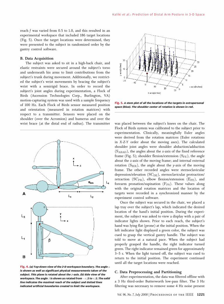
reach f was varied from 0.5 to 1.0, and this resulted in anexperimental workspace that included 186 target locations
(Fig. 5). Once the target locations were determined, they
were presented to the subject in randomized order by the
gantry control software.
B. Data AcquisitionThe subject was asked to sit in a high-back chair, and
elastic restraints were secured around the subject’s torsoand underneath his arms to limit contributions from the
subject’s trunk during movement. Additionally, we restrict-
ed the subject’s wrist movements by bracing the subject’s
wrist with a semirigid brace. In order to record the
subject’s joint angles during experimentation, a Flock of
Birds (Ascension Technologies Corp., Burlington, VA)
motion-capturing system was used with a sample frequency
of 100 Hz. Each Flock of Birds sensor measured positionand orientation (measured in rotation matrices) with
respect to a transmitter. Sensors were placed on the
shoulder (over the Acromion) and humerus and over the
wrist brace (at the distal end of radius). The transmitter was placed between the subject’s knees on the chair. The
Flock of Birds system was calibrated to the subject prior to
experimentation. Clinically, meaningfully Euler angleswere derived from the rotation matrices (Euler rotations
in X-Z-Y order about the moving axes). The calculated
shoulder joint angles were shoulder abduction/adduction
ðSABADÞ, the angles about the x-axis of the fixed reference
frame (Fig. 5); shoulder flexion/extension ðSFEÞ, the angleabout the z-axis of the moving frame; and internal external
rotation ðSIERÞ, the angle about the y-axis of the moving
frame. The other recorded angles were sternoclaviculardepression/elevation ðSCDEÞ, sternoclavicular protraction/retraction ðSCPRÞ, elbow flexion/extension ðEFEÞ, and
forearm pronation/supination ðFPSÞ. These values along
with the original rotation matrices and the location of
targets were recorded in a synchronized manner by the
experiment control software.
Once the subject was secured in the chair, we placed a
lap tray over the subject’s lap, which indicated the desiredlocation of the hand’s initial position. During the experi-
ment, the subject was asked to view a display with a pair of
indicator lights shown. Prior to each reach, the subject’s
hand was lying flat (prone) at the initial position. When the
left indicator light displayed a green color, the subject was
cued to grasp the vertical gantry handle. The subject was
told to move at a natural pace. When the subject had
properly grasped the handle, the right indicator turnedgreen. The right indicator remained green for approximately
3–5 s. When the light turned off, the subject was cued to
return to the initial position. The experiment continued
until all the target locations were reached.
C. Data Preprocessing and PartitioningAfter experimentation, the data was filtered offline with
a 3 Hz third-order Butterworth low-pass filter. The 3 Hzfiltering was necessary to remove some 4 Hz noise present
Fig. 4. (a) Top-down view of the 2-Dworkspace boundary. The angle �
is shown as well as significant physical measurements taken of the
subject. This plane is rotated about the z-axis. (b) Side view of the
workspace. The angle � is shown as rotated from��=2 to �=2. The solid
line indicates the maximal reach of the subject and dotted lines
indicated artificial boundaries created to limit the workspace.
Fig. 5. A stem plot of all the locations of the targets in extrapersonal
space (blue). The shoulder center of rotation is shown in red.
Kaliki et al.: Prediction of Distal Arm Posture in 3-D Space
Vol. 96, No. 7, July 2008 | Proceedings of the IEEE 1221

in our motion-capturing hardware, as evidenced by the
presence of the noise in the sensors prior to attachment tothe subject. The data were then down-sampled to 8 Hz to
reduce the data size and normalized by subtracting the mean
from each channel and dividing by the standard deviation.
Finally, data recorded during resting periods between target
reaches were removed in order to limit the contribution of
the initial posture to the neural network from each data set.
Prior to data partitioning, three different primary sets
of target reach data were created. The first primary set(labeled Ba[) included data from the entire trajectories
(reach plus hold period) to each of the 186 targets. In
the second primary set (b), we removed all the target
reach information that contained reaches in which the
elbow angle while grasping the target handle was less
than 10� different from the initial elbow angle. Shoulder
motion accounted for most of the movement to these
targets, so they were not useful for training elbow angleoutput. After we removed these target reaches, the data
set included 146 target reaches. The third primary data
set (c) included only those targets from set b that were
at f 9 0:8. This limited the data set to 50 target reaches.
Prior to ANN training, the primary data sets were
divided into two sets: a secondary working set, with which
the ANN was trained, and a validation set, which included
novel data to evaluate the performance of the ANN. Twentypercent of the data from the primary data set were randomly
chosen and set aside as the validation set. The remaining
data were designated the secondary working set. From thesecondary working set, 70% of the data were randomly
distributed in a training set and the remaining values
designated as the test set. The training set included data
with which the ANN would be trained via backpropagation,
and the test set was novel data used to measure the ANN’s
ability to generalize during ANN training.
D. Neural Network TrainingTwo-layer perceptron ANNs were created in Neural-
Works Predict (NeuralWare). This software employed an
adaptive gradient backwards propagation algorithm to tune
the weights and biases of the ANN to maximize the
correlation between the model predictions and the recorded
data. Hidden units had hyperbolic tangent activation
functions. The output units were logistic sigmoid activation
functions. Hidden layer size was determined through acascade learning algorithm developed by Fahlman and
Lebiere [10]. This algorithm adds hidden units incremen-
tally to the hidden layer until performance on the test set is
no longer improved. The software also used early stopping
to prevent the ANN from overfitting and to improve
generalization. Early stopping examines the performance of
the ANN during training by examining its performance on
the test set. If the network’s performance on the test set isno longer improved, then training is stopped.
Fig. 6. A schematic of the subject during experimentation. The locations of the Flock of Birds sensors are shown. The coordinate system
used indetermining the clinicallymeaningful Euler joint angles is shown. The recorded joint angleswere shoulder abduction/adduction ðSABADÞ,the angles about the x-axis of the fixed reference frame; shoulder flexion/extension ðSFEÞ, the angle about the y-axis of the moving frame;
internal external rotation ðSIERÞ, the angle about the z-axis of the moving frame; sternoclavicular depression/elevation ðSCDEÞ, the translation
along the y-axis of the shoulder frame; sternoclavicular protraction/retraction ðSCPRÞ, the translation along the x-axis of the shoulder frame;
elbow flexion/extension ðEFEÞ, the angle about the z-axis of the elbow frame; and forearm pronation/supination ðFPSÞ, the angle about
the x-axis of the elbow frame.
Kaliki et al. : Prediction of Distal Arm Posture in 3-D Space
1222 Proceedings of the IEEE | Vol. 96, No. 7, July 2008

ANNs were constructed with three different input/output (I/O) relationships to examine the efficacy of
different inputs and whether the addition of multiple distal
angle outputs significantly degraded ANN performance. The
first set of I/Os examined the ability of the three rotational
joint angles at the shoulder joint (SFE, SIER, and SABAD) to
predict the elbow angle ðEFEÞ during reaches in 3-D. ANNs
trained with this set of I/Os were labeled ANN1x, where x is
a place holder for the primary data set type (a, b, or c). Thenext set of I/Os incorporated shoulder translation move-
ments (SCDE and SCPR) as inputs in addition to SFE, SIER,and SABAD and evaluated whether these additional inputs
improved predictability of the EFE. These sets of ANNs
were labeled ANN2x. The third and final set of I/Os used
the same input, the five DOFs at the shoulder, as the
previous set but added the forearm pronation/supination
ðFPSÞ in addition to EFE as the outputs of the ANNs. Thisset of I/Os was created to examine the potential to predict
FPS for further study and whether predictive performance
significantly degraded for prediction of the EFE. The
coefficient of determination ðR2Þ between the predicted
output and recorded output was measured for all the ANNs.
Any R2 value above 0.7 was considered a strong correlation.
Additionally, the root mean squared (rms) error between
the predicted and recorded outputs was measured. Becausethe data were normalized by the standard deviation prior to
training, the error is unitless.
III . RESULTS
An example of target reach data used to train neural
networks is shown in Fig. 7. Table 1 shows the tabulated
R2 and rms errors for neural networks ANN1a, ANN1b,
and ANN1c. Inputs to these neural networks were the
three rotational angles at the shoulder while the output
was the elbow angle. The errors and R2-values are shownfor each of the data sets: training, test, and validation sets.
The R2-values for each of the ANNs on the validation set
were greater than 0.70 and, therefore, are considered
strong correlations. The neural network trained with the
distal targets ðf 9 0:8Þ reported the highest R2 and lowest
rms error for each of the data sets.
Table 2 summarizes the performance of neural
networks ANN2a, ANN2b, and ANN2c. The inputs tothese neural networks were the three rotational angles
and two translational movements at the shoulder. The
output was the elbow angle. The R2-values and rms
error values are tabulated for the training, test, and
validation sets. The R2-values for all the ANNs were
high. Neural networks trained with the distal targets
ðf 9 0:8Þ achieved the highest R2 for both the training
and test data sets and lowest rms error for all data sets.ANN2b had a slightly higher R2-value (0.8677) than
ANN2c (0.8630) on the validation set.
Table 3 summarizes data from the final set of trained
neural networks. These networks were trained with the
Fig. 7. Typical example of Euler angles recorded during a reach to a
target (� ¼ 33:5�, � ¼ 21:3�, f ¼ 1:0). The black, blue, and red plots
correspond to shoulder rotational angles, shoulder translational
movements, and distal arm angles.
Table 1 Summary Data Shown for Neural Networks Trained With Three
Rotational Shoulder Angles as Inputs Predicting the Elbow Angle as the
Output. R2 and Root Mean Squared Errors Shown for Train, Test, and
Validation Sets
Table 2 Summary Data Shown for Neural Networks Trained With Three
Rotational Shoulder Angles and Two Translational Angles as Inputs
Predicting the Elbow Angle as the Output. R2 and Root Mean Squared
Errors Shown for Train, Test, and Validation Sets
Kaliki et al.: Prediction of Distal Arm Posture in 3-D Space
Vol. 96, No. 7, July 2008 | Proceedings of the IEEE 1223

three shoulder rotational angles and translational move-
ments as the inputs and both the elbow flexion extension
and forearm pronation/supination as the output. The R2-
values and rms errors are tabulated for each data set.
ANN3c achieved the highest R2 and lowest rms error for
both inputs on all data sets and was the only ANN able to
predict the outputs with strong correlations to the
recorded outputs for all three data sets. The FPS wasconsistently predicted accurately across all trained ANNs
and data sets. The correlation for the predicted elbow
output was relatively low for all data sets of ANN3a and on
the training and test sets of ANN3b.
IV. DISCUSSION
From the results of ANN1a, -b, and -c, it is clear that neuralnetworks using three shoulder rotational angles to predict
the elbow angle during reaching in two-dimensions can be
extended to adequately predict the elbow angle for
reaching movements in a large three-dimensional extra-
personal space. Additionally, removing both weaker
synergies and proximal targets improves the performance
of the network.
Reaching in two dimensions did not require theadditional shoulder translational movements because all
targets were in the horizontal plane. Presenting targets in
three dimensions and fixing the wrist forced the subject to
make scapuloclavicular movements. Adding those shoul-
der translational movements as inputs to the neural
networks resulted in an increase in performance.
As more and more degrees of freedom are added toboth the input and the output of the neural network, it is
often expected that the increased complexity will result in
a decrease in performance. Adding the additional output of
forearm pronation/supination did not significantly hinder
the performance of a neural network on both outputs when
an ANN was trained with just distal targets. The
predictability of the network for the elbow angle decreased
only slightly, while theR2-value for the forearm pronation/supination output was extremely high ðR2 ¼ 0:95Þ for
ANN3c. As more targets were added to the primary data
set, the performance on the EFE output gradually
decreased. Perhaps two separately trained networks for
each output are required to reach a high level of
performance on each output.
It should be noted that this experiment was highly
constrained in terms of forearm movement. The forearmmovement from the initial position to the target position
was stereotyped in that the subject was always forced to
grasp the handle in the same vertical orientation in space,
but this actually requires a different anatomical pronation/
supination posture depending on shoulder angles. These
preliminary results suggest that there are useful synergies
among the five shoulder inputs and the forearm angle, and
that further studies are required to understand the fullextent and potential clinical utility of this relationship.
In the study reported in this paper, we used Euler
rotations to represent the joint movements in the ANN
training data. This commonly used method of joint motion
representation produces motion data that is easier to
understand and interpret. But the trigonometric conver-
sion from rotation matrices to Euler angles is prone to
singularities in some areas of the workspace, which occurfrequently in unconstrained 3-D reaching movements.
During preprocessing of the data, we removed any
singularities present in the data prior to neural network
training. To prevent the singularity problem, we have also
trained neural networks on the complete rotation matrices
as inputs/outputs. There was no significant difference in
the performance, but the nine-dimensional results are
more difficult to visualize and interpret. In future studies,we plan to use quaternions to alleviate the problem of
singularities.
Achieving a high level of offline neural network
performance is encouraging, but these results do not
provide any information about the tractability of these
neural networks in a prosthetic system. In order to
determine whether these predictive algorithms are actually
stable and useful as a basis for real-time control, furtherstudies are required to examine how these trained ANNs
behave in real-time virtual reality simulations analogous to
real-world prostheses and FES systems [11]. Informal tests
of a subject’s ability to use ANN3c to control a kinematic
simulation of a prosthetic arm were encouraging. The
subject was able to make stable and reasonably accurate
reaches in the workspace with little training, but methods to
Table 3 Summary Data Shown for Neural Networks Trained With Three
Rotational Shoulder Angles and Two Translational Angles as Inputs
Predicting the Elbow Angle and Forearm Angle as the Output. R2 and Root
Mean Squared Errors Shown for Train, Test, and Validation Sets
Kaliki et al. : Prediction of Distal Arm Posture in 3-D Space
1224 Proceedings of the IEEE | Vol. 96, No. 7, July 2008

quantify such performance and learning remain to beimplemented. Furthermore, it remains to be seen whether
forcing the subject into a nonnatural reaching strategy by
fixing the wrist movements has limited the tractability of
these neural networks. Further studies need to be conducted
to examine the significance of this constraint.
Eventually, we plan to embed these synergy models
into controllers for FES and powered prosthetic limbs. In
these systems, the residual shoulder movements would actas command signals to drive the movements of the distal
limb or the prosthesis. Implanted and wearable sensors for
the shoulder kinematics are under development [12]. Weplan to use our virtual reality environment as a training
tool with which a patient using stereogoggles can train to
operate a simulation of their FES arm or transhumeral
prosthetic. For FES patients, we plan to restore movement
to the distal limb with injectable, wireless, FES devices
called BIONs [13]. Command signals specifying desired
joint angles would have to be converted into desired
muscle activations according to an inverse model ofmusculoskeletal dynamics and any available sources of
kinematic feedback. h
REFERENCES
[1] M. D. Serruya, N. G. Hatsopoulos,L. Paninski, M. R. Fellows, andJ. P. Donoghue, BInstant neural control of amovement signal,[ Nature, vol. 416,pp. 141–142, 2002.
[2] J. K. Chapin, K. A. Moxon, R. S. Markowitz,and M. A. Nicolelis, BReal-time control of arobot arm using simultaneously recordedneurons in the motor cortex,[ NatureNeurosci., vol. 2, pp. 664–670, 1999.
[3] N. A. Bernstein, The Coordination andRegulation of Movements. Oxford, U.K.:Pergamon, 1967.
[4] K. L. Kilgore, P. H. Peckham, G. B. Thrope,M. W. Keith, and K. A. Gallaher-Stone,BSynthesis of hand grasp using functionalneuromuscular stimulation,[ IEEE Trans.Biomed. Eng, vol. 36, pp. 761–770, 1989.
[5] K. L. Kilgore, P. H. Peckham, F. W. Montague,R. L. Hart, A. M. Bryden, M. W. Keith,
H. Hoyen, and N. Bhadra, BAn implantedupper extremity neuroprosthesis utilizingmyoelectric control,[ in Proc. 2nd Annual Int.Conf. IEEE EMBS, 2005.
[6] D. B. Popovic and M. B. Popovic, BTuning of anonanalytic hierarchical control system forreaching with FES,[ IEEE Trans. Biomed. Eng,vol. 45, pp. 203–212, 1998.
[7] M. B. Popovic and D. B. Popovic, BCloningbiological synergies improved control ofelbow neuroprostheses,[ IEEE Eng. Med. Biol.Mag., vol. 20, pp. 74–81, 2001.
[8] S. D. Iftime, L. L. Egsgaard, andM. B. Popovic, BAutomatic determination ofsynergies by radial basis function artificialneural networks for the control of a neuralprosthesis,[ IEEE Trans. Neural Syst. Rehabil.Eng., vol. 13, pp. 482–489, 2005.
[9] R. R. Kaliki, R. Davoodi, and G. E. Loeb,BPrediction of elbow trajectory from shoulder
angles using neural networks,[ submitted forpublication.
[10] S. E. Fahlman and C. Lebiere, BThecascade-correlation learning architecture,[ inAdvances in Neural Information ProcessingSystems, vol. 2, D. S. Touretzky, Ed.San Mateo, CA: Morgan Kaufmann, 1990,pp. 524–532.
[11] M. Hauschild, R. Davoodi, and G. E. Loeb,BA virtal reality environment for designingand fitting neural prosthetic limbs,[ IEEETrans. Rehabil. Eng., vol. 15, pp. 9–15, 2007.
[12] G. E. Loeb and R. Davoodi, BThe functionalreanimation of paralyzed limbs,[ IEEE Eng.Med. Biol. Mag., vol. 24, pp. 45–51, 2005.
[13] W. Tan, Q. Zou, E. S. Kim, and G. E. Loeb,BSensing human arm posture withimplantable sensors,[ in Proc. EMBS. Conf.2004, 2004, vol. 6, pp. 4290–4293.
ABOUT THE AUTHORS
Rahul R. Kaliki received the B.S. degree in
biomedical engineering (premedical) from the
University of California, San Diego, in 2004. He is
currently pursuing the Ph.D. degree at the Uni-
versity of Southern California, Los Angeles.
He is studying the use of shoulder kinematics
as a means to predict and control distal joint
angles of the upper limb to restore functionality to
transhumeral amputees and C5/C6 spinal cord
injury patients. His research interests also include
neural prosthetics, reanimation of paralyzed limbs, upper limb prosthe-
ses, and motor control.
Rahman Davoodi received the B.S. degree in
mechanical engineering and the M.Sc. degree in
biomechanical engineering from Sharif University
of Technology, Tehran, Iran, and the Ph.D. degree
in biomedical engineering from the University of
Alberta, Edmonton, AB, Canada.
He is currently a Research Assistant Professor
in the Department of Biomedical Engineering,
University of Southern California, Los Angeles.
His current research is focused on the use of
neural prostheses to restore normal daily activities such as standing,
walking, reaching, and grasping to the paralyzed and amputee patients.
He has developed machine-learning control techniques to enable
coordinated man–machine interactions in these neural prosthetic
systems. He has also developed several publicly available software tools
to enable other researchers and engineers to easily model, simulate, and
virtually prototype complex neural prosthetic systems for the paralyzed
and amputee patients.
Gerald E. Loeb (Member, IEEE) received the B.A.
and M.D. degrees from The Johns Hopkins Univer-
sity, Baltimore, MD, in 1969 and 1972, respectively.
He completed one year of surgical residency at
the University of Arizona before joining the
Laboratory of Neural Control, National Institutes
of Health (NIH) (1973–1988). He was a Professor of
physiology and biomedical engineering at Queen’s
University, Kingston, ON, Canada (1988–1999). He
is now a Professor of biomedical engineering and
neurology and Director of the Medical Device Development Facility,
A. E. Mann Institute for Biomedical Engineering, University of
Southern California, Los Angeles. He was one of the original developers
of the cochlear implant to restore hearing to the deaf and was Chief
Scientist for Advanced Bionics Corp. (1994–1999), manufacturers of the
Clarion cochlear implant. He has received 43 issued U.S. patents and is
the author of more than 200 scientific papers. Most of his current
research is directed toward neural prosthetics to reanimate paralyzed
muscles and limbs using a new technology that he and his collaborators
developed called BIONs. This work is supported by an NIH Bioengineering
Research Partnership and is one of the testbeds in the National Science
Foundation’s Engineering Research Center on Biomimetic MicroElec-
tronic Systems, for which he is Deputy Director. These clinical applica-
tions build on his long-standing basic research into the properties and
natural activities of muscles, motoneurons, proprioceptors, and spinal
reflexes.
Prof. Loeb is a Fellow of the American Institute of Medical and
Biological Engineers.
Kaliki et al.: Prediction of Distal Arm Posture in 3-D Space
Vol. 96, No. 7, July 2008 | Proceedings of the IEEE 1225