Embed Size (px)
Citation preview
398
Sociedad Chilena de Pediatría
Keywords: Fever of unknown origin; panniculitis; panniculitis like T cell lymphoma;children.
Abstract
Introduction: Fever of unknown origin (FUO) is defined as fever over 7 to 10 days without a diag-nosis despite a complete initial study. The most frequent causes are infections, autoimmune and tu-mors. Even though most cases are self-limited there is a minority that has an underlying etiology with an ominous forecast, encouraging a systematized study. Objective: To report a rare case of a boy who presented fever of unknown origin associated to panniculitis and was diagnosed of subcutaneous panniculitis-like-T cell lymphoma and to emphasis the importance of a sequential study of FUO, in order to reach a diagnosis in patients who need a timely intervention. Clinical case: A ten year old boy, previously healthy, presented subcutaneous nodular lesions of 2 month of evolution, located in abdominal region and extremities, given few symptoms, associated with prolonged fever. He was hospitalized for proper study, in first instance infectious and immune causes were discarded and through lesions biopsy the diagnose of subcutaneous panniculitis-like-T cell lymphoma was reached. Conclusion: When FUO is diagnosed, most prevalent causes must be discarded. Then, differential diagnosis, such as immune and neoplasic etiologies, have to be considered. If FUO is associated to elemental nodular lesions, biopsy must be indicated early, in order to find potential malignant cases, avoiding therapeutic delay.
Rev Chil Pediatr. 2017;88(3):398-403DOI: 10.4067/S0370-41062017000300014
ClInICAl CASE
Paniculitis as manifestation of prolonged febrile syndrome: Case report
Paniculitis como manifestación de síndrome febril prolongado en pediatría. Caso clínico
Alexa Puchi Silvaa, Paulina López Radrigánb, Montserrat Zapico Lafuenteb, Sergio Tapia Carrerec, Sergio González Bombardiered
aSchool of Medicine, Faculty of Medicine, Andres Bello University, Viña del Mar, Chile. Medical Doctor, Pediatrician, Dr Gustavo Fricke Hospital, Viña del Mar, ChilebSchool of Medicine, Faculty of Medicine, Andres Bello University, Viña del Mar, Chile. Medicine students. cDivision of Pediatrics, Faculty of Medicine, University of Valparaiso, Chile. Medical Doctor, Pediatric hematologist oncologistdDepartment of Anatomical Pathology, Faculty of Medicine, Pontifical University Catholic of Chile, Santiago, Chile. Medical Doctor, Dermopatho-logist
Received: 27-7-2016; Accepted: 14-11-2016
Correspondence:Alexa Puchi Silva [email protected]
399
ClInICAl CASE
Introduction
In the 1960s, Petersdorf and Beeson conducted a prospective study at Yale University with a sample of 100 patients with body temperature above 38.3 ° C for more than 3 weeks without diagnosis, despite a week of etiological study hospitalized, coining the term “fever of unknown origin” (FOD)1,2; The FOD is equivalent to the concept of prolonged febrile syndrome (PFS), which in adults maintains the 3-week temporality cri-terion, but in pediatrics it is currently defined as a fe-brile episode of at least 7-10 days of evolution, without etiological diagnosis, despite an initial study consisting of anamnesis and complete physical examination to-gether with basic laboratory tests2-4.
As to the etiology of this syndrome, infectious, im-munoreumatological, neoplastic and miscellaneous causes are distinguished1-4. After an exhaustive study, in 10-32% of cases, according to geographic location, no etiology is determined2-9. It should be mentioned that identifying the etiological diagnosis can be complex, due to atypical presentations of frequent clinical pictures3-5.
The epidemiological situation of the pediatric PFS in Chile shows a great preponderance of infectious cause, being described in 68% of the cases, among which we find cat scratching, typhoid fever, urinary in-fection and infectious mononucleosis. Secondly, there are neoplastic and immunoreumatological etiologies, which are important in the differential diagnosis, with a prevalence of 4.8% for each2,4. Although the majority is progressing favorably, one out of ten patients has a final etiological diagnosis with ominous prognosis2.
Hence the importance of close monitoring of the patient until the resolution of his symptoms.
Establishing that the PFS is a diagnostic challen-ge for the medical team, it is necessary to carry out a systematic study, following a logical order that allows optimizing resources and avoiding diagnostic delays, to the benefit of the patient3,4,10.
It is relevant to carry out a thorough and comple-te anamnesis to parents or caregivers, emphasizing epidemiological antecedents and a thorough physical examination, evaluating the patient at least daily, es-pecially during febrile increases, due to the possible appearance of new clinical signs that could guide to the diagnosis3.
Panniculitis is described within the rare causes of PFS in pediatrics. This entity corresponds to an hete-rogeneous group of diseases that present with inflam-mation of the subcutaneous fat11-13. Childhood panni-culitis exist, such as neonatal subcutaneous fat necro-sis, neonatal scleroderma, post-steroidal panniculitis and cold panniculitis. In addition, cases of pediatric presentations of panniculitis described in adults, such as erythema nodosum, have been described12,13.
The objective of this work is to present the clinical case of a schoolboy with SFP associated with pannicu-litis and to emphasize the importance of a sequential study of SFP to investigate patients requiring timely intervention.
Clinical case
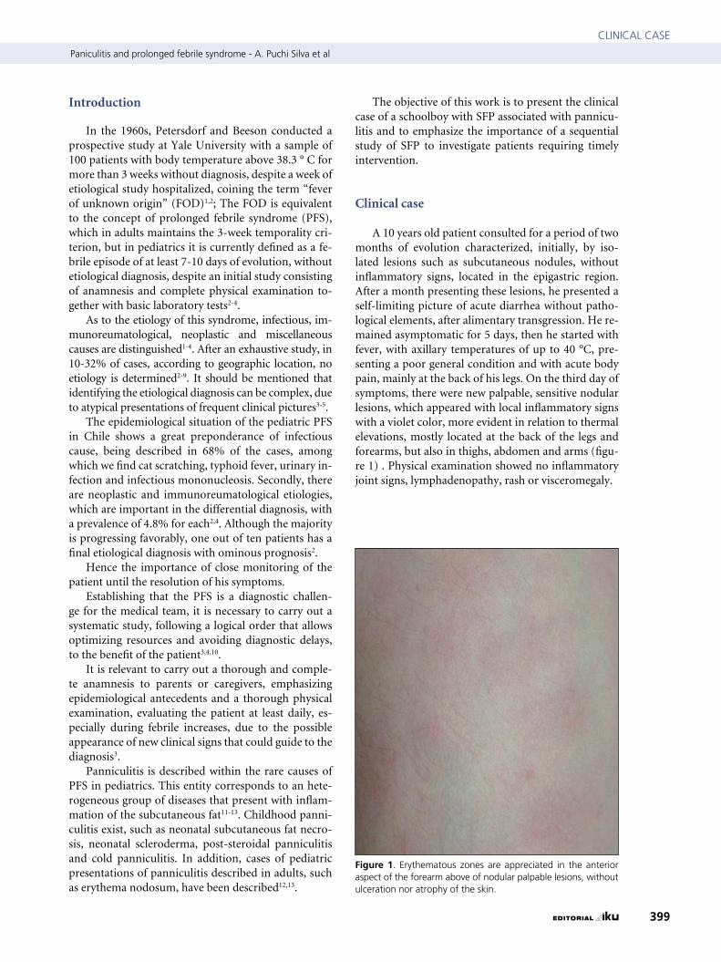
A 10 years old patient consulted for a period of two months of evolution characterized, initially, by iso-lated lesions such as subcutaneous nodules, without inflammatory signs, located in the epigastric region. After a month presenting these lesions, he presented a self-limiting picture of acute diarrhea without patho-logical elements, after alimentary transgression. He re-mained asymptomatic for 5 days, then he started with fever, with axillary temperatures of up to 40 °C, pre-senting a poor general condition and with acute body pain, mainly at the back of his legs. On the third day of symptoms, there were new palpable, sensitive nodular lesions, which appeared with local inflammatory signs with a violet color, more evident in relation to thermal elevations, mostly located at the back of the legs and forearms, but also in thighs, abdomen and arms (figu-re 1) . Physical examination showed no inflammatory joint signs, lymphadenopathy, rash or visceromegaly.
Paniculitis and prolonged febrile syndrome - A. Puchi Silva et al
Figure 1. Erythematous zones are appreciated in the anterior aspect of the forearm above of nodular palpable lesions, without ulceration nor atrophy of the skin.
400
ClInICAl CASE
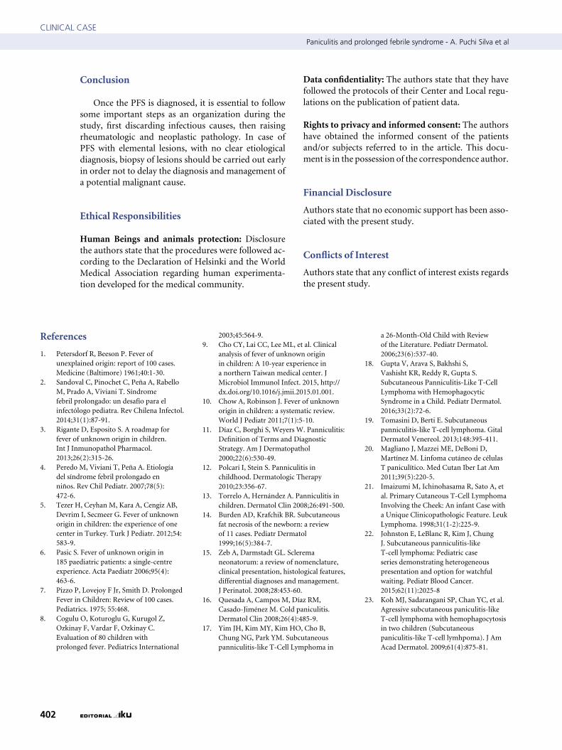
Figure 3. Mycro photografy 200x original magnification, showing atypical lymphocytes , of irregular nucleus, surrounding mature adipocytes. Hematoxylin-eosin stain.
He had a background of current vaccination without administration of recent vaccines; A 3-year-old cat as a pet and presented an azithromycin intake of 250 mg/day for three days, which was self-medicated at the start of the fever and other mentioned symptoms.
He consulted a pediatrician on the seventh day of fever, who indicated hospitalization, with a diagnosis of SFP and panniculitis, also considering an observation of erythema nodosum. Among the results of labora-tory tests performed were: normocytic-normochromic mild anemia, normal white and platelet series, ESR 39 mm/hr, normal PCR and procalcitonin, glycemia, cal-cemia, phosphemia, magnesemia, liver profile and al-buminemia were normal; Negative microalbuminuria with normal renal function and complete urinalysis.
Chest x-ray and abdominal ultrasound without patho-logical findings. Infectious study: Negative serology for Bartonella henselae, Mycoplasma pneumoniae, Epstein-Barr Virus, Cytomegalovirus, Parvovirus, To-xoplasma gondii, Brucella spp., HIV, quantiferon-TBC and antistreptolysin, blood cultures and coproculture were negative; The echocardiogram showed a normal cardiac structure, with minimal mitral insufficiency. Study with Autoantibodies (Anti-nuclear, Anti-DNA, Anti-neutrophil cytoplasm, Antibodies Extracts from the nucleus –ENA- and Rheumatoid Factor), negative.
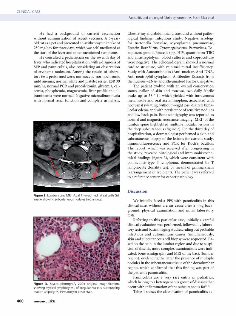
The patient evolved with an overall conservation status, pallor of skin and mucosa, two daily febrile peaks up to 38 ° C, which yielded with intravenous metamizole and oral acetaminophen, associated with nocturnal sweating, without weight loss, discrete bima-lleolar edema and with persistence of sensitive nodules and low back pain. Bone scintigraphy was reported as normal and magnetic resonance imaging (MRI) of the lumbar spine highlighted multiple nodular lesions in the deep subcutaneous (figure 2). On the third day of hospitalization, a dermatologist performed a skin and subcutaneous biopsy of the lesions for current study, immunofluorescence and PCR for Koch’s bacillus. The report, which was received after progressing in the study, revealed histological and immunohistoche-mical findings (figure 3), which were consistent with panniculitis-type T-lymphoma, demonstrated by T lymphocyte clonality test, by means of gamma chain rearrangement in recipients. The patient was referred to a reference center for cancer pathology.
Discussion
We initially faced a PFS with panniculitis in this clinical case, without a clear cause after a long back-ground, physical examination and initial laboratory tests.
Referring to this particular case, initially a careful clinical evaluation was performed, followed by labora-tory tests and basic imaging studies, ruling out probable infectious and autoimmune causes. Simultaneously, skin and subcutaneous cell biopsy were requested. Ba-sed on the pain in the lumbar region and due to suspi-cion of discitis, more complex examinations were indi-cated: bone scintigraphy and MRI of the back (lumbar region), evidencing the latter the presence of multiple nodules in the subcutaneous tissue of the dorsolumbar region, which confirmed that this finding was part of the patient’s panniculitis.
Panniculitis are a very rare entity in pediatrics, which belong to a heterogeneous group of diseases that occur with inflammation of the subcutaneous fat11-13.
Table 1 shows the classification of panniculitis ac-
Figure 2. lumbar spine MRI. Axial t1-weighted fat sat with Gd. Image showing subcutaneous nodules (red arrows).
Paniculitis and prolonged febrile syndrome - A. Puchi Silva et al

401
ClInICAl CASE
nocturnal diaphoresis and weight loss) and possible hematological alterations20,21. Histologically, atypical lymphocytes are commonly seen forming a ring-like around necrotic adipocytes as a loop13, 17-21. In the im-munohistochemistry, two subtypes are described: the alpha/beta with an indolent course and favorable prog-nosis and the gamma/delta, which has an aggressive course and a greater association with hematopagocytic syndrome. The estimated survival of the first is 80% and the second 10% to 5 years21.
We reviewed 9 pediatric cases described in the li-terature17-23, and we found some similar features with the present clinical case, as the presence of erythema-tous-violaceous non-ulcerated few sensitive nodules in the anterior abdominal wall, dorsum and extremi-ties, nodules of insidious installation and evolution in outbreaks. In 6 of the 9 cases there was concomitance with PFS and 7 of them, kept their weight17,18,22,23. The absence of lymphadenopathy and visceromegaly were all described. Regarding to age, only 2 of the cases oc-curred in schoolchildren18,23. From the laboratory co-llected data, the presence of mild anemia and elevation of ESR was observed in 4 of the cases, right as it was presented in the current case17,18,23. The biopsy revealed the presence of panniculitis compatible with subcuta-neous T-cell lymphoma, showing an association with hematophagocytosis in only one single patient, which did not occur in the present case18.
Table 1. Classification of the panniculitis of children
Specific panniculitis of children Subcutaneous fat necrosis of the newborn Sclerema neonatorum Cold panniculitis Poststeroid paniculitis
Adult-type panniculitis appearing in children
Erythema nodosum
Enzymatic panniculitis Pancreatic panniculitis
Infectious panniculitis Bacterial Mycobacterial Fungal
Panniculitis in connective tissue disease lupus panniculitis , Dermatomyositis, Polyarteritis nodosa
Granulomatous panniculitis: Sarcoidosis
Physical panniculitis Facticial, Blunt trauma
Malignant paniculitis: Subcutaneous panniculitic t-cell lymphoma Histiocytic cytophagic panniculitis
Idiopathic panniculitides
Adapted from torrelo3.
cording to etiology. Childhood panniculitis are descri-bed, such as neonatal subcutaneous fat necrosis, neo-natal scleroderma, post-steroidal panniculitis and cold panniculitis12-16.
There are very infrequent reports on pediatric pre-sentations of panniculitis that are well described in adults, being the most frequent, the erythema nodo-sum, a septal panniculitis without vasculitis, clinically characterized as the appearance of inflammatory and painful cutaneous nodules that mainly compromise the pretibial region of the lower limbs, which usually solve spontaneously in 1 to 6 weeks, without ulceration or cutaneous atrophy. It is a plurietiological syndrome that occurs frequently secondary to a hypersensitivity reaction associated with streptococcal, tuberculous or enteric disease in children and also to the use of beta-lactams and macrolides12,13.
Panniculitis are presented clinically as deep plaques or nodules, regardless its origin or cause. Thus, it is ne-cessary to complement its study with an histopatholo-gical study in order to approach to a correct diagno-sis11. Even so, its histology is not simple to interpret, considering the limited ability of adipose tissue to react with different patterns12,13. The best diagnostic tool is the skin biopsy and it is the role of the physician to de-termine the appropriate time to perform it, being advi-sable to perform it in early stages to avoid evolution to lesions with less specific histological changes13.
In order to identify the specific cause of panniculi-tis in the study, a trained dermatologist in the biopsy is fundamental as well as a good clinical pathological co-rrelation. To reach a good diagnostic, it is essential to obtain a representative sample of subcutaneous tissue, so the biopsy must be excisional and deep13.
In addition, panniculitis associated with malignant disease are described, which, although very rare, are important to consider when presenting atypical lesions or associated with constitutional symptoms12.
In this clinical case, the diagnosis of Non-Hodgkin’s lymphoma of T cells type panniculitis was reached through the biopsy. This represents less than 1% of non-Hodgkin’s lymphomas13,17, with cases reported in children only exceptionally, according to Vishal et al18, who reported 26 cases. Any cases have been reported in Chile.
Subcutaneous panniculitis-like T-cell lymphoma is defined as the primary infiltration of subcutaneous cellular tissue by pleomorphic T cells of small, medium and large size. It is usually presented in young adults, with a mean of 36 years, and presenting a slight female tendency19, as one or more subcutaneous nodules or plaques, usually not ulcerated, predominating in the legs. It could also compromise the trunk, upper limbs and face17,18. Up to half of the cases are associated with constitutional commitment with B symptoms (fever,
Paniculitis and prolonged febrile syndrome - A. Puchi Silva et al
402
ClInICAl CASE
Conclusion
Once the PFS is diagnosed, it is essential to follow some important steps as an organization during the study, first discarding infectious causes, then raising rheumatologic and neoplastic pathology. In case of PFS with elemental lesions, with no clear etiological diagnosis, biopsy of lesions should be carried out early in order not to delay the diagnosis and management of a potential malignant cause.
Ethical Responsibilities
Human Beings and animals protection: Disclosure the authors state that the procedures were followed ac-cording to the Declaration of Helsinki and the World Medical Association regarding human experimenta-tion developed for the medical community.
Data confidentiality: The authors state that they have followed the protocols of their Center and Local regu-lations on the publication of patient data.
Rights to privacy and informed consent: The authors have obtained the informed consent of the patients and/or subjects referred to in the article. This docu-ment is in the possession of the correspondence author.
Financial Disclosure
Authors state that no economic support has been asso-ciated with the present study.
Conflicts of Interest
Authors state that any conflict of interest exists regards the present study.
References
1. Petersdorf R, Beeson P. Fever of unexplained origin: report of 100 cases. Medicine (Baltimore) 1961;40:1-30.
2. Sandoval C, Pinochet C, Peña A, Rabello M, Prado A, Viviani T. Síndrome febril prolongado: un desafío para el infectólogo pediatra. Rev Chilena Infectol. 2014;31(1):87-91.
3. Rigante D, Esposito S. A roadmap for fever of unknown origin in children. Int J Inmunopathol Pharmacol. 2013;26(2):315-26.
4. Peredo M, Viviani T, Peña A. Etiología del síndrome febril prolongado en niños. Rev Chil Pediatr. 2007;78(5): 472-6.
5. Tezer H, Ceyhan M, Kara A, Cengiz AB, Devrim I, Secmeer G. Fever of unknown origin in children: the experience of one center in Turkey. Turk J Pediatr. 2012;54: 583-9.
6. Pasic S. Fever of unknown origin in 185 paediatric patients: a single-centre experience. Acta Paediatr 2006;95(4): 463-6.
7. Pizzo P, Lovejoy F Jr, Smith D. Prolonged Fever in Children: Review of 100 cases. Pediatrics. 1975; 55:468.
8. Cogulu O, Koturoglu G, Kurugol Z, Ozkinay F, Vardar F, Ozkinay C. Evaluation of 80 children with prolonged fever. Pediatrics International
2003;45:564-9.9. Cho CY, Lai CC, Lee ML, et al. Clinical
analysis of fever of unknown origin in children: A 10-year experience in a northern Taiwan medical center. J Microbiol Immunol Infect. 2015, http:// dx.doi.org/10.1016/j.jmii.2015.01.001.
10. Chow A, Robinson J. Fever of unknown origin in children: a systematic review. World J Pediatr 2011;7(1):5-10.
11. Díaz C, Borghi S, Weyers W. Panniculitis: Definition of Terms and Diagnostic Strategy. Am J Dermatopathol 2000;22(6):530-49.
12. Polcari I, Stein S. Panniculitis in childhood. Dermatologic Therapy 2010;23:356-67.
13. Torrelo A, Hernández A. Panniculitis in children. Dermatol Clin 2008;26:491-500.
14. Burden AD, Krafchik BR. Subcutaneous fat necrosis of the newborn: a review of 11 cases. Pediatr Dermatol 1999;16(5):384-7.
15. Zeb A, Darmstadt GL. Sclerema neonatorum: a review of nomenclature, clinical presentation, histological features, differential diagnoses and management. J Perinatol. 2008;28:453-60.
16. Quesada A, Campos M, Díaz RM, Casado-Jiménez M. Cold paniculitis. Dermatol Clin 2008;26(4):485-9.
17. Yim JH, Kim MY, Kim HO, Cho B, Chung NG, Park YM. Subcutaneous panniculitis-like T-Cell Lymphoma in
a 26-Month-Old Child with Review of the Literature. Pediatr Dermatol. 2006;23(6):537-40.
18. Gupta V, Arava S, Bakhshi S, Vashisht KR, Reddy R, Gupta S. Subcutaneous Panniculitis-Like T-Cell Lymphoma with Hemophagocytic Syndrome in a Child. Pediatr Dermatol. 2016;33(2):72-6.
19. Tomasini D, Berti E. Subcutaneous panniculitis-like T-cell lymphoma. Gital Dermatol Venereol. 2013;148:395-411.
20. Magliano J, Mazzei ME, DeBoni D, Martínez M. Linfoma cutáneo de células T paniculítico. Med Cutan Iber Lat Am 2011;39(5):220-5.
21. Imaizumi M, Ichinohasama R, Sato A, et al. Primary Cutaneous T-Cell Lymphoma Involving the Cheek: An infant Case with a Unique Clinicopathologic Feature. Leuk Lymphoma. 1998;31(1-2):225-9.
22. Johnston E, LeBlanc R, Kim J, Chung J. Subcutaneous panniculitis-like T-cell lymphoma: Pediatric case series demonstrating heterogeneous presentation and option for watchful waiting. Pediatr Blood Cancer. 2015;62(11):2025-8
23. Koh MJ, Sadarangani SP, Chan YC, et al. Agressive subcutaneous paniculitis-like T-cell lymphoma with hemophagocytosis in two children (Subcutaneous paniculitis-like T-cell lymhpoma). J Am Acad Dermatol. 2009;61(4):875-81.
Paniculitis and prolonged febrile syndrome - A. Puchi Silva et al