Embed Size (px)
Citation preview

255
Acta Veterinaria-Beograd 2020, 70 (2), 255-266UDK: 636.7.09:616.37-073
DOI: 10.2478/acve-2020-0018Short communication
*Corresponding author: e-mail: [email protected]
PANCREATIC EVALUATION IN DOGS USINGDIFFERENT ULTRASONOGRAPHIC TECHNIQUES – PRELIMINARY RESULTS
LOPES Michelle Avante1, FELICIANO Marcus Antônio Rossi1,2*, USCATEGUI Ricardo Andres Ramirez1,3, MARONEZI Marjury Cristina1, SILVA Priscila Del Aguila1, POZZOBON Ricardo2, SIMÕES Ana Paula Rodrigues1, SILVA Priscila1, GASSER Beatriz1, PAVAN Letícia1, AIRES Luiz Paulo Nogueira1, CANOLA Júlio Carlos1
1Faculdade de Ciências Agrárias e Veterinárias, Universidade Estadual Paulista (FCAV/UNESP), Jaboticabal, São Paulo, Brazil; 2Universidade Federal de Santa Maria (UFSM), Rio Grande do Sul, Brazil;3Instituto de Ciências Agrárias, Universidade Federal dos Vales do Jequitinhonha e Mucuri (UFVJM), Unaí, Minas Gerais, Brazil
(Received 27 August, Accepted 13 November 2019)
The aim of this study is to describe the preliminary results on the accuracy of ultrasonographic techniques such as elastography, contrast enhanced ultrasound (CEUS) and Doppler in determining pancreatic changes. Twenty-fi ve dogs, males and females, aged 1-14 years, were studied. Sixteen animals had no clinical signs of pancreatic disease (GS) and nine presented signs of pancreatic disorders (GD). All animals from GD presented sonographic changes in B-mode and qualitative elastography, with shear-wave velocity (SWV) higher (2.4±0.5m/s) in GD (p=0.014) than GS (1.9±0.3m/s) resulting in 78% sensitivity and 69% specifi city in the identifi cation of pancreatic changes. Regarding Doppler mode, no differences were observed between groups with color mapping or pulsed wave Doppler. The values obtained with CEUS did not differ between groups. Elastography is a promising technique for differential diagnosis of pancreatic changes because of its sensitivity and specifi city, while the other techniques did not show diagnostic accuracy.Key words: Ultrasonography, hemodynamics, pancreatic diseases, elastography
INTRODUCTION
Among pancreatic diseases, pancreatitis is the one that most affects the exocrine pancreas of dogs. The disease is characterized by nonspecifi c and intermittent gastrointestinal signs that may hinder the diagnosis [1,2], causing complications such as cell death, reduced blood supply and it might trigger systemic infl ammation, leading to pancreatic dysfunction with a high mortality rate [3,4]. One report mentions a case

Acta Veterinaria-Beograd 2020, 70 (2), 255-266
256
of pancreatic destruction in a dog, in which it was only possible to fi nd remnants of the parenchyma and infl ammatory cells under the microscope [2].Mild pancreatitis is a disease with low morbidity and mortality that can be reversed, if rapidly diagnosed [5].The diagnosis of pancreatitis is still challenging, especially in mild cases, where patients present discreet clinical signs. Routinely, serum activities of amylase and lipase are used for diagnosis, because they are indicators of pancreatic infl ammation, however, these assays have low specifi city and sensitivity in the identifi cation of pancreatitis in dogs and their usefulness in clinical practice is questionable [6]. Nowadays, rapid tests that measure the level of the specifi c pancreatic lipase in the blood are being used. Even though they have good sensitivity and specifi city in cases of acute pancreatitis, they may not detect subclinical phases or they might even provide false positive results [7]Defi nitive diagnosis is made by histopathological examination, however, its execution in the ante-mortem organ is rare [5]. Cytological examination by fi ne-needle aspiration was widely used in medicine, but nowadays its use is restricted and controversial. There are reports about secondary contamination that may occur in patients with pancreatic necrosis and the detection of the etiological agent becomes more diffi cult [8].Computed tomography (CT) and Magnetic Resonance Imaging (MRI) are widely used in medicine, however, in veterinary medicine, they are not easily accessible and require general anesthesia [9].Due to the diffi culty in concluding the diagnosis of pancreatitis, the variety of differential diagnoses of acute abdomen in dogs and lack of precise assays, ultrasonography is the method of choice for the evaluation of patients with suspected pancreatitis. Despite its sensitivity of 68% [1,6], the exam provides important information about changes in the pancreatic parenchyma, already identifi ed by other authors [5]. It is important to emphasize that a normal ultrasound examination does not rule out pancreatitis. If the disease is suspected, but there are no deviations in the fi rst exam, ultrasound should be repeated in a few days, as the severity increases and sonographic changes tend to appear with time [6].Although diagnostic accuracy of B mode ultrasonography is acceptable, this method cannot distinguish infl ammation, necrosis and neoplasia [5], thus, the use of new non-invasive ultrasonographic techniques, such as elastography and contrast enhanced ultrasound (CEUS) could contribute in the detection of pancreatic lesions and help the diagnosis of the patients.CEUS is a new advancement in imaging diagnosis in medicine and its application in veterinary medicine is constantly growing to assess organ perfusion, detect areas of necrosis and aid in the diagnosis of pancreatic neoplasms [4,9]. Elastography is an imaging modality that evaluates the stiffness of the organs. Studies performed with other tissues have shown a positive correlation between shear wave velocity and the

Lopes et al: Pancreatic evaluation in dogs using different ultrasonographic techniques – preliminary results
257
stiffness of the tissue evaluated, thus, the faster the shear wave velocity, the stiffer the tissue [10].The aim of this preliminary study was to evaluate accuracy of different ultrasonographic techniques (B mode, Doppler, CEUS and Elastography) in assessing pancreatic changes in dogs and to describe the fi ndings of each method in different lesions.
MATERIALS AND METHODS
The study was performed and approved by the Ethics Committee in the Use of Animals (protocol n. 007976/18). Twenty-fi ve dogs of different breeds, aged between 1-14 years and weighing between 5-29kg were selected for this study. Dogs were allocated in two groups: GS (n=16) healthy animals and GD (n=9) non-healthy animals, with suspicion of pancreatic disease. All dogs came from the clinical practice of the Veterinary Hospital of the same institution, informed and written consent were obtained from client-owned animals included in this study.Animals were classifi ed based on the results of physical and clinical examination, hematological results and B mode ultrasonography. GS animals did not present any alterations in these tests and GD presented pain in the epigastric region, abdominal discomfort and hematological changes, such as leukocytosis with left shift.For ultrasound examination, all animals fasted for 8-12 hours prior to the exam, so there would not be superposition of the gastrointestinal content on the pancreas. The abdomen was clipped and acoustic gel was applied to facilitate the procedure and promote more contact between the transducer and the skin. Patients were positioned in dorsal recumbency and during the scan they were shifted to lateral (right and left) recumbency, in order to locate all portions of the organ under study (pancreas).Scanning was performed by a single operator, with nine years of experience, using ACUSON S2000 – SIEMENS (Siemens, Munich, Germany) and linear and convex multifrequency transducers (7.5 to 9.0MHz).B mode scanning was performed prior to elastography, aiming to evaluate the entire pancreatic parenchyma of the animals in its different anatomical portions (right and left lobes and body) and also to assess their characteristics: echogenicity (in comparison to adjacent structures), echotexture (homogenous and heterogenous), contours (regular and irregular) and dimensions (normal, increased and reduced). To locate the right pancreatic lobe, the anatomical landmarks used were right kidney, descending duodenum (coursing through the right abdominal wall) and the pancreaticoduodenal vein, located parallel to the descending duodenum.In this study, all techniques were standardized on the right pancreatic lobe, because in GS it was the portion that could be identifi ed in all animals. In GD, all portions were assessed, however, only the values of the right lobe were considered.

Acta Veterinaria-Beograd 2020, 70 (2), 255-266
258
Elastography was performed with the same equipment, using the software for Acoustic Radiation Force Impulse (ARFI) qualitative characterization and quantifi cation (Virtual Touch Tissue Quantifi cation, 2D-SWE technique). The qualitative ARFI method resulted in colored images of the pancreatic tissue (elastogram) for the evaluation of tissue deformity (with or without deformation), in which lighter tones (bluish) represent more elastic tissues (soft) and darker tones (reddish) represent stiffer tissues (hard). Image quality was tested using the exhibition mode, in which greenish and homogenous images indicated high quality of the technique and yellowish and heterogenous images indicated low quality.In the quantitative ARFI method, it was possible to measure shear wave velocity (SWV m/s). In this study, fi ve measurements of the right pancreatic lobe were obtained and mean values were calculated for statistical analysis.Using Color Doppler, vascular characteristics of the parenchyma were evaluated. In patients with suspected pancreatic disease, it was verifi ed whether neovascularization was present. In order to obtain vascular indices of both healthy and diseased tissue, after identifi cation of the pancreaticoduodenal vein with color mapping, the sample volume was positioned in the central portion, then, Pulsed Doppler was activated, obtaining the results of resistivity index (RI) automatically by the equipment.For microbubble contrast enhanced ultrasound (CEUS), contrast-specifi c software was used (CADENCE®, Siemens, Munich, Germany), with secondary harmonic imaging, pulse inversion technique and a 9.0MHz linear transducer. The images obtained were evaluated in a specifi c imaging software. The contrast agent (SonoVue®, Bracco, Milan, Italy), was administered in the dosage of 0.1 mL for each animal, using a venous catheter that was maintained in the cephalic vein, followed by administration of 5.0 mL of saline solution (NaCl 0.9%).Video clips lasting 5 minutes after injection of the contrast were obtained and registered in the internal system storage and then, they were analyzed.This contrast exam defi ned parameters related to the fi lling (homogenous or heterogenous) of the organ by microbubbles. Additionally, the times of vascular fi lling were analyzed, since administration of the contrast in the bloodstream until the beginning of the organ perfusion (wash-in); peak of contrast (enhancement); and time of the contrast total output from the parenchyma (wash-out).Statistical analysis was performed using the R software (RTM Foundation for Stastistical Computing, Vienna, Austria). Sonographic variables were compared between clinical classifi cations using Student’s T- test. Subsequently, parameters that presented a signifi cant difference were submitted to a discriminant analysis using ROC curves and cutoff values (CV), sensitivity, specifi city and area under the curve were calculated using the logistic regression model. A signifi cance of 5% (p=0.05) was established for all tests.

Lopes et al: Pancreatic evaluation in dogs using different ultrasonographic techniques – preliminary results
259
RESULTS AND DISCUSSION
B mode ultrasonography and elastography were performed without diffi culties and intercurrences. However, Spectral Doppler evaluation was very diffi cult in six GS patients (37.5%), because of the narrow nature of the organ and its small evaluation window. In the GD group, this evaluation was not possible in three patients (33.3%), due to the relentlessness and abdominal discomfort of the patients. In CEUS, it was not possible to evaluate fi ve patients from GS (31.25%), due to the localization and dimension of the organ. These limiting factors of our study reduced the number of patients evaluated in the described techniques.In B mode scanning of GS dogs, the mean thickness was 0.67cm and it was possible to observe the right pancreatic lobe of normal size, preserved echogenicity (isoechoic to the mesenteric fat and the adjacent hepatic lobe) homogenous echotexture and regular contours. The characteristics found in the pancreas from the GS group were compatible to a healthy tissue.All nine sick patients (GD) showed nonspecifi c signs, such as anorexia, apathy, vomiting, diarrhea, “praying position”, pain in the epigastric region and sonographic alterations such as changes in echogenicity, echotexture, dimensions and contours. In seven cases (77.8%) changes in the adjacent organs were observed, reinforcing the suspicion of pancreatic disease (Table 1).
The sonographic alterations found in this study (reduced echogenicity, increased or normal size of the organ, heterogenous echotexture and adjacent changes, such as reactive mesentery, free fl uid in the abdomen or duodenitis) are images suggestive of acute pancreatitis, corroborating with several studies [4–7,11,12]. All alterations were noted in the right lobe, in some patients in the pancreatic body and only one patient presented alterations in all portions of the organ, which reinforces data from the literature that states that the right pancreatic lobe is more easily located in dogs [6].Pancreatic edema and adjacent free fl uid were seen in two patients. This was also observed in other studies that describe acute pancreatitis with swollen parenchyma, however, in these cases, it is necessary to consider differential diagnosis such as portal hypertension and hypoalbuminemia [12].No sonographic signs of neoplasia were seen in any patient. This might be because of the small number of patients in this study. Although Bailey and Page [13] reported that pancreatic neoplasms (exocrine and endocrine) are rare in veterinary medicine in comparison to human medicine, pancreatitis, in contrast, is routinely detected in the canine pancreas, as observed in the present study.In the qualitative elastography, GS group presented homogenous pancreatic parenchyma and of bluish colors (deformable), being characterized as soft. In eight GD patients, the elastogram presented deformable parenchyma, with predominantly bluish colors and some discreet stiff areas (small areas in yellowish color), suggesting

Acta Veterinaria-Beograd 2020, 70 (2), 255-266
260
Tab
le 1
. Cha
nges
in th
e pa
ncre
atic
pare
nchy
ma
and
adjac
ent t
issue
s in
patie
nts i
n B-
mod
e ul
traso
nogr
aphy
(Jab
otica
bal,
2018
)
Bre
edA
geE
chog
enic
ity
Ech
otex
ture
Th
ickn
ess
(cm
)C
onto
urs
Oth
ers
Qu
alit
ativ
e E
last
ogra
phy
Qu
anti
tati
ve
Ela
stog
rap
hy
1- M
ongr
el 17
Kg
4Re
duce
d H
eter
ogen
eous
1.78
Regu
larH
ypoe
choi
c les
ions
Pred
omin
antly
blu
ish a
nd g
reen
ish2.
26 m
/s
2- M
ongr
el20
Kg
7Re
duce
dH
omog
enou
s1.
20Re
gular
Reac
tive
Mes
ente
ryPr
edom
inan
tly b
luish
1.98
m/s
3- M
ongr
el5
Kg
7Re
duce
dH
omog
enou
s1.
40Re
gular
Reac
tive
Mes
ente
ryPr
edom
inan
tly b
luish
and
gre
enish
, w
ith a
small
yell
owish
are
a2.
82 m
/s
4- D
almat
ian 1
8 K
g8
Redu
ced
Hom
ogen
ous
2.40
Irre
gular
Reac
tive
Mes
ente
ry
and
Duo
deni
tisPr
edom
inan
tly g
reen
ish, w
ith la
rge
redd
ish a
reas
3.48
m/s
5- M
ongr
el29
Kg
10Re
duce
dH
omog
enou
s1.
0Re
gular
Reac
tive
Mes
ente
ryPr
edom
inan
tly b
luish
and
gre
enish
, w
ith a
small
redd
ish a
rea
2.19
m/s
6-Yo
rksh
ire
7 K
g 10
Redu
ced
Het
erog
eneo
us1.
0Ir
regu
larRe
activ
e M
esen
tery
Pred
omin
antly
blu
ish a
nd g
reen
ish,
with
small
yell
owish
and
redd
ish a
reas
1.94
m/s
7- M
ongr
el 28
Kg
11Re
duce
d H
eter
ogen
eous
1.25
Regu
larRe
activ
e M
esen
tery
Pred
omin
antly
blu
ish a
nd g
reen
ish,
with
small
yell
owish
and
redd
ish a
reas
2.24
m/s
8- P
itbul
l 2
6 K
g11
Redu
ced
Het
erog
eneo
us1.
47Ir
regu
lar
Ede
ma
and
free
fl u
idPr
edom
inan
tly b
luish
and
gre
enish
, w
ith a
small
yell
owish
are
a2.
43 m
/s
9- C
ocke
r Spa
niel
12,
5 K
g 14
Redu
ced
Het
erog
eneo
us1.
26Re
gular
Ede
ma,
reac
tive
mes
ente
ry a
nd fr
ee
fl uid
Pred
omin
antly
blu
ish, w
ith so
me
gree
nish
are
as a
nd a
small
yell
owish
ar
ea1.
70 m
/s

Lopes et al: Pancreatic evaluation in dogs using different ultrasonographic techniques – preliminary results
261
acute pancreatic alterations. In only one case (11%), i.e. animal 4, the parenchyma showed to be heterogeneous with reddish areas, demonstrating he stiffness of the tissue, suggesting a chronic alteration (Figure 1). Many studies point that this parenchymal characteristic is related to tissue malignancy, such as in mammary neoplasms, for example [14–16], however, these changes were also seen in chronic infl ammatory processes of the pancreatic parenchyma [17,18].
Quantitative elastography presented mean shear wave velocity of 1.91±0.32m/s in GS and 2.35 ±0.53m/s in the GD group. This difference was considered signifi cant (P = 0.014), thus, when pancreatic SWV is higher than 1.98m/s it might indicate pancreatic disease, with a 78% sensitivity, 69% specifi city and 78% AUC (Figure 2). Among GD, the changes caused by acute infl ammatory processes apparently resulted in lower SWV than in chronic alterations.Goertz et al. [18] cited that, in human patients, chronic infl ammation leads to fi brotic changes of the pancreatic tissue and, in some cases, calcifi cations. These characteristics decrease elasticity of the diseased pancreas, promoting elastographic fi ndings of greater stiffness [19], as observed in the present study. When tissues with sonographic
Figure 1. Qualitative and quantitative ARFI elastography images of the pancreatic parenchyma of the dog. A. and B. Healthy pancreas, with a homogenous pattern in bluish colors. C., D., and E. Images of the parenchyma with acute alterations, deformable with predominantly bluish colors and small areas of stiffness F. Images of the parenchyma with chronic alterations, heterogenous pattern, with reddish areas, characterizing the tissue’s stiffness.

Acta Veterinaria-Beograd 2020, 70 (2), 255-266
262
aspects of acute (infl ammatory) cases were observed, they presented discreet areas in yellowish and reddish colors in eight cases. It suggests an increase in parenchyma stiffness in comparison to the healthy group, which presented bluish and greenish on the elastogram (not hard). In one patient with fi ndings compatible with chronicity, a higher stiffness was observed, with bigger and more frequent reddish areas. Although in the present study there was no suspicion of neoplastic lesions, it is suspected that stiffness of these tissues may be higher than in chronic lesions, which might aid in the differential diagnosis of chronic pancreatitis and pancreatic neoplasms, which is still challenging [20]. Further studies are necessary in order to contribute for diagnosis of these alterations.
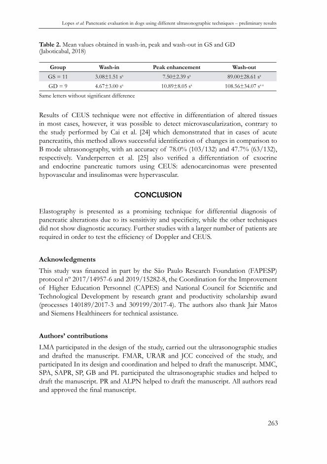
In both GS and GD, Color Doppler ultrasound did not show neovascularizations and the vascular pattern was homogenous. In Spectral Doppler, mean value of resistive index was 0.74±0.09 in GS and 0.79±0.13 in GD, with no signifi cant difference between these groups (P=0.386). Changes in these parameters were observed in cases of lesions suggestive of malignance in other tissues, such as presence of neovascularization, vessels with a tortuous aspect, elevated systolic and diastolic velocities and changes in the resistive index [21–23].Microbubble contrast-enhanced ultrasonography presented homogenous fi lling of the pancreas. Both groups had no side effects due to the use of the contrast agent. Mean values obtained in the contrast fi lling times did not have signifi cant differences between groups; wash in (P=0.128), peak enhancement (P=0.181) and wash-out (P=0.169) (Table 2). Although there was no difference between groups, animal 4 (Dalmatian) from the GD presented a markedly prolonged time in the three moments: 12s for the entrance, 32s peak enhancement and 110s the contrast agent was still present in the parenchyma. Studies performed by Rademacher et al. [4] and Lim et al. [9] also presented an increase in the time of entrance and peak enhancement in dogs with pancreatitis.
Figure 2. ROC curves representing sensitivity and specifi city (%) of each ultrasonographic method in determining pancreatic lesions.
Lopes et al: Pancreatic evaluation in dogs using different ultrasonographic techniques – preliminary results
263
Table 2. Mean values obtained in wash-in, peak and wash-out in GS and GD (Jaboticabal, 2018)
Group Wash-in Peak enhancement Wash-out
GS = 11 3.08±1.51 sa 7.50±2.39 sa 89.00±28.61 sa
GD = 9 4.67±3.00 sa 10.89±8.05 sa 108.56±34.07 sa a
Same letters without signifi cant difference
Results of CEUS technique were not effective in differentiation of altered tissues in most cases, however, it was possible to detect microvascularization, contrary to the study performed by Cai et al. [24] which demonstrated that in cases of acute pancreatitis, this method allows successful identifi cation of changes in comparison to B mode ultrasonography, with an accuracy of 78.0% (103/132) and 47.7% (63/132), respectively. Vanderperren et al. [25] also verifi ed a differentiation of exocrine and endocrine pancreatic tumors using CEUS: adenocarcinomas were presented hypovascular and insulinomas were hypervascular.
CONCLUSION
Elastography is presented as a promising technique for differential diagnosis of pancreatic alterations due to its sensitivity and specifi city, while the other techniques did not show diagnostic accuracy. Further studies with a larger number of patients are required in order to test the effi ciency of Doppler and CEUS.
Acknowledgments
This study was fi nanced in part by the São Paulo Research Foundation (FAPESP)protocol nº 2017/14957-6 and 2019/15282-8, the Coordination for the Improvement of Higher Education Personnel (CAPES) and National Council for Scientifi c and Technological Development by research grant and productivity scholarship award (processes 140189/2017-3 and 309199/2017-4). The authors also thank Jair Matos and Siemens Healthineers for technical assistance.
Authors’ contributions
LMA participated in the design of the study, carried out the ultrasonographic studies and drafted the manuscript. FMAR, URAR and JCC conceived of the study, and participated In its design and coordination and helped to draft the manuscript. MMC, SPA, SAPR, SP, GB and PL participated the ultrasonographic studies and helped to draft the manuscript. PR and ALPN helped to draft the manuscript. All authors read and approved the fi nal manuscript.

Acta Veterinaria-Beograd 2020, 70 (2), 255-266
264
Declaration of confl icting interests
The author(s) declared no potential confl icts of interest with respect to the research, authorship, and/or publication of this article.
REFERENCES
1. Watson P. Chronic pancreatitis in dogs. Top Companion Anim Med 2012;27:133–139. 2. Câmara BOS, Viana FAB, Ribeiro BNT, Ocarino NM, Nepomuceno AC, Serakides R. Um
caso raro de destruição total do pâncreas por pancreatite em cão. Arq Bras Med Veterinária e Zootec 2018;70:1655–1659.
3. Xenoulis PG., Steiner JM. Necrosis and Infl ammation: Canine. In: Canine and Feline Gastroenterology., St. Louis, MI, USA: Elsevier; 2013, p. 812–821.
4. Rademacher N, Schur D, Gaschen F, Kearney M, Gaschen L. Contrast-enhanced ultrasonography of the pancreas in healthy dogs and in dogs with acute pancreatitis. Vet Radiol Ultrasound 2016;57:58–64.
5. Mansfi eld C. Acute Pancreatitis in Dogs: Advances in Understanding, Diagnostics, and Treatment. Top Companion Anim Med 2012;27:123–132.
6. Hecht S, Henry G. Sonographic Evaluation of the Normal and Abnormal Pancreas. Clin Tech Small Anim Pract 2007;22:115–121.
7. Cridge H, MacLeod AG, Pachtinger GE, Mackin AJ, Sullivant AM, Thomason JM, Archer TM, Lunsford KV, Rosenthal K, Wills RW. Evaluation of SNAP cPL, Spec cPL, VetScan cPL Rapid Test, and Precision PSL Assays for the Diagnosis of Clinical Pancreatitis in Dogs. J Vet Intern Med 2018;32:658–664.
8. Rasslan R, Novo FCF, Bitran A, Utiyama EM, Rasslan S. Management of infected pancreatic necrosis: state of the art. Rev Col Bras Cir 2017;44:521–529.
9. Lim SY, Nakamura K, Morishita K, Sasaki N, Murakami M, Osuga T, Yokoyama N, Ohta H, Yamasaki M, Takiguchi M. Quantitative Contrast-enhanced Ultrasonographic Assessment of Naturally Occurring Pancreatitis in Dogs. J Vet Intern Med 2015;29:71–78.
10. Kawada N, Tanaka S. Elastography for the pancreas: Current status and future perspective. World J Gastroenterol 2016;22:3712–3724.
11. Ruaux CG. Diagnostic approaches to acute pancreatitis. Clin Tech Small Anim Pract 2003;18:245–249.
12. Penninck D, D’Anjou MA. Pancreas. In: Atlas of Small Animal Ultrasonography. 2nd ed., John Wiley & Sons, 2015; p. 309–330.
13. Bailey DB., Page RL. Tumors of the endocrine system. In: Withrow and MacEwens’s Small Animal Clinical Oncology. 4th ed., Philadelphia: Saunders; 2007, p. 583–609.
14. Tozaki M, Isobe S, Fukuma E. Preliminary study of ultrasonographic tissue quantifi cation of the breast using the acoustic radiation force impulse (ARFI) technology. Eur J Radiol 2011;80:182–187.
15. Bai M, Du L, Gu J, Li F, Jia X. Virtual Touch Tissue Quantifi cation Using Acoustic Radiation Force Impulse Technology. J Ultrasound Med 2012;31:289–294.

Lopes et al: Pancreatic evaluation in dogs using different ultrasonographic techniques – preliminary results
265
16. Feliciano MAR, Maronezi MC, Pavan L, Castanheira TL, Simões APR, Carvalho CF, Canola JC, Vicente WRR. ARFI elastography as a complementary diagnostic method for mammary neoplasia in female dogs - preliminary results. J Small Anim Pract 2014;55:504–508.
17. D’Onofrio M, Crosara S, De Robertis R, Canestrini S, Demozzi E, Pozzi Mucelli R. Elastography of the pancreas. Eur J Radiol 2014;83:415–419.
18. Goertz RS, Schuderer J, Strobel D, Pfeifer L, Neurath MF, Wildner D. Acoustic radiation force impulse shear wave elastography (ARFI) of acute and chronic pancreatitis and pancreatic tumor. Eur J Radiol 2016;85:2211–2216.
19. Yashima Y, Sasahira N, Isayama H, Kogure H, Ikeda H, Hirano K, Mizuno S, Yagioka H, Kawakubo, K, Sasaki T, Nakai Y, Tada M, Yoshida H, Omata M, Koike K. Acoustic radiation force impulse elastography for noninvasive assessment of chronic pancreatitis. J Gastroenterol 2012;47:427–432.
20. Dyrla P, Gil J, Florek M, Saracyn M, Grala B, Jedrzejewski E, Wojtuń S, Lubas A. Elastography in pancreatic solid tumours diagnoses. Prz Gastroenterol 2015;10:41–46.
21. Schroeder RJ, Bostanjoglo M, Rademaker J, Maeurer J, Felix R. Role of power Doppler techniques and ultrasound contrast enhancement in the differential diagnosis of focal breast lesions. Eur Radiol 2003;13:68–79.
22. Davoudi Y, Borhani B, Rad MP, Matin N. The role of doppler sonography in distinguishing malignant from benign breast lesions. J Med Ultrasound 2014;22:92–95.
23. Feliciano MAR, Uscategui RAR, Maronezi MC, Simões APR, Silva P, Gasser B, Pavan L, Carvalho CF, Canola JC, Vicente WRR. Ultrasonography methods for predicting malignancy in canine mammary tumors. PLoS One 2017;12:1–14.
24. Cai D, Parajuly SS, Wang H, Wang X, Ling W, Song B, Li Y, Luo Y. Accuracy of contrast-enhanced ultrasound compared with conventional ultrasound in acute pancreatitis: Diagnosis and complication monitoring. Exp Ther Med 2016;12:3189–3194.
25. Vanderperren K, Haers H, Van der Vekens E, Stock E, Paepe D, Daminet S, Saunders JH. Description of the use of contrast-enhanced ultrasonography in four dogs with pancreatic tumours. J Small Anim Pract 2014;55:164–169.
PROCENA PANKREASA KOD PASA UPOTREBOM RAZLIČITIH ULTRAZVUČNIH TEHNIKA- PRELIMINARNI REZULTATI
LOPES Michelle Avante, FELICIANO Marcus Antônio Rossi, USCATEGUI Ricardo Andres Ramirez, MARONEZI Marjury Cristina, SILVA Priscila Del Aguila, POZZOBON Ricardo, SIMÕES Ana Paula Rodrigues, SILVA Priscila, GASSER Beatriz, PAVAN Letícia, AIRES Luiz Paulo Nogueira, CANOLA Júlio Carlos
Cilj ove studije je da se opišu preliminarni rezultati tačnosti ultrazvučnih tehnika kao što su elastografi ja, kontrastno pojačani ultrazvuk (CEUS) i Doppler prilikom procen-jivanja promena na pankreasu. Ispitano je ukupno dvadeset i pet pasa, mužjaka i ženki, starosti 1-14 godina. Šesnaest pasa nije imalo kliničke znake oboljenja pankreasa (GS), dok je devet imalo znakove poremećaja pankreasa (GD). Sve životinje GD grupe su imale sonografske promene u B-modusu i kvalitativnoj elastografi ji, sa brzinom talasa
Acta Veterinaria-Beograd 2020, 70 (2), 255-266
266
(SWV) većom (2,4±0,5m/s) u GD grupi (p=0,014) u odnosu na GS (1,9±0,3m/s) grupu, što je rezultiralo osetljivošću od 78% i specifi čnošću određivanja promena na pankreasu od 69%. Što se tiče rezultata dobijenih putem Dopplera, nisu uočene razlike među grupama primenom kolor ili pulsnog Doppler-a. Vrednosti dobijene pomoću CEUS-a se nisu razlikovale između grupa. Obzirom na njenu osetljivost i specifi čnost, elastografi ja je tehnika koja obećava sa aspekta diferencijalne dijagnoze promena na pankreasu, dok ostale tehnike nisu pokazale dijagnostičku tačnost.