Embed Size (px)
Citation preview
257© Springer International Publishing Switzerland 2016 H. Ulrich, P. Davidson Negraes, Working with Stem Cells, DOI 10.1007/978-3-319-30582-0_15
Chapter 15 Pancreatic Differentiation from Human Pluripotent Stem Cells
Nicholas Vinckier , Jinzhao Wang , and Maike Sander
N. Vinckier • J. Wang Departments of Pediatrics and Cellular and Molecular Medicine, Pediatric Diabetes Research Center , University of California San Diego , La Jolla , CA 92093 , USA
M. Sander (*) Departments of Pediatrics and Cellular and Molecular Medicine, Pediatric Diabetes Research Center , University of California San Diego , La Jolla , CA 92093 , USA
Sanford Consortium for Regenerative Medicine , 2880 Torrey Pines Scenic Dr. – Room 3006 , La Jolla , CA 92093 , USA e-mail: [email protected]
Abstract The ability to produce human pancreatic cells in vitro would open new possibilities for developing improved therapies through cell transplantation, disease modeling, and drug screening. Of particular medical importance are the insulin- producing beta cells of the pancreas, which are lost or dysfunctional in diabetes. Furthermore, an in vitro model of human exocrine cells could help devise new ther-apies for pancreatic exocrine disease, most notably pancreatic cancer. In the past decade much progress has been made in developing protocols to generate multipo-tent pancreatic progenitor cells from human pluripotent stem cells (hPSCs) that are capable of differentiating into both endocrine and exocrine cells. The sole approach that has proven successful is to reproduce essential steps of in vivo development in vitro through directed step-wise differentiation of hPSCs. The directed differentia-tion entails sequential exposure of hPSCs to different signaling factors, thereby moving cells through several developmental intermediates towards the pancreatic fate. Upon implantation into mice, hPSC-derived pancreatic progenitor cells spon-taneously differentiate into endocrine and exocrine cells. Here, we describe a detailed protocol for the generation of pancreatic progenitor cells from hPSCs. We provide methods for the directed differentiation as well as the characterization of pancreatic progenitor cells and lineage intermediates by immunofl uorescence stain-ing and fl ow cytometry. With recently developed protocols, these pancreatic pro-genitor cells can be further differentiated in vitro into beta-like cells that functionally resemble immature human beta cells.
Keywords Human pluripotent stem cell (hPSC) • Human embryonic stem cell (hESC) • Defi nitive endoderm (DE) • Gut tube (GT) • Posterior foregut (FG) • Pancreatic endoderm (PE) • SOX17 • HNF4A • PDX1 • SOX9
258
15.1 Introduction
The development of protocols for deriving mature cell types from hPSCs has opened opportunities for modeling human disease ex vivo as well as for discovery of novel drugs (Merkle and Eggan 2013 ). In addition, functional cell types derived from hPSCs could provide an unlimited source to replace tissue cells lost in disease through cell transplantation. hPSC-based cell models are particularly promising in the context of diseases where a single cell type is affected. One such example is dia-betes, which is characterized by loss or dysfunction of the insulin-producing beta cells in the pancreas (Nathan 2015 ). There are two major forms of diabetes , type 1 and type 2 diabetes. While the causes of both types of diabetes are distinct, the mani-festation is similar: insuffi cient production of insulin results in inadequate glucose uptake by peripheral tissues, which in turn leads to elevated blood glucose levels. Type 1 diabetes is caused by autoimmune-mediated beta cell destruction and com-prises 5 % of diabetes cases (Cnop et al. 2005 ). The remaining 95 % of diabetes cases are classifi ed as type 2, and arise due to a combination of insuffi cient insulin produc-tion by the beta cells and insulin resistance of insulin target tissues (Johnson and Luciani 2010 ; Kahn et al. 2006 ; Stumvoll et al. 2005 ). Because both type 1 and type 2 diabetes eventually result in beta cell loss, transplantation of beta cells produced in vitro from hPSCs could help normalize blood glucose levels in patients with diabe-tes. In the pancreas, beta cells reside in so-called islets of Langerhans , where beta cells form functional units with other hormone-producing cells involved in blood glucose regulation, most notably the glucagon-producing alpha cells and somatosta-tin-producing delta cells. Since there is paracrine signaling between the endocrine cell types within the islet (Caicedo 2013 ), transplantation of functional islets might be clinically more benefi cial than transplanting beta cells alone. There is already precedence that transplantation of cadaveric islets can be curative for diabetes, at least in the short term (Meloche 2007 ; Robertson et al. 2000 ). hPSC- derived beta cells or islets could obviate both the limited supply of cadaveric islets and the need for immunosuppression owing to the allogeneic origin of cadaveric islets. In fact, the fi rst clinical trial to determine the safety and effi cacy of a hPSC- based islet cell replacement therapy is currently ongoing (ViaCyte 2014 ). The current clinical trial involves encapsulation of human embryonic stem cell (hESC)-derived multipotent pancreatic progenitor cells in small devices that are implanted subcutaneously. Animal studies have provided proof-of principle that these pancreatic progenitor cells will further differentiate into islet-like structures containing all endocrine cell types when implanted under the skin (Kelly et al. 2011 ; Kroon et al. 2008 ; Nostro et al. 2015 ; Rezania et al. 2012 ; Schulz et al. 2012 ; Xie et al. 2013 ).
While beta cell loss is the hallmark of type 1 diabetes , type 2 diabetes, at least in its early stages, is characterized by beta cell dysfunction rather than destruction. Type 2 diabetes has a strong genetic component; yet how genetic risk factors cause beta cell dysfunction remains largely unknown. While mouse models have provided important insight into the regulation of insulin production and secretion, compari-sons between mouse models of diabetes and the human disease have also revealed species differences. One condition that exemplifi es such differences is a monogenetic
N. Vinckier et al.
259
form of type 2 diabetes, called maturity onset diabetes of the young (MODY). In humans, inheritance of one mutant allele is suffi cient to cause MODY, whereas mice carrying the same heterozygous mutation do not acquire the disease (Haumaitre et al. 2006 ; Lee et al. 1998 ; Mayer et al. 2008 ; Yamagata et al. 1996 ). In vitro - generated beta cells and their precursors would provide a powerful model to study how MODY-associated gene mutations cause diabetes. With recent breakthroughs in genome editing technologies, known mutations, like those seen in MODY, could be introduced into hPSCs and beta cell differentiation and function could be ana-lyzed in vitro . Alternatively, induced hPSCs could be derived from MODY patients to create patient-specifi c disease models. Similar strategies could be implemented to determine how genetic variants, identifi ed via genome-wide association studies, affect beta cell development and/or function and cause disease.
A scalable differentiation system for human beta cells or islets would also provide an ideal drug-screening platform. In addition to enabling new drug discovery, such a system could help customize diabetes treatment in a patient-specifi c manner. It is known that not all patients with type 2 diabetes respond similarly to existing anti-dia-betic drugs (Standl and Fuchtenbusch 2003 ). Responsiveness to different drugs could be tested by deriving induced hPSCs from patients and producing patient-specifi c beta cells or islets in vitro . In the long term, the collective data from such experiments might allow us to predict drug responsiveness based on the absence or presence of specifi c genetic markers, as already used to predict responsiveness to cancer therapy.
The past decade has seen remarkable progress in the development of protocols to generate pancreatic cells from hPSCs (Mfopou et al. 2010 ; Pagliuca and Melton 2013 ). Multiple differentiation protocols aimed at deriving pancreatic progenitors and beta cells have been published with mounting levels of success. The underlying principle of all successful approaches is to closely mimic embryonic development in vitro . Effi cacious protocols have taken a step-wise approach in which cells are guided through defi ned developmental stages by sequential exposure to the growth factors that are also relevant during in vivo development. Earlier protocols yielded insulin-expressing cells in vitro that were not functional, expressed multiple hor-mones, and failed to restore blood glucose levels when implanted into diabetic mice (D’Amour et al. 2006 ; Kelly et al. 2011 ; Kroon et al. 2008 ; Nostro et al. 2011 ; Rezania et al. 2012 ). In contrast, when pancreatic progenitor cells were implanted into mice, the implants rescued diabetes within ~16 weeks after implantation (Kroon et al. 2008 ; Nostro et al. 2015 ; Rezania et al. 2012 ; Schulz et al. 2012 ). During this time, the progenitors differentiate into islet-like structures, containing mature func-tional beta cells as well as other endocrine cell types. A small fraction of pancreatic progenitors also differentiates into exocrine acinar and ductal cells after implanta-tion (Kroon et al. 2008 ; Xie et al. 2013 ).
Here, we present a step-wise protocol for the generation of pancreatic progenitor cells by directed differentiation of hESCs. The protocol was adapted from several pre-viously published protocols (Schulz et al. 2012 ; Rezania et al. 2013 , 2014 ). The proto-col is optimized for the H1 hESC line from WiCell (WiCell Research Institute, WA01) and employs a planar culture system in which the cells are adhered to Matrigel-coated tissue culture plates throughout differentiation. Recently, protocols have been published
15 Pancreatic Differentiation from Human Pluripotent Stem Cells
260
that report strategies to further differentiate hPSC-derived pancreatic progenitor cells into functional beta-like cells entirely in vitro (Pagliuca et al. 2014 ; Rezania et al. 2014 ; Russ et al. 2015 ). After implantation into diabetic mice, these beta-like cells lead to normalization of blood glucose levels within 2 weeks.
15.2 Materials
15.2.1 Cell Culture Supplies and Equipment
• Biological safety cabinet. • CO 2 incubator capable of maintaining 5 % CO 2 and >95 % humidity at 37 °C. • 37 °C water bath. • Sterile plastic tissue culture treated cell culture dishes:
– 6-Well plate; 9.5 cm 2 cell growth area (Corning, Cat. No. 3516). – 12-Well plate; 3.8 cm 2 cell growth area (Corning, Cat. No. 3513). – 10 cm plate; 56.5 cm 2 cell growth area (Corning, Cat. No. 351029).
• Sterile ultra-low attachment 6-well plates (Corning, Cat. No. 3471), for alterna-tive suspension culture (see Sect. 15.3.5 and Note 5 ).
• Cell lifter/scraper (VWR, Cat. No. 3008). • Hemocytometer or other cell counting tool. • Sterile 15 ml conical tubes. • Benchtop centrifuge capable of spinning 15 ml conical tubes. • Sterile 1.5 ml microcentrifuge tubes. • Benchtop microcentrifuge capable of spinning 1.5 ml microcentrifuge tubes. • Serological pipette controller and 10 ml sterile pipettes. • 2, 20, 200, and 1000 μl pipettes and accompanying sterile pipette tips. • Stericup-GP, 0.22 μm, polyethersulfone, 500 ml, radio-sterilized vacuum fi lters
(Millipore, SCGPU05RE). • Falcon 5 ml round bottom polystyrene test tube with cell strainer snap cap
(Corning, Cat. No. 352235). • Inverted light microscope (Zeiss Axio Vert or equivalent). • Inverted fl uorescence microscope (Zeiss Axio Observer or equivalent). • Flow cytometer (BD FACSCanto™ or equivalent).
15.2.2 Reagents
• Matrigel Growth Factor Reduced Basement Membrane Matrix (Corning, Cat. No. 356231).
• RHO/ROCK Pathway Inhibitor Y-27632 (StemCell, Cat. No. 72307). • TrypLE™ Express Enzyme (Life Technologies, Cat. No. 12604-021).
N. Vinckier et al.
261
• Accutase (eBioscience, Cat. No. 00-4555-56). • Sterile DPBS (Dulbecco’s Phosphate Buffered Saline) without Ca 2+ and Mg 2+
(VWR, Cat. No. 45000-434). • DMEM/F12 50:50 mix without glutamine (VWR, Cat. No. 45000-346). • Bovine Serum Albumin (BSA) Fraction V (7.5 % solution) (Life Technologies, Cat.
No. 15260-037), for alternative suspension culture (see Sect. 15.3.5 and Note 6 ). • Reagent Grade BSA—pH 7.0, > 98 % purity (Lampire Biological Laboratories,
Cat. No. 7500804), for use in differentiation media. • H1 hESC line (WiCell Research Institute, WA01). Other hPSC lines may be
used, but may require further optimization. • Essential 8 (E8) pluripotency maintenance medium (Life Technologies, Cat. No.
A1517001). The E8 culture medium is a xeno-free pluripotency maintenance medium (Chen et al. 2011 ) used here for feeder-free growth and expansion of hESCs.
• MCDB 131 cell culture medium (Life Technologies, Cat. No. 10372-019). The MCDB 131 medium is used here as the base medium throughout differentiation and is supplemented with different components and factors depending on the stage of differentiation. See Sect. 15.2.3 for proper stage-specifi c base-medium formulations and Table 15.1 for additional factors to be added on each day of differentiation.
• Supplements and factors used for endocrine differentiation:
– Sodium bicarbonate (NaHCO 3 ) (Sigma, Cat. No. S6297). – Recombinant Activin A (AA) (R&D Systems, Cat. No. 338-AC/CF). – Recombinant Wnt-3a (R&D Systems, Cat. No. 1324-WN/CF). – GlutaMAX (Life Technologies, Cat. No. 35050061). – BSA (Fisher, Cat. No. 7500804). – L-Ascorbic Acid (Vitamin C, VIT-C) (Sigma, Cat. No. A4544). – Recombinant KGF/FGF-7 (R&D Systems, Cat. No. 251-KG/CF). – D -Glucose (Fisher Scientifi c, Cat. No. D161). – Retinoic Acid (RA) (Sigma, Cat. No. R2625). – LDN193189 (Stemgent, Cat. No. 04-0074). – ITS-X (Life Technologies, Cat. No. 51500056). – SANT-1 (Sigma, Cat. No. S4572). – TPB (EMD Millipore, Cat. No. 565740).
• Cell fi xation solution for immunofl uorescence (IF) analysis: 4 % paraformalde-hyde (PFA) (96 % extra pure, Acros Organics, Cat. No. 41678) in DPBS.
• Cell permeabilization and blocking buffer for IF analysis: 0.15 % Triton X-100 (Fisher, Cat. No. BP151) and 1 % donkey serum (Gemini Bio-Products, Cat. No. 100-151) in DPBS.
• Superfrost Plus microscope slides (Fisher Scientifi c, Cat. No. 12-550-15). • VECTASHIELD Antifade mounting medium (Vector Laboratories, Cat. No.
H-1000). • FACS buffer: 0.2 % (w/v) BSA (Lampire, Cat. No. 7500804) in DPBS, keep at
2–8 °C. • BD fi xation/ permeabilization solution (BD Biosciences, Cat. No. 554714) for
fl ow cytometry analysis, keep at 2–8 °C.
15 Pancreatic Differentiation from Human Pluripotent Stem Cells
262
• BD perm/wash buffer (BD Biosciences, Cat. No. 554723) for fl ow cytometry analysis, keep at 2–8 °C.
15.2.3 Stage-Specifi c Medium Formulations
The supplements listed below are stable in the MCDB 131 medium for 1 month if kept at 2–8 °C. Because sodium bicarbonate (NaHCO 3 ) and BSA are supplied as a non-sterile powder, the medium must be sterile fi ltered (0.22 μm vacuum fi lter) after NaHCO 3 and BSA are added.
Stage 1 Medium
• MCDB 131 medium. • 1.5 g/l NaHCO 3 . • 1× GlutaMAX. • 10 mM D -Glucose (fi nal concentration; MCDB 131 already contains 5.55 mM). • 0.5 % BSA.
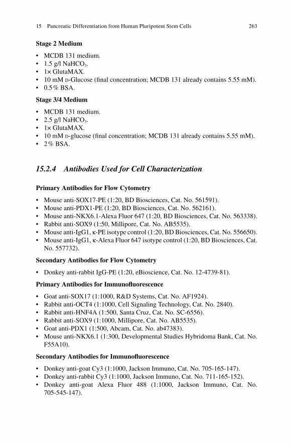
Table 15.1 Medium formulations used for each day of differentiation. See Sect. 15.2.3 for stage- specifi c base-medium formulations
Day Base medium Added factors
1 Stage 1 medium 100 ng/ml activin A 25 ng/ml Wnt-3a
2–3 Stage 1 medium 100 ng/ml activin A 4–5 Stage 2 medium 0.25 mM VIT-C
50 ng/ml FGF7 6–7 Stage 3 medium 0.25 mM VIT-C
50 ng/ml FGF7 0.25 μM SANT-1 1 μM retinoic acid 100 nM LDN193189 1:200 ITS-X 200 nM TPB
8–10 Stage 4 medium 0.25 mM VIT-C 2 ng/μl FGF7 0.25 μM SANT-1 0.1 μM retinoic acid 200 nM LDN193189 1:200 ITS-X 100 nM TPB
The same base media formulations and factors are used for either adherent cultures or suspension cultures
N. Vinckier et al.
263
Stage 2 Medium
• MCDB 131 medium. • 1.5 g/l NaHCO 3 . • 1× GlutaMAX. • 10 mM D - Glucose (fi nal concentration; MCDB 131 already contains 5.55 mM). • 0.5 % BSA.
Stage 3/4 Medium
• MCDB 131 medium. • 2.5 g/l NaHCO 3 . • 1× GlutaMAX. • 10 mM D - glucos e (fi nal concentration; MCDB 131 already contains 5.55 mM). • 2 % BSA.
15.2.4 Antibodies Used for Cell Characterization
Primary Antibodies for Flow Cytometry
• Mouse anti- SOX17 -PE (1:20, BD Biosciences, Cat. No. 561591). • Mouse anti- PDX1 -PE (1:20, BD Biosciences, Cat. No. 562161). • Mouse anti-NKX6.1-Alexa Fluor 647 (1:20, BD Biosciences, Cat. No. 563338). • Rabbit anti- SOX9 (1:50, Millipore, Cat. No. AB5535). • Mouse anti-IgG1, κ-PE isotype control (1:20, BD Biosciences, Cat. No. 556650). • Mouse anti-IgG1, κ-Alexa Fluor 647 isotype control (1:20, BD Biosciences, Cat.
No. 557732).
Secondary Antibodies for Flow Cytometry
• Donkey anti-rabbit IgG-PE (1:20, eBioscience, Cat. No. 12-4739-81).
Primary Antibodies for Immunofl uorescence
• Goat anti- SOX17 (1:1000, R&D Systems, Cat. No. AF1924). • Rabbit anti-OCT4 (1:1000, Cell Signaling Technology, Cat. No. 2840). • Rabbit anti- HNF4A (1:500, Santa Cruz, Cat. No. SC-6556). • Rabbit anti- SOX9 (1:1000, Millipore, Cat. No. AB5535). • Goat anti- PDX1 (1:500, Abcam, Cat. No. ab47383). • Mouse anti-NKX6.1 (1:300, Developmental Studies Hybridoma Bank, Cat. No.
F55A10).
Secondary Antibodies for Immunofl uorescence
• Donkey anti-goat Cy3 (1:1000, Jackson Immuno, Cat. No. 705-165-147). • Donkey anti-rabbit Cy3 (1:1000, Jackson Immuno, Cat. No. 711-165-152). • Donkey anti-goat Alexa Fluor 488 (1:1000, Jackson Immuno, Cat. No.
705-545-147).
15 Pancreatic Differentiation from Human Pluripotent Stem Cells
264
15.3 Methods
Cell culture and passaging of hESCs Proper handling and preparation of hESCs is important to ensure successful and effi cient differentiation. The protocol is optimized for H1 cells, which can be obtained from WiCell Research Institute (WA01). Below are the recommended methods for proper culture and expansion of H1 cells prior to differentiation.
15.3.1 Preparation of Matrigel-Coated Tissue Culture Dishes
Expansion is performed in 6-well plates and differentiation in 12-well plates. If different sizes of culture dishes are desired, adjust volumes as necessary.
1. Thaw Matrigel on ice and keep cold at all times; 2. Following the manufacturer’s instructions dilute Matrigel in the appropriate
volume of cold DMEM/F12 medium and keep on ice. The dilution used in this protocol is 1:100, but may vary from lot to lot. Unused diluted Matrigel can be stored at 2–8 °C, but should be used within 2 weeks from the time of dilution;
3. Load diluted Matrigel into each well of the tissue culture plate to be used. Ensure there is suffi cient volume to completely cover the entire well surface (~2 ml per well of a 6-well plate and ~1 ml per well of a 12-well plate);
4. Incubate the plate at 37 °C for 30 min before use.
15.3.2 Culturing hESCs
1. Aspirate excess Matrigel from pre-coated plates before seeding cells; 2. Thaw frozen cells in 37 °C water bath until just a small amount of ice is left in the vial; 3. Wash cells by resuspending in ~5 ml of E8 medium and spin at 200 × g for 4 min
at room temperature; 4. Aspirate supernatant carefully to not disrupt the cell pellet; 5. Resuspend cells in E8 medium (~5 × 10 5 cells/ml) containing freshly added
ROCK inhibitor (10 μM); 6. Add 2 ml cell suspension per well of the Matrigel-coated 6-well plate and place
in a 37 °C incubator at 5 % CO 2 ; 7. Replace medium with fresh E8 medium (w/o ROCK inhibitor) every 24 h; 8. Cells are passaged at ~80 % confl uency.
15.3.3 Passaging hESCs with TrypLE
1. Aspirate medium from wells and wash with DPBS without Ca 2+ and Mg 2+ ; 2. Aspirate DPBS and load 0.5–1 ml of room temperature TrypLE in each well
and incubate at 37 °C for 1 min;
N. Vinckier et al.
265
3. After 1 min examine cells under a microscope to ensure suffi cient detachment from plate. Cells should appear balled up at the edges of colonies, but not free- fl oating. If more time is required for cell detachment, place cells back in 37 °C and examine under microscope every 1 min until cells are suffi ciently detached (see Note 1 );
4. Stop enzymatic reaction by adding 4 volumes of E8 medium; 5. Use a cell scraper to lift cell clusters off plate; 6. Use a serological pipette to transfer the cell suspension into sterile 15 ml coni-
cal tubes; 7. Centrifuge for 4 min at 200 × g to pellet cells; 8. Aspirate media and resuspend cells in appropriate volume of E8 medium contain-
ing 10 μM ROCK inhibitor (Y-27632) to achieve desired dilution (see Note 2 ); 9. Add 2 ml of cell suspension per well of a Matrigel-coated 6-well plate; 10. Replace medium with fresh E8 medium (w/o ROCK inhibitor) every 24 h.
15.3.4 Cell Differentiation
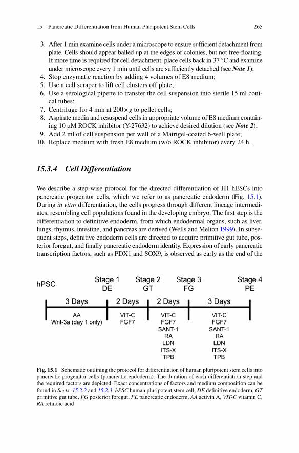
We describe a step-wise protocol for the directed differentiation of H1 hESCs into pancreatic progenitor cells, which we refer to as pancreatic endoderm (Fig. 15.1 ). During in vitro differentiation, the cells progress through different lineage intermedi-ates, resembling cell populations found in the developing embryo. The fi rst step is the differentiation to defi nitive endoderm, from which endodermal organs, such as liver, lungs, thymus, intestine, and pancreas are derived (Wells and Melton 1999 ). In subse-quent steps, defi nitive endoderm cells are directed to acquire primitive gut tube , pos-terior foregut , and fi nally pancreatic endoderm identity. Expression of early pancreatic transcription factors, such as PDX1 and SOX9 , is observed as early as the end of the
Fig. 15.1 Schematic outlining the protocol for differentiation of human pluripotent stem cells into pancreatic progenitor cells (pancreatic endoderm). The duration of each differentiation step and the required factors are depicted. Exact concentrations of factors and medium composition can be found in Sects. 15.2.2 and 15.2.3 . hPSC human pluripotent stem cell, DE defi nitive endoderm, GT primitive gut tube , FG posterior foregut , PE pancreatic endoderm, AA activin A, VIT-C vitamin C, RA retinoic acid
15 Pancreatic Differentiation from Human Pluripotent Stem Cells
266
posterior foregut stage. When implanted subcutaneously or under the kidney capsule into mice, pancreatic endoderm differentiates into fully functional beta cells, other endocrine cell types, as well as a small percentage of pancreatic ductal cells (Kelly et al. 2011 ; Kroon et al. 2008 ; Rezania et al. 2013 ; Schulz et al. 2012 ; Xie et al. 2013 ). The in vivo differentiation and maturation process of implanted pancreatic endoderm takes ~16 weeks, regardless of the protocol and cell line used to generate pancreatic endoderm. Although the application of the protocol we describe is not limited to H1 hESCs, when other hPSC lines are used, the protocol needs to be adapted to achieve similar differentiation effi ciencies as observed with H1 hESCs. Key variables include the concentration of the factors and the duration of each differentiation step.
15.3.4.1 Preparation of Coverslips for Culturing hESCs
Optional: If analysis will be performed later, it is necessary to load coverslips into the culture dish wells prior to coating with Matrigel. To prepare coverslips for cul-turing hESCs follow the procedure below, otherwise continue with the next Section.
1. Put coverslips into a sterile 10 cm petri dish with lid; 2. Pour enough 70 % ethanol into the dish to completely submerge all coverslips
and place the lid on the petri dish; 3. Allow coverslips to soak in 70 % ethanol overnight; 4. The following day, aspirate 70 % ethanol and replace it with 100 % ethanol,
ensuring all coverslips are submerged; 5. Allow coverslips to soak for 5 min; 6. Aspirate 100 % ethanol and replace it with fresh 100 % ethanol, again submerg-
ing all coverslips; 7. Allow coverslips to soak for 5 min; 8. Place petri dish lid upside down inside the biological safety cabinet; 9. Using sterilized tweezers, remove coverslips from 100 % ethanol and place
inside the inverted lid; 10. Lean each coverslip against the inside wall of the lid at an angle so that both
sides of the coverslip are exposed to air; 11. Allow the coverslips to dry completely; 12. Once coverslips have dried, using sterilized tweezers, place one coverslip on
the bottom of each well of the 12-well culture dish; 13. Proceed to coat the now sterilized coverslips with Matrigel as previously
explained.
15.3.4.2 Seeding hESCs for Differentiation
H1 cells must be plated at an appropriate density before beginning in vitro differen-tiation. Proper starting density will ensure effi cient and reproducible differentia-tions. After expanding cells to reach the desired cell numbers, the cells are passaged
N. Vinckier et al.
267
and plated on Matrigel-coated 12-well plates. To allow for accurate counting, the cells must be dissociated into a single cell suspension before seeding wells to pre-pare for the in vitro differentiation.
1. Aspirate medium from wells and rinse with DPBS without Ca 2+ and Mg 2+ (~2 ml); 2. Place 0.5–1 ml of room temperature TrypLE in each well and incubate at 37 °C
for 3–5 min; 3. After the fi rst 2 min, examine cells under a microscope to ensure suffi cient
detachment from plate (see Note 3 ); 4. Continue to incubate cells at 37 °C and examine under microscope every 1 min
until cells appear balled and many are free-fl oating; 5. Stop enzymatic reaction by adding 4 volumes of E8 medium; 6. Use a 1 ml pipette to triturate the detached cells several times to break up
remaining clumps, leaving a single-cell suspension (see Note 4 ); 7. Use a serological pipette to transfer the cell suspension into sterile 15 ml coni-
cal tubes; 8. Reserve a small aliquot of cell suspension for cell counting; 9. Centrifuge for 4 min at 200 × g to pellet cells and count cells during
centrifugation; 10. Aspirate media and resuspend cells in E8 medium (~3 × 10 5 cells/ml) contain-
ing freshly added ROCK inhibitor (10 μM); 11. Add 1.5 ml of cell suspension per well of a Matrigel-coated 12-well plate; 12. After 24–48 h cells should be roughly 90 % confl uent and ready to begin in
vitro differentiation.
15.3.5 Differentiation to Pancreatic Endoderm (Adherent Culture)
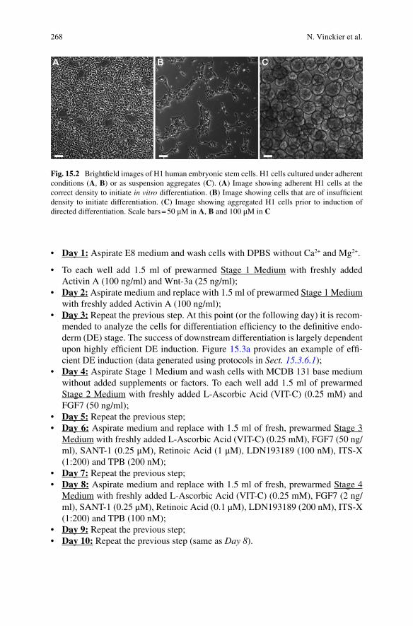
Figure 15.1 outlines the step-wise differentiation process and key factors contained in the differentiation medium at each stage of differentiation. Once cells have reached 90 % confl uency (Fig. 15.2a ), the differentiation process can be started. Figure 15.2b shows an example of cells that need to be expanded further before dif-ferentiation can be initiated. Stage-specifi c base-media are stable at 2–8 °C for up to 1 month and can therefore be mixed ahead of time. The formulations for stage- specifi c base-media are listed in Sect. 15.2.3 . Table 15.1 shows the fi nal concentra-tions of the added factors at each day of differentiation. These factors are not stable at 2–8 °C for long periods of time and must be added to stage-specifi c base-medium immediately prior to warming the medium on each day of differentiation.
Described here is a method in which the cells are adhered to 12-well plates in planar culture. However, this protocol can also be performed in suspension culture (Fig. 15.2c ), as previously described in detail (Schulz et al. 2012 ). Suspension cul-ture requires scaling up medium volumes and cell numbers, as well as orbital rotation (see Notes 5 and 6 ).
15 Pancreatic Differentiation from Human Pluripotent Stem Cells
268
• Day 1: Aspirate E8 medium and wash cells with DPBS without Ca 2+ and Mg 2+ .
• To each well add 1.5 ml of prewarmed Stage 1 Medium with freshly added Activin A (100 ng/ml) and Wnt-3a (25 ng/ml);
• Day 2: Aspirate medium and replace with 1.5 ml of prewarmed Stage 1 Medium with freshly added Activin A (100 ng/ml);
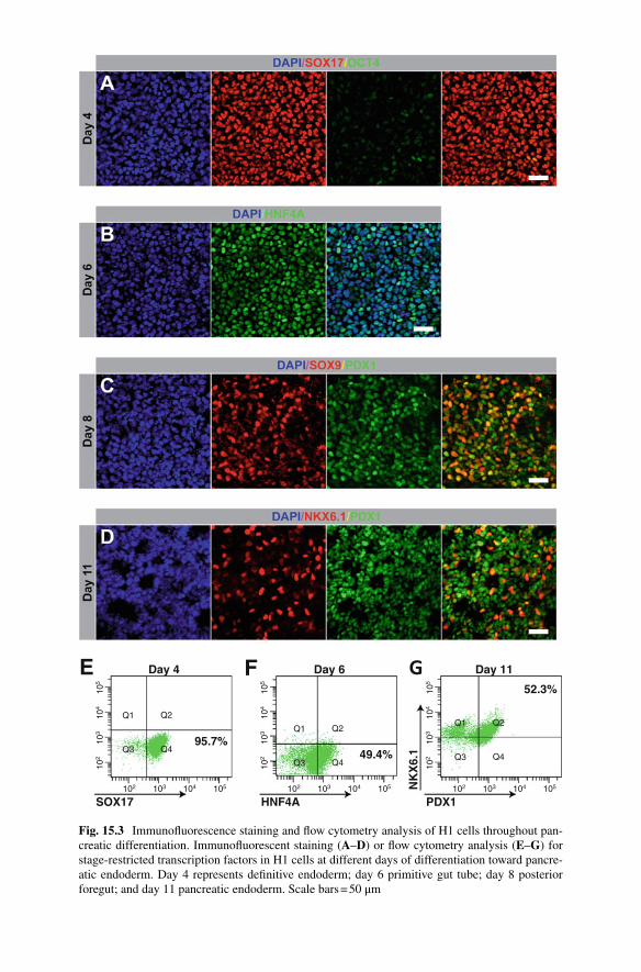
• Day 3: Repeat the previous step. At this point (or the following day) it is recom-mended to analyze the cells for differentiation effi ciency to the defi nitive endo-derm (DE) stage. The success of downstream differentiation is largely dependent upon highly effi cient DE induction. Figure 15.3a provides an example of effi -cient DE induction (data generated using protocols in Sect. 15.3.6.1 );
• Day 4: Aspirate Stage 1 Medium and wash cells with MCDB 131 base medium without added supplements or factors. To each well add 1.5 ml of prewarmed Stage 2 Medium with freshly added L-Ascorbic Acid (VIT-C) (0.25 mM) and FGF7 (50 ng/ml);
• Day 5: Repeat the previous step; • Day 6: Aspirate medium and replace with 1.5 ml of fresh, prewarmed Stage 3
Medium with freshly added L-Ascorbic Acid (VIT-C) (0.25 mM), FGF7 (50 ng/ml), SANT-1 (0.25 μM), Retinoic Acid (1 μM), LDN193189 (100 nM), ITS-X (1:200) and TPB (200 nM);
• Day 7: Repeat the previous step; • Day 8: Aspirate medium and replace with 1.5 ml of fresh, prewarmed Stage 4
Medium with freshly added L-Ascorbic Acid (VIT-C) (0.25 mM), FGF7 (2 ng/ml), SANT-1 (0.25 μM), Retinoic Acid (0.1 μM), LDN193189 (200 nM), ITS-X (1:200) and TPB (100 nM);
• Day 9: Repeat the previous step; • Day 10: Repeat the previous step (same as Day 8 ).
Fig. 15.2 Brightfi eld images of H1 human embryonic stem cells. H1 cells cultured under adherent conditions ( A , B ) or as suspension aggregates ( C ). ( A ) Image showing adherent H1 cells at the correct density to initiate in vitro differentiation. ( B ) Image showing cells that are of insuffi cient density to initiate differentiation. ( C ) Image showing aggregated H1 cells prior to induction of directed differentiation. Scale bars = 50 μM in A , B and 100 μM in C
N. Vinckier et al.
269
At the end of the differentiation protocol the cells have adopted characteristics of pancreatic progenitor cells. A recent report by Rezania et al. describes a method to further differentiate these H1-derived pancreatic endoderm cells into beta-like cells in vitro (Rezania et al. 2014 ). The method involves culturing the cells in a liquid-air interface after the pancreatic endoderm stage. An alternative method to the liquid- air interface culture is to perform the differentiation into beta-like cells in suspen-sion culture. However, the differentiation effi ciency is reported to be lower in suspension culture than in liquid-air interface culture (Rezania et al. 2014 ). Two other recent reports utilized suspension culture methods to derive beta-like cells, similar to the ones reported by Rezania et al. from hPSC-derived pancreatic endo-derm (Pagliuca et al. 2014 ; Russ et al. 2015 ).
15.3.6 Cell Characterization
15.3.6.1 Characterization of Cells by Flow Cytometry and Immunofl uorescence Analysis
To ensure effi cient differentiation, it is important to characterize the cells at various stages throughout the differentiation. Flow cytometry analysis and IF staining for stage-specifi c proteins are the hallmark methods to assess differentiation effi cien-cies. Figure 15.3 shows example images of IF analysis as well as fl ow cytometry plots of stage-specifi c markers from a successful differentiation. For a complete list of antibodies used for fl ow cytometry and IF analysis see Sect. 15.2.4 . Below are recommended protocols for both fl ow cytometry and IF analysis.
Flow cytometry analysis protocol
1. Thaw Accutase at room temperature or 2–8 °C prior to use; 2. Aspirate medium from each well to be analyzed; 3. Wash cells with DPBS (~1.5 ml per well of 12-well plate) and aspirate; 4. Add enough Accutase® to cover the surface of each well (0.3–0.5 ml per well
of 12-well plate); 5. Incubate at 37 °C for 5 min, then check briefl y under microscope to ensure cells
appear balled up and many are detached. If needed, place back at 37 °C and check every 1 min for good detachment (see Note 7 );
6. Add ~1 ml cold FACS buffer (0.2 % BSA in DPBS) to each well (fi nal volume ~1.5 ml) to stop Accutase activity;
7. Using a 1000 μl pipette triturate cells to break up any clumps and obtain a sin-gle cell suspension;
8. Label a 5 ml polystyrene tube with cell strainer cap for each sample and add ~250 μl FACS buffer to each cap to pre-wet the cell strainer. Ensure the buffer fl ows through the cap into the tube;
9. Load each sample into the cap of the appropriately labeled tube and allow the entire suspension to fl ow through the cell strainer. This will ensure good cell
15 Pancreatic Differentiation from Human Pluripotent Stem Cells
102
95.7%
Day 4 Day 6 Day 11
SOX17
E F G
HNF4A PDX1
NK
X6.
149.4%
52.3%
103 104 105
102
103
104
105
102
Q1 Q2
Q3 Q4
Q1 Q2
Q3 Q4
Q1 Q2
Q3 Q4
103 104 105
102
103
104
105
102 103 104 105
102
103
104
105
Fig. 15.3 Immunofl uorescence staining and fl ow cytometry analysis of H1 cells throughout pan-creatic differentiation. Immunofl uorescent staining ( A – D ) or fl ow cytometry analysis ( E – G ) for stage-restricted transcription factors in H1 cells at different days of differentiation toward pancre-atic endoderm. Day 4 represents defi nitive endoderm; day 6 primitive gut tube; day 8 posterior foregut; and day 11 pancreatic endoderm. Scale bars = 50 μm
271
separation, which will provide better fl ow cytometry results. If the suspension does not easily fl ow through the cell strainer it may be necessary to pull the sample through. To do so, place the tip of a 1000 μl pipette on the underside of the cell strainer cap containing the sample. Slowly release the pipette plunger to gently pull the sample through the fi lter top. Carefully eject the sample from the 1000 μl pipette into the bottom of the tube;
10. Transfer the strained cells into new 1.5 ml microcentrifuge tubes and centrifuge at room temperature and 200 × g for 5 min;
11. Resuspend each cell pellet with cold BD fi xation/permeabilization solution (300 μl per tube);
12. Incubate cells for 20 min at 2–8 °C (i.e. refrigerate— do not place on ice ); 13. Wash cells by adding ~1.1 ml cold 1× BD Perm/Wash™ Buffer to each tube
following fi xation/permeabilization (1.5 ml per well of 12-well plate) and cen-trifuge at 10 °C and 200 × g for 5 min;
14. Aspirate supernatant and repeat step 11 ; 15. Aspirate supernatant and resuspend cells in 50 μl cold 1× BD Perm/Wash
Buffer for each staining and isotype control to be used (e.g. add 150 μl total for two different staining samples and one isotype control sample);
16. Aliquot 50 μl of cell suspension into separate microcentrifuge tubes for each individual reaction. Co-staining with PE and AlexaFluor 647 conjugated anti-bodies can be performed together in one 50 μl aliquot;
17. For analysis of SOX17 expression (DE marker) load 2.5 μl SOX17-PE anti-body into one 50 μl cell suspension aliquot. Load 2.5 μl PE isotype into a sepa-rate 50 μl cell suspension aliquot to serve as a control;
18. For analysis of PDX1 expression (pancreatic endoderm marker) load 2.5 μl PDX1-PE antibody into one 50 μl cell suspension aliquot. Load 2.5 μl PE iso-type into a separate 50 μl cell suspension aliquot to serve as a control;
19. For analysis of NKX6.1 expression (pancreatic endoderm marker) load 2.5 μl NKX6.1-AlexaFluor 647 antibody into one 50 μl cell suspension aliquot. Load 2.5 μl AlexaFluor 647 isotype into a separate 50 μl cell suspension aliquot to serve as a control;
20. After the antibodies and matching isotypes have been loaded into cell suspensions, incubate cells in the dark for 1 h at 2–8 °C (i.e. refrigerate— do not place on ice );
21. Wash cells by adding 1.25 ml cold 1× BD Wash Buffer to each tube and centri-fuge at room temperature and 200 × g for 5 min;
22. Aspirate supernatant and resuspend each sample in 300 μl cold FACS buffer; 23. The samples are now stained and ready for analysis on a fl ow cytometer, such
as the FACSCanto used to generate the plots in Fig. 15.3 .
Immunofl uorescence analysis protocol
1. Aspirate medium from each well to be analyzed; 2. Wash cells with DPBS (~1.5 ml per well of 12-well plate) and aspirate; 3. Repeat step 2 ; 4. Add 1 ml of 4 % PFA to each well and incubate at 2–8 °C overnight; 5. The following day, aspirate PFA and wash with DPBS (~1.5 ml per well of
12-well plate);
15 Pancreatic Differentiation from Human Pluripotent Stem Cells
272
6. Aspirate DPBS and add 1 ml of blocking buffer (0.15 % Triton X-100, 1 % normal donkey serum in DPBS) and incubate at room temperature for 1 h;
7. Prepare primary antibody solutions by mixing appropriate volumes of each anti-body in blocking buffer (1 ml per well of a 12 well plate) and vortex gently. See Sect. 15.2.4 for appropriate working dilutions. For example, to analyze SOX17 expression, add 1 μl of goat anti-SOX17 antibody to 1 ml of blocking buffer;
8. Aspirate buffer from each well and load 1 ml of pre-mixed primary antibody solution to the appropriate wells and incubate at 2–8 °C overnight;
9. The following day, aspirate antibody solution and wash with DPBS (~1.5 ml per well of 12-well plate);
10. Aspirate DPBS and repeat step 9 three times; 11. Prepare secondary antibody solutions by mixing appropriate volumes of each
antibody in blocking buffer (1 ml per well of 12 well plate) and vortex gently. See Sect. 15.2.4 for appropriate working dilutions;
12. Aspirate DPBS from each well and add 1 ml of pre-mixed secondary antibody solution to the appropriate wells and incubate at room temperature for 1 h in the dark;
13. Aspirate antibody solution and wash with DPBS (~1.5 ml per well of 12-well plate);
14. Prepare nuclear staining solution by mixing Hoescht 33342 in DPBS (1:3000, 1 ml per well of 12-well plate);
15. Aspirate DPBS from each well and add 1 ml of nuclear staining solution and incubate at room temperature for 5 min in the dark;
16. Aspirate nuclear staining solution and wash with DPBS (~1.5 ml per well of 12-well plate);
17. Aspirate DPBS and wash once more with DPBS (~1.5 ml per well of 12-well plate);
18. The coverslips containing stained cells are now ready to be mounted on slides. Gently remove coverslips with tweezers and rinse by briefl y dipping each cov-erslip into distilled water;
19. Allow fl uid to run off the coverslip and remove excess fl uid by blotting the edge of the coverslip with a paper towel. Capillary action should help remove the excess;
20. Place one drop of VECTASHIELD Antifade Mounting Medium (or desired mounting solution) on the coverslip on the side containing the cells;
21. Gently invert coverslip while placing it cell side down on a slide. Be careful to avoid any air bubbles between the slide and coverslip;
22. Seal the edges of the coverslip to the slide with clear nail polish and allow it to fully dry in the dark before analyzing the slides. The nail polish prevents the slides from drying out if stored for extended periods of time;
23. The slide containing the coverslip with the stained cells is now ready to be analyzed on an inverted fl uorescent microscope, such as the Zeiss Axio Observer used to generate the images in Fig. 15.3 ;
24. Slides can be stored at 4 °C for short periods of time and at −20 °C for extended periods of time .
N. Vinckier et al.
273
15.4 Notes
1. During expansion of H1 cells it is better to passage cells as clusters rather than as a single cell suspension. This will enhance health and survival of the cells.
2. Typical passaging dilutions for expanding H1 cells is 1:6 (i.e. 1 well split into 6 wells).
3. Avoid incubating with TrypLE for longer than necessary as this can reduce cell viability.
4. Avoid excessive pipetting as this can reduce cell viability. 5. Performing differentiation in suspension culture —To culture cells in suspension,
use ultra-low attachment 6-well plates and rotate on an orbital shaker at 100 rpm. This ensures proper cell aggregation, which is necessary for cell survival and differentiation (Fig. 15.2c ). The culture volume is 5.5 ml/well and base media formulations and concentrations of factors are the same as used for differentia-tion in adherent cultures (Table 15.1 ). Following expansion and dissociation of hESCs with TrypLE, seed wells with 1 × 10 6 cells/ml, which is a total of 5.5 × 10 6 cells/well. Add 10 μM ROCK inhibitor to medium when seeding cells to promote cell survival and aggregation. 48 h following cell seeding the now aggregated cells (Fig. 15.2c ) are ready to initiate the in vitro differentiation. The differentiation protocol is the same as described for adherent culture, but the volume of culture medium has to be adjusted.
6. Handling cell aggregates in suspension culture —A solution of 0.2 % BSA in DPBS should be used to coat pipettes in order to prevent aggregates from adher-ing to plastic surfaces during medium exchanges. To properly exchange culture medium, draw PBS/BSA solution into the pipette and eject the solution. Swirl the 6-well plate in a circular motion to bring all aggregates to the center. Tilt the plate and draw medium off the side of each well with the pre-coated pipette, removing most of the medium. Leave a very small amount of medium to ensure no aggregates are removed. Load each well with 5.5 ml of the appropriate cul-ture medium for that day. Place the plate back on the orbital shaker at 100 rpm inside a 37 °C incubator.
7. Avoid prolonged exposure to Accutase. Detachment of monolayer H1 cells should occur after 5 min, however if the Accutase has started to become inactive, more time may be necessary. To ensure swift cell detachment use freshly thawed Accutase.
Acknowledgements The authors would like to thank Andrea Carrano and Allen Wang for com-ments. This work was supported by National Institutes of Health grants (U01-DK089567 and UC4-DK104202), California Institute for Regenerative Medicine grants (RB5-07236 and RB4- 06144), and Helmsley Charitable Trust grant 2012PG-T1D074 to M.S. N.V. was supported by the National Cancer Institute Cancer Cell Biology training grant 5T32 CA067754.
15 Pancreatic Differentiation from Human Pluripotent Stem Cells
274
References
Caicedo A (2013) Paracrine and autocrine interactions in the human islet: more than meets the eye. Semin Cell Dev Biol 24:11–21
Chen G, Gulbranson DR, Hou Z, Bolin JM, Ruotti V, Probasco MD, Smuga-Otto K, Howden SE, Diol NR, Propson NE et al (2011) Chemically defi ned conditions for human iPSC derivation and culture. Nat Methods 8:424–429
Cnop M, Welsh N, Jonas JC, Jorns A, Lenzen S, Eizirik DL (2005) Mechanisms of pancreatic beta-cell death in type 1 and type 2 diabetes: many differences, few similarities. Diabetes 54(Suppl 2):S97–S107
D'Amour KA, Bang AG, Eliazer S, Kelly OG, Agulnick AD, Smart NG, Moorman MA, Kroon E, Carpenter MK, Baetge EE (2006) Production of pancreatic hormone-expressing endocrine cells from human embryonic stem cells. Nat Biotechnol 24:1392–1401
Haumaitre C, Fabre M, Cormier S, Baumann C, Delezoide AL, Cereghini S (2006) Severe pan-creas hypoplasia and multicystic renal dysplasia in two human fetuses carrying novel HNF1beta/MODY5 mutations. Hum Mol Genet 15:2363–2375
Johnson JD, Luciani DS (2010) Mechanisms of pancreatic beta-cell apoptosis in diabetes and its therapies. Adv Exp Med Biol 654:447–462
Kahn SE, Hull RL, Utzschneider KM (2006) Mechanisms linking obesity to insulin resistance and type 2 diabetes. Nature 444:840–846
Kelly OG, Chan MY, Martinson LA, Kadoya K, Ostertag TM, Ross KG, Richardson M, Carpenter MK, D'Amour KA, Kroon E et al (2011) Cell-surface markers for the isolation of pancreatic cell types derived from human embryonic stem cells. Nat Biotechnol 29:750–756
Kroon E, Martinson LA, Kadoya K, Bang AG, Kelly OG, Eliazer S, Young H, Richardson M, Smart NG, Cunningham J et al (2008) Pancreatic endoderm derived from human embryonic stem cells generates glucose-responsive insulin-secreting cells in vivo. Nat Biotechnol 26:443–452
Lee YH, Sauer B, Gonzalez FJ (1998) Laron dwarfi sm and non-insulin-dependent diabetes melli-tus in the Hnf-1alpha knockout mouse. Mol Cell Biol 18:3059–3068
Mayer C, Bottcher Y, Kovacs P, Halbritter J, Stumvoll M (2008) Phenotype of a patient with a de novo mutation in the hepatocyte nuclear factor 1beta/maturity-onset diabetes of the young type 5 gene. Metabolism 57:416–420
Meloche RM (2007) Transplantation for the treatment of type 1 diabetes. World J Gastroenterol 13:6347–6355
Merkle FT, Eggan K (2013) Modeling human disease with pluripotent stem cells: from genome association to function. Cell Stem Cell 12:656–668
Mfopou JK, Chen B, Sui L, Sermon K, Bouwens L (2010) Recent advances and prospects in the differentiation of pancreatic cells from human embryonic stem cells. Diabetes 59:2094–2101
Nathan DM (2015) Diabetes: advances in diagnosis and treatment. JAMA 314:1052–1062 Nostro MC, Sarangi F, Ogawa S, Holtzinger A, Corneo B, Li X, Micallef SJ, Park IH, Basford C,
Wheeler MB et al (2011) Stage-specifi c signaling through TGFbeta family members and WNT regulates patterning and pancreatic specifi cation of human pluripotent stem cells. Development 138:861–871
Nostro MC, Sarangi F, Yang C, Holland A, Elefanty AG, Stanley EG, Greiner DL, Keller G (2015) Effi cient generation of NKX6-1+ pancreatic progenitors from multiple human pluripotent stem cell lines. Stem Cell Rep 4:591–604
Pagliuca FW, Melton DA (2013) How to make a functional beta-cell. Development 140:2472–2483
Pagliuca FW, Millman JR, Gurtler M, Segel M, Van Dervort A, Ryu JH, Peterson QP, Greiner D, Melton DA (2014) Generation of functional human pancreatic beta cells in vitro. Cell 159:428–439
Rezania A, Bruin JE, Riedel MJ, Mojibian M, Asadi A, Xu J, Gauvin R, Narayan K, Karanu F, O'Neil JJ et al (2012) Maturation of human embryonic stem cell-derived pancreatic progenitors
N. Vinckier et al.
275
into functional islets capable of treating pre-existing diabetes in mice. Diabetes 61:2016–2029
Rezania A, Bruin JE, Xu J, Narayan K, Fox JK, O'Neil JJ, Kieffer TJ (2013) Enrichment of human embryonic stem cell-derived NKX6.1-expressing pancreatic progenitor cells accelerates the maturation of insulin-secreting cells in vivo. Stem Cells 31:2432–2442
Rezania A, Bruin JE, Arora P, Rubin A, Batushansky I, Asadi A, O’Dwyer S, Quiskamp N, Mojibian M, Albrecht T et al (2014) Reversal of diabetes with insulin-producing cells derived in vitro from human pluripotent stem cells. Nat Biotechnol 32:1121–1133
Robertson RP, Davis C, Larsen J, Stratta R, Sutherland DE (2000) Pancreas and islet transplanta-tion for patients with diabetes. Diabetes Care 23:112–116
Russ HA, Parent AV, Ringler JJ, Hennings TG, Nair GG, Shveygert M, Guo T, Puri S, Haataja L, Cirulli V et al (2015) Controlled induction of human pancreatic progenitors produces func-tional beta-like cells in vitro. EMBO J 34:1759–1772
Schulz TC, Young HY, Agulnick AD, Babin MJ, Baetge EE, Bang AG, Bhoumik A, Cepa I, Cesario RM, Haakmeester C et al (2012) A scalable system for production of functional pan-creatic progenitors from human embryonic stem cells. PLoS One 7:e37004
Standl E, Fuchtenbusch M (2003) The role of oral antidiabetic agents: why and when to use an early-phase insulin secretion agent in Type II diabetes mellitus. Diabetologia 46(Suppl 1):M30–M36
Stumvoll M, Goldstein BJ, van Haeften TW (2005) Type 2 diabetes: principles of pathogenesis and therapy. Lancet 365:1333–1346
ViaCyte I (2014) ViaCyte, Inc. Announces FDA acceptance of IND to commence clinical trial of VC-01™ candidate cell replacement therapy for type 1 diabetes. [Press release online] http://viacyte.com/press-releases/viacyte-inc-announces-fda-acceptance-of-ind-to-commence-clinical-trial-of-vc-01-candidate- cell-replacement-therapy-for-type-1-diabetes/
Wells JM, Melton DA (1999) Vertebrate endoderm development. Annu Rev Cell Dev Biol 15:393–410
Xie R, Everett LJ, Lim HW, Patel NA, Schug J, Kroon E, Kelly OG, Wang A, D'Amour KA, Robins AJ et al (2013) Dynamic chomatin remodeling mediated by polycomb proteins orches-trates pancreatic differentiation of human embryonic stem cells. Cell Stem Cell 12:224–237
Yamagata K, Furuta H, Oda N, Kaisaki PJ, Menzel S, Cox NJ, Fajans SS, Signorini S, Stoffel M, Bell GI (1996) Mutations in the hepatocyte nuclear factor-4alpha gene in maturity-onset diabe-tes of the young (MODY1). Nature 384:458–460
15 Pancreatic Differentiation from Human Pluripotent Stem Cells