Embed Size (px)
Citation preview

ORIGINAL ARTICLE
Classification of acute pancreatitis—2012:revision of the Atlanta classification and definitionsby international consensusPeter A Banks,1 Thomas L Bollen,2 Christos Dervenis,3 Hein G Gooszen,4
Colin D Johnson,5 Michael G Sarr,6 Gregory G Tsiotos,7 Santhi Swaroop Vege,8
Acute Pancreatitis Classification Working Group
▸ Additional data are publishedonline only. To view these filesplease visit the journal online(http://dx.doi.org/10.1136/gutjnl-2012-302779).1Division of Gastroenterology,Hepatology, and Endoscopy,Harvard Medical School,Brigham and Women’sHospital, Boston,Massachusetts, USA2Department of Radiology,St Antonius Hospital,Nieuwegein, the Netherlands3First Department of Surgery,Agia Olga Hospital, Athens,Greece4Evidence-Based SurgeryResearch Unit, University ofNijmegen, Nijmegen,the Netherlands5University HospitalSouthampton, Southampton,UK6Department of Surgery, MayoClinic, Rochester, Minnesota,USA7Division of Digestive Surgery,Metropolitan Hospital, Athens,Greece8Department of Medicine,Division of Gastroenterologyand Hepatology, Mayo Clinic,Rochester, Minnesota, USA
Correspondence toMichael G Sarr, Departmentof Surgery, Mayo Clinic(GU 10-01), 200 1st St SW,Rochester, MN 55905, USA;[email protected]
Revised 29 August 2012Accepted 29 August 2012Published Online First25 October 2012
ABSTRACTBackground and objective The Atlanta classificationof acute pancreatitis enabled standardised reporting ofresearch and aided communication between clinicians.Deficiencies identified and improved understanding of thedisease make a revision necessary.Methods A web-based consultation was undertaken in2007 to ensure wide participation of pancreatologists.After an initial meeting, the Working Group sent a draftdocument to 11 national and international pancreaticassociations. This working draft was forwarded to allmembers. Revisions were made in response tocomments, and the web-based consultation wasrepeated three times. The final consensus was reviewed,and only statements based on published evidence wereretained.Results The revised classification of acute pancreatitisidentified two phases of the disease: early and late.Severity is classified as mild, moderate or severe. Mildacute pancreatitis, the most common form, has no organfailure, local or systemic complications and usuallyresolves in the first week. Moderately severe acutepancreatitis is defined by the presence of transient organfailure, local complications or exacerbation of co-morbiddisease. Severe acute pancreatitis is defined bypersistent organ failure, that is, organ failure >48 h.Local complications are peripancreatic fluid collections,pancreatic and peripancreatic necrosis (sterile orinfected), pseudocyst and walled-off necrosis (sterile orinfected). We present a standardised template forreporting CT images.Conclusions This international, web-based consensusprovides clear definitions to classify acute pancreatitisusing easily identified clinical and radiologic criteria. Thewide consultation among pancreatologists to reach thisconsensus should encourage widespread adoption.
BACKGROUNDThe Atlanta Symposium in 1992 attempted to offera global ‘consensus’ and a universally applicable clas-sification system for acute pancreatitis.1 Althoughthe Atlanta Classification has been useful, some ofthe definitions proved confusing.2 Better understand-ing of the pathophysiology of organ failure andnecrotising pancreatitis and their outcomes, as wellas improved diagnostic imaging, have made it neces-sary to revise the Atlanta Classification. This revi-sion includes a clinical assessment of severity and
provides more objective terms to describe the localcomplications of acute pancreatitis.The goal of this report is to present the updated
revision of the Atlanta Classification of acute pan-creatitis in adults (>18 years). This revision wasdesigned to incorporate modern concepts of thedisease, to address areas of confusion, to improveclinical assessment of severity, to enable standardisedreporting of data, to assist the objective evaluationof new treatments, and to facilitate communicationamong treating physicians and between institutions.This consensus classification defines criteria for thediagnosis of acute pancreatitis, differentiates the two
▸ http://dx.doi.org/10.1136/gutjnl-2012-303724http://dx.doi.org/10.1136/gutjnl-2012-303725http://dx.doi.org/10.1136/gutjnl-2012-304051
Significance of this study
What is already known on this subject?▸ The original Atlanta Classification of acute pan-
creatitis of 1992 is outdated.▸ Two types of acute pancreatitis have been
described: acute oedematous pancreatitis andacute necrotising pancreatitis.
▸ The description of pancreatic and peripancreaticcollections is confusing and not universal.
What are the new findings?▸ This current global consensus classification of
acute pancreatitis offers a comprehensive clas-sification of acute pancreatitis, severity andperipancreatic collections.
▸ New information of aetiology, pathophysiology,severity and radiologic descriptions of pancre-atic and peripancreatic collections are provided.
▸ This classification differentiates acute peripan-creatic fluid, pancreatic pseudocyst, acute nec-rotic collections and walled-off necrosis.
How might it impact on clinical practice inthe foreseeable future?▸ This classification of acute pancreatitis will
allow a consistent, worldwide classification.▸ The description of pancreatic and peripancreatic
collections on cross-sectional imaging will allowa consistent terminology across all studies.
▸ This classification of acute pancreatitis shouldavoid the confusion in terminology seen overthe last 20 years.
102 Gut 2013;62:102–111. doi:10.1136/gutjnl-2012-302779
Pancreas
on Novem
ber 13, 2021 by guest. Protected by copyright.
http://gut.bmj.com
/G
ut: first published as 10.1136/gutjnl-2012-302779 on 25 October 2012. D
ownloaded from
on N
ovember 13, 2021 by guest. P
rotected by copyright.http://gut.bm
j.com/
Gut: first published as 10.1136/gutjnl-2012-302779 on 25 O
ctober 2012. Dow
nloaded from
on Novem
ber 13, 2021 by guest. Protected by copyright.
http://gut.bmj.com
/G
ut: first published as 10.1136/gutjnl-2012-302779 on 25 October 2012. D
ownloaded from
types of acute pancreatitis (interstitial oedematous pancreatitisand necrotising pancreatitis), classifies the severity of acute pan-creatitis into three categories, and defines the morphology seen onimaging of pancreatic and peripancreatic collections that arise ascomplications of acute pancreatitis. This revision is not intendedto be a management guideline.
METHODSThis classification was generated by an iterative, web-based con-sultation process led by a working group and incorporatingresponses from the members of 11 national and internationalpancreatic societies. All responses were reviewed by the workinggroup, and the process was repeated by a web-based approachuntil the current fourth draft, which was then finalised for sub-mission. A full description of the methods is shown in onlinesupplementary appendix 1. There are many substantial andimportant differences in the current document when comparedto our preliminary working draft that appeared on the PancreasClub website3 and which has been referred to by otherauthors.4–8
Revised definitions and classification of acute pancreatitisThe following definitions and classifications are proposedfor use in clinical and research communications.
Definition of diagnosis of acute pancreatitisThe diagnosis of acute pancreatitis requires two of the followingthree features: (1) abdominal pain consistent with acute pancrea-titis (acute onset of a persistent, severe, epigastric pain often radi-ating to the back); (2) serum lipase activity (or amylase activity) atleast three times greater than the upper limit of normal; and (3) charac-teristic findings of acute pancreatitis on contrast-enhancedcomputed tomography (CECT) and less commonly magnetic res-onance imaging (MRI) or transabdominal ultrasonography.9–13
If abdominal pain suggests strongly that acute pancreatitis ispresent, but the serum amylase and/or lipase activity is less thanthree times the upper limit of normal, as may be the case withdelayed presentation, imaging will be required to confirm thediagnosis.13 14 If the diagnosis of acute pancreatitis is establishedby abdominal pain and by increases in the serum pancreaticenzyme activities, a CECT is not usually required for diagnosisin the emergency room or on admission to the hospital.
Definition of onset of acute pancreatitisThe onset of acute pancreatitis is defined as the time of onsetof abdominal pain (not the time of admission to the hospital).The time interval between onset of abdominal pain and firstadmission to the hospital should be noted. When patients withsevere disease are transferred to a tertiary hospital, the intervalsbetween onset of symptoms, first admission and transfershould be noted. Data recorded from a tertiary care hospitalshould be stratified to allow separate consideration of theoutcomes of patients who were admitted directly and thoseadmitted by transfer from another hospital (see online supple-mentary appendix 2 for suggested recording of data).
Definition of types of acute pancreatitisAcute pancreatitis can be subdivided into two types: interstitialoedematous pancreatitis and necrotising pancreatitis.
Interstitial oedematous pancreatitisThe majority of patients with acute pancreatitis have diffuse(or occasionally localised) enlargement of the pancreas due toinflammatory oedema. On CECT, the pancreatic parenchyma
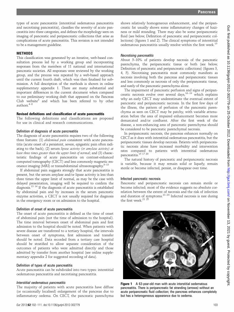
shows relatively homogeneous enhancement, and the peripan-creatic fat usually shows some inflammatory changes of hazi-ness or mild stranding. There may also be some peripancreaticfluid (see below, Definition of pancreatic and peripancreatic col-lections) (figures 1 and 2). The clinical symptoms of interstitialoedematous pancreatitis usually resolve within the first week.15
Necrotising pancreatitisAbout 5–10% of patients develop necrosis of the pancreaticparenchyma, the peripancreatic tissue or both (see below,Definition of pancreatic and peripancreatic collections) (figures 3,4, 5). Necrotising pancreatitis most commonly manifests asnecrosis involving both the pancreas and peripancreatic tissuesand less commonly as necrosis of only the peripancreatic tissue,and rarely of the pancreatic parenchyma alone.
The impairment of pancreatic perfusion and signs of peripan-creatic necrosis evolve over several days,16–19 which explainswhy an early CECT may underestimate the eventual extent ofpancreatic and peripancreatic necrosis. In the first few days ofthe illness, the pattern of perfusion of the pancreatic paren-chyma as seen on CECT may be patchy, with variable attenu-ation before the area of impaired enhancement becomes moredemarcated and/or confluent. After the first week of thedisease, a non-enhancing area of pancreatic parenchyma shouldbe considered to be pancreatic parenchymal necrosis.
In peripancreatic necrosis, the pancreas enhances normally onCECTas it does with interstitial oedematous pancreatitis, but theperipancreatic tissues develop necrosis. Patients with peripancrea-tic necrosis alone have increased morbidity and interventionrates compared to patients with interstitial oedematouspancreatitis.15 17 20
The natural history of pancreatic and peripancreatic necrosisis variable, because it may remain solid or liquefy, remainsterile or become infected, persist, or disappear over time.
Infected pancreatic necrosisPancreatic and peripancreatic necrosis can remain sterile orbecome infected; most of the evidence suggests no absolute cor-relation between the extent of necrosis and the risk of infectionand duration of symptoms.21–24 Infected necrosis is rare duringthe first week.21 25
Figure 1 A 63-year-old man with acute interstitial oedematouspancreatitis. There is peripancreatic fat stranding (arrows) without anacute peripancreatic fluid collection; the pancreas enhances completelybut has a heterogeneous appearance due to oedema.
Gut 2013;62:102–111. doi:10.1136/gutjnl-2012-302779 103
Pancreas
on Novem
ber 13, 2021 by guest. Protected by copyright.
http://gut.bmj.com
/G
ut: first published as 10.1136/gutjnl-2012-302779 on 25 October 2012. D
ownloaded from
The diagnosis of infected pancreatic necrosis is importantbecause of the need for antibiotic treatment and likely activeintervention.22 The presence of infection can be presumedwhen there is extraluminal gas in the pancreatic and/or peri-pancreatic tissues on CECT (figure 6) or when percutaneous,image-guided, fine-needle aspiration (FNA) is positive for bac-teria and/or fungi on Gram stain and culture.26 There may be avarying amount of suppuration (pus) associated with theinfected pancreatic necrosis, and this suppuration tends toincrease with time with liquefaction. The original AtlantaClassification proposed the term ‘pancreatic abscess’ to define a‘localised collection of purulent material without significant nec-rotic material’.1 This finding is extremely uncommon, andbecause the term is confusing and has not been adoptedwidely,27 the term ‘pancreatic abscess’ is not used in thecurrent classification.
The development of secondary infection in pancreatic necro-sis is associated with increased morbidity and mortality.28
Complications of acute pancreatitisDefinition of organ failureThree organ systems should be assessed to define organ failure:respiratory, cardiovascular and renal. Organ failure is defined as
a score of 2 or more for one of these three organ systems usingthe modified Marshall scoring system29 (table 1). The modifiedMarshall scoring system has the merit of simplicity, universalapplicability across international centres, and the ability tostratify disease severity easily and objectively.10 The modifiedMarshall scoring system is preferred to the SOFA scoringsystem,30 which is for patients managed in a critical care unitand which takes into account the use of inotropic and respira-tory support. Both scoring methods have the advantage ofbeing able to be used on presentation and repeated daily.30 31
They also allow stratification of the severity of organ failure,although that is not part of the current classification.
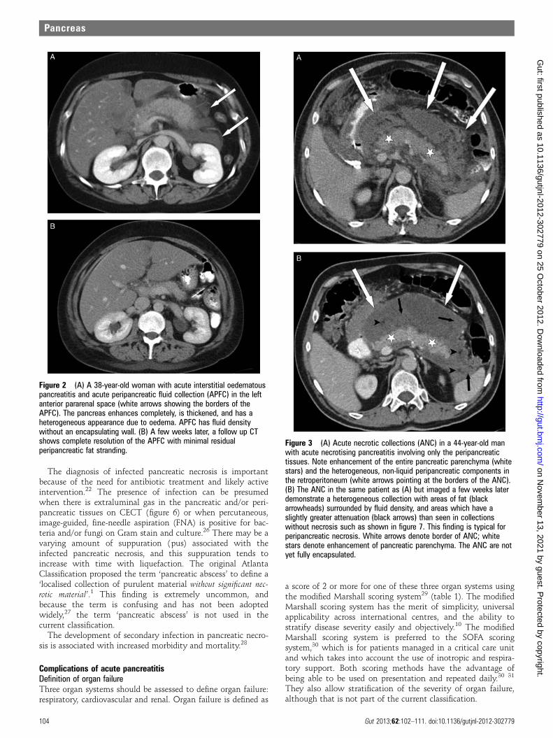
Figure 3 (A) Acute necrotic collections (ANC) in a 44-year-old manwith acute necrotising pancreatitis involving only the peripancreatictissues. Note enhancement of the entire pancreatic parenchyma (whitestars) and the heterogeneous, non-liquid peripancreatic components inthe retroperitoneum (white arrows pointing at the borders of the ANC).(B) The ANC in the same patient as (A) but imaged a few weeks laterdemonstrate a heterogeneous collection with areas of fat (blackarrowheads) surrounded by fluid density, and areas which have aslightly greater attenuation (black arrows) than seen in collectionswithout necrosis such as shown in figure 7. This finding is typical forperipancreatic necrosis. White arrows denote border of ANC; whitestars denote enhancement of pancreatic parenchyma. The ANC are notyet fully encapsulated.
Figure 2 (A) A 38-year-old woman with acute interstitial oedematouspancreatitis and acute peripancreatic fluid collection (APFC) in the leftanterior pararenal space (white arrows showing the borders of theAPFC). The pancreas enhances completely, is thickened, and has aheterogeneous appearance due to oedema. APFC has fluid densitywithout an encapsulating wall. (B) A few weeks later, a follow up CTshows complete resolution of the APFC with minimal residualperipancreatic fat stranding.
104 Gut 2013;62:102–111. doi:10.1136/gutjnl-2012-302779
Pancreas
on Novem
ber 13, 2021 by guest. Protected by copyright.
http://gut.bmj.com
/G
ut: first published as 10.1136/gutjnl-2012-302779 on 25 October 2012. D
ownloaded from
Definition of local complicationsThe original Atlanta Classification1 distinguished betweenuncomplicated interstitial pancreatitis and acute pancreatitisassociated with ‘local complications’. This distinction (localcomplications being absent or present) is useful. The natural
history and clinical consequences of different local complica-tions are now better understood and described. Local complica-tions are acute peripancreatic fluid collection, pancreaticpseudocyst, acute necrotic collection and walled-off necrosis.The morphologic features of these local complications aredescribed in detail later in this document (see below, Definitionof pancreatic and peripancreatic collections). Other local com-plications of acute pancreatitis include gastric outlet dysfunc-tion, splenic and portal vein thrombosis, and colonic necrosis.
Local complications should be suspected when there is per-sistence or recurrence of abdominal pain, secondary increases inserum pancreatic enzyme activity, increasing organ dysfunc-tion, and/or the development of clinical signs of sepsis, such asfever and leucocytosis. These developments usually promptimaging to detect local complications. The morphologic fea-tures of acute pancreatitis are well delineated by high reso-lution, multi-detector CECT and form the basis of the new,more objective definitions for the local complications of acutepancreatitis (box 1).
Pancreatic and peripancreatic collections should be describedon the basis of location (pancreatic, peripancreatic, other), thenature of the content (liquid, solid, gas), and the thicknessof any wall (thin, thick). The pattern and extent of impairedpancreatic parenchymal perfusion, if present, should also bedescribed.27 The morphologic description of local complicationsis necessary for accurate diagnosis. Local complications alone,however, do not define the severity of acute pancreatitis (seebelow, Definition of severity of acute pancreatitis).32 33
Definition of systemic complicationsExacerbation of pre-existing co-morbidity, such as coronaryartery disease or chronic lung disease, precipitated by the acutepancreatitis is defined as a systemic complication. In this docu-ment, we distinguish between persistent organ failure (thedefining feature of severe acute pancreatitis) and other systemiccomplications, which are an exacerbation of pre-existingco-morbid disease.
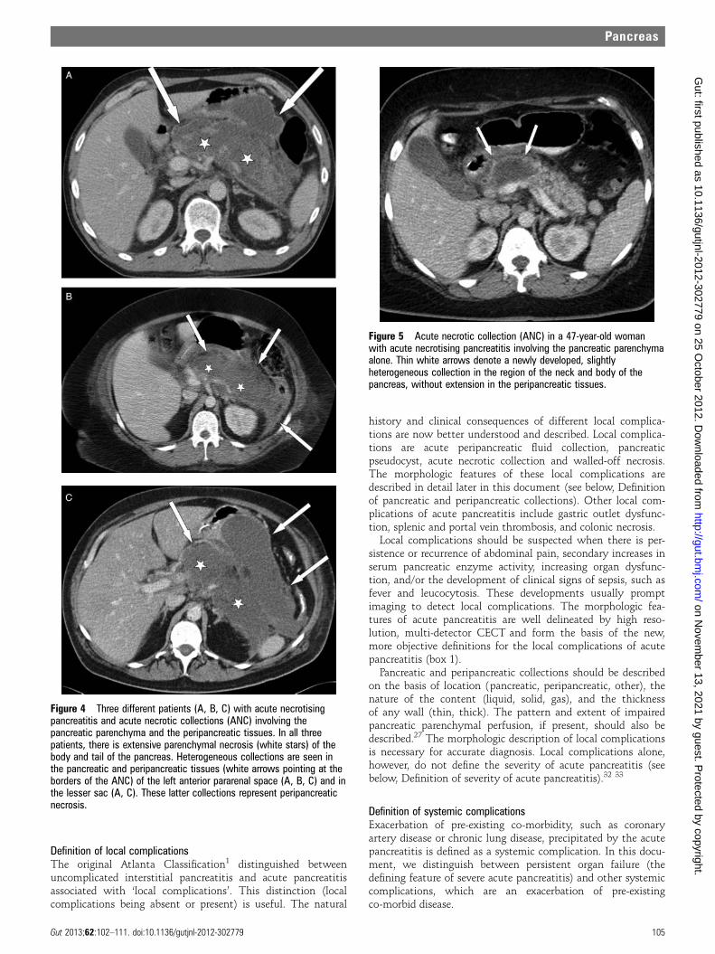
Figure 4 Three different patients (A, B, C) with acute necrotisingpancreatitis and acute necrotic collections (ANC) involving thepancreatic parenchyma and the peripancreatic tissues. In all threepatients, there is extensive parenchymal necrosis (white stars) of thebody and tail of the pancreas. Heterogeneous collections are seen inthe pancreatic and peripancreatic tissues (white arrows pointing at theborders of the ANC) of the left anterior pararenal space (A, B, C) and inthe lesser sac (A, C). These latter collections represent peripancreaticnecrosis.
Figure 5 Acute necrotic collection (ANC) in a 47-year-old womanwith acute necrotising pancreatitis involving the pancreatic parenchymaalone. Thin white arrows denote a newly developed, slightlyheterogeneous collection in the region of the neck and body of thepancreas, without extension in the peripancreatic tissues.
Gut 2013;62:102–111. doi:10.1136/gutjnl-2012-302779 105
Pancreas
on Novem
ber 13, 2021 by guest. Protected by copyright.
http://gut.bmj.com
/G
ut: first published as 10.1136/gutjnl-2012-302779 on 25 October 2012. D
ownloaded from
Phases of acute pancreatitisThere are two overlapping phases in this dynamic diseaseprocess with two peaks of mortality: early and late.34–37 Theearly phase, which usually lasts for the first week, is followedby a second later phase which can run a protracted course fromweeks to months. It is helpful to consider these two phasesseparately.
Early phaseDuring the early phase, systemic disturbances result from thehost response to local pancreatic injury. This early phase isusually over by the end of the first week but may extend intothe second week. Cytokine cascades are activated by the pan-creatic inflammation which manifest clinically as the systemicinflammatory response syndrome (SIRS)38–40 (box 2). WhenSIRS is persistent,41 42 there is an increased risk of developingorgan failure (table 1). The determinant of the severity of acutepancreatitis during the early phase is primarily the presenceand duration of organ failure. This is described as ‘transientorgan failure’ if the organ failure resolves within 48 h or as‘persistent organ failure’ if organ failure persists for
>48 h.39 41 43 If organ failure affects more than one organsystem, it is termed multiple organ failure (MOF).
Although local complications may be identified during theearly phase, they are not the predominant determinants ofseverity,32 and it may be unreliable to determine the extent ofnecrosis during the first few days of disease. In addition, theextent of morphologic changes is not directly proportional tothe severity of organ failure.24 Therefore, the definition ofsevere or moderately severe acute pancreatitis in the early phasedepends on the presence and duration of organ failure (seebelow, Definition of severity of acute pancreatitis).
Late phaseThe late phase is characterised by persistence of systemic signs ofinflammation or by the presence of local complications, and soby definition (see below), the late phase occurs only in patientswith moderately severe or severe acute pancreatitis. Local compli-cations evolve during the late phase. It is important to distinguishthe different morphologic characteristics of the local complica-tions by radiologic imaging, because these local complicationsmay have direct implications for management. Persistent organfailure, however, remains the main determinant of severity, so
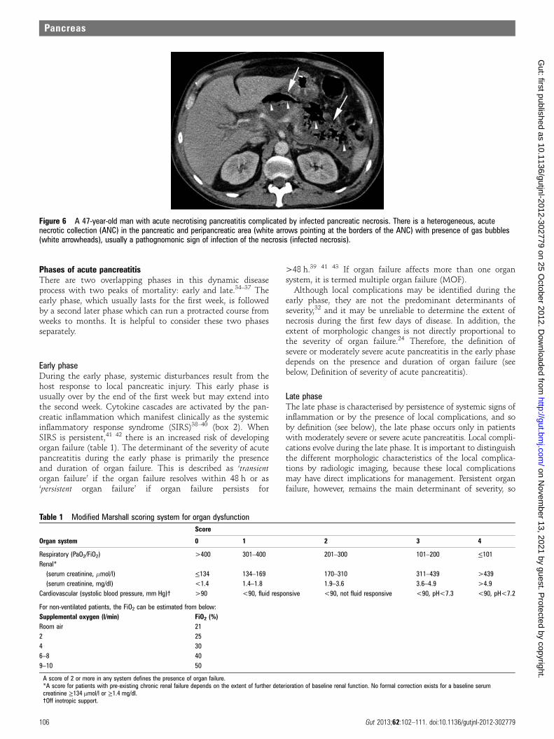
Figure 6 A 47-year-old man with acute necrotising pancreatitis complicated by infected pancreatic necrosis. There is a heterogeneous, acutenecrotic collection (ANC) in the pancreatic and peripancreatic area (white arrows pointing at the borders of the ANC) with presence of gas bubbles(white arrowheads), usually a pathognomonic sign of infection of the necrosis (infected necrosis).
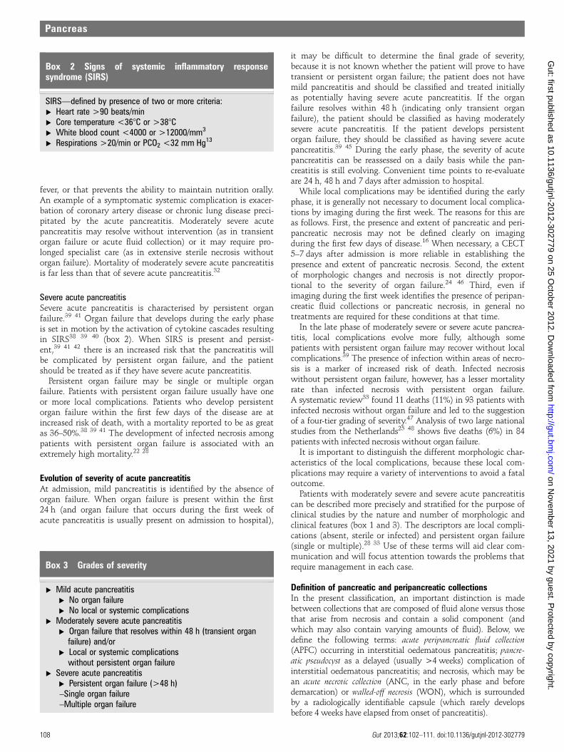
Table 1 Modified Marshall scoring system for organ dysfunctionScore
Organ system 0 1 2 3 4
Respiratory (PaO2/FiO2) >400 301–400 201–300 101–200 ≤101Renal*
(serum creatinine, �mol/l) ≤134 134–169 170–310 311–439 >439(serum creatinine, mg/dl) <1.4 1.4–1.8 1.9–3.6 3.6–4.9 >4.9
Cardiovascular (systolic blood pressure, mm Hg)† >90 <90, fluid responsive <90, not fluid responsive <90, pH<7.3 <90, pH<7.2
For non-ventilated patients, the FiO2 can be estimated from below:Supplemental oxygen (l/min) FiO2 (%)Room air 212 254 306–8 409–10 50
A score of 2 or more in any system defines the presence of organ failure.*A score for patients with pre-existing chronic renal failure depends on the extent of further deterioration of baseline renal function. No formal correction exists for a baseline serumcreatinine ≥134 μmol/l or ≥1.4 mg/dl.†Off inotropic support.
106 Gut 2013;62:102–111. doi:10.1136/gutjnl-2012-302779
Pancreas
on Novem
ber 13, 2021 by guest. Protected by copyright.
http://gut.bmj.com
/G
ut: first published as 10.1136/gutjnl-2012-302779 on 25 October 2012. D
ownloaded from

characterisation of acute pancreatitis in the late phase requiresboth clinical and morphologic criteria.
The SIRS of the early phase may be followed by a compensa-tory, anti-inflammatory response syndrome (CARS) that maycontribute to an increased risk of infection; however, theseevents are complex and poorly understood.44
Definition of severity of acute pancreatitisThere are important reasons to define and stratify the severityof acute pancreatitis. First, on admission, it is important toidentify patients with potentially severe acute pancreatitis whorequire aggressive early treatment. Second, in a secondary caresetting, clinicians need to identify such patients for possibletransfer to specialist care. Third, for specialists who receivesuch referrals, there are advantages to stratifying these patientsinto subgroups based on the presence of persistent organ failureand local or systemic complications.
This classification defines three degrees of severity: mildacute pancreatitis, moderately severe acute pancreatitis, andsevere acute pancreatitis.32 33 Terminology that is important inthis classification includes transient organ failure, persistentorgan failure, and local or systemic complications (boxes 1and 3). Transient organ failure is organ failure that is presentfor <48 h. Persistent organ failure is defined as organ failurethat persists for >48 h. Local complications include peripan-creatic fluid collections and acute necrotic collections13 14 39 41
(box 1), while systemic complications can be related to exacer-bations of underlying co-morbidities related to the acutepancreatitis.
Mild acute pancreatitisMild acute pancreatitis is characterised by the absence of organfailure and the absence of local or systemic complications.Patients with mild acute pancreatitis will usually be dischargedduring the early phase. Patients with mild acute pancreatitisusually do not require pancreatic imaging, and mortality isvery rare.15
Moderately severe acute pancreatitisModerately severe acute pancreatitis is characterised by thepresence of transient organ failure or local or systemic compli-cations in the absence of persistent organ failure. An exampleof a symptomatic local complication is a peripancreatic collec-tion resulting in prolonged abdominal pain, leucocytosis and
Box 1 (continued)Box 1 Revised definitions of morphological featuresof acute pancreatitis
1. Interstitial oedematous pancreatitisAcute inflammation of the pancreatic parenchyma and peri-pancreatic tissues, but without recognisable tissue necrosisCECT criteria▸ Pancreatic parenchyma enhancement by intravenous
contrast agent▸ No findings of peripancreatic necrosis (see below)▸ See figures 1 and 2
2. Necrotising pancreatitisInflammation associated with pancreatic parenchymal necro-sis and/or peripancreatic necrosisCECT criteria▸ Lack of pancreatic parenchymal enhancement by
intravenous contrast agent and/or▸ Presence of findings of peripancreatic necrosis (see
below—ANC and WON)▸ See figures 3, 4, 5 and 8
3. APFC (acute peripancreatic fluid collection)Peripancreatic fluid associated with interstitial oedematouspancreatitis with no associated peripancreatic necrosis. Thisterm applies only to areas of peripancreatic fluid seen withinthe first 4 weeks after onset of interstitial oedematous pan-creatitis and without the features of a pseudocyst.CECT criteria▸ Occurs in the setting of interstitial oedematous
pancreatitis▸ Homogeneous collection with fluid density▸ Confined by normal peripancreatic fascial planes▸ No definable wall encapsulating the collection▸ Adjacent to pancreas (no intrapancreatic extension)▸ See figure 2
4. Pancreatic pseudocystAn encapsulated collection of fluid with a well definedinflammatory wall usually outside the pancreas with minimalor no necrosis. This entity usually occurs more than 4 weeksafter onset of interstitial oedematous pancreatitis to mature.CECT criteria▸ Well circumscribed, usually round or oval▸ Homogeneous fluid density▸ No non-liquid component▸ Well defined wall; that is, completely encapsulated▸ Maturation usually requires >4 weeks after onset of
acute pancreatitis; occurs after interstitial oedematouspancreatitis
▸ See figure 75. ANC (acute necrotic collection)
A collection containing variable amounts of both fluid andnecrosis associated with necrotising pancreatitis; the necro-sis can involve the pancreatic parenchyma and/or the peri-pancreatic tissuesCECT criteria▸ Occurs only in the setting of acute necrotising
pancreatitis▸ Heterogeneous and non-liquid density of varying degrees
in different locations (some appear homogeneous earlyin their course)
▸ No definable wall encapsulating the collection▸ Location—intrapancreatic and/or extrapancreatic▸ See figures 3–5
6. WON (walled-off necrosis)A mature, encapsulated collection of pancreatic and/or peri-pancreatic necrosis that has developed a well defined inflam-matory wall. WON usually occurs >4 weeks after onset ofnecrotising pancreatitis.CECT criteria▸ Heterogeneous with liquid and non-liquid density with
varying degrees of loculations (some may appearhomogeneous)
▸ Well defined wall, that is, completely encapsulated▸ Location—intrapancreatic and/or extrapancreatic▸ Maturation usually requires 4 weeks after onset of acute
necrotising pancreatitis▸ See figure 8
Gut 2013;62:102–111. doi:10.1136/gutjnl-2012-302779 107
Pancreas
on Novem
ber 13, 2021 by guest. Protected by copyright.
http://gut.bmj.com
/G
ut: first published as 10.1136/gutjnl-2012-302779 on 25 October 2012. D
ownloaded from
fever, or that prevents the ability to maintain nutrition orally.An example of a symptomatic systemic complication is exacer-bation of coronary artery disease or chronic lung disease preci-pitated by the acute pancreatitis. Moderately severe acutepancreatitis may resolve without intervention (as in transientorgan failure or acute fluid collection) or it may require pro-longed specialist care (as in extensive sterile necrosis withoutorgan failure). Mortality of moderately severe acute pancreatitisis far less than that of severe acute pancreatitis.32
Severe acute pancreatitisSevere acute pancreatitis is characterised by persistent organfailure.39 41 Organ failure that develops during the early phaseis set in motion by the activation of cytokine cascades resultingin SIRS38 39 40 (box 2). When SIRS is present and persist-ent,39 41 42 there is an increased risk that the pancreatitis willbe complicated by persistent organ failure, and the patientshould be treated as if they have severe acute pancreatitis.
Persistent organ failure may be single or multiple organfailure. Patients with persistent organ failure usually have oneor more local complications. Patients who develop persistentorgan failure within the first few days of the disease are atincreased risk of death, with a mortality reported to be as greatas 36–50%.38 39 41 The development of infected necrosis amongpatients with persistent organ failure is associated with anextremely high mortality.22 28
Evolution of severity of acute pancreatitisAt admission, mild pancreatitis is identified by the absence oforgan failure. When organ failure is present within the first24 h (and organ failure that occurs during the first week ofacute pancreatitis is usually present on admission to hospital),
it may be difficult to determine the final grade of severity,because it is not known whether the patient will prove to havetransient or persistent organ failure; the patient does not havemild pancreatitis and should be classified and treated initiallyas potentially having severe acute pancreatitis. If the organfailure resolves within 48 h (indicating only transient organfailure), the patient should be classified as having moderatelysevere acute pancreatitis. If the patient develops persistentorgan failure, they should be classified as having severe acutepancreatitis.39 45 During the early phase, the severity of acutepancreatitis can be reassessed on a daily basis while the pan-creatitis is still evolving. Convenient time points to re-evaluateare 24 h, 48 h and 7 days after admission to hospital.
While local complications may be identified during the earlyphase, it is generally not necessary to document local complica-tions by imaging during the first week. The reasons for this areas follows. First, the presence and extent of pancreatic and peri-pancreatic necrosis may not be defined clearly on imagingduring the first few days of disease.16 When necessary, a CECT5–7 days after admission is more reliable in establishing thepresence and extent of pancreatic necrosis. Second, the extentof morphologic changes and necrosis is not directly propor-tional to the severity of organ failure.24 46 Third, even ifimaging during the first week identifies the presence of peripan-creatic fluid collections or pancreatic necrosis, in general notreatments are required for these conditions at that time.
In the late phase of moderately severe or severe acute pancrea-titis, local complications evolve more fully, although somepatients with persistent organ failure may recover without localcomplications.39 The presence of infection within areas of necro-sis is a marker of increased risk of death. Infected necrosiswithout persistent organ failure, however, has a lesser mortalityrate than infected necrosis with persistent organ failure.A systematic review33 found 11 deaths (11%) in 93 patients withinfected necrosis without organ failure and led to the suggestionof a four-tier grading of severity.47 Analysis of two large nationalstudies from the Netherlands25 48 shows five deaths (6%) in 84patients with infected necrosis without organ failure.
It is important to distinguish the different morphologic char-acteristics of the local complications, because these local com-plications may require a variety of interventions to avoid a fataloutcome.
Patients with moderately severe and severe acute pancreatitiscan be described more precisely and stratified for the purpose ofclinical studies by the nature and number of morphologic andclinical features (box 1 and 3). The descriptors are local compli-cations (absent, sterile or infected) and persistent organ failure(single or multiple).28 33 Use of these terms will aid clear com-munication and will focus attention towards the problems thatrequire management in each case.
Definition of pancreatic and peripancreatic collectionsIn the present classification, an important distinction is madebetween collections that are composed of fluid alone versus thosethat arise from necrosis and contain a solid component (andwhich may also contain varying amounts of fluid). Below, wedefine the following terms: acute peripancreatic fluid collection(APFC) occurring in interstitial oedematous pancreatitis; pancre-atic pseudocyst as a delayed (usually >4 weeks) complication ofinterstitial oedematous pancreatitis; and necrosis, which may bean acute necrotic collection (ANC, in the early phase and beforedemarcation) or walled-off necrosis (WON), which is surroundedby a radiologically identifiable capsule (which rarely developsbefore 4 weeks have elapsed from onset of pancreatitis).
Box 3 Grades of severity
▸ Mild acute pancreatitis▸ No organ failure▸ No local or systemic complications
▸ Moderately severe acute pancreatitis▸ Organ failure that resolves within 48 h (transient organ
failure) and/or▸ Local or systemic complications
without persistent organ failure▸ Severe acute pancreatitis
▸ Persistent organ failure (>48 h)–Single organ failure–Multiple organ failure
Box 2 Signs of systemic inflammatory responsesyndrome (SIRS)
SIRS—defined by presence of two or more criteria:▸ Heart rate >90 beats/min▸ Core temperature <36°C or >38°C▸ White blood count <4000 or >12000/mm3
▸ Respirations >20/min or PCO2 <32 mm Hg13
108 Gut 2013;62:102–111. doi:10.1136/gutjnl-2012-302779
Pancreas
on Novem
ber 13, 2021 by guest. Protected by copyright.
http://gut.bmj.com
/G
ut: first published as 10.1136/gutjnl-2012-302779 on 25 October 2012. D
ownloaded from
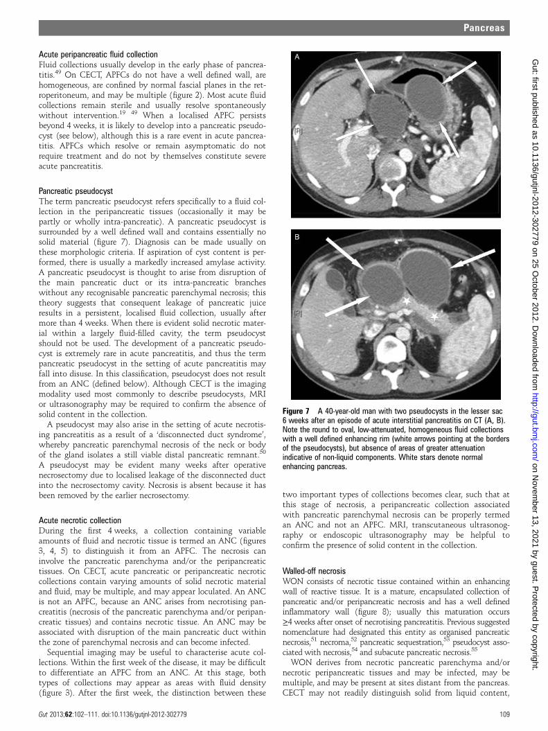
Acute peripancreatic fluid collectionFluid collections usually develop in the early phase of pancrea-titis.49 On CECT, APFCs do not have a well defined wall, arehomogeneous, are confined by normal fascial planes in the ret-roperitoneum, and may be multiple (figure 2). Most acute fluidcollections remain sterile and usually resolve spontaneouslywithout intervention.19 49 When a localised APFC persistsbeyond 4 weeks, it is likely to develop into a pancreatic pseudo-cyst (see below), although this is a rare event in acute pancrea-titis. APFCs which resolve or remain asymptomatic do notrequire treatment and do not by themselves constitute severeacute pancreatitis.
Pancreatic pseudocystThe term pancreatic pseudocyst refers specifically to a fluid col-lection in the peripancreatic tissues (occasionally it may bepartly or wholly intra-pancreatic). A pancreatic pseudocyst issurrounded by a well defined wall and contains essentially nosolid material (figure 7). Diagnosis can be made usually onthese morphologic criteria. If aspiration of cyst content is per-formed, there is usually a markedly increased amylase activity.A pancreatic pseudocyst is thought to arise from disruption ofthe main pancreatic duct or its intra-pancreatic brancheswithout any recognisable pancreatic parenchymal necrosis; thistheory suggests that consequent leakage of pancreatic juiceresults in a persistent, localised fluid collection, usually aftermore than 4 weeks. When there is evident solid necrotic mater-ial within a largely fluid-filled cavity, the term pseudocystshould not be used. The development of a pancreatic pseudo-cyst is extremely rare in acute pancreatitis, and thus the termpancreatic pseudocyst in the setting of acute pancreatitis mayfall into disuse. In this classification, pseudocyst does not resultfrom an ANC (defined below). Although CECT is the imagingmodality used most commonly to describe pseudocysts, MRIor ultrasonography may be required to confirm the absence ofsolid content in the collection.
A pseudocyst may also arise in the setting of acute necrotis-ing pancreatitis as a result of a ‘disconnected duct syndrome’,whereby pancreatic parenchymal necrosis of the neck or bodyof the gland isolates a still viable distal pancreatic remnant.50
A pseudocyst may be evident many weeks after operativenecrosectomy due to localised leakage of the disconnected ductinto the necrosectomy cavity. Necrosis is absent because it hasbeen removed by the earlier necrosectomy.
Acute necrotic collectionDuring the first 4 weeks, a collection containing variableamounts of fluid and necrotic tissue is termed an ANC (figures3, 4, 5) to distinguish it from an APFC. The necrosis caninvolve the pancreatic parenchyma and/or the peripancreatictissues. On CECT, acute pancreatic or peripancreatic necroticcollections contain varying amounts of solid necrotic materialand fluid, may be multiple, and may appear loculated. An ANCis not an APFC, because an ANC arises from necrotising pan-creatitis (necrosis of the pancreatic parenchyma and/or peripan-creatic tissues) and contains necrotic tissue. An ANC may beassociated with disruption of the main pancreatic duct withinthe zone of parenchymal necrosis and can become infected.
Sequential imaging may be useful to characterise acute col-lections. Within the first week of the disease, it may be difficultto differentiate an APFC from an ANC. At this stage, bothtypes of collections may appear as areas with fluid density(figure 3). After the first week, the distinction between these
two important types of collections becomes clear, such that atthis stage of necrosis, a peripancreatic collection associatedwith pancreatic parenchymal necrosis can be properly termedan ANC and not an APFC. MRI, transcutaneous ultrasonog-raphy or endoscopic ultrasonography may be helpful toconfirm the presence of solid content in the collection.
Walled-off necrosisWON consists of necrotic tissue contained within an enhancingwall of reactive tissue. It is a mature, encapsulated collection ofpancreatic and/or peripancreatic necrosis and has a well definedinflammatory wall (figure 8); usually this maturation occurs≥4 weeks after onset of necrotising pancreatitis. Previous suggestednomenclature had designated this entity as organised pancreaticnecrosis,51 necroma,52 pancreatic sequestration,53 pseudocyst asso-ciated with necrosis,54 and subacute pancreatic necrosis.55
WON derives from necrotic pancreatic parenchyma and/ornecrotic peripancreatic tissues and may be infected, may bemultiple, and may be present at sites distant from the pancreas.CECT may not readily distinguish solid from liquid content,
Figure 7 A 40-year-old man with two pseudocysts in the lesser sac6 weeks after an episode of acute interstitial pancreatitis on CT (A, B).Note the round to oval, low-attenuated, homogeneous fluid collectionswith a well defined enhancing rim (white arrows pointing at the bordersof the pseudocysts), but absence of areas of greater attenuationindicative of non-liquid components. White stars denote normalenhancing pancreas.
Gut 2013;62:102–111. doi:10.1136/gutjnl-2012-302779 109
Pancreas
on Novem
ber 13, 2021 by guest. Protected by copyright.
http://gut.bmj.com
/G
ut: first published as 10.1136/gutjnl-2012-302779 on 25 October 2012. D
ownloaded from

and, for this reason, pancreatic and peripancreatic necrosis maybe misdiagnosed as a pancreatic pseudocyst. For this purpose,MRI, transabdominal ultrasonography or endoscopic ultrason-ography may be required for this distinction. Demonstration ofthe presence or absence of pancreatic ductal communication isnot necessary in this classification, although determination ofsuch ductal communication is of potential importance, becauseit may affect management.
Infected necrosisThe diagnosis of infection (infected necrosis) of an ANC orof WON can be suspected by the patient’s clinical course orby the presence of gas within the collection seen on CECT(figure 6). This extraluminal gas is present in areas of necrosisand may or may not form a gas/fluid level depending on theamount of liquid content present at that stage of the disease.In cases of doubt, fine needle aspiration for culture may be per-formed, but some series have shown that the large majority ofpatients can be managed without FNA, especially if percutan-eous drainage is part of the management algorithm.25
CONCLUSIONThis classification revises and updates the definitions from theAtlanta Classification of acute pancreatitis. An importantfeature is the recognition that acute pancreatitis is an evolving,dynamic condition and that the severity may change duringthe course of the disease. Early in the disease, SIRS or organfailure indicate potentially severe disease. If the patientimproves rapidly during the early phase without organ failureand without local or systemic complications, the disease isdefined as mild acute pancreatitis. If the patient develops localor systemic complications and has no persistent organ failure,the disease is defined as moderately severe acute pancreatitis. Ifthe patient develops persistent organ failure, the disease isdefined as severe acute pancreatitis and is associated with veryhigh morbidity and mortality rates.
The accurate description of local complications, including thepresence of fluid or necrosis in or around the pancreas, the time
course of progression, and the presence or absence of infection,will improve the stratification of patients both for clinical care inspecialised centres and for reporting of clinical research.
Acknowledgements The Working Group wishes to thank: Deborah Frank for herexpert assistance in organising and coordinating electronically all the communicationsbetween national and international societies, collating the suggestions made by multiplereaders of this new classification on its three internet-based mailings, and finally in thepreparation of this manuscript; Drs John A. Windsor, Karen D. Horvath, KoenraadJ. Mortele, Timothy B. Gardner, Georgios Papachristou, Pramod Garg, Marc Besselink,Hjalmar van Santvoort, Mario Pelaez-Luna, Dhiraj Yadav, Gerasimos Stefanidis, StergiosDelakidis, Desiree E. Morgan and Ruedi F. L. Thoeni, and the secretaries and otherofficers of the 11 societies (International Association of Pancreatology (IAP), AmericanPancreatic Association (APA), European Pancreatic Club (EPC) and from the EPC to itsaffiliated societies, Pancreatic Disorders Section of the American GastroenterologicalAssociation, Society for Surgery of the Alimentary Tract (SSAT), Pancreas Club, AmericanHepato-Pancreato-Biliary Association (AHPBA), Japanese Association ofHepato-Biliary-Pancreatic Surgery, Pancreas Network of New Zealand, AustralasianPancreatic Club, and the Japanese Pancreas Society) who facilitated the dissemination ofdraft documents; and the following people who responded to one or more of the threeworking drafts circulated via the web-based process: Acosta, Juan M.; Amann, StephenT.; Andren-Sandberg, Ake; Aranha, Gerard V.; Asciutti, Stefania; Banks, Peter A.;Barauskas, Giedrius; Baron, Todd. H. Sr; Bassi, Claudio; Behrman, Steven; Behrns, KevinE.; Belliappa, Vikram; Berzin, Tyler M.; Besselink, Marc G.H.; Bhasin, Deepak Kumar;Biankin, Andrew; Bishop, Michele D.; Bollen, Thomas L.; Bonini, Claudio J.; Bradley.Edward L. III; Buechler, Markus; Carter, Ross; Cavestro, Giulia Martina; Chari, Suresh T.;Chavez-Rodriguez, Juan J.; da Cunha, Jose Eduardo; D’Agostino, Horatio; De Campos,Tercio; Delakidis, Stergios; de-Madaria, Enrique; Deprez, Pierre H.; Dervenis, Christos;DiSario, James A.; Doria, Cataldo; Falconi, Massimo; Fernandez-del Castillo, Carlos;Freeny, Patrick C.; Frey, Charles F.; Friess, Helmut; Frossard, Jean Louis; Fuchshuber,Pascal; Gallagher, Scott F.; Gardner, Timothy B.; Garg, Pramod Kumar; Ghattas, Georges;Glasgow, Robert; Gonzalez, Jose A.; Gooszen, Hein G.; Gress, Thomas M.; Gumbs,Andrew A.; Hallibruton, Cory; Helton, Scott; Hill, Michael C.; Horvath, Karen D.; Hoyos,Sergio; Imrie, Clem W.; Isenmann, Ranier; Izbicki, Jakob R.; Johnson, Colin D.;Karagiannis, John A.; Klar, Ernst; Kolokythas, Orpheus; Lau, Joseph; Litvin, Andrey A.;Longnecker, Daniel S.; Lowenfels, Albert B.; Mackey, Richard; Mah’Moud, Mitchell;Malangoni, Mark; McFadden, David W.; Mishra, Girish; Moody, Frank G.; Morgan, DesireeE.; Morinville, Veronique; Mortele, Koenraad J.; Neoptolemos, John P.; Nordback, Isto; Pap,Akos; Papachristou, Georgios I.; Parks, Rowan; Pedrazolli, Sergio; Pelaez-Luna, Mario; Pezzilli,Raffaele; Pitt, Henry A.; Prosanto, C.; Ramesh, H.; Ramirez, Francisco C.; Raper, Steven E.;Rasheed, Ashraf; Reed, Donald N. Jr; Romangnuolo, Joseph; Rossaak, Jeremy; Sanabria,Juan; Sarr, Michael G.; Schaefer, Claus; Schmidt, Johannes; Schmidt, Palle Nordblad;Serrablo, Alejandro; Senkowski, Christopher K.; Sharma, Manik; Sigman, Ken M.; Singh,Pankaj; Stefanidis, Gerasimos; Steinberg, William; Steiner, Joerg; Strasberg, Steven; Strum,Williamson; Takada, Tadahiro; Tanaka, Masao; Thoeni, Ruedi F. L.; Tsiotos, Gregory G.; VanSantvoort, Hjalmar; Vaccaro, Maria; Vege, Santhi Swaroop; Villavicencio, Roberto L.;
Figure 8 (A–C) Three differentpatients with walled-off necrosis(WON) after an acute attack ofnecrotising pancreatitis. In all threepatients, a heterogeneous, fullyencapsulated collection is noted in thepancreatic and peripancreatic area. (A)Non-liquid components of highattenuation (black arrowheads) in thecollection are noted. The collection hasa thin, well defined, and enhancingwall (thick white arrows). (B, C) Alargely liquefied collection in the bed ofthe pancreas is observed withnon-liquid components representingareas of trapped fat (blackarrowheads). (D) represents thecorresponding T2-weighted MRI to (C),showing the true heterogeneity of thecollection. Black arrowheads denoteareas of necrotic debris surrounded byfluid (white on T2-weighted image).
110 Gut 2013;62:102–111. doi:10.1136/gutjnl-2012-302779
Pancreas
on Novem
ber 13, 2021 by guest. Protected by copyright.
http://gut.bmj.com
/G
ut: first published as 10.1136/gutjnl-2012-302779 on 25 October 2012. D
ownloaded from

Vrochides, Dionisios; Wagner, Markus; Warshaw, Andrew L.; Wilcox, Charles M.; Windsor,John A.; Wysocki, Peter; Yadav, Dhiraj; Zenilman, Michael E.; Zyromski, Nicholas J.
Contributions Originators and coordinators (MGS, PAB, SVV). Writing committee(present authors—draft prepared by MGS, PAB, CDJ, and reviewed/commented onby all). Review of drafts—invited expert review; those named below for final review.Individual comments on drafts (long list of responders).
Competing interests None.
Provenance and peer review Not commissioned; externally peer reviewed.
REFERENCES1. Bradley EL III. A clinically based classification system for acute pancreatitis.
Summary of the International Symposium on Acute Pancreatitis, Atlanta, GA,September 11 through 13, 1992. Arch Surg 1993;128:586–90.
2. Bollen TL, van Santvoort HC, Besselink MG, et al. The Atlanta classification ofacute pancreatitis revisited. Br J Surg 2008;95:6–21.
3. Acute Pancreatitis Classification Working Group. Revision of the Atlantaclassification of acute pancreatitis. 2008. http://www.pancreasclub.com/resources/AtlantaClassification.pdf
4. Harrison S, Kakade M, Varadarajula S, et al. Characteristics and outcomes ofpatients undergoing debridement of pancreatic necrosis. J Gastrointest Surg2010;14:245–51.
5. Brun A, Agarwal N, Pitchumoni CS. Fluid collections in and around the pancreas inacute pancreatitis. J Clin Gastroenterol 2011;45:614–25.
6. Bharwani N, Patel S, Prabhudesai S, et al. Acute pancreatitis: the role of imagingin diagnosis and management. Clin Radiol 2011;66:164–75.
7. Sheu Y, Furlan A, Almusa O, et al. The revised Atlanta classification for acutepancreatitis: a CT imaging guide for radiologists. Emerg Radiol 2012;19:37–43.
8. Thoeni RF. The revised Atlanta classification of acute pancreatitis: itsimportance for the radiologist and its effect on treatment. Radiology2012;262:751–64.
9. Banks PA, Freeman ML. Practice guidelines in acute pancreatitis. Am JGastroenterol 2006;101:2379–400.
10. UK Working Party on Acute Pancreatitis. UK guidelines for the management ofacute pancreatitis. Gut 2005;54:iii1–9.
11. Uhl W, Warshaw A, Imrie C, et al. IAP Guidelines for the surgical management ofacute pancreatitis. Pancreatology 2002;2:565–73.
12. Arvanitakis M, Delhaye M, De MV, et al. Computed tomography and magneticresonance imaging in the assessment of acute pancreatitis. Gastroenterology2004;126:715–23.
13. Bollen TL, van Santvoort HC, Besselink MG, et al. Update on acute pancreatitis:ultrasound, computed tomography, and magnetic resonance imaging features. SeminUltrasound CT MRI 2007;28:371–83.
14. Morgan DE. Imaging of acute pancreatitis and its complications. Clin GastroenterolHepatol 2008;6:1077–85.
15. Singh VK, Bollen TL, Wu BU, et al. An assessment of the severity of interstitialpancreatitis. Clin Gastroenterol Hepatol 2011;9:1098–103.
16. Spanier BWM, Nio Y, van der Hulst RWN, et al. Practice and yield of early CT scanin acute pancreatitis: a Dutch observational multicenter study. Pancreatology2010;10:222–8.
17. Bollen TL, Singh VK, Maurer R, et al. A comparative evaluation of radiologic andclinical scoring systems in the early prediction of severity of acute pancreatitis. AmJ Gastroenterol 2012;107:612–19.
18. Isenmann R, Buechler M, Uhl W, et al. Pancreatic necrosis: an early finding insevere acute pancreatitis. Pancreas 1993;8:358–61.
19. Balthazar EJ, Robinson DL, Megibow AJ, et al. Acute pancreatitis: value of CT inestablishing prognosis. Radiology 1990;174:331–6.
20. Sakorafas GH, Tsiotos GG, Sarr MG. Extrapancreatic necrotizing pancreatitis with viablepancreas: a previously under-appreciated entity. J Am Coll Surg 1999;188:643–8.
21. Besselink MG, van Santvoort HC, Boermeester MA, et al. Timing and impact ofinfections in acute pancreatitis. Br J Surg 2009;96:267–73.
22. van Santvoort HC, Bakker OJ, Bollen TL, et al. A conservative and minimallyinvasive approach to necrotizing pancreatitis improves outcome. Gastroenterology2011;141:1254–63.
23. Beger HG, Bittner R, Block S, et al. Bacterial contamination of pancreatic necrosis.A prospective clinical study. Gastroenterology 1986;91:433–8.
24. Perez A, Whang EE, Brooks DC, et al. Is severity of necrotizing pancreatitisincreased in extended necrosis and infected necrosis? Pancreas 2002;25:229–33.
25. van Santvoort HC, Besselink MG, Bakker OJ, et al. A step-up approach or opennecrosectomy for necrotizing pancreatitis (PANTER trial). N Engl J Med2010;362:1491–502.
26. Banks PA, Gerzof SG, Langevin RE, et al. CT-guided aspiration of suspected pancreaticinfection: bacteriology and clinical outcome. Int J Pancreatol 1995;18:265–70.
27. van Santvoort HC, Bollen TL, Besselink MG, et al. Describing peripancreaticcollections in severe acute pancreatitis using morphologic terms: an internationalinterobserver agreement study. Pancreatology 2008;8:593–9.
28. Petrov MS, Shanbhag S, Chakraborty M, et al. Organ failure and infection ofpancreatic necrosis as determinants of mortality in patients with acute pancreatitis.Gastroenterology 2010;139:813–20.
29. Marshall JC, Cook DJ, Christou NV, et al. Multiple organ dysfunction score: areliable descriptor of a complex clinical outcome. Crit Care Med1995;23:1638–52.
30. Vincent JL, Moreno R, Takala J, et al. The SOFA (Sepsis-related Organ FailureAssessment) score to describe organ dysfunction/failure. On behalf of the workinggroup on sepsis-related problems of the European Society of Intensive CareMedicine. Intensive Care Med 1996;22:707–10.
31. Halonen KI, Pettila V, Leppaniemi AK, et al. Multiple organ dysfunction associatedwith severe acute pancreatitis. Crit Care Med 2002;30:1274–9.
32. Vege SS, Gardner TB, Chari ST, et al. Low mortality and high morbidity in severeacute pancreatitis without organ failure: a case for revising the Atlanta classificationto include “moderately severe acute pancreatitis”. Am J Gastroenterol2009;104:710–15.
33. Petrov MS, Windsor JA. Classification of the severity of acute pancreatitis: howmany categories make sense? Am J Gastroenterol 2010;105:74–6.
34. McKay CJ, Imrie CW. The continuing challenge of early mortality in acutepancreatitis. Br J Surg 2004;91:1243–4.
35. Renner IG, Savage WT, Pantoja JL, et al. Death due to acute pancreatitis: aretrospective analysis of 405 autopsy cases. Dig Dis Sci 1995;30:1005–18.
36. Widdison AL, Karanja ND. Pancreatic infection complicating acute pancreatitis. Br JSurg 1993;80:148–54.
37. Blum T, Maisonneuve P, Lowenfels AB, et al. Fatal outcome in acute pancreatitis: itsoccurrence and early prediction. Pancreatology 2001;1:237–41.
38. Buter A, Imrie CW, Carter CR, et al.Dynamic nature of early organ dysfunctiondetermines outcome in acute pancreatitis. Br J Surg 2002;89:298–302.
39. Johnson CD, Abu-Hilal M. Persistent organ failure during the first week as amarker of fatal outcome in acute pancreatitis. Gut 2004;53:1340–4.
40. Muckart DJ, Bhagwanjee S. American College of Chest Physicians/Society ofCritical Care Medicine Consensus Conference definitions of the systemicinflammatory response syndrome and allied disorders in relation to critically injuredpatients. Crit Care Med 1997;25:1789–95.
41. Mofidi R, Duff MD, Wigmore SJ, et al. Association between early systemicinflammatory response, severity of multiorgan dysfunction and death in acutepancreatitis. Br J Surg 2006;93:738–44.
42. Singh VK,Wu BU, Bollen TL, et al. Early systemic inflammatory response syndrome isassociated with severe acute pancreatitis. Clin Gastroenterol Hepatol 2009;7:1247–51.
43. Lytras D, Manes K, Triantopoulou C, et al. Persistent early organ failure: definingthe high risk group of patients with severe acute pancreatitis. Pancreas2008;36:249–54.
44. Cobb JP, O’Keefe GE. Injury research in the genomic era. Lancet2004;363:2076–83.
45. Johnson CD, Kingsnorth AN, Imrie CW, et al. Double blind, randomised, placebocontrolled study of a platelet activating factor antagonist, lexipafant, in the treatment andprevention of organ failure in predicted severe acute pancreatitis. Gut 2001;48:62–9.
46. Tenner S, Sica G, Hughes M, et al. Relationship of necrosis to organ failure insevere acute pancreatitis. Gastroenterology 1997;113:899–903.
47. Dellinger EP, Forsmark CE, Layer P, et al. Determinant-based classification of acutepancreatitis severity: an international multidisciplinary consultation. Ann Surg.Published Online First: 24 Sept 2012. doi: 10.1097/SLA.0b013e318256f778.
48. Besselink MG, van Santvoort HC, Buskens E, et al. Probiotic prophylaxis inpredicted severe acute pancreatitis: a randomized, double-blind, placebo-controlledtrial. Lancet 2008;371:651–9.
49. Lenhart DK, Balthazar EJ. MDCT of acute mild (nonnecrotizing pancreatitis):abdominal complications and fate of fluid collections. Am J Roentgenol2008;190:643–9.
50. Pelaez-Luna M, Vege SS, Petersen BT, et al. Disconnected pancreatic ductsyndrome in severe acute pancreatitis: clinical and imaging characteristics andoutcomes in a cohort of 31 cases. Gastrointest Endosc 2008;68:91–7.
51. Baron TH, Thaggard WG, Morgan DE, et al. Endoscopic therapy for organizedpancreatic necrosis. Gastroenterology 1996;111:755–64.
52. Bradley EL III. Atlanta redux. Pancreas 2003;26:105–6.53. Yeo CJ, Sarr MG. Cystic and pseudocystic diseases of the pancreas. Current Probl
Surg 1994;XXXI:165–252.54. Hariri M, Slivka A, Carr-Locke DL, et al. Pseudocyst drainage predisposes to
infection when pancreatic necrosis is unrecognized. Am J Gastroenterol1994;8:1781–4.
55. Petrakis I, Vrachassotakis N, Kogerakis N, et al. Subacute pancreatic necrosis.Panminerva Med 2000;42:279–86.
Gut 2013;62:102–111. doi:10.1136/gutjnl-2012-302779 111
Pancreas
on Novem
ber 13, 2021 by guest. Protected by copyright.
http://gut.bmj.com
/G
ut: first published as 10.1136/gutjnl-2012-302779 on 25 October 2012. D
ownloaded from

APPENDIX 1 (online version only)
An international, web-based, multiply reiterative process was designed to
obtain a consensus supported by evidence from a broad representation of physicians
from many disciplines who were interested in acute pancreatitis. Three sequential
drafts were sent to 11 major national and international organizations interested in
acute pancreatitis, so that an international consensus classification could be
developed.
All members of these organizations were invited to participate. After collation
of responses, each revision was sent again to the entire memberships of these
organizations regardless of whether they participated or not in a previous revision, so
all members of these 11 organizations had three opportunities to contribute.
After circulation of the first draft, responses were reviewed and incorporated
in a second draft, and this was sent out again. This process was repeated a third
time until a consensus document was obtained.
Initially, a select group of about 40 pancreatologists and pancreatic surgeons
met to agree on the process and areas for revision. Participants were chosen for
their defined interest and publication record in pancreatitis. Participants gave up
their own time during Digestive Disease Week 2007. A Working Group of 7
individuals (3 pancreatic surgeons, 2 pancreatologists, and 1 pancreatic radiologist)
from USA, the Netherlands, and Greece developed the first working document of a
revised classification of acute pancreatitis. This first working document was
discussed, revised, and edited by the Working Group and sent initially to the original
participating pancreatologists. These individuals sent back their suggestions on how
to improve the document.
All suggestions were reviewed by all members of the Working Group, and the
document was revised to the satisfaction of all members of the Working Group.
Subsequent correspondence (see below) was then coordinated by one of the
Working Group (MGS). This first document was defined clearly to be a “working

Page 2
document” and by no means a final draft; indeed, no formal “publication” was made
or even suggested—this was a working draft. This draft was sent electronically to all
members of the following national and international organizations through their
secretariat: the International Association of Pancreatology (IAP), American
Pancreatic Association (APA), European Pancreatic Club (EPC) and from the EPC to
its affiliated societies, Pancreatic Disorders Section of the American
Gastroenterological Association, Society for Surgery of the Alimentary Tract (SSAT),
Pancreas Club, American Hepato-Pancreato-Biliary Association (AHPBA), Japanese
Association of Hepato-Biliary-Pancreatic Surgery, Pancreas Network of New
Zealand, Australasian Pancreatic Club, and the Japanese Pancreas Society.
A cover letter accompanying the first draft asked the recipient of the e-mail to
read the draft document and return suggestions for improvement or criticisms of the
classification by e-mail.
This stimulated 57 individuals to respond with a wide variety of suggestions
for improvement. All responses were read and discussed by each member of the
Working Group. A revised second working draft (again defined clearly as such) was
prepared and discussed by the working group in a conference call. A revised second
draft acceptable to all of the Working Group was again sent electronically to all
members of the 11 national and international organizations listed above.
This time, 58 responses were received. The process of review of responses
and revision of the draft was repeated, and a third working draft (again defined
clearly as such) was sent to the same organizations. This third draft generated 36
responses, most all of which were minor; none of the suggestions led to any
substantive changes in the classification. This third draft was edited by the Working
Group and reviewed further by a select group of pancreatologists from 7 countries
(including 3 well known radiologists) (see acknowledgments).
In response to the comments received by the journal review (document too
long, consideration be given to 3 levels of severity, further review, etc), the working
Page 3
group was enlarged by addition of one other member chosen specifically for his
expertise from UK (CDJ) and another person from New Zealand (JAW) was queried
specifically for his input as well. The document was revised and shortened after
which the entire working group prepared a fourth version of the document.
Appropriate suggestions were included and discussed fully; a final draft was sent to
the working group, and the final version of the classification was prepared for
submission for scientific peer review. In this final version, opinion previously agreed
by consensus in earlier rounds was either supported by robust evidence or rejected.
APPENDIX 2 (online version only)
DATA RECORDING FOR CLINICAL MANAGEMENT AND CLINICAL RESEARCH STUDIES
Clinical Data
In addition to demographic and relevant clinical information (including etiology of
pancreatitis), the interval between onset of abdominal pain and first admission to the hospital
should be noted. Time of admission to hospital should be recorded. For the purpose of
standardizing data, the first hospital day should be designated as day 1. In order to permit
standardized reporting, day 2 should start at 8 AM on the following day and last for 24 hours.
The presence of organ failure should be documented on each day through day 7. The interval
from the onset of symptoms to the onset of persistent organ failure should also be documented.
Data originating from a tertiary care hospital should be stratified to allow separate consideration
of outcomes of patients who are transferred from other hospitals versus those who are admitted
directly to the tertiary care hospital.
Risk factors and markers of severity
Several potential risk factors, markers of severity, and measurements related to the
acute pancreatitis that may reflect severity should be recorded and evaluated prospectively, but
these risk factors do not in themselves define severity (17,W1). Risk factors include
comorbidities, age, and body mass index; markers of severity at admission or within the first 3
days include APACHE II scores and other scoring systems (W2,W3,W4,W5), pleural effusion or
Page 2
pulmonary opacities on chest radiography, and serum levels of C-reactive protein (CRP) (9,W6).
C-reactive protein is one of the most highly studied and valuable serum markers, but increases
in serum CRP levels have a delayed onset and are most predictive at 48-72 hours after onset of
disease. Procalcitonin may be useful particularly for early identification of infected necrosis.
Other markers of severity which have been used in clinical studies include CT severity index
(17,19), modified CT severity index (17,W7), urinary concentration of trypsinogen activating
peptide (TAP), hematocrit on admission (W8), BISAP score =/>3 (W9,W10), serial
measurements of serum BUN (W11,W12) and creatinine (W13), and serum levels of lactate
dehydrogenase (LDH), amyloid protein A, CAPAP-B, IL-6, and other markers of acute phase
injury. Not all these markers are available for clinical use. It should be stressed that plasma or
serum amylase and lipase activities, while important in the diagnosis of acute pancreatitis, are
not of any clinical importance in defining the severity of acute pancreatitis; moreover, it is not
necessary or useful to repeat these measurements every day (9,W6).
Radiologic Evaluation On CECT
Cross-sectional imaging usually with CECT is essential for the clinical management of
pancreatitis in the late phase. Good management depends on systematic reporting of all
abnormalities combined with close collaboration between clinician and radiologist. The
framework shown in Table 5 will assist in complete reporting.
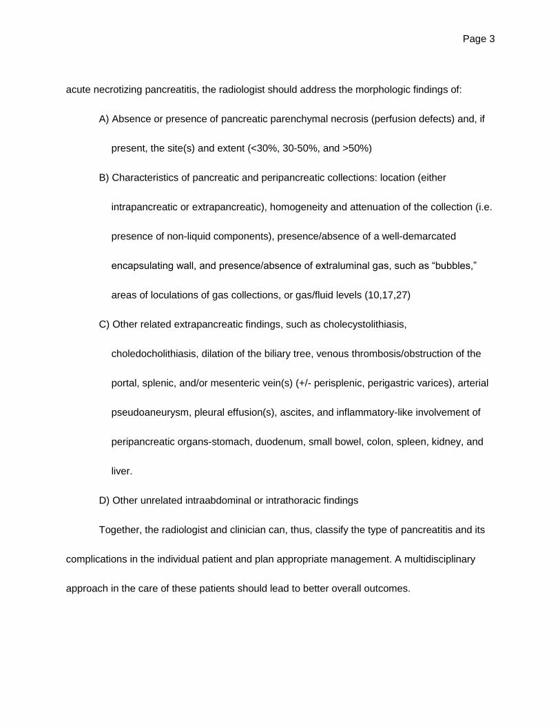
In addition to the diagnosis of interstitial edematous pancreatitis versus all three forms of
Page 3
acute necrotizing pancreatitis, the radiologist should address the morphologic findings of:
A) Absence or presence of pancreatic parenchymal necrosis (perfusion defects) and, if
present, the site(s) and extent (<30%, 30-50%, and >50%)
B) Characteristics of pancreatic and peripancreatic collections: location (either
intrapancreatic or extrapancreatic), homogeneity and attenuation of the collection (i.e.
presence of non-liquid components), presence/absence of a well-demarcated
encapsulating wall, and presence/absence of extraluminal gas, such as “bubbles,”
areas of loculations of gas collections, or gas/fluid levels (10,17,27)
C) Other related extrapancreatic findings, such as cholecystolithiasis,
choledocholithiasis, dilation of the biliary tree, venous thrombosis/obstruction of the
portal, splenic, and/or mesenteric vein(s) (+/- perisplenic, perigastric varices), arterial
pseudoaneurysm, pleural effusion(s), ascites, and inflammatory-like involvement of
peripancreatic organs-stomach, duodenum, small bowel, colon, spleen, kidney, and
liver.
D) Other unrelated intraabdominal or intrathoracic findings
Together, the radiologist and clinician can, thus, classify the type of pancreatitis and its
complications in the individual patient and plan appropriate management. A multidisciplinary
approach in the care of these patients should lead to better overall outcomes.
Page 4
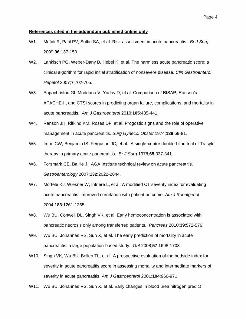
References cited in the addendum published online only W1. Mofidi R, Patil PV, Suttie SA, et al. Risk assessment in acute pancreatitis. Br J Surg
2009;96:137-150.
W2. Lankisch PG, Weber-Dany B, Hebel K, et al. The harmless acute pancreatic score: a
clinical algorithm for rapid initial stratification of nonsevere disease. Clin Gastroenterol
Hepatol 2007;7:702-705.
W3. Papachristou GI, Muddana V, Yadav D, et al. Comparison of BISAP, Ranson’s
APACHE-II, and CTSI scores in predicting organ failure, complications, and mortality in
acute pancreatitis. Am J Gastroenterol 2010;105:435-441.
W4. Ranson JH, Rifkind KM, Roses DF, et al. Progostic signs and the role of operative
management in acute pancreatitis. Surg Gynecol Obstet 1974;139:69-81.
W5. Imrie CW, Benjamin IS, Ferguson JC, et al. A single-centre double-blind trial of Trasylol
therapy in primary acute pancreatitis. Br J Surg 1978;65:337-341.
W6. Forsmark CE, Baillie J. AGA Institute technical review on acute pancreatitis.
Gastroenterology 2007;132:2022-2044.
W7. Mortele KJ, Wiesner W, Intriere L, et al. A modified CT severity index for evaluating
acute pancreatitis: improved correlation with patient outcome. Am J Roentgenol
2004;183:1261-1265.
W8. Wu BU, Conwell DL, Singh VK, et al. Early hemoconcentration is associated with
pancreatic necrosis only among transferred patients. Pancreas 2010;39:572-576.
W9. Wu BU, Johannes RS, Sun X, et al. The early prediction of mortality in acute
pancreatitis: a large population-based study. Gut 2008;57:1698-1703.
W10. Singh VK, Wu BU, Bollen TL, et al. A prospective evaluation of the bedside index for
severity in acute pancreatitis score in assessing mortality and intermediate markers of
severity in acute pancreatitis. Am J Gastroenterol 2001;104:966-971
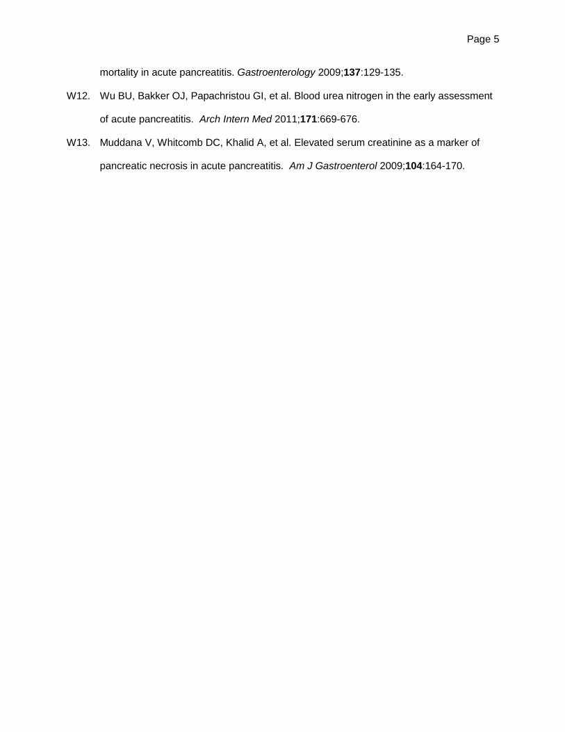
W11. Wu BU, Johannes RS, Sun X, et al. Early changes in blood urea nitrogen predict
Page 5
mortality in acute pancreatitis. Gastroenterology 2009;137:129-135.
W12. Wu BU, Bakker OJ, Papachristou GI, et al. Blood urea nitrogen in the early assessment
of acute pancreatitis. Arch Intern Med 2011;171:669-676.
W13. Muddana V, Whitcomb DC, Khalid A, et al. Elevated serum creatinine as a marker of
pancreatic necrosis in acute pancreatitis. Am J Gastroenterol 2009;104:164-170.
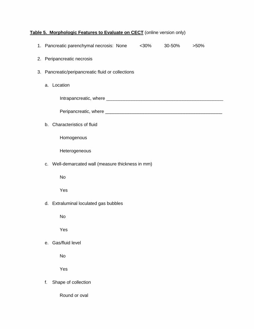
Table 5. Morphologic Features to Evaluate on CECT (online version only)
1. Pancreatic parenchymal necrosis: None <30% 30-50% >50%
2. Peripancreatic necrosis
3. Pancreatic/peripancreatic fluid or collections
a. Location
Intrapancreatic, where ______________________________________________
Peripancreatic, where ______________________________________________
b. Characteristics of fluid
Homogenous
Heterogeneous
c. Well-demarcated wall (measure thickness in mm)
No
Yes
d. Extraluminal loculated gas bubbles
No
Yes
e. Gas/fluid level
No
Yes
f. Shape of collection
Round or oval
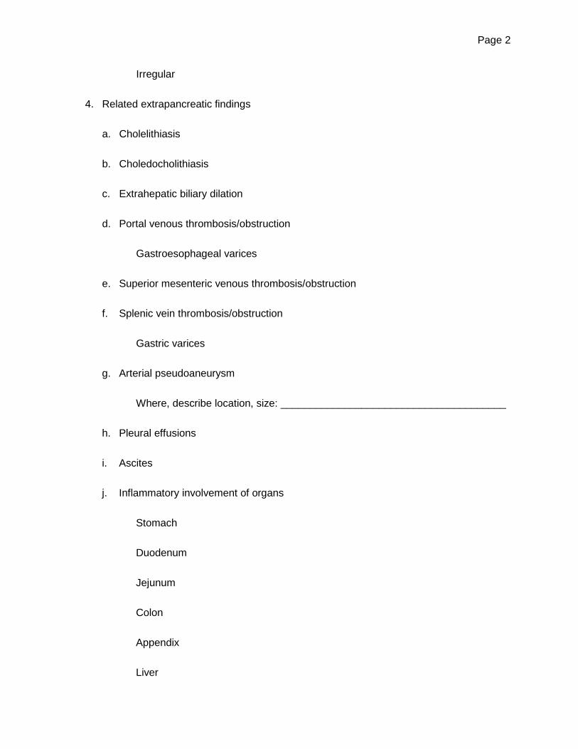
Page 2
Irregular
4. Related extrapancreatic findings
a. Cholelithiasis
b. Choledocholithiasis
c. Extrahepatic biliary dilation
d. Portal venous thrombosis/obstruction
Gastroesophageal varices
e. Superior mesenteric venous thrombosis/obstruction
f. Splenic vein thrombosis/obstruction
Gastric varices
g. Arterial pseudoaneurysm
Where, describe location, size: _______________________________________
h. Pleural effusions
i. Ascites
j. Inflammatory involvement of organs
Stomach
Duodenum
Jejunum
Colon
Appendix
Liver

Page 3
Kidney (right / left)
Ureter (right / left)
k. Colonic necrosis
l. Signs of chronic pancreatitis – pancreatic calcification
5. Unrelated intraabdominal or intrathoracic findings
Describe findings _____________________________________________________