Embed Size (px)
Citation preview
Pain Assessment and Management after Abdominal Surgery or Parturition in Dairy Cattle
by
Nathalie Christine Newby
A Thesis
presented to The University of Guelph
In partial fulfilment of requirements
for the degree of Doctor of Philosophy
in Population Medicine
Guelph, Ontario, Canada
© Nathalie Christine Newby, October, 2012
ABSTRACT
PAIN ASSESSMENT AND MANAGEMENT AFTER ABDOMINAL SURGERY OR
PARTURITION IN DAIRY CATTLE Nathalie Christine Newby Advisors: University of Guelph, 2012 Professors T. F. Duffield and
D. L. Pearl This thesis is an investigation of the impact of abdominal surgeries and assisted
parturition in dairy cows on physiological and behavioural parameters, and the potential
management of pain through the use of non-steroidal inflammatory drugs (NSAIDs) or a
mechanical brush. This research is novel and necessary because of the paucity of pain
research in dairy cows.
Three abdominal surgery studies were conducted. The first was a randomized clinical
field trial, conducted on commercial dairy herds in southern Ontario, Canada, to evaluate
the effect of ketoprofen following correction of left displaced abomasum. The second and
third studies were randomized clinical trials evaluating NSAIDs following the first stage
of a two-stage fistulation surgery. The second tested ketoprofen versus saline, while the
third compared ketoprofen and meloxicam. The key findings from these studies were that
there were indicators of pain following surgery (such as decreased milk production, dry
matter intake, and changes in lying behavior) and that there were beneficial effects of
administering NSAIDs following abdominal surgery(improved eating and lying
behavior), although these effects were not sufficient to alleviate all of the surgical pain.
Two trials were conducted in parturient cows. The first trial examined the effects of
meloxicam administration 24 h following assisted calving. There were beneficial effects
of NSAID on feeding behavior, however, further research is needed to investigate the full
potential of providing an NSAID as a post-calving pain therapy. The second trial
described the use of a mechanical brush by parturient cows. This study yielded insight on
the brush use of these cows, as well as on their maternal, auto-grooming, and scratching
behaviors. Cows used the brush before parturition, and when the calf was present, auto-
grooming and scratching behaviors were significantly reduced, and calf licking time was
greater in the brush group compared to the no brush group.
The findings described in this dissertation provide insights into the expression and
assessment of pain and its management following abdominal surgery in dairy cattle. This
study has also identified areas of future research for both assessment and management of
pain following abdominal surgery and following assisted calving.
iv
ACKNOWLEDGMENTS
Many individuals have contributed to the completion of this thesis, and whether
these contributions were big or small, I wish to extend my sincere gratitude to all who
played a role. I am grateful to my parents, Jean-Pierre and Monique Newby, for their
encouragement throughout my entire academic career, their unwavering moral support, as
well as their editorial help with this thesis.
Special thanks are extended to my committee: my advisor Dr. Todd F. Duffield,
my co-advisor Dr. David L. Pearl, as well as my committee members Dr. Marina A. G.
von Keyserlingk, Dr. Stephen J. LeBlanc, and Dr. Ken E. Leslie. I thank Todd for his
patience, his support, his understanding and his wisdom. He took a chance on taking me
on as a PhD, and his confidence in me to do these projects means a great deal. David is to
be thanked for his tremendous statistical help as well as editing throughout this thesis.
Nina was a great help by providing her knowledge about animal behavior and how to
study it. Stephen is to be thanked for teaching me about dairy health management and for
all of his help with the clinical field trial. And finally, I am grateful to Ken who
introduced me to the world of dairy research when I was his office assistant, introduced
me to Todd, and suggested I do my PhD in the Department with Todd. To all the
members of my committee, thank you very much for believing in me and allowing me to
bring to life my own project, which is part of this thesis in the 6th chapter. It was an
absolutely remarkable experience that increased my confidence in my ability to do
research.
Many others contributed through their collaboration. Laura Wright, at the Elora
Dairy Research Center, has played an instrumental role in a few of the projects from this
v
study and I am very grateful for all of her help and hard work. Thank you to all of the
staff at the Elora and Ponsonby Dairy Research Centres for all of your help, as well as for
making these past years quite memorable. I would also like to extend my gratitude to
Nelson Dinn and all of the staff at the University of British Columbia Dairy Education
and Research Centre for all of their hard work and help with my brush project. The
realization of the great deal of data collection and organization would not have been
possible without the help of my summer students: Anneliese Heinrich, Crystal Throop,
Allyson Cole-Duffield, Colleen Szentimrey, Rebecca Egan, Kailee Price, Theresa
Campbell, and Jessica Ford. Thanks to the participating herds and veterinary clinics as
well as the University of Guelph, Ontario Veterinary College, Ruminant Field Service for
making part of this project come to life through the collection of excellent data. I would
like to thank my collaborators at Boehringer-Ingelheim. I thank Paul Doig, Robert
Tremblay, Laurent Goby and Michael Witt for their valuable insights and support. A
special thanks to the ladies in the Vetmedica office in Burlington for their moral support
and many enjoyable lunches. Thanks to Helen Kocmarek, Animal Health Laboratories,
University of Guelph, to Domenico Barillari, Environemental Health and Safety,
University of Guelph, to Gosia Zobel, Faculty of Land and Food Systems, University of
British Columbia, to Dr. Cassandra B. Tucker, University of California in Davis, and to
OVC IT staff for all of their technical support.
I would like to extend many thanks to my office mates and dear friends (the list is
rather long, but you know who you are) for their moral support and for making these past
4 years memorable.
Thank you all for being part of this beautiful and enlightening journey.
vi
TABLE OF CONTENTS
ABSTRACT ……………………………………………………………………. ii
ACKNOWLEDGMENTS ..………………………………………………….. iv
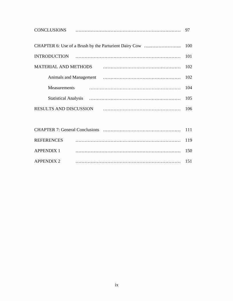
LIST OF TABLES ……………………………………………………………... x
LIST OF FIGURES …………………………………………………………… xiii
CHAPTER 1: Introduction and Literature Review …………………………… 1
INTRODUCTION …………………………………………………………… 1
LITERATURE REVIEW …………………………………………………… 2
Pain and Nociception ………………………………………………….... 2
Inflammation Mechanism …………………………………………… 3
Pain Assessment in Cattle …………………………………………… 3
Pain Scales ...…………………………………………………. 4
Assessment of Pain during Management Procedures ……..……. 6
Therapeutic Approaches to Pain …………………………………… 8
Management of Painful Procedures in Cattle ..……..……………..…….. 11
Castration ………………………………….....……………..…… 11
Dehorning …………………………………………………… 12
Painful Illnesses or Potential Painful Surgeries …………… 15
Research Objectives …………………………………………………… 21
CHAPTER 2: Pain and Displaced Abomasum Surgery in Cattle …..……….. 23
INTRODUCTION …………………………………………………………… 24
MATERIAL AND METHODS ………………………………………….... 26
Physical examination …………………………………………………… 28
vii
Behavioral Assessment ………………………………………….... 29
Milk Production and Culling …………………………………………… 29
Statistical Analysis ……………………………………………........... 30
RESULTS …………………………………………………………………… 31
Physical Examination and Blood Parameters …………………………… 31
Behavioral Assessment …………………………………………… 32
Production and Culling …………………………………………… 33
DISCUSSION …………………………………………………………… 34
CONCLUSIONS …………………………………………………………… 37
CHAPTER 3: Pain and Fistulation Surgery in Cattle …………………… 44
INTRODUCTION …………………………………………………………… 45
MATERIAL AND METHODS …………………………………………… 46
Animals and General Information …………………………………… 46
Fistulation Surgery …………………………………………………… 48
Experimental Protocol …………………………………………… 49
Statistical Analysis …………………………………………………… 51
RESULTS …………………………………………………………………… 53
DISCUSSION …………………………………………………………… 54
CONCLUSIONS …………………………………………………………… 58
CHAPTER 4: NSAIDs and Fistulation Surgery in Cattle …..……………….. 68
INTRODUCTION …………………………………………………………… 69
viii
MATERIAL AND METHODS …………………………………………… 71
General Information …………………………………………………… 71
Experimental Protocol …………………………………………… 72
Statistical Analysis …………………………………………………… 73
RESULTS AND DISCUSSION …………………………………………… 74
CONCLUSIONS …………………………………………………………… 76
CHAPTER 5: NSAIDs and Assisted Calving in Dairy Cattle …..……….. 85 INTRODUCTION …………………………………………………………… 86
MATERIAL AND METHODS …………………………………………… 87
General Information …………………………………………………… 87
Dry Matter Intake, Milk and Body Weight Data …..……………….. 88
Blood Collection and Analysis …………………………………… 89
Health Events …………………………………………………… 90
Behavior …………………………………………………………… 90
Statistical Analysis …………………………………………………… 91
RESULTS …………………………………………………………………… 93
Body Weight …………………………………………………………… 93
Feed Intake and Milk Production data …………………………… 93
Blood Analysis …………………………………………………… 93
Health Events …………………………………………………… 94
Lying and Feeding Behavior …………………………………………… 94
DISCUSSION …………………………………………………………… 94
ix
CONCLUSIONS …………………………………………………………… 97
CHAPTER 6: Use of a Brush by the Parturient Dairy Cow …..……………….. 100
INTRODUCTION …………………………………………………………… 101
MATERIAL AND METHODS …………………………………………… 102
Animals and Management …………………………………………… 102
Measurements …………………………………………………… 104
Statistical Analysis …………………………………………………… 105
RESULTS AND DISCUSSION …………………………………………… 106
CHAPTER 7: General Conclusions …………………………………………… 111 REFERENCES …………………………………………………………… 119 APPENDIX 1 …………………………………………………………… 150 APPENDIX 2 …………………………………………………………… 151
x
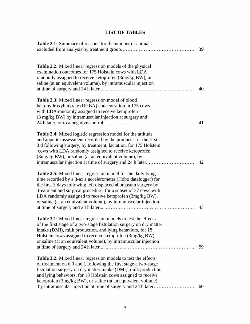
LIST OF TABLES
Table 2.1: Summary of reasons for the number of animals excluded from analysis by treatment group……………………………………… 39 Table 2.2: Mixed linear regression models of the physical examination outcomes for 175 Holstein cows with LDA randomly assigned to receive ketoprofen (3mg/kg BW), or saline (at an equivalent volume), by intramuscular injection at time of surgery and 24 h later.………………………………………………… 40 Table 2.3: Mixed linear regression model of blood beta-hydroxybutyrate (BHBA) concentration in 175 cows with LDA randomly assigned to receive ketoprofen (3 mg/kg BW) by intramuscular injection at surgery and 24 h later, or to a negative control...…………………………………………….. 41 Table 2.4: Mixed logistic regression model for the attitude and appetite assessment recorded by the producer for the first 3 d following surgery, by treatment, lactation, for 175 Holstein cows with LDA randomly assigned to receive ketoprofen (3mg/kg BW), or saline (at an equivalent volume), by intramuscular injection at time of surgery and 24 h later. ………………………. 42 Table 2.5: Mixed linear regression model for the daily lying time recorded by a 3-axis accelerometer (Hobo datalogger) for the first 3 days following left displaced abomasum surgery by treatment and surgical procedure, for a subset of 37 cows with LDA randomly assigned to receive ketoprofen (3mg/kg BW), or saline (at an equivalent volume), by intramuscular injection at time of surgery and 24 h later.....……………………………………………… 43 Table 3.1: Mixed linear regression models to test the effects of the first stage of a two-stage fistulation surgery on dry matter intake (DMI), milk production, and lying behaviors, for 18 Holstein cows assigned to receive ketoprofen (3mg/kg BW), or saline (at an equivalent volume), by intramuscular injection at time of surgery and 24 h later.......…………………………………………….. 59 Table 3.2: Mixed linear regression models to test the effects of treatment on d 0 and 1 following the first stage a two-stage fistulation surgery on dry matter intake (DMI), milk production, and lying behaviors, for 18 Holstein cows assigned to receive ketoprofen (3mg/kg BW), or saline (at an equivalent volume), by intramuscular injection at time of surgery and 24 h later……………………. 60
xi
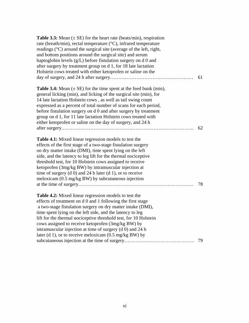
Table 3.3: Mean (± SE) for the heart rate (beats/min), respiration rate (breath/min), rectal temperature (°C), infrared temperature readings (°C) around the surgical site (average of the left, right, and bottom positions around the surgical site) and serum haptoglobin levels (g/L) before fistulation surgery on d 0 and after surgery by treatment group on d 1, for 18 late lactation Holstein cows treated with either ketoprofen or saline on the day of surgery, and 24 h after surgery…………………………………………… 61 Table 3.4: Mean (± SE) for the time spent at the feed bunk (min), general licking (min), and licking of the surgical site (min), for 14 late lactation Holstein cows , as well as tail swing count expressed as a percent of total number of scans for each period, before fistulation surgery on d 0 and after surgery by treatment group on d 1, for 11 late lactation Holstein cows treated with either ketoprofen or saline on the day of surgery, and 24 h after surgery.…………………………………….……………………………….. 62 Table 4.1: Mixed linear regression models to test the effects of the first stage of a two-stage fistulation surgery on dry matter intake (DMI), time spent lying on the left side, and the latency to leg lift for the thermal nociceptive threshold test, for 10 Holstein cows assigned to receive ketoprofen (3mg/kg BW) by intramuscular injection at time of surgery (d 0) and 24 h later (d 1), or to receive meloxicam (0.5 mg/kg BW) by subcutaneous injection at the time of surgery.……………………………………………………………. 78 Table 4.2: Mixed linear regression models to test the effects of treatment on d 0 and 1 following the first stage a two-stage fistulation surgery on dry matter intake (DMI), time spent lying on the left side, and the latency to leg lift for the thermal nociceptive threshold test, for 10 Holstein cows assigned to receive ketoprofen (3mg/kg BW) by intramuscular injection at time of surgery (d 0) and 24 h later (d 1), or to receive meloxicam (0.5 mg/kg BW) by subcutaneous injection at the time of surgery……………………………………. 79
xii
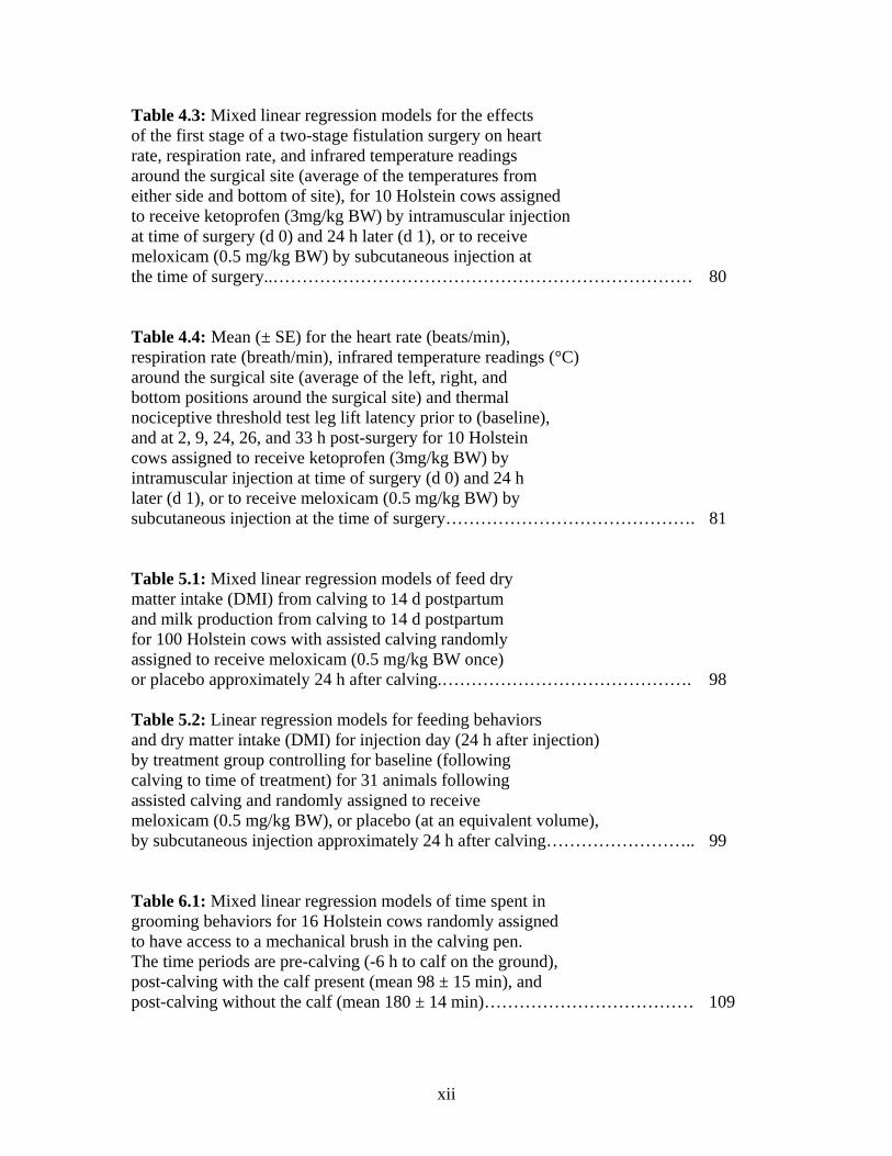
Table 4.3: Mixed linear regression models for the effects of the first stage of a two-stage fistulation surgery on heart rate, respiration rate, and infrared temperature readings around the surgical site (average of the temperatures from either side and bottom of site), for 10 Holstein cows assigned to receive ketoprofen (3mg/kg BW) by intramuscular injection at time of surgery (d 0) and 24 h later (d 1), or to receive meloxicam (0.5 mg/kg BW) by subcutaneous injection at the time of surgery..……………………………………………………………… 80
Table 4.4: Mean (± SE) for the heart rate (beats/min), respiration rate (breath/min), infrared temperature readings (°C) around the surgical site (average of the left, right, and bottom positions around the surgical site) and thermal nociceptive threshold test leg lift latency prior to (baseline), and at 2, 9, 24, 26, and 33 h post-surgery for 10 Holstein cows assigned to receive ketoprofen (3mg/kg BW) by intramuscular injection at time of surgery (d 0) and 24 h later (d 1), or to receive meloxicam (0.5 mg/kg BW) by subcutaneous injection at the time of surgery……………………………………. 81
Table 5.1: Mixed linear regression models of feed dry matter intake (DMI) from calving to 14 d postpartum and milk production from calving to 14 d postpartum for 100 Holstein cows with assisted calving randomly assigned to receive meloxicam (0.5 mg/kg BW once) or placebo approximately 24 h after calving.……………………………………. 98 Table 5.2: Linear regression models for feeding behaviors and dry matter intake (DMI) for injection day (24 h after injection) by treatment group controlling for baseline (following calving to time of treatment) for 31 animals following assisted calving and randomly assigned to receive meloxicam (0.5 mg/kg BW), or placebo (at an equivalent volume), by subcutaneous injection approximately 24 h after calving…………………….. 99 Table 6.1: Mixed linear regression models of time spent in grooming behaviors for 16 Holstein cows randomly assigned to have access to a mechanical brush in the calving pen. The time periods are pre-calving (-6 h to calf on the ground), post-calving with the calf present (mean 98 ± 15 min), and post-calving without the calf (mean 180 ± 14 min)……………………………… 109
xiii
LIST OF FIGURES Figure 3.1: Picture of the rumen clamp …………………………………… 63 Figure 3.2: Dry matter intake for ketoprofen and saline treated
fistulated cows …………………………………………………… 64 Figure 3.3: Milk production for ketoprofen and saline treated
fistulated cows …………………………………………………… 65 Figure 3.4: Tail flick count for ketoprofen and saline treated
fistulated cows …………………………………………………… 66 Figure 3.5: Time lying left for ketoprofen and saline treated
fistulated cows …………………………………………………… 67 Figure 4.1: Dry matter intake for ketoprofen and meloxicam treated
fistulated cows …………………………………………………… 82
Figure 4.2: Time lying left for ketoprofen and meloxicam treated fistulated cows …………………………………………………… 83 Figure 4.3: Percent time lying left for ketoprofen and meloxicam treated
fistulated cows ………………………………………………….. 84
Figure 6.1: Auto-grooming and scratching/rubbing behaviors and brush use around calving …………………………………… 110
1
Chapter 1 INTRODUCTION AND LITERATURE REVIEW
Introduction
The recognition of pain in cattle, and other farm production animals, has lagged
behind that of companion animals, horses, and humans. However, pain research in cattle
has been increasing over the past 15-20 years (Hudson et al., 2008). As part of good
animal welfare, the freedom from pain, injury, and disease is just one of the 5 freedoms
outlined by the Farm Animal Welfare Council (2009). One of the driving forces for this
research is society’s expectations to consider animal welfare a priority in animal
production (Rushen, 2003). The other driving force is the paucity of data on pain
assessment and management in dairy cows following abdominal surgeries and parturition.
The assessment of pain in these species has been challenging perhaps due to their stoic
nature, as an adaptation selected to hide their signs of pain or discomfort (Dobromylskyj
et al., 2000; Anil et al., 2005; Hudson et al., 2008). The Webster’s 1913 Dictionary
(2012) definition of the adjective stoic states “bearing pain, suffering, …, without
complaint”. Since cattle can be relatively undemonstrative when hurt (Broom, 2000), the
visible and more easily detected clinical signs in cattle may be an indication of severe
pain caused by advanced progression of a disease (O’Callaghan, 2002). The purpose of
this thesis is to explore methods of assessing, and managing, pain in dairy cows following
abdominal surgeries as well as following assisted parturition. This first chapter will
review the basis of pain mechanism in mammals and give an overview of the assessment
and management of pain in animals in general. This chapter will also provide some
examples of painful procedures, as well as some pain management strategies currently
found in the literature for dairy calves and adult dairy cows.
2
Literature Review
Pain and Nociception
Pain has been defined as “an unpleasant sensory and emotional experience
associated with actual or potential tissue damage, or described in terms of such damage”
(International Association for the Study of Pain, 1994). Since most of the mammalian
neural elements and biological consequences of pain are virtually the same, it is
important to recognize the need to better understand and appreciate the potential pain that
can be experienced by animals when they are ill or during painful procedures (Anderson
and Muir, 2005a). There are two kinds of fibers that innervate nociceptors in mammals:
small myelinated A-delta fibers and smaller unmyelinated C-fibres (Anderson and Muir,
2005a). The A-delta fibers are fast conducting and are involved in the first ‘pain’ or acute
response to a noxious stimulus, while the C-fibers are slower conducting, and are
involved in the second ‘pain’ or chronic response to a noxious stimulus. These nerve
fibers are involved in the ascending pain pathway which includes the transduction of the
electrical signal from a noxious stimulus. This electrical signal is then transmitted to the
superficial layer of the spinal cord, where it is modulated by local and descending
facilitatory and inhibitory neurons, and then, finally, this signal is projected to the brain to
be perceived by the animal (Anderson and Muir, 2005a). Pathologic pain is generally
produced by tissue or nerve damage but also frequently involves the development of
peripheral or central sensitization, as well as structural reorganization of neural elements
within the central nervous system leading to allodynia, and finally, disinhibition, which
can lead to hyperalgesia and allodynia (Anderson and Muir, 2005a). Allodynia is defined
as pain that is caused by a stimulus that is normally non painful, such as touch, while
3
hyperalgesia is an increased response to a stimulus that is painful (Anderson and Muir,
2005a).
Inflammation Mechanism
Tissue damage leads to the inflammatory cascade (Maroon et al., 2010), where in
general damaged cell walls lead to the breakdown of membrane phospholipids which get
converted to arachidonic acid through phospholipase A2 action. The arachidonic acid
undergoes oxidation and one of two processes occurs leading to inflammation in the
tissue: 1) cyclooxygenase converts it to prostaglandins, thromboxanes, prostacyclins; 2)
lipoxygenase converts it to leukotrienes (Maroon et al., 2010). The tissue damage and the
inflammatory response produce various chemicals, or sensitizers such as potassium ions,
prostaglandins, histamine, bradykinin, nerve growth factor, cytokines and chemokines,
which are involved in the activation of the peripheral nociceptors (Anderson and Muir,
2005b).
The main consequences of pain are negative stress which, if left untreated, can be
detrimental to an animal’s health, and in turn production, and which may even lead to
death (Anderson and Muir, 2005a; Anil et al., 2005). For these reasons, the identification
and the assessment of pain, especially following potentially painful procedures, are
crucial for pain management (Dobromylskyj et al., 2000; Anil et al., 2005; Hudson et al.,
2008).
Pain Assessment in Cattle
Pain assessment methods in animals can generally be categorized into objective
and subjective methods (Weary et al., 2006). Objective methods are those that measure
physiological parameters (e.g., heart rate, respiration rate, temperature, changes in
4
biochemical markers such as acute phase proteins, stress response through cortisol levels,
and daily activity) or production parameters (e.g., feed intake, weight gain, and milk
production) (Weary et al., 2006). Subjective methods are observations and
categorizations of behaviors and postures and can be measured in the form of scales, such
as the gait scoring scale for lameness, but are more prone to poor reliability because the
evaluation differs between observers (Weary et al., 2006). Behavioral subjective methods
can become a form of objective measure if used in a quantifiable way that is repeatable
and reliable, especially with experience and training of the observers. These behavioral
measures can be quantified to create objective measures that will give insight into
changes in behaviors. For example, lame dairy cattle spend more time lying and less time
feeding per day (Galindo and Broom, 2002). A combination of objective and subjective
methods may be optimal to gain more insight on what the animal is experiencing. The
observation of behaviors to interpret what the animal is experiencing is important in pain
assessment (Dobromylskyj et al., 2000).
Pain Scales
Pain scales, using objective and/or subjective methods, have been developed in
humans and in companion animals to assess pain and effective analgesia (Stevens et al.,
1996; Hansen, 2003). The visual analogue scale, which is a 100mm line with no pain at
one end and worst pain imaginable at the other end (Murrin and Rosen, 1985), has been
validated for use of pain assessment in humans (Price et al., 1983), and is also used to
assess pain in animals (Welsh et al., 1993; Hudson et al., 2004). However, this scale is
quite subjective and needs to be used with caution. In general, objective measures are
well accepted for the overall assessment of pain, stress or decreased health. Sprecher et
al. (1997) developed a lameness scoring system, in which gait and posture of the animal
5
are both assessed, because lameness is an indicator of pain felt by the animal
(O’Callaghan, 2002). This scoring system is well established and validated, and is used in
the Code of Practice for Dairy Cattle (National Farm Animal Care Council, 2009).This
scale is one of the few well established assessment tools available for dairy cattle (Flower
and Weary, 2006). The visual analogue scale used for human infants has been used to
evaluate pain in animals by clinicians (Dobromylskyj et al., 2000). There is, therefore, a
need for further development of pain scales for farm animals, especially cattle, to refine
the too few pain management methods. Mathews (1996) developed a visual analogue
pain scale from 0 to 10 for dogs and cats in conjunction with non-steroidal anti-
inflammatory drug recommendations at pain levels 3 and higher. Morton et al. (2005)
established and validated an interval measurement scale from 0 to 10 for acute pain in
dogs in 4 different groups: control dogs, dogs with medical conditions, dogs undergoing
soft tissue surgery, or dogs undergoing orthopedic surgery. Graubner et al. (2011) have
begun to validate a post abdominal surgery pain assessment scale (PASPAS) based on a
total pain index calculated from physiological (heart rate and respiration rate) and
behavioral (general subjective pain assessment on a scale from 1 to 5 and postural,
interactive, response to food, colic behavior, muscle stimulation, response to palpation of
incisional area) parameters following laparotomy surgery in horses using 8 different
observers on 8 different horses. They found good inter-observer reliability and also found
the scale to be a useful tool to evaluate post-surgical pain. A study by van Loon et al.
(2010) investigated the application of a composite pain scale to monitor horses with
somatic and visceral pain. This scale was composed of behavioral data (appearance of the
animal, sweating, kicking at the abdomen, pawing on the floor, posture, head movement,
appetite, and response to observer) and physiological data (heart rate, respiration rate,
6
digestive sounds, and rectal temperature). This composite scale was efficient at showing
low baseline values for healthy animals with non-painful conditions. There was a high
inter-observer reliability and the conclusion was that the composite pain scale is a
promising tool for the day-to-day assessment of pain status in equine patients. Thus far,
researchers have begun to identify pain assessment methods in dairy calves and adult
cows that are both objective and subjective during the examination for suspected diseases
as well as during management and/or surgical procedures. There has been a movement in
calf research toward comparing an analgesic treated group with a control group in order
to better understand pain behavior associated with painful procedures (Faulkner and
Weary, 2000; Sutherland et al., 2002; Ting et al., 2003; Stafford and Mellor, 2005;
Stilwell et al., 2008; Heinrich et al., 2009). With the aid of these studies, we can start
thinking about the development of a pain scale, like the one by Morton et al. (2005), to
assess acute pain, as well as a scale involving analgesia similar to the one developed for
dogs and cats by Mathews (1996) in order to assist in the management of pain.
Assessment of Pain during Management Procedures
There are no current common standard operating procedures in Canada for
detection of pain in cattle or for the management of potentially painful procedures such as
abdominal surgery. There are only guidelines and recommendations, based on current
research, which are not enforced as of yet (National Farm Animal Care Council, 2009). In
a British survey of bovine practitioners concerning their attitudes to pain and analgesia in
cattle, the general list for signs of pain they identified included: anorexia, vocalisation,
grinding of teeth, dull and depressed attitude, abnormal movements, abnormal posture,
increased heart rate, changes in respiration, flinching, recumbency, reduced rumination,
and reduced milk yield (Watts, 2000).
7
Pain during and after dehorning and castration has been extensively studied and,
as a result, some objective and subjective methods are accepted as valid methods for pain
assessment (Mellor et al., 1991; Molony et al., 1995; Petrie et al., 1996; Molony and
Kent, 1997; Sylvester et al., 1998; Graf and Senn, 1999; McMeekan et al., 1999;
Faulkner and Weary, 2000; Capucille et al., 2002; Earley and Crowe, 2002; Sutherland et
al., 2002; Milligan et al., 2004; Aubry, 2005; Stafford, 2007; Heinrich et al., 2009). These
studies used physiological measures (e.g., respiration rate, heart rate, and plasma cortisol
levels), as well as behavioral measures (e.g., tail wagging, head and ear movement,
rearing, tripping, feet stomping, licking at the site, and vocalization) as indicators of pain
at the time of and following a painful procedure.
Special tools, such as algometry and heat sources (e.g., laser), to evaluate
nociceptive thresholds or hyperalgesia have been used in some studies to assist in the
assessment of pain and to detect differences between pain vs no pain or between
analgesia vs control groups. In a lameness study, objective assessment of claw and soft
tissue pain was performed using a hoof tester (e.g., a pincer like instrument that is used to
squeeze the hoof of an animal to test the flinch response to pain at the pressure site) and
an algometer, which measured the pressure exerted on the area until a withdrawal
response of the foot (Dyer et al., 2007). In other studies, pressure nociceptive thresholds
using an algometer were measured in lame animals treated with, or without, analgesics
(ketoprofen or tolfenamic acid). The thresholds were increased in animals treated with
analgesics, showing that the pain from the lameness was attenuated by the analgesic
(Whay et al., 2005; Laven et al. 2008).
Fitzpatrick et al. (2003) used algometry as a method of pain assessment in animals
with mastitis. They found that there was increased sensitivity to pressure in the leg on the
8
same side as the mastitic quarter when compared to the leg on the other side of the
mastitic quarter. Heinrich et al. (2010) used an algometer to assess pain sensitivity around
the horn bud following dehorning in calves treated with and without the non-steroidal
anti-inflammatory drug NSAID meloxicam. Following dehorning, all calves had
increased sensitivity to pressure from the algometer around the disbudding site. However,
calves not treated with meloxicam were twice as sensitive as those treated with
meloxicam, suggesting that meloxicam was effective at alleviating some, but not all of
the pain associated with dehorning. Algometry is a novel objective method, however, and
further research is warranted to apply this method to pain assessment in cattle.
Pinheiro Machado et al. (1997) evaluated the role of amniotic fluid ingestion in
alleviating calving pain. This group used a thermal test from a laser, which created a
ramped radiating heat stimulus, in order to assess pain and the effectiveness of amniotic
fluids as a potential analgesic agent. Amniotic fluids did increase the thermal threshold of
the animal, suggesting that this fluid had analgesic properties in the animals. This laser
test is novel in dairy cows and further research is warranted to confirm these findings.
Therapeutic Approaches to Pain
The main purpose of using anesthetic and/or analgesic agents around a surgical
procedure, or to treat a painful condition, is to intercept the pain pathways in order to
prevent transduction or transmission of the nociceptive signal, thus stopping perception
of pain. In the UK there are a limited number of analgesic drugs available for use in dairy
cattle. These include: carprofen, flunixin meglumine, ketoprofen, meloxicam, and
tolfenamic acid (non-steroidal anti-inflammatory drugs (NSAID)), xylazine and
detomidine (alpha-2-agonists), and procaine (local nerve block), ketamine (anesthetic)
and the barbiturate thiopental (Hudson et al., 2008). In Canada, the analgesic list is
9
limited to: aspirin, flunixin meglumine, ketoprofen (the only one with a pain claim label),
and meloxicam (approved in calves only) (NSAID), xylazine sedative, and lidocaine
hydrochloride (HCl) nerve block (Compendium of Veterinary Products, 2012). Opioids
are not yet approved in cattle in Canada, perhaps due to the concern of residue in milk
and/or meat products sold to the consumer. Opioid drugs act at the spinal and central
nervous system sites to modulate incoming pain information from the transmission of the
signal, and can produce profound analgesia in cattle (Jenkins, 1987; Dafny, 1997).
Steroids are thought to have some local analgesic properties based on their anti-
inflammatory action, and their ability to inhibit prostaglandin synthesis, and are most
commonly used for short-term pain relief (Wong et al., 2010). There may be safety
concerns and complications with repeated administration of steroids, and thus these may
not be appropriate for chronic pain (Wong et al., 2010).
There are different types of mechanisms among the approved analgesic drugs for
dairy cattle. The two most commonly used analgesic agents for surgery in cattle are
xylazine and lidocaine HCL (Hewson et al., 2007). Xylazine is an alpha-2 adrenergic
agonist sedative with analgesic properties related to central nervous system depression
(Compendium of Veterinary Products, 2012). More specifically, the analgesic effect of an
alpha-2 adrenergic agonist, such as xylazine, stems from the descending control of pain,
which intercepts the pain pathway at the spinal cord level and prevents modulation,
projection and perception of pain (Millan, 2002). It is noteworthy that the sedative effects
of xylazine last up to 1-2 h, while its analgesia effects last only 15-30 min (Compendium
of Veterinary Products, 2012). Lidocaine HCL is a local anesthetic which blocks the local
nerves fibers (touch B fibers, A-delta, and C fibers) and prevents transmission of the pain
signal (Anderson and Muir, 2005a). The serum half-life of lidocaine HCL by inverted L
10
block was found to be 4.19 ± 1.69 h in dairy cows (Sellers et al., 2009), and the analgesic
action last about 1.5 h (Bourne, 2001).
Non-steroidal anti-inflammatory drugs (NSAID) are used as part of post-operative
care, but this use is not common in dairy cattle production (Hewson et al., 2007; Newman
et al., 2008; Croney and Anthony, 2011). NSAID effects are exerted both at local
inflammatory sites (for example, piroxicam scavenges free radicals, while flunixin and
ketoprofen both have anti-bradykinin properties and both can also inhibit the β-
glucuronidase enzyme release), as well as centrally through the inhibition of
cyclooxygenase isoforms, COX-1 and/or COX-2, which in turn mediate prostaglandins in
the inflammation cascade (Lees et al., 2004). COX-2 inhibition is thought to account for
most, possibly all, of the therapeutic effects of NSAIDs, while the inhibition of COX-1
likely accounts for most of the undesirable side-effects of NSAIDs such as
gastrointestinal irritation, renal toxicity, and inhibition of blood clotting (Lees et al.,
2004). Aspirin (acetylsalicylic acid) is primarily a COX-1 inhibitor (Schror, 1997), with a
biological half-life of 32 min when given orally to cattle at a dosage of 100 mg/kg
(Gingerich et al., 1975). When aspirin was administered orally to cattle prior to
castration, it failed to achieve plasma salicylate concentrations above 10 μg/mL, which is
just above the sensitivity assay limit of detection, suggesting limited absorption from the
gut, and it also failed to mitigate the acute cortisol increase caused by castration pain,
indicating minimal efficacy (Coetzee et al., 2007). Ketoprofen, the only NSAID with a
current pain label claim in cattle in Canada, is predominantly a COX-1 inhibitor (Cryer
and Feldman, 1998), with a plasma half-life of 2 h at a dose of 3 mg/kg (Compendium of
Veterinary Products, 2012). Flunixin meglumine, also approved in Canada, inhibits both
COX-1 and COX-2, but is more selective for COX-1 (Beretta et al., 2005), and has a
11
terminal half-life from 3.14 to 8.12 h (Compendium of Veterinary Products, 2012).
Meloxicam, solution for cattle currently approved for calves only in Canada, has a strong
anti-COX-2 activity and a weak COX-1 activity (Beretta et al., 2005), and has a half-life
of 23-27 h for low milk yield cows, and 17.5 h for high milk yield cows (EMEA, 2007).
Management of Painful Procedures in Cattle
Castration
Thuer et al. (2007) compared the Burdizzo castration method, which crushes the
spermatid cord mechanically from the outside, to the rubber ring castration method,
which uses a rubber ring at the base of the testicles to cut off circulation and over time the
testicles fall off, with, and without, the local anesthetic lidocaine. Their results suggested
that local anesthesia helped reduce the pain perception on the day of castration by
reducing the magnitude of change in cortisol level over the first h following the
procedures compared to the control group. However, the use of local anesthesia was not
effective at reducing the foot stamping and kicking, restlessness, and licking at the site of
lesion for the Burdizzo method but it did help during the rubber ring procedure by
decreasing the first 2 behaviors. The Burdizzo method is preferred to the rubber ring
method as it causes pain for a shorter time for the animal as shown by a significantly
lower proportion of abnormal postures (abnormal standing by walking unsteadily,
standing hunched back or with hind limbs further back than normal, and abnormal lying
with full or partial extension of hind legs) in the first week. Stilwell et al. (2008)
investigated the effects of providing carprofen or flunixin meglumine, two NSAIDs, with
2% lidocaine epidural anesthesia during a Burdizzo castration. The cortisol levels of the
control calves (given a saline injection subcutaneously and no epidural) 6 hours after
12
castration were significantly higher than for calves treated with an epidural and carprofen
and for calves treated with an epidural and flunixin meglumine. However, at 24 and 48
hours following castration, only the calves treated with an epidural and carprofen had
significantly lower cortisol levels compared to the control calves. Furthermore, carprofen
treated calves also had significantly less gait and postural abnormalities compared to
those of the control calves and they arrived sooner at the trough to feed. The calves
treated with flunixin meglumine were intermediate between the control calves and the
carprofen treated calves in terms of gait and posture abnormalities as well as time to
arrival at the feeding trough. These results suggest that a combination of lidocaine
epidural anesthesia with an NSAID, especially carprofen, 5 minutes prior to castration
clamp improved the well-being of 5 month old calves for at least 48 hours by reducing
signs of pain. Ting et al. (2003) studied the effects of ketoprofen around surgical
castration in 11 mo old Holstein × Friesian bulls. They found that ketoprofen reduced
plasma cortisol levels after 1.5 h, but failed to decrease the peak levels from 0.5 to 1.5 h.
Furthermore, ketoprofen had no effect on acute-phase proteins, immune response or
DMI, but did prevent the abnormal standing activity observed in the surgical control
group. Coetzee et al. (2012) did not find any effects of oral meloxicam on DMI in bull
calves surgically castrated on arrival at the feedlot, but did observe that meloxicam
reduced the pen-level first pull rate (i.e., when an animal is pulled from the group pen
sometimes due to health issues and can result in culling of the animal) as well as bovine
respiratory disease morbidity rate.
Dehorning
Graf and Senn (1999) evaluated the effects of a 2% lidocaine cornual nerve block
compared to saline or no injection (control) prior to heat cauterization dehorning on
13
physiological and behavioral responses in young calves. These researchers found that the
administration of lidocaine significantly decreased the physiological responses of plasma
adrenocorticotropic hormone and cortisol levels compared to those of saline treated or
control animals. Furthermore, the administration of lidocaine helped reduce behavioral
responses to pain, such as tail wagging, head moving, head shaking, tripping, rearing, and
abnormal backward locomotion for up to an hour post dehorning. Lidocaine
administration also helped calves resume feeding faster compared to the saline and
control animals. It was concluded that local anesthetic administration reduced the pain
and stress involved with heat cauterization dehorning for up to 2 hours.
Faulkner and Weary (2000), Sutherland et al. (2002), Stafford and Mellor (2005),
and Heinrich et al. (2009) investigated the alleviation of pain in calves using a
combination of local anesthetic and analgesia with an NSAID. Faulkner and Weary
(2000) concluded that the administration of ketoprofen given in milk 2 h before
dehorning and again 7 h after dehorning, compared to control, reduced head shaking, ear
flicking, head rubbing up to 24 hours after dehorning; it is noteworthy that the head
rubbing was reduced less than the other two. It should be noted that sedation with
xylazine delayed the behavioral responses for the first 2 h following dehorning, and these
calves also had a local anesthetic, lidocaine, injected around the cornual nerve in addition
to a ring block. Any NSAID effects during that time would have been masked, or made
unimportant, by the xylazine and lidocaine. Furthermore, they found that the calves with
the repeated doses of ketoprofen given with the two milk meals tended to gain more
weight during the 24 hour period following dehorning compared to the control calves.
The combination of the behavioral and weight gain results are a good start to conclude
that ketoprofen mitigated dehorning pain, however, further research is needed to
14
determine whether this weight gain can be truly associated with pain relief, and
physiological measures (e.g., cortisol level or heart rate) in conjunction with behavioral
measure should have also been used. Sutherland et al. (2002) compared the cortisol
response in calves treated with the NSAID phenylbutazone or with ketoprofen in
conjunction with nerve block anesthesia at dehorning. Phenylbutazone was not successful
at reducing the cortisol response once the nerve block wore off, but ketoprofen reduced
the plasma cortisol response for an extra hour after the anesthetic lidocaine wore off at 5
hours, and maintained reduced cortisol levels up to 8 hours following dehorning
compared to the group that only received lidocaine prior to dehorning. Stafford and
Mellor (2005) concluded that the use of a local anesthetic alone eliminated the spike in
plasma cortisol levels for up to 2 to 4 hours, while the combination of a local anesthetic
and ketoprofen significantly reduced the spike in plasma cortisol levels for up to 24 hours
after dehorning, suggesting potential adequate pain relief was provided for that period.
Finally, Heinrich et al. (2009) concluded that dehorning produced a stress response that
lasted 6 to 24 hours even with local anesthesia. In that study, the meloxicam reduced the
stress response associated with dehorning; the meloxicam treated calves had lower heart
and respiration rates after dehorning compared to control calves, and lower plasma
cortisol concentrations, supporting the analgesic effects of meloxicam. Furthermore,
Heinrich et al. (2010) demonstrated that the pressure threshold from an algometer was
higher in the meloxicam-treated group than for the saline-treated group in dehorned
calves thus further supporting the analgesic effects of this NSAID.
15
Painful Illnesses or Potentially Painful Surgeries
Potential causes for pain in adult cows may include disease and illnesses, such as
chronic arthritis, lameness, mastitis, and displaced abomasum, also include surgical
procedures, such as displaced abomasum surgery or rumen fistulation surgery, and may
include calving, especially dystocia. These are potential causes of pain because there is
activation of peripheral nociceptors from tissue damage or trauma. For example, chronic
arthritis can be caused by two types of joint lesions, inflammatory or degenerative, which
lead to lameness (Shupe, 1961). A small percentage of lame cows (between 33.3% for
acute lameness and 29.7% for chronic lameness) have been reported to receive analgesia
in the form of either aspirin or ketoprofen (Hewson et al., 2007). Whay et al. (2005)
observed an improved, although not significant, locomotion score in lame animals that
were administered ketoprofen compared to saline-treated animals. This result suggests
that there may be some benefits to administering ketoprofen to lame cows, and perhaps a
different dosing of ketoprofen or a stronger analgesic need to be tested. Furthermore, the
nociceptive threshold, tested with an algometer, of cows that received ketoprofen was
significantly reduced on days 3, 8, and 28 after initial examination, drug administration
and treatment of lesions. Mastitis triggers inflammation of the mammary gland (Gronlund
et al., 2003) and leads to the release of bradykinins, proteins known to cause severe pain
in humans, and is likely to result in pain in cattle as well (Fitzpatrick et al., 1998). A
displaced abomasum is likely to be painful because it cuts off the circulation and causes
pressure on the surrounding mesentery in the viscera, which will activate the visceral
nociceptors (Cervero, 1994; Anderson and Muir, 2005b).
16
Surgery is accepted as being painful, as the procedure deliberately results in tissue
damage. It is well established that laparotomies are painful procedures in small animals,
for example in the rat, and that NSAIDs provided analgesia following surgery (Roughan
and Flecknell, 2001). Because there is a paucity of published studies that assess pain
following abdominal surgeries and management of this pain in cows, it is logical to
investigate pain and its management in these animals. The mean pain score given by
respondents to a displaced abomasum omentopexy was a 7.2 out of 10 in a Canadian
survey from 2004-2005 (Hewson et al., 2007), and respondents to a New Zealand survey
gave a median pain score of 9 out of 10 for left displaced abomasum surgery (Laven et
at., 2009). The survey by Hewson et al. (2007) revealed that 96.9% of cows that
underwent a displaced abomasum omentopexy received analgesia, and the drugs most
commonly administered were lidocaine and xylazine. However, these drugs provide
analgesia for the acute pain phase only. The British survey by Watts (2000) reported that
the pain score for a laparotomy in cattle was 3-7 out of 10, and that 57% of cases received
an NSAID (e.g., ketoprofen or flunixin meglumine) following surgery. Watts (2000) also
reported that an epidural with lidocaine was the most common analgesic drug used
followed by xylaxine. To date, no published studies have successfully assessed pain
associated with abdominal laparotomy in dairy cattle (Walker et al., 2011). There are few
published studies which have investigated analgesia following flank or abdominal
surgery in cattle. Flerheller et al. (2004) concluded that a combination of romifidine and
morphine provided significant analgesia, based on an electrical avoidance response test,
compared to a saline control group in dairy cattle that underwent flank surgery. Newman
et al. (2008) reviewed minimally invasive field abomasopexy techniques to correct left
displaced abomasum in dairy cows in which sedation with xylazine and local anesthesia
17
with lidocaine were the most common form of pain management during surgery with no
post-surgical pain management. These authors recommended the administration of either
flunixin meglumine or ketoprofen following these procedures. Fistulation surgery is used
as a research tool, and although analgesic drugs are sometimes used for this type of
surgery, it is not common practice. Some studies have described using analgesic drugs in
the methods for the fistulation surgery, but have not investigated the effects of a post-
operative pain and its management (e.g., phenylbutazone postoperatively (Anil et al.,
1993), flunixin meglumine pre-operatively and butorphanol post-operatively (Sams and
Fubini, 1993), ketoprofen (T. F. Duffield, University of Guelph, personal
communication)). Caulkett et al. (1993) reported that epidural xylazine HCL was an
adequate sedative and analgesic agent during caesarean section in cows in addition to a
local nerve block, such as lidocaine, as a paravertebral or field block. Newman and
Anderson (2005) have suggested the administration of flunixin meglumine following
caesarean section in cattle, but only as a prevention for abdominal adhesion formation
rather than for pain management. Frame (2006) recommended caudal epidural analgesia
with xylazine for the different management practices of dystocia (i.e., episiotomy,
fetotomy, caesarean section). Waelchli et al. (1999) did not investigate the analgesic
properties of flunixin meglumine after a caesarean section in dairy cattle, but they
reported that there was a high probability of retained placenta in cows administered
flunixin meglumine compared to saline-treated animals. From these studies, there seems
to be a common consensus that a form of analgesia should be used, but there is a lack of
research regarding the post-operative or dystocia pain and its management. Thus,
research is sorely needed to investigate the pain in these animals and how to mitigate it.
18
It has been well established that the birthing process is painful in humans (often local
anesthetic and/or narcotics are used), and a review by Bonica (1979) summarized the
peripheral mechanisms and pathways of parturition pain. Considering that mammalian
neural elements and biological consequences of pain are virtually the same (Anderson
and Muir, 2005b), it follows that cows feel parturition pain in a similar manner to
humans; calving leads to inflammation which causes pain (Bionaz et al., 2007). Dystocic
parturition is likely to be a painful event, scoring a 5-8 out of 10 by UK veterinarians and
5.3 out of 10 by Canadian veterinarians, and the pain is mainly due to the requirement of
heavy traction to assist the dam during the calving process which increases risk of injury,
trauma and inflammation (Watts, 2000; Huxley and Whay, 2006; Hewson et al., 2007;
Richards et al., 2009). Dystocia is well known to reduce productivity as well as to raise
welfare concerns for both the dam and the calf (Lombard et al., 2007; Mee, 2008).
Kolkman et al. (2010) assessed pain in double muscled Belgian Blue cows following
natural calving versus caesarean section (CS) and found that CS cows had a lower overall
activity and had more transitions in posture compared to cows that calved naturally.
Previous studies have reported behavioral and physiological differences between cows
(and their calves) experiencing eutocia and dystocia (Erb et al., 1981; Lombard et al.,
2007; Tenhagen et al., 2007; Vasseur et al., 2008; Proudfoot et al., 2009). For example,
dams experiencing dystocia consumed less feed and water during the 48 hours before
calving compared to cows with normal deliveries (Proudfoot et al., 2009). Dystocia
decreased calf viability, milk production, and fertility and increased the risk of culling in
the study by Tenhagen et al. (2007). Wehrend et al. (2006) observed that a large
proportion of animals performed grooming (i.e., self-licking) behavior during the first
stage of labor (87% of cows and 70% of heifers). They failed, however, to observe any
19
difference in grooming behavior between the eutocic (87%) and the dystocic (89%)
groups. The results of this study should be viewed with caution as they only observed a
limited number of animals experiencing dystocia (9 cows and 0 heifers) compared to 78
animals experiencing eutocia (68 cows and 10 heifers). The authors did observe that a
greater proportion of cows that experienced dystocia rubbed against the wall compared to
eutocic cows and they concluded that rubbing against a surface may be an indicator of
dystocia. It could be speculated that the dystocic cows rub against walls to mitigating the
pain or the discomfort associated with dystocia.
There is limited work on the effects of analgesia at calving in dairy cows. Pinheiro
Machado et al. (1997) conducted 2 experiments to evaluate the effects of ingesting
afterbirth on thermal nociceptive threshold in cows. The first experiment was a 2 x 2
factorial design with a drug factor of morphine or saline i.v. and with a fluid factor of
orogastric infusion of amniotic fluid or water. Although the morphine-water and
morphine-amniotic fluid groups did not show an increase in thermal nociceptive
threshold, the saline-amniotic fluid group had a significantly higher thermal threshold
compared to the saline-water group. In the second experiment, the thermal nociceptive
threshold test revealed a linear rise in threshold in the 72 h before parturition. Following
calving, the 2 x 2 factorial design included the presence of amniotic fluid or the removal
of amniotic fluid and the presence of the placenta or the removal of the placenta. Cows
that consumed the placenta and the placenta and amniotic fluid had no differences in
thermal nociceptive thresholds. However, cows that ingested amniotic fluid had a
significantly higher thermal threshold 1 h post-calving compared to 6 and 36 h post-
calving, while cows that did not ingest amniotic fluid had no changes in thermal threshold
across the 3 time periods tested. Thus, the main conclusion of this paper was that
20
consumption of the amniotic fluid by the cow was shown to provide some analgesic
effect from endogenous opioids (Pinheiro Machado et al, 1997). This analgesic effect of
amniotic fluid has also been documented in rats (Kristal et al, 1990). Current
recommended management practices, such as removing the calf immediately after birth
(National Farm Animal Care Council, 2009) combined with the relatively high incidence
of dystocia, greater than 10% in North America (Mee, 2008), likely result in many cows
not being able to benefit from the ingestion of the amniotic fluid by licking their calf.
Shwartz et al. (2009) administered flunixin meglumine for the first 3 d following calving
and observed an increase in rectal temperature for the first 7 DIM, a decrease in dry
matter intake by over 2.0 kg/d and no milk yield impact for the first 35 DIM. In another
study, flunixin meglumine was administered within 1 h, and again 24 h, after calving, and
resulted in a significant increased odds of retained placenta and metritis compared to
saline controlled animals, but no differences in milk production, serum metabolic
parameters or acute phase proteins between treatment groups (Duffield et al., 2009).
Richards et al. (2009) administered ketoprofen immediately after calving and 24 h later
and observed that ketoprofen-treated animals were less likely to incur a retained placenta
compared to untreated cows, but there was no impact on other measures of uterine or
reproductive health or milk yield. However, the result was possibly confounded by parity
since a greater proportion of primiparous animals were administered ketoprofen.
Manteca et al. (2010) administered meloxicam within 12 h following calving in both
heifers and cows and found no difference in milk yield, but greater activity in heifers
treated with meloxicam compared to heifers treated with the placebo. Finally, another
study involving the administration of meloxicam shortly after calving revealed no effect
21
of treatment on milk production or the risk of retained placenta (T. F. Duffield,
University of Guelph, Guelph, Ontario, personal communication).
A potential alternative strategy to drug treatment for pain control following calving
could be the provision of a mechanical brush to assist cows in coping with the
physiological changes associated with parturition. Not only would the brush provide a
tool to scratch themselves in hard-to-reach places such as the neck, back and tail
(DeVries et al., 2007), but if given prior to calving it would also provide a massage tool
to alleviate pain and discomfort associated with calving; this effect is similar to the use of
massage therapy in humans during the time preceding parturition. Massage therapy has
been reported to decrease pain, duration of labor, hospital stay and postpartum depression
(Field et al., 1997; Field et al., 1999; Chang et al., 2002).
Research Objectives
Pain research and its management in dairy cattle following abdominal surgeries and
around calving is needed greatly to improve the welfare of cows. The purpose of this
thesis is to attempt to contribute to this growing field in order to gain a better
understanding of pain assessment and management in dairy cows. The overall objective
of this study was to examine the impact of NSAIDs or other tools to decrease pain
following abdominal surgery or parturition. This main objective is described in more
detail in the following 4 objectives.
The first objective of this thesis was to evaluate the effect of ketoprofen for post-
operative pain management in cows undergoing left displaced abomasal surgery. The
hypothesis was that the impact of surgery on physiological and behavioral parameters
would be reduced with the use of an NSAID.
22
The second objective was to investigate the impact of the first stage of a two-stage
fistulation surgery on possible indicators of post-surgical pain, as well as to evaluate the
effect of NSAIDs on these indicators following surgery. In order to test this, two studies
were conducted in which ketoprofen was compared to a saline control in the first trial and
in which ketoprofen was compared to meloxicam in the second trial. The hypotheses
were that the first stage of the fistulation surgery would affect physiological and
behavioral parameters and that the administration of NSAIDs would decrease the signs of
pain, as reflected in changes to behavior, physiology and/or production.
The third objective was to identify the effects of meloxicam on behavioural,
physiological, production, and pain indicators following assisted parturition in dairy
cows. The hypothesis was that the administration of meloxicam following assisted
calving would improve feed intake, milk production, as well as reduce inflammation and
pain in dairy cows.
The fourth objective was to describe the effects of a mechanical brush used by cows
or heifers around calving on auto-grooming and scratching behavior. The hypotheses
were that cows will use the brush around calving and that the stress-induced grooming
and scratching behaviors will be reduced in the brush group.
23
Chapter 2
The effect of administering ketoprofen on the physiology and behavior of
dairy cows following surgery to correct a left displaced abomasum
This Chapter has been submitted to the Journal of Dairy Science (MS# JDS-12-5566.R1)
24
INTRODUCTION
There is widespread and growing attention to animal welfare issues in food
animal production (Hudson et al., 2008). Currently, there is little information on pain
management following routine surgeries in dairy cows, likely due to the lack of
understanding of means to measure pain in these animals. Surgical intervention for
abomasal displacement is the most common surgery that occurs in adult dairy cows
(Hewson et al., 2007). Given that the incidence risk of left displaced abomasum (LDA)
has been reported to be between 3 and 7% of calvings (Grohn, 2000; LeBlanc et al.,
2002a, Chapinal et al., 2011), it is estimated that over 30,000 of the 1 million dairy cows
in Canada (Canadian Dairy Information Centre, 2011) will experience an LDA and
undergo abdominal surgery each year. There are few analgesics approved for use in
lactating dairy cows in the USA and Canada, and many that are approved, or for which
prescribing data are available, have milk and meat withdrawal periods. While some
veterinary practitioners may include the use of non-steroidal anti-inflammatory drugs
(NSAID) as part of post-operative care, there is limited use of analgesics in dairy cattle
production (Hewson et al., 2007; Newman et al., 2008; Croney and Anthony, 2011).
Minimizing pain in companion animals has been an integral part of veterinary
practice (Hansen, 2003), and has long been recognized as a key component when
evaluating animal welfare (Fraser et al., 1997). However, the assessment of pain in cattle
has been challenging as their stoic nature likely masks many of the common pain
behaviors often used to assess pain in non-human animals (Dobromylskyj et al., 2000;
Hudson et al., 2008; Weary et al., 2009). As a consequence of their stoic nature, the
visible and more easily detectable clinical signs, such as lameness, may only occur with
severe pain caused by the advanced progression of disease (O’Callaghan, 2002).
25
Although traditional post-surgical ancillary treatments of LDA include antibiotics,
intravenous dextrose, and propylene glycol drench, few veterinarians include post-
operative pain medications. Non-steroidal anti-inflammatory drugs (NSAID) have been
successfully administered in rodents after surgery for pain relief and to reduce
inflammation and prostaglandin synthesis (Pairet and Ruckebusch, 1989; De Winter et
al., 1998). NSAID therapy has also been shown to alleviate signs of visceral pain in cattle
(Constable et al., 1997), and as a preoperative treatment to make an animal less likely to
lie down during a standing laparotomy (Van Metre et al., 2005). Treated cows did
sometimes lie down at the time of painful manipulation of viscera, indicating that the
NSAID therapy was not adequate (Constable et al., 1997; Van Metre et al., 2005).
Ketoprofen (Anafen, Merial, Baie d’Urfé, Québec, Canada) is approved for the
symptomatic treatment of fever, pain and inflammation associated with a variety of
conditions. The conditions listed on the label include respiratory tract infections, mastitis,
udder edema, downer cow syndrome, endotoxemia, simple gastrointestinal disorders,
arthritis and traumatic musculoskeletal injuries (Merial-Canada, 2002).
Ketoprofen has also been used for managing postsurgical pain following
dehorning. A single intramuscular injection of ketoprofen at the time of a cornual nerve
block resulted in reduced serum cortisol in young calves dehorned with a butane dehorner
(Milligan et al., 2004). A follow-up study of the same design, but in older calves
dehorned with an electric cautery device, resulted in reductions in pain-related behavior
(ear flicks) and improved feed intake (Duffield et al., 2010).
The type of surgical procedure used has also been shown to affect pain-related
behaviors. For instance, Seeger et al. (2006) observed that cows that underwent LDA
correction by laparoscopic abomasopexy had a more rapid return to pre-surgery feed
26
intake levels and more rapid increase in milk yield compared to cows that underwent a
standing right flank surgery. The laparoscopic technique was found to be less invasive
and suggested to result in less post-operative pain based on the feed intake results, but no
formal pain assessment was done. To date, there has been no published work on post-
surgical pain management following LDA surgery, or different approaches used in the
field (e.g., right flank versus paramedian approaches).
The objectives of this research were to assess the effects of ketoprofen on
physiological, behavioral and production parameters in dairy cattle following LDA
surgery. We predicted that the administration of ketoprofen following LDA surgery
would result in increased appetite and reduced blood beta-hydroxybutyrate (BHBA),
which in turn would result in increased milk production compared to cows provided a
placebo.
MATERIAL AND METHODS
The experiment began in May 2009 and ended in June 2010 and was conducted in
association with four veterinary practices within 100 km of Guelph, Ontario, Canada (3
private clinics and 1 university field service). Holstein cattle (n=198) from 118 farms that
had been clinically diagnosed with left displaced abomasum (LDA) and that received
surgical abomasopexy correction by a veterinarian were enrolled in this study. Table 2.1
summarizes the reasons and the number of animals per treatment group that were
excluded from the statistical analysis. Of the animals included in the statistical analysis,
there were 107 multiparous cows with an average lactation number of 3.4 (± 1.3 SD) and
42 primiparous cows. These animals underwent either a standing right flank (RF)
27
(n=107) or a ventral paramedian (P) (n=68) laparotomy. Surgery occurred at 15 ± 1 DIM
d (mean ± SD).
The type of surgery was largely dependent on which veterinary clinic enrolled the
animal (two private clinics performed only RF; the third private clinic performed 100%
P; the university field service performed 50% RF and 50% P). All animals belonged to
herds on milk recording with CanWest DHI (Guelph, Ontario, Canada). Each animal
enrolled in this trial was allocated to 1 of 2 treatment groups in a triple blind manner
following surgery. The treatment group (n=88 cows) was randomly given ketoprofen
(Anafen, Merial, Baie d’Urfé, Québec, Canada) at the label dose of 3 mg/kg BW by
intramuscular injection at the time of surgery (d 0) followed by a second injection
approximately 24 h later (d 1). The placebo group (n=87 cows) was given an equivalent
volume of saline solution intramuscularly on day 0 and approximately 24 h post-surgery
(d 1). The BW was estimated by the veterinarian at time of surgery to determine the
approximate dose to be given (between 20 – 25 mL of solution).
The participating veterinary practices were provided with packages that were
randomly numbered, and were used in numerical order. Each package consisted of a vial
of either ketoprofen or saline solution for 2 injections per cow (50mL), and an enrolment
sheet for the veterinarian that was filled out at the time of surgery plus a follow-up
questionnaire for the producer. Each veterinary practice was also provided with a
handheld glucometer (Precision Xtra™, Abbott Laboratories LTD, Québec, Canada) and
ketone test strips for measuring beta-hydroxybutyrate (BHBA) in blood (previously
validated by Iwersen et al., 2009). The veterinarian, or the study technician, collected a
blood sample at the time of surgery to measure BHBA, administered the first injection,
28
and then recorded and sent the data to the trial manager. Producers administered the
second dose of the experimental treatment approximately 24 h after surgery. The
producer or primary caregiver of the cow recorded qualitative data on each cow’s attitude
and appetite for 3 d after surgery. A simple ethogram, described in the behavioral
assessment section below, was used by the veterinarian or study technician to assess the
animal’s demeanour and pain level at the time of enrolment (surgery day) and in the days
following surgery. The study technician visited the farm twice: between 2 and 4 d (visit
1) and again between 8 and 10 d (visit 2) after surgery to collect follow-up observational
data (behavioral assessment, physical examination (see description below)), and blood
samples to measure BHBA to detect ketosis (based on the cut-off of ≥1.4 mmol/L;
Geishauser et al., 2001).
All cows enrolled in the study were provided 300-500 mL/day of propylene
glycol for 3 d after surgery. Veterinarians and producers were requested to document all
other treatments given to the cow (e.g., dextrose, minerals or vitamins, and antibiotics)
throughout the course of the surgical follow-up (10 d).
Physical Examination
Prior to surgery the veterinarian or the study technician gave all cows a basic
routine physical examination, and this was repeated by the study technician at visits 1 and
2. This physical examination consisted of measuring respiration rate, heart rate, rumen
motility (number of contractions in 1 min and contraction strength (0=none, 1=poor,
2=moderate, 3=strong)), and rectal temperature. Assessment of the incision site was done
by the study technician at each post-surgical visit and a photograph was taken for
reference purposes. Visual assessment consisted of the following scores: 1-no
29
abnormalities noted (healing well), 2- swelling and redness at suture site, 3- swelling with
infection (pus present) at suture site, 4- dehiscence of the incision, 5- comment from the
study technician if appearance differed from 1 to 4 and was abnormal.
Behavioral Assessment
A simple ethogram was developed using a modified Delphi technique (Linstone
and Turoff, 1975) and applied to the cow immediately before surgery by the veterinarian,
and by the study technician at visit 1 and visit 2. The ethogram included the following
characteristics and descriptors to assess the animal’s demeanour: BAR= bright, alert and
responsive; QAR= quiet, alert and responsive; D= dull/depressed; NR= non-responsive.
The producer was also requested to undertake an assessment for whether the cow was
BAR or QAR daily for 3 d post-surgery.
A subset of cows (n=37) (randomly chosen from all veterinary clinics) were fitted
with a 3-axis accelerometer (Hobo Pendant G loggers, Hoskin Scientific LTD, British-
Columbia, Canada) on the right hind leg on the day of surgery to access the lying
behavior of the cow at 1 min/interval (validated by Legerwood et al., 2010). Surgical
procedure and housing for these animals was: 9 freestall paramedian, 7 tie-stall
paramedian, 14 freestall right flank, 7 tie-stall right flank, and one unknown housing right
flank. The device was removed at visit 2 by the study technician. The data from the
loggers were downloaded onto a computer and analyzed.
Milk Production and Culling
Dairy herd improvement (DHI) test to measure milk production and milk
components and these tests were used to assess response to treatment. The first 2 tests
30
following the DA surgery were utilized for this purpose. In addition, culling data (up to
200 d following surgery) for the lactation were also collected for 149 of the total animals
enrolled in the study.
Statistical Analysis
All descriptive statistics, model building, and analyses were performed using
STATA Intercooled 10.1 (StataCorp, College Station, TX, USA). Mixed multivariable
models were built using a random intercept to account for multiple measurements being
taken from each cow. Mixed linear models were performed for the outcomes of heart
rate, respiration rate, rumen motility, BHBA concentration, daily lying time, and milk
production, using the following independent variables: treatment, time, surgical
procedure, and parity (1 and ≥2) (for the respiration rate and milk production models
only). Mixed logistic regression models were used for the binary outcomes of incision
appearance, attitude assessment by the veterinarian or study technician, attitude
assessment by the producer, and appetite assessment by the producer (with and without
subsequent culling as a covariate), using the independent variables: treatment, time,
surgical procedure, and parity number (for the producer’s attitude and appetite
assessment of the cow). A logistic regression analysis for the odds of an animal being
culled was conducted with treatment, surgical procedure and parity as independent
variables. All tests were 2-sided and significance was based on P < 0.05. All variables
were screened in univariable models and were kept in the final model if they were
significant at P < 0.05, acted as a confounder, or were part of a significant interaction
term. A confounder variable was a non-intervening variable that made a 20% or greater
change in the coefficient of significant variables in the final model (Dohoo et al., 2003).
31
Interactions between treatment and any significant covariates in the final model were
tested. Linearity of continuous explanatory variables was assessed using a lowess (locally
weighted scatterplot smoothing) curve: no transformations of the data were required.
To identify outliers, we examined standardized residuals at the observation level
for the mixed linear regression models, and Pearson and deviance residuals were
examined for both the logistic and mixed logistic regression models. Best linear unbiased
predictors (BLUPS) were also examined for outliers in all random effects models.
Normality and homogeneity of variance were assessed for the observation-level
standardized residuals for mixed linear models and for the cow-level BLUPS of all mixed
models. A Pearson goodness-of-fit test was performed for the culling model using logistic
regression. Predicted values for heart rate, respiration rate, rumen motility, and daily
lying time were estimated while fixing all the independent variables to their referent
categories.
RESULTS
Physical Examination and Blood Parameters
For follow-up visit 1, 22 and 28% of cows were at 2 d after surgery, 31 and 36%
at 3 d, and 38 and 34% at 4 d in the ketoprofen and control groups, respectively. The
remainder in each group were sampled outside of the predefined visit time (d 2-4) by 1 or
2 d but retained in the analysis for this visit time. Final regression models for the physical
examination components for day of surgery, as well as both follow-up visits (Mean ± SD:
visit 1= 3 d ± 0.9 and visit 2= 9 d ± 1.2 post- surgery) are summarized in Table 2.2. The
physical examination parameters were not significantly different between treatment
groups. Furthermore, the heart rates of the cows that underwent a right flank correction
32
(Predicted HR= 71 beats/min; 95% C.I.: 69, 74) were significantly lower than the heart
rates of those that underwent a paramedian surgical procedure (Predicted HR= 81
beats/min; 95% C.I.: 78, 84) (Table 2.2). The rumen contraction rate per min significantly
increased over time and was significantly lower for the right flank procedure compared to
the paramedian surgical procedure (Table 2.2). Rectal temperatures were not significantly
different between treatments, surgical techniques or time period and averaged 38.5 ± 0.6
(Mean ± SD) across all cows.
The blood BHBA levels were not different between treatment groups for the study
period, but the BHBA levels were significantly lower on the follow-up visits compared to
the day of surgery (Table 2.3). The concentration of BHBA (accounting for treatment,
time and surgical procedure) was 2.9 μmol/L (95% C.I.: 2.6, 3.2) on the day of surgery,
1.0 μmol/L (95% C.I.: 0.8, 1.22) at visit 1, and 0.9 μmol/L (95% C.I.: 0.7, 1.1) at visit 2.
The proportion of cows that were ketotic at time of surgery was 71%, 16% at visit 1 and
12% at visit 2.
The odds of the incision sites being abnormal (swollen and red), based on visual
assessment, for the ketoprofen and the placebo groups were not significantly different
after controlling for treatment, surgical procedure, and time (OR= 2.01; 95% C.I.: 0.66,
6.17; P = 0.22). There was, significantly lower odds of swelling and redness for the right
flank surgical technique compared to the paramedian procedure (OR= 0.24; 95% C.I.:
0.07, 0.8; P= 0.02).
Behavioral Assessment
The odds of being bright, alert, and responsive (BAR) assessed by the veterinarian
and/or study technician, were significantly greater at the second follow-up visit than the
33
day of surgery (OR=3.18; 95% C.I.: 1.67, 6.09; P=0.001), but there was no significant
treatment effect (OR=1.48; 95% C.I.: 0.85, 2.57; P=0.167). There was no difference
between treatment groups in the odds of being assessed BAR by the producer (Table 2.4).
The odds of being BAR on d 3 following surgery compared to d 1 following surgery was
significantly greater, and animals that underwent right flank surgery had significantly
lower odds of being BAR compared to animals that underwent paramedian surgery
(Table 2.4). The odds of being BAR significantly increased with parity (Table 2.4).
In the appetite assessment model, the odds of eating at fresh feed delivery for
animals in the ketoprofen group was greater compared to the animals in the saline group
(P =0.054) (Table 2.4). It should be noted that lactation number acted as a confounder by
changing the treatment coefficient by 38% and thus was retained in the model, but no
interactions were present between lactation number and treatment (Table 2.4).
The final mixed linear regression model for total daily lying time for the 3 days
following surgery is summarized in Table 2.5. We observed no differences in total daily
lying time between cows treated with or without ketoprofen. However, animals that
underwent a right flank surgery lay down significantly more per day over the first 3 d
compared to the animals with a paramedian surgery (Table 2.5). Lying time over the first
3 d post-surgery (accounting for treatment, time and surgical procedure) for the animals
that underwent right flank surgery was 11.6 h/d (95% C.I.: 9.8, 13.3), compared to those
animals that underwent paramedian surgery was 7.74 h/d (95% C.I.: 6.1, 9.4).
Production and Culling
There were no differences between treatment groups or surgical procedures in
milk production for the first and second DHI tests following surgery (β= 1.78; 95% C.I.: -
34
1.60, 5.16; P=0.30). With respect to the odds of being culled during the study period,
there were no differences between treatment groups or surgical procedures, but animals
that ate at fresh feed delivery on d 1 had significantly lower odds of being culled (OR=
0.33, 95% C.I.: 0.12, 0.91; P= 0.03).
We did not identify any outliers in the residual analyses. Based on the
examination of observation-level residuals and BLUPS in the mixed models, model fit
was adequate. In the logistic regression model for culling, the Pearson goodness-fit-test
indicated that the model fit the data (Chi-square= 8.05; P=0.71).
DISCUSSION
Ketoprofen is currently one of only two NSAIDs approved in Canada for lactating
dairy cows, and has a label claim for pain in dairy cattle with no milk withdrawal period
(Compendium of Veterinary Products, 2012). Ketoprofen is, therefore, an analgesic of
choice for veterinarians who choose to administer one to lactating dairy cattle following
left displaced abomasum (LDA) surgery. However, this is the first study to investigate the
effects of ketoprofen as a post-operative drug in the days following left displaced
abomasum.
Collectively the results from this study indicate that ketoprofen did not
significantly improve the quantitative measures included in this study. However, the
qualitative assessment by the producer, who was blinded to treatment, indicated that
ketoprofen injections increased the odds of the animal eating at fresh feed delivery over
the first 3 d post-surgery compared to cows that received saline. It should be noted that
the P-value was close to the significant cut-off of 0.05. Consequently, the improvement
shown by the larger odds of eating at fresh feed delivery is worth investigating further.
35
In the present field study, the first visit was scheduled for 2-4 d following the
surgery to accommodate the study technician’s schedules. Because there were no
differences between d 2, 3, and 4 for any of the outcomes, they were modeled as one
category – visit 1. The purpose of this visit was to see if there were any long lasting
effects of ketoprofen. The failure to note any differences between treatment groups in the
respiration rate, heart rate, and the BHBA levels were not unexpected as the plasma half-
life of ketoprofen is 2 hr, and 80% of the dose is eliminated in the urine within 24 hr of
administration (Merial-Canada, 2002). We did not find differences in rumen contraction
and strength between the treated and untreated animals. Wittek et al. (2008) observed an
improvement in rumen contraction rate on the day following abomasal correction surgery
in animals that received flunixin meglumine compared to the control group. Those cows
visited on d 2 may have still had circulating levels of ketoprofen but there were too few
cows (16 in the ketoprofen group and 21 in the placebo group) visit on d 2 in each
treatment group to detect differences between treatment groups. We encourage future
research to include a controlled trial where animals are observed during the time period
when ketoprofen is known to be active. Also, further research is warranted to evaluate the
heart and respiration rates in the hours around surgery as potential post-operative pain
indicators.
Although the attitude assessments completed by the veterinarian and the study
technician did not identify any treatment effects, we did observe a difference in the daily
assessment by the producer. The producer’s observations noted that animals that received
ketoprofen were more likely to eat immediately when fresh feed was delivered for the
first time of the day over the first 3 d compared to the animals in the saline group. It is
noteworthy that the producer was blinded to treatment. This apparent improvement in
36
feeding behavior could result in an increase in dry matter intake. Therefore, we suggest a
follow up study that monitors individual feed intake over the first few days after surgery
to verify this observation, as improving dry matter intake would likely be important to
recovery from surgery. Interestingly, the animals that were culled within the first 200
DIM also had decreased odds of eating at the time of fresh feed delivery, a time when
cows are known to be highly motivated to access the feed bunk (DeVries et al. 2005a).
Given that the cows were operated on approximately 2 wk post-partum, it could be
speculated that those cows that received the ketoprofen were more likely to consume dry
matter intake and thus were able to better mitigate the negative effects of prolonged NEB
compared to those cows that only received saline. We encourage further research to test
this hypothesis.
The failure to detect differences in milk production and culling between the
ketoprofen and saline treated animals may be due to the timing of the ketoprofen
administration and short duration of effect compared to these measures which were taken
well after the drug was cleared from the animal. The milk data were from DHI milk tests
(i.e., whole herd testing at approximately 5 wk intervals during the year), and not from
daily milk weights. Although we had hoped to collect daily milk weight for the first 10 d
following surgery, the majority of farms in the area (> 85%, D. Kelton, University of Guelph,
Guelph, Ontario, personal communication) do not have daily milk weights. Furthermore, because
we could not predict where displaced abomasum surgery would occur, we only had 19 cows
with daily milk weight data, which was insufficient for analysis. We suggest that future
work in this area monitor daily milk weights collection following surgery as this would
provide a more direct measure of the effects of surgery and post-operative pain
management on production.
37
The decreased blood BHBA levels observed on both of the follow-up visits
compared to the levels recorded on the surgical day, the latter levels being above the
elevated predicted level of BHBA ketosis based on the cut-off of 1.4 mmol/L reported by
Geishauser et al. (2001), suggest that the surgical correction itself was effective at
improving BHBA levels, and thus returning the animal to a more normal production.
Animals that underwent a right flank surgery compared to animals that underwent
a paramedian surgical correction had a lower heart rate and a lower rumen contraction
rate. We encourage further studies to determine whether these surgical technique
differences are biologically meaningful. Animals that underwent a right flank surgery lay
down for approximately 11 h, which was 30 % longer lying than animals that underwent
paramedian surgery. The average lying time for transition cows has been reported to be
from approximately10 h/d (Huzzey et al., 2005) and 12 h/d (Dechamps et al, 1989), for
free stall and tie stall housed animals, respectively. Interestingly, farmers assessed the
cow’s attitude as more likely to be quiet than bright for animals that underwent right
flank surgery. Perhaps this could be explained by the fact that cows were lying down
more, as opposed to more time standing, like in the paramedian group. The low lying
time, compared to normal lying times for cattle, for the paramedian surgical group may
be an indication of reluctance to lie down due to pain from the incision site. The lower
lying time in the paramedian correction group could be useful as an assessment of pain
following abdominal surgery in cows in future studies.
CONCLUSIONS
This study did not find differences in physiologic measures, lying time, ketosis,
milk production in early lactation, or culling, between ketoprofen and placebo groups. As
38
used and measured in this study, ketoprofen administered following post-surgery did not
appear to have benefits for pain management. However, cows that were administered
ketoprofen did show increased odds of eating at fresh feed delivery, which could indicate
some benefit, and further research is recommended. Future research should investigate
shorter term changes, in physiological and behavioral parameters in combination with
more frequent and/or longer duration of administration of ketoprofen or with different
analgesics with longer half-lives. The increased heart rate and respiration rates at the
follow-up visits could suggest post-surgical pain in these animals. Furthermore, the
decreased lying time observed in the animals that underwent a paramedian procedure
provides evidence of reluctance to lie on the incision site, and thus a potential need for
post-operative pain management in these animals.
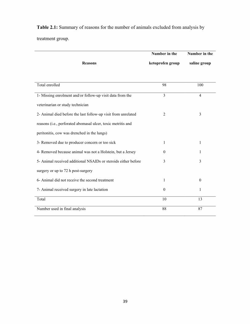
39
Table 2.1: Summary of reasons for the number of animals excluded from analysis by
treatment group.
Reasons
Number in the
ketoprofen group
Number in the
saline group
Total enrolled 98 100
1- Missing enrolment and/or follow-up visit data from the
veterinarian or study technician
3 4
2- Animal died before the last follow-up visit from unrelated
reasons (i.e., perforated abomasal ulcer, toxic metritis and
peritonitis, cow was drenched in the lungs)
2 3
3- Removed due to producer concern or too sick 1 1
4- Removed because animal was not a Holstein, but a Jersey 0 1
5- Animal received additional NSAIDs or steroids either before
surgery or up to 72 h post-surgery
3 3
6- Animal did not receive the second treatment 1 0
7- Animal received surgery in late lactation 0 1
Total 10 13
Number used in final analysis 88 87
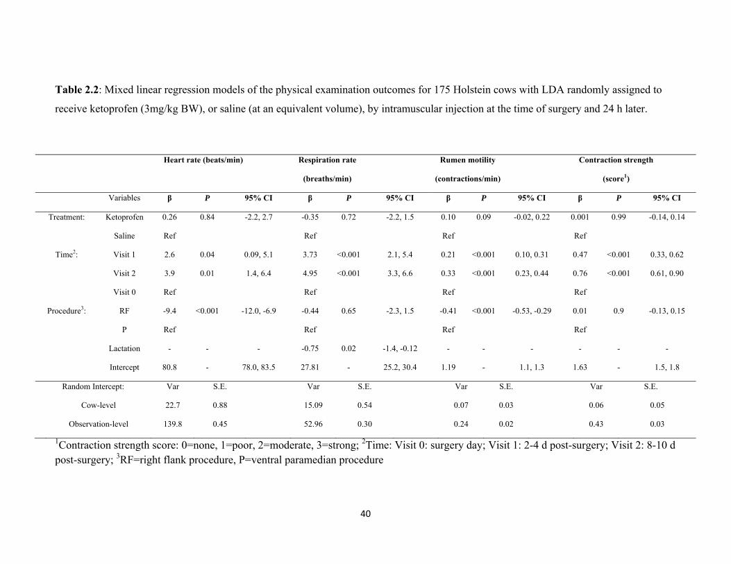
40
Table 2.2: Mixed linear regression models of the physical examination outcomes for 175 Holstein cows with LDA randomly assigned to
receive ketoprofen (3mg/kg BW), or saline (at an equivalent volume), by intramuscular injection at the time of surgery and 24 h later.
1Contraction strength score: 0=none, 1=poor, 2=moderate, 3=strong; 2Time: Visit 0: surgery day; Visit 1: 2-4 d post-surgery; Visit 2: 8-10 d post-surgery; 3RF=right flank procedure, P=ventral paramedian procedure
Heart rate (beats/min) Respiration rate
(breaths/min)
Rumen motility
(contractions/min)
Contraction strength
(score1)
Variables β P 95% CI β P 95% CI β P 95% CI β P 95% CI
Treatment: Ketoprofen 0.26 0.84 -2.2, 2.7 -0.35 0.72 -2.2, 1.5 0.10 0.09 -0.02, 0.22 0.001 0.99 -0.14, 0.14
Saline Ref Ref Ref Ref
Time2: Visit 1 2.6 0.04 0.09, 5.1 3.73 <0.001 2.1, 5.4 0.21 <0.001 0.10, 0.31 0.47 <0.001 0.33, 0.62
Visit 2 3.9 0.01 1.4, 6.4 4.95 <0.001 3.3, 6.6 0.33 <0.001 0.23, 0.44 0.76 <0.001 0.61, 0.90
Visit 0 Ref Ref Ref Ref
Procedure3: RF -9.4 <0.001 -12.0, -6.9 -0.44 0.65 -2.3, 1.5 -0.41 <0.001 -0.53, -0.29 0.01 0.9 -0.13, 0.15
P Ref Ref Ref Ref
Lactation - - - -0.75 0.02 -1.4, -0.12 - - - - - -
Intercept 80.8 - 78.0, 83.5 27.81 - 25.2, 30.4 1.19 - 1.1, 1.3 1.63 - 1.5, 1.8
Random Intercept: Var S.E. Var S.E. Var S.E. Var S.E.
Cow-level 22.7 0.88 15.09 0.54 0.07 0.03 0.06 0.05
Observation-level 139.8 0.45 52.96 0.30 0.24 0.02 0.43 0.03
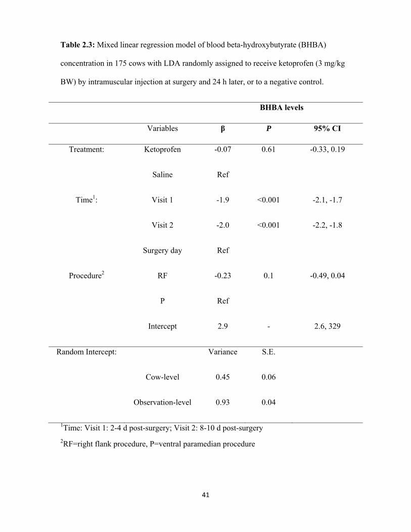
41
Table 2.3: Mixed linear regression model of blood beta-hydroxybutyrate (BHBA)
concentration in 175 cows with LDA randomly assigned to receive ketoprofen (3 mg/kg
BW) by intramuscular injection at surgery and 24 h later, or to a negative control.
BHBA levels
Variables β P 95% CI
Treatment: Ketoprofen -0.07 0.61 -0.33, 0.19
Saline Ref
Time1: Visit 1 -1.9 <0.001 -2.1, -1.7
Visit 2 -2.0 <0.001 -2.2, -1.8
Surgery day Ref
Procedure2 RF -0.23 0.1 -0.49, 0.04
P Ref
Intercept 2.9 - 2.6, 329
Random Intercept: Variance S.E.
Cow-level 0.45 0.06
Observation-level 0.93 0.04
1Time: Visit 1: 2-4 d post-surgery; Visit 2: 8-10 d post-surgery
2RF=right flank procedure, P=ventral paramedian procedure
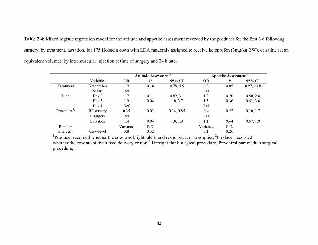
42
Table 2.4: Mixed logistic regression model for the attitude and appetite assessment recorded by the producer for the first 3 d following
surgery, by treatment, lactation, for 175 Holstein cows with LDA randomly assigned to receive ketoprofen (3mg/kg BW), or saline (at an
equivalent volume), by intramuscular injection at time of surgery and 24 h later.
Attitude Assessment1 Appetite Assessment2 Variables OR P 95% CI OR P 95% CI
Treatment Ketoprofen 1.9 0.16 0.78, 4.5 4.8 0.05 0.97, 23.8 Saline Ref Ref
Time: Day 2 1.7 0.11 0.89, 3.1 1.2 0.70 0.50, 2.8 Day 3 1.9 0.04 1.0, 3.7 1.5 0.36 0.62, 3.6 Day 1 Ref Ref
Procedure3: RF surgery 0.35 0.02 0.14, 0.85 0.4 0.22 0.10, 1.7 P surgery Ref Ref
Lactation 1.4 0.04 1.0, 1.9 1.1 0.64 0.67, 1.9 Random Variance S.E. Variance S.E. Intercept: Cow-level 3.8 0.32 7.1 0.26
1Producer recorded whether the cow was bright, alert, and responsive, or was quiet; 2Producer recorded whether the cow ate at fresh feed delivery or not; 3RF=right flank surgical procedure, P=ventral paramedian surgical procedure;
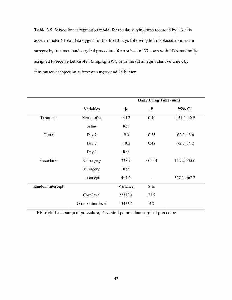
43
Table 2.5: Mixed linear regression model for the daily lying time recorded by a 3-axis
accelerometer (Hobo datalogger) for the first 3 days following left displaced abomasum
surgery by treatment and surgical procedure, for a subset of 37 cows with LDA randomly
assigned to receive ketoprofen (3mg/kg BW), or saline (at an equivalent volume), by
intramuscular injection at time of surgery and 24 h later.
Daily Lying Time (min)
Variables β P 95% CI
Treatment Ketoprofen -45.2 0.40 -151.2, 60.9
Saline Ref
Time: Day 2 -9.3 0.73 -62.2, 43.6
Day 3 -19.2 0.48 -72.6, 34.2
Day 1 Ref
Procedure1: RF surgery 228.9 <0.001 122.2, 335.6
P surgery Ref
Intercept 464.6 - 367.1, 562.2
Random Intercept: Variance S.E.
Cow-level 22310.4 21.9
Observation-level 13473.6 9.7
1RF=right flank surgical procedure, P=ventral paramedian surgical procedure
44
Chapter 3
Ketoprofen provides some pain relief following rumen fistulation surgery in
lactating dairy cows
Chapter to be submitted to Journal of Dairy Science
45
INTRODUCTION
Fistulation surgery of the rumen has been used to investigate nutrition (Hayes et
al., 1964) and effects of diet changes on rumen fluid in cattle (Willes, 1972; Thyfault et
al., 1975; Hernandez-Urdaneta et al., 1976; Anil et al., 1993). The overall objective of
this type of surgery is to facilitate access to the rumen by scientists interested in
understanding the function of the ruminant digestive system. Ruminants that have
undergone fistulation surgery often also serve as important teaching tools at many
university institutions, particularly for students interested in ruminant digestion, anatomy,
and physiology. This study was initiated because nutritionists at the University of Guelph
required fistulated cows for their intensive nutritional research projects conducted at the
Elora Dairy Research Centre, University of Guelph, Guelph, Ontario. It presented a
perfect opportunity to evaluate pain following surgery in a controlled research
environment. The first component of this surgery is a left-flank laparotomy conducted
under local anesthesia and clamping of the rumen wall to the skin. As such, studying pain
following this stage can become a model for other laparotomy surgeries, such as left
displaced abomasum correction or caesarean section. In the second stage of the fistulation
surgery 1 wk later the clamp is removed and a rumen cannula is fitted into the fistula. It
has been reported that laparotomies conducted on humans causes less pain compared to
more invasive surgeries, yet requires pain management (Luks et al., 1999; Nicholson et
al., 2001; Rosen et al., 2001). The administration of a non-steroidal anti-inflammatory
drug (NSAID), such as ketoprofen pre-operatively in humans improved post-operative
analgesia following a laparoscopic cholecystectomy compared to post-operative
administration and to the administration of opioids (propacetamol, Boccara et al., 2005).
46
Similarly, it is well established that laparotomies are painful procedures in small animals
(Roughan and Flecknell, 2001) and pain relief agents are used more and more frequently
(Mathews, 2000).
Before fistulation surgery in cattle, it is common to use a sedative (xylazine) and a
local anesthetic (or nerve block), with lidocaine for example, to block the acute pain
sensation of the surgery from the tissue damage that will be inflicted. Although pain relief
for this surgery has been reported [e.g., Tomanol postoperatively (Anil et al., 1993),
flunixin meglumine pre-operatively and butorphanol post-operatively (Sams and Fubini,
1993)] it is not common practice. Furthermore, insofar as we know, there have been no
studies examining the effect of a post-operative analgesic following fistulation on cow
behavior and performance. In addition there is a paucity of data on control of this pain
with appropriate medication.
The aims of this study was to evaluate the effects of providing an NSAID
immediately following the first stage of a two-stage fistulation surgery on animal
physiological, behavioral, and production parameters.
MATERIALS AND METHODS
Animals and General Information
Approval for this study was granted by the Animal Care Committee, University of
Guelph (AUP# 09R044). Animals were housed at the Elora Dairy Research Centre
(EDRC), the University of Guelph, Guelph, Ontario. Cows were housed in an individual
tie-stall (1.98 x 1.22 m; bedding of wood shavings over a pasture mat) in the main barn
with feed dividers to allow for feed intake recording beginning 7 d before surgery. This
47
study was conducted from April to August 2009. Cow pairs (because two surgical pens
were available at a time) were then moved to individual pens (3.5 x 3.1 m; with either
straw pack bedding or wood shavings over pasture mat) in the morning of the surgery and
remained in the pen for 7 d. All cow pairs were operated between the hours of 09:00 and
11:00 on their respective day of first stage of the fistulation surgery. The cows were then
moved to the physiology wing (tie-stall with feed dividers) immediately following the
second stage (cannula placement) for an additional week of observation before returning
to their designated tie-stall in the main barn.
Cows were fed twice daily at 07:30 and 13:00 according to routine feeding
procedure at EDRC, except for the day of surgery when the morning feeding was
withheld in the morning until after surgery completion (both for the first and the second
stages). Diets were fed as a total mixed ration and contained haylage, corn silage and hay
for the forage base; and high moisture corn, protein and mineral supplement for the
concentrate. Samples of the diet were collected twice weekly and frozen at -20°C for later
analysis. Dry matter intake (DMI) calculations were based on amount offered, orts, and
dry matter analysis of sampled diets. These data were measured and calculated daily
beginning 7 d before surgery until 14 d following the first stage of the surgical fistulation
procedure.
All clinical health events and treatments that may have occurred outside the
surgery were recorded for each cow enrolled in the study. All cows were weighed upon
enrolment in the study.
48
Fistulation Surgery
The fistulation surgery for each animal was completed as a two-stage procedure.
The first stage consisted of a rumen clamp procedure (a laparotomy through which the
rumen was pulled through and clamped in a wooden clamp and secured with 6 vertical
mattress sutures in the skin). This procedure began with clipping the hair and surgically
washing the left paralumbar fossa with iodine and alcohol solutions on the left side of the
animal. A sedative (xylazine, 0.015-0.02 mg/kg IV) was administered 30 minutes before
surgery; followed by administration of a proximal paravertebral block with 2% lidocaine.
Following confirmation of an adequate block (accomplished by touching and penetrating
the skin a few millimeters with an 18 gauge needle to the area, where the laparotomy
incision was to be made, to see if the cow reacted to the needle pain by moving or kicking
or twitching the skin around the site) and final skin preparation, a 20 cm vertical incision
was made immediately behind the rib cage and approximately 40-50 cm down from the
vertebrae. The rumen was pulled through the incision with towel clamps. Three vertical
mattress sutures were place on either side of the incision at the top, middle, and bottom,
to later hold the wooden clamp in place. The rumen was held by two towel clamps at
each end of the incision before being pulled through the wooden clamp, and the clamp
was then sutured tightly into place to the skin. The wooden clamp was tightened around
the rumen and any metal parts from the clamp were covered in gauze and tape (Figure
3.1). The area around the clamp was cleaned and petroleum jelly was rubbed on the hide
to prevent sores. Before releasing the cow from the head-gate, all cows received a dose of
penicillin given intramuscularly (20,000 iu/kg) and were either given the NSAID (see
experimental protocol below) or the saline injection.
49
The second stage of the fistulation procedure occurred 1 wk later and consisted of
removing the clamp (leaving the rumen open to the outside, while the rumen wall and the
skin wall were healed together) to allow for the cannula to be placed through the fistula
and into the rumen. The cow was sedated with xylazine in preparation for the second
stage of the fistulation. A scalpel blade was used to cut the dead rumen portion right
behind the wooden clamp. Once the clamp was removed, the fistula was scraped with a
surgical scrub brush to remove any excess dead tissue and examined for signs of
infection. A 7.62 cm diameter cannula (Bar Diamond, Inc., Parma, Idaho) was placed in
extremely hot water to soften the rubber; the cannula was allowed to cool slightly (to
avoid any burning) and then placed through the fistula and into the rumen. The cannula
was immediately closed off with the corresponding lid. Hibitane veterinary ointment
(Wyeth Animal Health, Division of Wyeth Canada, Guelph, Ontario) was liberally
applied to the incision site and petroleum jelly was rubbed on the hide under the cannula
and around it to prevent sores.
Experimental Protocol
Eighteen healthy first lactation Holstein cows, that were 293 ± 151 DIM and
weighed 609 ± 52 kg (Mean ± SD), were subjected to the two-stage fistulation surgery.
Enrolled cows were randomly assigned to 1 of 2 treatments: 1) intramuscular injection of
3 mg/kg BW ketoprofen (plasma half-life= 2 h; Anafen, Merial, Baie d’Urfé, Québec)
or placebo (3 mg/kg BW of saline). Each cow received her respective treatment at the
time of surgery completion (d 0) and 24 h post-surgery (d 1). The observer was blinded to
treatment throughout the experiment and for statistical analysis.
50
Daily measurements included: daily feed intake and milk weights for 7 d pre-
surgery and on d 0 and 1 following stage 1 surgery. We also measured heart rate,
respiration rate, and rectal temperature on d 0 immediately before surgery and 24 h post-
surgery following the second injection. Infrared temperature readings at 4 points around
the surgery site (top, bottom, right and left, at 5 cm from the surgical site) and at a control
point (at the edge of the shaved area by the L2 and L3 transverse processes) were also
taken using a digital temperature reader (Mastercraft, Canadian Tire Corporation Limited,
Ontario, Canada). Serum blood samples were obtained by coccygeal venipuncture, a pre-
surgical blood sample was taken, and all subsequent blood samples were taken in the
morning at approximately 2 h post-feeding and post-injection, allowed 1 h to clot, and
then centrifuged at 7,000 g (International Clinical centrifuge, Model CL, International
Equipment Co., Massachusetts). Blood serum was frozen and submitted for haptoglobin
analysis to the Animal Health Laboratory, University of Guelph, Guelph, Ontario,
Canada. All analyses were conducted using a Roche Cobas 6000 c501 automated
chemistry analyzer (Roche Canada, Laval, Quebec). Haptoglobin concentrations were
measured using the haemoglobin binding capacity, using a methemoglobin reagent made
on-site according to a method described elsewhere (Skinner et al., 1991; Skinner and
Roberts, 1994). The analytical sensitivity of the haptoglobin assay was 0.03 g/L. The
inter- and intra-assay coefficients of variation were 5.6 and 3.5%, respectively.
Behaviors were selected based on initial observations made from continuous
video footage of two cows used in this study as well as behaviors identified from work
undertaken on cattle during parturition (see Wehrend et al., 2006) and on behaviors
previously identified as indicative of laboratory animal pain, such as tail flick in response
51
to heat test or licking of acetic injection site in rats (Le Bars et al, 2001a, b). These
behaviors were monitored while the cows were housed in the individual pen using
continuous video recordings obtained from a wide angle camera lens (Panasonic camera
WV-CP504 and lens WV-LZA61/2S, Panasonic Canada Inc., Mississauga, Ontario,
Canada) connected to a recording system (GeoVision, UVS 1240E2, GeoVision Inc,
USAVisionSys, California). The individuals (n=3) analyzing the video footage were
blind to treatment (inter-observer reliability R2=0.90; intra-observer reliability R2=0.99).
Feeding behavior, licking around the surgical site, and general licking times were
quantified continuously for 48 h following surgery. The tail flick and tail swing counts
were analyzed during a 1 min continuous scan every 20 min from completion of surgery
to 21:00 on d 0 and from 06:00 to 18:00 on d 1, and the presence of these events was
converted to percent based on the total number of scans done during each period (24
scans for d 0 and 36 for d 1). Total daily lying time and the number of transitions from
standing to lying (or vice versa) were recorded using a 3-axis accelerometer (Hobo
Pendant G loggers, Hoskin Scientific LTD, British-Columbia, Canada; Ledgerwood et
al., 2010) attached to the right hind leg and recording at 1 min intervals 7 d prior to
surgery and on day of surgery and d 1. Days started at 05:00 when the cow was required
to stand up for milking.
Statistical Analysis
All descriptive statistics, model building, and analyses were performed with
STATA Intercooled 10.1 (StataCorp, College Station, Texas). Mixed multivariable
models were built and included a random intercept for animals to account for multiple
measurements being taken from each cow. Mixed linear regression models were
52
performed for the outcomes of DMI, milk production, and lying behavior, while linear
regression models were built for serum haptoglobin, physiological measurements (heart
rate, respiration rate, rectal temperature), behaviors from video analysis, and infrared
temperature measurements around the surgical site. All tests were 2-sided and
significance was based on α < 0.05. For all models, time and treatment were forced in as
covariate and were modeled as categorical variables. Univariable models were screened
and kept in the final model if they were significant at α < 0.05 level, acted as a
confounder (defined as a non-intervening variable whose inclusion in the model made a
20% or greater change in the coefficient of significant variables in the final model
(Dohoo et al., 2003)), or were part of a significant interaction term. Interactions between
treatment and any significant covariates in the final model were tested. Two types of
models were built for each outcome. The first set of models was designed to test for the
effects of the first stage of surgery on the outcome and as such, the models included time,
cow weight for the DMI and milk production models, and cow as a random effect to
account for repeated measures. The second set of models was designed to test for the
effect of treatment (administered on d 0 and d 1) following the first stage and as such, the
pre-surgery average values were included as a covariate in the mixed multivariable
models and the models included time, treatment, cow weight for the DMI and milk
production models, and cow as a random effect. For the linear regression models,
treatment and time were the covariates tested. For the infrared readings, the 4 reading
positions were tested individually against the control position, and only the positions
(bottom, right, and left) that were significantly different from the control position were
averaged and used as the infrared reading outcome.
53
We examined standardized residuals to identify outliers at the observation-level
for the linear and mixed linear models, and best linear unbiased predictors (BLUPS) for
any outliers at the cow-level for the mixed linear models. Normality and homogeneity of
variance were assessed for the observation-level standardized residuals for mixed linear
models, and for the cow-level models, BLUPS were examined.
RESULTS
The surgery caused a significant decrease in feed intake on d 0 and 1 (Table 3.1,
Figure 3.2) from the pre-surgical average, but there were no differences between
treatment groups on d 0 or d 1 (Table 3.2). Not surprisingly, the surgery completed
during stage 1 caused a decrease in milk production on d 0 and 1 (Table 3.1, Figure 3.3)
compared to pre-surgery average, regardless of treatment. However, cows that were
injected with saline tended to show a lower milk production compared to cows that
received ketoprofen on d 0 and 1 (Table 3.2).
Table 3.3 summarizes the means (± SE) for the heart rate (HR), respiration rate
(RR), rectal temperature (RT), infrared temperature (IRT) readings around the surgical
site (average of the left, right, and bottom positions around the surgical site), and serum
haptoglobin levels, with d 0 as the pre-surgical baseline. There were no significant
treatment effects for any of these outcomes, however, the HR, RT, IRT, and haptoglobin
levels were significantly higher following surgery compared to baseline (Table 3.3).
We observed no differences in time spent at the feed bunk, general licking, licking
of the surgical site, or tail swing count percent between the two treatments (Table 3.4).
We also did not observe any differences in time spent at the feed bunk as well as tail
swing count percent from d 0 to d 1 (Table 3.4). There was a significant decrease in
54
licking around the surgical site, and a tendency of increase in general licking, on d1
compared to d 0 (Table 3.4). There was an increase in tail flicks count percent on d 1
compared to d 0 (β= 3.5%; 95% C.I.: 1.7, 5.3; P < 0.001), and on d 1, saline-treated
animals had higher tail flick count percent compared to those that received ketoprofen
(β= 5.8%; 95% C.I.: 1.5, 10.1; P = 0.01;Figure 3.4).
The total daily lying time did not change as a result of surgery or in response to
treatment, with cows on average spending 12.3 h/d (± 0.78 SE) lying down on d 0 and 1
(Tables 3.1 and 3.2). There was a decrease in the amount of lying time spent on the left
side following surgery compared to the pre-surgical average (Table 3.1, Figure 3.5).
However, animals treated with saline spent less time lying on their left side compared to
animals treated with ketoprofen on d 0 and 1 (Table 3.2, Figure 3.5).
DISCUSSION
The acute effects of the first stage of a two-stage fistulation surgery resulted in a
decrease in DMI and milk production, and an increase in heart rate, respiration rate,
serum haptoglobin levels, and infrared temperature readings around the surgical site. The
first stage of the two-stage fistulation surgery arguably resulted in pain as defined by the
International Association for the Study of Pain (IASP) (1994): an unpleasant sensory and
emotional experience associated with actual or potential tissue damage. It is generally
accepted that visceral pain in cattle is characterized by total or partial inappetence, and
increased respiratory and heart rates (Dobromylskyj, et al., 2000) which explain the
results seen in this study.
Surgery caused other changes, such as in serum haptoglobin. The baseline values
corresponded to normal haptoglobin levels in healthy cows (i.e. ≤ 0.10 g/L; Panndorf et
55
al., 1976, Conner et al., 1986) and the increase in haptoglobin associated with fistulation
can potentially be attributed to the inflammatory process of the deliberate tissue damage
from surgery (Gabay, 2006). Indeed, the increased infrared temperature readings around
the surgical site could have corresponded to the inflammatory processes around the
surgical site. Increase in haptoglobin following surgery has also been reported in cats
(Kajikawa et al., 1999). There is merit in further investigating infrared temperature
reading of site of tissue trauma as a non-invasive method of assessing inflammation and
pain levels.
Despite no changes in overall lying time associated with the surgery or drug
treatment, we observed that the total time spent lying on the left side on d 0 and 1 was
much reduced compared to pre-surgery averages; but this latter observation was mitigated
to some degree by the administration of ketoprofen. This is the first study to describe the
laterality of lying immediately following the first stage of fistulation surgery. Grant et al.
(1990) observed that left-sided ruminally cannulated animals spent less lying on the left
side (70% increase in right side laterality over a 48 h observation period) compared to
intact animals (47% increase in right side laterality over a 48 h observation period), while
unaltered cattle will typically spend approximately 50% of their daily lying time on the
left side (Tucker et al., 2009). This is consistent with the current work where cattle spent
55% of their lying time on the left side before surgery. This was dramatically reduced to
31% in the days following the first stage of the fistulation procedure but this difference
was partly mitigated by ketoprofen (40%), perhaps because this NSAID has a plasma
half-life of 2 h (Merial-Canada, 2002) . Furthermore, ketoprofen administered i.v. at a
dose of 3 mg/kg to beef calves 20 min before castration has been shown to decrease pain-
56
associated behavioral responses during approximately 6 h following treatment (Ting et
al., 2003) thus suggesting that ketoprofen is a short acting drug. This short acting effect
of ketoprofen is reflected by the result that post-surgical time spent on the left side did
not return to pre-surgical levels, suggesting that there is still pain present due to the
reluctance of lying on the left side (the surgical side) following surgery. The findings
from Grant et al. (1990) indicate that this pain may either continue in the months
following the procedure, or that other aspects of cannulation have longer lasting effects
and deserve future study.
In addition to lying position, other behavioral changes are also considered part of
an overall integrated response to pain (Livingston and Chambers, 2000). In this study, the
feeding times on d 1 varied between 124 (saline group) and 187 (ketoprofen group)
min/d, which is less than the 191 to 216 min/d previously reported for healthy cows by
DeVries and von Keyserlingk (2005b). Perhaps the lower feeding time can be attributed
to post-surgical pain, as well as to the short half-life of ketoprofen. Unfortunately, we
were unable to obtain feeding behavior measures in the days before surgery as a
comparison, although we did find that DMI was depressed by surgery. Thus, we can
speculate that pre-surgical feeding times might have been higher than post-surgical times,
perhaps similar to feeding times reported for healthy cows.
The lower percent of tail flick count on d 1 in ketoprofen-treated animals
compared to animals that received saline may be an indicator that these NSAID-treated
animals were in less pain. The beneficial analgesic effects of ketoprofen following known
painful procedures, such as dehorning, have been investigated. For example, Faulkner
and Weary (2000) concluded that ketoprofen reduced head shaking and rubbing, and ear
57
flicking compared to control animals following hot-iron dehorning.
This study is one of the first to examine the use of an NSAID as a post-surgical
pain management tool and provides a number of lines of evidence that fistulation surgery
is indeed painful. Cows treated with ketoprofen tended to have higher milk production
and spent more time lying on the side of the surgery on d 0 and 1 compared to those
treated with saline. Furthermore, cows treated with ketoprofen had a lower number of tail
flicks on d 1 compared to those treated with saline. These results for the first stage of the
fistulation surgery suggest that ketoprofen, administered on d 0 and d 1 at a dose of 3
mg/kg, had a beneficial effect on the changes seen in production and time spent lying on
the left side following surgery compared to pre-surgical averages. Unfortunately, it
appears that ketoprofen did not completely mitigate the negative consequences of the
surgery on milk production and time spent lying on the left side,
In this study, ketoprofen was administered following a two-stage fistulation
surgery. Perhaps the minimal effect of ketoprofen at mitigating post-operative pain in
this study was due to the plasma half-life of 2 h and the elimination of 80 % of the dose
in the urine within 24 h of administration (Merial-Canada, 2002). Thus, because we only
provided ketoprofen on two occasions (24 h a part), it could be argued that the animals
were possibly feeling pain between the first and second injections. Therefore, future
research should investigate pre-operative administration of ketoprofen, as this improves
post-operative analgesia after laparoscopic cholecystectomy in humans (Boccara et al.,
2005), multiple daily injections of ketoprofen at the current label dose, or perhaps test a
more powerful or longer lasting NSAID.
58
CONCLUSIONS
This study is one of the first to examine the use of a non-steroidal anti-
inflammatory drug as a post-surgical pain management tool in cattle. Our results provide
clear evidence that the first stage of a two-stage fistulation surgery is painful and
ketoprofen administered at a label dose of 3 mg/kg BW at the time of surgery, and 24 h
following surgery, alleviated some, but not all, of the post-surgical pain. Our
recommendation is that scientists to include pain mitigation when fistulating cattle used
for research.
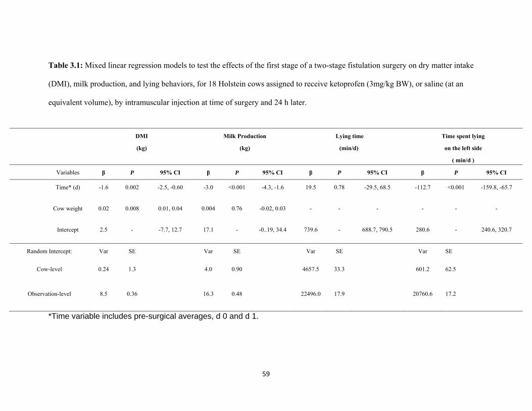
59
Table 3.1: Mixed linear regression models to test the effects of the first stage of a two-stage fistulation surgery on dry matter intake
(DMI), milk production, and lying behaviors, for 18 Holstein cows assigned to receive ketoprofen (3mg/kg BW), or saline (at an
equivalent volume), by intramuscular injection at time of surgery and 24 h later.
*Time variable includes pre-surgical averages, d 0 and d 1.
DMI
(kg)
Milk Production
(kg)
Lying time
(min/d)
Time spent lying
on the left side
( min/d )
Variables β P 95% CI β P 95% CI β P 95% CI β P 95% CI
Time* (d) -1.6 0.002 -2.5, -0.60 -3.0 <0.001 -4.3, -1.6 19.5 0.78 -29.5, 68.5 -112.7 <0.001 -159.8, -65.7
Cow weight 0.02 0.008 0.01, 0.04 0.004 0.76 -0.02, 0.03 - - - - - -
Intercept 2.5 - -7.7, 12.7 17.1 - -0..19, 34.4 739.6 - 688.7, 790.5 280.6 - 240.6, 320.7
Random Intercept: Var SE Var SE Var SE Var SE
Cow-level 0.24 1.3 4.0 0.90 4657.5 33.3 601.2 62.5
Observation-level 8.5 0.36 16.3 0.48 22496.0 17.9 20760.6 17.2
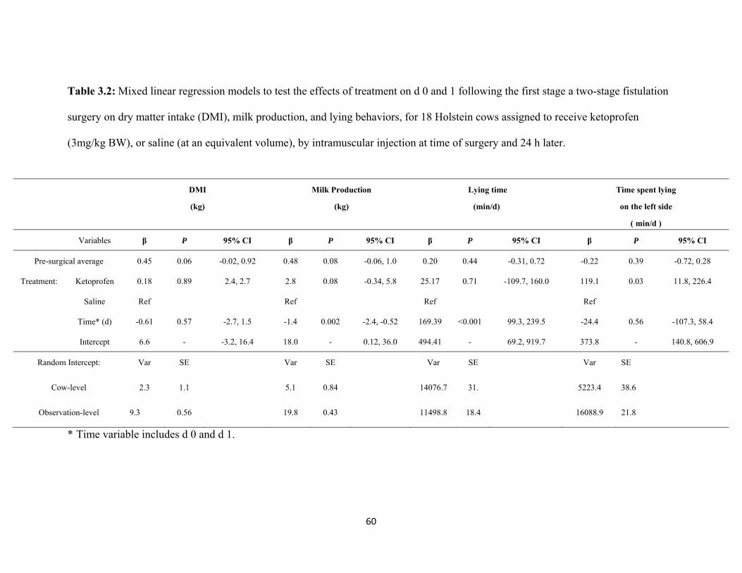
60
Table 3.2: Mixed linear regression models to test the effects of treatment on d 0 and 1 following the first stage a two-stage fistulation
surgery on dry matter intake (DMI), milk production, and lying behaviors, for 18 Holstein cows assigned to receive ketoprofen
(3mg/kg BW), or saline (at an equivalent volume), by intramuscular injection at time of surgery and 24 h later.
* Time variable includes d 0 and d 1.
DMI
(kg)
Milk Production
(kg)
Lying time
(min/d)
Time spent lying
on the left side
( min/d )
Variables β P 95% CI β P 95% CI β P 95% CI β P 95% CI
Pre-surgical average 0.45 0.06 -0.02, 0.92 0.48 0.08 -0.06, 1.0 0.20 0.44 -0.31, 0.72 -0.22 0.39 -0.72, 0.28
Treatment: Ketoprofen 0.18 0.89 2.4, 2.7 2.8 0.08 -0.34, 5.8 25.17 0.71 -109.7, 160.0 119.1 0.03 11.8, 226.4
Saline Ref Ref Ref Ref
Time* (d) -0.61 0.57 -2.7, 1.5 -1.4 0.002 -2.4, -0.52 169.39 <0.001 99.3, 239.5 -24.4 0.56 -107.3, 58.4
Intercept 6.6 - -3.2, 16.4 18.0 - 0.12, 36.0 494.41 - 69.2, 919.7 373.8 - 140.8, 606.9
Random Intercept: Var SE Var SE Var SE Var SE
Cow-level 2.3 1.1 5.1 0.84 14076.7 31. 5223.4 38.6
Observation-level 9.3 0.56 19.8 0.43 11498.8 18.4 16088.9 21.8
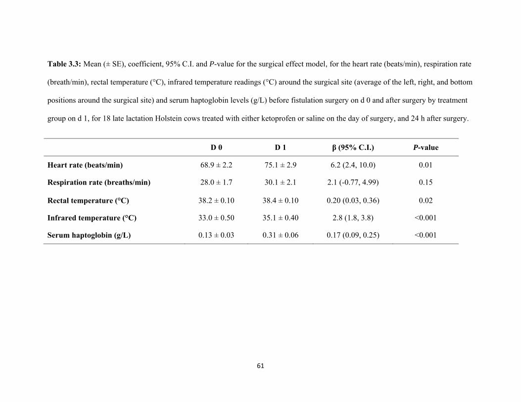
61
Table 3.3: Mean (± SE), coefficient, 95% C.I. and P-value for the surgical effect model, for the heart rate (beats/min), respiration rate
(breath/min), rectal temperature (°C), infrared temperature readings (°C) around the surgical site (average of the left, right, and bottom
positions around the surgical site) and serum haptoglobin levels (g/L) before fistulation surgery on d 0 and after surgery by treatment
group on d 1, for 18 late lactation Holstein cows treated with either ketoprofen or saline on the day of surgery, and 24 h after surgery.
D 0 D 1 β (95% C.I.) P-value
Heart rate (beats/min) 68.9 ± 2.2 75.1 ± 2.9 6.2 (2.4, 10.0) 0.01
Respiration rate (breaths/min) 28.0 ± 1.7 30.1 ± 2.1 2.1 (-0.77, 4.99) 0.15
Rectal temperature (°C) 38.2 ± 0.10 38.4 ± 0.10 0.20 (0.03, 0.36) 0.02
Infrared temperature (°C) 33.0 ± 0.50 35.1 ± 0.40 2.8 (1.8, 3.8) <0.001
Serum haptoglobin (g/L) 0.13 ± 0.03 0.31 ± 0.06 0.17 (0.09, 0.25) <0.001
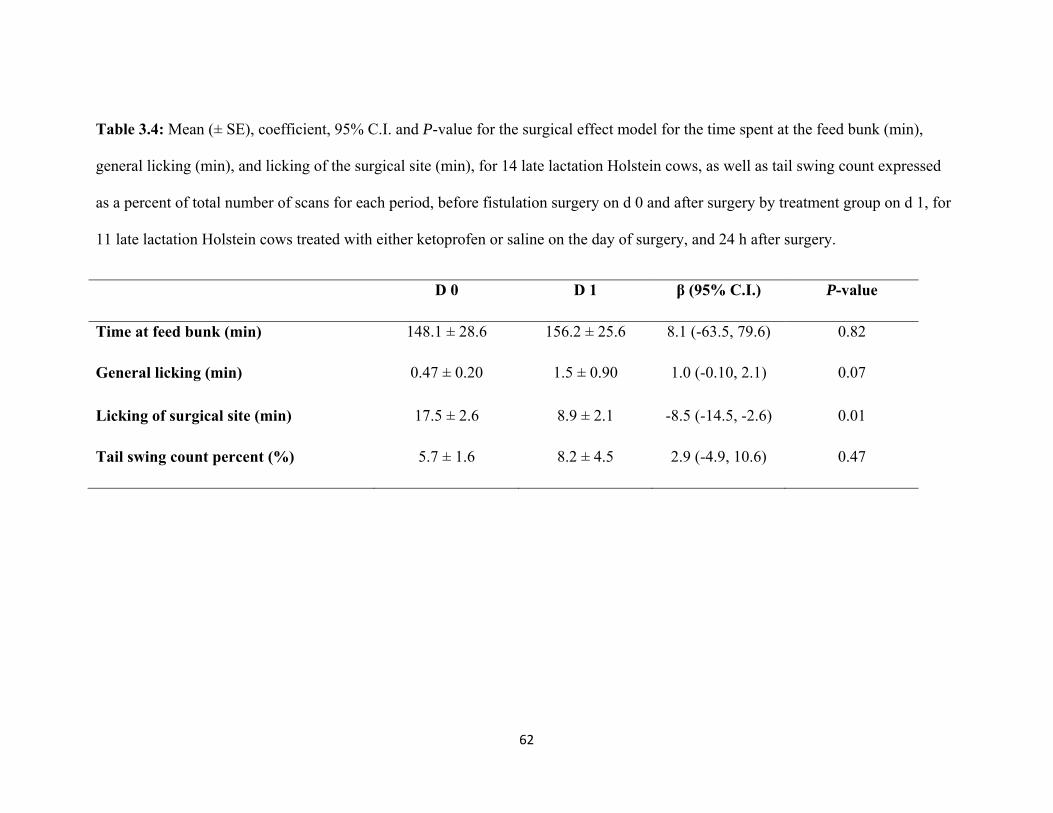
62
Table 3.4: Mean (± SE), coefficient, 95% C.I. and P-value for the surgical effect model for the time spent at the feed bunk (min),
general licking (min), and licking of the surgical site (min), for 14 late lactation Holstein cows, as well as tail swing count expressed
as a percent of total number of scans for each period, before fistulation surgery on d 0 and after surgery by treatment group on d 1, for
11 late lactation Holstein cows treated with either ketoprofen or saline on the day of surgery, and 24 h after surgery.
D 0 D 1 β (95% C.I.) P-value
Time at feed bunk (min) 148.1 ± 28.6 156.2 ± 25.6 8.1 (-63.5, 79.6) 0.82
General licking (min) 0.47 ± 0.20 1.5 ± 0.90 1.0 (-0.10, 2.1) 0.07
Licking of surgical site (min) 17.5 ± 2.6 8.9 ± 2.1 -8.5 (-14.5, -2.6) 0.01
Tail swing count percent (%) 5.7 ± 1.6 8.2 ± 4.5 2.9 (-4.9, 10.6) 0.47
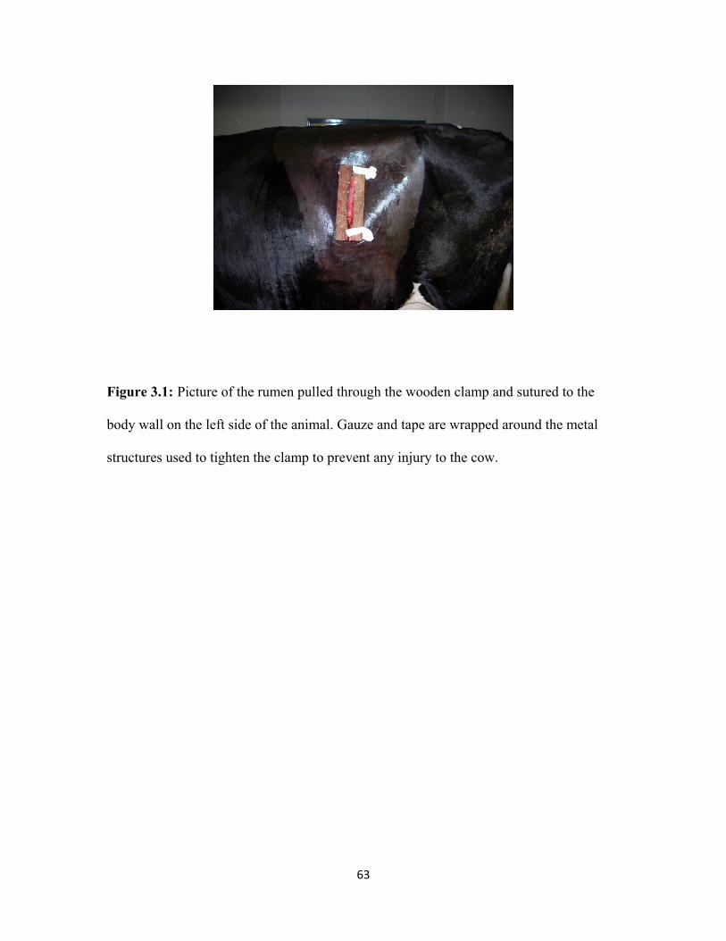
63
Figure 3.1: Picture of the rumen pulled through the wooden clamp and sutured to the
body wall on the left side of the animal. Gauze and tape are wrapped around the metal
structures used to tighten the clamp to prevent any injury to the cow.
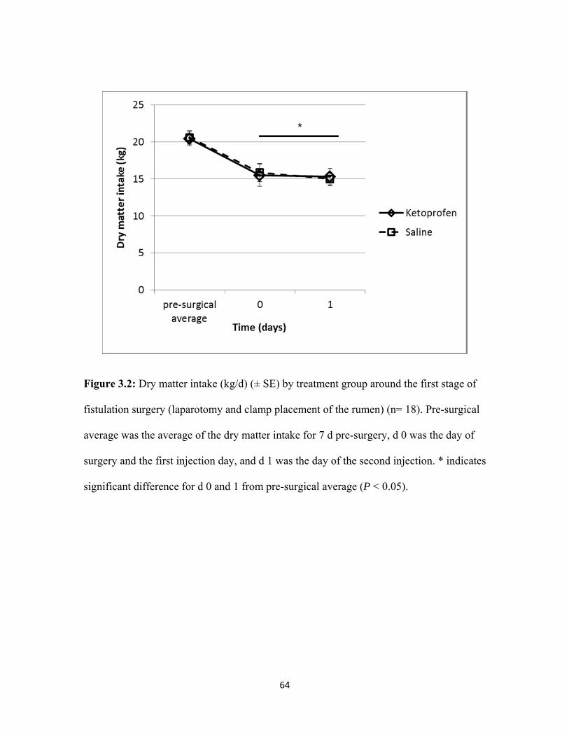
64
Figure 3.2: Dry matter intake (kg/d) (± SE) by treatment group around the first stage of
fistulation surgery (laparotomy and clamp placement of the rumen) (n= 18). Pre-surgical
average was the average of the dry matter intake for 7 d pre-surgery, d 0 was the day of
surgery and the first injection day, and d 1 was the day of the second injection. * indicates
significant difference for d 0 and 1 from pre-surgical average (P < 0.05).
*
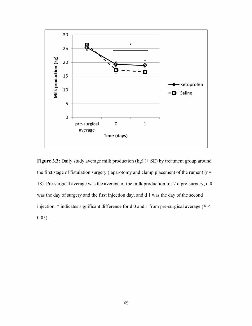
65
Figure 3.3: Daily study average milk production (kg) (± SE) by treatment group around
the first stage of fistulation surgery (laparotomy and clamp placement of the rumen) (n=
18). Pre-surgical average was the average of the milk production for 7 d pre-surgery, d 0
was the day of surgery and the first injection day, and d 1 was the day of the second
injection. * indicates significant difference for d 0 and 1 from pre-surgical average (P <
0.05).
*
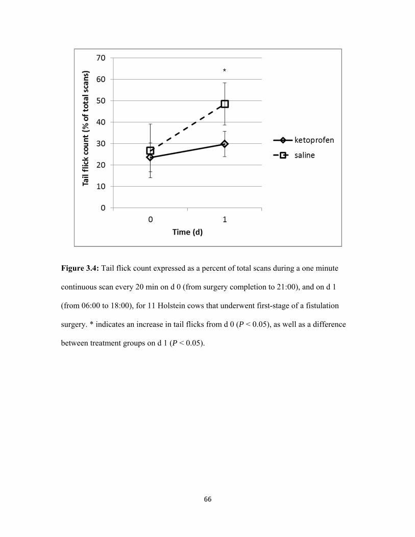
66
Figure 3.4: Tail flick count expressed as a percent of total scans during a one minute
continuous scan every 20 min on d 0 (from surgery completion to 21:00), and on d 1
(from 06:00 to 18:00), for 11 Holstein cows that underwent first-stage of a fistulation
surgery. * indicates an increase in tail flicks from d 0 (P < 0.05), as well as a difference
between treatment groups on d 1 (P < 0.05).
*
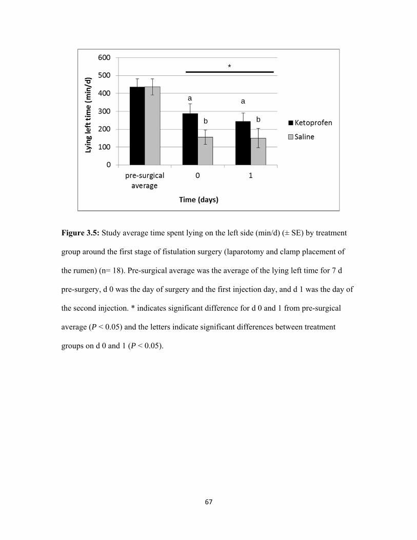
67
Figure 3.5: Study average time spent lying on the left side (min/d) (± SE) by treatment
group around the first stage of fistulation surgery (laparotomy and clamp placement of
the rumen) (n= 18). Pre-surgical average was the average of the lying left time for 7 d
pre-surgery, d 0 was the day of surgery and the first injection day, and d 1 was the day of
the second injection. * indicates significant difference for d 0 and 1 from pre-surgical
average (P < 0.05) and the letters indicate significant differences between treatment
groups on d 0 and 1 (P < 0.05).
b b
aa
*
68
Chapter 4
SHORT COMMUNICATION: A comparison of two NSAIDs
following the first stage of a two-stage fistulation surgery in dry dairy cows
Chapter to be submitted to Journal of Dairy Science
69
INTRODUCTION
One approach to fistulation surgery for placement of a permanent rumen fistula
involves a two stage process. First, a left-flank laparotomy is performed under local
anesthesia while clamping of the rumen. One week later, a rubber cannula is fitted into
the fistula. It has been reported that laparotomy in humans is a procedure that requires
pain management, even though it may be less painful compared to more invasive
surgeries (Luks et al., 1999; Nicholson et al., 2001; Rosen et al., 2001). In the previous
chapter (Chapter 3), the effects of ketoprofen versus saline were evaluated following
fistulation surgery in lactating dairy cows. In this Chapter 3, it was found that the surgery
itself proved to be painful as shown by a decrease in DMI, milk production, and in time
spent lying on the surgical side (the left side). Further evidence of the painful effects of
the first stage of the fistulation surgery were an increase in heart and respiration rates, in
infrared temperature readings around the surgical site as well as an increase in serum
haptoglobin protein concentration, as a result of the tissue damage caused by the
laparotomy (Chapter 3). It was observed that the administration of a label dose of
ketoprofen once following surgery on d 0 and once 24 h later provided some beneficial
analgesic effects to the animals on those days. The ketoprofen treated animals had a
significantly greater time spent lying on the left side on d 0 and 1, significantly lower tail
flick count percent on d 1, as well as a tendency toward higher milk production,
compared to control animals. However, even though the effects of post-surgical pain
were not completely alleviated by this dose of ketoprofen, it is clear that analgesia is
required following the first stage of a two stage fistulation surgery. Because nutritionist
researchers at the University of Guelph required 10 fistulated cows in their last trimester,
70
it presented the perfect opportunity to compare two NSAIDs following surgery and
provide a follow-up study to the previous fistulation study in Chapter 3.
The administration of non-steroidal anti-inflammatory drugs (NSAID) for pain
relief has been successful following surgery in rodents (Pairet and Ruckebusch, 1989; De
Winter et al., 1998). NSAID therapy has also been used to alleviate signs of visceral pain
in cattle (Constable et al., 1997). In 2003, Roughan and Flecknell evaluated the analgesic
effects of meloxicam or carprofen compared to saline, administered 1 h pre-operatively,
following a midline laparotomy in rats. They concluded that a dose of 1-2 mg/kg of
meloxicam effectively reduced the pain behavior observed in the 0.50 mg/kg meloxicam,
carprofen and saline groups.
The plasma half-life of ketoprofen is 2 h, and 80% of the dose is eliminated in the
urine within 24 h of administration (Merial-Canada, 2002), while the plasma half-life of
meloxicam is 23-27 h for low milk yield cows, and 17.5 h for high milk yield cows
(EMEA, 2007). The analgesic effects of ketoprofen compared to a saline control
following first stage of fistulation surgery were still not enough to completely alleviate
post-surgical pain (Newby et al., unpublished data). It is hypothesized that a longer
lasting NSAID, such as meloxicam, will further alleviate post-surgical pain, compared to
a shorter lasting one. Thus, the purpose of this study was to compare a 3 mg/kg dose of
ketoprofen administered on d 0 and 1 following the first stage of a two stage fistulation
surgery to a 0.5 mg/kg dose of meloxicam administered once only on d 0, in dry Holstein
cows.
71
MATERIAL AND METHODS
General Information
This study was conducted at the Elora Dairy Research Centre (EDRC), the
University of Guelph, Guelph, Ontario, and was approved by the University’s Animal
Care Committee (AUP#11R093). Cows were housed in individual tie-stall (2.0 x 1.2 m;
bedding of wood shavings over a pasture mat) in the main barn with feed dividers to
allow for feed intake recording 4 d prior to surgery. Cow pairs were then moved to
individual pens (3.5 x 3.1 m; straw pack bedding) on the day of the first stage of the
fistulation surgery and remained in the pen for the week following the first stage. The
cows were then moved to the physiology wing (tie-stall with feed dividers) following the
second stage (cannula placement) for 1 wk before returning to their designated stall in the
main barn. Cows were fed according to routine feeding procedure at EDRC, except for
the morning prior to surgery, when they did not receive their first feeding until after
surgery completion. Diets were fed as a total mixed ration and contained haylage, corn
silage and hay for the forage base; and high moisture corn, protein and mineral
supplement for the concentrate. Cows were fed twice daily. Samples of the diet were
collected twice weekly and frozen at -20°C for later analysis. DMI calculations were
based on amount offered, orts and dry matter analysis of sampled diets. All clinical health
events and treatments that may have occurred outside the surgery were recorded for each
cow enrolled in the study. In order to administer NSAID treatments, all cows weights
were estimated by the veterinarian upon enrolment in the study and the mean estimated
weight (± S.D.) for the ketoprofen group was 713 kg (± 56), and for the meloxicam group
was 632 kg (± 33).
72
Experimental Protocol
A randomized clinical trial was conducted with 10 healthy dry Holstein cows, at
the end of their first lactation, in their third trimester of gestation, and which underwent a
two-stage fistulation surgery (described elsewhere, Chapter 3) on average 53 d (± 12
S.D.) prior to calving. Enrolled cows were randomly assigned to one of two treatments:
ketoprofen or meloxicam. The ketoprofen group received 3 mg/kg body weight
ketoprofen (Anafen, Merial, Baie d’Urfé, Québec, Canada) intramuscularly at the time
of surgery completion (d 0), and 24 h following surgery . The meloxicam group received
0.5 mg/kg body weight of meloxicam (Metacam, Boehringer Ingelheim (Canada) Ltd.
Burlington, Ontario) subcutaneously once, as per product label instructions (EMEA,
2007) at the time of surgery completion. The cows were monitored daily 4 d prior to
surgery, and post-surgical measurements included daily DMI (d 0 and 1). Measurements
taken on d 0 prior to surgery, and at 2, 9, 24, 26, and 33 h post-surgery included
physiologic outcomes (heart rate, respiration rate, rectal temperature), and infrared
temperature readings at 4 points around the surgery site (top, bottom, right and left of the
surgical site) and at a control point (at the edge of the shaved area by the L2 and L3
transverse processes) using a digital temperature reader (Mastercraft, Canadian Tire
Corporation Limited, Ontario, Canada) 20 cm away from the skin. All cows were fitted
with a 3-axis accelerometer (Hobo Pendant G logger, Hoskin Scientific LTD, British-
Columbia; Ledgerwood et al., 2010) attached to the right hind leg and recording at 1 min
intervals 4 d prior to surgery and on d 0 and 1 post-surgery. The time for d 0 started
following surgery completion until 24 h post-surgery, and d 1 started at 24 h post-surgery
until 48 h. Furthermore, a thermal nociceptive threshold test (adapted from Pinheiro
73
Machado et al., 1997) using a class 4 carbon dioxide laser (MPB Technologies Inc.,
Dorval, Québec) was performed 200 cm from the end of the laser where the laser beam
comes out of the cover box to the lateral aspect of the hind leg, between the hock and
fetlock. This test consisted of timing how long it took for the cow to lift her leg once the
laser test was started and was done prior to surgery (baseline), and again at 2, 9, 24, 26,
33 h post-surgery.
Statistical Analysis
All descriptive statistics, model building and analyses were performed with
STATA Intercooled 10.1 (StataCorp, College Station, TX, USA). Mixed linear regression
models for treatment and time were built, including a random intercept for animals to
account for multiple measurements being taken from each cow. Time was modeled as a
continuous variable, unless it was not linear, in which case it was categorized. Models
were built for the outcomes of DMI, physiological measurements (heart rate, respiration
rate, and rectal temperature), lying activity (daily lying time and daily total time spent
lying on the left side), infrared temperature measurements around the surgical site, and
laser nociceptive threshold tests. All tests were 2-sided and significance was based on α <
0.05. For all models, time and treatment were forced in as covariates and were modeled
as categorical variables. For the DMI models, cow weight was offered as a covariate and
kept if it was significant. Interactions between treatment and any significant covariates in
the final model were tested. Two types of models were built for each outcome. The first
sets of models were designed to test for the effects of the first stage of surgery on the
outcome and as such, the pre-surgery average values were set as a referent variable within
the time period. The second sets of models were designed to test for the effect of
74
treatment (administered on d 0 and d 1, when applicable) following the first stage and as
such, the pre-surgery average values were included as a covariate in the model. The time
periods tested were d 0 and 1 for the DMI and lying activity, while the time periods tested
were 2, 9, 24, 26, and 33 h for the heart and respiration rates. For the infrared readings,
the four reading positions were tested against the control position and only the positions
that were significantly different from the control position were used in the infrared
reading average. Standardized residuals were examined to identify outliers at the
observation-level, and best linear unbiased predictors (BLUPS) for any outliers at the
cow-level. Normality and homogeneity of variance were assessed for the observation-
level standardized residuals, as well as the BLUPs.
RESULTS AND DISCUSSION
The acute effects of the first stage of a two-stage fistulation surgery resulted in a
significant decrease in DMI on day 1 (Table 4.1; Figure 4.1), and in a decrease in time
spent lying on the left side on d 0 and 1 (Table 4.1; Figure 4.2). The negative effects of
the surgery on DMI and lying left time were similar to the ones seen in the fistulation
surgery study that compared saline and ketoprofen post-operatively (Newby et al.,
unpublished data). Interestingly, the DMI on d 0 was not significantly different compared
to pre-surgery average (Table 4.1, Figure 4.1), suggesting that there may have been a
beneficial effect of NSAID administration on d 0. The meloxicam-treated animals had a
higher DMI compared to ketoprofen-treated animals on d 0 and d 1 (Table 4.2; Figure
4.1). Todd et al. (2010) reported that meloxicam-treated calves with neonatal calf
diarrhea complex consumed more milk as well as consumed calf starter earlier and at a
faster rate compared to placebo-treated calves with diarrhea. Heinrich et al. (2010)
75
observed a trend for meloxicam-treated calves to consume more feed following cautery
dehorning compared to placebo-treated calves. Cows that received ketoprofen following
left displaced abomasum surgery had greater odds of going to the feed bunk at fresh feed
delivery, for the first 3 d post-surgery, compared to the saline-treated animals (Newby et
al., unpublished data submitted to Journal of Dairy Science). The results from this study,
as well as other studies investigating NSAIDs, suggest that there are some benefits of
NSAID administration to manage post-operative, or post-calving, pain in dairy cows.
Because meloxicam-treated cows ate more compared to ketoprofen-treated cows
following surgery in the present study there may be benefits to choosing meloxicam as a
post-operative pain management therapy as opposed to ketoprofen.
The lying activity revealed that the first stage of this two-stage fistulation surgery
negatively affected the total daily time spent lying on the left side on d 0 and 1 compared
to pre-surgery average (Table 4.1; Figure 4.2). In another rumen fistulation study by
Newby et al. (unpublished data) comparing saline and ketoprofen, cows also significantly
decreased their time spent lying on the left side following surgery on d 0 and 1. However,
in the current experiment, there was a tendency for animals treated with meloxicam to
spend less time lying (mean ± SE d 0 meloxicam = 455.0 ± 130.1 min/d, ketoprofen =
721.4 ± 48.0; P = 0.08) as well as to spent less time lying on their left side compared to
animals treated with ketoprofen (Table 4.2; Figure 4.2). There were no differences in the
percent of time lying on the left side, while the cow was lying down, between treatment
groups (β= -1.9 %; P = 0.78; 95% C.I.: -15.7, 11.8, Figure 4.3). Further, it is noteworthy
that the meloxicam-treated animals had a numerically higher percentage of time spent on
the left on d 0 compared to ketoprofen-treated animals (Figure 4.3). A possible
76
explanation could be that the time budget on d 0 was different for animals in each
treatment group. The meloxicam-treated animals spent less time lying and as a result laid
less on their left side compared to the ketoprofen-treated animals. A possible explanation
the difference in lying times is that the meloxicam-treated cows ate more and thus spent
more time standing compared to the ketoprofen-treated cows. However, when the
meloxicam-treated animals did lay down on d 0, they chose to lie on their left side
numerically more than ketoprofen-treated animals (Figure 4.3). Further investigation with
an increased sample size would be required to compare the time budget between cows
treated with meloxicam or ketoprofen following surgery
In the present study, there was a general trend toward increasing following
surgery for the heart rate (HR), for the respiration rate (RR), and for the infrared
temperature (IRT) reading (Table 4.3 and 4.4) and no differences were found between
treatment groups (Table 4.3). These trends were also observed in the fistulation study
comparing saline and ketoprofen (Newby et al., unpublished data). The laser nociceptive
threshold was unsuccessful at detecting post-surgical pain threshold difference (Table 4.1
and 4.4) or pain threshold differences between treatment groups (Table 4.2). This result
was not surprising considering the fact that there were only 5 cows in each group and
very little power to this test, and the fact that individual pain thresholds (Sawyer, 1998)
may be different and play a big role in the variations seen for this test.
CONCLUSIONS
In conclusion, the first stage of a two-stage fistulation surgery was considered
painful due to the decreases in DMI and the decrease in time spent lying on the left side
as well as the increases in heart and respiration rates, and infrared temperature readings
77
around surgical site following deliberate tissue damage caused by the laparotomy. This
study compared ketoprofen versus meloxicam following surgery. Meloxicam-treated
animals ate better than ketoprofen-treated animals following the first stage, while
ketoprofen-treated animals had a tendency to spend more time lying left compared to
meloxicam-treated animals. NSAIDs should be considered following fistulation surgery
since there seems to be beneficial effects on DMI and preference of lying on the surgical
side in the day following surgery. However, further research is needed to determine the
appropriate dosage of NSAID required for sufficient pain relief.
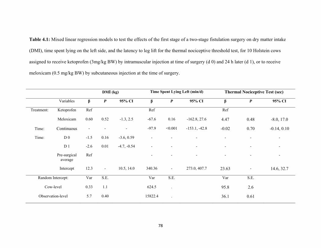
78
Table 4.1: Mixed linear regression models to test the effects of the first stage of a two-stage fistulation surgery on dry matter intake
(DMI), time spent lying on the left side, and the latency to leg lift for the thermal nociceptive threshold test, for 10 Holstein cows
assigned to receive ketoprofen (3mg/kg BW) by intramuscular injection at time of surgery (d 0) and 24 h later (d 1), or to receive
meloxicam (0.5 mg/kg BW) by subcutaneous injection at the time of surgery.
DMI (kg) Time Spent Lying Left (min/d) Thermal Nociceptive Test (sec)
Variables β P 95% CI β P 95% CI β P 95% CI
Treatment: Ketoprofen Ref Ref Ref
Meloxicam 0.60 0.52 -1.3, 2.5 -67.6 0.16 -162.8, 27.6 4.47 0.48 -8.0, 17.0
Time: Continuous - - - -97.9 <0.001 -153.1, -42.8 -0.02 0.70 -0.14, 0.10
Time: D 0 -1.5 0.16 -3.6, 0.59 - - - - - -
D 1 -2.6 0.01 -4.7, -0.54 - - - - - -
Pre-surgical average
Ref - - - - - -
Intercept 12.3 - 10.5, 14.0 340.36 - 273.0, 407.7 23.63 - 14.6, 32.7
Random Intercept: Var S.E. Var S.E. Var S.E.
Cow-level 0.33 1.1 624.5 . 95.8 2.6
Observation-level 5.7 0.40 15822.4 . 36.1 0.61
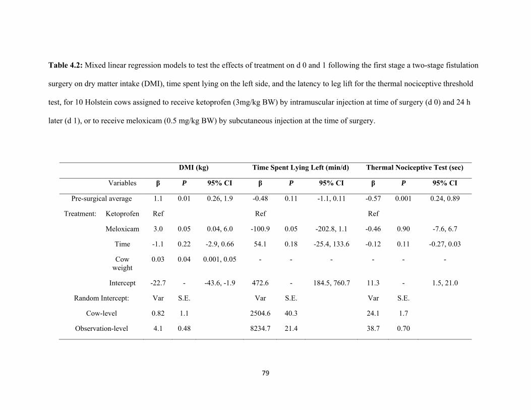
79
Table 4.2: Mixed linear regression models to test the effects of treatment on d 0 and 1 following the first stage a two-stage fistulation
surgery on dry matter intake (DMI), time spent lying on the left side, and the latency to leg lift for the thermal nociceptive threshold
test, for 10 Holstein cows assigned to receive ketoprofen (3mg/kg BW) by intramuscular injection at time of surgery (d 0) and 24 h
later (d 1), or to receive meloxicam (0.5 mg/kg BW) by subcutaneous injection at the time of surgery.
DMI (kg) Time Spent Lying Left (min/d) Thermal Nociceptive Test (sec)
Variables β P 95% CI β P 95% CI β P 95% CI
Pre-surgical average 1.1 0.01 0.26, 1.9 -0.48 0.11 -1.1, 0.11 -0.57 0.001 0.24, 0.89
Treatment: Ketoprofen Ref Ref Ref
Meloxicam 3.0 0.05 0.04, 6.0 -100.9 0.05 -202.8, 1.1 -0.46 0.90 -7.6, 6.7
Time -1.1 0.22 -2.9, 0.66 54.1 0.18 -25.4, 133.6 -0.12 0.11 -0.27, 0.03
Cow weight
0.03 0.04 0.001, 0.05 - - - - - -
Intercept -22.7 - -43.6, -1.9 472.6 - 184.5, 760.7 11.3 - 1.5, 21.0
Random Intercept: Var S.E. Var S.E. Var S.E.
Cow-level 0.82 1.1 2504.6 40.3 24.1 1.7
Observation-level 4.1 0.48 8234.7 21.4 38.7 0.70
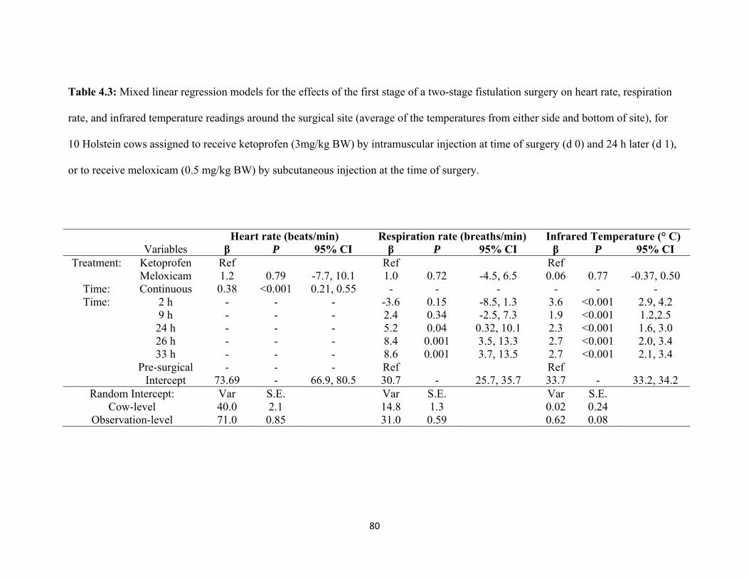
80
Table 4.3: Mixed linear regression models for the effects of the first stage of a two-stage fistulation surgery on heart rate, respiration
rate, and infrared temperature readings around the surgical site (average of the temperatures from either side and bottom of site), for
10 Holstein cows assigned to receive ketoprofen (3mg/kg BW) by intramuscular injection at time of surgery (d 0) and 24 h later (d 1),
or to receive meloxicam (0.5 mg/kg BW) by subcutaneous injection at the time of surgery.
Heart rate (beats/min) Respiration rate (breaths/min) Infrared Temperature (° C) Variables β P 95% CI β P 95% CI β P 95% CI
Treatment: Ketoprofen Ref Ref Ref Meloxicam 1.2 0.79 -7.7, 10.1 1.0 0.72 -4.5, 6.5 0.06 0.77 -0.37, 0.50
Time: Continuous 0.38 <0.001 0.21, 0.55 - - - - - - Time: 2 h - - - -3.6 0.15 -8.5, 1.3 3.6 <0.001 2.9, 4.2
9 h - - - 2.4 0.34 -2.5, 7.3 1.9 <0.001 1.2,2.5 24 h - - - 5.2 0.04 0.32, 10.1 2.3 <0.001 1.6, 3.0 26 h - - - 8.4 0.001 3.5, 13.3 2.7 <0.001 2.0, 3.4
33 h - - - 8.6 0.001 3.7, 13.5 2.7 <0.001 2.1, 3.4 Pre-surgical - - - Ref Ref Intercept 73.69 - 66.9, 80.5 30.7 - 25.7, 35.7 33.7 - 33.2, 34.2
Random Intercept: Var S.E. Var S.E. Var S.E. Cow-level 40.0 2.1 14.8 1.3 0.02 0.24
Observation-level 71.0 0.85 31.0 0.59 0.62 0.08
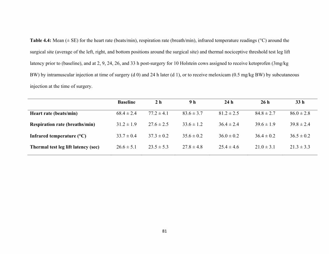
81
Table 4.4: Mean (± SE) for the heart rate (beats/min), respiration rate (breath/min), infrared temperature readings (°C) around the
surgical site (average of the left, right, and bottom positions around the surgical site) and thermal nociceptive threshold test leg lift
latency prior to (baseline), and at 2, 9, 24, 26, and 33 h post-surgery for 10 Holstein cows assigned to receive ketoprofen (3mg/kg
BW) by intramuscular injection at time of surgery (d 0) and 24 h later (d 1), or to receive meloxicam (0.5 mg/kg BW) by subcutaneous
injection at the time of surgery.
Baseline 2 h 9 h 24 h 26 h 33 h
Heart rate (beats/min) 68.4 ± 2.4 77.2 ± 4.1 83.6 ± 3.7 81.2 ± 2.5 84.8 ± 2.7 86.0 ± 2.8
Respiration rate (breaths/min) 31.2 ± 1.9 27.6 ± 2.5 33.6 ± 1.2 36.4 ± 2.4 39.6 ± 1.9 39.8 ± 2.4
Infrared temperature (°C) 33.7 ± 0.4 37.3 ± 0.2 35.6 ± 0.2 36.0 ± 0.2 36.4 ± 0.2 36.5 ± 0.2
Thermal test leg lift latency (sec) 26.6 ± 5.1 23.5 ± 5.3 27.8 ± 4.8 25.4 ± 4.6 21.0 ± 3.1 21.3 ± 3.3
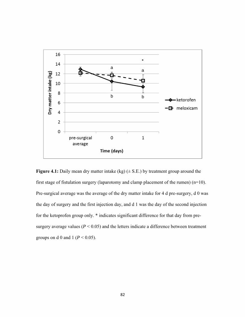
82
Figure 4.1: Daily mean dry matter intake (kg) (± S.E.) by treatment group around the
first stage of fistulation surgery (laparotomy and clamp placement of the rumen) (n=10).
Pre-surgical average was the average of the dry matter intake for 4 d pre-surgery, d 0 was
the day of surgery and the first injection day, and d 1 was the day of the second injection
for the ketoprofen group only. * indicates significant difference for that day from pre-
surgery average values (P < 0.05) and the letters indicate a difference between treatment
groups on d 0 and 1 (P < 0.05).
*
a
b b
a
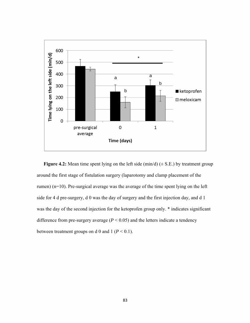
83
Figure 4.2: Mean time spent lying on the left side (min/d) (± S.E.) by treatment group
around the first stage of fistulation surgery (laparotomy and clamp placement of the
rumen) (n=10). Pre-surgical average was the average of the time spent lying on the left
side for 4 d pre-surgery, d 0 was the day of surgery and the first injection day, and d 1
was the day of the second injection for the ketoprofen group only. * indicates significant
difference from pre-surgery average (P < 0.05) and the letters indicate a tendency
between treatment groups on d 0 and 1 (P < 0.1).
a
b
b
a
*
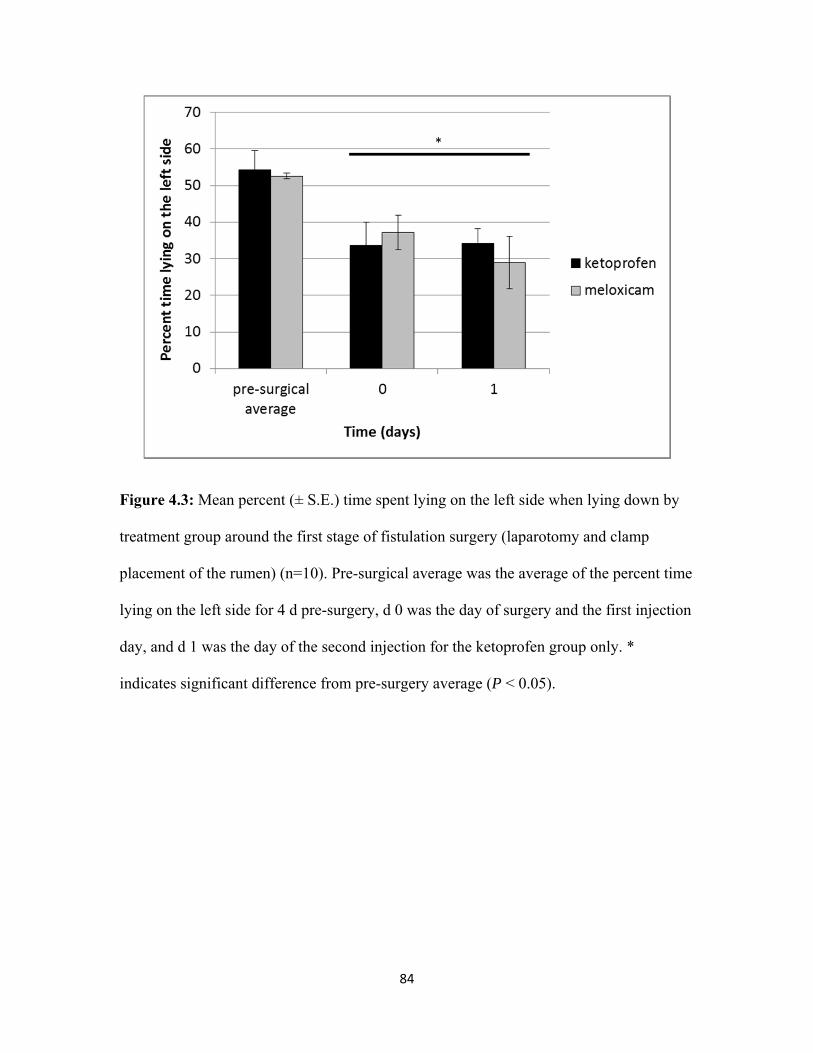
84
Figure 4.3: Mean percent (± S.E.) time spent lying on the left side when lying down by
treatment group around the first stage of fistulation surgery (laparotomy and clamp
placement of the rumen) (n=10). Pre-surgical average was the average of the percent time
lying on the left side for 4 d pre-surgery, d 0 was the day of surgery and the first injection
day, and d 1 was the day of the second injection for the ketoprofen group only. *
indicates significant difference from pre-surgery average (P < 0.05).
*
85
Chapter 5
Effects of Meloxicam on Milk Production, Behavior and Feed Intake
in Dairy Cows following an Assisted Calving
This Chapter has been submitted to the Journal of Dairy Science (MS#: JDS-12-6214)
86
INTRODUCTION
Parturition is necessary for dairy production and assisted calving is common.
Calving is a painful event that leads to inflammation (Bionaz et al., 2007). Dystocia rates
are higher in North America (>10%) compared with other parts of the world (<5%), and
regardless of country, are much higher in primiparous animals (Mee, 2008). Severe
dystocia (i.e., cases requiring either heavy tractive force with a calf puller, extensive
corrections of malpositions or caesarean section) was associated with reduced calf
viability, fertility, and survival of the dam (Tenhagen et al., 2007). Dystocia negatively
affected early lactation performance in Holstein cows, with peak yield lowered by 0.39,
2.2, 2.2, and 2.5 kg for parity 1 to 4, respectively (Atashi et al., 2012). Dystocia is a risk
factor for metritis and for purulent vaginal discharge (Dubuc et al., 2010).
In a questionnaire to cattle veterinarians in the UK, the median estimate of pain of
dystocia was 7 out of 10 (Huxley and Whay, 2006). Sixty-six percent of respondents
indicated using a non-steroidal anti-inflammatory drug in some cases of dystocia (Huxley
and Whay, 2006). In a survey on analgesic use among Canadian veterinarians in 2004-
2005, the mean estimate of pain level was 5.3 out of 10 (Hewson et al., 2007). Thirty-four
percent of veterinarians provided analgesia to some or all cases of dystocia (Hewson et
al., 2007). Despite the apparent recognition that dystocia is painful little is known about
the effects of pain resulting from difficulty or assistance at calving on production or
health and thus more work is warranted. In addition there is a paucity of data on
approaches to control this pain with medication.
87
Consumption of the amniotic fluid by the cow was shown to provide some
analgesic effect from endogenous opioids (Pinheiro Machado et al, 1997). Current
recommended management practices such as removing the calf immediately after birth
(National Farm Animal Care Council, 2009) and the relatively high incidence of dystocia
likely result in many cows not being able to benefit from the ingestion of amniotic fluid.
Therefore, some analgesic assistance in the form of an anti-inflammatory drug may prove
to be beneficial to these animals. The objective of this study was to evaluate the effect of
meloxicam on behavior, health, and production in cows with assisted calving.
MATERIAL AND METHODS
General Information
This randomized controlled trial involved Holstein cows (n=42) and heifers
(n=61) that experienced assisted parturition at the Elora and Ponsonby Dairy Research
Centers, University of Guelph, Guelph, Ontario, Canada, with enrolment into the study
taking place between January 2009 and January 2011. Calving difficulty was scored as 1:
an easy pull by one person with no mechanical assistance; or 2: a difficult pull with more
than one person, with mechanical assistance, or a combination of both. Cows that had a
fetotomy or Caesarean section were excluded. Whether the animal had retained fetal
membranes at 24 h post-calving was recorded systematically. Animals with assisted
calving were blocked into primiparous and multiparous groups, and into calving
difficulty, and randomly assigned within blocks to receive meloxicam (n = 51; 0.50
mg/kg BW Metacam® 20mg/mL solution for injection (Boehringer Ingelheim (Canada)
Ltd.) subcutaneously once 25.4 h (± 2.9 SD) following calving) or to be negative controls
88
(n = 52; injection of a similar volume of placebo (the medication vehicle solution with no
active ingredient once 25.0 h (± 2.8 SD)). Personnel administering the treatments,
recording data, and performing statistical analyses were blinded to treatment assignments.
Cows were housed in tie-stalls through the previous lactation and late gestation
and heifers were loose housed until 9 wk before expected calving when they were moved
to a tie-stall. All animals were moved to individual calving pens (7.0 x 3.1 m; straw pack
bedding in one half, and wood shavings over mattress filled with rubber crumbs in the
other half) 2 d (± 1.8 SD) before expected calving based on their due date and appearance
of early calving signs (e.g., filling of the udder). Cows remained in the maternity pen for
2 d (± 0.69 SD) after parturition, before moving to the tie-stall for lactation. All animals
in both barns were fed the same TMR twice daily (07:30 and 13:00) for ad libitum intake.
The diet in the calving pens was composed of (DM basis) 1 kg hay, 1.6 kg haylage, 6.6
kg corn silage, 0.80 kg high moisture corn, 2 kg commercial protein and mineral
supplement for dry cows. The lactation diet was composed of 1.5 kg hay, 6.2 kg haylage,
6.2 kg corn silage, 4.8 kg high moisture corn, and 5 kg containing soybeans, vitamin,
protein and mineral supplement and formulated to meet requirements (NRC, 2001) for a
650 kg producing 35 kg/d of milk at 3.8 % fat. Tie-stalls had feed dividers between cows
so that individual feed intakes could be measured.
Dry Matter Intake, Milk and Body Weight Data
Individual daily feed intakes were recorded from the time the cow entered the
maternity pen and then in the tie stalls until 14 DIM. Samples of the pre-partum diet and
the early-lactation diet were collected twice weekly and frozen at –20°C for later
analysis. Intake calculations were based on the amount fed to the animal, the orts
89
recovered, and the DMI analysis of the samples. Milk yield was recorded twice daily
until 14 DIM. All cows were weighed at enrolment in the study (2 d before expected
calving at movement into the calving pen) and at 60 DIM.
Blood Collection and Analysis
Blood samples were obtained by coccygeal venipuncture immediately after
calving and 1, 3, 6, 9 and 12 DIM. Day 1 blood sample was taken at the time of treatment
administration and all other blood samples were taken in the morning, approximately 3 h
after feeding, allowed to clot for 1 h, and centrifuged at 7000 g (International Clinical
centrifuge, Model CL, International Equipment Co., Massachusetts) to collect the serum.
Blood serum was frozen and submitted for analysis to the Animal Health Laboratory,
University of Guelph. Serum was analyzed for beta-hydroxybutyrate (BHBA), non-
esterified fatty acids (NEFA), glucose, calcium, and haptoglobin. All analyses were
conducted using a Roche Cobas 6000 c501 automated chemistry analyzer (Roche
Canada, Laval, Quebec). The NEFA and BHBA concentrations were determined using
Randox NEFA and Randox BHBA kits (Randox Laboratories Canada Ltd., Mississauga,
ON, Canada). The analytical sensitivity was 0.10 mmol/L for both the NEFA and BHBA
assays. The inter- and intra-assay coefficients of variation were 1.0 and 6.3%,
respectively for NEFA, and 0.50 and 1.6%, respectively for BHBA. Glucose
concentrations were measured using the Roche GLUC3 kit (Roche Diagnostics). The
analytical sensitivity of the glucose assay was 0.10 mmol/L, and the inter- and intra-assay
coefficients of variation were 2.9 and 2.4% respectively. Calcium concentrations were
measured with a commercial kit (Roche Diagnostics). The analytical sensitivity of the
calcium assay was 0.10-5.0 mmol/L, and the inter- and intra-assay coefficients of
90
variation were 2.1 and 3.3%, respectively. Haptoglobin concentrations were measured by
determining the haemoglobin binding capacity, using a methemoglobin reagent made on-
site (see Skinner et al., 1991; Skinner and Roberts, 1994). The analytical sensitivity of the
haptoglobin assay was 0.03 g/L. The inter- and intra-assay coefficients of variation were
5.9 and 5.3%, respectively.
Health Events
Rectal temperatures for the first 10 d post-calving were taken in the morning
using a digital rectal thermometer (Digital Fever Thermometer, Omron Healthcare, Inc.,
Vernon Hills, Illinois). All clinical health events and treatments were recorded for each
cow enrolled in the study up to 60 d post-calving. Diseases of interest for their possible
confounding effect included the occurrence of a retained placenta, milk fever, clinical
mastitis, clinical ketosis (clinical signs include anorexia, depression, and positive ketone
test in the milk (BHB ≥ 100 μmol/L) or urine ketone test (“small” or greater on Ketostix
strips; Geishauser et al., 1998), metritis (systemic illness including fever >39.5 °C with
fetid discharge from the vulva; LeBlanc et al., 2002b) and displaced abomasum. All cows
enrolled in the study were assessed for the presence of purulent vaginal discharge (PVD;
Dubuc et al 2010) once between d 30 and 36 postpartum by visual examination of the tail
for purulent debris and vaginoscopy. A diagnosis of PVD was recorded if either of the
two conditions were present.
Behavior
Feeding and lying behavior measures were collected between January 2010 and
January 2011 for a subset of animals. Daily feeding behaviors for the first 2 d following
91
calving were captured for 31 animals from continuous video recording using a wide angle
camera lens (Panasonic camera WV-CP504 and lens WV-LZA61/2S, Panasonic Canada
Inc., Mississauga, Ontario, Canada) connected to a recording system (GeoVision, UVS
1240E2, GeoVision Inc, USAVisionSys, California). Each animal’s feeding behavior was
analyzed by an individual blind to treatment and was defined as: 1) feeding visit - a visit
consisted of the animal inserting her head past the head-gate in the manger up to her
withers until the animal removed her head from the head-gate completely; 2) total feeding
time - the total amount of time (min) that the animal spent with her head though the head-
gate up to the withers, without being locked up in the head-gate for other reasons (e.g.,
for milking, for treatment, or for blood sampling). Total daily lying time and the number
of transitions from standing to lying (or vice versa) were recorded using a 3-axis
accelerometer (Hobo Pendant G loggers, Hoskin Scientific LTD, British-Columbia,
Canada; Ledgerwood et al., 2010) attached to the right hind leg on the day each animal (n
= 38) entered the maternity pen recording at 1 min intervals. Lying behavior for the first 3
d following calving was analyzed.
Statistical Analysis
Sample size for treatment groups (n= 48 per group) was based on detection of a
difference of 2.4 kg milk production with a S.D of 3.6 kg, and a difference of 0.50 kg
DMI with a SD of 0.75 kg/d for the first 14 DIM with a 95% confidence and 90% power.
Data were stored in a spreadsheet using Microsoft Office Excel (2010, Microsoft
Corporation, Redmond, Washington) and all descriptive statistics and analyses were
performed with STATA Intercooled 10.1 (StataCorp, College Station, TX, USA). Mixed
multivariable models were built including a random intercept for animals to account for
92
multiple measurements being taken from each cow. Mixed linear regression models were
for the outcomes of DMI, milk production, and serum metabolites (BHBA, NEFA,
glucose, calcium, and haptoglobin). Linear regressions were used for the BW differences
from calving to 60 d post-calving, and for feeding behavior (visit numbers, total time at
the feed bunk, and DMI). A logistic regression model was used for the binary outcome of
presence or absence of PVD. All tests were 2-sided and significance was based on α <
0.05. For all outcomes, each predictor variable was screened individually and offered to
the multivariable model if P < 0.20. Variables were kept in the final model if P < 0.05, if
they acted as a confounder, or were part of a significant interaction term. A confounder
variable was defined as a non-intervening variable whose inclusion in the model made a
20% or greater change in the coefficient of significant variables in the final model
(Dohoo et al., 2003). Interactions between treatment and any significant covariates in the
final model were tested. Linearity of predictors was assessed using a lowess (locally
weighted scatterplot smoothing) curve for the continuous independent variables. If the
linearity assumption was violated, the continuous outcome was either transformed, a
quadratic term was included in the model, or the variable was categorized. To account for
the curvilinear relationship between haptoglobin and time, a quadratic term was added to
the model. The treatment variable was retained in all models and time was forced into all
mixed models.
We examined standardized residuals at the observation level to identify outliers
for the linear and mixed linear regression models. For identification of outliers in the
logistic model, Pearson and deviance residuals were examined. In all random effect
models, we examined best linear unbiased predictors (BLUPS) for any outliers.
93
Normality and homogeneity of variance were assessed for the observation-level
standardized residuals for mixed linear models, and for the cow-level models, BLUPS
were examined. A Pearson goodness-of-fit test was performed for the regular logistic
regression model for the PVD outcome.
RESULTS
Body Weight
The mean (± SD) BW prior to calving for heifers was 588 kg ± 19 and for cows
was 699 kg ± 21. There were no differences between treatment groups in BW change
from calving to 60 DIM (β= 2.5 kg; P = 0.80; 95% C.I.: -17.6, 22.7), however there was a
decrease in BW at 60 DIM compared to calving (β= -34.4 kg; P < 0.001; 95% C.I.: -43.3
to -25.6).
Feed Intake and Milk Production data
Final regression models for DMI and milk production are summarized in Table
5.1. There were no differences between meloxicam and placebo groups in DMI (overall
mean SD, 12 3 kg/d) or milk production (22 8 kg/d) over the first 14 DIM (Table
5.1). Both the DMI and the milk production increased over the first 14 d and heifers ate
less and produced less milk than multiparous cows (Table 5.1). Animals that had a
retained placenta produced less milk (Table 5.1) compared to animals that expelled their
fetal membranes within the first 24 h.
Blood Analysis
There were no differences between the meloxicam and placebo groups for any of
the serum metabolites measured. Although there were time and parity effects for BHBA
94
and NEFA, there was no effect of having a retained placenta. Cows with a retained
placenta had higher haptoglobin concentrations (β= 0.25 g/L; P < 0.05; 95% C.I.: 0.07,
0.42).
Health Events
There were no differences (P = 0.84) in rectal temperature (38.8 °C ± 0.61 SD)
over the first 10 DIM between treatment groups nor was there a difference in the odds of
PVD between treatment groups at 30 to 36 DIM (OR= 1.23; P = 0.83).
Lying and Feeding Behavior
Animals in the meloxicam group visited the feed bunk more often than the
placebo group in the 24 h following treatment (52 ± 56 SD and 28 ± 13 SD, respectively;
Table 5.2). Similarly, the total time spent feeding for meloxicam treated animals was
greater than for animals in the placebo group (223.4 min ± 112.8 SD and 156.6 min ±
73.0 SD, respectively; Table 5.2).
Total daily lying time (666.9 min ± 182.5 SD), number of lying bouts (14.57 ± 5.2
SD), and average lying time per bout (51.2 min ± 21.4 SD) over the first 3 d post-calving
were not different between treatment groups (P > 0.05).
DISCUSSION
The administration of meloxicam subcutaneously 24 h following assisted
parturition in Holstein cows failed to improve DMI or milk production for the first 14
DIM. NSAIDs have been shown to improve DMI in calves following dehorning and
castration (Ting et al., 2003; Heinrich et al., 2010). Richards et al. (2009) did not find any
effects of ketoprofen administration following calving on milk yield. Administration of
95
meloxicam immediately following calving (rather than 24 h post-calving as in the present
study) should be investigated. We also observed no treatment differences in metabolic
indicators over the first 12 d, or the probability of PVD at 5 weeks after calving.
The decision to treat at 24 h was made to ensure that meloxicam would not
interfere with the prostaglandin response that may contribute to uterine contractions to
expel fetal membranes (Horta, 1984). Duffield et al. (2009) reported an increase in the
odds of having a retained placenta in animals that received flunixin meglumine
approximately 2 h after calving. Similarly, Waelchli et al. (1999) reported that there was
a high probability of retained placenta in cows administered flunixin meglumine
following a caesarean section. On the other hand, treatment with two doses of ketoprofen,
one immediately following calving and the second 24 h later, tended to reduce the
incidence of retained fetal membranes in dairy cows (Richards et al., 2009). Future
investigation should consider administering meloxicam sooner (i.e., immediately
following calving) to test its tocolytic properties and ability to mitigate pain from calving.
Given that the placental separation process usually takes less than 6 h (Roberts, 1986)
future work should consider administering meloxicam 6 h post-calving rather than the 24
h used in the present study. Furthermore, it is likely that natural levels of endorphins
associated with assisted parturition are still elevated after calving (Hydbring et al., 1999).
Thus, giving a NSAID to further help with pain management by decreasing inflammation
shortly after calving may prove to be beneficial. The plasma half-life of meloxicam in
cattle is 23-27 h for low milk yield cows, and 17.5 h for high milk yield cows (EMEA,
2007) so repeated administration of meloxicam following calving should be investigated.
96
Cows that received meloxicam spent more time at the manger the day after
treatment, although there was no difference in DMI in the 24 h after injection. The
increased number of feeding visits and total time spent feeding for the first 24 h after
calving in the meloxicam treated animals suggests that administration of this NSAID
could be alleviating some post-calving pain. Other work has shown that lame cows at
pasture grazed for shorter periods and had longer lying times than non-lame cows
(Hassall et al., 1993; Winckler and Brill, 2004). Since this was not observed in the
current study, the potential benefits may have been of short duration or alternatively the
timing of the treatment should have been closer to calving. Perhaps the cows that
received meloxicam and spent more time at the feed bunk ate smaller but more frequent
meals. Cows that received ketoprofen following left displaced abomasum surgery had
greater odds of going to the feed bunk at fresh feed delivery for the first 3 d post-surgery
compared to control animals based on observations by producers blinded to treatment
(Newby et al., submitted to JDS). Administration of NSAID following abdominal surgery
or assisted calving may have a beneficial effect on feeding behavior but not in DMI or
milk production. Further research is needed to investigate the effects of longer NSAID
administration on feeding behavior because return to a more normal feeding behavior
may improve rumen health with smaller and more frequent meals (Krause and Oetzel,
2006). The present study was done in a non-competitive environment for the cows, where
they had access to their own feed bunk. It would be interesting to investigate the potential
benefits of meloxicam administration following calving on the feeding behavior of cows
in a free stall system.
97
In the present study, the lack of effect of meloxicam on DMI, milk production,
blood parameters, and lying behavior following an assisted calving could be due to
insufficient attenuation of inflammation or pain or to too short a duration of effect as used
in this study. Conversely, the differences in feeding behavior suggest that effects were
produced and that further investigation of anti-inflammatory treatment strategies is
warranted.
CONCLUSIONS
In conclusion, this is one of the first studies to investigate the effects of
administering the NSAID meloxicam following assisted calving. There were no
differences associated with treatment in DMI, milk production, or metabolic indicators,
but short term effects on feeding behavior merit further investigation. We recommend
future research to investigate different analgesic strategies (timing and duration) to
mitigate the effects of assisted calving.
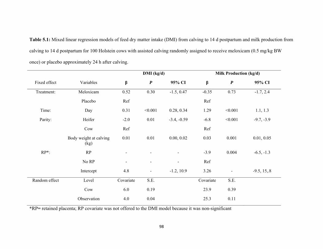
98
Table 5.1: Mixed linear regression models of feed dry matter intake (DMI) from calving to 14 d postpartum and milk production from
calving to 14 d postpartum for 100 Holstein cows with assisted calving randomly assigned to receive meloxicam (0.5 mg/kg BW
once) or placebo approximately 24 h after calving.
DMI (kg/d) Milk Production (kg/d)
Fixed effect Variables β P 95% CI β P 95% CI
Treatment: Meloxicam 0.52 0.30 -1.5, 0.47 -0.35 0.73 -1.7, 2.4
Placebo Ref Ref
Time: Day 0.31 <0.001 0.28, 0.34 1.29 <0.001 1.1, 1.3
Parity: Heifer -2.0 0.01 -3.4, -0.59 -6.8 <0.001 -9.7, -3.9
Cow Ref Ref
Body weight at calving (kg)
0.01 0.01 0.00, 0.02 0.03 0.001 0.01, 0.05
RP*: RP - - - -3.9 0.004 -6.5, -1.3
No RP - - - Ref
Intercept 4.8 - -1.2, 10.9 3.26 - -9.5, 15,.8
Random effect Level Covariate S.E. Covariate S.E.
Cow 6.0 0.19 23.9 0.39
Observation 4.0 0.04 25.3 0.11
*RP= retained placenta; RP covariate was not offered to the DMI model because it was non-significant
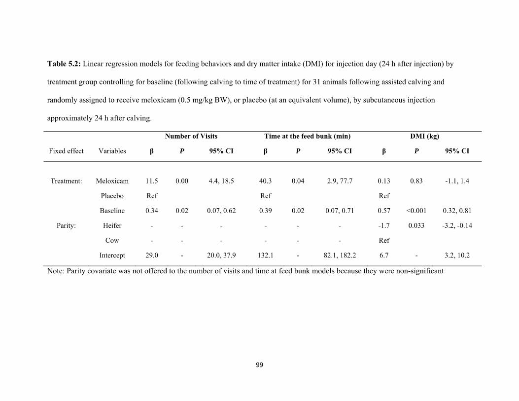
99
Table 5.2: Linear regression models for feeding behaviors and dry matter intake (DMI) for injection day (24 h after injection) by
treatment group controlling for baseline (following calving to time of treatment) for 31 animals following assisted calving and
randomly assigned to receive meloxicam (0.5 mg/kg BW), or placebo (at an equivalent volume), by subcutaneous injection
approximately 24 h after calving.
Number of Visits Time at the feed bunk (min) DMI (kg)
Fixed effect Variables β P 95% CI β P 95% CI β P 95% CI
Treatment:
Meloxicam
11.5
0.00
4.4, 18.5
40.3
0.04
2.9, 77.7
0.13
0.83
-1.1, 1.4
Placebo Ref Ref Ref
Baseline 0.34 0.02 0.07, 0.62 0.39 0.02 0.07, 0.71 0.57 <0.001 0.32, 0.81
Parity: Heifer - - - - - - -1.7 0.033 -3.2, -0.14
Cow - - - - - - Ref
Intercept 29.0 - 20.0, 37.9 132.1 - 82.1, 182.2 6.7 - 3.2, 10.2
Note: Parity covariate was not offered to the number of visits and time at feed bunk models because they were non-significant
100
Chapter 6
Use of a mechanical brush by Holstein dairy cattle around parturition
This Chapter has been submitted to the Journal of Dairy Science (MS#: JDS-12-6010)
101
INTRODUCTION
Grooming activity is an innate behavior in most animal species, and grooming
one’s own body is called auto-grooming (Spruijt et al., 1992). Studies have shown a
decreased (Hart, 1998; Kruk et al., 1998; Anil et al., 2005) or increased (Van Erp et al.,
1994) auto-grooming in animals when they are sick or in pain. Cattle put in social
isolation or deprived of the ability to lie down increased their auto-grooming frequency
(Munksgaard and Simonsen, 1996).
Mid-lactation dairy cows have been shown to spend on average 5.0 min/d
grooming, either by auto-grooming or by scratching themselves against walls and fixtures
in a stall (Munksgaard and Simonsen, 1996). Georg and Totschek (2001) concluded that
mechanical brushes available in an intensive housing system promoted natural grooming
behavior in lactating dairy cattle. DeVries et al. (2007) investigated the use of mechanical
brushes in a free-stall barn and found that installation of these brushes increased the time
spent grooming and performing scratching behaviors in mid-lactation cows, especially in
hard-to-reach places by the cow, such as the neck, back and tail. In addition, providing
cows with access to brushes decreased the time spent scratching against other structures
located in the pen, such as the wall and water trough. It has been suggested that grooming
is a way to relieve physiological stress (Spruijt et al., 1992; Van Erp et al., 1994). If so,
providing a mechanical brush might help the cow cope with stressors associated with
parturition.
Parturition in mammals is associated with a peak in corticotropin-releasing
hormone leading to a peak in cortisol levels (Smith, 2007). Elevated corticotropin,
102
adrenocorticotropic hormone, and cortisol levels have been shown to be associated with
increased auto-grooming (van Erp et al., 1994; Munksgaard and Simonsen, 1996; Kruk et
al., 1998). It follows that periparturient cattle may demonstrate increased auto-grooming.
A large proportion of cows performed grooming (self-licking) behavior during the first
stage of labor (87% of cows and 70% of heifers; Wehrend et al., 2006). Providing cows
access to a mechanical brush around calving may help to meet a behavioral need.
The aims of this study were to describe grooming and scratching behaviors in the
periparturient period, and to determine if provision of a mechanical brush altered
grooming and scratching behaviors in the calving pen, as well as the maternal behavior of
licking the calf.
MATERIAL AND METHODS
Animals and Management
This was a preliminary study in which the magnitude of the expected treatment
and time effects were unknown. Twenty-two multiparous non-lactating Holstein cows in
late pregnancy were enrolled in this study, which was conducted at the University of
British Columbia’s Dairy Education and Research Centre (Agassiz, BC, Canada). All
animals were cared for according to the guidelines of the Canadian Council on Animal
Care (2009). All cows entered the pre-partum pen 25 ± 2 d before their expected calving
date where they were housed in groups of 12. Each pen contained 12 free stalls with a
mattress (Pasture Mat, Promat Inc., Ontario, Canada) covered with 5 cm of sand bedding.
Pre-partum cows in the group-housed pens and individual maternity pens were fed twice
daily at approximately 0800 and 1600 h. The diet was a TMR provided ad libitum,
103
consisting of 21.3% corn silage, 42.8% alfalfa hay, and 35.9% concentrate and mineral
mix on a DM basis (DM: 50.8 ± 1.2%, CP: 14.4 ± 1.0% DM, ADF: 35.0 ± 2.7% DM,
NDF: 45.6 ± 2.6% DM, and NEL: 1.4 ± 0.1 Mcal/kg). In the pre-partum pens, cows were
fed through a headlock feed barrier, with one headlock per cow spaced at 60 cm on
center. Post-partum, cows were fed a TMR consisting of 21.3% grass silage, 14.7% corn
silage, 12.3% alfalfa hay, and 51.7% concentrate and mineral mix on a DM basis (DM:
51.1 ± 1.8%, CP: 17.7 ± 1.0% DM, ADF: 23.7 ± 1.4% DM, NDF: 36.1 ± 1.8% DM, and
NEL: 1.7 ± 0.02 Mcal/kg). Pens had vulcanized rubber floors in the alleys and crossovers
(Red Barn Dairy Mat, North West Rubber Mats, Ltd., Abbotsford, British Columbia,
Canada). A mechanical brush (Lely Luna, Lely Industries, NV, Maasland, the
Netherlands) was installed in the alley between the feed bunk and the stalls of the pre-
partum pen to allow cows to habituate to it. The brush was present at all times for all
cows while in the pre-partum group pen.
At the first signs of imminent calving, such as udder enlargement, milk let-down,
relaxation of sacrosciatic ligament and increased restlessness (Huzzey et al., 2005),
abdominal contractions, mucus discharge from the vulva, and/or raised tail, cows were
moved to an individual maternity pen (3.5 by 4.6 m) containing a sand-bedded pack. The
two maternity pens were each fitted with a mechanical brush identical to those in the pre-
partum pens. Once a week, the brushes were randomly rotated on their stand to either be
in or out of the pen to alternate treatment for the pens (brush versus no brush) in order to
control for any potential pen effect. While out of the pen, the brush was covered by a
cardboard box in order to be hidden from the animal. Cows were randomly assigned to
pens with or without a brush. Animals remained in the maternity pens for 24 h after
104
calving. Calving scores were allocated as 0=unassisted (eutocia) or 1= assisted manually
(dystocia). Cows that had twins, were mechanically assisted with a calving jack, or had a
Caesarean section were excluded (n=3). Three cows in the brush group were removed
from the statistical analyses. For 1 cow, the brush was too high to reach. In the case of the
other 2 cows, video footage was missing following calving. Thus, there were 7 cows in
the brush group and 9 in the no brush group.
Measurements
Behavior of the cows in the pre-calving pen and maternity pen was recorded using
24 h video surveillance with a wide angle camera lens (Super Dynamic III Panasonic
camera, WV-CP484, http://www.Panasonic.com), connected to a recording system
(GeoVision, UVS 1240E2, GeoVision Inc, USAVisionSys, California). In the pre-partum
pen, individual animals were identified by their hide pattern, or by hair dyed symbols in
order to determine if they used the brush (and if the habituation to the brush was
successful, by using the brush for at least 1 min continuously). Brush use was defined
when the cow first touched the brush until the cow moved away, no longer touching the
brush. The total time spent using the brush during the period 72 to 48 h before calving
(defined based on the actual time of delivery of the calf) was determined from video. In
the maternity pen, video was used to measure auto-grooming (i.e., self-licking),
rubbing/scratching (when the cow rubbed against the walls or fixtures of the pen), and the
use of the mechanical brush (when it was present in the pen). The cumulative durations of
these behaviors were recorded while cows were housed in the maternity pens for 3
periods: pre-calving (351 ± 38 min; mean SD), post-calving with the calf present (98 ±
62 min), and post-calving after removal of the calf (180 ± 59 min). The time spent away
105
from the maternity pen for milking during the post-calving period was removed from the
relevant time period, and behaviors were expressed as min/h in each period.
Statistical Analysis
All analyses were performed with STATA Intercooled 10.1 (StataCorp, College
Station, TX, USA). Mixed linear regression models were used to determine the impact of
the brush for the time periods of pre-calving, post-calving with calf, and post-calving
without calf for the following dependent variables: auto-grooming behavior and
scratching/rubbing behavior, with the following independent categorical variables offered
to the model: treatment and time. A mixed linear regression model was also used to look
at the brush use in the maternity pen, for the cows that had access to a brush, for the time
periods of pre-calving, post-calving with calf, and post-calving without calf. A linear
regression model of time spent calf licking during the first h post-calving to identify any
treatment effects (i.e. brush vs. no brush) while in the maternity pen. The animal was
considered as the experimental unit, and was included as a random intercept in mixed
regression models to account for repeated measures. All tests were 2-sided and
significance was based on α < 0.05. Interactions between treatment and period in the final
model were tested. Standardized residuals were examined to verify model assumptions of
normality and homoscedasticity, and to identify outliers at the observation-level for both
linear and mixed linear regression models, and best linear unbiased predictors (BLUPS)
were examined to identify any outliers at the cow-level for mixed linear regression
models. Normality and homogeneity of variance were assessed for the observation-level
standardized residuals, and the BLUPS for the mixed models.
106
RESULTS AND DISCUSSION
When in the group pre-calving pen, all cows made use of the mechanical brush on
average 31.5 ± 17.7 (Mean ± SD) min/d in the 72 to 48 h before calving. This study was
the first to describe brush use by Holstein cows around the time of calving, which was far
more time than the 5 to 7 min/d reported for mid-lactation dairy cows (DeVries et al.,
2007).
No differences were noted in the amount of time spent auto-grooming in the
maternity pen in the time period before parturition and when the calf was present, with
cows spending approximately 0.86 min/h (± 0.77 SD) and 0.67 min/h (± 1.0 SD) engaged
in these behaviors, respectively. However, auto-grooming increased to 2.3 min/h (± 2.3
SD) following calf removal (Table 6.1; Figure 6.1A). The amount of time spent
scratching/rubbing on the pen walls was not different between the brush and no brush
treatment groups over the 3 time periods (Table 6.1; Figure 6.1B). In the time period
before calving, cows spent approximately 0.15 min/h (± 0.10 SD) scratching/rubbing
themselves. However, there was virtually no scratching/rubbing behaviors observed in
cows while the calf was present (Table 6.1; Figure 6.1B). In the hours immediately
following removal of the calf, cows increased the scratching/rubbing time to 0.08 min/h
(± 0.07 SD) (Table 6.1; Figure 6.1B). However, the time spent scratching/rubbing failed
to return to the level observed during the pre-calving period (Table 6.1; Figure 6.1B). The
time spent scratching/rubbing before calving, as well as post-calving without the calf
present, was greater than that previously reported by DeVries et al. (2007). A possible
explanation for this difference is that DeVries et al (2007) observed mid to late lactation
cows, while the cows used in the present study were parturient cows. It could be argued
107
that the cows in the present study were preoccupied with the active stages of calving,
which may have resulted in an increase in scratching behavior (Neary and Hepworth,
2005; Wehrend et al., 2006; Smith, 2007).
It would appear that the maternal behavior of calf licking took precedence over
the cow’s auto-grooming and scratching/rubbing. Auto-grooming by the dam increased
immediately following calf removal. This increase in auto-grooming behavior after cow-
calf separation has been previously reported by Houwing et al., (1990), when the calf was
removed at approximately 10 h post-partum. Behavioral and physiological changes
following cow-calf separation are well established, and include increasing vocalization,
activity (Hudson and Mullord, 1977; Lidfors, 1996; Flower and Weary, 2001), and heart
rate (Hopster et al., 1995). Thus, the results observed in this study were not surprising.
The increases in auto-grooming for the remaining 3 h of observations following calf
removal may be in response to the acute stress arising from the removal of the calf.
Hudson and Mullord (1977) investigated the influence of time spent together by the cow
and her calf immediately following parturition on the establishment of the maternal bond
through licking. They showed that just 5 min of contact immediately after birth resulted
in the formation of a maternal bond that was strong enough to withstand 12 h (but not 24
h) of separation.
The current study examined the effects of having a cow in an individual maternity
pen 6 h before calving and up to 6 h after calving on scratching and grooming behaviors.
When given access to a brush around calving, cows used the brush, both prior to and
following calving (Table 6.1; Figure 6.1C). Although there were no effects of having a
brush on auto-grooming or on scratching/rubbing behavior, interestingly, cows that had
108
access to a brush spent more time engaged in calf licking behavior during the first h after
parturition (β= 8.7 min; P = 0.02; 95% C.I.: 1.5, 15.8; observed average brush = 43.3 min
± 9.9 SD and no brush = 31.0 min ± 12.9 SD) compared to the cows who calved in
maternity pens without a brush. A possible explanation for the increase in calf licking for
the brush group was that giving access to a brush allowed the cows to easily scratch
themselves, instead of attempting to scratch on the walls of the pen. This ease of
scratching would allow the dam to be able to concentrate on the licking of the calf, which
aids in the establishment of the maternal bond (for review see von Keyserlingk and
Weary, 2007), and may also provide an analgesic effect through increased time spent
licking the amniotic fluids from the calf (Pinheiro Machado et al., 1997).
Future work should investigate a brush versus no brush group for longer periods
of time before and after calving, particularly in the case of cows that experience difficult
deliveries and might have a higher level of stress due to the effects of dystocia.
Unfortunately, there were too few dystocia cows in the present study to evaluate the
effects of dystocia. The fact that the brush caused an increase in maternal behavior,
shown by the greater calf licking time in the first h following calving, compared to
animals without access to a brush, also warrants further investigation.
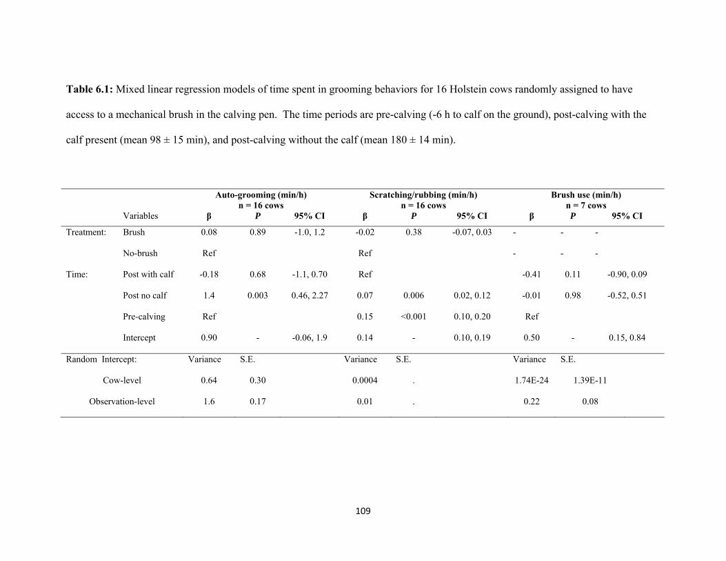
109
Table 6.1: Mixed linear regression models of time spent in grooming behaviors for 16 Holstein cows randomly assigned to have
access to a mechanical brush in the calving pen. The time periods are pre-calving (-6 h to calf on the ground), post-calving with the
calf present (mean 98 ± 15 min), and post-calving without the calf (mean 180 ± 14 min).
Auto-grooming (min/h) n = 16 cows
Scratching/rubbing (min/h) n = 16 cows
Brush use (min/h) n = 7 cows
Variables β P 95% CI β P 95% CI β P 95% CI
Treatment: Brush 0.08 0.89 -1.0, 1.2 -0.02 0.38 -0.07, 0.03 - - -
No-brush Ref Ref - - -
Time: Post with calf -0.18 0.68 -1.1, 0.70 Ref -0.41 0.11 -0.90, 0.09
Post no calf 1.4 0.003 0.46, 2.27 0.07 0.006 0.02, 0.12 -0.01 0.98 -0.52, 0.51
Pre-calving Ref 0.15 <0.001 0.10, 0.20 Ref
Intercept 0.90 - -0.06, 1.9 0.14 - 0.10, 0.19 0.50 - 0.15, 0.84
Random Intercept: Variance S.E. Variance S.E. Variance S.E.
Cow-level 0.64 0.30 0.0004 . 1.74E-24 1.39E-11
Observation-level 1.6 0.17 0.01 . 0.22 0.08
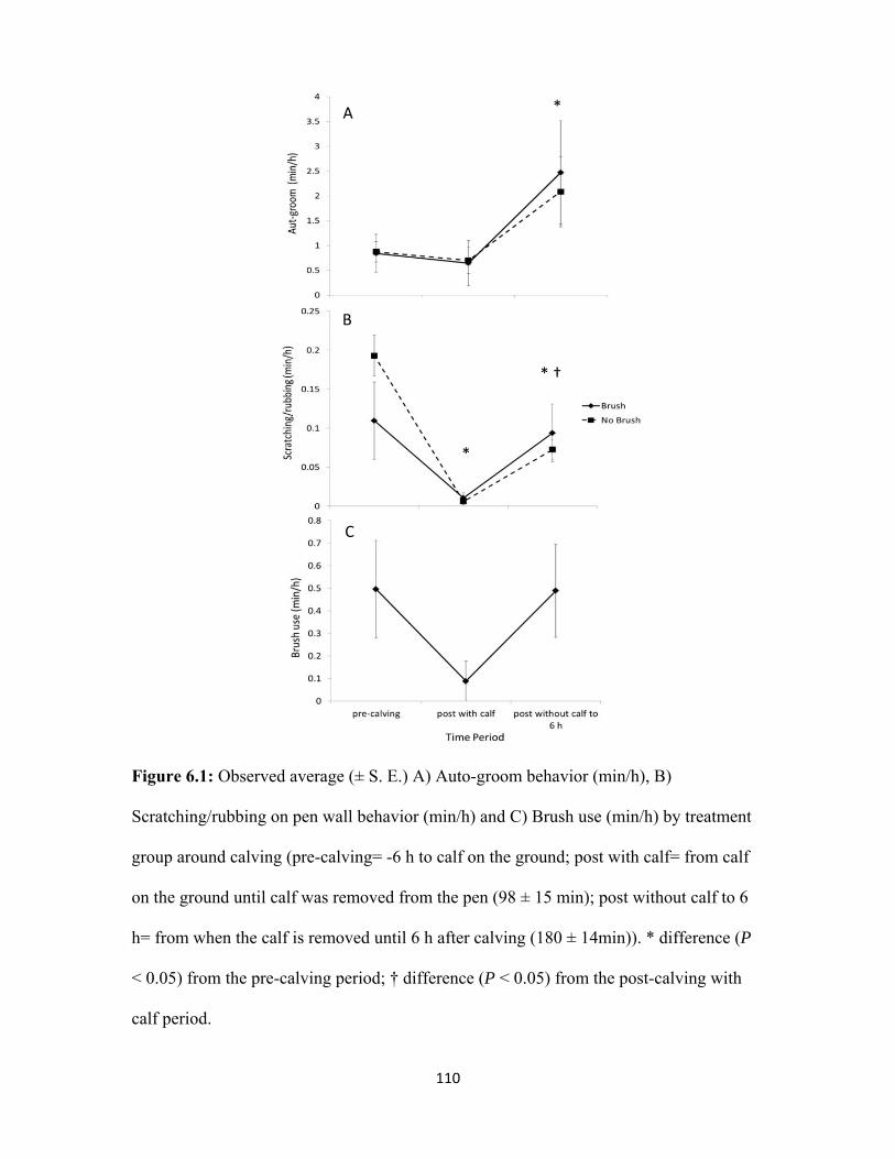
110
Figure 6.1: Observed average (± S. E.) A) Auto-groom behavior (min/h), B)
Scratching/rubbing on pen wall behavior (min/h) and C) Brush use (min/h) by treatment
group around calving (pre-calving= -6 h to calf on the ground; post with calf= from calf
on the ground until calf was removed from the pen (98 ± 15 min); post without calf to 6
h= from when the calf is removed until 6 h after calving (180 ± 14min)). * difference (P
< 0.05) from the pre-calving period; † difference (P < 0.05) from the post-calving with
calf period.
A
B
C
* †
*
*
111
Chapter 7 GENERAL CONCLUSIONS
A recent review about US dairy production stated that the public concern about
farm animal production, including animal welfare, was currently higher than at any point
in recent history (Croney and Anthony, 2011). Good animal welfare includes freedom
from pain, injury, and disease (Farm Animal Welfare Council, 2009). The recognition of
pain in cattle has led to a rise in research on pain in the past 15-20 years (Hudson et al.,
2008). From this research, parts of a code of practice document for the care and handling
of dairy cattle has been developed for dairy farmers pertaining to potentially painful
procedures, such as dehorning and castration in calves (National Farm Animal Care
Council, 2009). However, there is limited research on pain in dairy cattle following
abdominal surgeries as well as assisted calving (Walker et al., 2011).
The main goals of this project were to assess pain and its management in dairy
cattle following abdominal surgeries and following assisted parturition. The objectives of
my thesis were to evaluate the analgesic effects of NSAIDs administered following either
abdominal surgery, or assisted parturition. A final objective was to determine whether or
not a mechanical brush would be beneficial in promoting scratching using the brush in
parturient cows and alter auto-grooming and maternal behaviors.
The first study objective, described in the second chapter, evaluated the effect of
ketoprofen versus saline following left displaced abomasum surgery in cows in a
commercial setting. The results from this study suggested that a paramedian surgical
technique to correct left displaced abomasum may have been more painful post-
operatively, based on the decreased daily lying time of a subset of animals, compared to a
112
standing right flank approach, thus contributing to our knowledge of pain behavior in
dairy cows. Further research is warranted to improve our knowledge of pain following
displaced abomasum surgery and its management, especially for the different approaches
to surgical correction (i.e., flank vs. paramedian). Furthermore, the apparent differences
in appetite or attitude in cows reported by producers following treatment with ketoprofen
suggested that this analgesic may have some beneficial effects following surgery. Finally,
the increases in heart rate and respiration rate in the follow-up visits in the days following
surgery may be indicators of post-surgical pain and merit further investigation.
There were limitations to this study. Firstly, the cows enrolled remained on their
respective farms during the study period. Since the incidence risk of displaced abomasum
has been reported to be between 3 and 7% of calvings (Grohn, 2000; LeBlanc et al.,
2002, Chapinal et al., 2011) and since the average herd size in Ontario has been reported
to be between 50 and 70 cows (Rodenburg, 2007) the epidemiological approach for this
randomized clinical field trial was to sample cows from several farms. Having multiple
farms on the trial made it difficult to control for housing and management practices.
Furthermore, it was logistically impossible to monitor all lying behavior on all cows. The
results from this study regarding the lying behavior differences between cows that
underwent right flank or paramedian surgical correction revealed two different
populations. There were too few cows, a total of 20 right flank animals monitored in this
trial, to detect lying side differences between treatment groups. Based on the differences
between the ketoprofen and the saline groups in the fistulated cows in Chapter 3 for the
time spent lying on their left side, a sample size calculation revealed that 18 animals per
treatment group, for a total of 36 cows, would be required to detect a difference in lying
113
on the surgical side for the right flank surgical approach. Furthermore, it was impossible
to predict which cow was going to need corrective surgery; therefore pre-surgical
baseline values could not be obtained. Future research should consider fitting data loggers
on all cows undergoing LDA surgery, thus providing additional lying data for each type
of surgery. Logistically, it was impossible to record daily feed intake and milk weights
for all of the cows enrolled on the trial. New research should ideally include daily feed
intake and milk weights in order to support the producer’s observation that ketoprofen-
treated animals had greater odds of eating at fresh feed delivery. Intensive and controlled
research should further explore heart and respiration rates closer to surgery time (e.g.,
every 6-12 h for the first 3 d) to support the trends seen thus far for the physiological and
behavioral parameters that revealed post-surgical pain.
The second study objective, in the third and fourth chapters, was to examine the
impact of the first stage of a two-stage fistulation surgery as well as the effect of NSAIDs
as post-operative pain management therapy tools. These studies were opportunistic
studies because nutritionist researchers required fistulated animals, and sample sizes for
the total number of cows needed for these trials was based on the nutritional research
sample size calculation. We, therefore, had no control of the number of animals for our
trials. Even with the minimal sample size from both studies there was consistent evidence
that the first stage of a two-stage fistulation surgery was painful based on the post-
surgical decreases in feed intake, less time spent lying on the left side, and increases in
heart rate, respiration rate, and infrared temperature readings around the surgical site.
Although the administration of NSAIDs post-surgery proved to be beneficial, it was not
sufficient as the animals failed to completely return to pre-surgical levels in the days that
114
they were monitored. The limitations of these trials were that given the limited number of
animals, only the label dose recommendations for ketoprofen and meloxicam could be
investigated. Future research is needed to investigate the magnitude of post-surgical pain.
Perhaps a good starting point would be to test different dosages of different NSAIDs and
test different repeated dosages over several days in order to find the optimal pain
management solution following the first stage of a two-stage fistulation surgery.
The third objective, in the fifth chapter, was to investigate the beneficial effects of
a meloxicam injection following assisted parturition on physiological and behavioral
parameters. Although meloxicam treatment did not have any effects on feed intake, milk
production, blood metabolites, or health events, it did increase the time spent at the feed
bunk, compared to the placebo treatment. These results contribute a valuable starting
point for future research on pain following calving. The limitations for this study were
that only assisted cows and heifers were enrolled and more heifers than multiparous cows
were enrolled, which was not anticipated. It will be important to increase samples size for
each of these parity groups in order to increase the power of statistical analyses.
Furthermore, the administration of meloxicam approximately 24 h following calving to
ensure that the NSAID did not interfere with the expulsion of fetal membranes was a
possible explanation for the lack of differences between treatment groups. Further
research is needed to investigate the beneficial effects of meloxicam administered sooner
following calving, because unlike flunixin meglumine, there does not seem to be any
negative effects of meloxicam on retained placenta (Manteca et al., 2010; T. F. Duffield,
University of Guelph, Guelph, Ontario, personal communication).
115
One of the objective tools used to assess pain and the efficacy of NSAIDs
following surgery in the fourth chapter, and following assisted calving presented in
Appendix 2, was the thermal nociceptive threshold test using a class 4 carbon dioxide
laser. The laser test start date in the assisted calving trial was delayed due to repairs
needed to the laser unit. As such, we only had a limited number of cows near the end of
the study that were tested. Unfortunately, the results of this thermal nociceptive threshold
test were inconclusive due to the great variability between cows from the reduced sample
size. The laser test in the 10 fistulated cows was also inconclusive because we were
limited in the number of cows we could operate. However, there is merit to further test
the thermal nociceptive threshold test as a research tool in animals that either undergo
surgery or assisted calving because other studies have had success with this test. It would
also provide insight on how the animal responds to pain. A mechanical nociceptive test
was successful in 3 different studies. In the first study, nociceptive threshold differences
caused by mastitis pain were identified in dairy cows using a ramped mechanical pressure
test (Kemp et al., 2008). In the second study, a mechanical test using hoof testers was
successful at identifying claw and soft tissue pain in lame cows (Dyer et al., 2007). In the
third study, a pressure algometer was used to test the mechanical nociceptive threshold in
dehorned calves and was successful in determining pain sensitivity and NSAID analgesia
(Heinrich et al., 2010). A thermal nociceptive threshold test was successful at identifying
threshold differences caused by the analgesic properties of amniotic fluid in post-
parturient cows (Pinheiro Machado et al., 1997). Further research to explore the
possibility of nociceptive threshold tests in cows following painful events, such as
abdominal surgeries or calving pain, are needed in order to assist in the identification and
116
proper management of pain in these animals. This may also help provide better insight on
what the animal is experiencing and thus, further our knowledge of pain in dairy cattle.
The final objective of this study was to investigate the use of a mechanical brush
by cows around calving. Cows used the brush, when available, both in the pre-calving
and maternity pens. While the calf was present, cows spent little time self -licking,
scratching themselves, or using the brush, if it was available in the maternity pen.
Furthermore, cows that had a brush in the maternity pen licked their calf more than cows
that did not have a brush. Cows that went on to experience a difficult calving used the
brush in the pre-calving pen for a longer period of time than cows that experienced a
normal delivery. Additional studies assessing the pre-calving brush use will be needed to
confirm this effect. The limitations of this study were that the sample size was small due
to time limit constraints, and that the brush vs no brush treatments were only for a short
amount of time around calving. A larger sample size will provide power to the statistical
analysis, and allow more detailed analysis of trends in grooming, scratching/rubbing and
brush use behavior. Perhaps also looking at these behaviors for a longer period of time
prior to calving would be good to get a better baseline and then do a within cow
comparison. An idea for future research could be if cows were given access to the brush
at least one month prior to calving, to then be subjected to a brush vs no brush treatment
approximately 1 wk prior to calving. Perhaps this could identify if there are true benefits
to having a brush around the time of calving. In addition to behavioral measurements,
there would be merit in monitoring physiological parameters (e.g., heart rate, cortisol
levels, feed intake, and haptoglobin protein levels in the blood) to gain some insight on
the pain state of the animal. A possible follow-up study should examine the use of a brush
117
in fresh cows, 1-3 wk post-calving, since this time period is still part of the transition
period when cows are vulnerable to diseases (Mulligan and Doherty, 2008). There is
merit for further investigations on the potential beneficial effects of a brush as a pain
management tool for cows around calving. There is much to be gained by this approach
as it could be regarded as homeotherapy for the cows that can be just as helpful as
chemicals in addition to being a non-invasive and auto-controlled tool for potential pain
or discomfort alleviation.
In conclusion, this thesis provided new data on pain assessment and management
techniques following abdominal surgery or parturition in dairy cattle. Firstly, NSAID
administration at the dosages reported in this thesis alleviated some, but not all, of the
post-surgical pain based on physiological (increased heart rate after surgery not different
between treatment groups in both LDA and fistulation trials and did not return to baseline
in the fistulation trial; in the fistulation trials increased infrared temperature around
surgical site and haptoglobin levels were not different between treatment groups or
returned to pre-surgical levels, decreased DMI and milk production following surgery did
not return to pre-surgical levels and were not different between treatment groups, except
for the meloxicam vs. ketoprofen trial where DMI was greater for the meloxicam group)
and behavioral parameters (lower total daily lying time for the paramedian surgery in the
LDA trial and no differences between treatment groups; for the fistulation trials,
decreased time spent lying on the surgical side post-surgery but the ketoprofen group lied
more on their left side compared to the saline group, and lower tail flick behavior on d 1
for the ketoprofen group compared to the saline group). Secondly, NSAID administration
following assisted parturition seemed to have some benefits on the feeding behavior
118
during this delicate transition period. However, the parturition study results and those
results from previous studies currently provide limited support for the use of NSAIDs
around calving. Further research is warranted to design appropriate pain scales for
parturition and abdominal surgeries in dairy cattle and to find appropriate analgesic
agents as well as dosages and regimens of these drugs that will be adequate for pain relief
in these animals. Finally, the provision of a mechanical brush demonstrated very possible
potential benefits to the parturient cow.

119
REFERENCES
Anderson, D. E., and W. W. Muir. 2005a. Pain management in ruminants. Vet. Clin.
Food. Anim. 21:19-31.
Anderson, D. E., and W. W. Muir. 2005b. Pain management in cattle. Vet. Clin. Food.
Anim. 21:623-635.
Anil, M. H., J. N. Mbanya, H. W. Symonds, and J. M. Forbes. 1993. Responses in the
voluntary intake of hay or silage by lactating cows to intraruminal infusions of sodium
acetate or sodium propionate, the tonicity of rumen fluid or rumen distension. Br. J. Nutr.
69:699-712.
Anil, L., S. S. Anil, and J. Deen. 2005. Pain detection and amelioration in animals on the
farm: issues and options. J. Appl. Anim. Welf. Sci. 8:261-278.
Atashi, H., A. R. Abdolmohammadi, A. Asaadi, A. Akhlaghi, M. Dadpasand, and Y.
Jafari Ahangari. 2012. Using an incomplete gamma function to quantify the effect of
dystocia on the lactation performance of Holstein dairy cows in Iran. J. Dairy Sci.
95:2718-2722.
Aubry, P. 2005. Routine surgical procedures in dairy cattle under field conditions:
abomasal surgery, dehorning, and tail docking. Vet. Clin. N. Am. Food Anim. 21:55-72.
120
Beretta, C., G. Garavaglia, and M. Cavalli. 2005. COX-1 and COX-2 inhibition in horse
blood by phenylbutazone, flunixin, carprofen and meloxicam: an in vitro analysis.
Pharmacol. Res. 52:302-306.
Bionaz, M., E. Trevisi, L. Calamari, F. Librandi, A. Ferrari, and G. Bertoni. 2007. Plasma
paraoxonase, health, inflammatory conditions, and liver function in transition dairy cows.
J. Dairy Sci. 90:1740-1750.
Boccara, G., A. Chaumeron, Y. Pouzeratte, and C. Mann. 2005. The preoperative
administration of ketoprofen improves analgesia after laparoscopic cholecystectomy in
comparison with propacetamol or postoperative ketoprofen. Brit. J. Anaesth. 94:347-351.
Bonica, J. J. 1979. Peripheral mechanisms and pathways of parturition pain. Br. J.
Aneasth. 51:3S-9S.
Bourne, D. 2001. Inverted L block in ruminants in Veterinary Anaesthesia Tenth Edition.
Hall, L. W., K. W. Clarke, and C. M. Trim. (eds).W. B. Saunder, London, UK.
http://wildpro.twycrosszoo.org/S/00Man/VeterinaryTechniques/PainRumIndTech/LBloc
k_Rum.htm. Accessed August, 24, 2012.
121
Broom, D. 2000. Welfare assessment and welfare problem areas during handling and
transportation. Pages 43-61 in Livestock Handling and Transport, 2nd Edition. T. Grandin
Ed.CAB International Publishing, New York, United States of America.
Canadian Council on Animal Care. 2009. CCAC guidelines on: the care and use of farm
animals in research, teaching and testing. Canadian Council on Animal Care, Ottawa,
Canada.
Canadian Dairy Information Centre. 2011. Number of dairy cows by province.
http://www.dairyinfo.gc.ca/pdf/dairy_cows_by_prov.pdf. Accessed June. 15, 2011.
Capucille, D. J., M. H. Poore, and G. M. Rogers. 2002. Castration in cattle: techniques
and animal welfare issues. Compendium. 24:S66-S73.
Caulkett, N., P. H. Cribb, and T. Duke. 1993. Short communication: Xylazine epidural
analgesia for caesarean section in cattle. Can. Vet. J. 34:674-676.
Cervero, F. 1994. Sensory innervations of the viscera: peripheral basis of visceral pain.
Physiol. Rev. 74:95-138.
122
Chang, M.-Y., S.-Y. Wang, and C.-H. Chen. 2002. Effects of massage on pain and
anxiety during labour: a randomized controlled trial in Taiwan. J. Adv. Nurs. 38:68-73.
Chapinal, N., M. Carson, T. F. Duffield, M. Capel, S. Godden, M. Overton, J. E. P.
Santos, and S. J. LeBlanc. 2011. The association of serum metabolites with clinical
disease during the transition period. J. Dairy Sci. 94:4897-4903.
Coetzee, J. F., R. Gehring, A. C. Bettenhausen, B. V. Lubbers, S. E. Toerber, D. U.
Thomson, B. KuKanich, and M. D. Apley. 2007. Attenuation of acute plasma cortisol
response in calves following intravenous sodium salicylate administration prior to
castration. J. Vet. Pharmacol. Therap. 30:305-313.
Coetzee, J. F., L. N. Edwards, R. A. Mosher, N. M. Bello, A. M. O’Connor, B. Wang, B.
KuKanich, and D. A. Blasi. 2012. Effect of oral meloxicam on health and performance of
beef steers relative to bulls castrated on arrival at the feedlot. J. Anim. Sci. 90:1026-1039.
Compendium of Veterinary Products. 2012. Product Category for Dairy Cattle.
http://cca.naccvp.com/?m=catindex_main. Accessed Jun. 15, 2012.
123
Conner, J. G., P. D. Eckersall, M. Doherty, and T. A. Douglas. 1986. Acute phase
response and mastitis in the cow. Res. Vet. Sci. 41:126–128.
Constable, P. D., G. St Jean, B. L. Hull, D. M. Rings, D. E. Morin, and D. R. Nelson.
1997. Intussusception in cattle: 336 cases (1964–1993). J. Am. Vet. Med. Assoc.
210:531–6.
Croney, C. C. and R. Anthony. 2011. Invited review: Ruminating conscientiously:
Scientific and socio-ethical challenges for US dairy production. J. Dairy Sci. 94:539-546.
Cryer, B., and M. Feldman. 1998. Cyclooxygenase-1 and cyclooxygenase-2 selectivity of
widely used nonsteroidal anti-inflammatory drugs. Am. J. Med. 104:413-421.
Dafny, N. 1997. Chapter 8: Pain modulation and mechanisms.
http://neuroscience.uth.tmc.edu/index.htm. Accessed Jun 18, 2012.
Dechamps, P., B. Nicks, B. Canabt, M. Gielen, and L. Istasse. 1989. A note on resting
behavior of cows before and after calving in two different housing systems. Appl. Anim.
Behav. Sci. 23:99–105.
124
DeVries, T. J., M. A. G. von Keyserlingk, and K. A. Beauchemin. 2005a. Frequency of
feed delivery affects the behavior of lactating dairy cows. J. Dairy Sci. 88:3553:3562.
DeVries, T. J., and M. A. G. von Keyserlingk. 2005b. Time of feed delivery affects the
feeding and lying patterns of dairy cows. J. Dairy Sci. 88:625-631.
DeVries, T. J., M. Vankova, D. M. Veira, and M. A. G. von Keyserlingk. 2007. Short
Communication: usage of mechanical brushes by lactating dairy cows. J. Dairy Sci.
90:2241-2245.
De Winter, B. Y., G. E. Boeckxstaens, J. G. de Man, T. G. Moreels, A. G. Herman, and
P. A. Pelckmans. 1998. Differential effect of indomethacin and ketorolac on
postoperative ileus in rats. Eur. J. Pharmacol. 344:71–76.
Dobromylskyj, P., P. A. Flecknell, B. D. Lascelles, A. Livingston, P. Taylor, and A.
Waterman-Pearson. 2000. Pain Assessment. Page 53-76 in Pain management in animals.
P. Flecknell and A. Waterman-Pearson, ed. WB Saunders, London, UK.
Dohoo, I., W. Martin, and H. Stryhn. 2003. Confounder bias: analytic control and
matching. Pages 235-272 in Veterinary Epidemiologic Research. S. M. McPike, ed. AVC
Inc., Prince Edward Island, Canada.
125
Dubuc, J., T. F. Duffield, K. E. Leslie, J. S.Walton, and S. J. LeBlanc. 2010. Risk factors
for postpartum uterine diseases in dairy cows. J. Dairy Sci. 93:5764-5771.
Duffield, T. F., H. Putnam-Dingwell, D. Weary, A. Skidmore, L. Neuder, W. Raphael, S.
Millman, N. Newby, and K. E. Leslie. 2009. Effect of flunixin meglumine treatment
following parturition on cow health and milk production. J. Dairy Sci. 92 (E-Suppl. 1),
118.
Duffield, T. F., A. Heinrich, S. T. Millman, A. DeHaan, S. James, and K. Lissemore.
2010. Reduction in pain response by combined use of local lidocaine anesthesia and
systemic ketoprofen in dairy calves dehorned by heat cauterization. Can. Vet. J. 51:283-
288.
Dyer, R. M., N. K. Neerchal, U. Tasch, Y. Wu, P. Dyer, and P. G. Rajkondawar. 2007.
Objective determination of claw pain and its relationship to limb locomotion score in
dairy cattle. J. Dairy Sci. 90:4592-4602.
Earley, B., and M. A. Crowe. 2002. Effects of ketoprofen alone and in combination with
local anesthesia during castration of bull calves on plasma cortisol, immunological, and
inflammatory responses. J. Anim. Sci. 80:1044-1052.
126
EMEA (European Agency for the Evaluation of Medicinal Products). 2007. Scientific
discussion, Metacam. CVMP/323/97, London, UK.
Erb, R. E., M. France D’Amico, B. P. Chew, P. V. Malven, and C. N. Zamet. 1981.
Variables associated with peripartum traits in dairy cows. VIII. Hormonal profiles
associate with dystocia. J. Anim. Sci. 52:346-358.
Farm Animal Welfare Council. 2009. Five Freedoms.
http://www.fawc.org.uk/freedoms.htm. Accessed June 22, 2012.
Faulkner, P. M., and D. M. Weary. 2000. Reducing pain after dehorning in dairy calves.
J. Dairy Sci. 83:2037-2041.
Field, T., M. Hernandez-Reif, S. Taylor, O. Quintino, and I. Burman. 1997. Labor pain is
reduced by massage therapy. J. Psychosom. Obstet. Gynecol. 18:286-291.
Field, T., M. Hernandez-Reif, S. Hart, H. Theakston, S. Schanberg, and C. Kuhn. 1999.
Pregnant women benefit from massage therapy. J. Psychocom. Obstet. Gynecol. 20:31-
38.
127
Fitzpatrick, J. L. 2003. Assessment and alleviation of pain in dairy cows with clinical
mastitis. Cattle Prac. 11:289-293.
Fitzpatrick, J. L., F. Y. Young, D. Eckersall, D. N. Logue, C. J. Knight, and A. Nolan.
1998. Recognising and controlling pain and inflammation in mastitis. Page 36 in Proc
British Mastitis Conference, Axient/Institute for Animal Health, Milk Development
Council/Novartis Animal Health, Berkshire, UK.
Flerheller, E. E., N. A. Caulkett, and J. V. Balley. 2004. A romifidine and morphine
combination for epidural analgesia of the flank in cattle. Can. Vet. J. 45:917-923.
Flower, F., and D. Weary. 2001. Effects of early separation on the dairy cow and calf. II:
Separation at 1 day and 2 weeks after birth. Appl. Anim. Behav. Sci. 70: 275-284.
Flower, F. C., and D. M. Weary. 2006. Effects of hoof pathologies on subjective
assessments of dairy cow gait. J. Dairy Sci. 89:139-146.
Frame, N. 2006. Management of dystocia in cattle. In Practice. 28:470-476.
Fraser, D., D. M. Weary, E. A. Pajor, and B. N. Milligan. 1997. A scientific conception
of animal welfare that reflects ethical concerns. Anim. Welfare. 6:187-205.
128
Gabay, C. 2006. Interleukin-6 and chronic inflammation. Arthritis Res. Ther. 8 (Suppl.
2): S3.
Graubner, C., V. Gerber, M. Doherr, and C. Spadavecchia. 2011. Clinical application and
reliability of a post abdominal surgery pain assessment scale (PASPAS) in horses. Vet. J.
188:178-183.
Geishauser, T., K. Leslie, D. Kelton, and T. Duffield. 1998. Evaluation of five cowside
tests for use with milk to detect subclinical ketosis in dairy cows. J. Dairy Sci. 81:438-
443.
Geishauser, T., K. Leslie, D. Kelton, and T. Duffield. 2001. Monitoring for subclinical
ketosis in dairy herds. Compend. Contin. Educ. Pract. Vet. 23:65–71.
Georg, H., and K. Totschek. 2001. Examining an automatic cow brush for dairy cows.
Landtechnik. 56:260:261.
Gingerich, D. A., J. D. Baggot, and R. A. Yeary. 1975. Pharmacokinetics and dosage of
aspirin in cattle. J. Am. Vet. Med. Assoc. 167:945-948.
129
Graf, B., and M. Senn. 1999. Behavioural and physiological responses of calves to
dehorning by heat cauterisation with or without local anaesthesia. Appl. Anim. Behav.
Sci. 62:153–171.
Grant, R. J., V. F. Colenbrander, and J. L. Albright. 1990. Effect of particle size of forage
and rumen cannulation upon chewing activity and laterality in dairy cows. J. Dairy Sci.
73:3158-3164.
Grohn, Y. T. 2000. Milk yield and disease: Towards optimizing dairy herd health and
management decisions. Bovine Pract. 34:32–40.
Gronlund, U., C. Hulten, P. D. Eckersall, C. Hogarth, and K. Persson Waller. 2003.
Haptoglobin and serum amyloid A in milk and serum during acute and chronic
experimentally induced Staphylococcus aureus mastitis. J. Dairy Res. 70:379-386.
Hansen, B. D. 2003. Assessment of pain in dogs: veterinary clinical studies. ILAR J.
44:197-205.
Hart, B. L. 1998. Biological basis of the behavior of sick animals. Neurosci. Biobehav.
Rev. 12:123-137.

130
Hassall, S. A., W. R. Ward, and R. D. Murray. 1993. Effects of lameness on the
behaviour of cows during the summer. Vet. Rec. 132:578-580.
Hayes, B. W., C. O. Little, and G. E. Mitchell Jr. 1964. Influence of ruminal, abomasal,
and intestinal fistulation on digestion in steers. J. Anim. Sci. 23:764-766.
Heinrich, A., T. F. Duffield, K. D. Lissemore, E. J. Squires, and S. T. Millman. 2009. The
impact of Meloxicam on postsurgical stress associated with cautery dehorning. J. Dairy
Sci. 92:540-547.
Heinrich, A., T. F. Duffield, K. D. Lissemore, and S. T. Millman. 2010. The effect of
meloxicam on behaviour and pain sensitivity of dairy calves following cautery dehorning
with a local anesthetic. J. Dairy Sci. 93:2450-2457.
Hernandez-Urdaneta, A., C. E. Coppock, R.E. McDowel, D. Gianola, and N. E. Smith.
1976. Changes in forage-concentrate ratio of complete feeds for dairy cows. J. Dairy Sci.
59:695-707.
Hewson, C. J., I. R. Dohoo, K. A. Lemke, and H. W. Barkema. 2007. Canadian
veterinarians’ use of analgesics in cattle, pigs, and horses in 2004-2005. Can. Vet. J.
48:155-164.
131
Hopster, H., J. M. O’Connell, J.H. Blokhuis. 1995. Acute effects of cow-calf separation
on heart rate, plasma cortisol and behaviour in multiparous dairy cows. Appl. Anim.
Behav. Sci. 44:1-8.
Horta, A. E. M. 1984. Efeitos das prostaglandinas E2 e F2 alpha sobre a retencao
placentaria induzida pelo acetilsalicilato de lisina em vacas leiteiras; estudo da motilidade
uterina. Zootechnia. 33:39-47.
Houwing, H., J. F. Hurnik, and N. J. Lewis. 1990. Behavior of periparturient dairy cows
and their calves. Can. J. Anim. Sci. 70:355-362.
Hudson, S., and M. Mullord. 1977. Investigations of maternal bonding in dairy cattle.
Appl. Anim. Behav. Sci. 3: 271-276.
Hudson, C., H. Whay, and J. Huxley. 2008. Recognition and management of pain in
cattle. In Prac. 30:126-134.
Huxley, J. N., and H. R. Whay. 2006. Current attitudes of cattle practitioners to pain and
the use of analgesics in cattle. Vet. Rec. 159:662-668.
132
Huzzey, J. M., M. A. G. Von Keyserlingk, and D. M. Weary. 2005. Changes in feeding,
drinking, and standing behavior of dairy cows during the transition period. J. Dairy Sci.
88:2454-2461.
Hydbring, E., A. Madej, E. MacDonal, G. Drugge-Boholm, B. Berglund, and K. Olsson.
1999. Hormonal changes during parturition in heifers and goats are related to the phases
and severity of labour. J. Endocrinol. 160:75-85.
International Association for the Study of Pain. 1994. Part III: Pain terms, a current list
with definitions and notes on usage. Pages 209-214 in Classification of chronic pain,
second edition, IASP task force on taxonomy. H. Merskey and N. Bogduk, ed. IASP
Press, Seattle, WA.
Iwersen, M., U. Falkenberg, R. Voigtsberger, D. Forderung, and W. Heuwieser. 2009.
Evaluation of an electronic cowside test to detect subclinical ketosis in dairy cows. J.
Dairy Sci. 92:2618-2624.
Jenkins, W. L. 1987. Pharmacologic aspects of analgesic drugs in animals: an overview.
J. Am. Vet. Med. Assoc. 191:1231-1240.
133
Kajikawa, T., A. Furuta, T. Onishi, T. Tajima, and S. Sugii. 1999. Short communication:
changes in concentrations of serum amyloid A protein, α1-acid glycoprotein, haptoglobin,
and C-reactive protein in feline sera due to induced inflammation and surgery. Vet.
Immunol. Immunop. 68:91-98.
Kemp, M. H., A. M. Nolan, P. J. Cripps, and J. L. Fitzpatrick. 2008. Animal-based
measurements of the severity of mastitis in dairy cows. Vet. Rec. 163:175-179.
Kolkman, I.,S. Aerts, H. Vervaecke, J. Vicca, J. Vandelook, A. de Kruif, G. Opsomer,
and D. Lips. 2010. Assessment of differences in some indicators of pain in double
muscled Belgian blue cows following naturally calving vs. caesarean section. Reprod.
Dom. Anim. 45, 160–167.
Krause, K. M., and G. R. Oetzel. 2006. Understanding and preventing subacute ruminal
acidosis in dairy herds: a review. Anim. Feed Sci. and Tech. 126:215-236.
Kristal, M. B., A. C. Thompson, P. Abbott, J. M. Di Pirro, E. J. Ferguson, and J. C.
Doerr. 1990. Amniotic fluid ingestion by parturient rates enhances pregnancy mediated
analgesia. Life Sci. 46, 693-698.
Kruk, M. R., K. G. C. Westphal, A. M. M. Van Erp, J. Van Asperen, B. J. Cave, E.
Slater, J. de Koning, and J. Haller. 1998. The hypothalamus: cross-roads of endocrine and
134
behavioural regulation in grooming and aggression. Neurosci. Biobehav. Rev. 23:163-
177.
Laven, R. A., K. E. Lawrence, J. F. Weston, K. R. Dowson, and K. J. Stafford. 2008.
Assessment of The duration of the pain response associated with lameness in dairy cows,
and the influence of treatment. New Zeal. Vet. J. 56:210-217.
Laven, R. A., J. N. Huxley, H. R. Whay, and K. J. Stafford. 2009. Results of a survey of
attitudes of dairy veterinarians in New Zealand regarding painful procedures and
conditions in cattle. New Zeal. Vet. J. 57:215-220.
Le Bars, D., M. Gozariu, and S. W. Cadden. 2001a. Evaluation de la douleur aigue chez
l’animal d’experience. Premiere partie. Ann. Fr. Anesth. Reanim. 20:347–36.
Le Bars, D., M. Gozariu, and S. W. Cadden. 2001b. Animal models of nociception.
Pharmacol. Rev. 53:597–652.
LeBlanc, S. J., T. F. Duffield, K. E. Leslie, K.G. Bateman, J. TenHag, J. S. Walton, and
W. H. Johnson. 2002a. The effect of prepartum injection of vitamin E on health in
transition dairy cows. J. Dairy Sci. 85:1416–1426.
135
LeBlanc, S. J., T. F. Duffield, K. E. Leslie, K. G. Bateman, G. P. Keefe, J. S. Walton, and
W. H. Johnson. 2002b. Defining and diagnosing postpartum clinical endometritis and its
impact on reproductive performance in dairy cows. J. Dairy Sci. 85:2223–2236.
Lees, P., M. F. Landoni, J. Giraudel, and P. L. Toutain. 2004. Pharmacodynamics and
pharmacokinetics of nonsteroidal anti-inflammatory drugs in species of veterinary
interest. J. Vet. Pharmacol. Therap. 27:479-490.
Ledgerwood, D. N., C. Winkler, and C. B. Tucker. 2010. Evaluation of data loggers,
sampling intervals, and editing techniques for measuring the lying behavior of dairy
cattle. J. Dairy Sci. 93:5129-5139.
Lidfors, L. 1996. Behavioural effects of separating the dairy calf immediately or 4 d post-
partum. Appl. Anim. Behav. Sci. 49: 269-283.
Linstone, H. A. and M. Turoff. 1975. The Delphi method: techniques and applications.
Addison-Wesley Publishing Company, Massachusetts, USA.
136
Livingston, A., and P. Chambers. 2000. The physiology of pain. Pages 9-19 in Pain
management in animals. P. Flecknell and A. Waterman-Pearson, ed. WB Saunders,
London, UK.
Lombard, J.E., F. B. Garry, S. M. Tomlinson, and L. P. Garber. 2007. Impacts of dystocia
on health and survival of dairy calves. J. Dairy Sci. 90:1751-1760.
Luks F. I., J. Logan, C. K. Breuer, A. G. Kurkchubasche, C. W. Wesselhoeft, and T. F.
Tracy. 1999. Cost effectiveness of laparoscopy in children. Arch. Pediatr. Adolesc. Med.
153:965–968.
Manteca, X. E., E. Mainau, A. Cuevas, and J. L. Ruiz de la Torre. 2010. The effect of
parity and time on pain and discomfort associated with normal calving in dairy cows. 3rd
Boehringer-Ingelheim Expert Forum on Farm Animal Well Being. Pages 37-39,
Barcelona, Spain.
Maroon, J. C., J. W. Bost, and A. Maroon. 2010. Natural anti-inflammatory agents for
pain relief. Surg. Neurol. Int. 1:80-89.
137
Mathews, K. A. 1996. Nonsteroidal anti-inflammatory analgesics in pain management in
dogs and cats. Can. Vet. J. 37:539-545.
McMeekan, C. M., K. J. Stafford, D. J. Mellor, R. A. Bruce, R. N. Ward, and N. G.
Gregory. 1999. Effects of a local anaesthetic and a nonsteroidal anti-inflammatory
analgesic on the Behavioural responses of calves to dehorning. New Zeal. Vet. J. 47:92–
96.
Mee, J.F. 2008. Prevalence and risk factors for dystocia in dairy cattle. Vet. J. 176:93-
101.
Mellor, D. J., V. Molony, and I.S. Robertson. 1991. Effects of castration on behaviour
and plasma cortisol concentrations in young lambs, kids, and calves. Res Vet. Sci.51:149-
154.
Merial-Canada. 2002. Anafen Injection (large animal) In Compendium of veterinary
products, CD ed. A. Arrioja-Dechert, ed. North American Compendiums, Inc. Port
Huron, MI.
Millan, M. J. 2002. Descending control of pain. Prog. Neurobiol. 66:355-474.
138
Milligan, B. N., T. Duffield, and K. Lissemore. 2004. The utility of ketoprofen for
alleviating pain following dehorning in young dairy calves. Can. Vet. J. 45:140-143.
Molony, V., J. E. Kent, and I. S. Robertson. 1995. Assessment of acute and chronic pain
after different methods of castration of calves. Appl. Anim. Behav. Sci. 46:33-48.
Morton, C. M., J. Reid, E. M. Scott, L. L. Holton, and A. M. Nolan. 2005. Application of
a scaling model to establish and validate an interval level pain scale for assessment of
acute pain in dogs. Am. J. Vet. Res. 66:2154-2166.
Mulligan, F. J., and M. L. Doherty. 2008. Production diseases of the transition cow. Vet.
J. 176:3-9.
Munksgaard, L., and H. B. Simonsen. 1996. Behavioral and pituitary adrenal-axis
responses of dairy cows to social isolation and deprivation of lying down. J. Anim. Sci.
74:769-778.
Murrin, K. R., and M. Rosen. 1985. Pain Measurement. Pages 104-132 in Acute Pain. G.
Smith and B. G. Covino, eds, Butterworths, London, UK.
139
National Farm Animal Care Council (NFACC). 2009. Health and welfare management.
Pages 25-26 in Code of practice for the care and handling of dairy cattle. Dairy Farmers
of Canada & National Farm Animal Care Council, Ottawa, ON.
Neary, M., Hepworth, K. 2005. Parturition in Livestock. Purdue Extension AS-561-W
Animal Sciences. Purdue University Cooperative Extension Service, West Lafayette, IN.
Newby, N.C., D. L. Pearl, S. J. LeBlanc, K. E. Leslie, M. A. G. von Keyserlingk, and T.
F. Duffield. The effect of administering ketoprofen on the physiology and behavior of
dairy cows following surgery to correct a left displaced abomasum. Submitted to J. Dairy
Sci. Mar 23, 2012. Manuscript # JDS-12-5566.
Newman, K. D., and D. E. Anderson. 2005. Cesarean section in cows. Vet. Clin. Food
Anim. 21:73-100.
Newman, K. D., D. Harvey, and J-P Roy. 2008. Minimally invasive field abomasopexy
techniques for correction and fixation of left displacement of the abomasum in dairy
cows. Vet. Clin. Food Anim. 24:359-382.
140
Nicholson T., and V. Tiruchelvam . 2001. Comparison of laparoscopic-assisted
appendectomy with intracorporal laparoscopic appendectomy and open appendectomy. J.
Soc. Laparoendosc. Surg. 5:47–51.
O’Callaghan, K. 2002. Lameness and associated pain in cattle – challenging traditional
perceptions. In Practice. 24:212-219.
Pairet, M., and Y. Ruckebusch. 1989. On the relevance of non-steroidal anti-
inflammatory drugs in the prevention of paralytic ileus in rodents. J.Pharm. Pharmacol.
41:757–761.
Panndorf, J., H. Richter, and B. Oittrich. 1976. Haptoglobin in domestic animals. V.
Plasma haptoglobin levels in cattle under pathological conditions. Arch. Exp.
Veterinarmed. 30:193–202.
Pinheiro Machado F°, L. C., J. F. Hurnik, and J. H. Burton. 1997. The effect of amniotic
fluid ingestion on the nociception of cows. Physiol. Behav. 62:1339-1344.
141
Price, D. D., P. A. McGrath, A. Rafii, and B. Buckingham. 1983. The validation of visual
analogue scales as ratio scale measures for chronic and experimental pain. Pain. 17:45-
56.
Proudfoot, K. L., J. M. Huzzey, and M. A. G. von Keyserlingk. 2009. The effect of
dystocia on the dry matter intake and behaviour of Holstein cows. J. Dairy Sci. 92:4937-
4944.
Richards, B. D., D. H. Black, R. M. Christley, M. D. Royal, R. F. Smith, and H. Dobson.
2009. Effects of the administration of ketoprofen at parturition on the milk yield and
fertility of Holstein-Friesian cattle. Vet. Rec. 165:102-106.
Roberts, S. J. 1986. Veterinary Obstetrics and Genital Diseases, Woodstock, Vt., pp 373.
Rodenburg, J. 2007. Precision dairy management and the future of dairy production in
Ontario. Ministry of Agriculture, Food and Rural Affairs Factsheet.
http://www.omafra.gov.on.ca/english/livestock/dairy/facts/07-065.htm. Accessed August
25, 2012.
Roughan, J. V., and P. A. Flecknell. 2001. Behavioral effects of laparotomy and analgesic
effects of ketoprofen and carprofen in rats. Pain. 90:65-74.
142
Roughan, J. V. and P. A. Flecknell. 2003. Evaluation of a short duration behavior-based
post-operative pain scoring system in rats. Eur. J. Pain. 7:397-406.
Rosen M., A. Garcia-Ruiz, J. Malm, J. T. Mayes, E. Steiger, and J. Ponsky. 2001.
Laparoscopic hernia repair enhances early return of physical work capacity. Surg.
Laparosc. Endosc. 11:28–33.
Rushen, J. 2003. Changing concepts of farm animal welfare: bridging the gap between
applied and basic research. Appl. Ani. Behav. Sci. 81:199-214.
Sams, A. E., and S. L. Fubini. 1993. Primary repair of abomasal fistulae resulting from
right paramedian abomasopexy in eight adult dairy cattle. Vet. Surg. 22:190-193.
Sawyer, D. C. 1998. Pain control in small-animal patients. Appl. Anim. Behav. Sci. 59:
135-146.
Schror, K. 1997. Aspirin and platelets: the antiplatelet action of aspirin and its role in
thrombosis treatment and prophylaxis. Semin. Thromb. Hemost. 23:349-356.
143
Seeger, T., H. Kumper, K. Failing, and K. Doll. 2006. Comparison of laparoscopic-
guided abomasopexy versus omentopexy via right flank laparotomy for the treatment of
left abomasal displacement in dairy cows. Am. J. Vet. Res. 67:472-478.
Sellers, G., H. C. Lin, M. G. Riddell, W. R. Ravis, S. H. Duran, and M. D. Givens. 2009.
Pharmacokinetics of lidocaine in serum and milk of mature Holstein cows. J. Vet.
Pharmacol. Therap. 32:446-450.
Shupe, J. L. 1961. Arthritis in cattle. Can. Vet. J. 2: 369-376.
Shwartz, G., K. L. Hill, M. J. VanBaale, and L. H. Baumgard. 2009. Effects of flunixin
meglumin on pyrexia and bioenergetics variables in postparturient dairy cows. J. Dairy
Sci. 92:1963-1970.
Skinner, J. G., R. A. L. Brown, and L. Roberts. 1991. Bovine haptoglobin response in
clinically defined field conditions. Vet. Rec. 128:147–149.
Skinner, J.G., and L. Roberts. 1994. Haptoglobin as an indicator of infection in sheep.
Vet. Rec. 134:33–36.
144
Smith, R. 2007. Parturition. New Engl. J. Med. 356:271-283.
Sprecher, D.J., D. E. Hostetler, and J. B. Kaneene. 1997. A lameness scoring system that
uses posture and gait to predict dairy cattle reproductive performance. Theriogenology.
47:1179-1187.
Spruijt, B. M., J. A. R. A. M. Van Hooff, and W. H. Gispen. 1992. Ethology and
neurobiology of grooming behaviour. Physiol. Rev. 72: 825-852.
Stafford, K. 2007. Alleviating the pain caused by the castration of cattle. Vet. J. 173:245-
247.
Stafford, K. J., and D. J. Mellor. 2005. Dehorning and disbudding distress and its
alleviation in calves. Vet. J. 169:337-349.
Stevens, B., C. Johnston, P. Petryshen, and A. Taddio. 1996. Premature infant pain
profile: development and initial validation. Clin. J. Pain. 12:13-22.
145
Stilwell, G., M. S. Lima, and D. M. Broom. 2008. Effects of nonsteroidal anti-
inflammatory drugs on long-term pain in calves castrated by use of an external clamping
technique following epidural anesthesia. Am. J. Vet. Res. 69: 744-750.
Sutherland, M.A., Mellor, D.J., Stafford, K.J., Gregory, N.G., Bruce, R.A., Ward, R.N.,
2002. Modification of cortisol responses to dehorning in calves using a 5-hour local
anaesthetic regimen plus phenylbutazone, ketoprofen or adrenocorticotropic hormone
injected prior to dehorning. Research in Veterinary Science 73, 115–123.
Sylvester, S. P., D. J.Mellor, K. J. Stafford, R. A. Bruce, and R. N. Ward. 1998. Acute
cortisol responses of calves to scoop dehorning using local anaesthesia and/or cautery of
the wound. Aust. Vet. J. 76:118–122.
Tenhagen, B.-A., A. Helmbold, and W. Heuwieser. 2007. Effect of various degrees of
dystocia in dairy cattle on calf viability, milk production, fertility and culling. J. Vet.
Med. A. 54:98-102.
Thuer, S., S. Mellema, M. G. Doherr, B. Wechsler, K. Nuss, and A. Steiner. 2007. Effect
of local anaesthesia on short- and long-term pain induced by two bloodless castration
methods in calves. Vet. J. 173:333-342.
146
Thyfault, H. A., E. C. Leffel, and M. Der Huang. 1975. Simplified method for producing
permanent ruminal fistulae. J. Dairy Sci. 58:1899-1901.
Ting, S. T. L., B. Earley, J. M. L. Hughes, and M. A. Crowe. 2003. Effect of ketoprofen,
lidocaine local anesthesia, and combined xylazine and lidocaine caudal epidural
anesthesia during castration of beef cattle on stress responses, immunity, growth, and
behavior. J. Anim. Sci. 81:1281-1293.
Todd, C. G., S. T. Millman, D. R. McKnight, T. F., Duffield, and K. E. Leslie. 2010.
Nonsteroidal anti-inflammatory drug therapy for neonatal calf diarrhea complex: effects
on calf performance. J. Anim. Sci. 88:2019-2028.
Tucker, C. B., N. R. Cox, D. M. Weary, and M. Spinka. 2009. Laterality of lying
behavior in dairy cattle. Appl. Anim. Behav. Sci. 120:125-131.
Van Erp, A. M. M., M. R. Kruk, W. Meelis, and D. C. Willekens-Bramer. 1994. Effect of
environmental stressors on time course, variability and form of self-grooming in the rat:
handling, social contact, defeat, novelty, restraint and fur moistening. Behav. Brain Res.
65:47-55.
147
Van Loon, J. P. A. M., W. Back, L. J. Hellebrekers, and P. R. van Weeren. 2010.
Application of a composite pain scale to objectively monitor horses with somatic and
visceral pain under hospital conditions. J. Equine Vet. Sci. 30:641-649.
Van Metre, D. C, R. J. Callan, T. N. Holt, and F. B. Garry. 2005. Abdominal
Emergencies in Cattle. Vet. Clin. N. Am.: Food Anim. Practice 21:655–696.
Vasseur, E., F. Borderas, R. Cue, A. M. de Passille, D. Lefebvre, D. Pellerin, J. Rushen,
and K. Wade. 2008. Dairy producers’ use of calf rearing practices that impact calf health
and welfare. Proceedings of the 29th World Veterinary Congress. Vancouver July 27th-
31st, 2008. p1135.
Von Keyserlingk, M. A. G., and D. M. Weary. 2007. Maternal behavior in cattle. Horm.
Behav. 52:106-113.
Waelchli, R. O., R. Thun, and H. Stocker. 1999. Effect of flunixin meglumine on
placental expulsion in dairy cattle after a caesarean. Vet. Rec. 144:702–703.
Walker, K. A., T. F. Duffield, and D. M. Weary. 2011. Identifying and preventing pain
during and after surgery in farm animals. Appl. Anim. Behav. Sci. 135:259-265.
148
Watts, S. A. 2000. A survey of bovine practitioners' attitudes to pain and analgesia in
cattle. Cattle Prac. 8:361-362.
Weary, D. M., L. Niel, F. C. Flower, and D. Fraser. 2006. Identifying and preventing pain
in animals. Appl. Anim. Behav. Sci. 100:64-76.
Weary, D. M., J. M. Huzzey, and M. A. G. con Keyserlingk. 2009. Board-invited review:
using behavior to predict and identify ill health in animals. J. Anim. Sci. 87:770-777.
Webster’s 1913 Dictionary. 2012. Stoic. http://www.webster-
dictionary.org/definition/Stoic. Accessed August, 24, 2012.
Wehrend, A., E. Hofmann, K. Failing, and H. Bostedt. 2006. Behaviour during the first
stage of labour in cattle: Influence of parity and dystocia. Appl. Ani. Behav. Sci. 100:
164-170.
Welsh, E. M., G. Gettinby, and A. M. Nolan. 1993. Comparison of a visual analogue
scale and a numerical rating scale for assessment of lameness, using sheep as a model.
Am. J. Vet. Res. 54:976-983.
149
Whay, H. R., A. J. F. Webster, and A. E. Waterman-Pearson. 2005. Role of ketoprofen in
the modulation of hyperalgesia associated with lameness in dairy cattle. Vet. Rec.
157:729-733.
Willes, R. F. 1972. Permanently installed digestive cannulae. J. Dairy Sci. 55:1188-1190.
Winckler, C., and G. Brill. 2004. Lameness prevalence and behavioural traits in cubicle
housed dairy herds – a field study. Pages 355 in Proc. 12th Int. Symp. Lameness in
Ruminants, Orlando, Fl.
Wittek, T., K. Tischer, T. Gieseler, M. Furll, and P. D. Constable. 2008. Effect of
preoperative administration of erythromycin or flunixin meglumine on postoperative
abomasal emptying rate in dairy cows undergoing surgical correction of left displacement
of the abomasum. J. Am. Vet. Med. Assoc. 232:418-423.
Wong, S. H. S., C. S. M. Wong, and T. T. L. Li. 2010. Steroids in regional analgesia.
Expert. Opin. Pharmacother. 11:2839-2848.
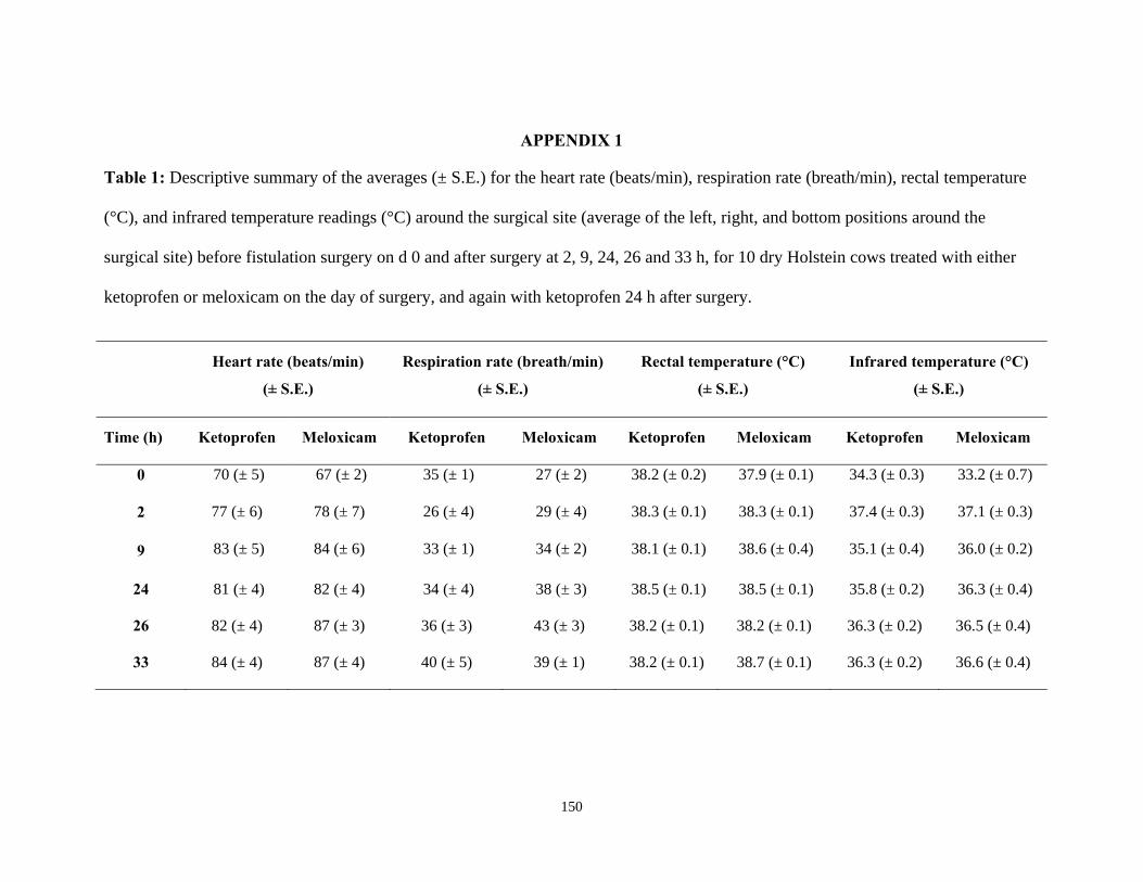
150
APPENDIX 1
Table 1: Descriptive summary of the averages (± S.E.) for the heart rate (beats/min), respiration rate (breath/min), rectal temperature
(°C), and infrared temperature readings (°C) around the surgical site (average of the left, right, and bottom positions around the
surgical site) before fistulation surgery on d 0 and after surgery at 2, 9, 24, 26 and 33 h, for 10 dry Holstein cows treated with either
ketoprofen or meloxicam on the day of surgery, and again with ketoprofen 24 h after surgery.
Heart rate (beats/min)
(± S.E.)
Respiration rate (breath/min)
(± S.E.)
Rectal temperature (°C)
(± S.E.)
Infrared temperature (°C)
(± S.E.)
Time (h)
Ketoprofen Meloxicam
Ketoprofen Meloxicam
Ketoprofen Meloxicam
Ketoprofen Meloxicam
0 70 (± 5) 67 (± 2) 35 (± 1) 27 (± 2) 38.2 (± 0.2) 37.9 (± 0.1) 34.3 (± 0.3) 33.2 (± 0.7)
2 77 (± 6) 78 (± 7) 26 (± 4) 29 (± 4) 38.3 (± 0.1) 38.3 (± 0.1) 37.4 (± 0.3) 37.1 (± 0.3)
9 83 (± 5) 84 (± 6) 33 (± 1) 34 (± 2) 38.1 (± 0.1) 38.6 (± 0.4) 35.1 (± 0.4) 36.0 (± 0.2)
24 81 (± 4) 82 (± 4) 34 (± 4) 38 (± 3) 38.5 (± 0.1) 38.5 (± 0.1) 35.8 (± 0.2) 36.3 (± 0.4)
26 82 (± 4) 87 (± 3) 36 (± 3) 43 (± 3) 38.2 (± 0.1) 38.2 (± 0.1) 36.3 (± 0.2) 36.5 (± 0.4)
33 84 (± 4) 87 (± 4) 40 (± 5) 39 (± 1) 38.2 (± 0.1) 38.7 (± 0.1) 36.3 (± 0.2) 36.6 (± 0.4)
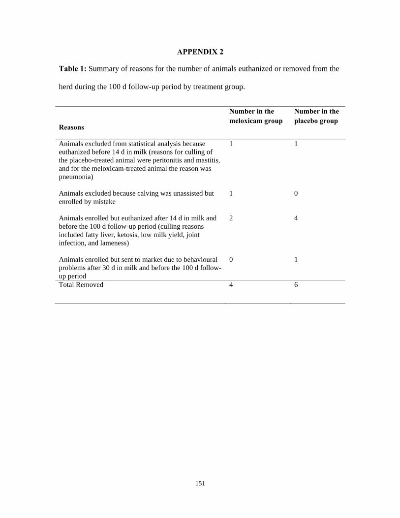
151
APPENDIX 2
Table 1: Summary of reasons for the number of animals euthanized or removed from the
herd during the 100 d follow-up period by treatment group.
Reasons
Number in the meloxicam group
Number in the placebo group
Animals excluded from statistical analysis because euthanized before 14 d in milk (reasons for culling of the placebo-treated animal were peritonitis and mastitis, and for the meloxicam-treated animal the reason was pneumonia)
1 1
Animals excluded because calving was unassisted but enrolled by mistake
1 0
Animals enrolled but euthanized after 14 d in milk and before the 100 d follow-up period (culling reasons included fatty liver, ketosis, low milk yield, joint infection, and lameness)
2 4
Animals enrolled but sent to market due to behavioural problems after 30 d in milk and before the 100 d follow-up period
0 1
Total Removed 4 6
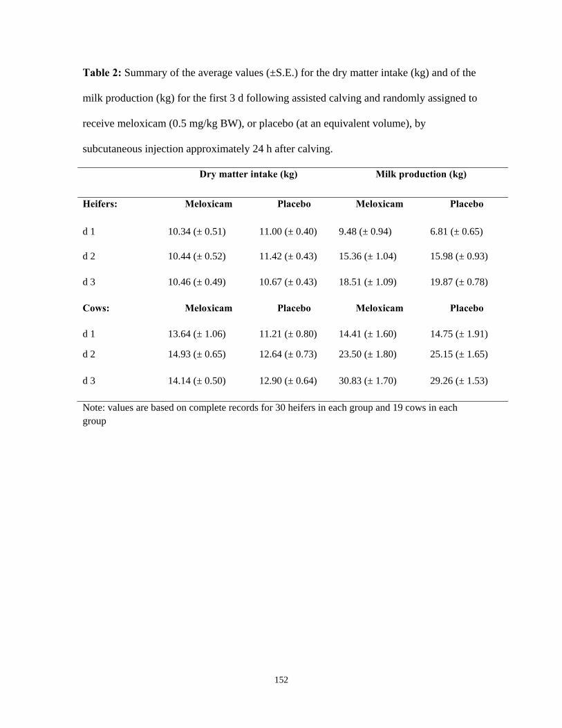
152
Table 2: Summary of the average values (±S.E.) for the dry matter intake (kg) and of the
milk production (kg) for the first 3 d following assisted calving and randomly assigned to
receive meloxicam (0.5 mg/kg BW), or placebo (at an equivalent volume), by
subcutaneous injection approximately 24 h after calving.
Dry matter intake (kg) Milk production (kg)
Heifers: Meloxicam Placebo Meloxicam Placebo
d 1 10.34 (± 0.51) 11.00 (± 0.40) 9.48 (± 0.94) 6.81 (± 0.65)
d 2 10.44 (± 0.52) 11.42 (± 0.43) 15.36 (± 1.04) 15.98 (± 0.93)
d 3 10.46 (± 0.49) 10.67 (± 0.43) 18.51 (± 1.09) 19.87 (± 0.78)
Cows: Meloxicam Placebo Meloxicam Placebo
d 1 13.64 (± 1.06) 11.21 (± 0.80) 14.41 (± 1.60) 14.75 (± 1.91)
d 2 14.93 (± 0.65) 12.64 (± 0.73) 23.50 (± 1.80) 25.15 (± 1.65)
d 3 14.14 (± 0.50) 12.90 (± 0.64) 30.83 (± 1.70) 29.26 (± 1.53)
Note: values are based on complete records for 30 heifers in each group and 19 cows in each group
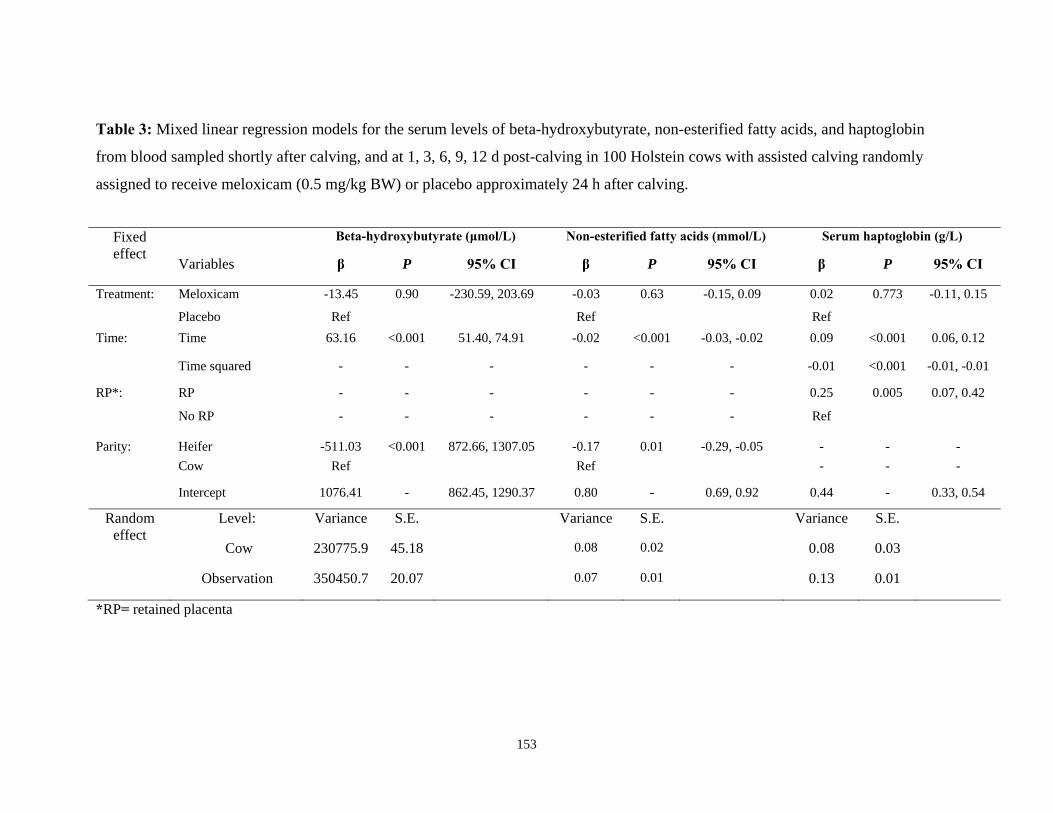
153
Table 3: Mixed linear regression models for the serum levels of beta-hydroxybutyrate, non-esterified fatty acids, and haptoglobin
from blood sampled shortly after calving, and at 1, 3, 6, 9, 12 d post-calving in 100 Holstein cows with assisted calving randomly
assigned to receive meloxicam (0.5 mg/kg BW) or placebo approximately 24 h after calving.
Fixed effect
Beta-hydroxybutyrate (μmol/L) Non-esterified fatty acids (mmol/L) Serum haptoglobin (g/L)
Variables β P 95% CI β P 95% CI β P 95% CI
Treatment: Meloxicam -13.45 0.90 -230.59, 203.69 -0.03 0.63 -0.15, 0.09 0.02 0.773 -0.11, 0.15
Placebo Ref Ref Ref
Time: Time 63.16 <0.001 51.40, 74.91 -0.02 <0.001 -0.03, -0.02 0.09 <0.001 0.06, 0.12
Time squared - - - - - - -0.01 <0.001 -0.01, -0.01
RP*: RP - - - - - - 0.25 0.005 0.07, 0.42
No RP - - - - - - Ref
Parity: Heifer -511.03 <0.001 872.66, 1307.05 -0.17 0.01 -0.29, -0.05 - - -
Cow Ref Ref - - -
Intercept 1076.41 - 862.45, 1290.37 0.80 - 0.69, 0.92 0.44 - 0.33, 0.54
Random effect
Level: Variance S.E. Variance S.E. Variance S.E.
Cow 230775.9 45.18 0.08 0.02 0.08 0.03
Observation 350450.7 20.07 0.07 0.01 0.13 0.01
*RP= retained placenta
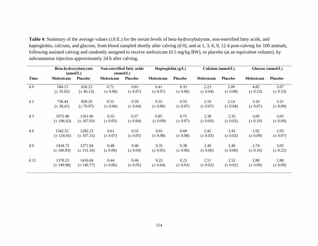
154
Table 4: Summary of the average values (±S.E.) for the serum levels of beta-hydroxybutyrate, non-esterified fatty acids, and haptoglobin, calcium, and glucose, from blood sampled shortly after calving (d 0), and at 1, 3, 6, 9, 12 d post-calving for 100 animals, following assisted calving and randomly assigned to receive meloxicam (0.5 mg/kg BW), or placebo (at an equivalent volume), by subcutaneous injection approximately 24 h after calving.
Beta-hydroxybutyrate (μmol/L)
Non-esterified fatty acids (mmol/L)
Haptoglobin (g/L) Calcium (mmol/L) Glucose (mmol/L)
Time Meloxicam Placebo Meloxicam Placebo Meloxicam Placebo Meloxicam Placebo Meloxicam Placebo
d 0 584.13 (± 35.92)
620.23 (± 40.13)
0.71 (± 0.06)
0.81 (± 0.07)
0.41 (± 0.07)
0.33 (± 0.06)
2.23 (± 0.04)
2.09 (± 0.08)
4.85 (± 0.23)
5.07 (± 0.33)
d 1
736.44 (± 38.41)
830.20 (± 70.97)
0.53 (± 0.06)
0.59 (± 0.04)
0.55 (± 0.06)
0.55 (± 0.07)
2.10 (± 0.07)
2.14 (± 0.04)
3.10 (± 0.07)
3.21 (± 0.09)
d 3 1072.48 (± 106.63)
1161.06 (± 107.03)
0.55 (± 0.05)
0.57 (± 0.04)
0.85 (± 0.09)
0.75 (± 0.07)
2.38 (± 0.03)
2.35 (± 0.02)
3.00 (± 0.10)
3.03 (± 0.09)
d 6 1342.52 (± 134.65)
1282.23 (± 107.15)
0.61 (± 0.07)
0.55 (± 0.05)
0.61 (± 0.08)
0.68 (± 0.08)
2.42 (± 0.03)
2.43 (± 0.02)
2.92 (± 0.09)
2.93 (± 0.07)
d 9 1434.73 (± 160.83)
1271.04 (± 131.16)
0.48 (± 0.06)
0.46 (± 0.04)
0.31 (± 0.05)
0.38 (± 0.06)
2.40 (± 0.06)
2.40 (± 0.06)
2.74 (± 0.10)
3.05 (± 0.22)
d 12 1378.23 (± 149.98)
1416.04 (± 140.77)
0.44 (± 0.06)
0.46 (± 0.05)
0.22 (± 0.04)
0.23 (± 0.03)
2.51 (± 0.02)
2.52 (± 0.02)
2.88 (± 0.09)
2.88 (± 0.09)
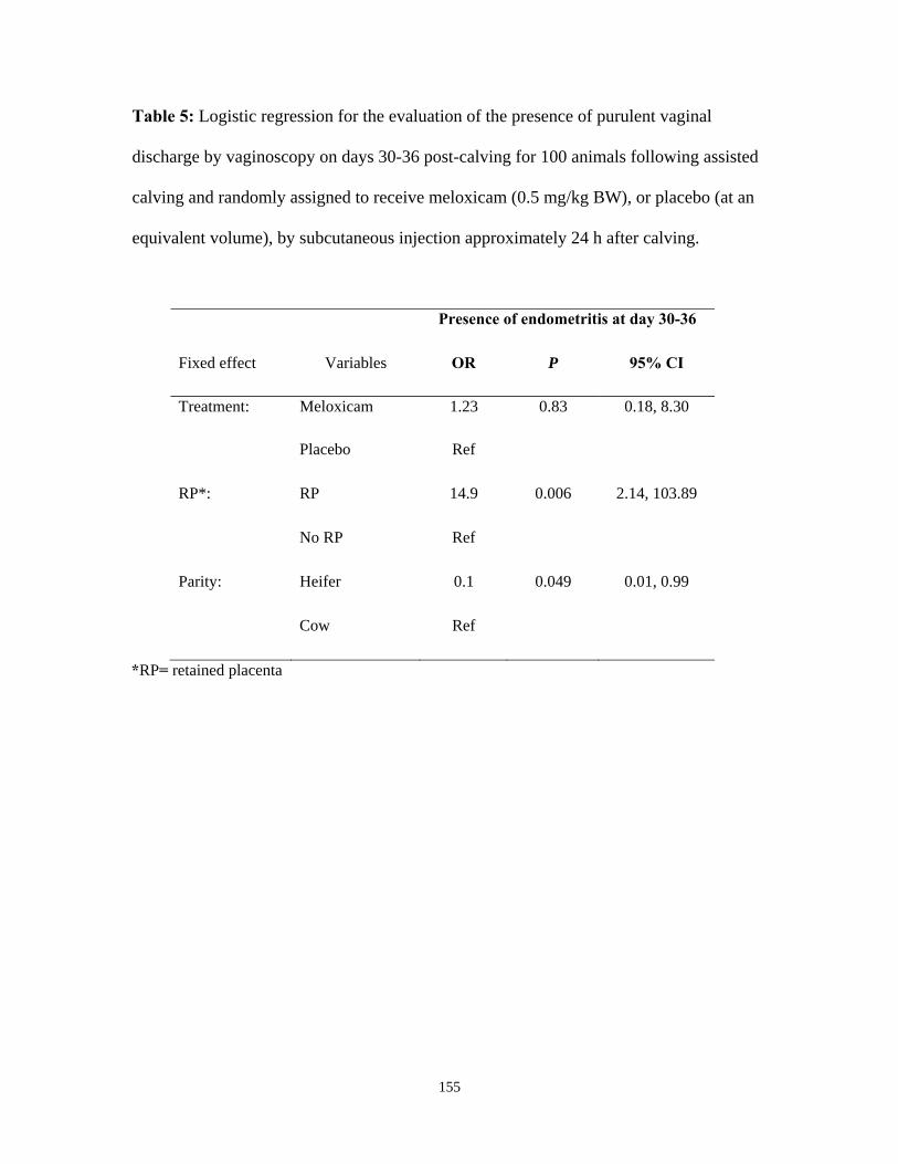
155
Table 5: Logistic regression for the evaluation of the presence of purulent vaginal
discharge by vaginoscopy on days 30-36 post-calving for 100 animals following assisted
calving and randomly assigned to receive meloxicam (0.5 mg/kg BW), or placebo (at an
equivalent volume), by subcutaneous injection approximately 24 h after calving.
Presence of endometritis at day 30-36
Fixed effect Variables OR P 95% CI
Treatment: Meloxicam 1.23 0.83 0.18, 8.30
Placebo Ref
RP*: RP 14.9 0.006 2.14, 103.89
No RP Ref
Parity: Heifer 0.1 0.049 0.01, 0.99
Cow Ref
*RP= retained placenta
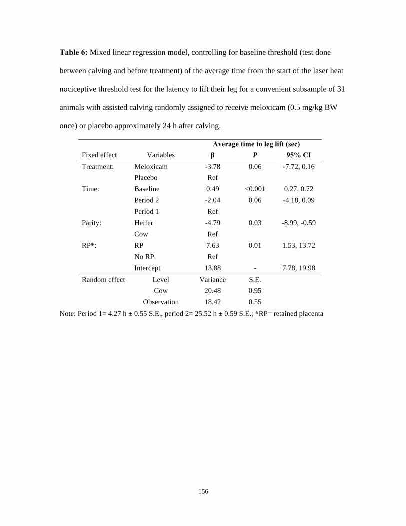
156
Table 6: Mixed linear regression model, controlling for baseline threshold (test done
between calving and before treatment) of the average time from the start of the laser heat
nociceptive threshold test for the latency to lift their leg for a convenient subsample of 31
animals with assisted calving randomly assigned to receive meloxicam (0.5 mg/kg BW
once) or placebo approximately 24 h after calving.
Average time to leg lift (sec)
Fixed effect Variables β P 95% CI
Treatment: Meloxicam -3.78 0.06 -7.72, 0.16
Placebo Ref
Time: Baseline 0.49 <0.001 0.27, 0.72
Period 2 -2.04 0.06 -4.18, 0.09
Period 1 Ref
Parity: Heifer -4.79 0.03 -8.99, -0.59
Cow Ref
RP*: RP 7.63 0.01 1.53, 13.72
No RP Ref
Intercept 13.88 - 7.78, 19.98
Random effect Level Variance S.E.
Cow 20.48 0.95
Observation 18.42 0.55
Note: Period 1= 4.27 h ± 0.55 S.E., period 2= 25.52 h ± 0.59 S.E.; *RP= retained placenta
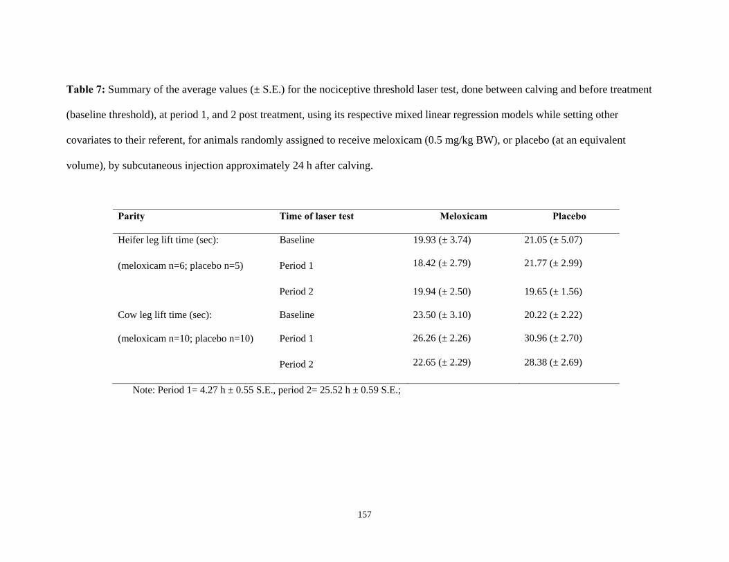
157
Table 7: Summary of the average values (± S.E.) for the nociceptive threshold laser test, done between calving and before treatment
(baseline threshold), at period 1, and 2 post treatment, using its respective mixed linear regression models while setting other
covariates to their referent, for animals randomly assigned to receive meloxicam (0.5 mg/kg BW), or placebo (at an equivalent
volume), by subcutaneous injection approximately 24 h after calving.
Parity Time of laser test Meloxicam Placebo
Heifer leg lift time (sec): Baseline 19.93 (± 3.74) 21.05 (± 5.07)
(meloxicam n=6; placebo n=5) Period 1 18.42 (± 2.79) 21.77 (± 2.99)
Period 2 19.94 (± 2.50) 19.65 (± 1.56)
Cow leg lift time (sec): Baseline 23.50 (± 3.10) 20.22 (± 2.22)
(meloxicam n=10; placebo n=10) Period 1 26.26 (± 2.26) 30.96 (± 2.70)
Period 2 22.65 (± 2.29) 28.38 (± 2.69)
Note: Period 1= 4.27 h ± 0.55 S.E., period 2= 25.52 h ± 0.59 S.E.;
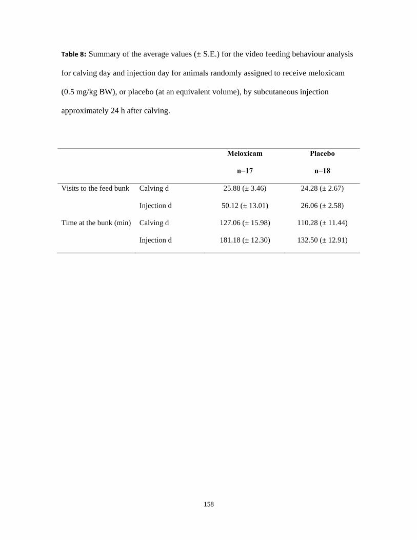
158
Table 8: Summary of the average values (± S.E.) for the video feeding behaviour analysis
for calving day and injection day for animals randomly assigned to receive meloxicam
(0.5 mg/kg BW), or placebo (at an equivalent volume), by subcutaneous injection
approximately 24 h after calving.
Meloxicam
n=17
Placebo
n=18
Visits to the feed bunk Calving d 25.88 (± 3.46) 24.28 (± 2.67)
Injection d 50.12 (± 13.01) 26.06 (± 2.58)
Time at the bunk (min) Calving d 127.06 (± 15.98) 110.28 (± 11.44)
Injection d 181.18 (± 12.30) 132.50 (± 12.91)
![World Journal of Emergency Surgery BioMed Central...abdominal pain but recurrent pain may suggest intermit-tent torsions [4,9]. On examination 50% of patients present with an abdominal](https://img.pdfslide.us/doc/110x75/6090bbaa04e64b13233c51e4/world-journal-of-emergency-surgery-biomed-central-abdominal-pain-but-recurrent.jpg)