Embed Size (px)
Citation preview
Med J Malaysia Vol 74 No 3 June 2019 229
SUMMARY‘Pai syndrome’ (PS) is a rare congenital syndrome.Presented here, a new-born baby-girl who exhibited thecharacteristic features of having a midline nasal (septal)polyp, an anterior alveolar process polyp, and a pericallosallipoma associated with corpus callosum dysgenesis of thebrain. Both polyps were lined with stratified-squamousepithelium. The overall features were largely consistent withthose described by Pai et al., in 1987. A midline cleft-lip (withor without cleft-alveolus) is one of the most commonfeatures of the syndrome which was however absent in thiscase. Instead, an anterior alveolar polyp is present, which isrelatively rare.
INTRODUCTIONPai syndrome is a rare syndrome of unknown cause,comprising a particular variety of congenital developmentalmalformation with variable phenotypical presentations. Itwas first reported by Pai et al., in 1987 when it was describedin a male new-born as an unusual combination of three rareanomalies, i.e., complete median cleft lip, cutaneous polyps,and developmental midline lipomas of the central nervoussystem.1 After that, over sixty similar cases have beenreported, although their phenotypic presentations were notvery similar, ranging from mild-facial dysmorphism to severefrontonasal dysplasia (FND). The perceived impression aboutthe underlying aetiology goes towards a multifactorial origin.No chromosomal abnormalities have been described inpatients with PS on karyotype. Diagnosis of the syndrome isbased on clinical signs and associated pathology, thecommon clinical component being midline facial skinmasses. Minimum diagnostic criteria as suggested by Castoriet al.2 and Lederer et al.3, one or more hamartomatous nasalpolyps plus median cleft lip (with or without cleft alveolus),and/or alveolar process congenital polyp, and/or peri-callosal lipoma. Pericallosal lipoma is a common finding(85%) in this syndrome which is frequently associated withvarious degrees of intracerebral malformations such asagenesis or dysgenesis of corpus callosum.3 Most of the casesof PS have been diagnosed postnatally after birth. Typically,the condition is still not found to have any negative impacton psychoneurological development. In a recent report of an8-year-long follow-up case, Imai et al., observed someattention-deficit disorder, but its definite relationship with thesyndrome is yet to be confirmed.4
CASE REPORTThe present case is that of a dysmorphic baby girl, born vialower-segment caesarean section (LSCS) for maternaleclampsia at the gestational age of 38 weeks. This is the firstchild of non-consanguineous Chinese parents. The mother(in her thirties) developed pregnancy-induced hypertension(PIH). Baby’s birth-weight was 3.31kg, length: 49cm, head-circumference: 34cm. She developed hypoxic-ischemicencephalopathy which was successfully managed byimmediate intensive care support. Except for PIH in latepregnancy, the mother had no other known obstetric illness.For this case, a prenatal diagnosis was not possible probablybecause of the non-compliance with regular follow-up.
Family history revealed the presence of Down syndrome fromthe paternal side. Drug history revealed nothingcontributory. The mother also denied any addictive habitssuch as alcohol, smoking or other harmful substances.
At birth, the baby exhibited two separate soft skin-coveredpolypoidal masses on the face (Fig. 1), one from the rightnasal cavity (nasal septal origin), measuring about 2cm at itsvisible exterior part, partially obstructing the ipsilateral nasalcavity. Another one arose from the mid-anterior alveolarprocess, a little smaller and associated with the high-archedpalate. No other orofacial deformity was detected. Othersystemic examinations were unremarkable. Nohypertelorism or other ocular problems were noted. Thepolyps were surgically removed, and the procedure wasuneventful. During the last review when the child was oneyear of age, she showed good achievement of all milestones.
Chromosomal study result showed a normal femalekaryotype: 46, XX.
DISCUSSIONLipoma is the most common soft tissue tumor in adults, butit is uncommon in children and even more rare as anintracranial lesion. However, pericallosal lipoma is acommon finding in PS which was present in our patient. It isthought to have originated from over-proliferation of the fatcells present in the leptomeninx, an embryological structurenormally differentiated from the meninx primitiva.5 Basedon imaging there are two types of pericallosal lipomas. Thetubulo-nodular is the most common type, frequentlyassociated with extensive callosal and often fronto-nasal
‘Pai Syndrome’ with anterior alveolar polyp: A variant of arare clinical entity
Kallyan Kishore Debnath, MSc1,2, Yogesvaran Kanapaty, MS2, Yong Dong Jieh, MS3, Shobashinni Chandran, MD2,Mohd. Adzreil Bakri, M2
1AIMST University, Kedah, Malaysia, 2Hospital Duchess of Kent, Sabah, Malaysia, 3Queen Elizabeth Hospital, Sabah, Malaysia
CASE REPORT
This article was accepted: 4 April 2019Corresponding Author: Kallyan Kishore DebnathEmail: [email protected]
8-Pai00145R1_3-PRIMARY.qxd 6/14/19 12:56 PM Page 229
Case Report
230 Med J Malaysia Vol 74 No 3 June 2019
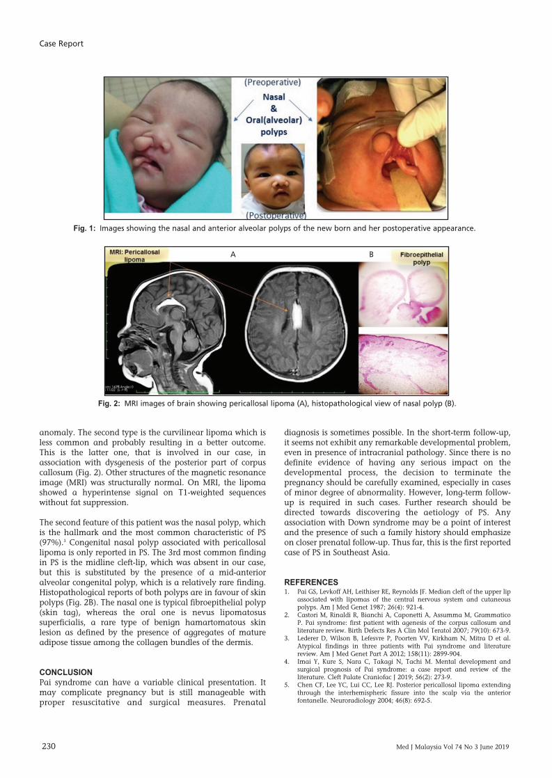
anomaly. The second type is the curvilinear lipoma which isless common and probably resulting in a better outcome.This is the latter one, that is involved in our case, inassociation with dysgenesis of the posterior part of corpuscallosum (Fig. 2). Other structures of the magnetic resonanceimage (MRI) was structurally normal. On MRI, the lipomashowed a hyperintense signal on T1-weighted sequenceswithout fat suppression.
The second feature of this patient was the nasal polyp, whichis the hallmark and the most common characteristic of PS(97%).3 Congenital nasal polyp associated with pericallosallipoma is only reported in PS. The 3rd most common findingin PS is the midline cleft-lip, which was absent in our case,but this is substituted by the presence of a mid-anterioralveolar congenital polyp, which is a relatively rare finding.Histopathological reports of both polyps are in favour of skinpolyps (Fig. 2B). The nasal one is typical fibroepithelial polyp(skin tag), whereas the oral one is nevus lipomatosussuperficialis, a rare type of benign hamartomatous skinlesion as defined by the presence of aggregates of matureadipose tissue among the collagen bundles of the dermis.
CONCLUSIONPai syndrome can have a variable clinical presentation. Itmay complicate pregnancy but is still manageable withproper resuscitative and surgical measures. Prenatal
diagnosis is sometimes possible. In the short-term follow-up,it seems not exhibit any remarkable developmental problem,even in presence of intracranial pathology. Since there is nodefinite evidence of having any serious impact on thedevelopmental process, the decision to terminate thepregnancy should be carefully examined, especially in casesof minor degree of abnormality. However, long-term follow-up is required in such cases. Further research should bedirected towards discovering the aetiology of PS. Anyassociation with Down syndrome may be a point of interestand the presence of such a family history should emphasizeon closer prenatal follow-up. Thus far, this is the first reportedcase of PS in Southeast Asia.
REFERENCES1. Pai GS, Levkoff AH, Leithiser RE, Reynolds JF. Median cleft of the upper lip
associated with lipomas of the central nervous system and cutaneouspolyps. Am J Med Genet 1987; 26(4): 921-4.
2. Castori M, Rinaldi R, Bianchi A, Caponetti A, Assumma M, GrammaticoP. Pai syndrome: first patient with agenesis of the corpus callosum andliterature review. Birth Defects Res A Clin Mol Teratol 2007; 79(10): 673-9.
3. Lederer D, Wilson B, Lefesvre P, Poorten VV, Kirkham N, Mitra D et al.Atypical findings in three patients with Pai syndrome and literaturereview. Am J Med Genet Part A 2012; 158(11): 2899-904.
4. Imai Y, Kure S, Nara C, Takagi N, Tachi M. Mental development andsurgical prognosis of Pai syndrome: a case report and review of theliterature. Cleft Palate Craniofac J 2019; 56(2): 273-9.
5. Chen CF, Lee YC, Lui CC, Lee RJ. Posterior pericallosal lipoma extendingthrough the interhemispheric fissure into the scalp via the anteriorfontanelle. Neuroradiology 2004; 46(8): 692-5.
Fig. 1: Images showing the nasal and anterior alveolar polyps of the new born and her postoperative appearance.
Fig. 2: MRI images of brain showing pericallosal lipoma (A), histopathological view of nasal polyp (B).
A B
8-Pai00145R1_3-PRIMARY.qxd 6/14/19 12:56 PM Page 230