Embed Size (px)
Citation preview
Pediatr Blood Cancer 2013;60:1826–1832
Paediatric and Adolescent Alveolar Soft Part Sarcoma:A Joint Series From European Cooperative Groups
D. Orbach, MD,1* B. Brennan, MD,2 M. Casanova, MD,3 C. Bergeron, MD,4 V. Mosseri, MD,5 N. Francotte, MD,6
M. Van Noesel, MD, PhD, MSc,7 A. Rey, MD,8 G. Bisogno, MD,9 G. Pierron, MD,10 and A. Ferrari, MD3
INTRODUCTION
Alveolar Soft Part Sarcoma (ASPS) is a rare, malignant,
mesenchymal tumour characterized by an unbalanced recurrent t
(X;17)(p11.2;q25) translocation, which leads to a chimeric
ASPSCR1-TFE3 transcription factor [1]. ASPS is characterized
by uniform, organoid nests of polygonal tumour cells, separated
by fibrovascular septa and delicate capillary-sized vascular
channels [2]. ASPS can occur at any age [1,3–5], with a peak
incidence in the third decade. About one third of ASPS occur in
children and adolescents. Paediatric oncologists usually classify
ASPS in the heterogeneous group of non-rhabdomyosarcoma soft
tissue sarcomas (NRSTS), in which ASPS represents 4.5% of all
tumour types [6,7]. In adulthood, ASPS accounts for 0.7% of all soft
tissue sarcomas [2,5]. Due to the rarity of this disease, no
standardised treatment guidelines have yet been defined. Surgery
remains the mainstay of treatment with an R0 resection critical for a
good outcome in localized ASPS. However, surgery alone may not
be sometime feasible or sufficient, as local recurrence is possible
after adequate resection, and metastases (lung or brain) may occur,
sometimes years after the initial diagnosis [4]. ASPS is reported
to be chemo insensitive, with a complete/partial remission rate of
less than 10% with conventional chemotherapy [5]. In a previous
paediatric series, only two out of seven evaluable patients
reportedly obtained a clinical partial response [3]. Targeted
therapies such as multiple tyrosine kinase receptor inhibitors
(RTKs) or anti-angiogenic therapy (i.e. sunitinib, cediranib
or bevacizumab) have been shown to be effective in adult
ASPS [8–11]. Although the extreme rarity of ASPS in children
precludes exclusively paediatric studies on this specific histotype,
recent data indicate that paediatric cases might be equally sensitive
to new targeted therapies [11,12], but further data are needed to
confirm this finding.
We therefore retrospectively analysed the clinical data of
various ASPS cases prospectively registered and treated in various
European paediatric cooperative groups and compared these
features to previously reported adult series.
METHODS
Study Population
The study concerned a series of 51 previously untreated patients
with a diagnosis of ASPS: 23 cases were prospectively registered in
Italian protocols, 17 cases enrolled in the International Society of
Paediatric Oncology Malignant Mesenchymal Tumour group
protocols; another 11 French cases were collected from the Institut
Curie Genetic Laboratory. All these patients were treated according
to the on-going European paediatric protocol at time of diagnosis.
Thirteen of these patients have been already included in previous
publications [3,10,13]. Inclusion criteria for this study were:
(1) study period: 1980–2009; (2) patient’s age: 0–21 years;
(3) histological diagnosis of ASPS after panel confirmation or
presence of ASPSCR1-TFE3 fusion transcript in a non-renal soft
tissue tumour [14].
Tumour site was defined as extremities (including the limb
girdles, i.e. inguinal region, hip, buttock and shoulder) or axial sites
Background. Alveolar soft part sarcomas (ASPS) are generallychemo- and radio-resistant mesenchymal tumours, with no stan-dardized treatment guidelines. We describe the clinical behaviour ofpaediatric ASPS and compare these features to previously reportedadult series. Patients and Methods. The clinical data of 51 childrenand adolescents with ASPS, prospectively enrolled in or treatedaccording to seven European Paediatric trials were analysed.Results.Median age was 13 years [range: 2–21]. Primary sites includedmostly limbs (63%). IRS post-surgical staging was: IRS-I (completeresection) 35%, II (microscopic residual disease) 20%, III (grossresidual disease) 18% and IV (metastases) 27%. Only 3 of the 18evaluable patients (17%) obtained a response to conventionalchemotherapy. After a median follow-up of 126 months (range: 9–
240), 14/18 patients with IRS-I tumour, 10/10 IRS-II, 7/9 IRS-III and 2/14 IRS-IV were alive in remission. Sunitinib treatment achieved twovery good partial responses in four patients. Ten-year overall survival(OS) and event free survival (EFS) was 78.0�7% and 62.8�7%respectively. Stage IV, size >5 cm and T2 tumours had a pooreroutcome, but only IRS staging was an independent prognostic factor.Conclusions. ASPS is a very rare tumour frequently arising inadolescents and in the extremities, and chemo resistant. Localsurgical control is critical. ASPS is a poorly chemo sensitive tumour.For IRS-III/IV tumours, delayed radical local therapies includingsurgery are essential. Metastatic patients had a poor prognosis buttargeted therapies showed promising results. Pediatr Blood Cancer2013;60:1826–1832. # 2013 Wiley Periodicals, Inc.
Key words: adolescence; Alveolar soft part sarcoma; chemotherapy; childhood; radiotherapy; target therapy
1Department of Paediatric Oncology, Institut Curie, Paris, France;2Department of Paediatric Oncology, Royal Manchester Children’s
Hospital, Manchester, United Kingdom; 3Paediatric Oncology Unit,
Fondazione IRCCS Istituto Nazionale Tumouri, Milano, Italy; 4Institut
d’Hematologie et d’Oncologie Pediatrie, Lyon, France; 5Department of
Biostatistics, Institut Curie, Paris, France; 6Department of Paediatrics,
CHC-Clin Esperance—rue Saint Nicolas, Montegnee, Belgium;7Department of Paediatric Oncology-Hematology, Erasmus MC/
Sophia Children’s Hospital, Rotterdam, The Netherlands; 8Department
of Biostatistics, Institut Gustave Roussy, Villejuif, France; 9Paediatric
Hematology and Oncology Division, Padova University, Padova, Italy;10Somatic Genetic Unit, Institut Curie, Paris, France
Conflict of interest: Nothing to declare.
�Correspondence to: Daniel Orbach, Department of Adolescent
Paediatric Oncology, Institut Curie, 26 rue d’Ulm, 75231 Paris Cedex
05, France. E-mail: [email protected]
Received 7 May 2013; Accepted 12 June 2013
�C 2013 Wiley Periodicals, Inc.DOI 10.1002/pbc.24683Published online 16 July 2013 in Wiley Online Library(wileyonlinelibrary.com).
(i.e. head and neck, lung and pleura, retroperitoneum, trunk
(thoracic and abdominal wall)). Staging in all protocols included
lung computed tomography (CT) scan of the lung, bone marrow
aspiration and biopsies, and Technetium bone scanning. Tumour
stage was defined according to the paediatric pretreatment TNM
system (T1 or T2 according to the invasion of contiguous organs, A
or B according to tumour diameter �5 or >5 cm, respectively; N0
or N1, and M0 or M1 according to the presence of lymph node or
distant metastases) and the Intergroup Rhabdomyosarcoma Study
(IRS) post-surgical grouping classification (group I: complete
resection, group-II: microscopic residual disease, group-III: gross
residual disease, group-IV: metastases at onset) [15]. As centralized
pathology review had already been set up in each collaborative
group, histological diagnoses were not specifically reviewed for this
analysis.
Treatment Modalities
Patients received multi-modal treatments including surgery,
radiotherapy and chemotherapy, according to the paediatric
treatment protocols in use at the time. The general treatment
strategy did not change substantially over time, or between the
various protocols. Attempted conservative surgery was the
mainstay of treatment: primary excision was planned when it
was expected to be complete and non-mutilating, otherwise biopsy
was performed. Primary re-excision was recommended prior to any
other therapy in the case of suspected microscopic residual tumour,
in an attempt to achieve a complete resection. If conservative
complete excision was considered unfeasible at the time of
diagnosis, surgery was delayed until after chemotherapy to induce
tumour shrinkage and improve resectability [7,13].
Radiotherapy was considered for all patients at high risk of local
relapse due to either tumour size (>5 cm) or incomplete
resection [7]. External beam irradiation to the primary tumour
was administered with conventional fractionation (1.8–2Gy/day)
for a total dose ranging from 40 to 50Gy. According to the protocol,
the irradiation target volume included the initial tumour plus
2–3 cm margin and the surgical scar.
Chemotherapy was used in selected cases, either in the presence
of microscopic or gross residual disease after surgery, according to
the various protocols, usually ifosfamide-doxorubicin based
regimens [6,13]. Response to chemotherapy in patients with
measurable disease was evaluated after three cycles and was based
on the radiological detectable reduction in the sum of the products
of the perpendicular diameters of all measurable lesions. Response
was defined as: complete (CR)¼ complete disappearance of
disease; partial (PR)¼ tumour reduction >50%; minor (MR)¼reduction >25%. Stable disease or tumour reduction <25% was
classified as no response, whilst an increase in tumour size or
detection of new lesions was considered to be progression.
Statistical Methods
Local control was defined as control of the primary tumour with
disappearance of all clinical and radiological signs of disease, or
stable residual radiographic images for at least 12 months after
completion of treatment. Outcome was defined by overall survival
(OS) and event-free survival (EFS). For EFS, events were defined as
relapse after CR, failure of tumour local control, progressive of
metastatic disease, or death from any cause. In the case of no initial
control of tumour the time-point taken into account for the event
was the date of the beginning of therapy. Survival curves were
computed by the Kaplan–Meier method. The statistical significance
of each variablewas tested by the log-rank test (univariate analysis).
Ten-year survival rates are expressed together with the standard
error.Multivariate analysis of 10-year EFSwas performed using the
Cox proportional hazards regression model.
RESULTS
Patient and Tumour Characteristics
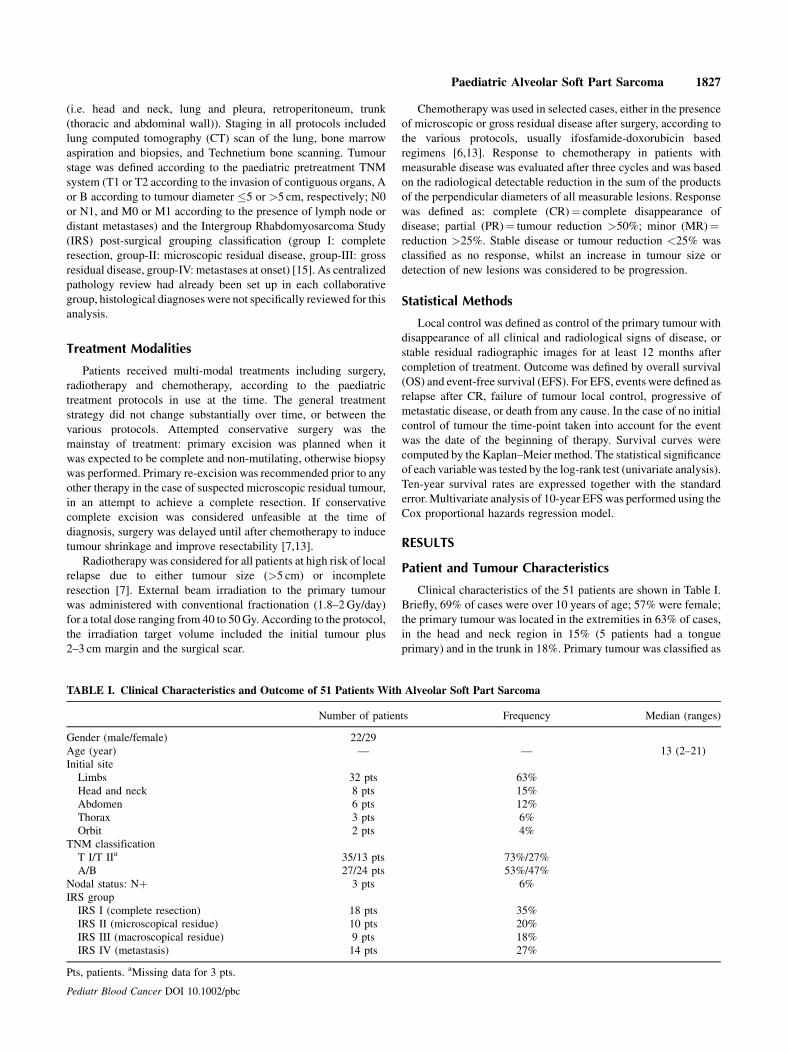
Clinical characteristics of the 51 patients are shown in Table I.
Briefly, 69% of cases were over 10 years of age; 57% were female;
the primary tumour was located in the extremities in 63% of cases,
in the head and neck region in 15% (5 patients had a tongue
primary) and in the trunk in 18%. Primary tumour was classified as
TABLE I. Clinical Characteristics and Outcome of 51 Patients With Alveolar Soft Part Sarcoma
Number of patients Frequency Median (ranges)
Gender (male/female) 22/29
Age (year) — — 13 (2–21)
Initial site
Limbs 32 pts 63%
Head and neck 8 pts 15%
Abdomen 6 pts 12%
Thorax 3 pts 6%
Orbit 2 pts 4%
TNM classification
T I/T IIa 35/13 pts 73%/27%
A/B 27/24 pts 53%/47%
Nodal status: Nþ 3 pts 6%
IRS group
IRS I (complete resection) 18 pts 35%
IRS II (microscopical residue) 10 pts 20%
IRS III (macroscopical residue) 9 pts 18%
IRS IV (metastasis) 14 pts 27%
Pts, patients. aMissing data for 3 pts.
Pediatr Blood Cancer DOI 10.1002/pbc
Paediatric Alveolar Soft Part Sarcoma 1827
T1aA in 54% of cases, T1B in 21%, T2A in 2% and T2B in 25%.
Regional lymph node involvement was reported in only three
cases (6%).
Subgroup Analysis
IRS-I group. This group represents 18 patients with initial
complete resection. All but one case had a T1 tumour (missing data
in 2 patients) and all but four cases had tumours less than 5 cm at
diagnosis. Adjuvant chemotherapy was administered to seven
patients, and in two patients adjuvant radiotherapy at a dose of 55.8
or 50Gy in addition to chemotherapy in one patient. At the time of
analysis, 14 patients were alive in first remission, 17–240 months
from diagnosis (median: 131months). Four patients had ametastatic
relapse (three in the lung only, one in lung and brain) at 18–110
months after diagnosis. One of these patients had received adjuvant
chemotherapy as a front-line treatment. One of the relapsed patients
died of disease; one developed a secondary acute leukaemia 2 years
after the second remission ofASPS and is alive 10 years from the end
of therapy; the third is currently receiving sunitinib therapy after
failure of various chemotherapy regimens, and the last patient was
alive in remission 18 months after relapse.
IRS-II group. Microscopic residual disease after first surgery
was present in 10 patients. All but one of the patients in this
subgroup had a T1A tumour. Seven patients received adjuvant
chemotherapy, in combination with radiotherapy (45–53Gy) for
five patients, and one patient received exclusive adjuvant
radiotherapy at 54Gy. All patients were alive in first remission at
the time of the report (follow-up: 11–199 months, median: 150
months).
IRS-III group. For nine patients, IRS grouping was classified
as III for gross residual disease after biopsy (eight cases) or initial
incomplete surgery (one case). In 6 cases, the tumour was larger
than 5 cm. All nine patients received chemotherapy, five underwent
delayed surgery (complete in three cases (i.e. margins negative) and
unknown in two cases), with radiotherapy for six patients (45–
60Gy, median: 55Gy). In this subgroup, 7/9 patients were in first
remission at the time of the report, 59–155 months after diagnosis
(median: 110 months). Two patients relapsed, one locally and one
locally with lung metastases, after 20 and 2 months, respectively.
First-line local treatment in these two patients was exclusive
radiotherapy. Both patients died of disease. Only one patient,
treated with definitive radiotherapy, had a durable local tumour
control.
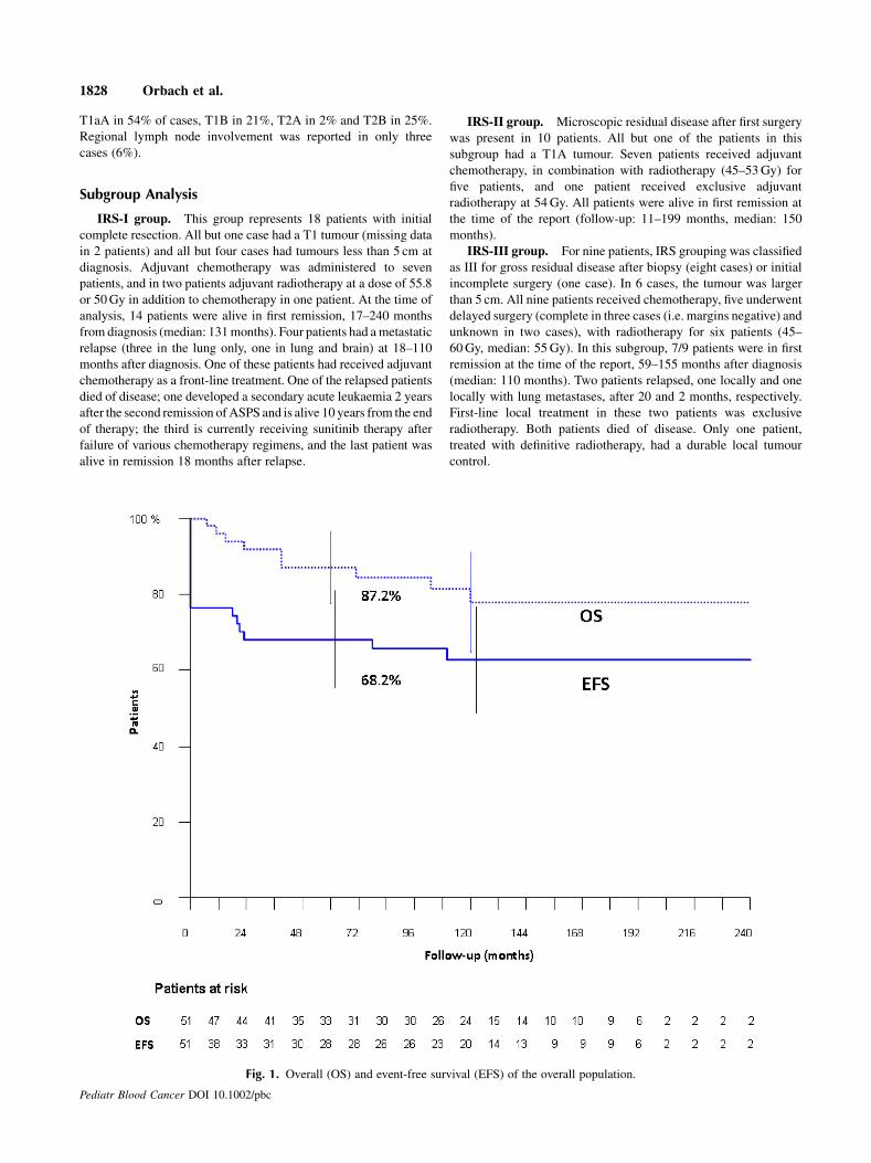
Fig. 1. Overall (OS) and event-free survival (EFS) of the overall population.
Pediatr Blood Cancer DOI 10.1002/pbc
1828 Orbach et al.
IRS-IV group. Distant metastases were present at diagnosis
in 14 cases. All but one case had a tumour larger than 5 cm. In 13
patients (93%), the lung was the site of metastases (associated with
bone metastases in two cases and liver metastases in one case); one
patient had isolated bone metastasis. Three patients also had
regional lymph node involvement. One patient refused treatment
and was lost to follow-up with progression. All other cases received
various chemotherapy regimens; nine received adjuvant radiother-
apy (36–50.4Gy). Eleven of the 13 evaluable patients experienced
tumour progression or relapse, two locally, three locally plus
metastases, six at metastatic sites only. Sunitinib was administered
on compassionate grounds to four patients with a very good partial
response of the lungmetastases in two patients, stable disease in one
patient still on therapy after 6 months and 1 failure due to patient
refusal after severe prolonged cutaneous adverse events despite
tumour stabilization after 12 months of therapy. Sunitinib therapy
was not strictly uniform. All four patients are alive at the end of
follow-up with stable residual pulmonary images. Six patients out
of the 14 died of disease. Overall, only two patients were in
continuous remission at the time of the report, 121 and 185 months
after diagnosis, respectively. These two patients received various
types of chemotherapy and a complete resection of all pulmonary
metastasis after bilateral thoracotomies. Another patient was in
second remission with surgery for lung metastases and various
chemotherapy regimens (six drugs regimen, 5-fluorouracil-cisplat-
inum, Interleukine 2) after pulmonary progression. Four other
patients were alive on treatment after various systemic salvage
therapies.
Overall Response Rate to Systemic Therapy
In summary, 3 of the 18 patients with evaluable IRS-stage III–IV
ASPS obtained a response to initial conventional chemotherapy
(17%) with one complete remission and two partial responses with
VACA/VAIA regimens. Twelve patients had a stable disease and
three patients a progressive disease. Response to sunitinib, given
after tumour relapse or progression, consisted of two very good
partial responses and two cases of stable disease.
Overall Outcome and Prognostic Variables
After a median follow-up of 126 months (range: 9–240 months),
33 patients were alive in first remission, 3 in second remission and 6
were alivewith disease. Nine patients died of disease. Among the 12
patients with progressive disease in which data are available, 4
developed brain metastasis during progression as well as lung
metastasis. Data was not specified for 5 patients. Only 3 of the 10
patients alive after relapse were disease-free after a long follow up.
In summary, 90% of patients with localized tumour had a grossly
resected tumour defined by IRS group I/II and group III with
complete delayed resection. Five- and 10-year EFS were 68.2� 7%
and 62.8� 7%; 5- and 10-year OS were 87.2� 5% and 78.0� 7%,
respectively (Fig. 1). Table II describes the statistical analysis. IRS
grouping, size and TNM classification were important prognostic
risk factors for EFS and OS. Age did not appear to be a prognostic
factor. Similar results were observed for 10-year survival rates (data
not shown). Initial tumour extension was the only significant
prognostic factor for EFS in the multivariable model (IRS group I–
II–III vs. IV, Relative risk: 12.50 [4.6–34.3]). Valid statistical
comparison of OS could not be performed due to the small numbers
of events.
DISCUSSION
This series of paediatric ASPS cases recruited by various
paediatric European cooperative groups can be considered to be the
most extensive clinical description of this tumour in children and
adolescents. The primary objective of this study was to describe the
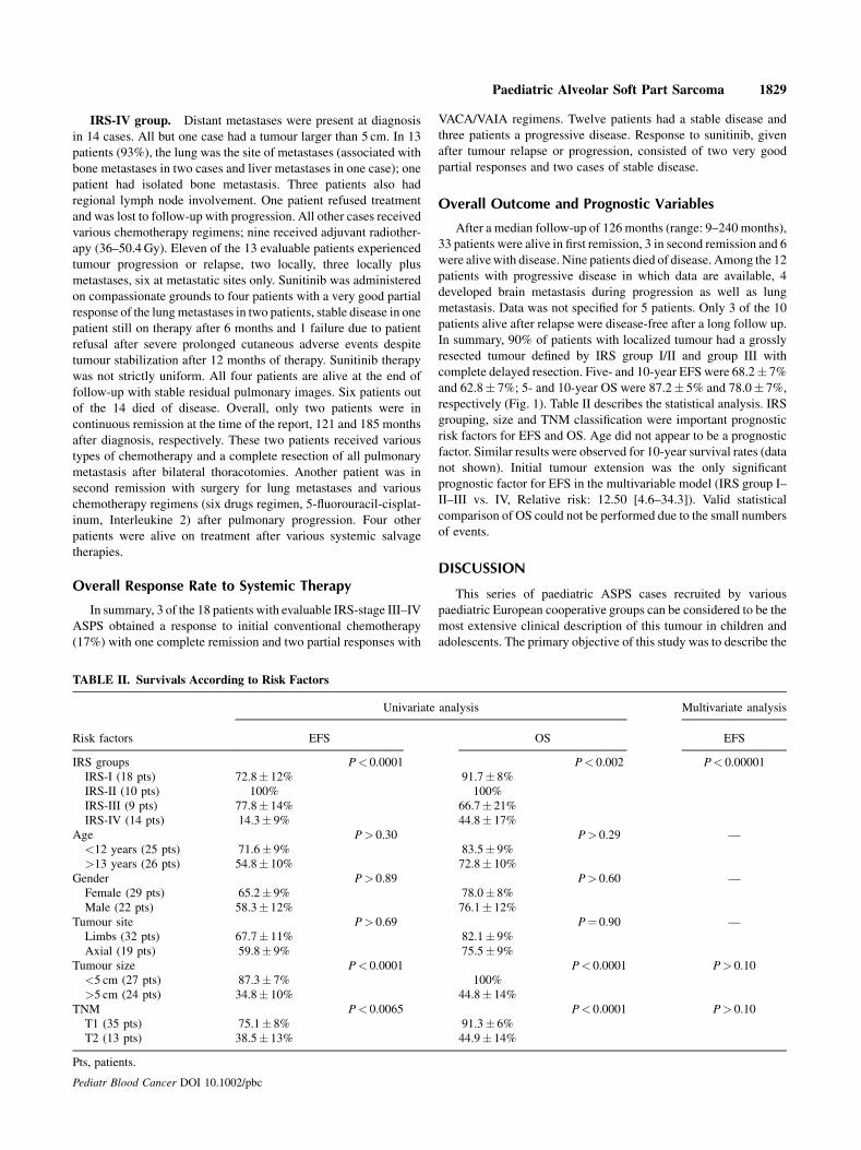
TABLE II. Survivals According to Risk Factors
Risk factors
Univariate analysis Multivariate analysis
EFS OS EFS
IRS groups P< 0.0001 P< 0.002 P< 0.00001
IRS-I (18 pts) 72.8� 12% 91.7� 8%
IRS-II (10 pts) 100% 100%
IRS-III (9 pts) 77.8� 14% 66.7� 21%
IRS-IV (14 pts) 14.3� 9% 44.8� 17%
Age P> 0.30 P> 0.29 —
<12 years (25 pts) 71.6� 9% 83.5� 9%
>13 years (26 pts) 54.8� 10% 72.8� 10%
Gender P> 0.89 P> 0.60 —
Female (29 pts) 65.2� 9% 78.0� 8%
Male (22 pts) 58.3� 12% 76.1� 12%
Tumour site P> 0.69 P¼ 0.90 —
Limbs (32 pts) 67.7� 11% 82.1� 9%
Axial (19 pts) 59.8� 9% 75.5� 9%
Tumour size P< 0.0001 P< 0.0001 P> 0.10
<5 cm (27 pts) 87.3� 7% 100%
>5 cm (24 pts) 34.8� 10% 44.8� 14%
TNM P< 0.0065 P< 0.0001 P> 0.10
T1 (35 pts) 75.1� 8% 91.3� 6%
T2 (13 pts) 38.5� 13% 44.9� 14%
Pts, patients.
Pediatr Blood Cancer DOI 10.1002/pbc
Paediatric Alveolar Soft Part Sarcoma 1829
TABLE
III.
Main
StudiesPublished
onAlveolarSoftPart
Sarcomas
Number
of
patients
Medianage
year
[ranges]
Lim
bs
primaryMetastatic
5-year
EFS
5-yearOS
Adverse
prognostic
factors
Notes
Paediatric
series
Currentseries,Europe,
2012
51
13[2–21]
63%
27%
63.8�7%
78�6.7%
Metastasis
—
KaytonM.USA,2006
20
16.5
[6–24]
50%
37%
22%
83%
Size>5cm
.Notage
Relativelygoodprognosisdespite
metastasisin
70%
ofcases
CasanovaM.Italy,
2000
19
12[4–18]
53%
21%
—80%
Metastasis,size
>5cm
.Notage,
gender,site
andtypeoftreatm
ent
Importance
ofsurgery
Pappo.USA,1996
11
9.6
[2.8–16]
36%
18%
—85%
—Im
portance
oftumourresectability
Adultseries
Daigeler
A.Germany,
2008
11
32[19–49]
75%
0%
—58%
—
Lazar
A.USA,2007
33
28
——
—74%
—Analysisofangiogenesis
promotinggene
patterns:
ASPSangiogenic
signature
is
specific
JongR.Canada,
1998
946[18–70]
86%
33%
——
—Clinicopathological
analysis:someunusual
histopathological
featuresarepossible
Combined
ageseries
Ogura
K.Japan,2012
26
27[2–46]
69%
62%
64%
Size,
AJC
Cstage
Age�1
8yearis
notaprognostic
factor
RekhiB.India,2012
47
24[2–54]
67%
48%
—100%
—ShortFU
ofonly
22pts.TFE3is
auseful
marker
fordiagnosis
William
sA.UK
andSweden,2011
18
23[3–46]
89%
39%
——
—ASPL/TFE3fusiontranscriptsarepowerful
toolsfordiagnosis
JunH.Korea,
2010
12
29[12–57]
50%
50%
—53%
Metastasis
Thehighlevel
ofexpressionofMETin
ASPL-TFE3supportpotential
role
of
targeted
agents
PennacchioliE.Italy,
2010
33
39[7–62]
79%
36%
—68.7%
Qualityofsurgery,
size
>5cm
Importance
oftumourresectability.
Distant
metastasesfairly
common,typical
indolent
course
PangL.China,
2008
16
26[3–58]
50%
12.5%
——
—
ReichardtP.
Germany,
2003
829[15–43]
88%
100%
——
—Highincidence
ofbrain
metastases.ASPS
patients
should
notbetreatedwithCT
exceptin
controlled
clinical
trials
Ogose
A.Japan,2003
57
25[7–75]
84%
64%
—56%
Metastasis,tumoursize
andbone
involvem
entat
primary.
Notage,
gender,siteandtypeofchem
otherapy
Nomultivariate
analysisperform
ed.No
advantagecould
bedem
onstratedforCT
withhigh-dose
ifosfam
ideorcisplatin
Van
Ruth
S.theNetherlands,2002
15
27[1–52]
73%
33%
—38%
—Im
portance
oftumourresectability.
Poor
prognosisin
thepresence
ofmetastasis
PorteraC.USA,2001
74
26[3–68]
60%
65%
L:71%
M:20%
L:88%
Metastasis
Relativelyindolentclinical
course.
Routine
intracranialim
agingshould
beused
Lieberman
P.USA,1989
102
27[5–56]
74%
23%
—62%
Metastasis,tumoursize,adults.
Notgender;norlaterality
Importance
oftumourresectability.
Long
survival
may
beobserved,even
with
metastases
EvansH.USA,1985
14
23[5–59]
50%
21%
NA
50%
Metastasis,tumoursize
CT,chem
otherapy;EFS,event-free
survival;OS,overallsurvival;L,localized;M,metastatic;
FU,follow-up.
Pediatr Blood Cancer DOI 10.1002/pbc
1830 Orbach et al.
clinical findings and behaviour of this rare malignancy. Further-
more this study was set up to analyse therapeutic considerations
according to initial tumour staging and resectability. It confirmed
the previous findings of chemo insensitivity in ASPSwith only 17%
of the evaluable patients responding to chemotherapy, other studies
reporting an overall response rate of between 0% and 30% [3–5,16–
19]. The role of adjuvant chemotherapy is unclear on the basis of
our results. One out of 14 patients with grossly resected tumour at
diagnosis (IRS-I or II) treated with chemotherapy had a metastatic
relapse versus 3 out of 14 who did not receive any adjuvant
chemotherapy. Other drugs in ASPSmay have an anti-angiogenesis
effect, such as bevacizumab [9], sunitinib [10–12] or interferon
alpha-2b [20]. Phase II trials with cediranib and a c-Met inhibitor
(ARQ-197) have recently confirmed the efficacy of these
drugs [8,21]. Data on efficacy of anti-angiogenesis drugs in
ASPS are limited in children and most of them are scared and
coming from retrospective study. Our limited experience would
suggest that sunitinibmay constitute a potential treatment option for
patients with unresectable or metastatic disease [10]. Sunitinib is a
small molecule that inhibits multiple receptor tyrosine kinases,
some of which are involved in tumour growth, angiogenesis and
metastatic progression of cancer. The use of sunitinib has a rational
basis, in that MET autophosphorylation, due to the ASPSCR1-
TFE3 fusion protein, activates cell signalling pathways governing
angiogenesis, cell division and growth, and cell survival [22,23].
Moreover, sunitinib has also been shown to be effective in the Xp11
renal cell carcinoma (RCC), which is known to sometimes share the
same transcript, with objective responses and prolonged progres-
sion-free survival. In RCC either, translocation involves the
transcription factor E3 (TFE3) gene located on Xp11.2. The
TFE3 protein encoded by this gene interacts with same transcription
cell regulator agents. In this latter tumour, the most common
translocations involve an alveolar soft part sarcoma locus (ASPL)-
TFE3 or renal cell carcinoma (RCC) papillary 1 gene (PRCC)-
TFE3 fusion [24].
Although this study analysed a large series of paediatric ASPS,
each subset comprised of only a few cases and the small sample size
therefore prevents any valid conclusions concerning treatment.
Surgery has been shown to play a critical role in achieving local
control. The fairly satisfactory survival rates observed in our cohort
of patients with localized tumours is related to the high resectability
rate, possibly due to their initial presentation with a relatively small
tumour (53% of cases <5 cm), and the majority non-invasive (73%
T1). In contrast, our analysis does not provide any arguments in
favour of the systematic use of radiotherapy in patients with IRS
group-II or -III tumours. In IRS II tumours no local relapse occurred
even though 72% of patients did not received any adjuvant
radiotherapy.
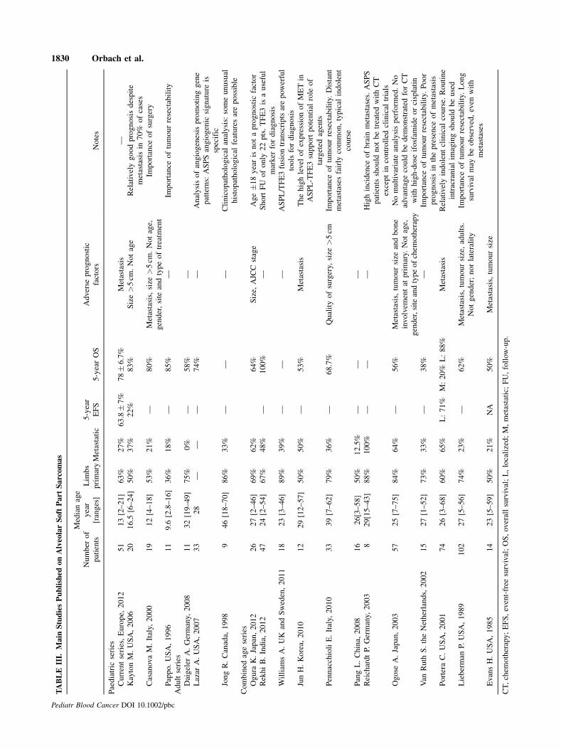
Comparison of this paediatric series with published series of
adult ASPS, as reported in Table III, did not demonstrate any major
differences compared to ASPS in children [1,3–5,16–19,25–34],
with identical female sex predominating in both populations.
Clinical findingswere also similar in terms of distribution of tumour
sites, chemo insensitivity, a high rate of resectability and the
development delayed pulmonary or cerebral metastasis. However,
adult patients present with a higher percentage of metastases at
diagnosis and larger tumours, two variables that may be associated
with a poorer overall prognosis. The present series also suggests a
relationship between large tumour size and the presence of
metastases, as all patients with metastases bar one had a tumour
larger than 5 cm. Tumour size larger than 5 cm was also correlated
to a worse prognosis in other studies in children or adults but
without multivariate analysis [3–4,30–31].
The study confirms the tendency of ASPS tometastasize, mainly
to the lung, with 27% of patients presenting with distant metastases,
compared to the 3% reported in large series of various adult-type
NRSTS [7]. In patients with localized disease, however, tumours
were often confined to the tissue/organ of origin, with no local
invasion and with small tumours. Due to the absence of bone
marrow spreading in this tumour, we do not recommend bone
marrow analysing for initial staging.
The relatively large difference between EFS and OS is probably
due to slow disease progression rather than the ability to salvage
relapsed patients, as only 3 of the 10 patients alive after relapsewere
disease-free. It is notable that some metastatic relapses occurred
more than 5-year after diagnosis requiring long term follow-up of
all these patients.
Disappointingly, the rarity of this tumour does not allow us to
plan future therapeutic trials. In the current NRSTS protocol of the
European paediatric Soft tissue sarcoma Study Group (EpSSG),
ASPS patients are now included in the large heterogeneous group of
adult-type NRSTS, together with other histotypes, managed by
surgery, radiotherapy and adjuvant chemotherapy according to the
already known risk factors (initial extension, tumour size and site
and histological grading) [35]. Due to their chemo insensitivity, new
guidelines should be tailored to stress the role of local therapies, in
particular surgery of the primary tumour and also for distant
metastases either at first presentation or after relapse. Novel
targeted therapies, in particular sunitinib, would appear to be a
potentially effective treatment option. However, due to its toxicity
profile and the lack of knowledge of its long term use, especially in
children with this indolent sarcoma, we recommend its use should
be restricted to cases of inoperable progressive disease or relapsed
tumours in prospective experimental study [36]. Nevertheless, the
weaknesses of data in children available until now do not allow us to
treat children with this new drug out of a prospective experimental
controlled study.
ACKNOWLEDGMENTS
The authors would like to thank all colleagues for their
contribution to the treatment of these children.
REFERENCES
1. Williams A, Bartle G, Sumathi VP, et al. Detection of ASPL/TFE3 fusion transcripts and the TFE3
antigen in formalin-fixed, paraffin-embedded tissue in a series of 18 cases of alveolar soft part sarcoma:
Useful diagnostic tools in cases with unusual histological features. Virchows 2011;458:291–300. Epub
2011 Jan 2029.
2. Folpe AL, Deyrup AT. Alveolar soft-part sarcoma: A review and update. J Clin Pathol 2006;59:1127–
1132.
3. CasanovaM, Ferrari A, Bisogno G, et al. Alveolar soft part sarcoma in children and adolescents: A report
from the Soft-Tissue Sarcoma Italian Cooperative Group. Ann Oncol 2000;11:1445–1449.
4. Kayton ML, Meyers P, Wexler LH, et al. Clinical presentation, treatment, and outcome of alveolar soft
part sarcoma in children, adolescents, and young adults. J Pediatr Surg 2006;41:187–193.
5. Reichardt P, Lindner T, PinkD, et al. Chemotherapy in alveolar soft part sarcomas.What dowe know? Eur
J Cancer 2003;39:1511–1516.
6. BisognoG, Compostella A, Ferrari A, et al. Rhabdomyosarcoma in adolescents: A report from theAIEOP
Soft Tissue Sarcoma Committee. Cancer 2011;118:821–827.
7. Ferrari A, Casanova M, Collini P, et al. Adult-type soft tissue sarcomas in pediatric-age patients:
Experience at the Istituto Nazionale Tumori in Milan. J Clin Oncol 2005;23:4021–4030.
8. Kummar S, Allen D, Monks A, et al. Cediranib for metastatic alveolar soft part sarcoma. J Clin Oncol
2013;2013:29.
9. Azizi AA, Haberler C, Czech T, et al. Vascular-endothelial-growth-factor (VEGF) expression and
possible response to angiogenesis inhibitor bevacizumab in metastatic alveolar soft part sarcoma. Lancet
Oncol 2006;7:521–523.
10. Hilbert M, Mary P, Larroquet M, et al. Alveolar soft part sarcoma in childhood: Is Sunitinib-Sutent(R)
treatment an effective approach? Pediatr Blood Cancer 2012;58:475–476.
Pediatr Blood Cancer DOI 10.1002/pbc
Paediatric Alveolar Soft Part Sarcoma 1831
11. Stacchiotti S, Tamborini E, Marrari A, et al. Response to sunitinib malate in advanced alveolar soft part
sarcoma. Clin Cancer Res 2009;15:1096–1104.
12. Ghose A, Tariq Z, Veltri S. Treatment of multidrug resistant advanced alveolar soft part sarcoma with
sunitinib. Am J Ther 2012;19:e56–e58.
13. Ferrari A, Miceli R, Rey A, et al. Non-metastatic unresected paediatric non-rhabdomyosarcoma soft
tissue sarcomas: Results of a pooled analysis from United States and European groups. Eur J cancer
2011;47:724–731.
14. Peter M, Gilbert E, Delattre O. A multiplex real-time PCR assay for the detection of gene fusions
observed in solid tumors. Lab Invest 2001;81:905–912.
15. Maurer HM, Beltangady M, Gehan EA, et al. The intergroup rhabdomyosarcoma study-I. A final report.
Cancer 1988;61:209–220.
16. Jun HJ, Lee J, Lim doH, et al. Expression ofMET in alveolar soft part sarcoma.MedOncol 2009;27:459–
465.
17. Pang LM, Roebuck DJ, Griffith JF, et al. Alveolar soft-part sarcoma: A rare soft-tissue malignancy with
distinctive clinical and radiological features. Pediatr Radiol 2001;31:196–199.
18. Pennacchioli E, Fiore M, Collini P, et al. Alveolar soft part sarcoma: Clinical presentation, treatment, and
outcome in a series of 33 patients at a single institution. Ann Surg Oncol 2010;17:3229–3233. Epub 2010
Jul 3221.
19. Rekhi B, Ingle A, Agarwal M, et al. Alveolar soft part sarcoma ‘revisited’: Clinicopathological review of
47 cases from a tertiary cancer referral centre, including immunohistochemical expression of TFE3 in 22
cases and 21 other tumours. Pathology 2012;44:11–17.
20. Roozendaal KJ, de Valk B, ten Velden JJ, et al. Alveolar soft-part sarcoma responding to interferon alpha-
2b. Br J Cancer 2003;89:243–245.
21. Wagner AJ, Goldberg JM, Dubois SG, et al. Tivantinib (ARQ 197), a selective inhibitor of MET, in
patients with microphthalmia transcription factor-associated tumors: Results of a multicenter phase 2
trial. Cancer 2012;118:5894–5902.
22. Stacchiotti S, Negri T, Zaffaroni N, et al. Sunitinib in advanced alveolar soft part sarcoma: Evidence of a
direct antitumor effect. Ann Oncol 2011;22:1682–1690.
23. Choueiri TK, Lim ZD, Hirsch MS, et al. Vascular endothelial growth factor-targeted therapy for the
treatment of adult metastatic Xp11.2 translocation renal cell carcinoma. Cancer 2010;116:5219–
5225.
24. OguraK, BeppuY, ChumanH, et al. Alveolar soft part sarcoma: A single-center 26-patient case series and
review of the literature. Sarcoma 2012;2012:907179.
25. Pappo AS, Parham DM, Cain A, et al. Alveolar soft part sarcoma in children and adolescents: Clinical
features and outcome of 11 patients. Med Pediatr Oncol 1996;26:81–84.
26. Daigeler A, Kuhnen C, Hauser J, et al. Alveolar soft part sarcoma: Clinicopathological findings in a series
of 11 cases. World J Surg Oncol 2008;6:71.
27. Jong R, Kandel R, Fornasier V, et al. Alveolar soft part sarcoma: Review of nine cases including two cases
with unusual histology. Histopathology 1998;32:63–68.
28. Lazar AJ, Das P, Tuvin D, et al. Angiogenesis-promoting gene patterns in alveolar soft part sarcoma. Clin
Cancer Res 2007;13:7314–7321.
29. Lieberman PH, Brennan MF, Kimmel M, et al. Alveolar soft-part sarcoma. A clinico-pathologic study of
half a century. Cancer 1989;63:1–13.
30. Ogose A, Yazawa Y, Ueda T, et al. Alveolar soft part sarcoma in Japan: Multi-institutional study of
57 patients from the Japanese Musculoskeletal Oncology Group. Oncology 2003;65:7–13.
31. Portera CA, Jr., Ho V, Patel SR, et al. Alveolar soft part sarcoma: Clinical course and patterns of
metastasis in 70 patients treated at a single institution. Cancer 2001;91:585–591.
32. van Ruth S, van Coevorden F, Peterse JL, et al. Alveolar soft part sarcoma. A report of 15 cases. Eur J
Cancer 2002;38:1324–1328.
33. Evans HL. Alveolar soft-part sarcoma. A study of 13 typical examples and one with a histologically
atypical component. Cancer 1985;55:912–917.
34. Ferrari A, Casanova M. New concepts for the treatment of pediatric nonrhabdomyosarcoma soft tissue
sarcomas. Expert Rev Anticancer Ther 2005;5:307–318.
35. Dubois SG, Shusterman S, Ingle AM, et al. Phase I and pharmacokinetic study of sunitinib in pediatric
patients with refractory solid tumors: A children’s oncology group study. Clin Cancer Res 2011;17:5113–
5122.
Pediatr Blood Cancer DOI 10.1002/pbc
1832 Orbach et al.