Embed Size (px)
Citation preview
For more information or to feedback to Northdoc please email Dr Mel Bates, Medical Director at [email protected]
Paediatric algorithms in D Doc
1. Acute otitis media
2. Acute bronchitis
3. Acute gastroenteritis
4. Colic in infancy
5. Constipation
6. retentive issues
7. Fits faints and funny turns
8. The limping child
9. Urinary tract infection
10. Childhood headache
11. Fever in young children
12/10 1
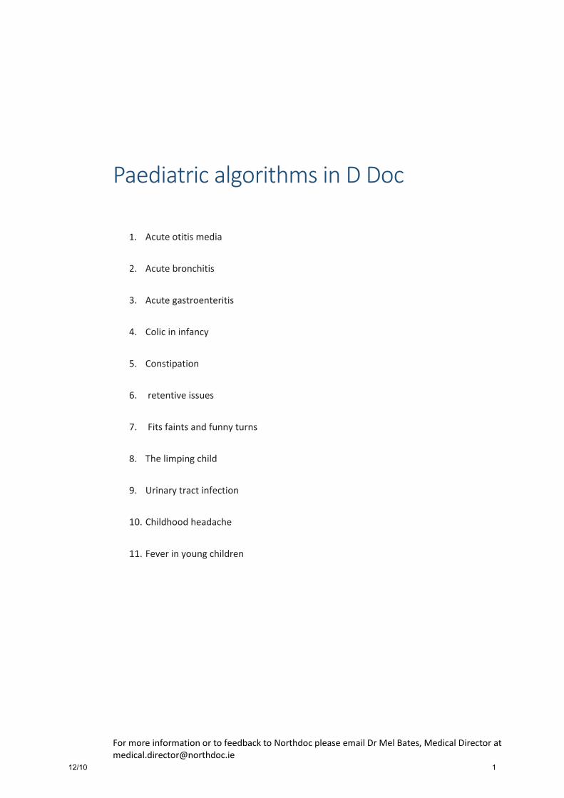
Acute Otitis Media
HISTORY Fever Ear pain/pulling ear Otorrhoea Lethargy/irritability/sleep
disturbance Protracted & severe crying
REFERRAL More than 4 episodes of
AOM in 6 months Complications of AOM Persistent serous otitis
media in children over 3 years of age with speech & language, developmental or behavioural problems
EXAMINATION Examine child in parent’s
arms Use a well-maintained
auroscope Visualize the eardrum
INVESTIGATIONS Acute otitis media is a clinical
diagnosis and no investigations are required
Tympanocentesis is not routinely performed
TREATMENT Do not prescribe antibiotics
as initial treatment Provide delayed antibiotic
prescription and advise treatment if no improvement after 3 days
Amoxicillin ± clavulanic acid Pain relief very important No role for decongestants
TAKE HOME MESSAGES Very common condition Often over-diagnosed Pain relief vital Delay antibiotics unless
discharging ear Refer to ENT if > 4 episodes
AOM in 6 months or if serous otitis media over 3 years old with hearing loss
BACKGROUND Acute otitis media (AOM) is
inflammation of the middle ear cavity with fluid collection (effusion) or discharge (otorrhoea)
Most cases are viral in origin and will resolve spontaneously in 10-14 days with adequate analgesia
RISK FACTORS Attending crèche First AOM before 6 months Not breast fed Passive smoking Food allergies Cleft palate Down syndrome Recurrent URTIs
PATHOGENESIS Preceding viral infection
Eustachian tube swelling Bacterial colonization (Strep/Staph/Moraxella)
COMPLICATIONS Perforation & discharge Conductive hearing loss Acute suppurative
labyrinthitis Facial nerve palsies Acute mastoiditis Intracranial spread of
infection Venous sinus
thrombosis Meningitis Subdural or extradural
abscess
SEROUS OTITIS MEDIA Also known as “glue ear” Many children will have
glue ear for up to 3 months following AOM
Prolonged course in Down syndrome and cleft palate
May lead to conductive hearing loss
Some children will require grommets
ACUTELY DRAINING EAR Usually indicates
perforated tympanic membrane
Need adequate pain relief Oral antibiotics Re-evaluate after 48-72
hours Watch for mastoiditis,
Bell’s palsy, intracranial extension
REFERENCES SIGN Guideline No. 66 Feb
2003: Diagnosis and management of childhood otitis media in primary care
12/10 2
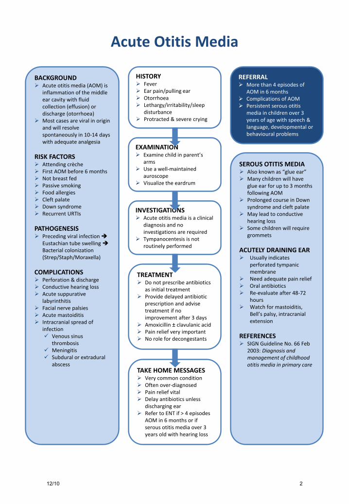
Acute Bronchiolitis
HISTORY Cough Breathing difficulty Audible wheeze Running nose Fever Poor feeding Apnoea (in very young)
EXAMINATION Respiratory rate Use of accessory muscles of
respiration Audible wheeze Pallor Head bobbing Apnoeic spells
INVESTIGATIONS Pulse oximetry NPA for RSV CXR only if severe
TREATMENT Maintain hydration – may
need NG feeds Oxygen via nasal cannulae if
oxygen saturation ≤92% or signs of respiratory distress
Hypertonic saline NO role for antibiotics,
steroids or inhalers
TAKE HOME MESSAGES Very common illness Treatment is supportive Most children have mild
disease that can be managed at home with primary care support
Wheeze may persist for 4 weeks post-illness
RSV is highly infectious, precautions must be taken to prevent spread
REFERRAL Poor feeding (<50% of usual
fluid intake in 24 hours) Lethargy Apnoea Respiratory rate >70/min Nasal flaring or grunting Severe chest wall recession Cyanosis Oxygen saturation ≤94% Uncertainty regarding
diagnosis N.B. Lower threshold for referral in children with significant comorbidities, age less than 3 months or born before 35 weeks gestation.
PROPHYLAXIS Pavalizumab may be
considered for use in infants <12 months old with: Extreme prematurity Acyanotic congenital
heart disease Congenital or acquired
significant orphan lung disease
Immune deficiency
EVIDENCE BASE The following treatments
are not recommended for use in acute bronchiolitis (A): Nebulised ribavirin Inhaled
bronchodilators Nebulised epinephrine Inhaled/oral
corticosteroids Chest physiotherapy
BACKGROUND Acute bronchiolitis a
clinically diagnosed respiratory condition that most commonly affects infants aged 3-6 months
RSV is the causative organism in 75% of cases
Seasonal: peak prevalence in November to March
Incubation period is 2-8 days
Children may develop a post-bronchiolitis wheeze
PATTERN OF ILLNESS Prior coryza for 2-3 days Severity peaks at 72 hours If fever >39oC, look for
other causes before diagnosing bronchiolitis
RISK FACTORS FOR SEVERE DISEASE Age Prematurity <35 weeks Congenital heart disease Chronic lung disease of
prematurity Immunodeficiency Down syndrome Severe hypotonia Parental smoking Breast feeding reduces risk
REFERENCES SIGN Guideline 91
November 2006: Bronchiolitis in children
12/10 3
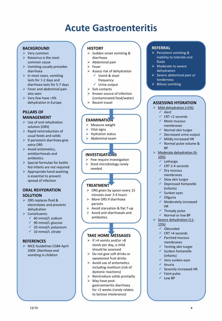
Acute Gastroenteritis
HISTORY Sudden onset vomiting &
diarrhoea Abdominal pain Fever Assess risk of dehydration Vomit & stool
frequency Urine output
Sick contacts Known source of infection
(contaminated food/water) Recent travel
REFERRAL Persistent vomiting &
inability to tolerate oral fluids
Moderate to severe dehydration
Severe abdominal pain or tenderness
Bilious vomiting
BACKGROUND Very common Rotavirus is the most
common cause Vomiting usually precedes
diarrhoea In most cases, vomiting
lasts for 1-2 days and diarrhoea lasts for 5-7 days
Fever and abdominal pain also seen
Very few have >3% dehydration in Europe
PILLARS OF MANAGEMENT Use of oral rehydration
solution (ORS) Rapid reintroduction of
usual feeds and solids If persistent diarrhoea give
extra ORS Avoid antiemetics,
antidiarrhoeals and antibiotics
Special formulae for bottle fed infants are not required
Appropriate hand-washing is essential to prevent spread of infection
ORAL REHYDRATION SOLUTION ORS replaces fluid &
electrolytes and prevents dehydration
Constituents: 60 mmol/L sodium 90 mmol/L glucose 20 mmol/L potassium 10 mmol/L citrate
REFERENCES NICE Guidelines CG84 April
2009: Diarrhoea and vomiting in children
EXAMINATION Measure weight Vital signs Hydration status Abdominal exam
INVESTIGATIONS Few require investigation Stool microbiology rarely
needed
TREATMENT ORS given by spoon every 15
minutes over 3-4 hours More ORS if diarrhoea
persists Avoid starvation & flat 7-up Avoid anti-diarrhoeals and
antibiotics
TAKE HOME MESSAGES If >4 vomits and/or >8
stools per day, a child should be assessed
Do not give soft drinks or sweetened fruit drinks
Avoid use of antiemetics including motilium (risk of dystonic reactions)
Reintroduce solids promptly May have post-
gastroenteritis diarrhoea for >2 weeks (rarely relates to lactose intolerance)
ASSESSING HYDRATION Mild dehydration (<5%) Alert CRT <2 seconds Moist mucous
membranes Normal skin turgor Decreased urine output Mildly increased HR Normal pulse volume &
BP Moderate dehydration (6-
10%) Lethargic CRT 2-4 seconds Dry mucous
membranes Slow skin turgor Depressed fontanelle
(infants) Sunken eyes Oliguria Moderately increased
HR Thready pulse Normal or low BP
Severe dehydration (11-15%) Obtunded CRT >4 seconds Parched mucous
membranes Tenting skin turgor Sunken fontanelle
(infants) Very sunken eyes Anuria Severely increased HR Faint pulse Low BP
12/10 4
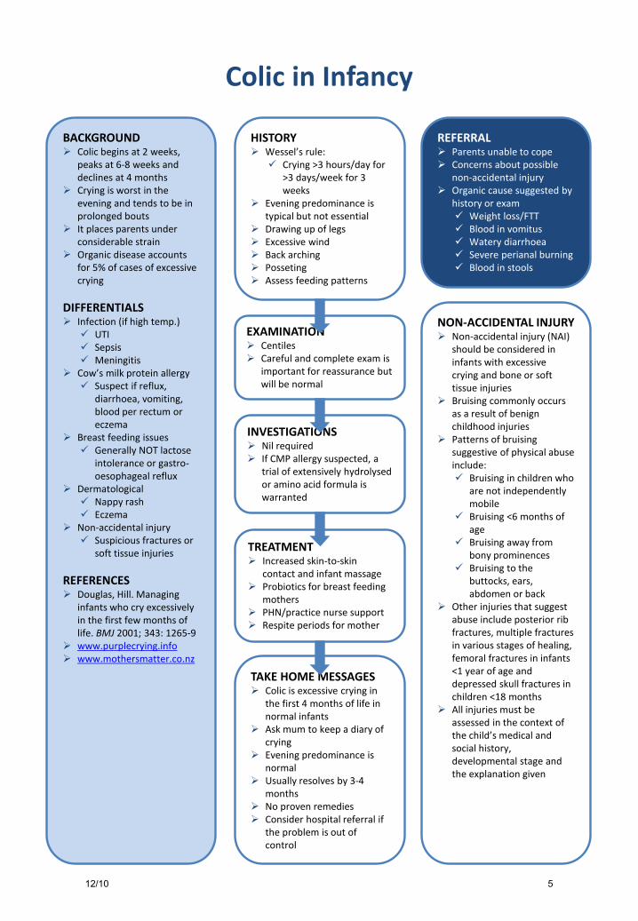
Colic in Infancy
HISTORY Wessel’s rule: Crying >3 hours/day for
>3 days/week for 3 weeks
Evening predominance is typical but not essential
Drawing up of legs Excessive wind Back arching Posseting Assess feeding patterns
EXAMINATION Centiles Careful and complete exam is
important for reassurance but will be normal
INVESTIGATIONS Nil required If CMP allergy suspected, a
trial of extensively hydrolysed or amino acid formula is warranted
BACKGROUND Colic begins at 2 weeks,
peaks at 6-8 weeks and declines at 4 months
Crying is worst in the evening and tends to be in prolonged bouts
It places parents under considerable strain
Organic disease accounts for 5% of cases of excessive crying
DIFFERENTIALS Infection (if high temp.) UTI Sepsis Meningitis
Cow’s milk protein allergy Suspect if reflux,
diarrhoea, vomiting, blood per rectum or eczema
Breast feeding issues Generally NOT lactose
intolerance or gastro-oesophageal reflux
Dermatological Nappy rash Eczema
Non-accidental injury Suspicious fractures or
soft tissue injuries
REFERENCES Douglas, Hill. Managing
infants who cry excessively in the first few months of life. BMJ 2001; 343: 1265-9
www.purplecrying.info www.mothersmatter.co.nz
TREATMENT Increased skin-to-skin
contact and infant massage Probiotics for breast feeding
mothers PHN/practice nurse support Respite periods for mother
TAKE HOME MESSAGES Colic is excessive crying in
the first 4 months of life in normal infants
Ask mum to keep a diary of crying
Evening predominance is normal
Usually resolves by 3-4 months
No proven remedies Consider hospital referral if
the problem is out of control
REFERRAL Parents unable to cope Concerns about possible
non-accidental injury Organic cause suggested by
history or exam Weight loss/FTT Blood in vomitus Watery diarrhoea Severe perianal burning Blood in stools
NON-ACCIDENTAL INJURY Non-accidental injury (NAI)
should be considered in infants with excessive crying and bone or soft tissue injuries
Bruising commonly occurs as a result of benign childhood injuries
Patterns of bruising suggestive of physical abuse include: Bruising in children who
are not independently mobile
Bruising <6 months of age
Bruising away from bony prominences
Bruising to the buttocks, ears, abdomen or back
Other injuries that suggest abuse include posterior rib fractures, multiple fractures in various stages of healing, femoral fractures in infants <1 year of age and depressed skull fractures in children <18 months
All injuries must be assessed in the context of the child’s medical and social history, developmental stage and the explanation given
12/10 5
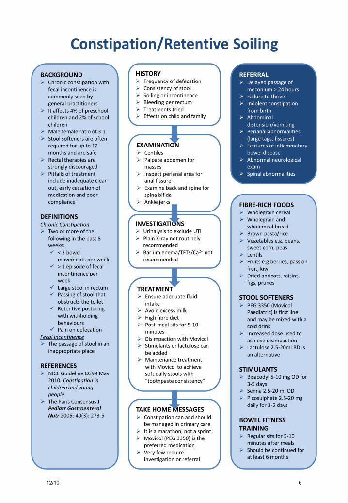
Constipation/Retentive Soiling
HISTORY Frequency of defecation Consistency of stool Soiling or incontinence Bleeding per rectum Treatments tried Effects on child and family
EXAMINATION Centiles Palpate abdomen for
masses Inspect perianal area for
anal fissure Examine back and spine for
spina bifida Ankle jerks
INVESTIGATIONS Urinalysis to exclude UTI Plain X-ray not routinely
recommended Barium enema/TFTs/Ca2+ not
recommended
TREATMENT Ensure adequate fluid
intake Avoid excess milk High fibre diet Post-meal sits for 5-10
minutes Disimpaction with Movicol Stimulants or lactulose can
be added Maintenance treatment
with Movicol to achieve soft daily stools with “toothpaste consistency”
TAKE HOME MESSAGES Constipation can and should
be managed in primary care It is a marathon, not a sprint Movicol (PEG 3350) is the
preferred medication Very few require
investigation or referral
REFERRAL Delayed passage of
meconium > 24 hours Failure to thrive Indolent constipation
from birth Abdominal
distension/vomiting Perianal abnormalities
(large tags, fissures) Features of inflammatory
bowel disease Abnormal neurological
exam Spinal abnormalities
BACKGROUND Chronic constipation with
fecal incontinence is commonly seen by general practitioners
It affects 4% of preschool children and 2% of school children
Male:female ratio of 3:1 Stool softeners are often
required for up to 12 months and are safe
Rectal therapies are strongly discouraged
Pitfalls of treatment include inadequate clear out, early cessation of medication and poor compliance
DEFINITIONS Chronic Constipation Two or more of the
following in the past 8 weeks: < 3 bowel
movements per week > 1 episode of fecal
incontinence per week
Large stool in rectum Passing of stool that
obstructs the toilet Retentive posturing
with withholding behaviours
Pain on defecation Fecal Incontinence The passage of stool in an
inappropriate place
REFERENCES NICE Guideline CG99 May
2010: Constipation in children and young people
The Paris Consensus J Pediatr Gastroenterol Nutr 2005; 40(3): 273-5
FIBRE-RICH FOODS Wholegrain cereal Wholegrain and
wholemeal bread Brown pasta/rice Vegetables e.g. beans,
sweet corn, peas Lentils Fruits e.g berries, passion
fruit, kiwi Dried apricots, raisins,
figs, prunes
STOOL SOFTENERS PEG 3350 (Movicol
Paediatric) is first line and may be mixed with a cold drink
Increased dose used to achieve disimpaction
Lactulose 2.5-20ml BD is an alternative
STIMULANTS Bisacodyl 5-10 mg OD for
3-5 days Senna 2.5-20 ml OD Picosulphate 2.5-20 mg
daily for 3-5 days
BOWEL FITNESS TRAINING Regular sits for 5-10
minutes after meals Should be continued for
at least 6 months
12/10 6
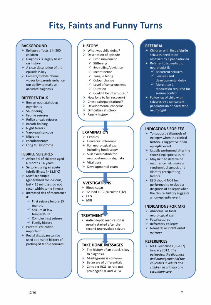
Fits, Faints and Funny Turns
HISTORY What was child doing? Description of episode Limb movement Stiffening Eye rolling/deviation Incontinence Tongue biting Colour change Level of consciousness Duration Could it be interrupted?
How long to full recovery? Chest pain/palpitations? Developmental concerns Difficulties at school Family history
EXAMINATION Centiles Head circumference Full neurological exam
including fundoscopy Skin examination for
neurocutaneous stigmata Vital signs Developmental exam
INVESTIGATIONS Blood sugar 12-lead ECG (calculate QTc) EEG MRI
TREATMENT Antiepileptic medication is
usually started after the second unprovoked seizure
TAKE HOME MESSAGES The history of an attack is key
to diagnosis Misdiagnosis is common Be aware of differentials Consider ECG to rule out
prolonged QT and WPW
BACKGROUND Epilepsy affects 1 in 200
children Diagnosis is largely based
on history A clear description of the
episode is key Camera/mobile phone
videos by parents enhance our ability to make an accurate diagnosis
DIFFERENTIALS Benign neonatal sleep
myoclonus Shuddering Febrile seizures Reflex anoxic seizures Breath-holding Night terrors Vasovagal syncope Migraine Pseudoseizures Long QT syndrome
FEBRILE SEIZURES Affect 3% of children aged
6 months – 6 years Seizure during an acute
febrile illness (> 38.5°C) Most are simple
(generalised tonic-clonic, last < 15 minutes, do not recur within same illness)
Increased risk of recurrence if: First seizure before 15
months Seizure at low
temperature Complex first seizure Family history
Parental education important
Rectal diazepam can be used at onset if history of prolonged febrile seizures
INDICATIONS FOR EEG To support a diagnosis of
epilepsy when the clinical history is suggestive of an epileptic event
Usually performed after the second epileptic seizure
May help to determine recurrence risk, make a syndromic diagnosis and identify precipitating factors
EEG should NOT be performed to exclude a diagnosis of epilepsy when the clinical history suggests a non-epileptic event
INDICATIONS FOR MRI Abnormal or focal
neurological exam Focal seizures Refractory epilepsy Neonatal or infant onset
epilepsy
REFERENCES NICE Guidelines (CG137)
January 2012: The epilepsies: the diagnosis and management of the epilepsies in adults and children in primary and secondary care
REFERRAL Children with first afebrile
seizures need to be assessed by a paediatrician
Referral to a paediatric neurologist if: Recurrent seizures Seizures and
developmental delay More than 1
medication required for seizure control
Follow up of child with seizures by a consultant paediatrician or paediatric neurologist
12/10 7
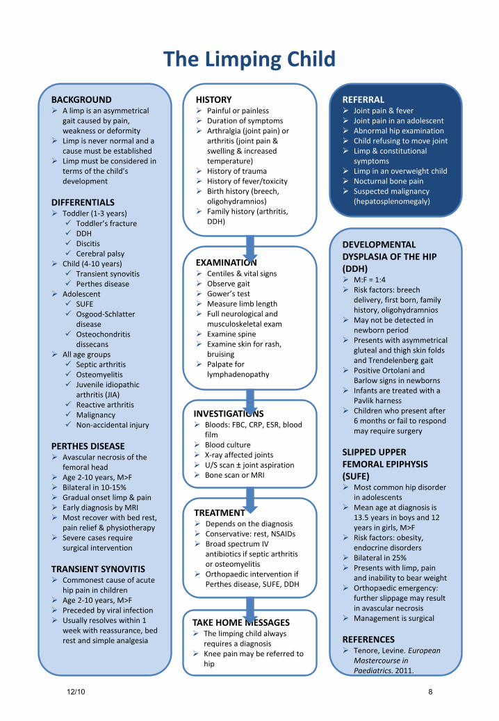
The Limping Child
HISTORY Painful or painless Duration of symptoms Arthralgia (joint pain) or
arthritis (joint pain & swelling & increased temperature)
History of trauma History of fever/toxicity Birth history (breech,
oligohydramnios) Family history (arthritis,
DDH)
EXAMINATION Centiles & vital signs Observe gait Gower’s test Measure limb length Full neurological and
musculoskeletal exam Examine spine Examine skin for rash,
bruising Palpate for
lymphadenopathy
INVESTIGATIONS Bloods: FBC, CRP, ESR, blood
film Blood culture X-ray affected joints U/S scan ± joint aspiration Bone scan or MRI
TREATMENT Depends on the diagnosis Conservative: rest, NSAIDs Broad spectrum IV
antibiotics if septic arthritis or osteomyelitis
Orthopaedic intervention if Perthes disease, SUFE, DDH
TAKE HOME MESSAGES The limping child always
requires a diagnosis Knee pain may be referred to
hip
REFERRAL Joint pain & fever Joint pain in an adolescent Abnormal hip examination Child refusing to move joint Limp & constitutional
symptoms Limp in an overweight child Nocturnal bone pain Suspected malignancy
(hepatosplenomegaly)
DEVELOPMENTAL DYSPLASIA OF THE HIP (DDH) M:F = 1:4 Risk factors: breech
delivery, first born, family history, oligohydramnios
May not be detected in newborn period
Presents with asymmetrical gluteal and thigh skin folds and Trendelenberg gait
Positive Ortolani and Barlow signs in newborns
Infants are treated with a Pavlik harness
Children who present after 6 months or fail to respond may require surgery
SLIPPED UPPER FEMORAL EPIPHYSIS (SUFE) Most common hip disorder
in adolescents Mean age at diagnosis is
13.5 years in boys and 12 years in girls, M>F
Risk factors: obesity, endocrine disorders
Bilateral in 25% Presents with limp, pain
and inability to bear weight Orthopaedic emergency:
further slippage may result in avascular necrosis
Management is surgical
REFERENCES Tenore, Levine. European
Mastercourse in Paediatrics. 2011.
BACKGROUND A limp is an asymmetrical
gait caused by pain, weakness or deformity
Limp is never normal and a cause must be established
Limp must be considered in terms of the child’s development
DIFFERENTIALS Toddler (1-3 years) Toddler’s fracture DDH Discitis Cerebral palsy
Child (4-10 years) Transient synovitis Perthes disease
Adolescent SUFE Osgood-Schlatter
disease Osteochondritis
dissecans All age groups Septic arthritis Osteomyelitis Juvenile idiopathic
arthritis (JIA) Reactive arthritis Malignancy Non-accidental injury
PERTHES DISEASE Avascular necrosis of the
femoral head Age 2-10 years, M>F Bilateral in 10-15% Gradual onset limp & pain Early diagnosis by MRI Most recover with bed rest,
pain relief & physiotherapy Severe cases require
surgical intervention
TRANSIENT SYNOVITIS Commonest cause of acute
hip pain in children Age 2-10 years, M>F Preceded by viral infection Usually resolves within 1
week with reassurance, bed rest and simple analgesia
12/10 8
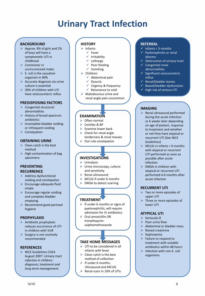
Urinary Tract Infection
HISTORY Infants: Fever Irritability Lethargy Poor feeding Vomiting
Children: Abdominal pain Dysuria Urgency & frequency Reluctance to void
Malodourous urine and renal angle pain uncommon
EXAMINATION Often normal Centiles & BP Examine lower back Check for renal angle
tenderness & renal masses Out rule constipation
INVESTIGATIONS Urinalysis Urine microscopy, culture
and sensitivity Renal ultrasound MCUG if under 6 months DMSA to detect scarring
TREATMENT If under 6 months or signs of
pyelonephritis, will require admission for IV antibiotics
Oral amoxicillin OR trimethoprim-sulphamethoxazole
TAKE HOME MESSAGES UTI to be considered in all
infants with fever Clean catch is the best
method of collection If under 6 months:
Ultrasound and MCUG Renal scars in 10% of UTIs
BACKGROUND Approx. 8% of girls and 2%
of boys will have a symptomatic UTI in childhood
Commoner in uncircumcised males
E. coli is the causative organism in 90%
Accurate diagnosis via urine culture is essential
30% of children with UTI have vesicoureteric reflux
PREDISPOSING FACTORS Congenital structural
abnormalities History of broad spectrum
antibiotics Incomplete bladder voiding
or infrequent voiding Constipation
OBTAINING URINE Clean catch is the best
method High contamination of bag
specimens
PREVENTING RECURRENCE Address dysfunctional
voiding and constipation Encourage adequate fluid
intake Encourage regular voiding
and complete bladder emptying
Recommend good perineal hygiene
PROPHYLAXIS Antibiotic prophylaxis
reduces occurrence of UTI in children with VUR
Surgery is not routinely recommended
REFERENCES NICE Guidelines CG54
August 2007: Urinary tract infection in children: diagnosis, treatment and long-term management.
REFERRAL Infants < 3 months Pyelonephritis or renal
abscess Obstruction of urinary tract Congenital renal
abnormalities Significant vesicoureteric
reflux Renal/bladder stones Bowel/bladder dysfunction High risk of serious UTI
IMAGING Renal ultrasound performed
during the acute infection or 6 weeks later depending on age of patient, response to treatment and whether or not they have atypical or recurrent UTI (See NICE Guidelines)
MCUG in infants < 6 months with atypical or recurrent UTI performed as soon as possible after acute infection
DMSA in children with atypical or recurrent UTI performed 4-6 months after acute infection
RECURRENT UTI Two or more episodes of
upper UTI Three or more episodes of
lower UTI
ATYPICAL UTI Seriously ill Poor urine flow Abdominal or bladder mass Raised creatinine Septicaemia Failure to respond to
treatment with suitable antibiotics within 48 hours
Infection with non-E. coli organisms
12/10 9
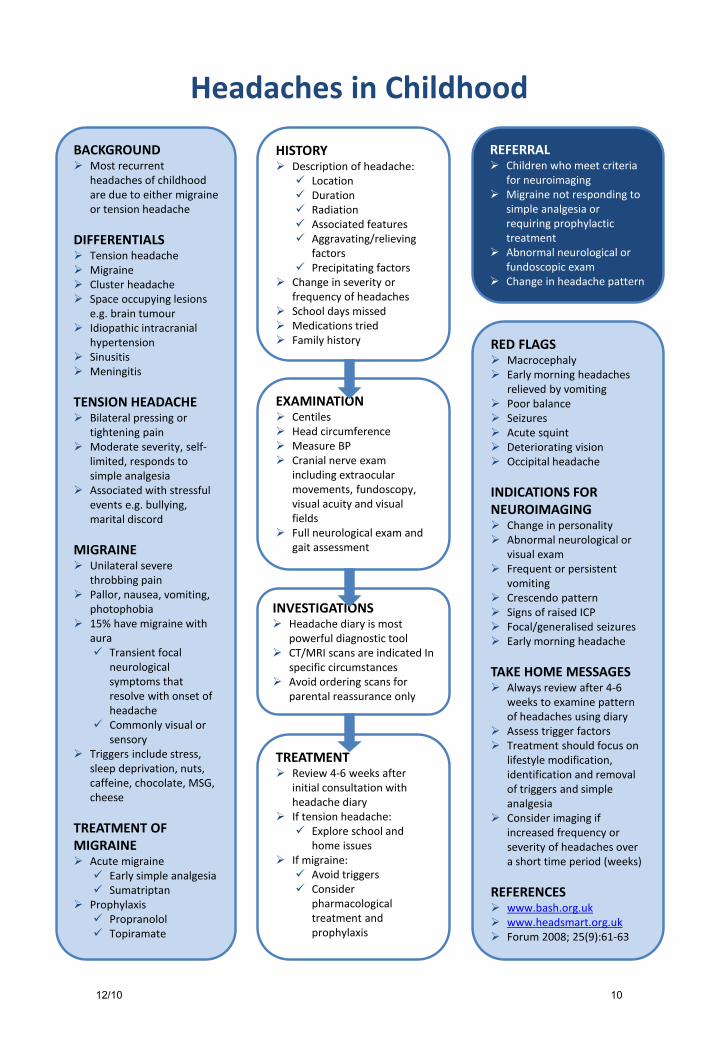
Headaches in Childhood
HISTORY Description of headache: Location Duration Radiation Associated features Aggravating/relieving
factors Precipitating factors
Change in severity or frequency of headaches
School days missed Medications tried Family history
INVESTIGATIONS Headache diary is most
powerful diagnostic tool CT/MRI scans are indicated In
specific circumstances Avoid ordering scans for
parental reassurance only
REFERRAL Children who meet criteria
for neuroimaging Migraine not responding to
simple analgesia or requiring prophylactic treatment
Abnormal neurological or fundoscopic exam
Change in headache pattern
EXAMINATION Centiles Head circumference Measure BP Cranial nerve exam
including extraocular movements, fundoscopy, visual acuity and visual fields
Full neurological exam and gait assessment
TREATMENT Review 4-6 weeks after
initial consultation with headache diary
If tension headache: Explore school and
home issues If migraine: Avoid triggers Consider
pharmacological treatment and prophylaxis
BACKGROUND Most recurrent
headaches of childhood are due to either migraine or tension headache
DIFFERENTIALS Tension headache Migraine Cluster headache Space occupying lesions
e.g. brain tumour Idiopathic intracranial
hypertension Sinusitis Meningitis
TENSION HEADACHE Bilateral pressing or
tightening pain Moderate severity, self-
limited, responds to simple analgesia
Associated with stressful events e.g. bullying, marital discord
MIGRAINE Unilateral severe
throbbing pain Pallor, nausea, vomiting,
photophobia 15% have migraine with
aura Transient focal
neurological symptoms that resolve with onset of headache
Commonly visual or sensory
Triggers include stress, sleep deprivation, nuts, caffeine, chocolate, MSG, cheese
TREATMENT OF MIGRAINE Acute migraine Early simple analgesia Sumatriptan
Prophylaxis Propranolol Topiramate
RED FLAGS Macrocephaly Early morning headaches
relieved by vomiting Poor balance Seizures Acute squint Deteriorating vision Occipital headache
INDICATIONS FOR NEUROIMAGING Change in personality Abnormal neurological or
visual exam Frequent or persistent
vomiting Crescendo pattern Signs of raised ICP Focal/generalised seizures Early morning headache
TAKE HOME MESSAGES Always review after 4-6
weeks to examine pattern of headaches using diary
Assess trigger factors Treatment should focus on
lifestyle modification, identification and removal of triggers and simple analgesia
Consider imaging if increased frequency or severity of headaches over a short time period (weeks)
REFERENCES www.bash.org.uk www.headsmart.org.uk Forum 2008; 25(9):61-63
12/10 10
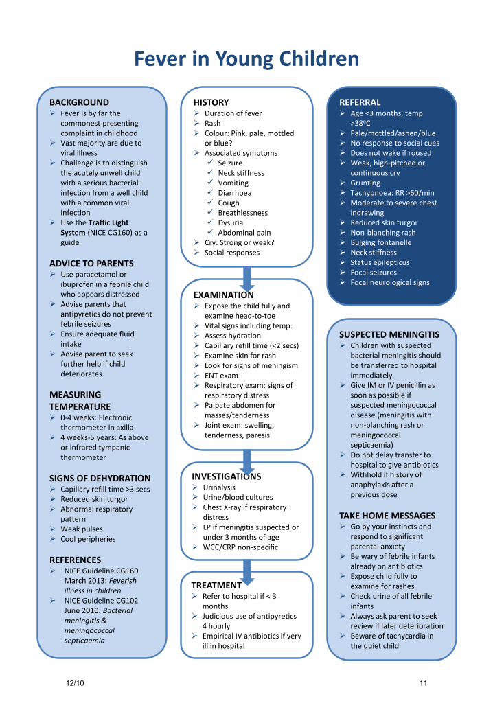
Fever in Young Children
HISTORY Duration of fever Rash Colour: Pink, pale, mottled
or blue? Associated symptoms Seizure Neck stiffness Vomiting Diarrhoea Cough Breathlessness Dysuria Abdominal pain
Cry: Strong or weak? Social responses
EXAMINATION Expose the child fully and
examine head-to-toe Vital signs including temp. Assess hydration Capillary refill time (<2 secs) Examine skin for rash Look for signs of meningism ENT exam Respiratory exam: signs of
respiratory distress Palpate abdomen for
masses/tenderness Joint exam: swelling,
tenderness, paresis
INVESTIGATIONS Urinalysis Urine/blood cultures Chest X-ray if respiratory
distress LP if meningitis suspected or
under 3 months of age WCC/CRP non-specific
TREATMENT Refer to hospital if < 3
months Judicious use of antipyretics
4 hourly Empirical IV antibiotics if very
ill in hospital
REFERRAL Age <3 months, temp
>38oC Pale/mottled/ashen/blue No response to social cues Does not wake if roused Weak, high-pitched or
continuous cry Grunting Tachypnoea: RR >60/min Moderate to severe chest
indrawing Reduced skin turgor Non-blanching rash Bulging fontanelle Neck stiffness Status epilepticus Focal seizures Focal neurological signs
SUSPECTED MENINGITIS Children with suspected
bacterial meningitis should be transferred to hospital immediately
Give IM or IV penicillin as soon as possible if suspected meningococcal disease (meningitis with non-blanching rash or meningococcal septicaemia)
Do not delay transfer to hospital to give antibiotics
Withhold if history of anaphylaxis after a previous dose
TAKE HOME MESSAGES Go by your instincts and
respond to significant parental anxiety
Be wary of febrile infants already on antibiotics
Expose child fully to examine for rashes
Check urine of all febrile infants
Always ask parent to seek review if later deterioration
Beware of tachycardia in the quiet child
BACKGROUND Fever is by far the
commonest presenting complaint in childhood
Vast majority are due to viral illness
Challenge is to distinguish the acutely unwell child with a serious bacterial infection from a well child with a common viral infection
Use the Traffic Light System (NICE CG160) as a guide
ADVICE TO PARENTS Use paracetamol or
ibuprofen in a febrile child who appears distressed
Advise parents that antipyretics do not prevent febrile seizures
Ensure adequate fluid intake
Advise parent to seek further help if child deteriorates
MEASURING TEMPERATURE 0-4 weeks: Electronic
thermometer in axilla 4 weeks-5 years: As above
or infrared tympanic thermometer
SIGNS OF DEHYDRATION Capillary refill time >3 secs Reduced skin turgor Abnormal respiratory
pattern Weak pulses Cool peripheries
REFERENCES NICE Guideline CG160
March 2013: Feverish illness in children
NICE Guideline CG102 June 2010: Bacterial meningitis & meningococcal septicaemia
12/10 11