Embed Size (px)
Citation preview
PACS - Picture Archiving and Communication System
Maximilian Hecht∗
(#0827459)Vienna University of Technology
University of Paderborn
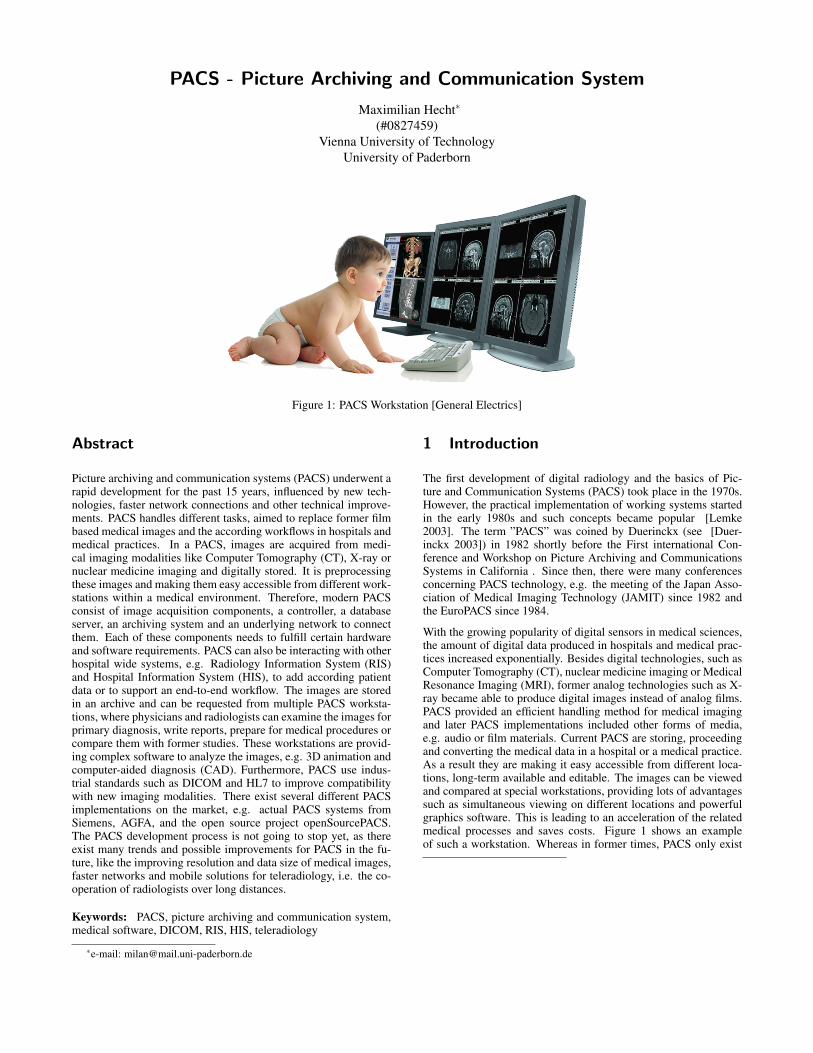
Figure 1: PACS Workstation [General Electrics]
Abstract
Picture archiving and communication systems (PACS) underwent arapid development for the past 15 years, influenced by new tech-nologies, faster network connections and other technical improve-ments. PACS handles different tasks, aimed to replace former filmbased medical images and the according workflows in hospitals andmedical practices. In a PACS, images are acquired from medi-cal imaging modalities like Computer Tomography (CT), X-ray ornuclear medicine imaging and digitally stored. It is preprocessingthese images and making them easy accessible from different work-stations within a medical environment. Therefore, modern PACSconsist of image acquisition components, a controller, a databaseserver, an archiving system and an underlying network to connectthem. Each of these components needs to fulfill certain hardwareand software requirements. PACS can also be interacting with otherhospital wide systems, e.g. Radiology Information System (RIS)and Hospital Information System (HIS), to add according patientdata or to support an end-to-end workflow. The images are storedin an archive and can be requested from multiple PACS worksta-tions, where physicians and radiologists can examine the images forprimary diagnosis, write reports, prepare for medical procedures orcompare them with former studies. These workstations are provid-ing complex software to analyze the images, e.g. 3D animation andcomputer-aided diagnosis (CAD). Furthermore, PACS use indus-trial standards such as DICOM and HL7 to improve compatibilitywith new imaging modalities. There exist several different PACSimplementations on the market, e.g. actual PACS systems fromSiemens, AGFA, and the open source project openSourcePACS.The PACS development process is not going to stop yet, as thereexist many trends and possible improvements for PACS in the fu-ture, like the improving resolution and data size of medical images,faster networks and mobile solutions for teleradiology, i.e. the co-operation of radiologists over long distances.
Keywords: PACS, picture archiving and communication system,medical software, DICOM, RIS, HIS, teleradiology
∗e-mail: [email protected]
1 Introduction
The first development of digital radiology and the basics of Pic-ture and Communication Systems (PACS) took place in the 1970s.However, the practical implementation of working systems startedin the early 1980s and such concepts became popular [Lemke2003]. The term ”PACS” was coined by Duerinckx (see [Duer-inckx 2003]) in 1982 shortly before the First international Con-ference and Workshop on Picture Archiving and CommunicationsSystems in California . Since then, there were many conferencesconcerning PACS technology, e.g. the meeting of the Japan Asso-ciation of Medical Imaging Technology (JAMIT) since 1982 andthe EuroPACS since 1984.
With the growing popularity of digital sensors in medical sciences,the amount of digital data produced in hospitals and medical prac-tices increased exponentially. Besides digital technologies, such asComputer Tomography (CT), nuclear medicine imaging or MedicalResonance Imaging (MRI), former analog technologies such as X-ray became able to produce digital images instead of analog films.PACS provided an efficient handling method for medical imagingand later PACS implementations included other forms of media,e.g. audio or film materials. Current PACS are storing, proceedingand converting the medical data in a hospital or a medical practice.As a result they are making it easy accessible from different loca-tions, long-term available and editable. The images can be viewedand compared at special workstations, providing lots of advantagessuch as simultaneous viewing on different locations and powerfulgraphics software. This is leading to an acceleration of the relatedmedical processes and saves costs. Figure 1 shows an exampleof such a workstation. Whereas in former times, PACS only exist
in major hospitals, today the decreasing costs for technical equip-ments and software are forwarding the installation of PACS in smallclinics as well. Faster wide area connections (WAN) and wirelesscommunications are channeling PACS research into the directionof teleradiology, i.e. the cooperation of medical institutes with ra-diologists over long distance, e.g. through mobile devices and rapiddata transfers.
Due to many different PACS products from varied vendors, manyPACS projects from the Universities or open source communitiesand plenty of scientific papers with sometimes diverging opinions,it is not easy to get a quick idea of the subject of PACS. This pa-per answers to this problem by giving a review on the current stateof the art of PACS. Therefore in the following sections, a generaldescription of the typical components, standards and technologiesof a PACS are presented. This includes related systems and anintroduction of the DICOM and the HL7 industrial standard. Toprovide a more practical view on PACS systems, the actual PACSsystems from Siemens, AGFA and the open source project open-SourcePACS are briefly introduced, outlining some practical pos-sibilities. Furthermore the current advantages and disadvantagesof a PACS implementation in a modern hospital are discussed in aconclusion. The scope of this discussion is to outline the existingeffects on the radiologic workflow and the cost benefits. Finallytrends and possible improvements of PACS in the future are deter-mined in the conclusion, like the improving resolution and data sizeof medical images and mobile solutions for telemedical access.
2 Components and architecture of PACS
Although there are many PACS solutions from different vendors,the basic components, standards and corresponding systems of aPACS are very similar. Hurlen et al. [Hurlen et al. 2008] definedthe properties of a PACS, as a system that typically acquire, store,transmit, display, and process digital images.
On the basis of this definition the components can be separated.For acquiring and preprocessing of the images an image acquisitioncomponent is needed. To store the image a database component ex-ists, controlled by the PACS controller, which is the central control-ling component in a PACS. Finally, to display the digital images, aviewing component is required, referred to as workstation. In thefollowing these components are specified in more details, regardingto the models given in [Huang 1996] and [Heitmann 2006].
2.1 Image acquisition component
The images of a PACS are produced by several radiologic imagingmodalities. While the images of CT, ultrasound, MRI and nuclearmedicine imaging (PET/SPECT) are digitally captured, the imagesof X-ray scanners have to be digitalized first. The images can betransmitted from the modalities using a specified interface. A DI-COM interface is the most frequently used standard at this point.[Heitmann 2006]
Due to the fact that many imaging equipments are not supporting in-dustrial standards, like the DICOM standard, acquisition computers(also called acquisition gateways) are needed to enable the digitalexchange of the images. Therefore a computer is placed betweenthe modalities and the PACS network. The computer receives thepicture from the imaging modality through its specified interface,preprocessing it and converting it to a standard, which is supportedby the PACS (i.e. the DICOM standard). For the image exchangebetween the modality and the acquisition gateway Huang [Huang
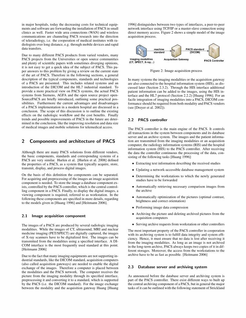
1996] distinguishes between two types of interfaces, a peer-to-peernetwork interface using TCP/IP or a master-slave connection usingdirect memory access. Figure 2 shows a simple model of the imageacquisition process.
Figure 2: Image acquisition process
In many systems the imaging modalities or the acquisition gatewayare also connected to the hospital information system (HIS), as dis-cussed later (Section 2.3.2). Through the HIS interface additionalpatient information can be added to the images, using the HIS in-terface and the HL7 protocol (Section 2.2.2) [Huang 1996]. For anfacile integration of imaging modalities into a PACS, DICOM con-formance should be required from both modality and PACS vendors(see [Dreyer et al. 2002]).
2.2 PACS controller
The PACS controller is the main engine of the PACS. It controlsall transactions in the system between components and its databaseserver and an archive system. The images and the patient informa-tion are transmitted from the imaging modalities or an acquisitioncomputer, the radiology information systems (RIS) and the hospitalinformation system (HIS) to the PACS controller. After receivingthe data the controller continuous the processing of the data, con-sisting of the following tasks [Huang 1996]:
• Extracting text information describing the received studies
• Updating a network-accessible database management system
• Determining the workstations to which the newly generatedstudies have to be forwarded
• Automatically retrieving necessary comparison images fromthe archive
• Automatically optimization of the pictures (optimal contrast,brightness and correct orientation)
• Performing image data compression
• Archiving the picture and deleting archived pictures from theacquisition computers
• Serving archive requests from workstation or other controllers
The most important property of the PACS controller in cooperationwith its archiving system is to fulfill data integrity and system effi-ciency. Hence, it must ensure that no data is lost after receiving itfrom the imaging modalities. As long as an image is not archivedin the long-term archive, PACS always keeps two copies of it in dif-ferent storages. Moreover, the access from the workstations to thearchive have to be as fast as possible. [Heitmann 2006]
2.3 Database server and archiving system
As announced before the database server and archiving system ispart of the PACS controller. There exist different ways to built upthe central archiving component of a PACS, but in general the majortasks of it can be outlined with the following statement of Strickland
[Strickland 2000]: ”The PACS database ensures that all images areautomatically grouped into the correct examination, are chronolog-ically ordered, correctly orientated and labeled, and can be easilyretrieved using a variety of criteria (for example, name, hospitalnumber, date, referring clinician, etc). All imaging studies of apatient are immediately available on the PACS which encouragesreview of examinations with preceding studies and intermodalitycomparisons”.
Therefore a PACS database server should consist of redundantdatabases with identical reliable commercial database software (e.g.Oracle, MySQL), supporting Structured Query Language (SQL).The systems should mirror the data in two database servers, to en-sure a stable data handling even in the case of system failure or diskcrashes. The PACS database system should be interfaced to the ra-diology information system (RIS, Section 2.7.2) and the HIS (seeSection 2.7.1), to allow gathering additional patient information.[Huang 1996]
The hardware of the database system should use an fast multiplecentral processing unit and performant interfaces, like SCSI (SmallComputer System Interface), S-ATA (Serial-Advanced TechnologyAttachments) and a fast network interface. With this configura-tion the system can support parallel processing and a simultaneoustransfer of images to different networks or network devices (see[Huang 1996]).
The storage components are normally separated in a fast short-termarchive and a slower, cheaper and bigger long-term archive.
2.3.1 Short-term archive
The short-term archive is used as the cache of the system. It is themost expensive storage in a PACS. Images from last recent stud-ies and examination are firstly stored there to provide a fast accessfrom viewing components. As said before the capacity of this cachehas been increasing in the last 20 years. For comparison: WhileHuang recommended at least 13.6 gigabyte RAID as a cache in1996 [Huang 1996], the PACS of the radiological institute of Mu-nich owns a 880 gigabyte cache pool (see [Wirth et al. 2005]). Thisis correlating with expanding data size of medical images throughhigher resolutions.
The short-term archive is realized with S-ATA or SCSI hard disksin a redundant array of inexpensive disks (RAID). Through the useof RAID transfer rates between 150 MB/s to 320 MB/s are possi-ble. The most frequently used RAID levels in a PACS are level 1and level 5. RAID level 1 mirrors the data on more than one disc,improving fault tolerance, while RAID level 5 is using the conceptof stripping and parity, to improve the transfer rates and to balanceworkload between discs. RAID level 1 and 5 can be combined.
The capacities of the short-term archive should at least be bigenough to keep all images produced within two weeks. However,by reason of decreasing costs for hard disks it is nowadays possibleto implement short-term archives with a better capacity, big enoughto store image data of two years. [Heitmann 2006]
For example: The 880 gigabyte cache pool of the radiological in-stitute of Munich is able to store the images from all examinationswithin the last 4-5 months (see [Wirth et al. 2005]).
2.3.2 Long-term archive
One of the most important tasks of a PACS is to ensure a properlong-term archival of image data. This is fulfilled in the long-termarchive, a specific archive with a high security level and a high fault
tolerance. A long-term archive need to guarantee a storage of theimages between 10-30 years, as regulated by law. By a PACS thisarchive is digitally realized and it replaces the former big archivingrooms and the human workers to sort it.
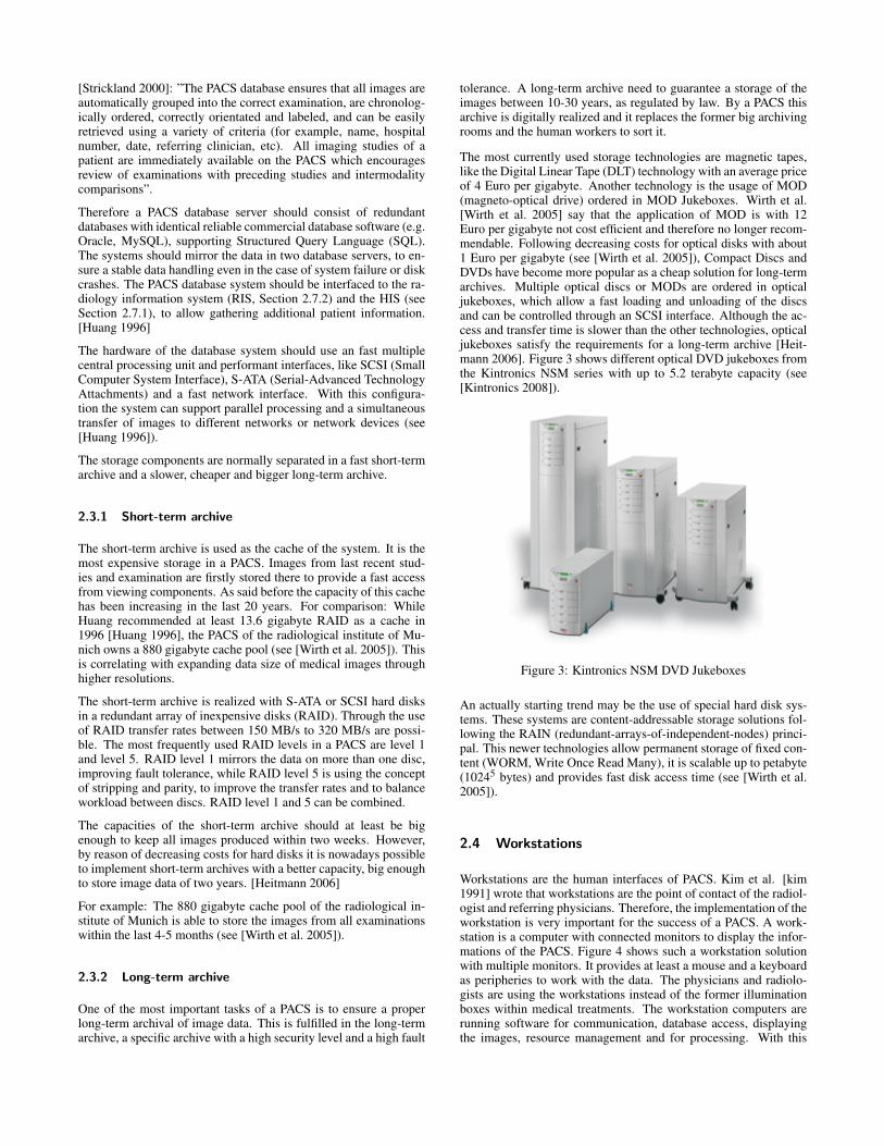
The most currently used storage technologies are magnetic tapes,like the Digital Linear Tape (DLT) technology with an average priceof 4 Euro per gigabyte. Another technology is the usage of MOD(magneto-optical drive) ordered in MOD Jukeboxes. Wirth et al.[Wirth et al. 2005] say that the application of MOD is with 12Euro per gigabyte not cost efficient and therefore no longer recom-mendable. Following decreasing costs for optical disks with about1 Euro per gigabyte (see [Wirth et al. 2005]), Compact Discs andDVDs have become more popular as a cheap solution for long-termarchives. Multiple optical discs or MODs are ordered in opticaljukeboxes, which allow a fast loading and unloading of the discsand can be controlled through an SCSI interface. Although the ac-cess and transfer time is slower than the other technologies, opticaljukeboxes satisfy the requirements for a long-term archive [Heit-mann 2006]. Figure 3 shows different optical DVD jukeboxes fromthe Kintronics NSM series with up to 5.2 terabyte capacity (see[Kintronics 2008]).
Figure 3: Kintronics NSM DVD Jukeboxes
An actually starting trend may be the use of special hard disk sys-tems. These systems are content-addressable storage solutions fol-lowing the RAIN (redundant-arrays-of-independent-nodes) princi-pal. This newer technologies allow permanent storage of fixed con-tent (WORM, Write Once Read Many), it is scalable up to petabyte(10245 bytes) and provides fast disk access time (see [Wirth et al.2005]).
2.4 Workstations
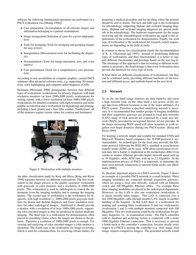
Workstations are the human interfaces of PACS. Kim et al. [kim1991] wrote that workstations are the point of contact of the radiol-ogist and referring physicians. Therefore, the implementation of theworkstation is very important for the success of a PACS. A work-station is a computer with connected monitors to display the infor-mations of the PACS. Figure 4 shows such a workstation solutionwith multiple monitors. It provides at least a mouse and a keyboardas peripheries to work with the data. The physicians and radiolo-gists are using the workstations instead of the former illuminationboxes within medical treatments. The workstation computers arerunning software for communication, database access, displayingthe images, resource management and for processing. With this
software the following fundamental operations are performed on aPACS workstation (see [Huang 1996]):
• Case preparation (Accumulation of all relevant images andinformation belonging to a patient examination)
• Image arrangement (Selection of cases for a given subpopula-tion)
• Tools for arranging (Tools for arranging and grouping imagesfor easy review)
• Interpretation (Measurement tools for facilitating the diagno-sis)
• Documentation (Tools for image annotation, text, and voicereports)
• Case presentation (Tools for a comprehensive case presenta-tion)
According to new possibilities in computer graphics, current PACSsolutions offer advanced software tools, e.g. supporting 3D anima-tions, color highlighting and computer-aided diagnosis (CAD).
Heitmann [Heitmann 2006] distinguishes between four differenttypes of workstation: workstations for primary diagnosis with highresolution monitors (at least 2500x2000 pixel), workstations forwriting reports with a lower resolution (at least 1000x1000 pixel),workstations for detailed evaluation with high resolution and fastergraphic acceleration and a workstation for digitalizing and printing,including a laser printer and a laser-film scanner. Furthermore, allof the monitors require certain values for contrast and luminance.
Figure 4: Workstation with multiple monitors
An other classification made by Knig and Klose [Knig and Klose1999] separates between six different workstations. The first work-station in the image process is the quality assurance workstationwith grayscale or color monitors and a resolution of 1000x1000pixels. This workstation is used by radiologists to control the ad-justments from the imaging modality and to manage the imagingquality. The second type of workstation is the workstation for di-agnosis, with high resolution (≥ 2000x2000 pixel) grayscale mon-itors for thorax and skeletal diagnosis and lower resolution mon-itors for other radiological images. Additional it should providea monitor with a resolution of at least 1000x1000 pixels, support-ing color-encoded images as they are produced in nuclear medicineimaging. The third type is a workstation for demonstration, oftenplaced in consulting rooms, where the images are shown to the pa-tients. Therefore a resolution of 1000x1000 pixels is enough. Forviewing and analyzing of color encoded images a color monitor iselemental. The fourth type is the workstation for image reviewing,which is used for communication, for reviewing certain studies, for
preparing a medical procedure and for deciding where the picturesshould be sent or stored. The next and fifth type is the workstationfor teleradiology, supporting biplane and sectional imaging diag-nosis. Biplane and sectional imaging diagnosis are special meth-ods in the teleradiology. The hardware requirements for the imagereviewing and the teleradiological workstation are equal to the re-quirements of the workstation for demonstration. Finally, the sixthtype of workstation is the workstation for research, which require-ments are depending on the field of study.
In contrast to theses two classification stands the recommendationof R. A. Glicksman [gli 1995]: Instead of predefining differenttypes of workstations and their functionalities it is better to sup-port different functionality and privilege based on the user log-in.The advantage of this approach is that no routing to different work-station is necessary, the disadvantage is that every workstation mustprovide the hardware for every type of work.
In brief there are different classifications of workstations, but theycan be combined easily, providing different hardware on the loca-tion and different access and methods based on the user level.
2.5 Network
As on the one hand image transfers are data intensive and causea high network load, on the other hand a fast access of the im-age data from different locations is one of the major attributes of aPACS system. Therefore a specified underlying network is neces-sary. It is recommended that smaller groups of imaging modalitiesand their acquisition gateways are grouped in local area networks(LAN), many of such network are connected by a wide area net-work (WAN), provided by an regional Internet service provider. Inthe future increasing WAN speed will allow hospitals to work to-gether over larger distances sharing one PACS system. [Knig andKlose 1999]
For keeping a network simple and scalable for standard UNIX andMicrosoft Windows based computers, the TCP/IP protocol is rec-ommendable. As technologies for the local area network, the Eth-ernet protocol following the IEEE 802.x standard or asynchronoustransfer mode (ATM) can be used. ATM allows priorisation of cer-tain data, but is harder to implement as the technologies differ fromvendor to vendor. Ethernet provides higher network speed with upto 10 Gigabit/s, while ATM runs with up to 2.2 Gigabit/s. In theimplementation process of PACS it is important, to determine themost used network connections to prevent bottle-necks (see [Heit-mann 2006]).
To illustrate important aspects in a PACS network, Figure 5 showsan example of a possible PACS network in a small hospital. Threeimaging modalities are connected through acquisition gateways,which are using a local area network, realized with an Ethernetswitch and 100 Megabit/s Ethernet cables. For example thesethree imaging modalities are placed in the radiological department.Moreover, in this LAN is also one workstation (workstation 4)to check recently taken images. This LAN is connected over afast 1000 Megabit/s cable through another LAN, maybe in anotherbuilding of the hospital. In that LAN there is a workstation forprinting and scanning films (workstation 3) and two workstations(workstation 1 and 2) with multiple monitors connected with 100Megabit/s. This two workstations could be the workstations for pri-mary diagnosis, i.e. in examination rooms. The PACS controllerwith its database and archiving system is connected with a faster1000 Gigabit/s Ethernet connection. This is necessary to preventa bottle-neck at the controller’s connection, because nearly everyrequest in a PACS is passing the controller (e.g. store image, loadimage, request comparison images). The presented network would
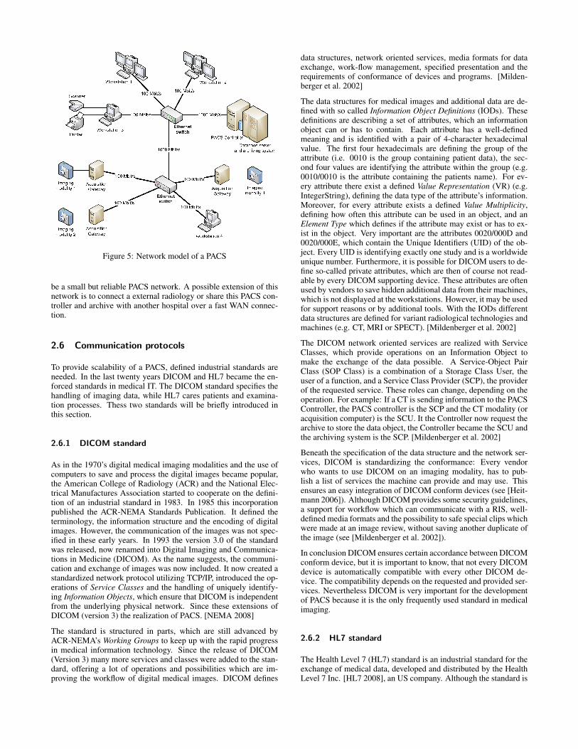
Figure 5: Network model of a PACS
be a small but reliable PACS network. A possible extension of thisnetwork is to connect a external radiology or share this PACS con-troller and archive with another hospital over a fast WAN connec-tion.
2.6 Communication protocols
To provide scalability of a PACS, defined industrial standards areneeded. In the last twenty years DICOM and HL7 became the en-forced standards in medical IT. The DICOM standard specifies thehandling of imaging data, while HL7 cares patients and examina-tion processes. Thess two standards will be briefly introduced inthis section.
2.6.1 DICOM standard
As in the 1970’s digital medical imaging modalities and the use ofcomputers to save and process the digital images became popular,the American College of Radiology (ACR) and the National Elec-trical Manufactures Association started to cooperate on the defini-tion of an industrial standard in 1983. In 1985 this incorporationpublished the ACR-NEMA Standards Publication. It defined theterminology, the information structure and the encoding of digitalimages. However, the communication of the images was not spec-ified in these early years. In 1993 the version 3.0 of the standardwas released, now renamed into Digital Imaging and Communica-tions in Medicine (DICOM). As the name suggests, the communi-cation and exchange of images was now included. It now created astandardized network protocol utilizing TCP/IP, introduced the op-erations of Service Classes and the handling of uniquely identify-ing Information Objects, which ensure that DICOM is independentfrom the underlying physical network. Since these extensions ofDICOM (version 3) the realization of PACS. [NEMA 2008]
The standard is structured in parts, which are still advanced byACR-NEMA’s Working Groups to keep up with the rapid progressin medical information technology. Since the release of DICOM(Version 3) many more services and classes were added to the stan-dard, offering a lot of operations and possibilities which are im-proving the workflow of digital medical images. DICOM defines
data structures, network oriented services, media formats for dataexchange, work-flow management, specified presentation and therequirements of conformance of devices and programs. [Milden-berger et al. 2002]
The data structures for medical images and additional data are de-fined with so called Information Object Definitions (IODs). Thesedefinitions are describing a set of attributes, which an informationobject can or has to contain. Each attribute has a well-definedmeaning and is identified with a pair of 4-character hexadecimalvalue. The first four hexadecimals are defining the group of theattribute (i.e. 0010 is the group containing patient data), the sec-ond four values are identifying the attribute within the group (e.g.0010/0010 is the attribute containing the patients name). For ev-ery attribute there exist a defined Value Representation (VR) (e.g.IntegerString), defining the data type of the attribute’s information.Moreover, for every attribute exists a defined Value Multiplicity,defining how often this attribute can be used in an object, and anElement Type which defines if the attribute may exist or has to ex-ist in the object. Very important are the attributes 0020/000D and0020/000E, which contain the Unique Identifiers (UID) of the ob-ject. Every UID is identifying exactly one study and is a worldwideunique number. Furthermore, it is possible for DICOM users to de-fine so-called private attributes, which are then of course not read-able by every DICOM supporting device. These attributes are oftenused by vendors to save hidden additional data from their machines,which is not displayed at the workstations. However, it may be usedfor support reasons or by additional tools. With the IODs differentdata structures are defined for variant radiological technologies andmachines (e.g. CT, MRI or SPECT). [Mildenberger et al. 2002]
The DICOM network oriented services are realized with ServiceClasses, which provide operations on an Information Object tomake the exchange of the data possible. A Service-Object PairClass (SOP Class) is a combination of a Storage Class User, theuser of a function, and a Service Class Provider (SCP), the providerof the requested service. These roles can change, depending on theoperation. For example: If a CT is sending information to the PACSController, the PACS controller is the SCP and the CT modality (oracquisition computer) is the SCU. It the Controller now request thearchive to store the data object, the Controller became the SCU andthe archiving system is the SCP. [Mildenberger et al. 2002]
Beneath the specification of the data structure and the network ser-vices, DICOM is standardizing the conformance: Every vendorwho wants to use DICOM on an imaging modality, has to pub-lish a list of services the machine can provide and may use. Thisensures an easy integration of DICOM conform devices (see [Heit-mann 2006]). Although DICOM provides some security guidelines,a support for workflow which can communicate with a RIS, well-defined media formats and the possibility to safe special clips whichwere made at an image review, without saving another duplicate ofthe image (see [Mildenberger et al. 2002]).
In conclusion DICOM ensures certain accordance between DICOMconform device, but it is important to know, that not every DICOMdevice is automatically compatible with every other DICOM de-vice. The compatibility depends on the requested and provided ser-vices. Nevertheless DICOM is very important for the developmentof PACS because it is the only frequently used standard in medicalimaging.
2.6.2 HL7 standard
The Health Level 7 (HL7) standard is an industrial standard for theexchange of medical data, developed and distributed by the HealthLevel 7 Inc. [HL7 2008], an US company. Although the standard is
in commercial hands of a company, it can be used for free. In thispaper HL7 is always used for the standards not for the company.The latest version approved as an ANSI standard is HL7 version2.6, but a draft of version 3 is available on their website (see [HL72008]).
The HL7 standard is build on top of the application layer, the sev-enth layer of the ISO/OSI reference model. It is defining the datastructure of textual messages to communicate between applicationsin a medical environment. This could be the exchange of data be-tween multiple departments in hospitals or between different hos-pitals and medical practices. Thereby the scope of the standard isto facilitate the interface implementation in computer applicationsfrom varied vendors. Most of the RIS and HIS implementationsare supporting the data exchange through HL7. Hence, a PACSshould provide an HL7 interface to communicate with these sys-tems. [Huang 1996]
Different to DICOM, HL7 is textual orientated and it is definingthe transfer of data through event based text messages. A typicalsituation for an HL7 message would be a new patient in a hospital,who needs to be examined in the radiology department. This eventwould trigger the sending of an HL7 message to the radiology de-partment, including the necessary patient data. In this case, HL7defines the data structure of the message and the meaning of thedifferent data parts. After a successful sending of this HL7 mes-sage, the radiological department has all the data needed to add theupcoming examination into the workflow of the radiology depart-ment. [Heitmann 2006]
The HL7 standard defines multiple events, message types and seg-ments. These events control when a message has to be broadcasted,for example when an ADT event (”Admission, Discharge, Trans-fer”) is raising, the connected HL7 messages with the updated pa-tient and study data will be sent. There exist several types of mes-sages depending on the triggered event. The type of a messageis specified through the segments the message is including. A seg-ment is the smallest data element in the HL7 standard. For example,a PID segment contains the patient identifying data. Every messagegot to include a message header segment in which certain constantsare defined, like the encoding of the separator and the type of themessage (see [HL7 1998]).
2.7 Related systems
This section is introducing two other computerized systems appear-ing in the context of medical information technology, the HospitalInformation System and the Radiology Information System.
2.7.1 HIS - Hospital information system
A Hospital Information System (HIS) is a computerized manage-ment system in a hospital to manage administrative, financial andclinical tasks within a hospital. It is supporting the organization ofpatients, workflows and employees. Huang [Huang 1996] wrote,that there are three major categories to be handled by the HIS: thesupport of clinical and medical patient care activities, the admin-istration of the hospital (financial, personnel, payroll, bed census,etc.), and the management and control operations to provide long-range planning and evaluation of hospital performances. It is con-trolling financial tasks, like an automatic printing of bills and calcu-lating costs. Since the early sixties there have been a lot of differentHIS systems, changing with IT development and newer program-ming languages. HIS systems are not very portable, as they are
strongly linked to national rules and culture of medical treatment.[rie 1993]
To provide a paperless integration in the workflow of the hospital,thePACS need to be connected to the HIS. Through this interface itis possible to combine images, patient data and examination infor-mation in the treatment processes.
2.7.2 RIS - Radiology information system
Radiology Information Systems (RIS) became popular, when radio-logical scanners like CT or MRI were turning the radiology depart-ment to a key diagnostic department in the seventies. Accordingly,to manage this huge and expensive imaging departments RIS wereimplemented all over the world [rie 1993]. Depending on the sit-uation of the hospitals, RIS systems were either realized as stand-alone systems or as an extension of their HIS. Parallel to a HIS,the RIS is coordinating the workflow in a radiologic department,like setting examination dates and examination structure, forward-ing results and the scheduling of radiologists, assistants, rooms andthe utilization of imaging modalities.
While a PACS is managing the imaging data, the RIS is managingadministrative tasks. To get reliable patient data and to integratethe radiological department into the workflows of the hospital, aninterface to the HIS is needed and nowadays implemented. Hence, amodern PACS is trying to combine images and the associated work-flows and documents, RIS and PACS are deeply connected. That isthe reason, why many vendors are selling their PACS as an inte-grated RIS/PACS product, because most of the modern PACS so-lutions are containing solution for administrative and managementtasks of the radiology department.
3 Current PACS products and software
After describing the abstract components, protocols and architec-ture of PACS, the following section is concentrating on currentPACS software available on the market, to outline the possibili-ties of actual PACS systems. Therefore, some PACS examples areshortly presented. Naturally, there are a lot of PACS solutions avail-able on the market. Hence, it was necessary to confine oneself ona few examples. The PACS AGFA IMPAX, Siemens syngo Suite,and the open source project openPACS were just randomly pickedwithout any intention in advertising them.
3.1 Agfa: IMPAX 6 R©
Agfa IMPAX 6 is the PACS system of the IMPAX Enterprise solu-tion, an integrated RIS/PACS and reporting system. Impax Enter-prise is build up on IMPAX 6, adding reporting tools, web-basedservices, system monitoring and 24x7 support [Agfa HealthCare2008b]. IMPAX 6 and IMPAX Enterprise are developed and dis-tributed by Agfa HealthCare, a global vendor in the market of in-tegrated IT and imaging systems. Agfa HealthCare is developingPACS system since 1991 and has installed more than 1,200 PACSsystems worldwide [Agfa Group 2006].
IMPAX 6 offers an role-based access based on IT standards, fittingto the users workflow. Furthermore, the interface can be adjusted bythe user. IMPAX 6 distinguishes between six different user types:radiologists, technologists, hospital administrators, PACS adminis-trators, clinicians and IT managers. Login is provided at installed
workstation and office desktops. In addition IMPAX 6 allows re-mote access over HTTPS. Thus, IMPAX 6 is supporting teleradiol-ogy and a secure access to patient information from locations out-side the clinic, e.g. the radiologists home (see [Agfa HealthCare2008a]).
Furthermore, the Agfa PACS contains applications for processing,storing and consulting complex image data files. This includes ”3Dimages, images obtained through combining data from MRI, PETand/or CT devices and images used for virtual colonoscopy, com-puter assisted detection (CAD) and nuclear medicine [Agfa Group2006]. The processing of complex images is done within the IM-PAX 6 workflow. As a result the productivity of users is improved,as the processing is already done when receiving the images.
The communication of IMPAX 6 is based on client-server technol-ogy, using Web services for a communication between the clientsand the core. Therefore, Agfa implemented a so-called Websurgetechnology to reduce problems with low-bandwidth networks. Thistechnology is sending only the pixel data for the actual required res-olution of the image. Moreover, a PACS is designed in a 3-layer ar-chitecture, consisting of a business logic layer (application server),a data layer (IMPAX core) and a presentation layer (IMPAX client).[Agfa HealthCare 2008b]
Overall, Agfa provides with IMPAX 6 a suitable PACS solution,fulfilling the requirements for the imaging workflow and teleradiol-ogy, e.g. speech reporting and Web access. Agfa offers besides IM-PAX and IMPAX Enterprise, a solution for small clinics and med-ical practices called IMPAX El, archiving solutions, e.g. IMPAXData Center, and workstation devices such as IMPAX EL DS1000and IMPAX MA3000. Agfa is also selling an IT system for hospitaladministration (Agfa ORBIS) and imaging modalities. Hence, anintegration of IMPAX 6 with these modalities or system is facile.
3.2 Siemens: syngo R©Suite
Siemens syngo Suite is the name for an integrated RIS/PACS so-lution from the Healthcare Sector of the Siemens AG. syngo Suitecan be partitioned in four different groups, syngo Portals, syngo Ap-plications, syngo Enablers and syngo Special Topics (see [SiemensHealthcare 2008]).
With syngo Portals, Siemens coined the portal software runningon the workstations, which enables a role-based login such as de-scribed in section 2.4. Siemens [Siemens Healthcare 2008] is pro-viding four different user interfaces, depending on the login: Firstlythe syngo Portal Radiologist offers access to relevant informationand tools. It is structured according to radiological workflow to im-prove task organization when checking requests, reading images,and signing reports. Secondly the syngo Portal Referring Physi-cian, offers workflow support and allows a secure interaction withthe radiology department. The next workstation environment isthe syngo Portal Executive, providing tools for quality metrics andother key performance indicators. Finally, the syngo Portal Tran-scriptionist, offers access to relevant information and tools for thetranscriptionist.
The second part of the suite is called syngo Applications. It is in-cluding different software applications, each of them developed forspecific tasks. The packages of syngo Applications can again bedistinguished in workflow applications, imaging applications andso-called knowledge applications. The workflow applications areincluding tools to organize the imaging workflows. Secondly theimaging applications are including image processing and editingtools, i.e. 2D applications, 3D applications, 4D applications, in-telligent post processing applications, dynamics applications and
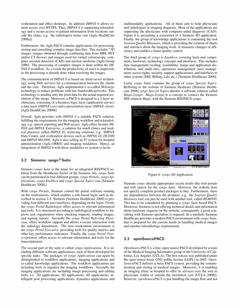
multimodality applications. All of them aim to help physiciansand radiologists in imaging diagnosis. Most of the applications aresupporting the physicians with computer-aided diagnosis (CAD).Figure 6 is presenting a screenshot of a Siemens 4D application.Finally, the group of knowledge applications is containing the toolSoarian Quality Measures, which is providing the creation of chartsand statistics about the imaging work. It measures changes of effi-ciency and enables a faster quality control.
The third group of syngo is Enablers, covering back-end compo-nents, hardware, technology concepts and interfaces. This includesdata management (scaling, availability, image and application dis-tribution, and multi-site), operations management (user manage-ment, access rights, security, support applications), and interfaces toother systems (HIS, Billing, Lab, etc.) [Siemens Healthcare 2008].
Lastly syngo Suite contains the group of syngo Special Topics.Reffering to the website of Siemens Healtcare [Siemens Health-care 2008] syngo Special Topics denotes a software solution calledMagic2syngo. Magic2syngo is a concept to connect the SiemensHIS solution Magic with the Siemens RIS/PACS syngo.
Figure 6: syngo 4D Application
Siemens syngo already appropriates recent trends like web portalsand web spaces for the syngo Suite. However, the website doesnot specify complete product packages to buy. Furthermore, thereare dependencies between the products, e.g. the Soarian QualityMeasures tool can just be used with another tool, called REMIND.This has to be considered by planning a syngo Suite based PACS.Moreover, Siemens is not offering technical details and informationabout hardware requests on the website, consequently a good con-sulting with Siemens specialists is required. In a nutshell, SiemensHealthcare provides a modern PACS environment with syngo Suite,that is prepared for the current needs in handling medical imagesand satisfies teleradiology requirements.
3.3 openSourcePACS
OpenSourcePACS is a free, open source PACS developed by a teamof the Medical Imaging Informatics group of the University of Cali-fornia, Los Angeles (UCLA). The first release was published underthe open source lesser GNU public license (LGPL) in 2005. Open-SourcePACS delivers a basic PACS system, providing the commu-nication and storing of medical images. Moreover, it is enablingan imaging clinic or hospital to offer its services over the web tophysicians within or outside the institution (see [UCLA 2008]).However, openSourcePACS is just handling the image flow and not
yet supporting all RIS tasks like dictation, transcription and re-porting. That means, that openSourcePACS is, in contrast to thetwo commercial PACS introduced before, not yet a fully integratedRIS/PACS solution (see [Bui et al. 2007]).
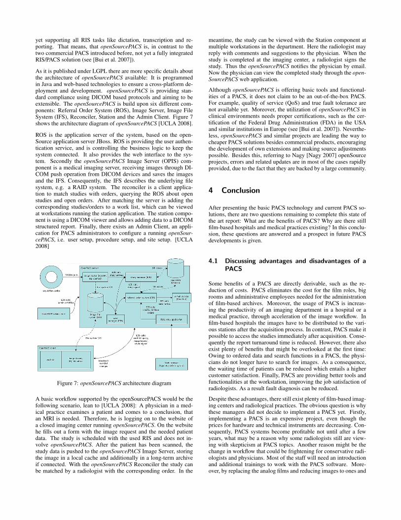
As it is published under LGPL there are more specific details aboutthe architecture of openSourcePACS available: It is programmedin Java and web-based technologies to ensure a cross-platform de-ployment and development. openSourcePACS is providing stan-dard compliance using DICOM based protocols and aiming to beextensible. The openSourcePACS is build upon six different com-ponents: Referral Order System (ROS), Image Server, Image FileSystem (IFS), Reconciler, Station and the Admin Client. Figure 7shows the architecture diagram of openSourcePACS [UCLA 2008].
ROS is the application server of the system, based on the open-Source application server JBoss. ROS is providing the user authen-tication service, and is controlling the business logic to keep thesystem connected. It also provides the web interface to the sys-tem. Secondly the openSourcePACS Image Server (OPIS) com-ponent is a medical imaging server, receiving images through DI-COM push operation from DICOM devices and saves the imagesand the IFS. Consequently, the IFS describes the underlying filesystem, e.g. a RAID system. The reconciler is a client applica-tion to match studies with orders, querying the ROS about openstudies and open orders. After matching the server is adding thecorresponding studies/orders to a work list, which can be viewedat workstations running the station application. The station compo-nent is using a DICOM viewer and allows adding data to a DICOMstructured report. Finally, there exists an Admin Client, an appli-cation for PACS administrators to configure a running openSour-cePACS, i.e. user setup, procedure setup, and site setup. [UCLA2008]
Figure 7: openSourcePACS architecture diagram
A basic workflow supported by the openSourcePACS would be thefollowing scenario, lean to [UCLA 2008]: A physician in a med-ical practice examines a patient and comes to a conclusion, thatan MRI is needed. Therefore, he is logging on to the website ofa closed imaging center running openSourcePACS. On the websitehe fills out a form with the image request and the needed patientdata. The study is scheduled with the used RIS and does not in-volve openSourcePACS. After the patient has been scanned, thestudy data is pushed to the openSourcePACS Image Server, storingthe image in a local cache and additionally in a long-term archiveif connected. With the openSourcePACS Reconciler the study canbe matched by a radiologist with the corresponding order. In the
meantime, the study can be viewed with the Station component atmultiple workstations in the department. Here the radiologist mayreply with comments and suggestions to the physician. When thestudy is completed at the imaging center, a radiologist signs thestudy. Thus the openSourcePACS notifies the physician by email.Now the physician can view the completed study through the open-SourcePACS web application.
Although openSourcePACS is offering basic tools and functional-ities of a PACS, it does not claim to be an out-of-the-box PACS.For example, quality of service (QoS) and true fault tolerance arenot available yet. Moreover, the utilization of openSourcePACS inclinical environments needs proper certifications, such as the cer-tification of the Federal Drug Administration (FDA) in the USAand similar institutions in Europe (see [Bui et al. 2007]). Neverthe-less, openSourcePACS and similar projects are leading the way tocheaper PACS solutions besides commercial products, encouragingthe development of own extensions and making source adjustmentspossible. Besides this, referring to Nagy [Nagy 2007] openSourceprojects, errors and related updates are in most of the cases rapidlyprovided, due to the fact that they are backed by a large community.
4 Conclusion
After presenting the basic PACS technology and current PACS so-lutions, there are two questions remaining to complete this state ofthe art report: What are the benefits of PACS? Why are there stillfilm-based hospitals and medical practices existing? In this conclu-sion, these questions are answered and a prospect in future PACSdevelopments is given.
4.1 Discussing advantages and disadvantages of aPACS
Some benefits of a PACS are directly derivable, such as the re-duction of costs. PACS eliminates the cost for the film roles, bigrooms and administrative employees needed for the administrationof film-based archives. Moreover, the usage of PACS is increas-ing the productivity of an imaging department in a hospital or amedical practice, through acceleration of the image workflow. Infilm-based hospitals the images have to be distributed to the vari-ous stations after the acquisition process. In contrast, PACS make itpossible to access the studies immediately after acquisition. Conse-quently the report turnaround time is reduced. However, there alsoexist plenty of benefits that might be overlooked at the first time:Owing to ordered data and search functions in a PACS, the physi-cians do not longer have to search for images. As a consequence,the waiting time of patients can be reduced which entails a highercustomer satisfaction. Finally, PACS are providing better tools andfunctionalities at the workstation, improving the job satisfaction ofradiologists. As a result fault diagnosis can be reduced.
Despite these advantages, there still exist plenty of film-based imag-ing centers and radiological practices. The obvious question is whythese managers did not decide to implement a PACS yet. Firstly,implementing a PACS is an expensive project, even though theprices for hardware and technical instruments are decreasing. Con-sequently, PACS systems become profitable not until after a fewyears, what may be a reason why some radiologists still are view-ing with skepticism at PACS topics. Another reason might be thechange in workflow that could be frightening for conservative radi-ologists and physicians. Most of the staff will need an introductionand additional trainings to work with the PACS software. More-over, by replacing the analog films and reducing images to ones and
zeros, with a PACS implementation medical images are not longersubstantial objects. As a result they can be lost or deleted, as stillno archive can guarantee a total fault-tolerance. The imaging ser-vice is essential for current medical environment, so there shouldalways be a computer engineer available, to serve daily faults andproblems.
To put things in a nutshell, the benefits of PACS are clearly visible,but there are some reasonable doubts left. To improve the numberof PACS installations, the vendors and developers of PACS systemsshould, besides the technical advancement, focus in helping the endusers to remove the last doubts. This could be reached with easierintroductions into the topic, even better adjustable systems to theclinical workflows and reliable archiving and backup solutions.
4.2 Future trends and developments
After this paper presented the current state and basics of PACS, fol-lowed by a short discussion of advantages, disadvantages and prob-lems of PACS, there remains the question about the future trendsand happenings in the domain of PACS. Firstly, digital imaging andcomputer graphics is still a field with rapid progress, developinghigher resolution images, saved in better data formats with highercompression rates. Furthermore, PACS are going to provide furthertechnologies for teleradiology, due to the fact that health systemsand health institutions are becoming closer connected and coop-erating. Besides already realized technologies, such as web accessand web viewers, newer technologies for low-bandwidth data trans-fer are currently enabling to view image streams on mobile devicessupporting volume rendering and 3D animations, e.g. on a smartphone or PDA. As a result, radiologists may work from all overthe world on a certain study. Consequently, experts do not have totravel to the patients, neither does the patient has to travel to theexpert. After successful PACS implementations in several hospitalson the world, the connection between the different PACS got to befaced. A secure way for communication between the hospitals andmedical practices would support a total paperless radiology work.As a result, a physician would be able to receive former digital im-ages over a large distance from a hospital, where his patient got anexamination a few years before. This would support the idea of theElectronic Health Record (EHR), which is currently discussed inmany countries. One concept regarding the EHR, is a patient cardequipped with a microchip, which is storing necessary patient datafor following examinations or emergencies. In the future it mightbe possible to store image materials on this microchip, acting likea mobile device of the PACS. Another concept is to connect everymedical institution in a country or nation with a centralized server.This solution would enable the exchange of images over the centralsever through PACS-to-PACS interfaces. To conclude, PACS havebecome an essential part in modern medical environments and arefacing plenty of different trends in the future.
References
AGFA GROUP. 2006. Agfa Annual Report 2006.
AGFA HEALTHCARE. 2008. IMPAX 6 Brochure (English).
AGFA HEALTHCARE. 2008. Website of Agfa HealthCare,http://www.agfa.com/en/he/home.jsp. Agfa HealthCare.
BUI, A. A. T., MORIOKA, C., DIONISIO, J., JOHNSON, D. B.,SINHA, U., ARDEKANI, S., TAIRA, R. K., ABERLE, D. R.,
EL-SADEN, S., AND KANGARLOO, H. 2007. openSource-PACS: An extensible infrastructure for medical image man-agement. IEEE Transactions on Information Technology inBiomedicine 11, 1, 94–109.
DREYER, K. J., MEHTA, A., AND THRALL, J. H. 2002. PACS: AGuide to the Digital Revolution. Springer-Verlag.
DUERINCKX, A. J. 2003. Introduction to two PACS ’82 Panel Dis-cussions edited by Andr J. Duerinckx, M.D., Ph.D.: ”EquipmentManufacturer’s View on PACS” and ”The Medical Community’sView on PACS”. Journal of Digital Imaging 16, 1, 29–31.
1995. Image management in a multi-hospital environment. In Inter-national Conference on Image Management and Communicationin Patient Care. IMAC’95, IEEE, R. A. Glicksman, F. W. Prior,and D. L. Wilson, Eds., 173–181.
HEITMANN, R. 2006. Auswahl und Konfiguration von PAC-Systemen fr radiologische Arztpraxen unter Bercksichtigung derEinfhrung der elektronischen Patientenkarte. Master’s thesis,FH Gießen.
HL7. 1998. HL7 Implementation Support Guide,http://www.hl7.org/Special/IG/final.pdf. Health Level Seven,Inc..
HL7. 2008. Website of the Health Level 7 Inc., http://www.hl7.org.Health Level Seven, German usergroup.
HUANG, H. K. 1996. PACS. VCH Verlag.
HURLEN, P., STBYE, T., BORTHNE, A., AND GULBRANDSEN, P.2008. Introducing PACS to the Late Majority. A LongitudinalStudy. Journal of Digital Imaging Online.
1991. Requirements for pacs workstations. In The Second Interna-tional Conference on Image Management and Communication(IMAC) in Patient Care: New Technologies for Better PatientCare, 1991, IEEE, Y. Kim, H. Park, and D. Haynor, Eds., 36–41.
KINTRONICS. 2008. NSM DVD Jukeboxes.http://www.kintronics.com/nsm.html.
KNIG, H., AND KLOSE, K. J. 1999. Anforderungsdefinition und-spezifikation fr PAC-Systeme. Der Radiologe, 39, 269–275.
LEMKE, H. U. 2003. Pacs developments in europe. ComputerizedMedical Imaging and Graphics 23, 3, 111–120.
MILDENBERGER, P., EICHELBERG, M., AND MARTIN, E. 2002.Introduction to the DICOM standard. European Radiology 12,4, 920–927.
NAGY, P. 2007. Open Source in Imaging Informatics. Journal ofDigital Imaging 20, 1, 1–10.
NEMA. 2008. DICOM Strategic Document - Version 8.1,http://medical.nema.org/dicom/geninfo/Strategy.pdf. NationalElectrical Manufacturers Association.
1993. HIS-RIS-PACS. In The Third International Conference onImage Management and Communication in Patient Care, 1993.IMAC’93., IEEE, O. Rienhoff, Ed., 29–32.
SIEMENS HEALTHCARE. 2008. Website of the Siemens syngoSuite, http://www.medical.siemens.com/syngo. Siemens Health-care, Siemens AG.
STRICKLAND, N. H. 2000. PACS (picture archiving and com-munication systems): filmless radiology. Archives of Disease inChildhood 83, 82–86.

UCLA. 2008. Website of OpenSourcePACS,http://www.mii.ucla.edu/opensourcepacs. UCLA MedicalImaging Informatics Group.
WIRTH, S., TREITL, M., VILLAIN, S., LUCKE, A., NISSEN-MEYER, S., AND MITTERMAIER, I. 2005. PACS: speicherungund abruf digitaler radiologischer bilddaten. Der Radiologe, 46.