Embed Size (px)
Citation preview
Pacific Island Burkitt Lymphoma Protocol v1.0 (2009 revised 2012) 1
PACIFIC ISLAND WORKSTREAM
BURKITT LYMPHOMA GUIDELINE
For The Treatment of
Burkitt Lymphoma
MODIFIED LMB PROTOCOL
DR ROB CORBETT
ASSOCIATE PROFESSOR MICHAEL SULLIVAN Children’s Haematology Oncology Centre
Christchurch New Zealand
On behalf of the NCCN Pacific Island Workstream
Revised
August 2012
Pacific Island Burkitt Lymphoma Protocol v1.0 (2009 revised 2012) 2
Table Of Contents
Background ............................................................................................................................... 4
Epidemiology: ........................................................................................................................ 4
Clinical presentation: ............................................................................................................. 4
Histopathology and immunocytochemistry: .......................................................................... 4
Cytogenetics and molecular studies: ..................................................................................... 6
Diagnosis ................................................................................................................................... 6
Clinical examination: .............................................................................................................. 6
Investigations: ........................................................................................................................ 6
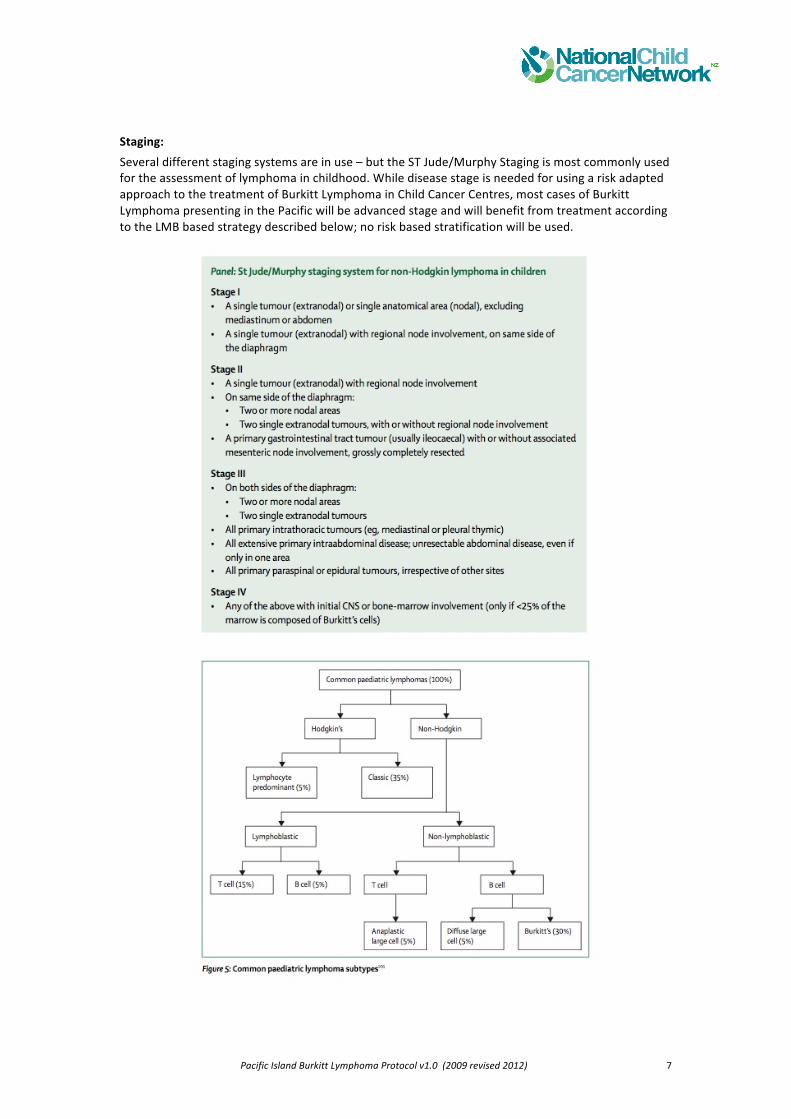
Staging: ...................................................................................................................................... 7
Treatment .................................................................................................................................. 8
Initial Supportive Care ............................................................................................................ 8
Steroid Prephase; ................................................................................................................... 8
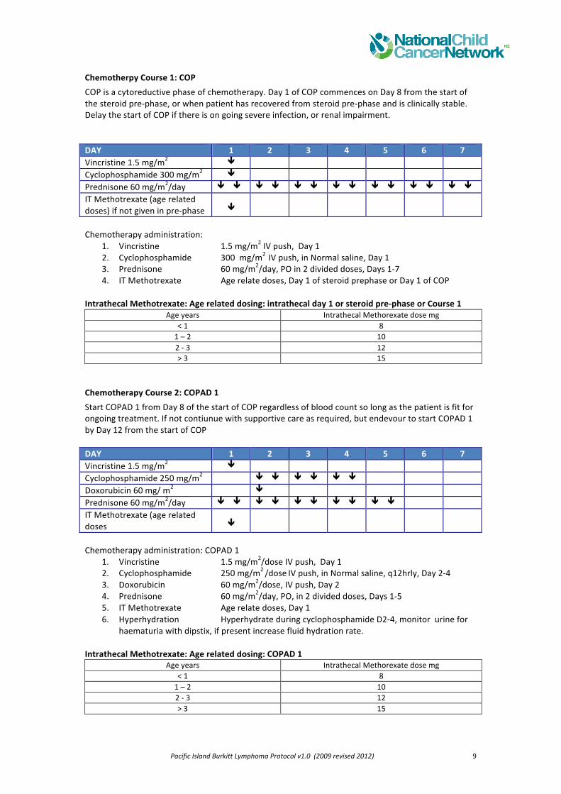
Chemotherpy Course 1: COP .................................................................................................. 9
Chemotherapy Course 2: COPAD 1 ........................................................................................ 9
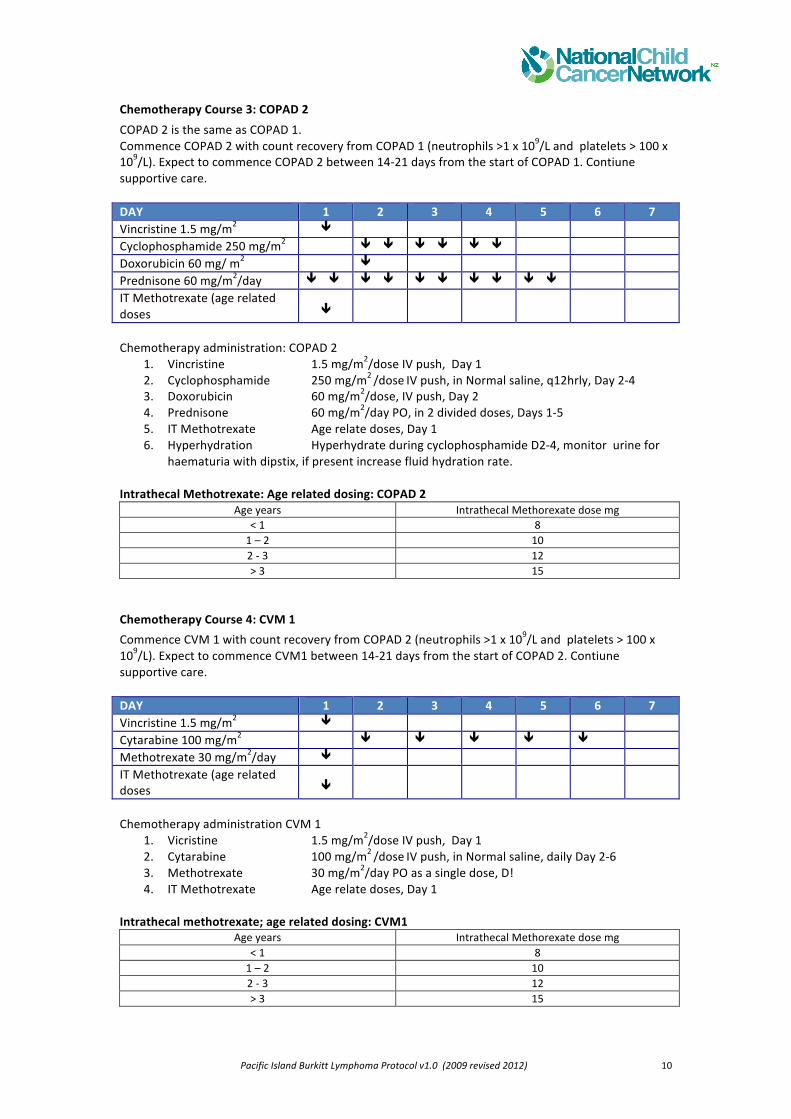
Chemotherapy Course 3: COPAD 2 ...................................................................................... 10
Chemotherapy Course 4: CVM 1 .......................................................................................... 10
Chemotherapy Course 5: CVM 2 .......................................................................................... 11
Follow-‐up ................................................................................................................................. 11
Pacific Island Burkitt Lymphoma Protocol v1.0 (2009 revised 2012) 3
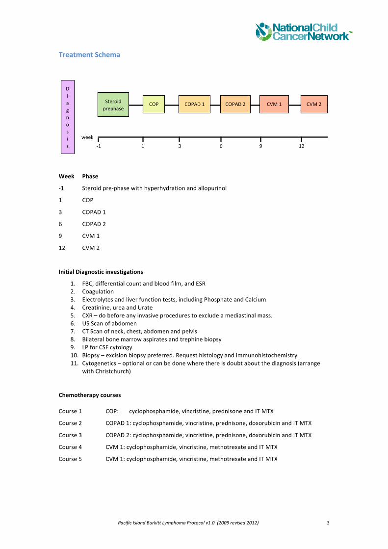
Treatment Schema
Steroid(prephase
COP COPAD(1 COPAD(2 CVM(1 CVM(2
161 3 6 9 12
week
Diagnosis
Week Phase
-‐1 Steroid pre-‐phase with hyperhydration and allopurinol
1 COP
3 COPAD 1
6 COPAD 2
9 CVM 1
12 CVM 2
Initial Diagnostic investigations
1. FBC, differential count and blood film, and ESR 2. Coagulation 3. Electrolytes and liver function tests, including Phosphate and Calcium 4. Creatinine, urea and Urate 5. CXR – do before any invasive procedures to exclude a mediastinal mass. 6. US Scan of abdomen 7. CT Scan of neck, chest, abdomen and pelvis 8. Bilateral bone marrow aspirates and trephine biopsy 9. LP for CSF cytology 10. Biopsy – excision biopsy preferred. Request histology and immunohistochemistry 11. Cytogenetics – optional or can be done where there is doubt about the diagnosis (arrange
with Christchurch) Chemotherapy courses Course 1 COP: cyclophosphamide, vincristine, prednisone and IT MTX
Course 2 COPAD 1: cyclophosphamide, vincristine, prednisone, doxorubicin and IT MTX
Course 3 COPAD 2: cyclophosphamide, vincristine, prednisone, doxorubicin and IT MTX Course 4 CVM 1: cyclophosphamide, vincristine, methotrexate and IT MTX
Course 5 CVM 1: cyclophosphamide, vincristine, methotrexate and IT MTX
Pacific Island Burkitt Lymphoma Protocol v1.0 (2009 revised 2012) 4
(These Background sections are reproduced, with permission, from Molyneax et al Lancet 2012)
Background The WHO classification of Burkitt’s lymphoma describes three clinical variants: endemic, sporadic (the predominant type found in non-‐malarial areas), and immunodeficiency-‐related. These types are similar in morphology, immunophenotype, and genetic features. The endemic variant is associated with malaria endemicity and EBV is found in almost all cases. The sporadic type occurs mainly throughout the rest of the world (predominantly North America and Europe), with no special climatic or geographical links, and is rarely associated with EBV infection. This sporadic-‐type Burkitt’s lymphoma accounts for 1–2% of adult lymphomas and 30–40% of childhood non-‐Hodgkin lymphomas in Europe and North America. The immunodeficiency-‐related type is seen most often in patients with HIV infection and less than 40% of US and European cases are associated with EBV.
Epidemiology: The distribution of endemic Burkitt’s lymphoma across Africa and Papua New Guinea corresponds to areas of holoendemic malaria and the early acquisition of EBV. The annual incidence has been estimated at 40–50 per million children younger than 18 years. In these high-‐risk areas endemic Burkitt’s lymphoma comprises about half of all childhood cancer diagnoses and up to 90% of lymphoma diagnoses. Incidence peaks at age 6 years and the disease is twice as common in boys as in girls. Sporadic Burkitt’s lymphoma occurs most commonly in children aged 3–12 years (median 6–8 years) and is 3·∙5 times more common in boys than in girls. Sporadic Burkitt’s lymphoma is found in low-‐risk areas such as North America, Northern and Eastern Europe, and East Asia at an annual incidence of 2 per million children younger than 18 years. Parts of South America, Southern Europe, North Africa, and the Middle East are areas of intermediate risk. Immunodeficiency-‐associated Burkitt’s lymphoma occurs at an incidence of 22 per 100 000 person-‐years in the USA.
Clinical presentation: The most common site of presentation in sporadic Burkitt’s lymphoma is the abdomen (60–80%). Presenting symptoms include abdominal pain (25% of patients have ileocaecal disease—either a right lower quadrant mass or pain from intussusception), distension, nausea and vomiting, and gastrointestinal bleeding. The next most common site is the head and neck, including lymphadenopathy and involvement of the nasal or oropharynx, tonsils, or sinuses. The jaw is infrequently implicated. Bone marrow is infiltrated in roughly 20% of patients. Some cases are classified as Burkitt’s leukaemia and are characterised by extensive marrow infiltration (more than 25% blasts), with possible bone pain as a presenting feature. Rare presenting sites include the mediastinum, CNS, skin, testes, breasts, and thyroid gland. Patients with endemic Burkitt’s lymphoma most frequently present with jaw or periorbital swellings, or abdominal involvement (of retroperitoneal tissue, gut, ovary, or kidney). Approximately 15% present with sudden paraplegia and incontinence. Infiltration of bone marrow is rare. Jaw involvement is common in young children (peak ages of incidence 3–7 years). In low-‐ income countries, such as in sub-‐Saharan Africa, many children present with advanced disease. In a study of 84 Malawian children with Burkitt’s lymphoma, 26 (31%) presented with facial disease only and 52 (62%) with abdominal disease; 58 (69%) had St Jude stage III or IV disease. Patients are commonly malnourished at diagnosis.
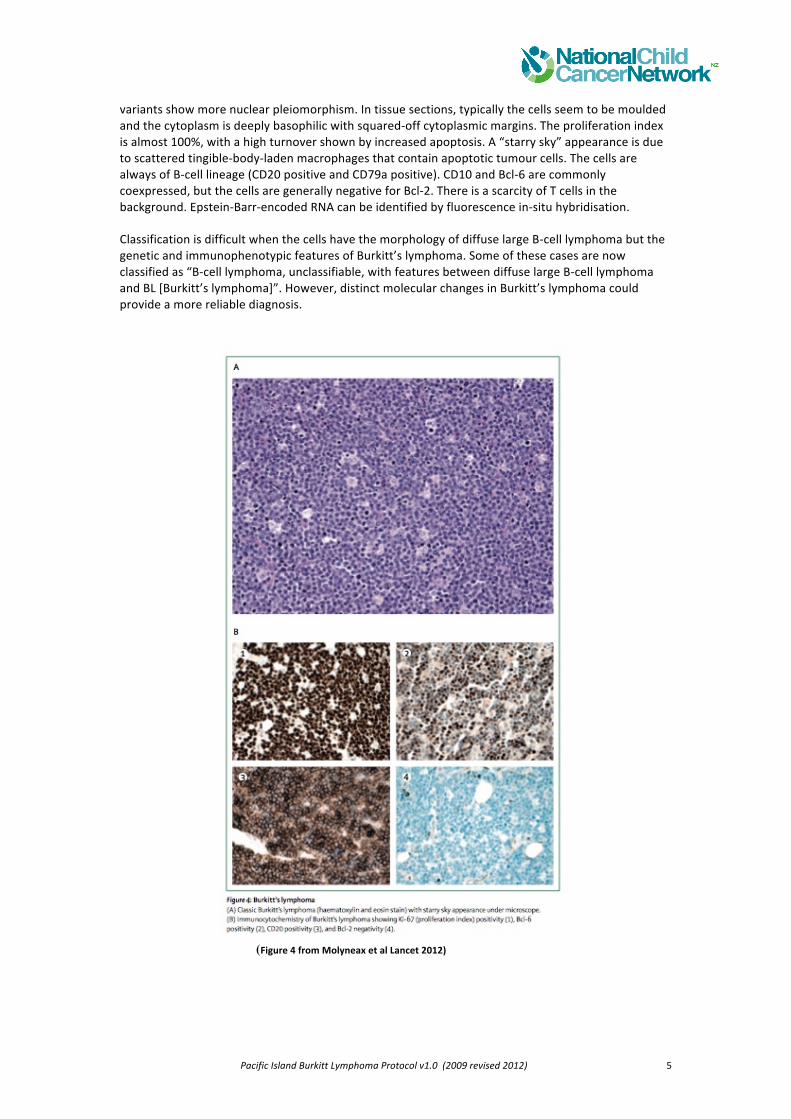
Histopathology and immunocytochemistry: Burkitt’s lymphoma is a highly aggressive B-‐cell non-‐ Hodgkin lymphoma characterised by monomorphic medium-‐sized cells with a very high proliferation rate. The cells are intermediate in size and contain coarse chromatin and prominent basophilic nucleoli. Some plasmacytoid and atypical
Pacific Island Burkitt Lymphoma Protocol v1.0 (2009 revised 2012) 5
variants show more nuclear pleiomorphism. In tissue sections, typically the cells seem to be moulded and the cytoplasm is deeply basophilic with squared-‐off cytoplasmic margins. The proliferation index is almost 100%, with a high turnover shown by increased apoptosis. A “starry sky” appearance is due to scattered tingible-‐body-‐laden macrophages that contain apoptotic tumour cells. The cells are always of B-‐cell lineage (CD20 positive and CD79a positive). CD10 and Bcl-‐6 are commonly coexpressed, but the cells are generally negative for Bcl-‐2. There is a scarcity of T cells in the background. Epstein-‐Barr-‐encoded RNA can be identified by fluorescence in-‐situ hybridisation. Classification is difficult when the cells have the morphology of diffuse large B-‐cell lymphoma but the genetic and immunophenotypic features of Burkitt’s lymphoma. Some of these cases are now classified as “B-‐cell lymphoma, unclassifiable, with features between diffuse large B-‐cell lymphoma and BL [Burkitt’s lymphoma]”. However, distinct molecular changes in Burkitt’s lymphoma could provide a more reliable diagnosis.
(Figure 4 from Molyneax et al Lancet 2012)
Pacific Island Burkitt Lymphoma Protocol v1.0 (2009 revised 2012) 6
Cytogenetics and molecular studies: Typically, Burkitt’s lymphoma has a simple karyotype with increasing genetic complexity linked to disease progression. The translocation t(8;14)(q24;q32) is the hallmark of Burkitt’s lymphoma and occurs in 70–80% of patients. The variant translocations, t(2;8)(p12;q24) and t(8;22) (q24;q11), occur in 10–15% of patients. The molecular consequence of the three translocations is deregulated expression of the MYC oncogene, which has an essential role in cell cycle control. Deregulated expression arises as a result of juxtaposition of MYC to the enhancer elements of one of the immunoglobulin genes: the heavy chain at 14q32; the kappa light chain at 2p12; or the lambda light chain at 22q11. The three translocations have different breakpoints; activation of MYC occurs on the derived chromosome 14 in t(8;14), with breakpoints centromeric of MYC, whereas it occurs on the derived chromosome 8 in cases with t(2;8) and t(8;22), with breakpoints telomeric of MYC. While cytogenetic studies are a routine part of the diagnostic assessment of children presenting to Child Cancer Centres such as Starship and CHOC, these do not inform current management and do not inform treatment planning. However, where there is doubt about the diagnosis, a FISH analysis of the diagnostic biopsy to detect the t(8;14) translocation may be helpful.
Diagnosis Diagnosis of Burkitt’s lymphoma should be confirmed by microscopy and immunocytological analysis (figure 5) if available. The recommended approach is to remove and examine the most accessible disease-‐containing tissue. This sample could be a superficial lymph node or malignant pleural fluid. Excision biopsy of a lymph node is preferable to fine-‐needle aspiration, which does not provide sufficient tissue for all the investigations required. In some cases a laparotomy or laparoscopy is necessary to obtain tissue.
Clinical examination: Thorough general examination; examine all nodal regions, ENT, orbits and fundi, CVS, Respiratory exam. Abdominal exam, document liver and spleen size, examine testes. CNS – cranial nerves and routine peripheral nerve examination. Establish nutritional status and consider clinical signs of concurrent infections such as TB and HIV (if at risk).
Investigations: Essential investigations are:
1. FBC, differential count and blood film, and ESR 2. Coagulation 3. Electrolytes and liver function tests, including Phosphate and Calcium 4. Creatinine, urea and Urate 5. CXR – do before any invasive procedures to exclude a mediastinal mass. 6. US Scan of abdomen 7. CT Scan of neck, chest, abdomen and pelvis 8. Bilateral bone marrow aspirates and trephine biopsy 9. LP for CSF cytology 10. Biopsy – excision biopsy is preferred. Request histology and immunohistochemistry 11. Cytogenetics – optional or can be done where there is doubt about the diagnosis
(arrange with Christchurch)
Pacific Island Burkitt Lymphoma Protocol v1.0 (2009 revised 2012) 7
Staging: Several different staging systems are in use – but the ST Jude/Murphy Staging is most commonly used for the assessment of lymphoma in childhood. While disease stage is needed for using a risk adapted approach to the treatment of Burkitt Lymphoma in Child Cancer Centres, most cases of Burkitt Lymphoma presenting in the Pacific will be advanced stage and will benefit from treatment according to the LMB based strategy described below; no risk based stratification will be used.
Pacific Island Burkitt Lymphoma Protocol v1.0 (2009 revised 2012) 8
Treatment This Pacific Island Burkitt Lymphoma guideline is based on the French LMB Burkitt Lymphoma protocol with several modifications to provide children a very good chance of cure within the resources available.
Initial Supportive Care The major clinical problems at diagnosis or immediately following the start of treatment are;
• Tumour Lysis Syndrome (with the potential for acute renal failure), -‐ spontaneous TLS may be present before the start of any treatment
• Infection – at diagnosis and during therapy • Complicated disease-‐related malnutrition • Other potential problems at diagnosis include, mediastinal mass with airway or SVC
obstruction, pericardial effusion, pleural effusion, and CNS disease
1. Admit to ward with barrier isolation 2. Treat fever with broad spectrum antibiotics – avoid aminoglycosides in the initial phase due
to the risk of renal impairment 3. Fluconazole and Cotrimoxazle prophylaxis as per ALL protocol 4. Transfuse if necessary 5. Nasogastric tube for early commencement of NG feeding. 6. Allopurinol – start 24 hrs before commencing corticosteroids and continue for 7 days 7. Hyperhydration at 125 ml/m2/hr (3L m2/day) to maintain a urine output >3ml/kg/hr as per
ALL induction protocol (1/2 Normal saline or 1/5th Saline – no KCL). If satisfactory urine output is not achieved, increase fluid rate to 150 ml/m2/hr
Steroid Prephase; Once pre-‐hydrated with adequate urine output start a steroid pre-‐phase with Prednisone 20mg/m2/day (Day 1-‐2), increase to 40mg/m2/day (Day 3-‐4) and 60mg/m2/day (Day 5-‐7) – give in 2 divided doses. 1. Diagnostic LP – give first Intrathecal Methotrexate in age related doses (see below) 2. Check Electrolytes, K+, PO4, Ca, Cr, Urea, Urate, twice daily – monitor for hyperkalaemia,
hyperphosphataemia, hypocalcaemia and hyperuricaemia; treat metabolic disturbance as per Tumour Lysis Syndrome protocol
3. Hyperhydration should continue until clinical resolution of lymph nodes, resolution of any metabolic evidence of tumour lysis syndrome and return of normal renal function.
4. Burkitt lymphoma usually responds to steroids with the rapid resolution of lymph nodes 5. If the child has significant co-‐morbidity such as a very poor nutritional state or concurrent
infection, complete the 7-‐day steroid pre-‐phase and reassess. 6. If there is control of disease as evidenced by resolving lymph nodes, consider a 1 week
period of nutritional support and treatment for infection before commencing chemotherapy Course 1 (COP) – But continue prednisone at 60mg/m2/day. This approach should reduce the risk of treatment related mortality from infection and malnutrition.
7. Continue NG feeding support throughout treatment if malnourished 8. Omeprazole or ranitidine concurrently with steroids
Intrathecal Methotrexate: Age related dosing: Day 1 of steroid pre-‐phase or Course 1
Age years Intrathecal Methorexate dose mg < 1 8 1 – 2 10 2 -‐ 3 12 > 3 15
Pacific Island Burkitt Lymphoma Protocol v1.0 (2009 revised 2012) 9
Chemotherpy Course 1: COP COP is a cytoreductive phase of chemotherapy. Day 1 of COP commences on Day 8 from the start of the steroid pre-‐phase, or when patient has recovered from steroid pre-‐phase and is clinically stable. Delay the start of COP if there is on going severe infection, or renal impairment. DAY 1 2 3 4 5 6 7 Vincristine 1.5 mg/m2 Cyclophosphamide 300 mg/m2 Prednisone 60 mg/m2/day
IT Methotrexate (age related doses) if not given in pre-‐phase
Chemotherapy administration:
1. Vincristine 1.5 mg/m2 IV push, Day 1 2. Cyclophosphamide 300 mg/m2 IV push, in Normal saline, Day 1 3. Prednisone 60 mg/m2/day, PO in 2 divided doses, Days 1-‐7 4. IT Methotrexate Age relate doses, Day 1 of steroid prephase or Day 1 of COP
Intrathecal Methotrexate: Age related dosing: intrathecal day 1 or steroid pre-‐phase or Course 1
Age years Intrathecal Methorexate dose mg < 1 8 1 – 2 10 2 -‐ 3 12 > 3 15
Chemotherapy Course 2: COPAD 1 Start COPAD 1 from Day 8 of the start of COP regardless of blood count so long as the patient is fit for ongoing treatment. If not contiunue with supportive care as required, but endevour to start COPAD 1 by Day 12 from the start of COP DAY 1 2 3 4 5 6 7 Vincristine 1.5 mg/m2 Cyclophosphamide 250 mg/m2 Doxorubicin 60 mg/ m2 Prednisone 60 mg/m2/day IT Methotrexate (age related doses
Chemotherapy administration: COPAD 1
1. Vincristine 1.5 mg/m2/dose IV push, Day 1 2. Cyclophosphamide 250 mg/m2 /dose IV push, in Normal saline, q12hrly, Day 2-‐4 3. Doxorubicin 60 mg/m2/dose, IV push, Day 2 4. Prednisone 60 mg/m2/day, PO, in 2 divided doses, Days 1-‐5 5. IT Methotrexate Age relate doses, Day 1 6. Hyperhydration Hyperhydrate during cyclophosphamide D2-‐4, monitor urine for
haematuria with dipstix, if present increase fluid hydration rate.
Intrathecal Methotrexate: Age related dosing: COPAD 1 Age years Intrathecal Methorexate dose mg
< 1 8 1 – 2 10 2 -‐ 3 12 > 3 15
Pacific Island Burkitt Lymphoma Protocol v1.0 (2009 revised 2012) 10
Chemotherapy Course 3: COPAD 2 COPAD 2 is the same as COPAD 1. Commence COPAD 2 with count recovery from COPAD 1 (neutrophils >1 x 10⁹/L and platelets > 100 x 109/L). Expect to commence COPAD 2 between 14-‐21 days from the start of COPAD 1. Contiune supportive care. DAY 1 2 3 4 5 6 7 Vincristine 1.5 mg/m2 Cyclophosphamide 250 mg/m2 Doxorubicin 60 mg/ m2 Prednisone 60 mg/m2/day IT Methotrexate (age related doses
Chemotherapy administration: COPAD 2
1. Vincristine 1.5 mg/m2/dose IV push, Day 1 2. Cyclophosphamide 250 mg/m2 /dose IV push, in Normal saline, q12hrly, Day 2-‐4 3. Doxorubicin 60 mg/m2/dose, IV push, Day 2 4. Prednisone 60 mg/m2/day PO, in 2 divided doses, Days 1-‐5 5. IT Methotrexate Age relate doses, Day 1 6. Hyperhydration Hyperhydrate during cyclophosphamide D2-‐4, monitor urine for
haematuria with dipstix, if present increase fluid hydration rate. Intrathecal Methotrexate: Age related dosing: COPAD 2
Age years Intrathecal Methorexate dose mg < 1 8 1 – 2 10 2 -‐ 3 12 > 3 15
Chemotherapy Course 4: CVM 1 Commence CVM 1 with count recovery from COPAD 2 (neutrophils >1 x 10⁹/L and platelets > 100 x 109/L). Expect to commence CVM1 between 14-‐21 days from the start of COPAD 2. Contiune supportive care. DAY 1 2 3 4 5 6 7 Vincristine 1.5 mg/m2 Cytarabine 100 mg/m2 Methotrexate 30 mg/m2/day IT Methotrexate (age related doses
Chemotherapy administration CVM 1
1. Vicristine 1.5 mg/m2/dose IV push, Day 1 2. Cytarabine 100 mg/m2 /dose IV push, in Normal saline, daily Day 2-‐6 3. Methotrexate 30 mg/m2/day PO as a single dose, D! 4. IT Methotrexate Age relate doses, Day 1
Intrathecal methotrexate; age related dosing: CVM1
Age years Intrathecal Methorexate dose mg < 1 8 1 – 2 10 2 -‐ 3 12 > 3 15
Pacific Island Burkitt Lymphoma Protocol v1.0 (2009 revised 2012) 11
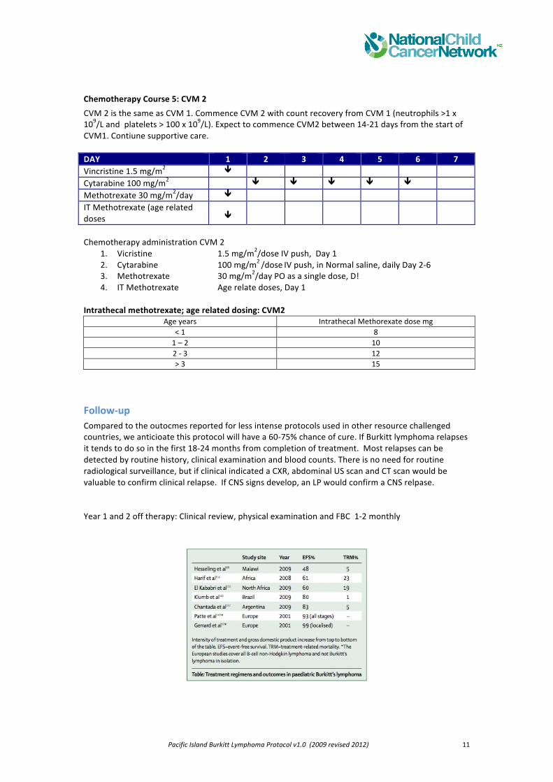
Chemotherapy Course 5: CVM 2 CVM 2 is the same as CVM 1. Commence CVM 2 with count recovery from CVM 1 (neutrophils >1 x 10⁹/L and platelets > 100 x 109/L). Expect to commence CVM2 between 14-‐21 days from the start of CVM1. Contiune supportive care. DAY 1 2 3 4 5 6 7 Vincristine 1.5 mg/m2 Cytarabine 100 mg/m2 Methotrexate 30 mg/m2/day IT Methotrexate (age related doses
Chemotherapy administration CVM 2
1. Vicristine 1.5 mg/m2/dose IV push, Day 1 2. Cytarabine 100 mg/m2 /dose IV push, in Normal saline, daily Day 2-‐6 3. Methotrexate 30 mg/m2/day PO as a single dose, D! 4. IT Methotrexate Age relate doses, Day 1
Intrathecal methotrexate; age related dosing: CVM2
Age years Intrathecal Methorexate dose mg < 1 8 1 – 2 10 2 -‐ 3 12 > 3 15
Follow-‐up Compared to the outocmes reported for less intense protocols used in other resource challenged countries, we anticioate this protocol will have a 60-‐75% chance of cure. If Burkitt lymphoma relapses it tends to do so in the first 18-‐24 months from completion of treatment. Most relapses can be detected by routine history, clinical examination and blood counts. There is no need for routine radiological surveillance, but if clinical indicated a CXR, abdominal US scan and CT scan would be valuable to confirm clinical relapse. If CNS signs develop, an LP would confirm a CNS relpase. Year 1 and 2 off therapy: Clinical review, physical examination and FBC 1-‐2 monthly