Embed Size (px)
Citation preview
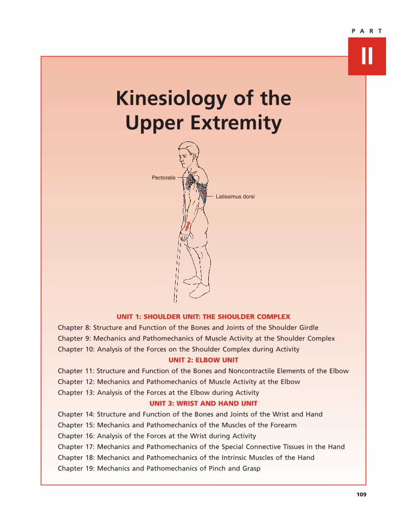
Kinesiology of theUpper Extremity
IIP A R T
109
UNIT 1: SHOULDER UNIT: THE SHOULDER COMPLEX
Chapter 8: Structure and Function of the Bones and Joints of the Shoulder Girdle
Chapter 9: Mechanics and Pathomechanics of Muscle Activity at the Shoulder Complex
Chapter 10: Analysis of the Forces on the Shoulder Complex during Activity
UNIT 2: ELBOW UNIT
Chapter 11: Structure and Function of the Bones and Noncontractile Elements of the Elbow
Chapter 12: Mechanics and Pathomechanics of Muscle Activity at the Elbow
Chapter 13: Analysis of the Forces at the Elbow during Activity
UNIT 3: WRIST AND HAND UNIT
Chapter 14: Structure and Function of the Bones and Joints of the Wrist and Hand
Chapter 15: Mechanics and Pathomechanics of the Muscles of the Forearm
Chapter 16: Analysis of the Forces at the Wrist during Activity
Chapter 17: Mechanics and Pathomechanics of the Special Connective Tissues in the Hand
Chapter 18: Mechanics and Pathomechanics of the Intrinsic Muscles of the Hand
Chapter 19: Mechanics and Pathomechanics of Pinch and Grasp
Latissimus dorsi
Pectoralis
The shoulder complex is the functional unit that results in movement of the arm with
respect to the trunk. This unit consists of the clavicle, scapula, and humerus; the ar-
ticulations linking them; and the muscles that move them. These structures are so
functionally interrelated to one another that studying their individual functions
is almost impossible. However, a careful study of the structures that compose the
shoulder unit reveals an elegantly simple system of bones, joints, and muscles that
together allow the shoulder an almost infinite number of movements (Figure). An
important source of patients’ complaints of pain and dysfunction at the shoulder
complex is an interruption of the normal coordination of these interdependent
structures.
The primary function of the shoulder complex is to position the upper extremity in
space to allow the hand to perform its tasks. The wonder of the shoulder complex
is the spectrum of positions that it can achieve; yet this very mobility is the source
of great risk to the shoulder complex as well. Joint instability is another important
UNIT 1: SHOULDER UNIT: THE SHOULDER COMPLEX
110
Scapula
Sternum
Clavicle
Humerus
The shoulder complex. The shouldercomplex consists of the humerus,clavicle, and scapula and includes thesternoclavicular, acromioclavicular,glenohumeral, and scapulothoracicjoints.
source of patients’ complaints of shoulder dysfunction. Thus an understanding of the
function and dysfunction of the shoulder complex requires an understanding of the
coordinated interplay among the individual components of the shoulder complex as
well as an appreciation of the structural compromises found in the shoulder that
allow tremendous mobility yet provide sufficient stability.
This three-chapter unit on the shoulder complex describes the structure of the shoul-
der complex and its implications for function and dysfunction. The purposes of this
unit are to
■ Provide the clinician with an understanding of the morphology of
the individual components of the complex
■ Identify the functional relationships among the individual
components
■ Discuss how the structures of the shoulder complex contribute to
mobility and stability
■ Provide insight into the stresses that the shoulder complex sustains
during daily activity
The unit is divided into three chapters. The first chapter presents the bony structures
making up the shoulder complex and the articulations that join them. The second
chapter presents the muscles of the shoulders and their contributions to function and
dysfunction. The third chapter investigates the loads to which the shoulder complex
and its individual components are subjected during daily activity.
UNIT 1: SHOULDER UNIT: THE SHOULDER COMPLEX
111

112
Structure and Function of the Bonesand Joints of the Shoulder Girdle
STRUCTURE OF THE BONES OF THE SHOULDER COMPLEX . . . . . . . . . . . . . . . . . . . . . . . .113
Clavicle . . . . . . . . . . . . . . . . . . . . . . . . . . . . . . . . . . . . . . . . . . . . . . . . . . . . . . . . . . . . .113
Scapula . . . . . . . . . . . . . . . . . . . . . . . . . . . . . . . . . . . . . . . . . . . . . . . . . . . . . . . . . . . . .113
Proximal Humerus . . . . . . . . . . . . . . . . . . . . . . . . . . . . . . . . . . . . . . . . . . . . . . . . . . .116
Sternum and Thorax . . . . . . . . . . . . . . . . . . . . . . . . . . . . . . . . . . . . . . . . . . . . . . . . . .118
STRUCTURE OF THE JOINTS AND SUPPORTING STRUCTURES OF
THE SHOULDER COMPLEX . . . . . . . . . . . . . . . . . . . . . . . . . . . . . . . . . . . . . . . . . . . . . . .119
Sternoclavicular Joint . . . . . . . . . . . . . . . . . . . . . . . . . . . . . . . . . . . . . . . . . . . . . . . . . .120
Acromioclavicular Joint . . . . . . . . . . . . . . . . . . . . . . . . . . . . . . . . . . . . . . . . . . . . . . .123
Scapulothoracic Joint . . . . . . . . . . . . . . . . . . . . . . . . . . . . . . . . . . . . . . . . . . . . . . . . . .125
Glenohumeral Joint . . . . . . . . . . . . . . . . . . . . . . . . . . . . . . . . . . . . . . . . . . . . . . . . . . .126
TOTAL SHOULDER MOVEMENT . . . . . . . . . . . . . . . . . . . . . . . . . . . . . . . . . . . . . . . . . . . . .132
Movement of the Scapula and Humerus during Arm–Trunk Elevation . . . . . . . . . . . .132
Sternoclavicular and Acromioclavicular Motion during Arm–Trunk Elevation . . . . . . .133
Impairments in Individual Joints and Their Effects on Shoulder Motion . . . . . . . . . . .134
SHOULDER RANGE OF MOTION . . . . . . . . . . . . . . . . . . . . . . . . . . . . . . . . . . . . . . . . . . . .137
SUMMARY . . . . . . . . . . . . . . . . . . . . . . . . . . . . . . . . . . . . . . . . . . . . . . . . . . . . . . . . . . . . . .138
This chapter describes the structure of the bones and joints of the shoulder complex as it
relates to the function of the shoulder. The specific purposes of this chapter are to
■ Describe the structures of the individual bones that constitute the shoulder complex
■ Describe the articulations joining the bony elements
■ Discuss the factors contributing to stability and instability at each joint
■ Discuss the relative contributions of each articulation to the overall motion of the
shoulder complex
■ Review the literature’s description of normal range of motion (ROM) of the shoulder
■ Discuss the implications of abnormal motion at an individual articulation to the
overall motion of the shoulder complex.
8C H A P T E R

113
STRUCTURE OF THE BONES OFTHE SHOULDER COMPLEX
The shoulder complex consists of three individual bones: theclavicle, the scapula, and the humerus. Each of these bonesis discussed in detail below. However, the complex itself isconnected to the axioskeleton via the sternum and rests onthe thorax, whose shape exerts some influence on the func-tion of the entire complex. Therefore, a brief discussion ofthe sternum and the shape of the thorax as it relates to theshoulder complex is also presented.
ClavicleThe clavicle functions like a strut to hold the shoulder com-plex and, indeed, the entire upper extremity suspended onthe axioskeleton [73]. Other functions attributed to the clav-icle are to provide a site for muscle attachment, to protectunderlying nerves and blood vessels, to contribute to in-creased ROM of the shoulder, and to help transmit muscleforce to the scapula [48,58]. This section describes the detailsof the clavicle that contribute to its ability to perform each ofthese functions. How these characteristics contribute to thefunctions of the clavicle and how they are implicated in in-juries to the clavicle are discussed in later sections of thischapter.
The clavicle lies with its long axis close to the transverseplane. It is a crank-shaped bone when viewed from above,with its medial two thirds convex anteriorly, approximatelyconforming to the anterior thorax, and its lateral one thirdconvex posteriorly (Fig. 8.1). The functional significance ofthis unusual shape becomes apparent in the discussion ofoverall shoulder motion.
The superior surface of the clavicle is smooth and readilypalpated under the skin. Anteriorly, the surface is roughenedby the attachments of the pectoralis major medially and thedeltoid laterally. The posterior surface is roughened on thelateral one third by the attachment of the upper trapezius.Inferiorly, the surface is roughened medially by attachmentsof the costoclavicular ligament and the subclavius muscle andlaterally by the coracoclavicular ligament. The latter producestwo prominent markings on the inferior surface of the lateralaspect of the clavicle, the conoid tubercle and, lateral to it,the trapezoid line.
The medial and lateral ends of the clavicle provide artic-ular surfaces for the sternum and acromion, respectively. Themedial aspect of the clavicle expands to form the head of theclavicle. The medial surface of this expansion articulates withthe sternum and intervening articular disc, or meniscus, aswell as with the first costal cartilage. The articular surface ofthe clavicular head is concave in the anterior posterior direc-tion and slightly convex in the superior inferior direction[80,87]. Unlike most synovial joints, the articular surface ofthe clavicle is covered by thick fibrocartilage. The lateral one
third of the clavicle is flattened with respect to the other twothirds and ends in a broad flat expansion that articulates withthe acromion at the acromioclavicular joint. The actual artic-ular surface is a small facet facing inferiorly and laterally.It too is covered by fibrocartilage rather than hyaline carti-lage. The medial and lateral aspects of the clavicle are easilypalpated.
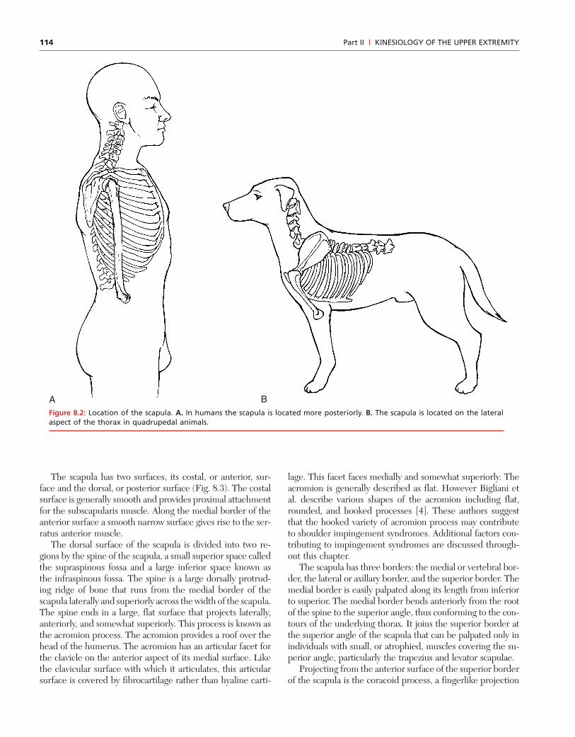
ScapulaThe scapula is a flat bone whose primary function is to pro-vide a site for muscle attachment for the shoulder. A total of15 major muscles acting at the shoulder attach to the scapula[53,87]. In quadrupedal animals, the scapula is long and thinand rests on the lateral aspect of the thorax. In primates, thereis a gradual mediolateral expansion of the bone along with agradual migration from a position lateral on the thorax to amore posterior location (Fig. 8.2). The mediolateral expan-sion is largely the result of an increased infraspinous fossa andcostal surface that provide attachment for three of the fourrotator cuff muscles as well as several other muscles of theshoulder [37,72]. These changes in structure and location ofthe scapula reflect the gradual change in the function of theupper extremity from its weight-bearing function to one ofreaching and grasping. These alterations in function requirea change in the role of muscles that now must position andsupport a scapula and glenohumeral joint that are no longerprimarily weight bearing and are free to move through a muchlarger excursion.
Chapter 8 | STRUCTURE AND FUNCTION OF THE BONES AND JOINTS OF THE SHOULDER GIRDLE
First thoracic vertebra
First rib
Sternum
A
B
Clavicle
Trapezoid line
Articular surface for acromion
Articular surface for sternum Conoid tubercle
Clavicle
Figure 8.1: Clavicle. A. View of the superior surface. B. View ofthe inferior surface.
114 Part II | KINESIOLOGY OF THE UPPER EXTREMITY
BAFigure 8.2: Location of the scapula. A. In humans the scapula is located more posteriorly. B. The scapula is located on the lateralaspect of the thorax in quadrupedal animals.
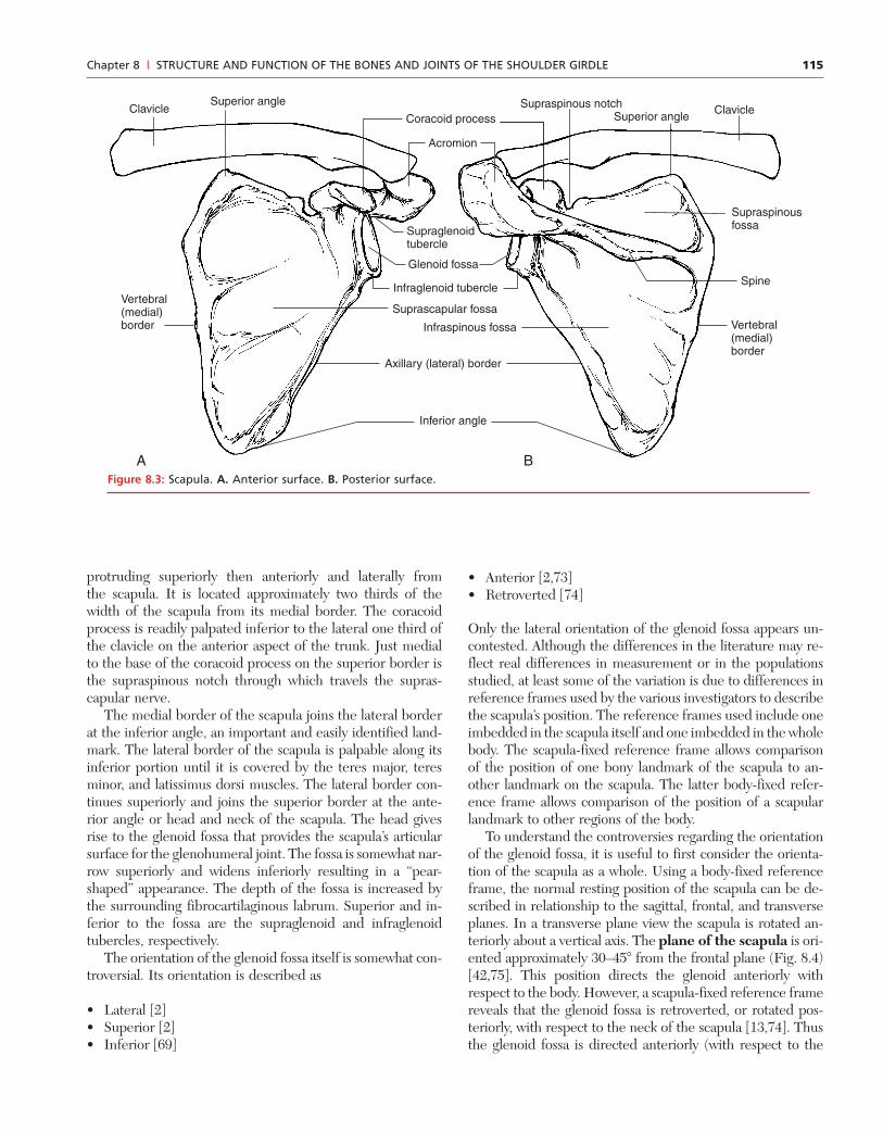
The scapula has two surfaces, its costal, or anterior, sur-face and the dorsal, or posterior surface (Fig. 8.3). The costalsurface is generally smooth and provides proximal attachmentfor the subscapularis muscle. Along the medial border of theanterior surface a smooth narrow surface gives rise to the ser-ratus anterior muscle.
The dorsal surface of the scapula is divided into two re-gions by the spine of the scapula, a small superior space calledthe supraspinous fossa and a large inferior space known asthe infraspinous fossa. The spine is a large dorsally protrud-ing ridge of bone that runs from the medial border of thescapula laterally and superiorly across the width of the scapula.The spine ends in a large, flat surface that projects laterally,anteriorly, and somewhat superiorly. This process is known asthe acromion process. The acromion provides a roof over thehead of the humerus. The acromion has an articular facet forthe clavicle on the anterior aspect of its medial surface. Likethe clavicular surface with which it articulates, this articularsurface is covered by fibrocartilage rather than hyaline carti-
lage. This facet faces medially and somewhat superiorly. Theacromion is generally described as flat. However Bigliani etal. describe various shapes of the acromion including flat,rounded, and hooked processes [4]. These authors suggestthat the hooked variety of acromion process may contributeto shoulder impingement syndromes. Additional factors con-tributing to impingement syndromes are discussed through-out this chapter.
The scapula has three borders: the medial or vertebral bor-der, the lateral or axillary border, and the superior border. Themedial border is easily palpated along its length from inferiorto superior. The medial border bends anteriorly from the rootof the spine to the superior angle, thus conforming to the con-tours of the underlying thorax. It joins the superior border atthe superior angle of the scapula that can be palpated only inindividuals with small, or atrophied, muscles covering the su-perior angle, particularly the trapezius and levator scapulae.
Projecting from the anterior surface of the superior borderof the scapula is the coracoid process, a fingerlike projection
115
protruding superiorly then anteriorly and laterally fromthe scapula. It is located approximately two thirds of thewidth of the scapula from its medial border. The coracoidprocess is readily palpated inferior to the lateral one third ofthe clavicle on the anterior aspect of the trunk. Just medialto the base of the coracoid process on the superior border isthe supraspinous notch through which travels the supras-capular nerve.
The medial border of the scapula joins the lateral borderat the inferior angle, an important and easily identified land-mark. The lateral border of the scapula is palpable along itsinferior portion until it is covered by the teres major, teresminor, and latissimus dorsi muscles. The lateral border con-tinues superiorly and joins the superior border at the ante-rior angle or head and neck of the scapula. The head givesrise to the glenoid fossa that provides the scapula’s articularsurface for the glenohumeral joint. The fossa is somewhat nar-row superiorly and widens inferiorly resulting in a “pear-shaped” appearance. The depth of the fossa is increased bythe surrounding fibrocartilaginous labrum. Superior and in-ferior to the fossa are the supraglenoid and infraglenoidtubercles, respectively.
The orientation of the glenoid fossa itself is somewhat con-troversial. Its orientation is described as
• Lateral [2]• Superior [2]• Inferior [69]
Chapter 8 | STRUCTURE AND FUNCTION OF THE BONES AND JOINTS OF THE SHOULDER GIRDLE
Superior angleSuperior angle
Supraspinous fossa
A B
Clavicle Clavicle Coracoid process
Glenoid fossa
Infraglenoid tubercle
Supraglenoid tubercle
Acromion
Vertebral (medial)border Vertebral
(medial)border
Infraspinous fossa
Suprascapular fossa
Spine
Axillary (lateral) border
Inferior angle
Supraspinous notch
Figure 8.3: Scapula. A. Anterior surface. B. Posterior surface.
• Anterior [2,73]• Retroverted [74]
Only the lateral orientation of the glenoid fossa appears un-contested. Although the differences in the literature may re-flect real differences in measurement or in the populationsstudied, at least some of the variation is due to differences inreference frames used by the various investigators to describethe scapula’s position. The reference frames used include oneimbedded in the scapula itself and one imbedded in the wholebody. The scapula-fixed reference frame allows comparisonof the position of one bony landmark of the scapula to an-other landmark on the scapula. The latter body-fixed refer-ence frame allows comparison of the position of a scapularlandmark to other regions of the body.
To understand the controversies regarding the orientationof the glenoid fossa, it is useful to first consider the orienta-tion of the scapula as a whole. Using a body-fixed referenceframe, the normal resting position of the scapula can be de-scribed in relationship to the sagittal, frontal, and transverseplanes. In a transverse plane view the scapula is rotated an-teriorly about a vertical axis. The plane of the scapula is ori-ented approximately 30–45� from the frontal plane (Fig. 8.4)[42,75]. This position directs the glenoid anteriorly withrespect to the body. However, a scapula-fixed reference framereveals that the glenoid fossa is retroverted, or rotated pos-teriorly, with respect to the neck of the scapula [13,74]. Thusthe glenoid fossa is directed anteriorly (with respect to the

116
body) and at the same time is retroverted (with respect to thescapula).
Rotation of the scapula in the frontal plane about a body-fixed anterior–posterior (AP) axis is also described (Fig. 8.5).This frontal plane rotation of the scapula is described by eitherthe upward or downward orientation of the glenoid fossa orby the medial or lateral location of the scapula’s inferior an-gle [2,22,69]. A rotation about this AP axis that tips the gle-noid fossa inferiorly, moving the inferior angle of the scapulamedially (i.e., closer to the vertebral column), is described asdownward or medial rotation of the scapula. A rotationthat tilts the glenoid fossa upward, moving the inferior anglelaterally away from the vertebral column is upward or lat-eral rotation. Two investigations report that the glenoid fossais upwardly inclined in quiet standing [2,55]. Two other stud-ies report a downward inclination of approximately 5� [22,69].The posture of the studies’ subjects may help to explain thesereported differences. Perhaps subjects who demonstrate anupward inclination are instructed to pull their shoulders backinto an “erect” posture while those who have a downward in-clination of the glenoid fossa have slightly drooping shoulders(Fig. 8.6). A final determination of the normal orientation ofthe scapulae in the frontal plane requires an accepted defi-nition of normal postural alignment of the shoulder. Thatdefinition unfortunately is presently lacking. Therefore, thecontroversy regarding the orientation of the scapula and itsglenoid fossa in the frontal plane continues.
Viewed sagittally, the scapula tilts forward from the frontalplane approximately 10� about a medial lateral axis (Fig. 8.7)[16]. This forward tilting is partly the result of the scapula’s
position on the superior thorax, which tapers toward its apex.Additional forward tilt of the scapula causes the inferior angleof the scapula to protrude from the thorax.
CLINICAL RELEVANCE: SCAPULAR POSITION INSHOULDER DYSFUNCTIONAbnormal scapular positions have been implicated in severalforms of shoulder dysfunction. Abnormal orientation of theglenoid fossa has been associated with instability of theglenohumeral joint [2,74]. In addition, excessive anteriortilting is found in individuals with shoulder impingementsyndromes during active shoulder abduction [55]. Carefulevaluation of scapular position is an essential componentof a thorough examination of patients with shoulder dys-function.
Proximal HumerusThe humerus is a long bone composed of a head, neck, andbody, or shaft. The body ends distally in the capitulum andtrochlea. This chapter presents only those portions of the
Part II | KINESIOLOGY OF THE UPPER EXTREMITY
1 23
1
2
3
Frontal plane
40°
Plane of scapula Scapula
Clavicle
Figure 8.4: Plane of the scapula. A transverse view of thescapula reveals that the plane of the scapula forms an angle ofapproximately 40� with the frontal plane.
Figure 8.5: Scapular rotation. Rotation of the scapula about ananterior–posterior (AP) axis causes the glenoid fossa to faceupward or downward.
117
humerus that are relevant to a discussion of the mechanicsand pathomechanics of the shoulder complex. The rest of thehumerus is discussed in Chapter 11 with the elbow. Thearticular surface of the head of the humerus is most oftendescribed as approximately half of an almost perfect sphere(Fig. 8.8) [36,77,85,87]. The humeral head projects medially,
Chapter 8 | STRUCTURE AND FUNCTION OF THE BONES AND JOINTS OF THE SHOULDER GIRDLE
Figure 8.6: Postural changes of the scapula. A. This individual is standing with drooping, or rounded, shoulders, andthe scapulae are rotated so that the glenoid fossa tilts downward. B. This individual stands with the shoulders pulledback and the scapulae tilted upward.
Figure 8.7: Scapular rotation. Rotation of the scapula about aML axis tilts the scapula anteriorly and posteriorly.
Humeral head
Greatertubercle
Superior facetsMiddle facetsInferior facets
Intertubercular groove
Lessertubercle
Deltoidtuberosity
A B
Humeral head
Figure 8.8: Proximal humerus. A. Anterior view. B. Posterior view.

118
superiorly, and posteriorly with respect to the plane formedby the medial and lateral condyles (Fig. 8.9) [37]. The humeralhead ends in the anatomical neck marking the end of thearticular surface.
On the lateral aspect of the proximal humerus is the greatertubercle, a large bony prominence that is easily palpated onthe lateral aspect of the shoulder complex. The greater tu-bercle is marked by three distinct facets on its superior andposterior surfaces. These facets give rise from superior to pos-terior to the supraspinatus, infraspinatus, and teres minormuscles, respectively. On the anterior aspect of the proximalhumerus is a smaller but still prominent bony projection, thelesser tubercle. It too has a facet that provides attachment forthe remaining rotator cuff muscle, the subscapularis. Sepa-rating the tubercles is the intertubercular, or bicipital, groovecontaining the tendon of the long head of the biceps brachii.The greater and lesser tubercles continue onto the body ofthe humerus as the medial and lateral lips of the groove. The
surgical neck is a slight narrowing of the shaft of the humerusjust distal to the tubercles.
CLINICAL RELEVANCE: THE DEPTH OFTHE BICIPITAL GROOVEThe depth of the bicipital groove varies. A shallow grooveappears to be a contributing factor in dislocations of thebiceps tendon [51,53].
Approximately midway distally on the body of the humerusis the deltoid tuberosity on the anterolateral surface. It pro-vides the distal attachment for the deltoid muscle. The spiralgroove is another important landmark on the body of thehumerus. It is found on the proximal half of the humerus,spiraling from proximal to distal and medial to lateral on theposterior surface. The radial nerve travels in the spiral groovealong with the profunda brachii vessels. The radial nerve isparticularly susceptible to injury as it lies in the spiral groove.
Sternum and ThoraxAlthough the sternum and thorax are not part of the shoul-der complex, both are intimately related to the shoulder;therefore, a brief description of their structure as it relates tothe shoulder complex is required. Both the sternum and tho-rax are covered in greater detail in Chapter 29. The superiorportion of the sternum, the manubrium, provides an articu-lar surface for the proximal end of each clavicle (Fig. 8.10).
Part II | KINESIOLOGY OF THE UPPER EXTREMITY
Medial epicondyle
Greatertubercle
Lateral epicondyle
Lessertubercle
Lateral
Medial-lateral axis of distal humerus
A
B
Humeral head
Anterior
Figure 8.9: Orientation of the head of the humerus. A. In thetransverse plane, the humeral head is rotated posteriorly withrespect to the condyles of the distal humerus. B. In the frontalplane the head of the humerus is angled medially and superiorlywith respect to the shaft of the humerus.
Leftclavicle
Sternum
Manubrium
1st rib
Right clavicle
Figure 8.10: The sternum’s articular surface. The sternumprovides a shallow articular surface for the head of the clavicle.
119
The articular surface is a shallow depression called the clav-icular notch covered with fibrocartilage like the clavicularhead with which it articulates. Each notch provides consid-erably less articular surface than the clavicular head that ar-ticulates with it. The two clavicular notches are separated bythe sternal or jugular notch on the superior aspect of themanubrium. This notch is very prominent and is a useful land-mark for identifying the sternoclavicular joints. Another reli-able and useful landmark is the angle formed by the junctionof the manubrium with the body of the sternum, known asthe sternal angle, or angle of Louis. This is also the site of theattachment of the second costal cartilage to the manubriumand body of the sternum.
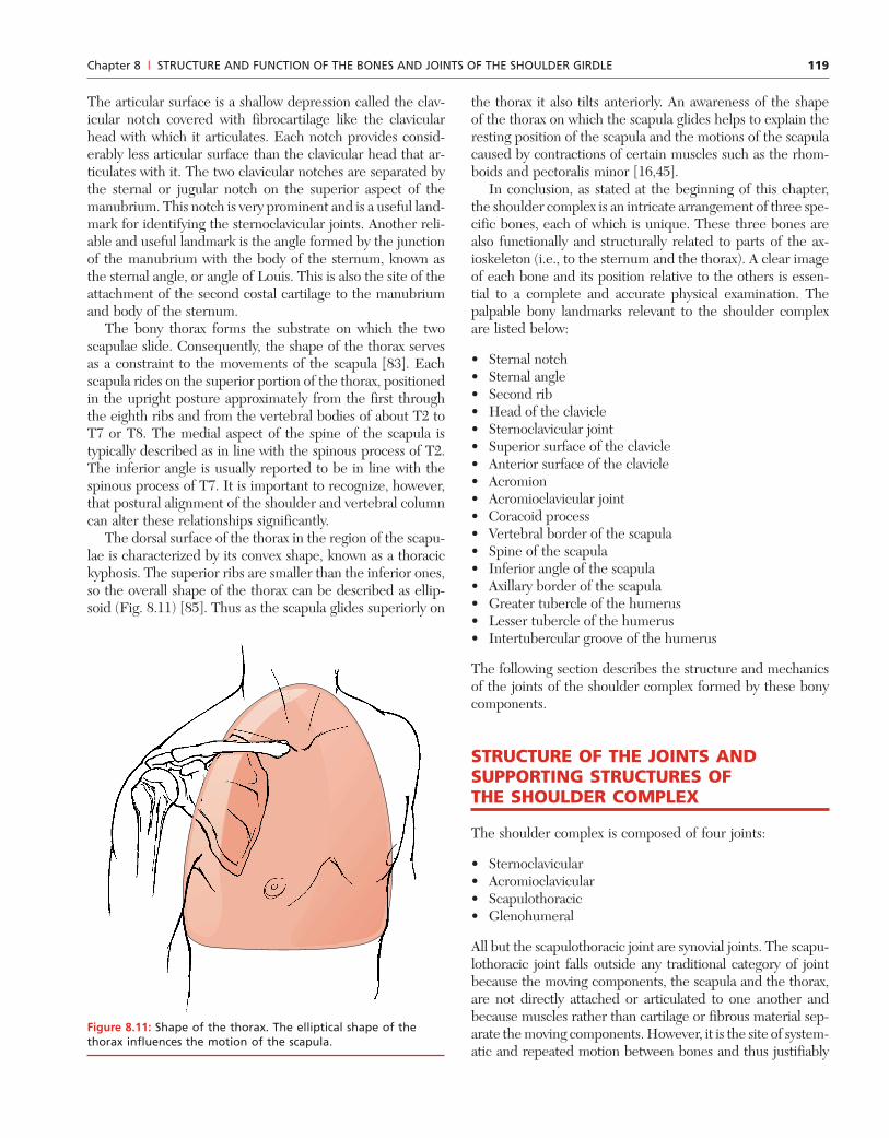
The bony thorax forms the substrate on which the twoscapulae slide. Consequently, the shape of the thorax servesas a constraint to the movements of the scapula [83]. Eachscapula rides on the superior portion of the thorax, positionedin the upright posture approximately from the first throughthe eighth ribs and from the vertebral bodies of about T2 toT7 or T8. The medial aspect of the spine of the scapula istypically described as in line with the spinous process of T2.The inferior angle is usually reported to be in line with thespinous process of T7. It is important to recognize, however,that postural alignment of the shoulder and vertebral columncan alter these relationships significantly.
The dorsal surface of the thorax in the region of the scapu-lae is characterized by its convex shape, known as a thoracickyphosis. The superior ribs are smaller than the inferior ones,so the overall shape of the thorax can be described as ellip-soid (Fig. 8.11) [85]. Thus as the scapula glides superiorly on
the thorax it also tilts anteriorly. An awareness of the shapeof the thorax on which the scapula glides helps to explain theresting position of the scapula and the motions of the scapulacaused by contractions of certain muscles such as the rhom-boids and pectoralis minor [16,45].
In conclusion, as stated at the beginning of this chapter,the shoulder complex is an intricate arrangement of three spe-cific bones, each of which is unique. These three bones arealso functionally and structurally related to parts of the ax-ioskeleton (i.e., to the sternum and the thorax). A clear imageof each bone and its position relative to the others is essen-tial to a complete and accurate physical examination. Thepalpable bony landmarks relevant to the shoulder complexare listed below:
• Sternal notch• Sternal angle• Second rib• Head of the clavicle• Sternoclavicular joint• Superior surface of the clavicle• Anterior surface of the clavicle• Acromion• Acromioclavicular joint• Coracoid process• Vertebral border of the scapula• Spine of the scapula• Inferior angle of the scapula• Axillary border of the scapula• Greater tubercle of the humerus• Lesser tubercle of the humerus• Intertubercular groove of the humerus
The following section describes the structure and mechanicsof the joints of the shoulder complex formed by these bonycomponents.
STRUCTURE OF THE JOINTS ANDSUPPORTING STRUCTURES OFTHE SHOULDER COMPLEX
The shoulder complex is composed of four joints:
• Sternoclavicular• Acromioclavicular• Scapulothoracic• Glenohumeral
All but the scapulothoracic joint are synovial joints. The scapu-lothoracic joint falls outside any traditional category of jointbecause the moving components, the scapula and the thorax,are not directly attached or articulated to one another andbecause muscles rather than cartilage or fibrous material sep-arate the moving components. However, it is the site of system-atic and repeated motion between bones and thus justifiably
Chapter 8 | STRUCTURE AND FUNCTION OF THE BONES AND JOINTS OF THE SHOULDER GIRDLE
Figure 8.11: Shape of the thorax. The elliptical shape of thethorax influences the motion of the scapula.
120
can be designated a joint. This section presents the struc-ture and mechanics of each of the four joints of the shouldercomplex.
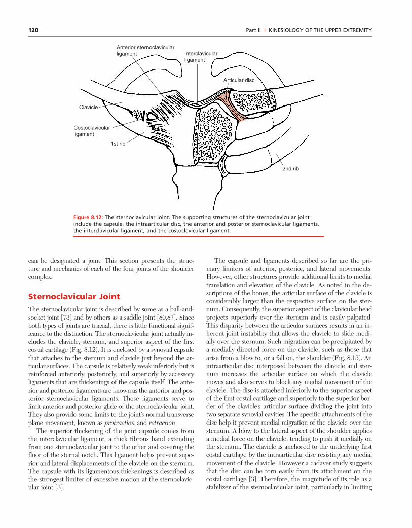
Sternoclavicular JointThe sternoclavicular joint is described by some as a ball-and-socket joint [73] and by others as a saddle joint [80,87]. Sinceboth types of joints are triaxial, there is little functional signif-icance to the distinction. The sternoclavicular joint actually in-cludes the clavicle, sternum, and superior aspect of the firstcostal cartilage (Fig. 8.12). It is enclosed by a synovial capsulethat attaches to the sternum and clavicle just beyond the ar-ticular surfaces. The capsule is relatively weak inferiorly but isreinforced anteriorly, posteriorly, and superiorly by accessoryligaments that are thickenings of the capsule itself. The ante-rior and posterior ligaments are known as the anterior and pos-terior sternoclavicular ligaments. These ligaments serve tolimit anterior and posterior glide of the sternoclavicular joint.They also provide some limits to the joint’s normal transverseplane movement, known as protraction and retraction.
The superior thickening of the joint capsule comes fromthe interclavicular ligament, a thick fibrous band extendingfrom one sternoclavicular joint to the other and covering thefloor of the sternal notch. This ligament helps prevent supe-rior and lateral displacements of the clavicle on the sternum.The capsule with its ligamentous thickenings is described asthe strongest limiter of excessive motion at the sternoclavic-ular joint [3].
The capsule and ligaments described so far are the pri-mary limiters of anterior, posterior, and lateral movements.However, other structures provide additional limits to medialtranslation and elevation of the clavicle. As noted in the de-scriptions of the bones, the articular surface of the clavicle isconsiderably larger than the respective surface on the ster-num. Consequently, the superior aspect of the clavicular headprojects superiorly over the sternum and is easily palpated.This disparity between the articular surfaces results in an in-herent joint instability that allows the clavicle to slide medi-ally over the sternum. Such migration can be precipitated bya medially directed force on the clavicle, such as those thatarise from a blow to, or a fall on, the shoulder (Fig. 8.13). Anintraarticular disc interposed between the clavicle and ster-num increases the articular surface on which the claviclemoves and also serves to block any medial movement of theclavicle. The disc is attached inferiorly to the superior aspectof the first costal cartilage and superiorly to the superior bor-der of the clavicle’s articular surface dividing the joint intotwo separate synovial cavities. The specific attachments of thedisc help it prevent medial migration of the clavicle over thesternum. A blow to the lateral aspect of the shoulder appliesa medial force on the clavicle, tending to push it medially onthe sternum. The clavicle is anchored to the underlying firstcostal cartilage by the intraarticular disc resisting any medialmovement of the clavicle. However a cadaver study suggeststhat the disc can be torn easily from its attachment on thecostal cartilage [3]. Therefore, the magnitude of its role as astabilizer of the sternoclavicular joint, particularly in limiting
Part II | KINESIOLOGY OF THE UPPER EXTREMITY
Interclavicular ligament
Articular disc
Anterior sternoclavicular ligament
Clavicle
1st rib
Costoclavicular ligament
2nd rib
Figure 8.12: The sternoclavicular joint. The supporting structures of the sternoclavicular jointinclude the capsule, the intraarticular disc, the anterior and posterior sternoclavicular ligaments,the interclavicular ligament, and the costoclavicular ligament.
121
medial translation of the clavicle on the sternum, remains un-clear. The disc may also serve as a shock absorber betweenthe clavicle and sternum [46].
Another important stabilizing structure of the sternoclav-icular joint is the costoclavicular ligament, an extracapsularligament lying lateral to the joint itself. It runs from the lateralaspect of the first costal cartilage superiorly to the inferior as-pect of the medial clavicle. Its anterior fibers run superiorlyand laterally, while the posterior fibers run superiorly and me-dially. Consequently, this ligament provides significant limitsto medial, lateral, anterior, and posterior movements of theclavicle as well as to elevation.
A review of the supporting structures of the sternoclavic-ular joint reveals that despite an inherently unstable joint sur-face, these supporting structures together limit medial, lateral,posterior, anterior, and superior displacements of the clavicleon the sternum. Inferior movement of the clavicle is limitedby the interclavicular ligament and by the costal cartilageitself. Thus it is clear that the sternoclavicular joint is soreinforced that it is quite a stable joint [61,82].
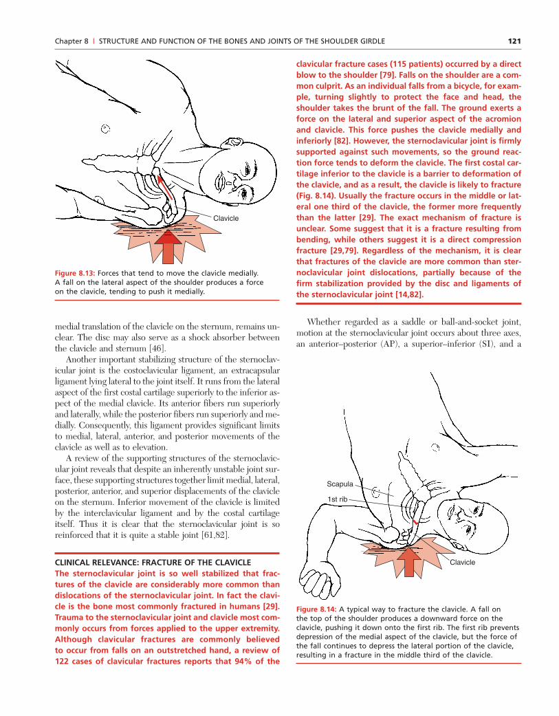
CLINICAL RELEVANCE: FRACTURE OF THE CLAVICLEThe sternoclavicular joint is so well stabilized that frac-tures of the clavicle are considerably more common thandislocations of the sternoclavicular joint. In fact the clavi-cle is the bone most commonly fractured in humans [29].Trauma to the sternoclavicular joint and clavicle most com-monly occurs from forces applied to the upper extremity.Although clavicular fractures are commonly believedto occur from falls on an outstretched hand, a review of122 cases of clavicular fractures reports that 94% of the
clavicular fracture cases (115 patients) occurred by a directblow to the shoulder [79]. Falls on the shoulder are a com-mon culprit. As an individual falls from a bicycle, for exam-ple, turning slightly to protect the face and head, theshoulder takes the brunt of the fall. The ground exerts aforce on the lateral and superior aspect of the acromionand clavicle. This force pushes the clavicle medially andinferiorly [82]. However, the sternoclavicular joint is firmlysupported against such movements, so the ground reac-tion force tends to deform the clavicle. The first costal car-tilage inferior to the clavicle is a barrier to deformation ofthe clavicle, and as a result, the clavicle is likely to fracture(Fig. 8.14). Usually the fracture occurs in the middle or lat-eral one third of the clavicle, the former more frequentlythan the latter [29]. The exact mechanism of fracture isunclear. Some suggest that it is a fracture resulting frombending, while others suggest it is a direct compressionfracture [29,79]. Regardless of the mechanism, it is clearthat fractures of the clavicle are more common than ster-noclavicular joint dislocations, partially because of thefirm stabilization provided by the disc and ligaments ofthe sternoclavicular joint [14,82].
Whether regarded as a saddle or ball-and-socket joint,motion at the sternoclavicular joint occurs about three axes,an anterior–posterior (AP), a superior–inferior (SI), and a
Chapter 8 | STRUCTURE AND FUNCTION OF THE BONES AND JOINTS OF THE SHOULDER GIRDLE
Clavicle
Figure 8.13: Forces that tend to move the clavicle medially.A fall on the lateral aspect of the shoulder produces a forceon the clavicle, tending to push it medially.
Clavicle
1st rib
Scapula
Figure 8.14: A typical way to fracture the clavicle. A fall onthe top of the shoulder produces a downward force on theclavicle, pushing it down onto the first rib. The first rib preventsdepression of the medial aspect of the clavicle, but the force ofthe fall continues to depress the lateral portion of the clavicle,resulting in a fracture in the middle third of the clavicle.

122
longitudinal axis through the length of the clavicle (Fig 8.15).Although these axes are described as slightly oblique to thecardinal planes of the body [80], the motions of the clavicletake place very close to these planes. Movement about theAP axis yields elevation and depression, which occur ap-proximately in the frontal plane. Movements about the SIaxis are known as protraction and retraction and occur inthe transverse plane. Rotations around the longitudinal axisare upward and downward rotation, defined by whetherthe anterior surface of the clavicle turns up (upward rota-tion) or down (downward rotation).
Although movement at the sternoclavicular joint is rota-tional, the prominence of the clavicular head and the loca-tion of the joint’s axes allow easy palpation of the head of theclavicle during most of these motions. This palpation fre-quently results in confusion for the novice clinician. Notethat retraction of the clavicle causes the head of the clavicleto move anteriorly on the sternum as the body of the clavi-cle rotates posteriorly (Fig. 8.16). Similarly in protraction theclavicular head rolls posteriorly as the body moves anteriorly.Likewise in elevation the body of the clavicle and theacromion rise, but the head of the clavicle descends on thesternum; depression of the sternoclavicular joint is the reverse.These movements of the proximal and distal clavicular sur-faces in opposite directions are consistent with rotations ofthe sternoclavicular joint and are the result of the location ofthe axes within the clavicle itself. The exact location of theaxes about which the movements of the sternoclavicularjoint occur are debated, but probably the axes lie somewhatlateral to the head of the clavicle [3,71]. This location explainsthe movement of the lateral and medial ends of the clavicle
in apparently opposite directions. With the axes of motionlocated between the two ends of the clavicle, pure rotationresults in opposite movements of the two ends, just as thetwo ends of a seesaw move in opposite directions during purerotation about the pivot point.
Few studies are available that investigate the availableROM of the sternoclavicular joint. The total excursion of ele-vation and depression is reportedly 50 to 60�, with depressionbeing less than 10� of the total [58,80]. Elevation is limited bythe costoclavicular ligament, and depression by the superiorportion of the capsule and the interclavicular ligament [3,80].Some suggest that contact between the clavicle and the firstrib also limits depression of the sternoclavicular joint [71].Facets found in some cadaver specimens between the clavicleand first costal cartilage provide strong evidence for contactbetween these structures in at least some individuals [3,71].
Protraction and retraction appear to be more equal in ex-cursion, with a reported total excursion ranging from 30 to60� [71,80]. Protraction is limited by the posterior stern-oclavicular ligament limiting the backward movement of theclavicular head and by the costoclavicular ligament limitingthe forward movement of the body of the clavicle. Retractionis limited similarly by the anterior sternoclavicular ligamentand by the costoclavicular ligament. The interclavicular liga-ment assists in limiting both motions [3].
Upward and downward rotations appear to be more lim-ited than the other motions, with estimates of upward rota-tion ROM that vary from 25 to 55� [3,37,71]. Although thereare no known studies of downward rotation ROM, it appearsto be much less than upward rotation, probably less than 10�.Regardless of the exact amount of excursion available at the
Part II | KINESIOLOGY OF THE UPPER EXTREMITY
SternumDownward
rotation
Upward
rotation
Vertical axis
AP axis
ML axis
Retraction
Elevation
Protraction
Depression
Figure 8.15: Axes of motion of the sternoclavicular joint.A. Elevation and depression of the sternoclavicular joint occurabout an anterior–posterior axis. B. Protraction and retractionof the sternoclavicular joint occur about a vertical axis.C. Upward and downward rotation of the sternoclavicularjoint occur about a medial–lateral axis.
ClavicleAcromion
Humerus
Figure 8.16: Movement of the head of the clavicle. Rotationof the sternoclavicular joint about an axis causes the head ofthe clavicle to move in a direction opposite the motion of therest of the clavicle just as the two ends of a seesaw move inopposite directions about a central pivot point.
123
sternoclavicular joint, it is well understood that motion atthe sternoclavicular joint is intimately related to motions ofthe other joints of the shoulder complex. How these motionsare related is discussed after each joint is presented.
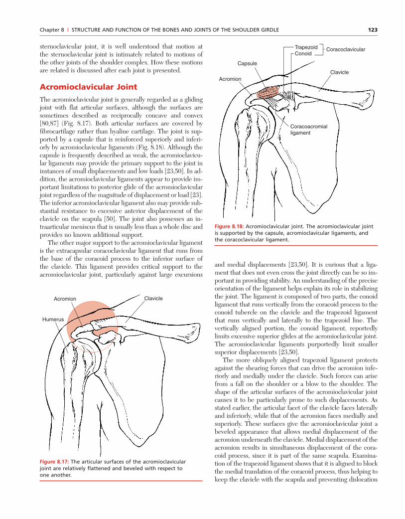
Acromioclavicular JointThe acromioclavicular joint is generally regarded as a glidingjoint with flat articular surfaces, although the surfaces aresometimes described as reciprocally concave and convex[80,87] (Fig. 8.17). Both articular surfaces are covered byfibrocartilage rather than hyaline cartilage. The joint is sup-ported by a capsule that is reinforced superiorly and inferi-orly by acromioclavicular ligaments (Fig. 8.18). Although thecapsule is frequently described as weak, the acromioclavicu-lar ligaments may provide the primary support to the joint ininstances of small displacements and low loads [23,50]. In ad-dition, the acromioclavicular ligaments appear to provide im-portant limitations to posterior glide of the acromioclavicularjoint regardless of the magnitude of displacement or load [23].The inferior acromioclavicular ligament also may provide sub-stantial resistance to excessive anterior displacement of theclavicle on the scapula [50]. The joint also possesses an in-traarticular meniscus that is usually less than a whole disc andprovides no known additional support.
The other major support to the acromioclavicular ligamentis the extracapsular coracoclavicular ligament that runs fromthe base of the coracoid process to the inferior surface ofthe clavicle. This ligament provides critical support to theacromioclavicular joint, particularly against large excursions
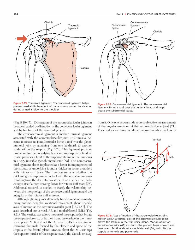
and medial displacements [23,50]. It is curious that a liga-ment that does not even cross the joint directly can be so im-portant in providing stability. An understanding of the preciseorientation of the ligament helps explain its role in stabilizingthe joint. The ligament is composed of two parts, the conoidligament that runs vertically from the coracoid process to theconoid tubercle on the clavicle and the trapezoid ligamentthat runs vertically and laterally to the trapezoid line. Thevertically aligned portion, the conoid ligament, reportedlylimits excessive superior glides at the acromioclavicular joint.The acromioclavicular ligaments purportedly limit smallersuperior displacements [23,50].
The more obliquely aligned trapezoid ligament protectsagainst the shearing forces that can drive the acromion infe-riorly and medially under the clavicle. Such forces can arisefrom a fall on the shoulder or a blow to the shoulder. Theshape of the articular surfaces of the acromioclavicular jointcauses it to be particularly prone to such displacements. Asstated earlier, the articular facet of the clavicle faces laterallyand inferiorly, while that of the acromion faces medially andsuperiorly. These surfaces give the acromioclavicular joint abeveled appearance that allows medial displacement of theacromion underneath the clavicle. Medial displacement of theacromion results in simultaneous displacement of the cora-coid process, since it is part of the same scapula. Examina-tion of the trapezoid ligament shows that it is aligned to blockthe medial translation of the coracoid process, thus helping tokeep the clavicle with the scapula and preventing dislocation
Chapter 8 | STRUCTURE AND FUNCTION OF THE BONES AND JOINTS OF THE SHOULDER GIRDLE
ClavicleAcromion
Humerus
Figure 8.17: The articular surfaces of the acromioclavicularjoint are relatively flattened and beveled with respect toone another.
ClavicleAcromion
Capsule
Coracoacromialligament
TrapezoidConoid
Coracoclavicular
Figure 8.18: Acromioclavicular joint. The acromioclavicular jointis supported by the capsule, acromioclavicular ligaments, andthe coracoclavicular ligament.
124
(Fig. 8.19) [71]. Dislocation of the acromioclavicular joint canbe accompanied by disruption of the coracoclavicular ligamentand by fractures of the coracoid process.
The coracoacromial ligament is another unusual ligamentassociated with the acromioclavicular joint. It is unusual be-cause it crosses no joint. Instead it forms a roof over the gleno-humeral joint by attaching from one landmark to anotherlandmark on the scapula (Fig. 8.20). This ligament providesprotection for the underlying bursa and supraspinatus tendon.It also provides a limit to the superior gliding of the humerusin a very unstable glenohumeral joint [53]. The coracoacro-mial ligament also is implicated as a factor in impingement ofthe structures underlying it and is thicker in some shoulderswith rotator cuff tears. The question remains whether thethickening is a response to contact with the unstable humerusresulting from the disrupted rotator cuff or whether the thick-ening is itself a predisposing factor for rotator cuff tears [76].Additional research is needed to clarify the relationship be-tween the morphology of the coracoacromial ligament and theintegrity of the rotator cuff muscles.
Although gliding joints allow only translational movements,many authors describe rotational movement about specificaxes of motion at the acromioclavicular joint [16,71,87]. Theaxes described are vertical, AP, and medial/lateral (ML) (Fig.8.21). The vertical axis allows motion of the scapula that bringsthe scapula closer to, or farther from, the clavicle in the trans-verse plane. Motion about the AP axis results in enlarging orshrinking the angle formed by the clavicle and spine of thescapula in the frontal plane. Motion about the ML axis tipsthe superior border of the scapula toward the clavicle or away
Part II | KINESIOLOGY OF THE UPPER EXTREMITY
Scapula
Trapezoidligament
Clavicle
F
Figure 8.19: Trapezoid ligament. The trapezoid ligament helpsprevent medial displacement of the acromion under the clavicleduring a medial blow to the shoulder.
Clavicle
Subacromialspace
Coracoacromialligament
Figure 8.20: Coracoacromial ligament. The coracoacromialligament forms a roof over the humeral head and helpscreate the subacromial space.
M-L
Vertical
A-P
Figure 8.21: Axes of motion of the acromioclavicular joint.Motion about a vertical axis of the acromioclavicular jointmoves the scapula in the transverse plane. Motion about ananterior–posterior (AP) axis turns the glenoid fossa upward anddownward. Motion about a medial–lateral (ML) axis tilts thescapula anteriorly and posteriorly.
from it. Only one known study reports objective measurementsof the angular excursion at the acromioclavicular joint [71].These values are based on direct measurements as well as on

125
mathematical models. These data suggest that the largest ex-cursion occurs about the vertical axis but is less than 10�. Mo-tion about the other two axes appears to be less than 5� in eachdirection. This study suggests that the primary source of scapu-lar movement on the thorax is the sternoclavicular joint.
Viewed in the context of the shoulder complex, the acromio-clavicular joint is responsible for maintaining articulation ofthe clavicle with the scapula, even as these two bones move inseparate patterns. Whether this results in systematic rotationalmotions or in a gliding reorientation of the bones is not criti-cal to the clinician, since in either case the motions cannot bereadily measured. What is essential is the recognition that al-though the clavicle and scapula move together, their contri-butions to whole shoulder motion require that they also movesomewhat independently of one another. This independentmovement requires motion at the acromioclavicular joint.
Scapulothoracic JointThe scapulothoracic joint, as stated earlier, is an atypicaljoint that lacks all of the traditional characteristics of a jointexcept one, motion. The primary role of this joint is to am-plify the motion of the glenohumeral joint, thus increasingthe range and diversity of movements between the arm and
trunk. In addition, the scapulothoracic joint with its sur-rounding musculature is described as an important shockabsorber protecting the shoulder, particularly during falls onan outstretched arm [46].
Primary motions of the scapulothoracic joint include twotranslations and two rotations (Fig. 8.22). Those motions are
• Elevation and depression• Abduction and adduction• Downward (medial) and upward (lateral) rotations• Scapular tilt
Elevation is defined as the movement of the entire scapulasuperiorly on the thorax. Depression is the opposite. Ab-duction is defined as the entire medial border of the scapulamoving away from the vertebrae, and adduction as move-ment toward the vertebrae. Abduction and adduction of thescapulothoracic joint are occasionally referred to as protrac-tion and retraction. However, protraction also is used bysome to refer to the combination of abduction and upwardrotation of the scapula. Others use the term protraction torefer to a rounded shoulder posture that may include ab-duction and downward rotation of the scapula. Thereforeto avoid confusion, this text describes scapular movements
Chapter 8 | STRUCTURE AND FUNCTION OF THE BONES AND JOINTS OF THE SHOULDER GIRDLE
A
C
B
Elevation
Depression
Abduction Adduction
Upward rotation
Downward rotation
Figure 8.22: Primary motions of the scapulothoracic joint. A. Elevation and depression. B. Abduction and adduction.C. Upward and downward rotation.
126 Part II | KINESIOLOGY OF THE UPPER EXTREMITY
discretely as flexion and extension, abduction and adduction,and upward and downward rotation. Protraction and retrac-tion refer solely to the motions of the sternoclavicular jointin the transverse plane.
Downward (medial) rotation of the scapula is definedas a rotation about an AP axis resulting in downward turn ofthe glenoid fossa as the inferior angle moves toward thevertebrae. Upward (lateral) rotation is the opposite. Thelocation of the axis of downward and upward rotation is con-troversial but appears to be slightly inferior to the scapularspine, approximately equidistant from the vertebral and axil-lary borders [83]. It is likely that the exact location varies withROM of the shoulder.
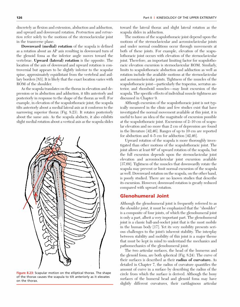
As the scapula translates on the thorax in elevation and de-pression or in abduction and adduction, it tilts anteriorly andposteriorly in response to the shape of the thorax as well. Forexample, in elevation of the scapulothoracic joint, the scapulatilts anteriorly about a medial lateral axis as it conforms to thenarrowing superior thorax (Fig. 8.23). It rotates posteriorlyabout the same axis. As the scapula abducts, it also exhibitsslight medial rotation about a vertical axis as the scapula slides
toward the lateral thorax and slight lateral rotation as thescapula slides in adduction.
The motions of the scapulothoracic joint depend upon themotions of the sternoclavicular and acromioclavicular jointsand under normal conditions occur through movements atboth of these joints. For example, elevation of the scapu-lothoracic joint occurs with elevation of the sternoclavicularjoint. Therefore, an important limiting factor for scapulotho-racic elevation excursion is sternoclavicular ROM. Similarly,limits to scapulothoracic abduction and adduction as well asrotation include the available motions at the sternoclavicularand acromioclavicular joints. Tightness of the muscles of thescapulothoracic joint—particularly the trapezius, serratus an-terior, and rhomboid muscles—may limit excursion of thescapula. The specific effects of individual muscle tightness arediscussed in Chapter 9.
Although excursion of the scapulothoracic joint is not typ-ically measured in the clinic and few studies exist that haveinvestigated the normal movement available at this joint, it isuseful to have an idea of the magnitude of excursion possibleat the scapulothoracic joint. Excursions of 2–10 cm of scapu-lar elevation and no more than 2 cm of depression are foundin the literature [42,46]. Ranges of up to 10 cm are reportedfor abduction and 4–5 cm for adduction [42,46].
Upward rotation of the scapula is more thoroughly inves-tigated than other motions of the scapulothoracic joint. Thejoint allows at least 60� of upward rotation of the scapula, butthe full excursion depends upon the sternoclavicular jointelevation and acromioclavicular joint excursion available[37,69]. Tightness of the muscles that downwardly rotate thescapula may prevent or limit normal excursion of the scapulaas well. Downward rotation on the scapula, on the other hand,is poorly studied. There are no known studies that describeits excursion. However, downward rotation is greatly reducedcompared with upward rotation.
Glenohumeral JointAlthough the glenohumeral joint is frequently referred to asthe shoulder joint, it must be emphasized that the “shoulder”is a composite of four joints, of which the glenohumeral jointis only a part, albeit a very important part. The glenohumeraljoint is a classic ball-and-socket joint that is the most mobilein the human body [17]. Yet its very mobility presents seri-ous challenges to the joint’s inherent stability. The interplaybetween stability and mobility of this joint is a major themethat must be kept in mind to understand the mechanics andpathomechanics of the glenohumeral joint.
The two articular surfaces, the head of the humerus andthe glenoid fossa, are both spherical (Fig. 8.24). The curve oftheir surfaces is described as their radius of curvature. Asdetailed in Chapter 7, the radius of curvature quantifies theamount of curve in a surface by describing the radius of thecircle from which the surface is derived. Although the bonysurfaces of the humeral head and glenoid fossa may haveslightly different curvatures, their cartilaginous articular
Figure 8.23: Scapular motion on the elliptical thorax. The shapeof the thorax causes the scapula to tilt anteriorly as it elevateson the thorax.

127
surfaces have approximately the same radius of curvature[36,77,85]. Because these surfaces have similar curvatures,they fit well together; that is, there is a high degree of con-gruence. Increased congruence spreads the loads applied tothe joint across a larger surface area and thus reduces the stress(force/area) applied to the articular surface. However, theamount of congruence is variable, even in healthy gleno-humeral joints [4]. In cadavers, decreased congruence leadsto an increase in the gliding motions between the humeralhead and the glenoid fossa [4,44]. Thus decreased congruencemay be a contributing factor in glenohumeral joint instability.
Although the articular surfaces of the glenohumeral jointare similarly curved, the actual areas of the articular surfacesare quite different from one another. While the head of thehumerus is approximately one half of a sphere, the surfacearea of the glenoid fossa is less than one half that of thehumeral head [41,48]. This disparity in articular surface sizeshas dramatic effects on both the stability and mobility of theglenohumeral joint. First, the difference in the size of the ar-ticular surfaces allows a large degree of mobility since thereis no bony limitation to the excursion. The size of the articu-lar surfaces is an important factor in making the glenohumeral
joint the most mobile in the body. However, by allowingtremendous mobility, the articular surfaces provide little orno stability for the glenohumeral joint [53]. The stability ofthe glenohumeral joint depends upon nonbony structures.
SUPPORTING STRUCTURES OFTHE GLENOHUMERAL JOINT
The supporting structures of the glenohumeral joint consistof the
• Labrum• Capsule• Three glenohumeral ligaments• Coracohumeral ligament• Surrounding musculature
The noncontractile supporting structures of the glenohumeraljoint are discussed in this section. The role of muscles in sup-porting the joint is discussed in Chapter 9.
The shallow glenoid fossa has already been identifiedas a contributing factor in glenohumeral joint instability.The stability is improved by deepening the fossa with thelabrum (Fig. 8.25). The labrum is a ring of fibrous tissue and
Chapter 8 | STRUCTURE AND FUNCTION OF THE BONES AND JOINTS OF THE SHOULDER GIRDLE
Axillary recess
Joint cavity
Articular cartilage of glenoid fossa
Articular cartilage of head of humerus
Articular capsule of shoulder joint
Long head of biceps muscle
Figure 8.24: Articular surfaces of the glenohumeral joint. Thehumeral head and the glenoid fossa possess similar curvatures.
Axillary recess
Joint cavity
Articular capsule of shoulder joint
Long head of biceps muscle
Glenoidlabrum
Glenoidlabrum
Figure 8.25: Glenoid labrum. The glenoid labrum deepens theglenoid fossa.

128
The remaining connective tissue supporting structures ofthe glenohumeral joint are known collectively as the capsu-loligamentous complex. It consists of the joint capsule andreinforcing ligaments. It encircles the entire joint and providesprotection against excessive rotation and translation in all di-rections. It is important to recognize that the integrity of thecomplex depends on the integrity of each of its components.
The fibrous capsule of the glenohumeral joint is inti-mately related to the labrum. The capsule attaches distally tothe anatomical neck of the humerus and proximally to theperiphery of the glenoid fossa and/or to the labrum itself.Inferiorly, it is quite loose, forming folds (Fig. 8.26). Thesefolds must open, or unfold, as the glenohumeral joint elevatesin abduction or flexion.
CLINICAL RELEVANCE: ADHESIVE CAPSULITISIn adhesive capsulitis, fibrous adhesions form in the gleno-humeral joint capsule, particularly in the inferior folds. Thecapsule then is unable to unfold to allow full flexion orabduction, resulting in decreased joint excursion. Onset isfrequently insidious, and the etiology is unknown.However, the classic physical findings are severe andpainful limitations in joint ROM [27,62].
The normal capsule is quite lax and, by itself, contributes lit-tle to the stability of the glenohumeral joint. However, it is
Part II | KINESIOLOGY OF THE UPPER EXTREMITY
A
Joint capsule
BJoint capsule
Joint cavity
Joint cavity
fibrocartilage surrounding the periphery of the fossa, ap-proximately doubling the depth of the articular surface of thefossa [35,59]. Besides increasing the depth of the articularsurface, the ring increases the articular contact area, whichalso decreases the stress (force/area) on the glenoid fossa. Thelabrum provides these benefits while being deformable,thereby adding little or no restriction to glenohumeral move-ment. Magnetic resonance imaging (MRI) shows consider-able variation in the shape of the labrum in asymptomaticshoulders, including notches and separations, particularly inthe anterior aspect of the ring. A small percentage of indi-viduals lack portions of the labrum [66].
Labral tears are well described in the clinical literature[15,65]. Mechanical tests of the ring demonstrate that it isweakest anteriorly and inferiorly, which is consistent with theclinical finding that anterior tears are the most common [28].However, the functional significance of a torn labrum in theabsence of other pathology remains controversial [16,65,68].The amount of dysfunction that results from a labral tearprobably depends upon the severity of the lesion. Small tearsmay have little or no effect, while large tears that extend toother parts of the joint capsule produce significant instability.The normal variability of the labrum in asymptomatic shoul-ders lends strength to the concept that small isolated labraltears do not result in significant dysfunction. However, addi-tional studies are needed to clarify the role of labral tears inglenohumeral dysfunction.
Figure 8.26: Glenohumeral joint capsule. A. When the shoulder is in neutral, the inferior portion of the capsule is lax andappears folded. B. In abduction the folds of the inferior capsule are unfolded, and the capsule is pulled more taut.
129Chapter 8 | STRUCTURE AND FUNCTION OF THE BONES AND JOINTS OF THE SHOULDER GIRDLE
Figure 8.27: Glenohumeral joint. The glenohumeral joint capsuleis reinforced by the superior, middle, and inferior glenohumeralligaments. The joint is also supported by the coracohumeralligament.
Coracohumeral ligament
Glenohumeral ligament:SuperiorMiddleInferior
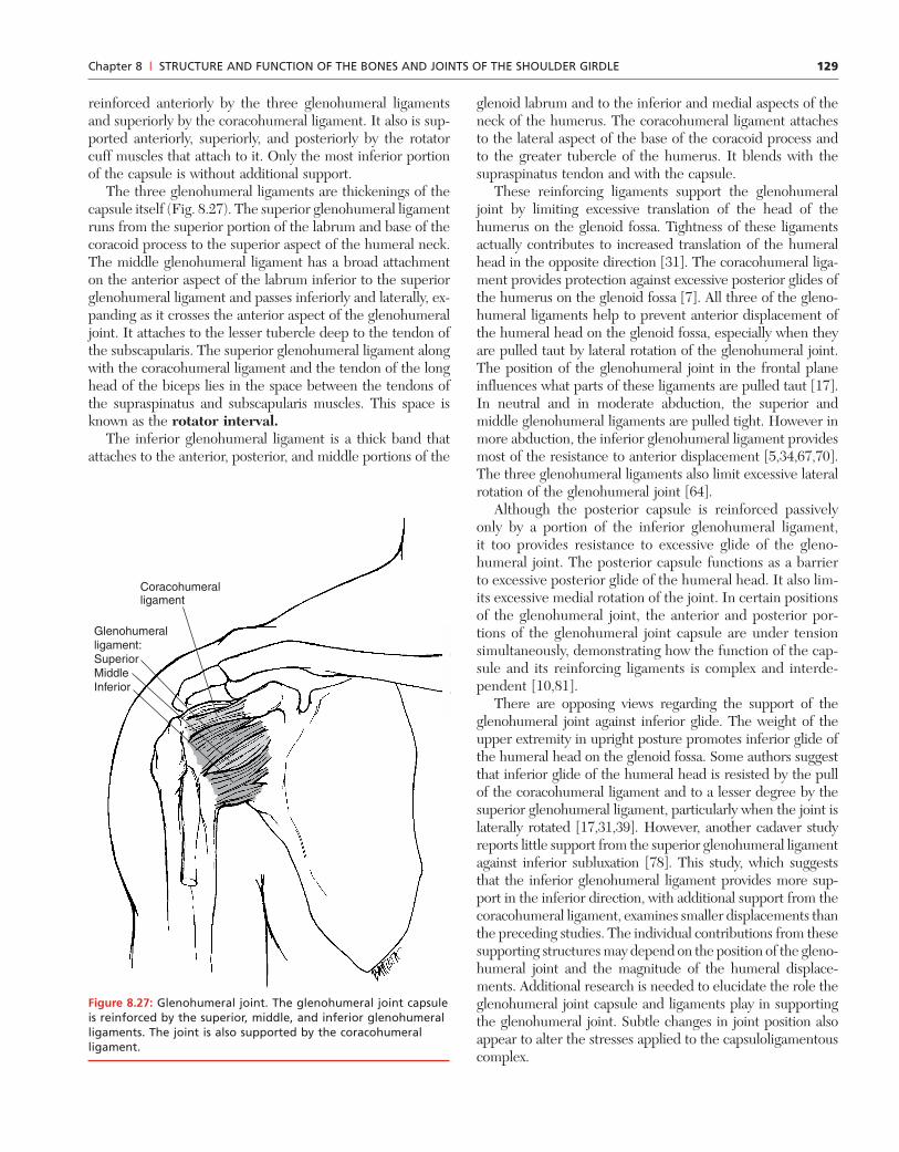
reinforced anteriorly by the three glenohumeral ligamentsand superiorly by the coracohumeral ligament. It also is sup-ported anteriorly, superiorly, and posteriorly by the rotatorcuff muscles that attach to it. Only the most inferior portionof the capsule is without additional support.
The three glenohumeral ligaments are thickenings of thecapsule itself (Fig. 8.27). The superior glenohumeral ligamentruns from the superior portion of the labrum and base of thecoracoid process to the superior aspect of the humeral neck.The middle glenohumeral ligament has a broad attachmenton the anterior aspect of the labrum inferior to the superiorglenohumeral ligament and passes inferiorly and laterally, ex-panding as it crosses the anterior aspect of the glenohumeraljoint. It attaches to the lesser tubercle deep to the tendon ofthe subscapularis. The superior glenohumeral ligament alongwith the coracohumeral ligament and the tendon of the longhead of the biceps lies in the space between the tendons ofthe supraspinatus and subscapularis muscles. This space isknown as the rotator interval.
The inferior glenohumeral ligament is a thick band thatattaches to the anterior, posterior, and middle portions of the
glenoid labrum and to the inferior and medial aspects of theneck of the humerus. The coracohumeral ligament attachesto the lateral aspect of the base of the coracoid process andto the greater tubercle of the humerus. It blends with thesupraspinatus tendon and with the capsule.
These reinforcing ligaments support the glenohumeraljoint by limiting excessive translation of the head of thehumerus on the glenoid fossa. Tightness of these ligamentsactually contributes to increased translation of the humeralhead in the opposite direction [31]. The coracohumeral liga-ment provides protection against excessive posterior glides ofthe humerus on the glenoid fossa [7]. All three of the gleno-humeral ligaments help to prevent anterior displacement ofthe humeral head on the glenoid fossa, especially when theyare pulled taut by lateral rotation of the glenohumeral joint.The position of the glenohumeral joint in the frontal planeinfluences what parts of these ligaments are pulled taut [17].In neutral and in moderate abduction, the superior andmiddle glenohumeral ligaments are pulled tight. However inmore abduction, the inferior glenohumeral ligament providesmost of the resistance to anterior displacement [5,34,67,70].The three glenohumeral ligaments also limit excessive lateralrotation of the glenohumeral joint [64].
Although the posterior capsule is reinforced passivelyonly by a portion of the inferior glenohumeral ligament,it too provides resistance to excessive glide of the gleno-humeral joint. The posterior capsule functions as a barrierto excessive posterior glide of the humeral head. It also lim-its excessive medial rotation of the joint. In certain positionsof the glenohumeral joint, the anterior and posterior por-tions of the glenohumeral joint capsule are under tensionsimultaneously, demonstrating how the function of the cap-sule and its reinforcing ligaments is complex and interde-pendent [10,81].
There are opposing views regarding the support of theglenohumeral joint against inferior glide. The weight of theupper extremity in upright posture promotes inferior glide ofthe humeral head on the glenoid fossa. Some authors suggestthat inferior glide of the humeral head is resisted by the pullof the coracohumeral ligament and to a lesser degree by thesuperior glenohumeral ligament, particularly when the joint islaterally rotated [17,31,39]. However, another cadaver studyreports little support from the superior glenohumeral ligamentagainst inferior subluxation [78]. This study, which suggeststhat the inferior glenohumeral ligament provides more sup-port in the inferior direction, with additional support from thecoracohumeral ligament, examines smaller displacements thanthe preceding studies. The individual contributions from thesesupporting structures may depend on the position of the gleno-humeral joint and the magnitude of the humeral displace-ments. Additional research is needed to elucidate the role theglenohumeral joint capsule and ligaments play in supportingthe glenohumeral joint. Subtle changes in joint position alsoappear to alter the stresses applied to the capsuloligamentouscomplex.
130
CLINICAL RELEVANCE: THERAPEUTIC STRETCH OFTHE GLENOHUMERAL JOINTBy altering the position of the glenohumeral joint, a clini-cian can direct treatment toward a particular portion ofthe capsuloligamentous complex. Anterior translation ofthe humerus while the glenohumeral joint is abducted hasa greater effect on the inferior glenohumeral ligamentthan on the superior or middle glenohumeral ligaments.The clinician can also use this information to reduce theloads on an injured or repaired structure.
One of the factors coupling the support of the glenohumeralligaments and capsule to each other is the intraarticularpressure that also helps to support the glenohumeral joint[38,39]. Puncturing, or venting, the rotator interval in ca-davers results in a reduction of the inferior stability of thehumeral head, even in the presence of an otherwise intactcapsule [39,88]. Isolated closure of rotator interval defects ap-pears to restore stability in young subjects who have no ad-ditional glenohumeral joint damage [20]. This supports thenotion that tears in this part of the capsule can destabilize thejoint not only by a structural weakening of the capsule itselfbut also by a disruption of the normal intraarticular pressure.
Thus the capsule with its reinforcing ligaments acts as abarrier to excessive translation of the humeral head and lim-its motion of the glenohumeral joint, particularly at the endsof glenohumeral ROM. It also contributes to the normal glideof the humerus on the glenoid fossa during shoulder motion.However, this complex of ligaments still is insufficient to sta-bilize the glenohumeral joint, particularly when externalloads are applied to the upper extremity or as the shoulder
moves through the middle of its full ROM. The role of themuscles in stabilization of the glenohumeral joint is discussedin Chapter 9.
MOTIONS OF THE GLENOHUMERAL JOINT
As a ball-and-socket joint, the glenohumeral joint has threeaxes of motion that lie in the cardinal planes of the body.Therefore the motions available at the glenohumeral joint are
• Flexion/extension• Abduction/adduction• Medial/lateral rotation
Abduction and flexion sometimes are each referred to as el-evation. Authors also distinguish between elevation of theglenohumeral joint in the plane of the scapula and that in thesagittal and frontal planes.
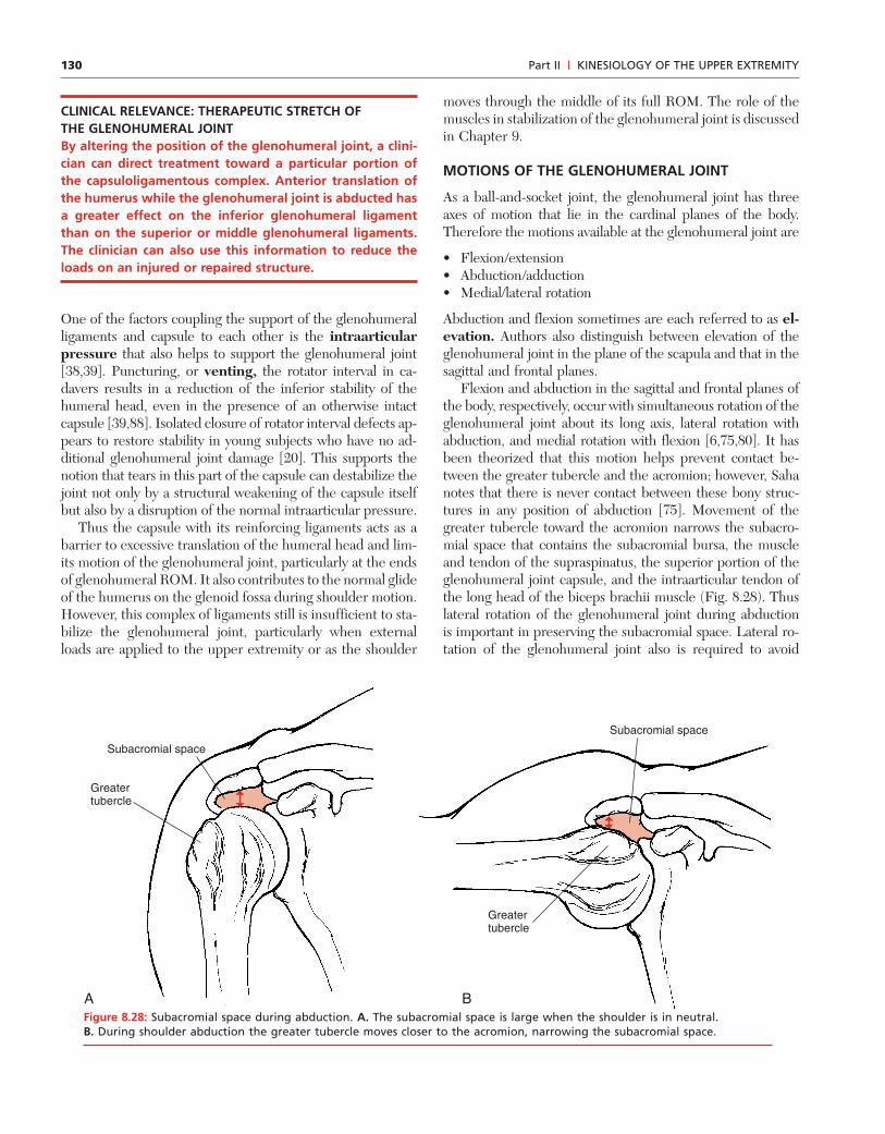
Flexion and abduction in the sagittal and frontal planes ofthe body, respectively, occur with simultaneous rotation of theglenohumeral joint about its long axis, lateral rotation withabduction, and medial rotation with flexion [6,75,80]. It hasbeen theorized that this motion helps prevent contact be-tween the greater tubercle and the acromion; however, Sahanotes that there is never contact between these bony struc-tures in any position of abduction [75]. Movement of thegreater tubercle toward the acromion narrows the subacro-mial space that contains the subacromial bursa, the muscleand tendon of the supraspinatus, the superior portion of theglenohumeral joint capsule, and the intraarticular tendon ofthe long head of the biceps brachii muscle (Fig. 8.28). Thuslateral rotation of the glenohumeral joint during abductionis important in preserving the subacromial space. Lateral ro-tation of the glenohumeral joint also is required to avoid
Part II | KINESIOLOGY OF THE UPPER EXTREMITY
Subacromial space
Subacromial space
Greatertubercle
Greatertubercle
A BFigure 8.28: Subacromial space during abduction. A. The subacromial space is large when the shoulder is in neutral.B. During shoulder abduction the greater tubercle moves closer to the acromion, narrowing the subacromial space.
131
impingement of the greater tubercle on the superior rim ofthe glenoid fossa [40]. Clearly, lateral rotation of the humerusis essential for full, pain-free abduction of the glenohumeraljoint in the frontal plane.
CLINICAL RELEVANCE: RESTORING SHOULDERABDUCTION ROMPatients with decreased abduction ROM frequently reportpain located in the superior aspect of the joint at the endof their available ROM of abduction. Assessment usuallyreveals diminished lateral rotation ROM as well.Restoration of lateral rotation ROM is essential to therecovery of abduction ROM. As lateral rotation rangeimproves, the patient usually notices decreased pain atthe end of abduction ROM. The decreased pain is attrib-uted to decreased impingement. Attempts to restoreabduction mobility without also restoring lateral rotationROM result in increased pain, patient dissatisfaction, andrarely any improvement in abduction ROM.
Rotation about the long axis of the humerus during abduc-tion and flexion disappears by the time the shoulder reachesapproximately 160� of flexion or abduction. Saha refers to thisposition as the “zero-position” and suggests that it resultsfrom an unwinding of the ligaments and muscles of the gleno-humeral joint, which occurs as the scapula and humerus movethrough the range [75]. Indeed, one of the characteristics offlexion and abduction in the plane of the scapula is that noglenohumeral rotation is required during the movement.Consequently, abduction in the plane of the scapula is morecomfortable for the patient with decreased rotation ROM.
Although flexion, abduction, and rotation of the gleno-humeral joint imply pure rotational movements, the asym-metrical articular areas of the humeral head and glenoid fossa,the pull of the capsuloligamentous complex, and the forcesfrom the surrounding muscles result in a complex combina-tion of rotation and gliding motions at the glenohumeral joint.If the motion of the glenohumeral joint consisted entirely ofpure rotation, the motion could be described as a rotationabout a fixed axis. When rotation is accompanied by gliding,the rotation can be described as occurring about a movingaxis. As described in Chapter 7, the degree of mobility of theaxis of rotation in the two-dimensional case is described bythe instant center of rotation (ICR). The ICR is the loca-tion of the axis of motion at a given joint position. The morestable the axis of motion, the more constant is the ICR. TheICR of the glenohumeral joint moves only slightly during flex-ion or abduction of the shoulder, indicating only minimaltranslation [86].
The amount of humeral head translation during shouldermotion has received considerable attention among cliniciansand researchers [25,30,31,48,86]. Glenohumeral translation isless during active shoulder motions when muscle contractions
help to stabilize the humeral head than during passive mo-tions [25]. In active elevation of the glenohumeral joint in theplane of the scapula, the humeral head undergoes minimalsuperior glide (�3 mm) and then remains fixed or glides in-feriorly no more than 1 mm [11,19,25,46,69,77]. Individualswith muscle fatigue or glenohumeral instability, however, con-sistently exhibit excessive superior glide during active shoul-der elevation [10,17,19,42].
The humeral head glides posteriorly in shoulder extensionand in lateral rotation; it translates anteriorly during abduc-tion and medial rotation [25,59,30,64,77]. These data contra-dict the so-called concave–convex rule, which states thatthe convex humeral head glides on the concave glenoid fossain directions opposite the humeral roll. For example, the con-cave–convex rule predicts that inferior glide of the humerusaccompanies its superior roll in flexion or abduction, and lat-eral rotation occurs with anterior glide [75,80]. Direct meas-urements reveal otherwise.
Although slight, joint glides appear to accompany gleno-humeral motions. This recognition supports the standardclinical practice of restoring translational movement to re-store full ROM at the glenohumeral joint. The concept ofjoint glide at the glenohumeral joint also forms the theoret-ical basis for many mobilization techniques used in the clinic.Reporting the amount of available passive humeral head glideas a percentage of the glenoid diameter in the direction ofthe glide, a study of anesthetized subjects without shoulderpathology reports that the humeral head can glide 17, 26,and 29% in the anterior, posterior, and inferior directions,respectively, with the glenohumeral joint in neutral [32]. Pas-sive glides of almost 1.5 cm are reported in subjects withoutshoulder impairments [9]. Patients with anterior instabilitiesdemonstrate significant increases in both anterior and infe-rior directions. Patients with multidirectional instabilities ex-hibit significantly increased excursions in all three directions[11,19]. It is essential for the clinician to understand thatslight translation occurs in normal glenohumeral joint mo-tion. Yet excessive translation may contribute to significantdysfunction.
Total glenohumeral joint elevation is most frequently de-scribed as a percentage of shoulder complex motion. Gleno-humeral flexion and abduction are reported to be 100–120�[37,69,84]; however, shoulder rotation comes solely from theglenohumeral joint. Although protraction of the sternoclav-icular joint and abduction of the scapulothoracic joint causethe humerus to face medially, these are substitutions for me-dial rotation of the shoulder rather than contributions to truemedial rotation. Similarly, retraction of the sternoclavicularjoint and adduction of the scapulothoracic joint can substi-tute for lateral rotation of the shoulder. True shoulder rota-tion ROM values range from approximately 70 to 90� for bothmedial and lateral rotation. There are no known studies thatidentify the contribution of the glenohumeral joint to shoul-der extension, but the glenohumeral joint is the likely sourceof most extension excursion, with only a minor contribution
Chapter 8 | STRUCTURE AND FUNCTION OF THE BONES AND JOINTS OF THE SHOULDER GIRDLE
132
from adduction and medial rotation of the scapulothoracicjoint.
In summary, this section reviews the individual joints thatconstitute the shoulder complex. Each joint has a uniquestructure that results in a unique pattern of mobility and sta-bility. The overall function of the shoulder complex dependson the individual contributions of each joint. A patient’s com-plaints to the clinician usually are focused on the function ofthe shoulder as a whole, such as an inability to reach over-head or the presence of pain in throwing a ball. The clinicianmust then determine where the impairment is within theshoulder complex. A full understanding of the role of eachjoint in the overall function of the shoulder complex is es-sential to the successful evaluation of the shoulder complex.The following section presents the role of each joint in theproduction of normal motion of the shoulder complex.
TOTAL SHOULDER MOVEMENT
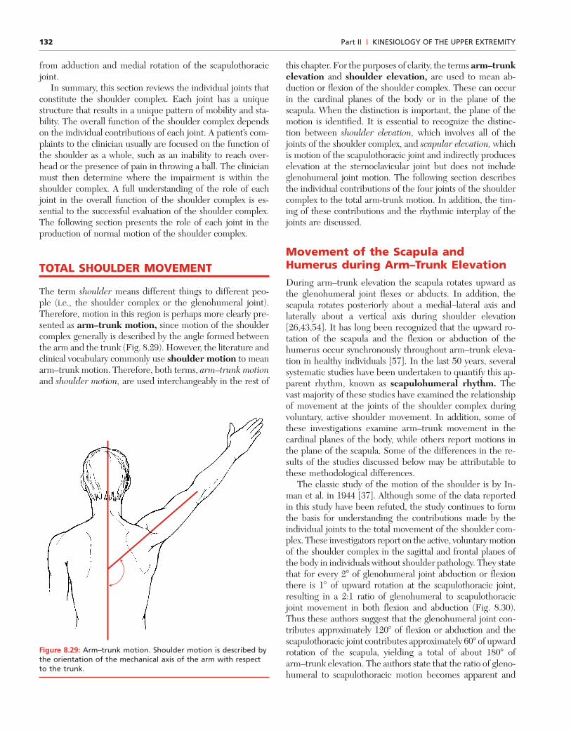
The term shoulder means different things to different peo-ple (i.e., the shoulder complex or the glenohumeral joint).Therefore, motion in this region is perhaps more clearly pre-sented as arm–trunk motion, since motion of the shouldercomplex generally is described by the angle formed betweenthe arm and the trunk (Fig. 8.29). However, the literature andclinical vocabulary commonly use shoulder motion to meanarm–trunk motion. Therefore, both terms, arm–trunk motionand shoulder motion, are used interchangeably in the rest of
this chapter. For the purposes of clarity, the terms arm–trunkelevation and shoulder elevation, are used to mean ab-duction or flexion of the shoulder complex. These can occurin the cardinal planes of the body or in the plane of thescapula. When the distinction is important, the plane of themotion is identified. It is essential to recognize the distinc-tion between shoulder elevation, which involves all of thejoints of the shoulder complex, and scapular elevation, whichis motion of the scapulothoracic joint and indirectly produceselevation at the sternoclavicular joint but does not includeglenohumeral joint motion. The following section describesthe individual contributions of the four joints of the shouldercomplex to the total arm-trunk motion. In addition, the tim-ing of these contributions and the rhythmic interplay of thejoints are discussed.
Movement of the Scapula andHumerus during Arm–Trunk ElevationDuring arm–trunk elevation the scapula rotates upward asthe glenohumeral joint flexes or abducts. In addition, thescapula rotates posteriorly about a medial–lateral axis andlaterally about a vertical axis during shoulder elevation[26,43,54]. It has long been recognized that the upward ro-tation of the scapula and the flexion or abduction of thehumerus occur synchronously throughout arm–trunk eleva-tion in healthy individuals [57]. In the last 50 years, severalsystematic studies have been undertaken to quantify this ap-parent rhythm, known as scapulohumeral rhythm. Thevast majority of these studies have examined the relationshipof movement at the joints of the shoulder complex duringvoluntary, active shoulder movement. In addition, some ofthese investigations examine arm–trunk movement in thecardinal planes of the body, while others report motions inthe plane of the scapula. Some of the differences in the re-sults of the studies discussed below may be attributable tothese methodological differences.
The classic study of the motion of the shoulder is by In-man et al. in 1944 [37]. Although some of the data reportedin this study have been refuted, the study continues to formthe basis for understanding the contributions made by theindividual joints to the total movement of the shoulder com-plex. These investigators report on the active, voluntary motionof the shoulder complex in the sagittal and frontal planes ofthe body in individuals without shoulder pathology. They statethat for every 2� of glenohumeral joint abduction or flexionthere is 1� of upward rotation at the scapulothoracic joint,resulting in a 2:1 ratio of glenohumeral to scapulothoracicjoint movement in both flexion and abduction (Fig. 8.30).Thus these authors suggest that the glenohumeral joint con-tributes approximately 120� of flexion or abduction and thescapulothoracic joint contributes approximately 60� of upwardrotation of the scapula, yielding a total of about 180� ofarm–trunk elevation. The authors state that the ratio of gleno-humeral to scapulothoracic motion becomes apparent and
Part II | KINESIOLOGY OF THE UPPER EXTREMITY
Figure 8.29: Arm–trunk motion. Shoulder motion is described bythe orientation of the mechanical axis of the arm with respectto the trunk.
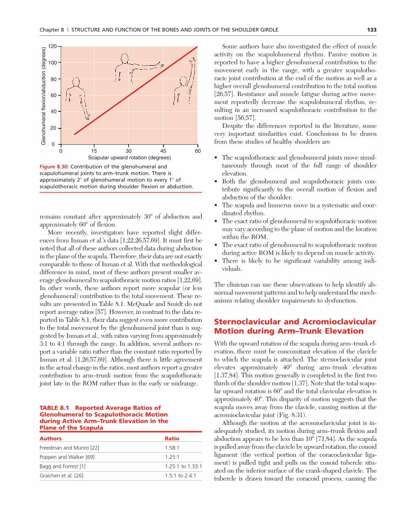
133
remains constant after approximately 30� of abduction andapproximately 60� of flexion.
More recently, investigators have reported slight differ-ences from Inman et al.’s data [1,22,26,57,69]. It must first benoted that all of these authors collected data during abductionin the plane of the scapula. Therefore, their data are not exactlycomparable to those of Inman et al. With that methodologicaldifference in mind, most of these authors present smaller av-erage glenohumeral to scapulothoracic motion ratios [1,22,69].In other words, these authors report more scapular (or lessglenohumeral) contribution to the total movement. These re-sults are presented in Table 8.1. McQuade and Smidt do notreport average ratios [57]. However, in contrast to the data re-ported in Table 8.1, their data suggest even more contributionto the total movement by the glenohumeral joint than is sug-gested by Inman et al., with ratios varying from approximately3:1 to 4:1 through the range. In addition, several authors re-port a variable ratio rather than the constant ratio reported byInman et al. [1,26,57,69]. Although there is little agreementin the actual change in the ratios, most authors report a greatercontribution to arm–trunk motion from the scapulothoracicjoint late in the ROM rather than in the early or midrange.
Some authors have also investigated the effect of muscleactivity on the scapulohumeral rhythm. Passive motion isreported to have a higher glenohumeral contribution to themovement early in the range, with a greater scapulotho-racic joint contribution at the end of the motion as well as ahigher overall glenohumeral contribution to the total motion[26,57]. Resistance and muscle fatigue during active move-ment reportedly decrease the scapulohumeral rhythm, re-sulting in an increased scapulothoracic contribution to themotion [56,57].
Despite the differences reported in the literature, somevery important similarities exist. Conclusions to be drawnfrom these studies of healthy shoulders are
• The scapulothoracic and glenohumeral joints move simul-taneously through most of the full range of shoulderelevation.
• Both the glenohumeral and scapulothoracic joints con-tribute significantly to the overall motion of flexion andabduction of the shoulder.
• The scapula and humerus move in a systematic and coor-dinated rhythm.
• The exact ratio of glenohumeral to scapulothoracic motionmay vary according to the plane of motion and the locationwithin the ROM.
• The exact ratio of glenohumeral to scapulothoracic motionduring active ROM is likely to depend on muscle activity.
• There is likely to be significant variability among indi-viduals.
The clinician can use these observations to help identify ab-normal movement patterns and to help understand the mech-anisms relating shoulder impairments to dysfunction.
Sternoclavicular and AcromioclavicularMotion during Arm–Trunk ElevationWith the upward rotation of the scapula during arm–trunk el-evation, there must be concomitant elevation of the clavicleto which the scapula is attached. The sternoclavicular jointelevates approximately 40� during arm–trunk elevation[1,37,84]. This motion generally is completed in the first twothirds of the shoulder motion [1,37]. Note that the total scapu-lar upward rotation is 60� and the total clavicular elevation isapproximately 40�. This disparity of motion suggests that thescapula moves away from the clavicle, causing motion at theacromioclavicular joint (Fig. 8.31).
Although the motion at the acromioclavicular joint is in-adequately studied, its motion during arm–trunk flexion andabduction appears to be less than 10� [71,84]. As the scapulais pulled away from the clavicle by upward rotation, the conoidligament (the vertical portion of the coracoclavicular liga-ment) is pulled tight and pulls on the conoid tubercle situ-ated on the inferior surface of the crank-shaped clavicle. Thetubercle is drawn toward the coracoid process, causing the
Chapter 8 | STRUCTURE AND FUNCTION OF THE BONES AND JOINTS OF THE SHOULDER GIRDLE
TABLE 8.1 Reported Average Ratios ofGlenohumeral to Scapulothoracic Motionduring Active Arm–Trunk Elevation in thePlane of the Scapula
Authors Ratio
Freedman and Munro [22] 1.58:1
Poppen and Walker [69] 1.25:1
Bagg and Forrest [1] 1.25:1 to 1.33:1
Graichen et al. [26] 1.5:1 to 2.4:1
Gle
nohu
mer
al fl
exio
n/ab
duct
ion
(deg
rees
)
Scapular upward rotation (degrees)0
20
0
40
60
80
120
100
15 30 45 60
Figure 8.30: Contribution of the glenohumeral andscapulohumeral joints to arm–trunk motion. There isapproximately 2� of glenohumeral motion to every 1� ofscapulothoracic motion during shoulder flexion or abduction.
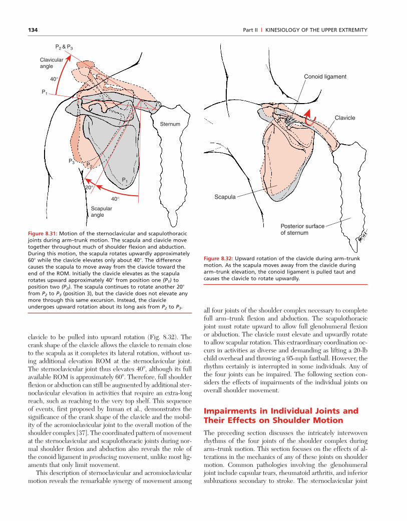
134
clavicle to be pulled into upward rotation (Fig. 8.32). Thecrank shape of the clavicle allows the clavicle to remain closeto the scapula as it completes its lateral rotation, without us-ing additional elevation ROM at the sternoclavicular joint.The sternoclavicular joint thus elevates 40�, although its fullavailable ROM is approximately 60�. Therefore, full shoulderflexion or abduction can still be augmented by additional ster-noclavicular elevation in activities that require an extra-longreach, such as reaching to the very top shelf. This sequenceof events, first proposed by Inman et al., demonstrates thesignificance of the crank shape of the clavicle and the mobil-ity of the acromioclavicular joint to the overall motion of theshoulder complex [37]. The coordinated pattern of movementat the sternoclavicular and scapulothoracic joints during nor-mal shoulder flexion and abduction also reveals the role ofthe conoid ligament in producing movement, unlike most lig-aments that only limit movement.
This description of sternoclavicular and acromioclavicularmotion reveals the remarkable synergy of movement among
all four joints of the shoulder complex necessary to completefull arm–trunk flexion and abduction. The scapulothoracicjoint must rotate upward to allow full glenohumeral flexionor abduction. The clavicle must elevate and upwardly rotateto allow scapular rotation. This extraordinary coordination oc-curs in activities as diverse and demanding as lifting a 20-lbchild overhead and throwing a 95-mph fastball. However, therhythm certainly is interrupted in some individuals. Any ofthe four joints can be impaired. The following section con-siders the effects of impairments of the individual joints onoverall shoulder movement.
Impairments in Individual Joints andTheir Effects on Shoulder MotionThe preceding section discusses the intricately interwovenrhythms of the four joints of the shoulder complex duringarm–trunk motion. This section focuses on the effects of al-terations in the mechanics of any of these joints on shouldermotion. Common pathologies involving the glenohumeraljoint include capsular tears, rheumatoid arthritis, and inferiorsubluxations secondary to stroke. The sternoclavicular joint
Part II | KINESIOLOGY OF THE UPPER EXTREMITY
P1
P2 & P3
P1
P2P3
Sternum
40°
40°
20°
Scapularangle
Clavicularangle
Figure 8.31: Motion of the sternoclavicular and scapulothoracicjoints during arm–trunk motion. The scapula and clavicle movetogether throughout much of shoulder flexion and abduction.During this motion, the scapula rotates upwardly approximately60� while the clavicle elevates only about 40�. The differencecauses the scapula to move away from the clavicle toward theend of the ROM. Initially the clavicle elevates as the scapularotates upward approximately 40� from position one (P1) toposition two (P2). The scapula continues to rotate another 20�
from P2 to P3 (position 3), but the clavicle does not elevate anymore through this same excursion. Instead, the clavicleundergoes upward rotation about its long axis from P2 to P3.
Conoid ligament
Clavicle
Scapula
Posterior surface of sternum
Figure 8.32: Upward rotation of the clavicle during arm–trunkmotion. As the scapula moves away from the clavicle duringarm–trunk elevation, the conoid ligament is pulled taut andcauses the clavicle to rotate upwardly.

135
can be affected by rheumatoid arthritis or by ankylosingspondylitis. The acromioclavicular joint is frequently dislo-cated and also is susceptible to traumatic osteoarthritis.Scapulothoracic joint function can be compromised by traumasuch as a gunshot wound or by scarring resulting from suchinjuries as burns. These are just examples to emphasize thateach joint of the shoulder complex is susceptible to patholo-gies that impair its function. Each joint is capable of losingmobility and thus affecting the mobility of the entire shoul-der complex. It is not possible to consider all conceivablepathologies and consequences. The purpose of this section isto consider the altered mechanics and potential substitutionsresulting from abnormal motion at each of the joints of theshoulder complex. Such consideration illustrates a frameworkfrom which to evaluate the function of the shoulder complexand the integrity of its components.
LOSS OF GLENOHUMERAL OR SCAPULOTHORACICJOINT MOTION
As discussed earlier in this chapter, the data from studies ofscapulohumeral rhythm suggest that the glenohumeral jointprovides more than 50% of the total shoulder flexion or ab-duction. Therefore, the loss of glenohumeral motion has aprofound effect on shoulder motion. However, it must be em-phasized that shoulder motion is not lost completely, evenwith complete glenohumeral joint immobility. The scapu-lothoracic and sternoclavicular joints with the acromioclav-icular joint combine to provide the remaining one third ormore motion. In the absence of glenohumeral movementthese joints, if healthy, may become even more mobile. Thuswithout glenohumeral joint motion and in the presence of
intact scapulothoracic, sternoclavicular, and acromioclavicu-lar joints, an individual should still have at least one third thenormal shoulder flexion or abduction ROM.
Complete loss of glenohumeral joint motion, however, re-sults in total loss of shoulder rotation. Yet even under theseconditions scapulothoracic motion can provide some substi-tution. Forward tipping of the scapula about a medial–lateralaxis is a common substitution for decreased medial rotationof the shoulder.
CLINICAL RELEVANCE: MEASUREMENT OFMEDIAL ROTATION ROM OF THE SHOULDERGoniometry manuals describe measurement of medialrotation of the shoulder with the subject lying supineand the shoulder abducted to 90� [63]. In this positionthe shoulder is palpated to identify anterior tilting of thescapula as the shoulder is medially rotated. Firm manualstabilization is usually necessary to prevent the scapulafrom tilting anteriorly to substitute for medial rotation(Fig. 8.33).
Conversely, the loss of scapulothoracic motion results in aloss of at least one third of full shoulder elevation ROM. Al-though this appears to be roughly true in passive ROM, Inmanet al. report that in the absence of scapulothoracic joint mo-tion, active shoulder abduction is closer to 90� of abductionrather than the expected 120� [37]. These authors hypothe-size that upward rotation of the scapula is essential to main-taining an adequate contractile length of the deltoid muscle.Scapular upward rotation lengthens the deltoid even as themuscle contracts across the glenohumeral joint during
Chapter 8 | STRUCTURE AND FUNCTION OF THE BONES AND JOINTS OF THE SHOULDER GIRDLE
Figure 8.33: Scapular substitutions in shoulder ROM. A. Standard goniometric measurement of medial rotation ROM of shoulderrequires adequate stabilization of the scapula. B. Inadequate stabilization allows anterior tilting of the scapula with an apparentincrease in medial rotation ROM of the shoulder.
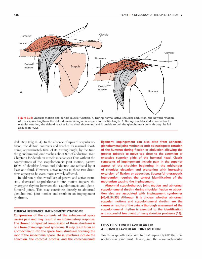
136
abduction (Fig. 8.34). In the absence of upward scapular ro-tation, the deltoid contracts and reaches its maximal short-ening, approximately 60% of its resting length, by the timethe glenohumeral joint reaches about 90� of abduction. (SeeChapter 4 for details on muscle mechanics.) Thus without thecontributions of the scapulothoracic joint motion, passiveROM of shoulder flexion and abduction are reduced by atleast one third. However, active ranges in these two direc-tions appear to be even more severely affected.
In addition to the overall loss of passive and active excur-sion, decreased scapulothoracic joint motion impairs thesynergistic rhythm between the scapulothoracic and gleno-humeral joints. This may contribute directly to abnormalglenohumeral joint motion and result in an impingementsyndrome.
CLINICAL RELEVANCE: IMPINGEMENT SYNDROMECompression of the contents of the subacromial spacecauses pain and may result in an inflammatory response.The chronic or repeated compression of these structures isone form of impingement syndrome. It may result from anencroachment into the space from structures forming theroof of the subacromial space. These structures include theacromion, the coracoid process, and the coracoacromial
ligament. Impingement can also arise from abnormalglenohumeral joint mechanics such as inadequate rotationof the humerus during flexion or abduction allowing thegreater tubercle to move too close to the acromion orexcessive superior glide of the humeral head. Classicsymptoms of impingement include pain in the superioraspect of the shoulder beginning in the midrangesof shoulder elevation and worsening with increasingexcursion of flexion or abduction. Successful therapeuticintervention requires the correct identification of themechanism causing the impingement.
Abnormal scapulothoracic joint motion and abnormalscapulohumeral rhythm during shoulder flexion or abduc-tion also are associated with impingement syndromes[46,49,54,55]. Although it is unclear whether abnormalscapular motions and scapulohumeral rhythm are thecauses or results of the pain, a thorough assessment of thescapulohumeral rhythm is essential to the identificationand successful treatment of many shoulder problems [12].
LOSS OF STERNOCLAVICULAR ORACROMIOCLAVICULAR JOINT MOTION
For the scapulothoracic joint to rotate upwardly 60�, the ster-noclavicular joint must elevate, and the acromioclavicular
Part II | KINESIOLOGY OF THE UPPER EXTREMITY
A
B
Clavicle
Deltoidmuscle Scapula
Deltoidmuscle
Clavicle
Scapula
Humerus
Humerus
Figure 8.34: Scapular motion and deltoid muscle function. A. During normal active shoulder abduction, the upward rotationof the scapula lengthens the deltoid, maintaining an adequate contractile length. B. During shoulder abduction withoutscapular rotation, the deltoid reaches its maximal shortening and is unable to pull the glenohumeral joint through its fullabduction ROM.
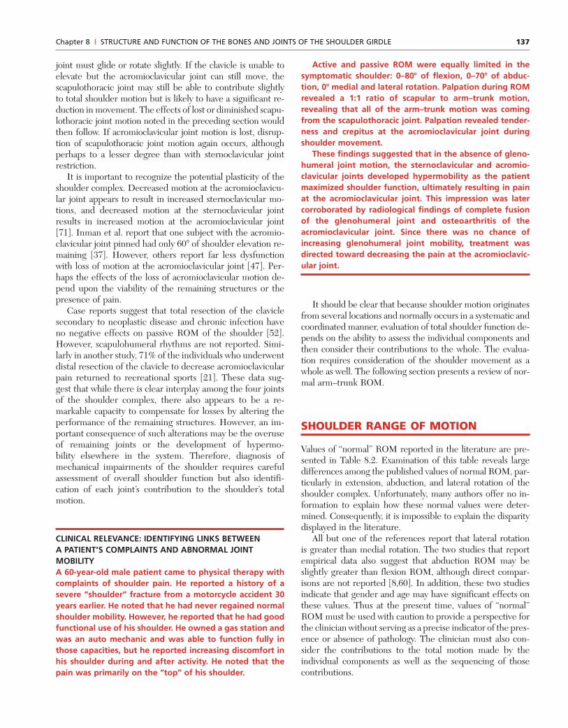
137
joint must glide or rotate slightly. If the clavicle is unable toelevate but the acromioclavicular joint can still move, thescapulothoracic joint may still be able to contribute slightlyto total shoulder motion but is likely to have a significant re-duction in movement. The effects of lost or diminished scapu-lothoracic joint motion noted in the preceding section wouldthen follow. If acromioclavicular joint motion is lost, disrup-tion of scapulothoracic joint motion again occurs, althoughperhaps to a lesser degree than with sternoclavicular jointrestriction.
It is important to recognize the potential plasticity of theshoulder complex. Decreased motion at the acromioclavicu-lar joint appears to result in increased sternoclavicular mo-tions, and decreased motion at the sternoclavicular jointresults in increased motion at the acromioclavicular joint[71]. Inman et al. report that one subject with the acromio-clavicular joint pinned had only 60� of shoulder elevation re-maining [37]. However, others report far less dysfunctionwith loss of motion at the acromioclavicular joint [47]. Per-haps the effects of the loss of acromioclavicular motion de-pend upon the viability of the remaining structures or thepresence of pain.
Case reports suggest that total resection of the claviclesecondary to neoplastic disease and chronic infection haveno negative effects on passive ROM of the shoulder [52].However, scapulohumeral rhythms are not reported. Simi-larly in another study, 71% of the individuals who underwentdistal resection of the clavicle to decrease acromioclavicularpain returned to recreational sports [21]. These data sug-gest that while there is clear interplay among the four jointsof the shoulder complex, there also appears to be a re-markable capacity to compensate for losses by altering theperformance of the remaining structures. However, an im-portant consequence of such alterations may be the overuseof remaining joints or the development of hypermo-bility elsewhere in the system. Therefore, diagnosis ofmechanical impairments of the shoulder requires carefulassessment of overall shoulder function but also identifi-cation of each joint’s contribution to the shoulder’s totalmotion.
CLINICAL RELEVANCE: IDENTIFYING LINKS BETWEENA PATIENT’S COMPLAINTS AND ABNORMAL JOINTMOBILITYA 60-year-old male patient came to physical therapy withcomplaints of shoulder pain. He reported a history of asevere “shoulder” fracture from a motorcycle accident 30years earlier. He noted that he had never regained normalshoulder mobility. However, he reported that he had goodfunctional use of his shoulder. He owned a gas station andwas an auto mechanic and was able to function fully inthose capacities, but he reported increasing discomfort inhis shoulder during and after activity. He noted that thepain was primarily on the “top” of his shoulder.
Active and passive ROM were equally limited in thesymptomatic shoulder: 0–80� of flexion, 0–70� of abduc-tion, 0� medial and lateral rotation. Palpation during ROMrevealed a 1:1 ratio of scapular to arm–trunk motion,revealing that all of the arm–trunk motion was comingfrom the scapulothoracic joint. Palpation revealed tender-ness and crepitus at the acromioclavicular joint duringshoulder movement.
These findings suggested that in the absence of gleno-humeral joint motion, the sternoclavicular and acromio-clavicular joints developed hypermobility as the patientmaximized shoulder function, ultimately resulting in painat the acromioclavicular joint. This impression was latercorroborated by radiological findings of complete fusionof the glenohumeral joint and osteoarthritis of theacromioclavicular joint. Since there was no chance ofincreasing glenohumeral joint mobility, treatment wasdirected toward decreasing the pain at the acromioclavic-ular joint.
It should be clear that because shoulder motion originatesfrom several locations and normally occurs in a systematic andcoordinated manner, evaluation of total shoulder function de-pends on the ability to assess the individual components andthen consider their contributions to the whole. The evalua-tion requires consideration of the shoulder movement as awhole as well. The following section presents a review of nor-mal arm–trunk ROM.
SHOULDER RANGE OF MOTION
Values of “normal” ROM reported in the literature are pre-sented in Table 8.2. Examination of this table reveals largedifferences among the published values of normal ROM, par-ticularly in extension, abduction, and lateral rotation of theshoulder complex. Unfortunately, many authors offer no in-formation to explain how these normal values were deter-mined. Consequently, it is impossible to explain the disparitydisplayed in the literature.
All but one of the references report that lateral rotationis greater than medial rotation. The two studies that reportempirical data also suggest that abduction ROM may beslightly greater than flexion ROM, although direct compar-isons are not reported [8,60]. In addition, these two studiesindicate that gender and age may have significant effects onthese values. Thus at the present time, values of “normal”ROM must be used with caution to provide a perspective forthe clinician without serving as a precise indicator of the pres-ence or absence of pathology. The clinician must also con-sider the contributions to the total motion made by theindividual components as well as the sequencing of thosecontributions.
Chapter 8 | STRUCTURE AND FUNCTION OF THE BONES AND JOINTS OF THE SHOULDER GIRDLE
138
SUMMARY
This chapter examines the bones and joints of the shouldercomplex, which allow considerable mobility but possess in-herent challenges to stability. The bones provide little limita-tion to the motion of the shoulder under normal conditions.The primary limits to normal shoulder motion are the cap-suloligamentous complex and the surrounding muscles of theshoulder. The normal function of the shoulder complex de-pends on the integrity of four individual joint structures andtheir coordinated contributions to arm–trunk motion. Theglenohumeral joint is the sole contributor to medial and lat-eral rotation of the shoulder and contributes over 50% of themotion in arm–trunk elevation. The remaining arm–trunk el-evation comes from upward rotation of the scapula. Thescapula also undergoes posterior tilting and lateral rotationabout a vertical axis during arm–trunk elevation. In additionto glenohumeral and scapulothoracic contributions toarm–trunk elevation, the sternoclavicular and acromioclavic-ular joints contribute important motions to allow full, pain-
free arm–trunk elevation. Impairments in the individual jointsof the shoulder complex produce altered arm–trunk move-ment and are likely contributors to complaints of pain in theshoulder complex.
Throughout this chapter the importance of muscularsupport to the shoulder is emphasized. The following chap-ter presents the muscles of the shoulder complex and dis-cusses their contributions to the stability and mobility of theshoulder.
References
1. Bagg SD, Forrest WJ: A biomechanical analysis of scapular rota-tion during arm abduction in the scapular plane. Am J Phys MedRehabil 1988; 238–245.
2. Basmajian JV, DeLuca CJ: Muscles Alive. Their FunctionRevealed by Electromyography. Baltimore: Williams & Wilkins,1985.
3. Bearn JG: Direct observations on the function of the capsule ofthe sternoclavicular joint in clavicular support. J Anat 1967; 101:159–170.
Part II | KINESIOLOGY OF THE UPPER EXTREMITY
TABLE 8.2 Normal ROM Values from the Literature (in Degrees)
Abduction Medial Lateral AbductionFlexion Extension Rotation Rotation Rotation in Scapular Plane
Steindler [80] 180 30–40 150
US Army/Air 180 60 180 70 90
Force [18]
Boone and 165.0 � 57.3 � 8.1 182.7 � 67.1 � 99.6 �
Azin [8]a 5.0 9.0 4.1 7.6
Hislop and 180 45 180 80 60 170
Montgomery
[33]b
Murray, et al 170 � 57 � 3c 178 � 1c 49 � 3c 94 � 2c
2c 58 � 3d 180 � 1d 53 � 3d 101 � 2d
[60] 172 � 55 � 2e 178 � 1e 59 � 2e 82 � 4e
1d 61 � 2f 178 � 1f 56 � 2f 94 � 2f
165 �
2e
170 �
1f
Gerhardt and 170 50 170 80 90
Rippstein [24]
Bagg and 168.1
Forrest [1]
Freedman and 167.17 �
Munro [22] 7.57
a Data from 56 adult males. These values are also used as “normal” values by the American Academy of Orthopedic Surgeons.b Reported wide ranges from the literature.c Data from 20 young adult males.d Data from 20 young adult females.e Data from 20 male elders.f Data from 20 female elders.

139
4. Bigliani L, Morrison DS, April EW: The morphology of theacromion and its relationship to rotator cuff tears. Orthop Trans1986; 10: 228 (Abstract).
5. Bigliani LU, Kelkar R, Flatow EL, et al.: Glenohumeral stabili-ty biomechanical properties of passive and active stabilizers.Clin Orthop 1996; 330: 13–30.
6. Blakely RL, Palmer ML: Analysis of rotation accompanyingshoulder flexion. Phys Ther 1984; 64: 1214–1216.
7. Blasier RB, Soslowsky LJ, Malicky DM, Palmer ML: Posteriorglenohumeral subluxation: active and passive stabilization in abiomechanical model. J Bone Joint Surg 1997; 79A: 433–440.
8. Boone DC, Azen SP: Normal range of motion of joints in malesubjects. J Bone Joint Surg 1979; 61-A: 756–759.
9. Borsa PA, Sauers EL, Herling DE, Manzour WF: In vivo quan-tification of capsular end-point in the noninjured glenohumeraljoint using an instrumented measurement system. J OrthopSports Phys Ther 2001; 31: 419–431.
10. Branch TP, Lawton RL, Iobst CA, Hutton WC: The role ofglenohumeral capsular ligaments in internal and external rota-tion of the humerus. Am J Sports Med 1995; 23: 632–637.
11. Chen SK, Simonian PT, Wickiewicz TL, et al.: Radiographicevaluation of glenohumeral kinematics: a muscle fatigue model.J Shoulder Elbow Surg 1999; 8: 49–52.
12. Cohen RB, Williams GR Jr: Impingement syndrome and rotatorcuff disease as repetitive motion disorders. Clin Orthop 1998;95–101.
13. Couteau B, Mansat P, Darmana R, et al.: Morphological andmechanical analysis of the glenoid by 3D geometric reconstruc-tion using computed tomography. Clin Biomech 2000; 15:S8–S12.
14. Crenshaw AH: Campbell’s Operative Orthopaedics. Vol 2. St.Louis: Mosby Year Book, 1992.
15. Crenshaw AH: Campbell’s Operative Orthopaedics. Vol 3. St.Louis: Mosby Year Book, 1992.
16. Culham E, Peat M: Functional anatomy of the shoulder com-plex. JOSPT 1993; 18: 342–350.
17. Curl LA, Warren RF: Glenohumeral joint stability selective cut-ting studies on the static capsular restraints. Clin Orthop 1996;54–65.
18. Departments of the U.S. Army and Air Force. US ArmyGoniometry Manual: Technical Manual No. 8-640. Air ForcePamphlet No. 160-14. 1-8-1968. Washington, DC: Departmentsof the Army and Air Force.
19. Deutsch A, Altchek DW, Schwartz E, et al.: Radiologic measure-ment of superior displacement of the humeral head in theimpingement syndrome. J Shoulder Elbow Surg 1996; 5: 186–193.
20. Field LD, Warren RF, O’Brien SJ, et al.: Isolated closure of rota-tor interval for shoulder instability. Am J Sports Med 1995; 23:557–563.
21. Flatow EL, Duralde XA, Nicholson GP, et al.: Arthroscopicresection of the distal clavicle with a superior approach. JShoulder Elbow Surg 1995; 4: 41–50.
22. Freedman L, Munro RR: Abduction of the arm in the scapularplane: scapular and glenohumeral movements a roentgeno-graphic study. J Bone Joint Surg 1966; 48A: 1503–1510.
23. Fukuda K, Craig EV, An K, et al.: Biomechanical study of theligamentous system of the acromioclavicular joint. J Bone JointSurg 1986; 68A: 434–440.
24. Gerhardt JJ, Rippstein J: Measuring and Recording of JointMotion Instrumentation and Techniques. Lewiston, NJ:Hogrefe & Huber, 1990.
25. Graichen H, Stammberger T, Bonel H, et al.: Glenohumeraltranslation during active and passive elevation of the shoulder—a 3D open-MRI study. J Biomech 2000; 33: 609–613.
26. Graichen H, Stammberger T, Bonel H, et al.: Magnetic reso-nance based motion analysis of the shoulder during elevation.Clin Orthop 2000; 370: 154–163.
27. Grubbs N: Frozen shoulder syndrome: a review of literature.JOSPT 1993; 18: 479–487.
28. Hara H, Ito N, Iwasaki K: Strength of the glenoid labrum andadjacent shoulder capsule. Shoulder Elbow Surg 1996; 5:263–268.
29. Harrington MA Jr, Keller TS, Seiler JGI, et al.: Geometric prop-erties and the predicted mechanical behavior of adult humanclavicles. J Biomech 1993; 26: 417–426.
30. Harryman DT II, Sidles JA, Clark JM, et al.: Translation of thehumeral head on the glenoid with passive glenohumeral motion.J Bone Joint Surg 1990; 72-A: 1334–1343.
31. Harryman DT II, Sidles JA, Harris SL, Matsen FA III: The roleof the rotator interval capsule in passive motion and stability ofthe shoulder. J Bone Joint Surg [AM] 1992; 74: 53–66.
32. Hawkins R, Schutte J, Janda D, Huckell G: Translation of theglenohumeral joint with the patient under anesthesia. JShoulder Elbow Surg 1996; 5: 286–292.
33. Hislop HJ, Montgomery J: Daniel’s and Worthingham’s MuscleTesting: Techniques of Manual Examination. Philadelphia: WBSaunders, 1995.
34. Hjelm R, Draper C, Spencer S: Anterior-inferior capsular lengthinsufficiency in the painful shoulder. JOSPT 1996; 23: 216–222.
35. Howell SM, Galinat BJ: The glenoid-labral socket. A con-strained articular surface. Clin Orthop 1989; 243: 122–125.
36. Iannotti JP, Gabriel JP, Scheck SL, et al.: The normal gleno-humeral relationships an anatomical study of one hundred andforty shoulders. J Bone Joint Surg 1992; 74A: 491–500.
37. Inman VT, Saunders JB, Abbott LC: Observations of the func-tion of the shoulder Joint. J Bone Joint Surg 1944; 42: 1–30.
38. Inokuchi W, lsen B, Ojbjerg J, Neppen O: The relation betweenthe position of the glenohumeral joint and the intraarticularpressure: an experimental study. J Shoulder Elbow Surg 1997; 6:144–149.
39. Itoi E, Berglund LJ, Grabowski JJ, et al.: Superior-inferiorstability of the shoulder: role of the coracohumeral ligamentand the rotator interval capsule. Mayo Clin Proc 1998; 73:508–515.
40. Jobe CM: Superior glenoid impingement. Orthop Clin NorthAm 1997; 28: 137–143.
41. Jobe CM, Iannotti JP: Limits imposed on glenohumeral motionby joint geometry. J Shoulder Elbow Surg 1995; 4: 281–285.
42. Kapandji IA: The Physiology of the Joints. Vol 1, The UpperLimb. Edinburgh: Churchill Livingstone, 1982.
43. Karduna AR, McClure PW, Michener L: Scapular kinematics:effects of altering the Euler angle sequence of rotations. JBiomech 2000; 33: 1063–1068.
44. Karduna AR, Williams GR, Williams JL, Iannotti JP:Glenohumeral joint translations before and after total shoulderarthroplasty. J Bone Joint Surg 1997; 79–A: 1166–1174.
45. Kebaetse M, McClure P, Pratt NA: Thoracic position effect onshoulder range of motion, strength, and three-dimensionalscapular kinematics. Arch Phys Med Rehabil 1999; 80: 945–950.
46. Kelley MJ: Biomechanics of the shoulder. In: OrthopedicTherapy of the Shoulder. Kelley MJ, Clark WA, eds.Philadelphia: JB Lippincott, 1995.
Chapter 8 | STRUCTURE AND FUNCTION OF THE BONES AND JOINTS OF THE SHOULDER GIRDLE
140
47. Kennedy JC, Cameron H: Complete dislocation of the acromio-clavicular joint. J Bone Joint Surg 1954; 36: 202–208.
48. Kent BE: Functional anatomy of the shoulder complex a review.Phys Ther 1971; 51: 867–888.
49. Kibler WB: The role of the scapula in athletic shoulder function.Am J Sports Med 1998; 26: 325–337.
50. Lee K, Debski RE, Chen C, et al.: Functional evaluation of theligaments at the acromioclavicular joint during anteroposteriorand superoinferior translation. Am J Sports Med 1997; 25:858–862.
51. Levinsohn EM, Santelli ED: Bicipital groove dysplasia andmedial dislocation of the biceps brachii tendon. Skeletal Radiol1991; 20: 419–423.
52. Lewis MM, Ballet FL, Kroll PG, Bloom N: En bloc clavicularresection: operative procedure and postoperative testing offunction case reports. Clin Orthop 1985; 214–220.
53. Lucas D: Biomechanics of the shoulder joint. Arch Surg 1973;107: 425–432.
54. Ludewig PM, Cook TM: Alterations in shoulder kinematics andassociated muscle activity in people with symptoms of shoulderimpingement. Phys Ther 2000; 80: 276–291.
55. Lukasiewicz AM, McClure P, Michener L, et al.: Comparison of3-dimensional scapular position and orientation between sub-jects with and without shoulder impingement. J Orthop SportsPhys Ther 1999; 29: 574–586.
56. McQuade KJ, Dawson J, Smidt GL: Scapulothoracic musclefatigue associated with alterations in scapulohumeral rhythmkinematics during maximum resistive shoulder elevation.JOSPT 1998; 28: 74–80.
57. McQuade KJ, Smidt GL: Dynamic scapulohumeral rhythm: theeffects of external resistance during elevation of the arm in thescapular plane. JOSPT 1998; 27: 125–133.
58. Moseley H: The clavicle: its anatomy and function. Clin Orthop1968; 58: 17–27.
59. Moseley H, Övergaard B: The anterior capsular mechanism inrecurrent anterior dislocation of the shoulder. J Bone Joint Surg1962; 44 B: 913–927.
60. Murray MP, Gore DR, Gardner GM, Mollinger LA: Shouldermotion and muscle strength of normal men and women in twoage groups. Clin Orthop 1985; 268–273.
61. Nettles JL, Linscheid RL: Sternoclavicular dislocations. JTrauma 1968; 8: 158–164.
62. Neviaser TJ: Adhesive capsulitis. Orthop Clin North Am 1987;18: 439–443.
63. Norkin CC, White DJ: Measurement of Joint Motion. A Guideto Goniometry. Philadelphia: FA Davis, 1995.
64. Novotny JE, Beynnon BD, Nichols CE: Modeling the stabilityof the human glenohumeral joint during external rotation. JBiomech 2000; 33: 345–354.
65. Novotny JE, Nichols CE, Beynnon BD: Kinematics of theglenohumeral joint with Bankart lesion and repair. J Bone JointSurg 1998; 16: 116–121.
66. Nuemann CH, Petersen SA, Jahnke AH: MR Imaging of thelabral-capsular complex: normal variations. Am J Roentgenol1991; 157: 1015–1021.
67. O’Connell PW, Nuber GW, Mileski RA, LautenschlagerE: The contribution of the glenohumeral ligaments to anteriorstability of the shoulder joint. Am J Sports Med 1990; 18:579–584.
68. Pagnani MJ, Deng XH, Warren RF, et al.: Effect of lesions of thesuperior portion of the glenoid labrum on glenohumeral trans-lation. J Bone Joint Surg 1995; 77A: 1003–1010.
69. Poppen NK, Walker PS: Normal and abnormal motion of theshoulder. J Bone Joint Surg 1976; 58A: 195–201.
70. Pratt NE: Anatomy and biomechanics of the shoulder. J HandTher 1994; 7: 65–76.
71. Pronk GM, van der Helm FCT, Rozendaal LA: Interactionbetween the joints in the shoulder mechanism: the function ofthe costoclavicular, conoid and trapezoid ligaments. Proc InstMech Eng 1993; 207: 219–229.
72. Roberts D: Structure and function of the primate scapula. In:Primate Locomotion. Jenkins FA Jr., ed. New York: AcademicPress, 1974; 171–200.
73. Romanes GJE: Cunningham’s Textbook of Anatomy. Oxford:Oxford University Press, 1981.
74. Saha AK: Mechanics of elevation of glenohumeral joint, its appli-cation in rehabilitation of flail shoulder in upper brachial plexusinjuries and poliomyelitis and in replacement of the upperhumerus by prosthesis. Acta Orthop Scand 1973; 44: 668–678.
75. Saha AK: The classic mechanism of shoulder movements and aplea for the recognition of “zero position” of glenohumeral joint.Clin Orthop 1983; 3–10.
76. Soslowsky LJ, An CH, Johnston SP, Carpenter JE: Geometric andmechanical properties of the coracoacromial ligament and theirrelationship to rotator cuff disease. Clin Orthop 1994; 10–17.
77. Soslowsky LJ, Flatow EL, Bigliani L, et al.: Quantitation of insitu contact areas at the glenohumeral joint: a biomechanicalstudy. J Orthop Res 1992; 10: 524–534.
78. Soslowsky LJ, Malicky DM, Blasier RB: Active and passive fac-tors in inferior glenohumeral stabilization: a biomechanicalmodel. J Shoulder Elbow Surg 1997; 6: 371–379.
79. Stanley D, Trowbridge EA, Norris SH: The mechanism of clav-icular fracture: a clinical and biomechanical analysis. J BoneJoint Surg 1988; 70B: 461–464.
80. Steindler A: Kinesiology of the Human Body under Normal andPathological Conditions. Springfield, IL: Charles C. Thomas,1955.
81. Terry GC, Hammon D, France P, Norwood LA: The stabilizingfunction of passive shoulder restraints. Am J Sports Med 1991;19: 26–34.
82. Thomas CB Jr, Friedman RJ: Case report ipsilateral sternoclav-icular dislocation and clavicle fracture. J Orthop Trauma 1989; 3:353–357.
83. van der Helm FCT: A finite element musculoskeletal model ofthe shoulder mechanism. J Biomech 1994; 27: 551–569.
84. van der Helm FCT, Pronk G: Three-dimensional recording anddescription of motions of the shoulder mechanism. J BiomechEng 1995; 117: 27–40.
85. van der Helm FCT, Veeger HEJ, Pronk GM: Geometry param-eters for musculoskeletal modelling of the shoulder mechanism.J Biomech 1992; 25: 129–144.
86. Veeger HEJ: The position of the rotation center of the gleno-humeral joint. J Biomech 2000; 33: 1711–1715.
87. Williams P, Bannister L, Berry M, et al: Gray’s Anatomy, TheAnatomical Basis of Medicine and Surgery, Br. ed. London:Churchill Livingstone, 1995.
88. Wuelker N, Korell M, Thren K: Dynamic glenohumeral jointstability. J Shoulder Elbow Surg 1998; 7: 43–52.
Part II | KINESIOLOGY OF THE UPPER EXTREMITY