Embed Size (px)
DESCRIPTION
To characterize the expression profile of p73 in human normal tissues by immunohistochemistry (IHC) and to analyze the correlation between p73 expression and bladder cancer progression. p73alpha plays a tumor suppressor role in bladder cancer, and its inactivation occurs through an epigenetic mechanism, most probably involving protein degradation.
Citation preview
2003;9:5642-5651. Published online December 3, 2003.Clin Cancer Res Pere Puig, Paola Capodieci, Marija Drobnjak, et al. in Bladder Cancer
Expression Is Associated with Tumor Progressionαof p73p73 Expression in Human Normal and Tumor Tissues : Loss
Updated Version http://clincancerres.aacrjournals.org/content/9/15/5642
Access the most recent version of this article at:
Cited Articles http://clincancerres.aacrjournals.org/content/9/15/5642.full.html#ref-list-1
This article cites 29 articles, 7 of which you can access for free at:
Citing Articles http://clincancerres.aacrjournals.org/content/9/15/5642.full.html#related-urls
This article has been cited by 9 HighWire-hosted articles. Access the articles at:
E-mail alerts related to this article or journal.Sign up to receive free email-alerts
SubscriptionsReprints and
[email protected] Department atTo order reprints of this article or to subscribe to the journal, contact the AACR
To request permission to re-use all or part of this article, contact the AACR Publications
American Association for Cancer Research Copyright © 2003 on August 10, 2012clincancerres.aacrjournals.orgDownloaded from
p73 Expression in Human Normal and Tumor Tissues: Loss ofp73� Expression Is Associated with Tumor Progression inBladder Cancer
Pere Puig,1 Paola Capodieci,1 Marija Drobnjak,1
David Verbel,1 Carol Prives,2
Carlos Cordon-Cardo,1 and Charles J. Di Como1
1Division of Molecular Pathology, Memorial Sloan-Kettering CancerCenter, New York, New York, and 2Department of BiologicalSciences, Columbia University, New York, New York
ABSTRACTPurpose: To characterize the expression profile of p73
in human normal tissues by immunohistochemistry (IHC)and to analyze the correlation between p73 expression andbladder cancer progression.
Experimental Design: CJDp73 was characterized forp73� detection in Western blot and IHC through its appli-cation to isoform-transfected 293 cells. Normal tissues wereanalyzed by IHC with the CJDp73 antiserum. Transitionalcell carcinoma (TCC)-derived cell lines were subjected toreverse transcription-PCR and Western blot. TCC tissuemicroarrays were analyzed for p73� expression by IHC, andthe results were statistically analyzed.
Results: p73 immunostaining was nuclear and re-stricted to epithelial cells of certain organs such as squamousepithelium of the epidermis and transitional epithelium ofthe bladder. The expression was also observed in certainspecialized glandular epithelia such as acinar cells of breastand parotid gland. Four of seven TCC-derived cell lines hadlow to undetectable p73� protein levels. We found undetect-able or low p73� expression in 104 of 154 (68%) TCC cases,this phenotype being more frequently observed in invasivetumors when compared with superficial lesions. This asso-ciation was statistically significant (P < 0.0001). We alsoobserved a significant association between p53, p63, andp73� alterations with bladder cancer progression (P <0.0001).
Conclusions: p73� plays a tumor suppressor role inbladder cancer, and its inactivation occurs through an epi-genetic mechanism, most probably involving protein degra-dation.
INTRODUCTIONp73 was identified as the first homologue of p53 and along
with p63 have added an additional level of complexity to theanalysis of p53 function (1, 2). The p73 gene maps to 1p36.33,a region reported to harbor frequent deletions in certain humantumors, including neuroblastoma and colon cancer (3, 4). Threedifferent promoters (P1, P2, and P3) have been described forp73 at the RNA level, giving rise to either the TA (transcrip-tionally active) or �N (dominant-negative) variants (5). Addi-tionally, p73 is subject to extensive COOH-terminal alternativesplicing of exons 11 through 14, yielding at least six isoforms(6–8).
p73 shares significant amino acid identity to p53 in thetransactivation, DNA binding, and oligomerization domains (6).Not surprisingly, TAp73 can transactivate many p53 targetgenes such as p21 and BAX, leading to cell cycle arrest orapoptosis, respectively (6–11). In opposition, �Np73 acts asdominant-negative inhibitor, not only toward TAp73, but alsotoward other members of the p53 family (2).
Initially, because of its similarities to TP53 and the chro-mosomal location in a region frequently deleted, p73 was hy-pothesized to be a suppressor gene. This observation has beenchallenged by the low frequency of p73 mutations found inprimary tumors, suggesting that the tumor suppression activityof p73 could be lost epigenetically and is not conforming toKnudson’s two-hit hypothesis (4). A recent study demonstratedthat the simultaneous absence of p73 and p63 affected theinduction of p53-dependent apoptosis in response to DNA dam-age in E1A-expressing cells and in the developing mouse centralnervous system (12). However, p73-deficient mice were nottumor prone or display an increase in tumor incidence (13).
Previous studies have shown that p73� and p73� tran-scripts are ubiquitously expressed at very low levels in somenormal human organs, including brain, heart, kidney, liver, andspleen (14). Bladder cancer is the fifth most common cancer inthe United States (15). Alterations in proto-oncogenes such asRas (16) and alterations of certain tumor suppressors such asTP53 and RB (17) have been associated with bladder cancerprogression. Approximately 50% of bladder tumors harborTP53 mutations or an altered p53 expression (18). A previousstudy from our group, centered in a closely related set of patientsto the here described, found p53 nuclear overexpression andTP53 mutations in �50% of the cases. p53 overexpression,associated with loss of p21 and mdm2 nuclear overexpression,was found to be a significant prognostic factor associated withpatient survival (19). More recently, we reported the significant
Received 4/21/03; revised 7/22/03; accepted 7/28/03.Grant support: Secretaria de Estado de Educacion y Universidades,MECD-Fulbright Grant 39351138R-EX2001 (to P. P.), NIH GrantsCA-87497, CA-47179, and DK-47650 (to C. C-C.), The Leukemia andLymphoma Society Grant 3956-01 (to C. J. D.).Present address: Paola Capodieci, David Verbel, and Charles J. DiComo, Aureon Biosciences Corporation, 28 Wells Avenue, Yonkers,NY 10701.The costs of publication of this article were defrayed in part by thepayment of page charges. This article must therefore be hereby markedadvertisement in accordance with 18 U.S.C. Section 1734 solely toindicate this fact.Requests for reprints: Dr. Carlos Cordon-Cardo, Division of Molec-ular Pathology, Memorial Sloan-Kettering Cancer Center, 1275 YorkAvenue, New York, NY 10021. Phone: (212) 639-7746; Fax: (212) 794-3186; E-mail: [email protected].
5642 Vol. 9, 5642–5651, November 15, 2003 Clinical Cancer Research
American Association for Cancer Research Copyright © 2003 on August 10, 2012clincancerres.aacrjournals.orgDownloaded from
association between loss of p63 expression and tumor progres-sion in bladder cancer, analyzing the same cohort of patientsstudied here. p63 expression is lost in most invasive bladderTCC,3 whereas papillary superficial tumors maintain p63 ex-pression (20).
In this study, we analyzed the expression of p73 in humannormal tissues at the protein level. We also examined p73�expression patterns in a cohort of patients with bladder tumorspreviously examined for p53 and p63 and studied the relation-ship between bladder cancer progression and the loss of p73�expression, both individually and related to p63 expression and
p53 status. A role for p73 as a tumor suppressor is hypothesizedfor bladder cancer.
PATIENTS AND METHODSHuman Tissue and Patient Characteristics. A broad
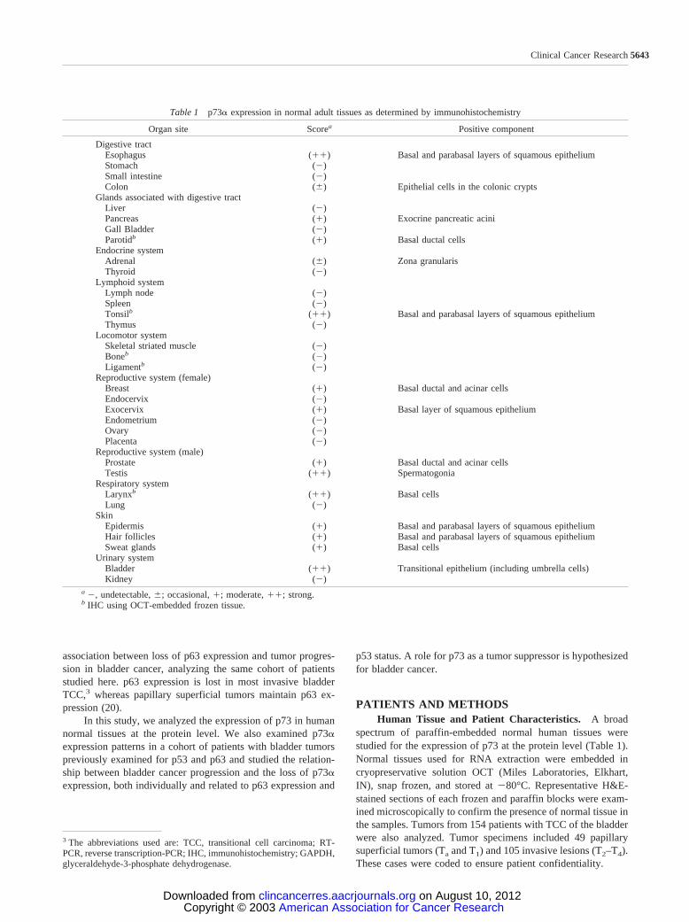
spectrum of paraffin-embedded normal human tissues werestudied for the expression of p73 at the protein level (Table 1).Normal tissues used for RNA extraction were embedded incryopreservative solution OCT (Miles Laboratories, Elkhart,IN), snap frozen, and stored at �80°C. Representative H&E-stained sections of each frozen and paraffin blocks were exam-ined microscopically to confirm the presence of normal tissue inthe samples. Tumors from 154 patients with TCC of the bladderwere also analyzed. Tumor specimens included 49 papillarysuperficial tumors (Ta and T1) and 105 invasive lesions (T2–T4).These cases were coded to ensure patient confidentiality.
3 The abbreviations used are: TCC, transitional cell carcinoma; RT-PCR, reverse transcription-PCR; IHC, immunohistochemistry; GAPDH,glyceraldehyde-3-phosphate dehydrogenase.
Table 1 p73� expression in normal adult tissues as determined by immunohistochemistry
Organ site Scorea Positive component
Digestive tractEsophagus (��) Basal and parabasal layers of squamous epitheliumStomach (�)Small intestine (�)Colon (�) Epithelial cells in the colonic crypts
Glands associated with digestive tractLiver (�)Pancreas (�) Exocrine pancreatic aciniGall Bladder (�)Parotidb (�) Basal ductal cells
Endocrine systemAdrenal (�) Zona granularisThyroid (�)
Lymphoid systemLymph node (�)Spleen (�)Tonsilb (��) Basal and parabasal layers of squamous epitheliumThymus (�)
Locomotor systemSkeletal striated muscle (�)Boneb (�)Ligamentb (�)
Reproductive system (female)Breast (�) Basal ductal and acinar cellsEndocervix (�)Exocervix (�) Basal layer of squamous epitheliumEndometrium (�)Ovary (�)Placenta (�)
Reproductive system (male)Prostate (�) Basal ductal and acinar cellsTestis (��) Spermatogonia
Respiratory systemLarynxb (��) Basal cellsLung (�)
SkinEpidermis (�) Basal and parabasal layers of squamous epitheliumHair follicles (�) Basal and parabasal layers of squamous epitheliumSweat glands (�) Basal cells
Urinary systemBladder (��) Transitional epithelium (including umbrella cells)Kidney (�)
a �, undetectable, �; occasional, �; moderate, ��; strong.b IHC using OCT-embedded frozen tissue.
5643Clinical Cancer Research
American Association for Cancer Research Copyright © 2003 on August 10, 2012clincancerres.aacrjournals.orgDownloaded from
CJDp73 Antibody Production. A 518-bp DNA frag-ment, spanning amino acids 428–599 and mapping to theCOOH terminus of simian p73�, was inserted downstream inframe of the polyhistidine (6xHIS) tag of the pRSET-C-T7expression vector (Invitrogen Life Technologies, Inc., Carlsbad,CA). The p73� fusion protein was expressed in BL21(DE3)Escherichia coli and purified on an 8% SDS-polyacrylamidegel. Crushed gel slices containing the fusion protein were usedto raise polyclonal antibodies in rabbits (Covance ResearchProducts, Inc., Denver, PA). The resultant hyperimmune serumwas designated CJDp73 and an aliquot was affinity purifiedusing recombinant 6xHIS-p73� protein attached to a Nickel-NTA column (21).
Cell Lines. Nine bladder cancer cell lines were studied,including RT4, which is derived from a superficial TCC; T24,J82, 5637, HT-1197, HT-1376, UM-UC-3, and TCC-SUP de-rived from invasive TCC; and SCaBER derived from an inva-sive squamous cell carcinoma of the bladder. For transfectionexperiments, transformed human primary embryonal kidney 293cells were used. Bladder tumor-derived cell lines and 293 cellswere obtained from and maintained as recommended from theAmerican Type Culture Collection (Manassas, VA). H24 celllines expressing tetracycline-repressible simian HA-p73� andmurine myc-�Np63� (for use in Western blots and RT-PCR)were a gift from Xinbin. Chen (22). These stable cell lines donot express p63 or p73 when grown in the presence of 1 �g/mltetracycline.
Plasmids, Cell Culture, and Transfection. For CJDp73characterization by Western blotting, transformed human pri-mary embryonal kidney 293 cells were transfected in 35-mmdishes with human HA-tagged p53, TAp73�, TAp73�, TAp73,TAp73, �Np73�, �Np73�, �Np73, and vector control(pcDNA3) by the Fugene method (Roche Diagnostics, Indian-apolis, IN). After 48 h, the cells were washed with a PBSsolution and pelleted by centrifugation. For the CJDp73 char-acterization by IHC, transformed human primary embryonalkidney 293 cells were transfected in 2-well chambered slides(Nalge Nunc International, Naperville, IL) with TAp73�,TAp73�, TAp73, TAp73, �Np73�, �Np73�, �Np73, andvector control (pcDNA3). After 48 h, the slides were washedtwice with PBS, fixed for 10 min in formalin, washed again withPBS, and subjected to the IHC protocol described below.
Tissue Array Construction. Normal and tumor tissueswere fixed with formalin and embedded in paraffin. Five-�msections stained with H&E were obtained to identify viable,representative areas of the specimen. From the defined areascore biopsies were taken with a precision instrument (BeecherInstruments, Silver Spring, MD), as described previously (23).Tissue cores with a diameter of 0.6 mm from each specimenwere punched and arrayed in triplicate on a recipient paraffinblock (24). Five-�m sections of these tissue array blocks werecut and placed on charged poly-lysine-coated slides. Thesesections were used for immunohistochemical analysis (25). Celllines known to express p73 were used as positive controls (seeabove).
IHC. Five-�m tissue sections were deparaffinized, rehy-drated in graded alcohols, and processed using the avidin-biotinimmunoperoxidase method. Briefly, sections were submitted toantigen retrieval by microwave oven treatment for 15 min in 10
mM citrate buffer (pH 6.0). Slides were subsequently incubatedin 10% normal serum for 30 min followed by the overnightincubation at 4°C with the appropriately diluted primary anti-body. The rabbit antisimian CJDp73 polyclonal antibody (�50ng/�l) was used at a 1:400 dilution to a final concentration of0.125 ng/�l. The mouse monoclonal antihuman p53 antibody(PAb1801 clone) recognizing an epitope located between aminoacids 46 and 55 was used at 0.2 �g/ml (Oncogene ResearchProducts, Boston, MA). p63 was analyzed with the monoclonal4A4 antibody (Santa Cruz Biotechnology, Santa Cruz, CA) asdescribed previously (20). After the primary antibody, sampleswere incubated with the biotinylated antirabbit or antimouseimmunoglobulins for 30 min, followed by avidin-biotin perox-idase complexes for 30 min (Vector Laboratories, Inc., Burl-ingame, CA). 3,3�-Diaminobenzidine was used as the chromo-gen and hematoxylin as the nuclear counterstain. Slides werereviewed by several investigators (P. P., P. C., M. D., C. C-C.,and C. J. D.). Some normal tissues were only available as frozensamples. These sections were cut, fixed on formalin for 10 minat 4°C, and analyzed as the paraffin embedded samples, exceptfor the deparaffinization step.
Results were scored in TCC lesions by estimating thepercentage of tumor cells showing characteristic nuclear stain-ing. An arbitrarily defined 10% cutoff was taken to classify theTCC data into two categorical groups (positive versus negative).TCC samples were considered to have a negative phenotype forthe p73� expression when �10% of nuclei showed staining.The cutoff point for p53, p53 � 20% immunoreactive tumorcells, was selected based on previous publications (19). Thecutoff point for p63 was chosen at 30% of tumor cells displayingnuclear immunostaining (20).
Western Blotting. For Western blotting, total cell ex-tracts of cultured cells were prepared as described previously(26). The rabbit antisimian CJDp73 polyclonal antibody wasused at 1:500 dilution. The anti-HA monoclonal antibody(HA.11) was obtained from Covance (Princeton, NJ) and used at1:1000 dilution. The anti-myc monoclonal antibody (S1826)was obtained from Clontech (Palo Alto, CA) and used at 1:500dilution. The goat anti-human Ran polyclonal antibody (C-20,sc-1156) was purchased from Santa Cruz Biotechnology andused at a dilution of 1:1000. Horseradish peroxidase-conjugatedantibodies (Amersham, Arlington Heights, IL, and Sigma, St.Louis, MO) were used as secondary antibodies at a 1:1000dilution. Proteins were visualized with an enhanced chemilumi-nescence plus detection system (Amersham).
RNA Isolation and RT-PCR. Total RNA was extractedfrom the nine bladder carcinoma cell lines using TRIzol (In-vitrogen Life Technologies, Inc.) according to manufacturer’sinstructions. Total RNA (250 ng) was then amplified using p73isoform-specific primers using the Superscript One-Step RT-PCR kit with Platinum Taq (Invitrogen Life Technologies, Inc.)using the manufacturer’s protocol (50 �l of reaction volume).All reverse transcription reactions were carried out for 30 min at50°C, then 4 min at 94°C, followed by isoform-specific PCRconditions for each primer set: A, TAp73 isoforms (nucleotides61–366 of TAp73�): 40 cycles at 94°C (30 s), 56°C (40 s), and72°C (30 s) using SKO53 (sense, 5�-TCTCTGGAACCAGA-CAGCAC-3�) and SKO54 (antisense, 5�-GGGGTAGTCGGT-GTTGGAG-3�); B, �Np73 isoforms (nucleotides 5–218 of
5644 p73 Expression in Bladder Cancer
American Association for Cancer Research Copyright © 2003 on August 10, 2012clincancerres.aacrjournals.orgDownloaded from
�Np73): 40 cycles at 94°C (30 s), 60°C (20 s), and 72°C (8 s)using SKO78 (sense, 5�-TGTACGTCGGTGACCCCG-3�) andSKO54 (antisense, 5�-GGGGTAGTCGGTGTTGGAG-3�); C,p73� COOH-terminal variant (nucleotides 1591–1897 ofTAp73�): 40 cycles at 94°C (30 s), 56°C (40 s), and 72°C (30s) using SKO57 (sense, 5�-CTGAAGATCCCCGAGCAGTA-3�) and SKO58 (antisense, 5�-CTCCGTGAACTCCTCCTTGA-3�); D, p73� and p73 COOH-terminal variants (nucleotides899-1203 of TAp73 ): 40 cycles at 94°C (30 s), 56°C (40 s), and72°C (30 s) using SKO61 (sense, 5�-GACCGAAAAGCTGAT-GAGGA-3�) and SKO62 (antisense, 5�-CCCCAGGTCCTCTG-TAGGAG-3�); and E, p73 COOH-terminal variant (nucleo-tides 1198–1411 of TAp73): 40 cycles at 94°C (30 s), 54°C (4s), and 72°C (6 s) using SKO59 (sense, 5�-CGGGATGCTCAA-CAACCAT-3�) and SKO60 (antisense, 5�-TGCAGGTGGTA-AATGCTCTG-3�). The GAPDH gene was chosen as an endog-enous expression RT-PCR standard using SKO36 (sense, 5�-GAAGGTGAAGGTCGGAGT-3�) and SKO37 (antisense, 5�-
GAAGATGGTGATGGGATTTC-3�). Isoform-specific RT-PCR (including GAPDH and a no-RNA control) was performedin triplicate. Forty-five �l of RT-PCR products were resolved in2.0% agarose gels. Total RNA from H24 cells expressing HA-TAp73� was used as positive control.
Statistical Analyses. In this study, the association be-tween p73� expression levels and histopathological variables ofthe cases analyzed was evaluated. p73� expression was treatedas both a continuous variable and a categorical variable (positiveversus negative taking 10% of positive cells as the cutoff point).p63 (positive versus negative) and p53 (wild-type versus mu-tant) were treated as categorical variables. These determinationswere made as a final call at the tumor, as opposed to the core,level. The difference on tumor stages between groups splitaccording to data from p53, p63, and p73� staining was ana-lyzed as categorical variables. p73� expression as a categoricalvariable was also individually analyzed versus tumor stage andgrade. A Fisher’s exact test was used to test the hypothesis of nodifference between the categorical groups analyzed. When treat-ing the p73� expression as continuous variable, each tumor wastreated as a random effect to account for the inherent butunknown correlation between the different cores of an individ-ual tumor. The data were then fit using the method of restrictedmaximum likelihood, and an F test was used to test the hypoth-esis of no difference between groups.
RESULTSCharacterization of the CJDp73 Antibody. A p73�
fusion protein was used for immunization of rabbits by standardmethods and an aliquot of the resultant hyperimmune serum,designated CJDp73, was affinity purified (see “Materials andMethods”). Western blot analysis of 293 cell extracts ectopically
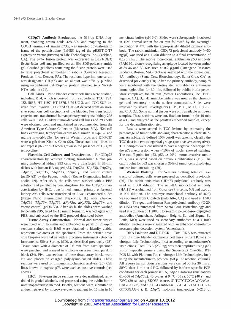
Fig. 1 p73 expression levels in 293-transfected cells. A panel of anti-bodies, including CJDp73, was used in Western blots of protein extractsfrom transfected 293 cells. In the first lane, “C” identifies transfectedcells with the vector control HA-tagged. The next seven lanes depict theexpression of seven different HA-tagged isoforms of p73 following thisorder from Lane 2 to Lane 8 (TAp73�, �Np73�, TAp73�, �Np73�,TAp73, �Np73, and TAp73). Two controls were added to ensurethe specificity of the detection; Lanes 9 and 10 (myc-tagged �Np63�and HA-tagged p53). The bottom panel shows the anti-Ran antibodyused in blots from protein extracts at an equivalent loading through alllanes. The middle panels illustrate the set of p53 family memberstransiently expressed on 293 cells. �Np63� is the only myc-taggedprotein showed after anti-myc blotting, whereas the other p53 familymembers are depicted when using the anti-HA antibody. Please note thespecificity of CJDp73 antibody for the two p73� isoforms and �Np73�,not reacting with �Np63� or p53.
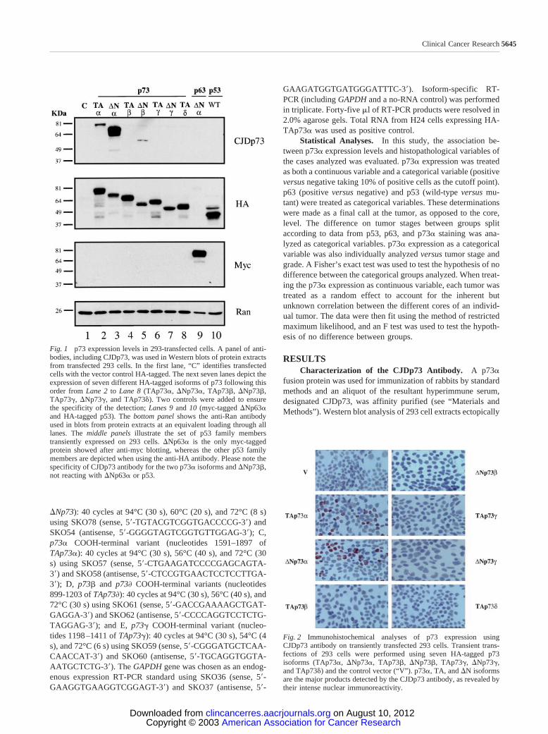
Fig. 2 Immunohistochemical analyses of p73 expression usingCJDp73 antibody on transiently transfected 293 cells. Transient trans-fections of 293 cells were performed using seven HA-tagged p73isoforms (TAp73�, �Np73�, TAp73�, �Np73�, TAp73, �Np73,and TAp73) and the control vector (“V”). p73�, TA, and �N isoformsare the major products detected by the CJDp73 antibody, as revealed bytheir intense nuclear immunoreactivity.
5645Clinical Cancer Research
American Association for Cancer Research Copyright © 2003 on August 10, 2012clincancerres.aacrjournals.orgDownloaded from
expressing HA-p73, HA-p53, myc-p63, and vector control con-structs demonstrated that CJDp73 recognized both TAp73� and�Np73�, as well as �Np73� but to a lesser extent (Fig. 1).Interestingly, although CJDp73 reacted weakly with �Np73�,this antibody does not react with TAp73� (Fig. 1). As expected,CJDp73 does not react with p73 and p73 (Fig. 1). Moreimportantly, CJDp73 does not recognize p53 and �Np63�(Fig. 1).
Immunoreactivity of CJDp73 was also examined in tissuesand cell lines by IHC. We first titrated the CJDp73 antibody onhuman normal skin sections and obtained an optimal concen-tration for IHC of 0.125 ng/�l. At this concentration, the skindisplayed intense p73 nuclear immunoreactivity in cells of thebasal and parabasal layers of the epidermis, hair follicles, andsweat glands (Table 1). We next examined 293 cells ectopicallyexpressing the seven p73 constructs used in the above Westernanalysis. Only those cells expressing TAp73� and �Np73�displayed intense p73 nuclear immunoreactivity with CJDp73(Fig. 2). The other p73 isoforms and vector control showed alack of immunoreactivity (Fig. 2). CJDp73 was raised againstp73� and p73� isoforms, the antiserum being mainly directed tothe �-derived isoforms, as revealed by both Western blot andIHC. The affinity for the � isoforms is much lower, and itappears to be only over the detection limit of immunoblottingfor the �Np73� isoform. Transient transfection experiments forIHC and Western blot were conducted in triplicate and inparallel. In all cases, the �Np73� isoform was detected byWestern blot but not by IHC. Thus, we conclude that CJDp73 is
an appropriate reagent to specifically discriminate betweenp73� and other p73 isoforms by IHC assays.
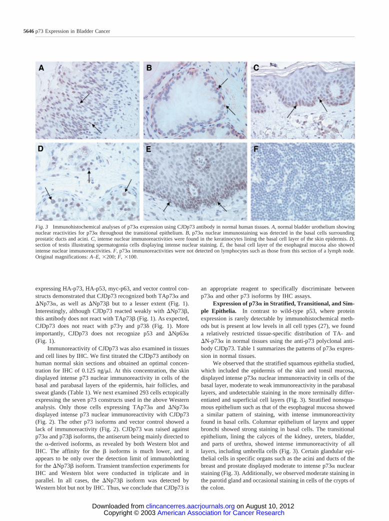
Expression of p73� in Stratified, Transitional, and Sim-ple Epithelia. In contrast to wild-type p53, where proteinexpression is rarely detectable by immunohistochemical meth-ods but is present at low levels in all cell types (27), we founda relatively restricted tissue-specific distribution of TA- and�N-p73� in normal tissues using the anti-p73 polyclonal anti-body CJDp73. Table 1 summarizes the patterns of p73� expres-sion in normal tissues.
We observed that the stratified squamous epithelia studied,which included the epidermis of the skin and tonsil mucosa,displayed intense p73� nuclear immunoreactivity in cells of thebasal layer, moderate to weak immunoreactivity in the parabasallayers, and undetectable staining in the more terminally differ-entiated and superficial cell layers (Fig. 3). Stratified nonsqua-mous epithelium such as that of the esophageal mucosa showeda similar pattern of staining, with intense immunoreactivityfound in basal cells. Columnar epithelium of larynx and upperbronchi showed strong staining in basal cells. The transitionalepithelium, lining the calyces of the kidney, ureters, bladder,and parts of urethra, showed intense immunoreactivity of alllayers, including umbrella cells (Fig. 3). Certain glandular epi-thelial cells in specific organs such as the acini and ducts of thebreast and prostate displayed moderate to intense p73� nuclearstaining (Fig. 3). Additionally, we observed moderate staining inthe parotid gland and occasional staining in cells of the crypts ofthe colon.
Fig. 3 Immunohistochemical analyses of p73� expression using CJDp73 antibody in normal human tissues. A, normal bladder urothelium showingnuclear reactivities for p73� throughout the transitional epithelium. B, p73� nuclear immunostaining was detected in the basal cells surroundingprostatic ducts and acini. C, intense nuclear immunoreactivities were found in the keratinocytes lining the basal cell layer of the skin epidermis. D,section of testis illustrating spermatogonia cells displaying intense nuclear staining. E, the basal cell layer of the esophageal mucosa also showedintense nuclear immunoreactivities. F, p73� immunoreactivities were not detected on lymphocytes such as those from this section of a lymph node.Original magnifications: A–E, �200; F, �100.
5646 p73 Expression in Bladder Cancer
American Association for Cancer Research Copyright © 2003 on August 10, 2012clincancerres.aacrjournals.orgDownloaded from
Expression of p73� in Other Normal Tissues. We ob-served intense p73� expression in spermatogonia cells of thetestis (Fig. 3). p73� was generally undetectable in mesenchymalelements, including smooth and striated muscle. Additionally,p73� was undetectable in lymphocytes. Other organs such asliver, spleen, thyroid, and placenta had undetectable levels ofp73� expression (Table 1).
Expression of p73 in Human Transitional Cell Carci-noma Cell Lines. We applied the CJDp73 antibody in immu-noblotting and the RT-PCR conditions previously described tothe characterization of p73� in a group of nine bladder tumorderived cell lines. Western blot analysis of the seven invasivebladder cell lines revealed a heterogeneous pattern of p73 ex-pression (Fig. 4). Interestingly, only three of the invasive TCCderivatives expressed p73�. They showed two prominent spe-cies migrating at Mr �75,000 and Mr �65,000 (T24, 5637, andH-1376). The upper band is likely to be TAp73� isoformbecause the HA-tagged TAp73� ran slightly higher in the samegel (Fig. 4). We further examined the samples with other anti-bodies to check for the identity of the lower bands (Mr �65,000and Mr �55,000). We could not find any other suggestion tomatch these bands with �Np73� or �Np73� (data not shown).In summary, the expression of TAp73� is lost in four of seveninvasive tumor-derived cell lines (Fig. 4).
In an attempt to further clarify the identity of the p73isoforms seen by Western blot, we performed RT-PCR for TA,�N, and the various COOH-terminal splice variants of p73 ontotal RNA isolated from the nine bladder cancer cell lines (Fig.5). We found that most of the nine cell lines expressed the TA,�N, and COOH-terminal splice variants tested (Fig. 5). TAp73RNA was found in all of the bladder cancer cell lines but inSCaBER and UM-UC-3. We observed a correlation betweenhigh expression of TAp73 RNA (Fig. 5) and TAp73� proteinaccumulation by Western blotting (Fig. 4). Additionally,whereas we detected �Np73 in all of the lines, we did not detect
the p73 splice variant and any TA RNA in the UM-UC-3 cellline, suggesting that these cells do not express any transcrip-tionally active isoform and only expresses some �Np73 isoformdifferent from gamma. SCaBER cells only expressed the �Np73isoforms.
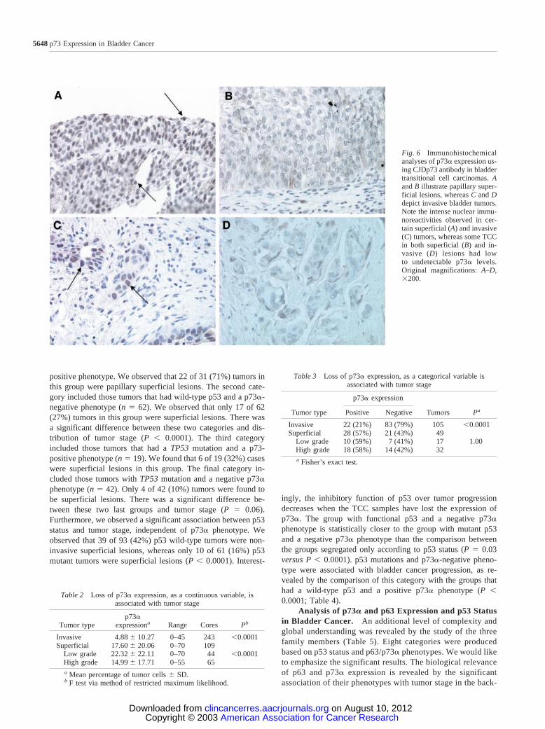
Expression of p73� in Bladder Cancer: Relation toTumor Stage and Grade. We examined 154 well-character-ized primary bladder tumors compiled onto three tissue arrays.TCC arrays were analyzed by IHC using CJDp73 antibody (Fig.6). p73� expression was reported both as continuous and cate-gorical data (positive versus negative). Cores with no tumor orinsufficient tumor cells were not used in the final analysis.Similar to what we observed in the invasive TCC-derived celllines, most invasive tumors lost p73� expression. Althoughmost superficial tumors (28 of 49; 57%) express p73�, only 22of 105 (21%) invasive TCC lesions retain p73� expression (Fig.6). p73� expression, as a continuous variable, was found to bestatistically associated with tumor stage (P � 0.0001; Table 2).Using Fisher’s exact test, p73� as a categorical variable wasalso found to be associated tumor stage (P � 0.0001; Table 3).
Analysis of p73 Expression and p53 Status in BladderCancer. p53 status was previously analyzed in these 154tumor samples. We examined the correlation between tumorstage (superficial versus invasive) and the combined data refer-ring to p53 status and p73� expression. Tumors were classifiedaccording to phenotypes into four categories (Table 4). The firstcategory included those tumors with wild-type p53 and p73�-
Fig. 4 p73 expression levels in bladder cancer cell lines. Protein ex-tracts from seven different bladder tumor-derived cell lines were ana-lyzed by Western blotting using CJDp73 and anti-Ran antibodies. Lanes1–7 depict levels of p73 on the following TCC lines J82, T24, HT-1197,5637, UM-UC-3, TCC-SUP, and HT-1376. A protein extract obtainedfrom 293 cells transiently expressing HA-tagged p73� was loaded ontoLane 10 and was used as a positive control for CJDp73. The bottompanel shows the anti-Ran antibody used in blots from protein extracts atan equivalent loading through all lanes.
Fig. 5 Analyses of p73 transcript levels in bladder cancer cell lines.Isoform-specific sets of primers were applied by RT-PCR to a panel ofnine bladder cancer cell lines. The first lane shows the water control.Lanes 2–10 show the bladder cell lines in the following order: SCaBER,J82, T24, HT-1197, 5637, UM-UC-3, TCC-SUP, HT-1376, and RT4.The positive control is RNA obtained from a TAp73�-overexpressingcells (last lane). GAPDH amplification is used as a control. Most bladdercancer cells, with the exception of UM-UC-3, displayed all p73 tran-scripts.
5647Clinical Cancer Research
American Association for Cancer Research Copyright © 2003 on August 10, 2012clincancerres.aacrjournals.orgDownloaded from
positive phenotype. We observed that 22 of 31 (71%) tumors inthis group were papillary superficial lesions. The second cate-gory included those tumors that had wild-type p53 and a p73�-negative phenotype (n � 62). We observed that only 17 of 62(27%) tumors in this group were superficial lesions. There wasa significant difference between these two categories and dis-tribution of tumor stage (P � 0.0001). The third categoryincluded those tumors that had a TP53 mutation and a p73-positive phenotype (n � 19). We found that 6 of 19 (32%) caseswere superficial lesions in this group. The final category in-cluded those tumors with TP53 mutation and a negative p73�phenotype (n � 42). Only 4 of 42 (10%) tumors were found tobe superficial lesions. There was a significant difference be-tween these two last groups and tumor stage (P � 0.06).Furthermore, we observed a significant association between p53status and tumor stage, independent of p73� phenotype. Weobserved that 39 of 93 (42%) p53 wild-type tumors were non-invasive superficial lesions, whereas only 10 of 61 (16%) p53mutant tumors were superficial lesions (P � 0.0001). Interest-
ingly, the inhibitory function of p53 over tumor progressiondecreases when the TCC samples have lost the expression ofp73�. The group with functional p53 and a negative p73�phenotype is statistically closer to the group with mutant p53and a negative p73� phenotype than the comparison betweenthe groups segregated only according to p53 status (P � 0.03versus P � 0.0001). p53 mutations and p73�-negative pheno-type were associated with bladder cancer progression, as re-vealed by the comparison of this category with the groups thathad a wild-type p53 and a positive p73� phenotype (P �0.0001; Table 4).
Analysis of p73� and p63 Expression and p53 Statusin Bladder Cancer. An additional level of complexity andglobal understanding was revealed by the study of the threefamily members (Table 5). Eight categories were producedbased on p53 status and p63/p73� phenotypes. We would liketo emphasize the significant results. The biological relevanceof p63 and p73� expression is revealed by the significantassociation of their phenotypes with tumor stage in the back-
Table 2 Loss of p73� expression, as a continuous variable, isassociated with tumor stage
Tumor typep73�
expressiona Range Cores Pb
Invasive 4.88 � 10.27 0–45 243 �0.0001Superficial 17.60 � 20.06 0–70 109
Low grade 22.32 � 22.11 0–70 44 �0.0001High grade 14.99 � 17.71 0–55 65a Mean percentage of tumor cells � SD.b F test via method of restricted maximum likelihood.
Table 3 Loss of p73� expression, as a categorical variable isassociated with tumor stage
Tumor type
p73� expression
Tumors PaPositive Negative
Invasive 22 (21%) 83 (79%) 105 �0.0001Superficial 28 (57%) 21 (43%) 49
Low grade 10 (59%) 7 (41%) 17 1.00High grade 18 (58%) 14 (42%) 32a Fisher’s exact test.
Fig. 6 Immunohistochemicalanalyses of p73� expression us-ing CJDp73 antibody in bladdertransitional cell carcinomas. Aand B illustrate papillary super-ficial lesions, whereas C and Ddepict invasive bladder tumors.Note the intense nuclear immu-noreactivities observed in cer-tain superficial (A) and invasive(C) tumors, whereas some TCCin both superficial (B) and in-vasive (D) lesions had lowto undetectable p73� levels.Original magnifications: A–D,�200.
5648 p73 Expression in Bladder Cancer
American Association for Cancer Research Copyright © 2003 on August 10, 2012clincancerres.aacrjournals.orgDownloaded from
ground of wild-type p53. More specifically, tumors that hada p53 wild-type and p63/p73�-positive phenotype (n � 25)comprised 21 (84%) superficial lesions, whereas wild-typep53 and p63/p73�-negative phenotype tumors (n � 31) in-cluded only 4 (13%) superficial lesions (P � 0.0001). Sim-ilarly, the cooperative effect of these three genes is revealedthrough the significant association to tumor stage. Tumorswith a wild-type p53 and positive p63/p73� phenotype (n �25) included 21 (84%) superficial lesions, whereas tumorscarrying p53 mutations and negative p63/p73� phenotype(n � 32) included only 2 superficial lesions (P � 0.0001).There is no statistical difference according to tumor stagebetween the group with wild-type p53 and negative p63/p73�expression and the samples with mutant p53 and a p63/p73�negative phenotype (P � 0.43).
DISCUSSIONUsing the affinity-purified anti-p73 polyclonal antiserum
CJDp73 in immunohistochemical assays, we found that it de-tects p73� isoforms specifically, not identifying any other p73isoforms tested. When applied to human normal adult tissues,we observed p73� immunoreactivities in basal cells lining glan-dular epithelia such as breast and prostate but not in the luminalcells of these organs. p73� was also expressed in basal celllayers of stratified epithelia such as the skin epidermis and in allcells of the transitional epithelium of the ureter and urinarybladder. Expression of p73� in proliferating cells such as sper-matogonia and basal epidermal cells suggests a potential role forp73� in mediating tumor suppression activity, as suggested byFlores et al. (12). Thus, accumulation of p73� might have a
Table 4 Altered expression of p53 and p73� is associated with tumor stage
p53 status p73� expression
Histology
Tumors Pa p53 status PaSuperficial Invasive
Wild type Positive 22 9 31�0.0001 Wild type
Wild type Negative 17 45 62Wild type 39 54 93
Mutant Positive 6 13 190.06 Mutant
�0.0001Mutant Negative 4 38 42
Mutant 10 51 61Total 49 105 154 154
Pa (p53–p73) Wild type-negative Mutant-positive Mutant-negative
Wild type-positive �0.0001 0.01 �0.0001
Pa (p53–p73) Mutant-negative
Wild type-negative 0.03a Fisher’s exact test.
Table 5 Altered expression of the p53 family members is associated with tumor stage
p53 status p63 expression p73� expression
Histology
Tumors PaSuperficial Invasive
Wild type Positive Positive 21 4 25 �0.0001Wild type Negative Negative 4 27 31Wild type Negative Positive 0 5 5Wild type Positive Negative 11 18 29Mutant Positive Positive 5 5 10 0.005Mutant Negative Negative 2 30 32Mutant Negative Positive 1 8 9Mutant Positive Negative 2 7 9
46 104 150
Pa (p53-p63-p73) Mutant-positive-positive Mutant-negative-negative
Wild type-positive-positive 0.08 �0.0001
Pa (p53-p63-p73) Mutant-negative-negative
Wild type-negative-negative
0.43
a Used Fisher’s exact test.
5649Clinical Cancer Research
American Association for Cancer Research Copyright © 2003 on August 10, 2012clincancerres.aacrjournals.orgDownloaded from
preventive function in the event of cellular stress, which couldmenace the integrity and viability of the cell. The accumulationof p73�, as well as that reported for p63 (20), may prepare thecell to react to genotoxic insults.
The expression pattern of p73� in normal tissue is similarto that reported for p63 (28). However, there are certain differ-ences, including the more predominant expression of p63 on thesuprabasal layer of stratified epithelia when compared with theintense basal expression of p73. In addition, p63 was not de-tected in umbrella cells of the bladder, whereas p73� is ex-pressed in these cells (Fig. 3). Similarly, p63 was not detected inthe pancreas, whereas we report herein the expression of p73�in exocrine pancreas.
Our previous observation that normal bladder urotheliumexpresses high levels of p63 and that the loss of p63 expressionis associated with tumor progression in bladder cancer (20)prompted us to investigate the role of p73 in transitional cellcarcinoma. TAp73� expression was lost in four of seven inva-sive derived cell lines (Fig. 4). We also observed that p73� wasmore frequently lost in invasive TCC cases than in superficialTCC lesions. Statistical analyses revealed that loss of p73�expression was a frequent event associated with advanced tumorstage in bladder cancer. Our findings are in agreement with astudy recently reported showing a decline in p73 expression inesophageal cancer (29).
In our study, we also compared p73�, p63, and p53 ex-pression patterns in the same cohort of bladder cancer patients(19, 20). Although the three genes have some shared functionstheir expression patterns in normal tissue are different. Althoughp53 levels are very low, as a matter of fact undetectable by IHC,p63 and p73 are usually expressed in the nuclei of certainnormal cells, including urothelium. Interestingly, it appears thatthe loss of p63 and p73 expression, as assessed by IHC, repre-sents an epigenetic tumor suppressor event, because tumor-specific mutations have been reported to be very rare (seebelow).
p73� expression and p53 status were both significantlyassociated with tumor stage. We observed that the impact of p53status in samples displaying the p73�-positive phenotype wasless significant than p73� expression in samples with wild-typep53. Thus, p73� activity appears to have a critical tumor sup-pression function. Moreover, p73� expression and p53 statuswere found to have a negative cooperative effect and to besignificantly associated with tumor stage.
We also observed a negative cooperative effect for thethree p53 family members. Tumors with wild-type p53 andpositive p63 and p73� phenotype had a significantly betterprognosis than those with wild-type p53 and undetectable p63and p73�. The dependence of p53 on p63 and p73� expressionis in concordance with the results of Flores et al. (12). Theseinvestigators have reported that p63 and p73 expression isneeded to induce a p53-dependent response to DNA damage inE1A-expressing cells and in the developing mouse central nerv-ous system. There is a strong association between p53, p63, andp73 alterations with bladder cancer progression, as revealed bycomparing the group with wild-type p53 and positive p63 andp73� phenotype versus the p53 mutant group lacking p63 andp73� expression with tumor stage (Table 5). The collaborativeeffect of p63 and p73� on the p53 function is also revealed by
comparing the TCC samples with wild-type p53 versus mutantp53 when both groups share a negative phenotype for p63 andp73�. The dependence of the wild-type p53 function on p63 andp73� to block bladder tumor progression can be hypothesizedbecause both groups have a highly similar fraction of invasivetumors, and the only difference is the p53 status. On the basis ofdata presented here, it can be postulated that assessment of p53status alone might not be as robust as if incorporating the statusof p63 and p73�.
In conclusion, this study reports the characterization of theCJDp73 antibody by Western blot and IHC on normal andtumor tissue, as well as the RT-PCR study of p73 expression innine bladder tumor-derived cell lines. It also reports the signif-icant association between p73� loss and advanced tumor stagein bladder cancer. In addition, it describes for the first time thenegative collaborative effect produced by alterations of the threep53 family members in bladder cancer progression using a largeand well-characterized cohort of patients. Results from thisstudy support a tumor suppressor role for p73�, where inacti-vation appears to be through epigenetic events in bladder cancer.
ACKNOWLEDGMENTSWe thank Maria Dudas and Carme Mir for their technical assist-
ance, Elizabeth Charytonowicz and Minglan Lu for providing bladdertumor arrays and Marta Sanchez-Carbayo for sharing unpublished data.We also thank Francesca Bernassola and Gerry Melino for providingexpression constructs encoding for �Np73�, �Np73�, and �Np73. Wethank Michel Herranz for the human �Np73 upstream sequence.
REFERENCES1. Yang, A., and McKeon, F. P63 and P73: P53 mimics, menaces andmore. Nat. Rev. Mol. Cell. Biol., 1: 199–207, 2000.2. Moll, U. M., Erster, S., and Zaika, A. p53, p63 and p73: solos,alliances and feuds among family members. Biochim. Biophys. Acta,1552: 47–59, 2001.3. Takeda, O., Homma, C., Maseki, N., Sakurai, M., Kanda, N.,Schwab, M., Nakamura, Y., and Kaneko, Y. There may be two tumorsuppressor genes on chromosome arm 1p closely associated with bio-logically distinct subtypes of neuroblastoma. Genes Chromosomes Can-cer, 10: 30–39, 1994.4. Irwin, M. S., and Kaelin, W. G. Role of the newer p53 familymembers in malignancy. Apoptosis, 6: 17–29, 2001.5. Douc-Rasy, S., Barrois, M., Echeynne, M., Kaghad, M., Blanc, E.,Raguenez, G., Goldschneider, D., Terrier-Lacombe, M. J., Hartmann,O., Moll, U., Caput, D., and Benard, J. [/Delta]N-p73� accumulates inhuman neuroblastic tumors. Am. J. Pathol., 160: 631–639, 2002.6. Kaghad, M., Bonnet, H., Yang, A., Creancier, L., Biscan, J. C.,Valent, A., Minty, A., Chalon, P., Lelias, J. M., Dumont, X., Ferrara, P.,McKeon, F., and Caput, D. Monoallelically expressed gene related top53 at 1p36, a region frequently deleted in neuroblastoma and otherhuman cancers. Cell, 90: 809–819, 1997.7. De Laurenzi, V., Costanzo, A., Barcaroli, D., Terrinoni, A., Falco,M., Annicchiarico-Petruzzelli, M., Levrero, M., and Melino, G. Twonew p73 splice variants, and , with different transcriptional activity.J. Exp. Med., 188: 1763–1768, 1998.8. De Laurenzi, V. D., Catani, M. V., Terrinoni, A., Corazzari, M.,Melino, G., Costanzo, A., Levrero, M., and Knight, R. A. Additionalcomplexity in p73: induction by mitogens in lymphoid cells and iden-tification of two new splicing variants � and �. Cell Death Differ., 6:389–390, 1999.9. Jost, C. A., Marin, M. C., and Kaelin, W. G., Jr. p73 is a simian[correction of human] p53-related protein that can induce apoptosis.Nature (Lond.), 389: 191–194, 1997.
5650 p73 Expression in Bladder Cancer
American Association for Cancer Research Copyright © 2003 on August 10, 2012clincancerres.aacrjournals.orgDownloaded from
10. Ishida, S., Yamashita, T., Nakaya, U., and Tokino, T. Adenovirus-mediated transfer of p53-related genes induces apoptosis of humancancer cells. Jpn. J. Cancer Res., 91: 174–180, 2000.11. Stiewe, T., and Putzer, B. M. Role of p73 in malignancy: tumorsuppressor or oncogene? Cell Death Differ., 9: 237–245, 2002.12. Flores, E. R., Tsai, K. Y., Crowley, D., Sengupta, S., Yang, A.,McKeon, F., and Jacks, T. p63 and p73 are required for p53-dependentapoptosis in response to DNA damage. Nature (Lond.), 416: 560–564,2002.13. Yang, A., Walker, N., Bronson, R., Kaghad, M., Oosterwegel, M.,Bonnin, J., Vagner, C., Bonnet, H., Dikkes, P., Sharpe, A., McKeon, F.,and Caput, D. p73-deficient mice have neurological, pheromonal andinflammatory defects but lack spontaneous tumours. Nature (Lond.),404: 99–103, 2000.14. Ikawa, S., Nakagawara, A., and Ikawa, Y. p53 family genes: struc-tural comparison, expression and mutation. Cell Death Differ., 6: 1154–1161, 1999.15. Zhou, J. H., Rosser, C. J., Tanaka, M., Yang, M., Baranov, E.,Hoffman, R. M., and Benedict, W. F. Visualizing superficial humanbladder cancer cell growth in vivo by green fluorescent protein expres-sion. Cancer Gene Ther., 9: 681–686, 2002.16. Fujita, J., Srivastava, S. K., Kraus, M. H., Rhim, J. S., Tronick,S. R., and Aaronson, S. A. Frequency of molecular alterations affectingras protooncogenes in human urinary tract tumors. Proc. Natl. Acad. Sci.USA, 82: 3849–3853, 1985.17. Cordon-Cardo, C., Zhang, Z. F., Dalbagni, G., Drobnjak, M.,Charytonowicz, E., Hu, S. X., Xu, H. J., Reuter, V. E., and Benedict,W. F. Cooperative effects of p53 and pRB alterations in primarysuperficial bladder tumors. Cancer Res., 57: 1217–1221, 1997.18. Goto, K., Konomoto, T., Hayashi, K., Kinukawa, N., Naito, S.,Kumazawa, J., and Tsuneyoshi, M. p53 mutations in multiple urothelialcarcinomas: a molecular analysis of the development of multiple carci-nomas. Mod. Pathol., 10: 428–437, 1997.19. Lu, M., Wikman, F., Orntoft, T. F., Charytonowicz, E., Rabbani, F.,Zhang, Z., Dalbagni, G., Pohar, K. S., Yu, G., and Cordon-Cardo, C.Impact of alterations affecting the p53 pathway in bladder cancer onclinical outcome, assessed by conventional and array-based methods.Clin. Cancer Res., 8: 171–179, 2002.
20. Urist, M. J., Di Como, C. J., Lu, M., Charytonowicz, E., Verbel, D.,Crum, C. P., Ince, T. A., McKeon, F. D., and Cordon-Cardo, C. Loss ofp63 expression is associated with tumor progression in bladder cancer.Am. J. Pathol., 161: 1199–1206, 2002.
21. Gu, J., Stephenson, C. G., and Iadarola, M. J. Recombinant proteinsattached to a nickel-NTA column: use in affinity purification of anti-bodies. Biotechniques, 17: 257–262, 1994.
22. Zhu, J., Jiang, J., Zhou, W., and Chen, X. The potential tumorsuppressor p73 differentially regulates cellular p53 target genes. CancerRes., 58: 5061–5065, 1998.
23. Kononen, J., Bubendorf, L., Kallioniemi, A., Barlund, M., Schraml,P., Leighton, S., Torhorst, J., Mihatsch, M. J., Sauter, G., and Kallion-iemi, O. P. Tissue microarrays for high-throughput molecular profilingof tumor specimens. Nat. Med., 4: 844–847, 1998.
24. Hoos, A., Urist, M. J., Stojadinovic, A., Mastorides, S., Dudas,M. E., Leung, D. H., Kuo, D., Brennan, M. F., Lewis, J. J., andCordon-Cardo, C. Validation of tissue microarrays for immunohisto-chemical profiling of cancer specimens using the example of humanfibroblastic tumors. Am. J. Pathol., 158: 1245–1251, 2001.
25. Cordon-Cardo, C., and Richon, V. M. Expression of the retinoblas-toma protein is regulated in normal human tissues. Am. J. Pathol., 144:500–510, 1994.
26. Di Como, C. J., Gaiddon, C., and Prives, C. p73 function isinhibited by tumor-derived p53 mutants in mammalian cells. Mol. Cell.Biol., 19: 1438–1449, 1999.
27. MacCallum, D. E., Hupp, T. R., Midgley, C. A., Stuart, D., Camp-bell, S. J., Harper, A., Walsh, F. S., Wright, E. G., Balmain, A., Lane,D. P., and Hall, P. A. The p53 response to ionising radiation in adult anddeveloping murine tissues. Oncogene, 13: 2575–2587, 1996.
28. Di Como, C. J., Urist, M. J., Babayan, I., Drobnjak, M., Hedvat,C. V., Teruya-Feldstein, J., Pohar, K., Hoos, A., and Cordon-Cardo, C.p63 expression profiles in human normal and tumor tissues. Clin.Cancer Res., 8: 494–501, 2002.
29. Masuda, N., Kato, H., Nakajima, T., Sano, T., Kashiwabara, K.,Oyama, T., and Kuwano, H. Synergistic decline in expressions of p73and p21 with invasion in esophageal cancers. Cancer Sci., 94: 612–617,2003.
5651Clinical Cancer Research
American Association for Cancer Research Copyright © 2003 on August 10, 2012clincancerres.aacrjournals.orgDownloaded from