Embed Size (px)
Citation preview

Cellular & MolecularBiology Letters
Liu et al. Cellular & Molecular Biology Letters (2016) 21:29 DOI 10.1186/s11658-016-0031-z
MINI REVIEW Open Access
p62 links the autophagy pathway and theubiqutin–proteasome system uponubiquitinated protein degradation
Wei Jing Liu1,2†, Lin Ye1†, Wei Fang Huang1, Lin Jie Guo1, Zi Gan Xu1, Hong Luan Wu1, Chen Yang1and Hua Feng Liu1*
* Correspondence: [email protected]†Equal contributors1The Institute of Nephrology,Guangdong Medical University,Zhanjiang, Guangdong 524001,ChinaFull list of author information isavailable at the end of the article
Abstract
The ubiquitin–proteasome system (UPS) and autophagy are two distinct and interactingproteolytic systems. They play critical roles in cell survival under normal conditions andduring stress. An increasing body of evidence indicates that ubiquitinated cargoes areimportant markers of degradation. p62, a classical receptor of autophagy, is amultifunctional protein located throughout the cell and involved in many signaltransduction pathways, including the Keap1–Nrf2 pathway. It is involved in theproteasomal degradation of ubiquitinated proteins. When the cellular p62 level ismanipulated, the quantity and location pattern of ubiquitinated proteins changewith a considerable impact on cell survival. Altered p62 levels can even lead tosome diseases. The proteotoxic stress imposed by proteasome inhibition canactivate autophagy through p62 phosphorylation. A deficiency in autophagy maycompromise the ubiquitin–proteasome system, since overabundant p62 delaysdelivery of the proteasomal substrate to the proteasome despite proteasomalcatalytic activity being unchanged. In addition, p62 and the proteasome canmodulate the activity of HDAC6 deacetylase, thus influencing the autophagicdegradation.
Keywords: p62, Autophagy, Ubiquitin–proteasome system (UPS), Ubiquitinatedprotein, Aggresome, Proteostasis, p62 phosphorylation, Keap1–Nrf2 pathway,Histone deacetylase 6 (HDAC6), Mechanistic target of rapamycin complex 1(mTORC1)
IntroductionNearly 30% of newly synthesized proteins in the cell are misfolded under normal condi-
tions [1]. Two systems that maintain cellular proteostasis are the ubiquitin–proteasome
system (UPS) and autophagy. These self-governed systems degrade various substrates,
and while they are distinct, a growing body of evidence indicates cooperation between
them. They share some ubiquitinated proteins, such as HttQ74, a huntingtin protein in
Huntington’s disease [2], but also degradation elements, such as p62.
p62 is an autophagy substrate that is used as a reporter of autophagy activity. Recently,
p62 was also shown to deliver ubiquitinated proteins, such as tau, to the proteasome for
degradation. In addition, it can shuttle between the nucleus and cytoplasm to bind with
ubiquitinated cargoes and facilitate nuclear and cytosolic protein quality control. Other
© The Author(s). 2017 Open Access This article is distributed under the terms of the Creative Commons Attribution 4.0 InternationalLicense (http://creativecommons.org/licenses/by/4.0/), which permits unrestricted use, distribution, and reproduction in any medium,provided you give appropriate credit to the original author(s) and the source, provide a link to the Creative Commons license, andindicate if changes were made. The Creative Commons Public Domain Dedication waiver (http://creativecommons.org/publicdomain/zero/1.0/) applies to the data made available in this article, unless otherwise stated.

Liu et al. Cellular & Molecular Biology Letters (2016) 21:29 Page 2 of 14
functions of p62 are gradually being revealed, emphasizing its importance in the proteo-
lytic system. This review focuses on the role of p62 in linking the ubiquitin–proteasome
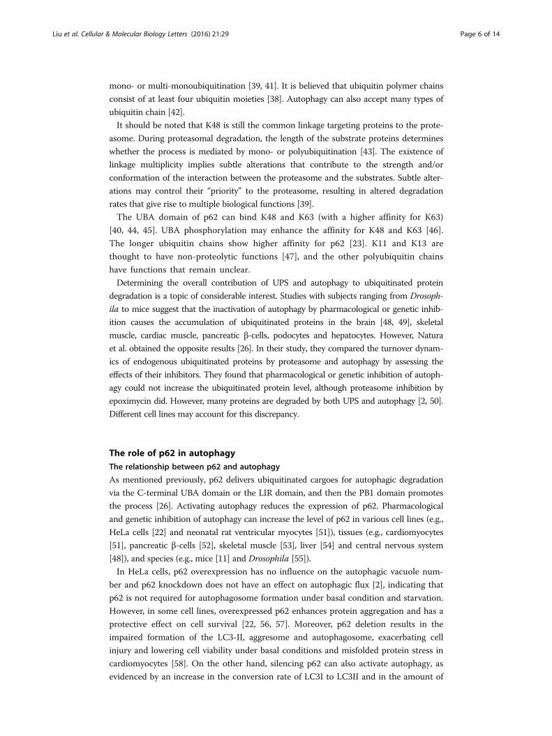
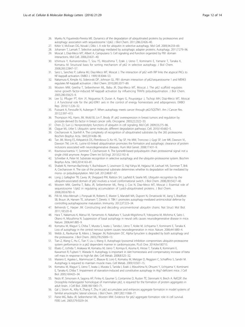
system and autophagy pathway upon ubiquitinated protein degradation (Fig. 1).
Ubiquitin–proteasome systemThe ubiquitin–proteasome system (UPS) plays a critical role in the degradation of
short-lived, misfolded and damaged proteins. This is necessary to maintain protein
homeostasis, cell cycle control [3], inflammation, oxidative stress, apoptosis [4] and
immunity [1]. It even serves a non-proteolytic function in the control of translation [5].
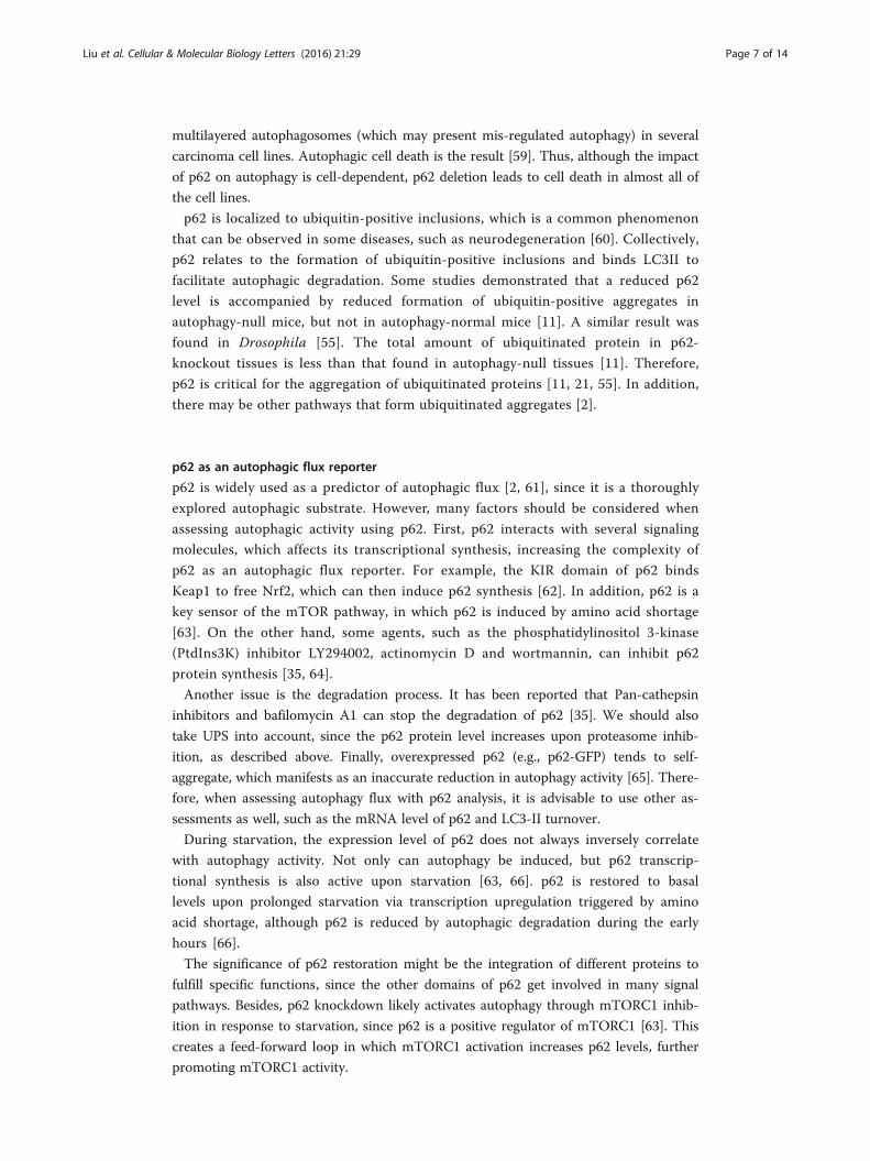
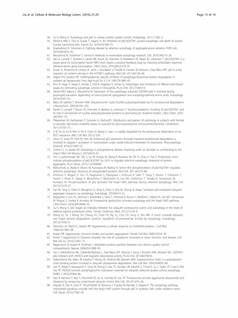
The proteasome is a highly conserved protease complex consisting of two moieties that
combine into a diversity of forms: the 20S catalytic core particle and the 19S or 11S
regulatory particle(s) (Fig. 2). 20S is a barrel-shaped complex that possesses two α-rings
and two β-rings, with each ring composed of seven subunits. β1, β2 and β5 are 3
subunits of each β-ring, respectively possessing peptidylglutamyl peptide-hydrolyzing
or caspase-like activity (PGPH or C-L); trypsin-like activity (T-L); and chymotrypsin-
like activity (CT-L) [6]. 19S consists of a lid and a base, which is involved in
substrate recognition, deubiquitination, unfolding and further translation into 20S
for degradation [7, 8].
p62 UPS
ubiquitinated-protein
aggregation
cellsurvival
diseases
PHDAC6
Nrf2
Over-expression
Deletion
ubiquitinated-protein
degradation
Autophagy-null tissues
Fig. 1 The interactions of p62 and the UPS, autophagy and ubiquitinated proteins. Upon UPS inhibition,p62 is upregulated and phosphorylated on S405 and S409, which can facilitate the degradation of ubiquitinatedcargoes via autophagy. p62 synthesis is induced by an increase in Nrf2 following UPS deficiency. The increasedp62 competes with Nrf2 for Keap1, and then a p62–Keap1 complex selectively facilitates the ubiquitinatedaggregate formation and creates a positive feedback loop with Nrf2. HDAC6 can be activated by the products inUPS (such as K63), but inhibited directly by p62. HDAC6 plays a critical role in ubiquitinated aggregate formationand autophagosome–lysosome fusion, while a ratio of p62 to HDAC6 maintains the homeostasis of autophagicprocess. Besides inhibiting the degradation of p62 and ubiquitinated proteins, a deficiency in autophagy alsocompromises UPS since the increased p62 delays ubiquitinated protein delivery to UPS for degradation. p62overexpression increases the aggregation of ubiquitinated proteins and has a protective effect on cell survival,while p62 deletion exacerbates cell injury and relates to some diseases by either facilitating ordamaging autophagic degradation dependent on the cell type

B
side view
top view
11S
A
19S
Rpn15
Rpn3
Rpn6
Rpn7
Rpn8
Rpn9 Rpn12
Rpt1Rpt2
Rpt3
Rpt4Rpt5
Rpn10
Rpn11
Rpn5
lid
base
C
degradation of non-ubiquitinated proteins
26S 26S(30S)
Hybrid
immunoproteasome
20S
IFN-
-ring
-rings
-ring
degradation of ubiquitinated proteins
poorly studied
5i
2i
1i
Fig. 2 Structures of the mammalian proteasome. a A simplified model of the proteasome regulatoryparticle 19S. The lid mainly de-ubiquitylates the captured substrates, while the base functions as substrateunfolding and translocation. Rpn11 serves as a de-ubiquitylating enzyme (DUB) en clon cleaving thepolyubiquitin chain of substrates. Rpt1-6, an ATPase ring, is involved in substrate protein unfolding andtranslocation into the channel of the 20S. Rpn13 and Rpn10 (a lid subunit) serve as ubiquitin receptors.Rpn1 can bind to the ubiquitin shuttle receptors and cytoplasmic deubiquitinases. b A simplified modelof the proteasome regulatory particle 11S, which is also termed PA28. It is an activator of the proteasome.c Assembly model of mammalian proteasome. 20S binding 19S at one or two ends generates the 26Sproteasome (or 30S), with an ATP-dependent degradation of ubiquitinated substrates. Upon stimulationof interferon-γ (INF-γ), all three active subunits (β1, β2 and β5) of the constitutive 20S proteasome arereplaced by close-proximity similar subunits (β1i, β2i and β5i, respectively) that bind to 11S to generatethe immunoproteasome. The immunoproteasome responds to antigen presentation with a non-ATP-dependent degradation of non-ubiquitinated proteins
Liu et al. Cellular & Molecular Biology Letters (2016) 21:29 Page 3 of 14
The progress of proteolysis also requires ubiquitin to covalently attach to substrates.
This 76-amino acid protein can form an isopeptide bond between its C-terminal
glycine (G76) and a lysine residue within the target molecules or ubiquitin itself [9].
Ubiquitination is completed via an enzymatic cascade involving E1 ubiquitin-activating
enzyme(s), E2 ubiquitin-conjugating enzyme(s) and E3 ubiquitin ligase(s). Then the
ubiquitinated proteins are recognized and degraded by the 26S proteasome, which
consists of a 20S unit with one or two 19S units at one or both ends [10].
AutophagyAutophagy is a highly evolutionarily conserved degradation system in eukaryotes [11].
It was first considered to be a non-selective bulk system for degrading long-lived
proteins and organelles to recycle nutrients and generate energy [12]. Later studies

Liu et al. Cellular & Molecular Biology Letters (2016) 21:29 Page 4 of 14
showed that autophagy selectively degrades protein aggregates (aggrephagy), peroxisomes
(pexophagy), damaged mitochondria (mitophagy), intracellular bacteria and viruses
(xenophagy), surplus endoplasmic reticulum (reticulophagy), ribosomes (ribophagy) and
mid-body ring structures [13].
The autophagic degradation model of eukaryotes is emerging through more recent
research [12]. Autophagy begins with the formation of a phagophore, which is a
crescent-shaped double membrane tightly associated with LC3II. The phagophore
engulfs adaptor-mediated ubiquitinated substrates to become an autophagosome, which
fuses with the lysosome to become an autolysosome with an internal acidic, hydrolytic
environment that helps to degrade the content [14]. For content outside the cell, the
cell membrane caves to envelop it. This is an endosome, which fuses with an autopha-
gosome to become an amphisome, which in turn fuses with a lysosome to become an
autolysosome.
Autophagy-related gene (Atg) proteins play essential roles in autophagy. They are
known as the ‘core machinery’ [15]. More than 40 Atg proteins have been identified as
participating in autophagy or autophagy-related processes [16]. p62 and NBR1 (neighbor
of BRCA1 gene 1) are two important cargo receptors involved in selective autophagy.
They are essential in the formation of ubiquitinated aggregates [17, 18]. NBR1 is twice as
large as p62, has a similar domain architecture, and shares several key features with it.
Cellular NBR1 is modulated by the autophagic process and does not seem to be influ-
enced by proteasomal degradation [17]. Recent studies have revealed a critical role for
autophagy in some human diseases, such as tumors [19], neurodegenerative diseases and
aging. Some cell lines, such as podocytes, have high basal autophagy [20].
p62p62 was the first selected autophagy adaptor discovered in mammals [11, 21, 22]. It
was termed sequestosome 1 (SQSTM 1) by Shin due to its ability to form aggregates
[23]. A170 and ZIP are the respective names for the mouse and rat variants.
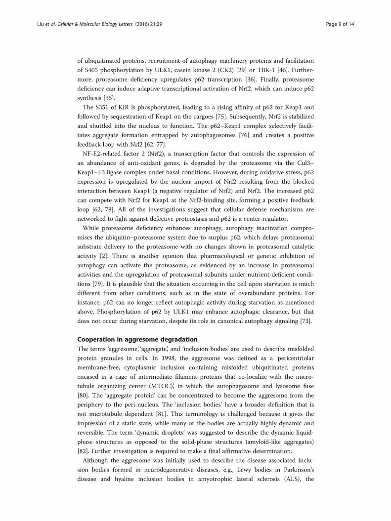
p62 is a multifunctional protein consisting of an N-terminal Phox-BEM1 domain (PB1),
a ZZ-type zinc finger domain, a nuclear localization signal (NLS), an export motif (NES),
an LC3-interacting region (LIR), a Keap1-interacting region (KIR), and a C-terminal
ubiquitin-associated domain (UBA) [24, 25] (Fig. 3). p62 interacts non-covalently with
ubiquitin or polyubiquitin chains via the UBA domain, and then delivers polyubiquiti-
nated cargoes to autophagy via the LIR domain (which is also known as the Atg8 family-
interacting motif), and to the proteasome via the PB1 domain [23, 26]. In addition to a
high potential for homo-oligomerization [27, 28], the PB1 domain can also hetero-
oligomerize with NBR1 or other PB1 domain-containing proteins, such as atypical protein
kinases Cs (αPKCs), MEKK3, MEK5, ERK1 and Rpt1, which modulate different signaling
pathways and get involved in osteoclastogenesis, angiogenesis and early cardiovascular
development or cell polarity [29].
The oligomerization of p62 via the PB1 domain is critical for ubiquitinated protein
accumulation in autophagy-null cells [30]. αPKCs and MEKK3 can activate NF-κB
signaling by binding the PB1 domain of p62, respectively with the assistance of the
receptor-interacting protein-1-binding (RIP-binding) ZZ domain and tumor necrosis
factor receptor-associated factor 6-binding (TRAF6-binding) TB domain [31, 32]. The
p62–TRAF6 complex appears to modulate the ubiquitination of the IKK complex [33].
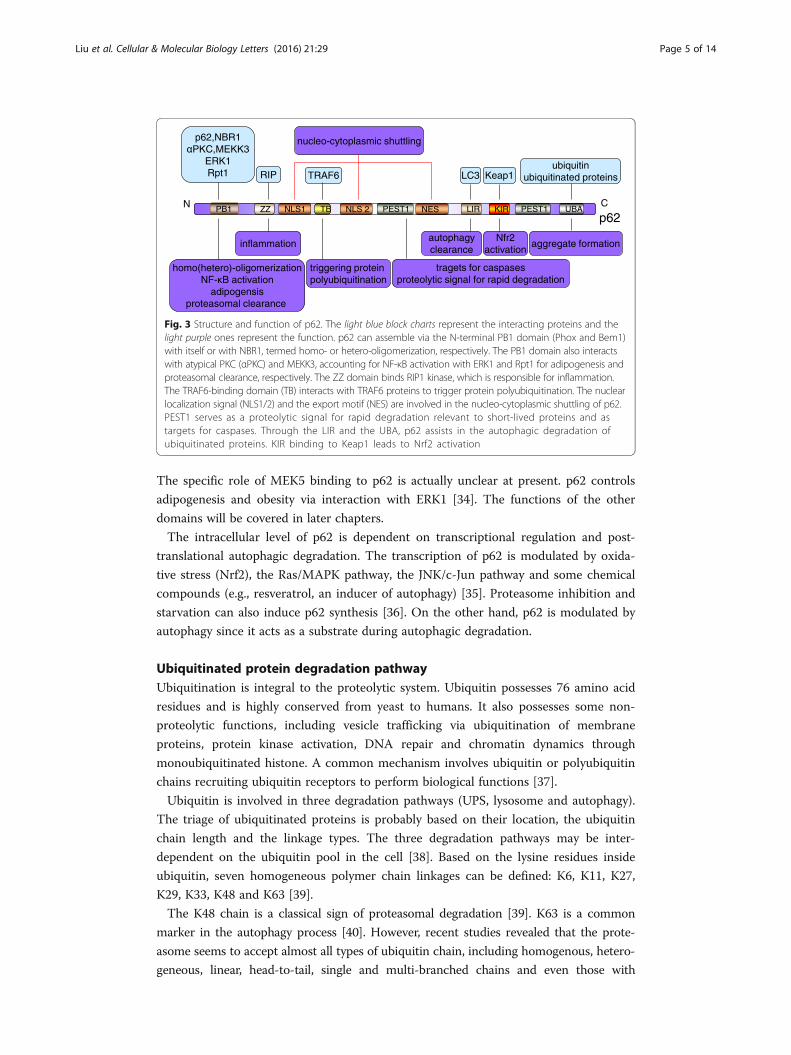
p62N C
LIR UBAPEST1NLS1 TBPB1 ZZ PEST1KIR
1RBN,26p3KKEM,CKPα
ERK1Rpt1 RIP TRAF6 LC3 Keap1
ubiquitinsnietorpdetanitiuqibu
homo(hetero)-oligomerizationNF-κB activation
adipogensisproteasomal clearance
inflammation
triggering protein polyubiquitination
autophagyclearance
Nfr2activation
tragets for caspasesnoitadargeddiparroflangiscityloetorp
aggregate formation
nucleo- gnilttuhscimsalpotyc
NLS 2 NES
Fig. 3 Structure and function of p62. The light blue block charts represent the interacting proteins and thelight purple ones represent the function. p62 can assemble via the N-terminal PB1 domain (Phox and Bem1)with itself or with NBR1, termed homo- or hetero-oligomerization, respectively. The PB1 domain also interactswith atypical PKC (αPKC) and MEKK3, accounting for NF-κB activation with ERK1 and Rpt1 for adipogenesis andproteasomal clearance, respectively. The ZZ domain binds RIP1 kinase, which is responsible for inflammation.The TRAF6-binding domain (TB) interacts with TRAF6 proteins to trigger protein polyubiquitination. The nuclearlocalization signal (NLS1/2) and the export motif (NES) are involved in the nucleo-cytoplasmic shuttling of p62.PEST1 serves as a proteolytic signal for rapid degradation relevant to short-lived proteins and astargets for caspases. Through the LIR and the UBA, p62 assists in the autophagic degradation ofubiquitinated proteins. KIR binding to Keap1 leads to Nrf2 activation
Liu et al. Cellular & Molecular Biology Letters (2016) 21:29 Page 5 of 14
The specific role of MEK5 binding to p62 is actually unclear at present. p62 controls
adipogenesis and obesity via interaction with ERK1 [34]. The functions of the other
domains will be covered in later chapters.
The intracellular level of p62 is dependent on transcriptional regulation and post-
translational autophagic degradation. The transcription of p62 is modulated by oxida-
tive stress (Nrf2), the Ras/MAPK pathway, the JNK/c-Jun pathway and some chemical
compounds (e.g., resveratrol, an inducer of autophagy) [35]. Proteasome inhibition and
starvation can also induce p62 synthesis [36]. On the other hand, p62 is modulated by
autophagy since it acts as a substrate during autophagic degradation.
Ubiquitinated protein degradation pathwayUbiquitination is integral to the proteolytic system. Ubiquitin possesses 76 amino acid
residues and is highly conserved from yeast to humans. It also possesses some non-
proteolytic functions, including vesicle trafficking via ubiquitination of membrane
proteins, protein kinase activation, DNA repair and chromatin dynamics through
monoubiquitinated histone. A common mechanism involves ubiquitin or polyubiquitin
chains recruiting ubiquitin receptors to perform biological functions [37].
Ubiquitin is involved in three degradation pathways (UPS, lysosome and autophagy).
The triage of ubiquitinated proteins is probably based on their location, the ubiquitin
chain length and the linkage types. The three degradation pathways may be inter-
dependent on the ubiquitin pool in the cell [38]. Based on the lysine residues inside
ubiquitin, seven homogeneous polymer chain linkages can be defined: K6, K11, K27,
K29, K33, K48 and K63 [39].
The K48 chain is a classical sign of proteasomal degradation [39]. K63 is a common
marker in the autophagy process [40]. However, recent studies revealed that the prote-
asome seems to accept almost all types of ubiquitin chain, including homogenous, hetero-
geneous, linear, head-to-tail, single and multi-branched chains and even those with
Liu et al. Cellular & Molecular Biology Letters (2016) 21:29 Page 6 of 14
mono- or multi-monoubiquitination [39, 41]. It is believed that ubiquitin polymer chains
consist of at least four ubiquitin moieties [38]. Autophagy can also accept many types of
ubiquitin chain [42].
It should be noted that K48 is still the common linkage targeting proteins to the prote-
asome. During proteasomal degradation, the length of the substrate proteins determines
whether the process is mediated by mono- or polyubiquitination [43]. The existence of
linkage multiplicity implies subtle alterations that contribute to the strength and/or
conformation of the interaction between the proteasome and the substrates. Subtle alter-
ations may control their “priority” to the proteasome, resulting in altered degradation
rates that give rise to multiple biological functions [39].
The UBA domain of p62 can bind K48 and K63 (with a higher affinity for K63)
[40, 44, 45]. UBA phosphorylation may enhance the affinity for K48 and K63 [46].
The longer ubiquitin chains show higher affinity for p62 [23]. K11 and K13 are
thought to have non-proteolytic functions [47], and the other polyubiquitin chains
have functions that remain unclear.
Determining the overall contribution of UPS and autophagy to ubiquitinated protein
degradation is a topic of considerable interest. Studies with subjects ranging from Drosoph-
ila to mice suggest that the inactivation of autophagy by pharmacological or genetic inhib-
ition causes the accumulation of ubiquitinated proteins in the brain [48, 49], skeletal
muscle, cardiac muscle, pancreatic β-cells, podocytes and hepatocytes. However, Natura
et al. obtained the opposite results [26]. In their study, they compared the turnover dynam-
ics of endogenous ubiquitinated proteins by proteasome and autophagy by assessing the
effects of their inhibitors. They found that pharmacological or genetic inhibition of autoph-
agy could not increase the ubiquitinated protein level, although proteasome inhibition by
epoximycin did. However, many proteins are degraded by both UPS and autophagy [2, 50].
Different cell lines may account for this discrepancy.
The role of p62 in autophagyThe relationship between p62 and autophagy
As mentioned previously, p62 delivers ubiquitinated cargoes for autophagic degradation
via the C-terminal UBA domain or the LIR domain, and then the PB1 domain promotes
the process [26]. Activating autophagy reduces the expression of p62. Pharmacological
and genetic inhibition of autophagy can increase the level of p62 in various cell lines (e.g.,
HeLa cells [22] and neonatal rat ventricular myocytes [51]), tissues (e.g., cardiomyocytes
[51], pancreatic β-cells [52], skeletal muscle [53], liver [54] and central nervous system
[48]), and species (e.g., mice [11] and Drosophila [55]).
In HeLa cells, p62 overexpression has no influence on the autophagic vacuole num-
ber and p62 knockdown does not have an effect on autophagic flux [2], indicating that
p62 is not required for autophagosome formation under basal condition and starvation.
However, in some cell lines, overexpressed p62 enhances protein aggregation and has a
protective effect on cell survival [22, 56, 57]. Moreover, p62 deletion results in the
impaired formation of the LC3-II, aggresome and autophagosome, exacerbating cell
injury and lowering cell viability under basal conditions and misfolded protein stress in
cardiomyocytes [58]. On the other hand, silencing p62 can also activate autophagy, as
evidenced by an increase in the conversion rate of LC3I to LC3II and in the amount of
Liu et al. Cellular & Molecular Biology Letters (2016) 21:29 Page 7 of 14
multilayered autophagosomes (which may present mis-regulated autophagy) in several
carcinoma cell lines. Autophagic cell death is the result [59]. Thus, although the impact
of p62 on autophagy is cell-dependent, p62 deletion leads to cell death in almost all of
the cell lines.
p62 is localized to ubiquitin-positive inclusions, which is a common phenomenon
that can be observed in some diseases, such as neurodegeneration [60]. Collectively,
p62 relates to the formation of ubiquitin-positive inclusions and binds LC3II to
facilitate autophagic degradation. Some studies demonstrated that a reduced p62
level is accompanied by reduced formation of ubiquitin-positive aggregates in
autophagy-null mice, but not in autophagy-normal mice [11]. A similar result was
found in Drosophila [55]. The total amount of ubiquitinated protein in p62-
knockout tissues is less than that found in autophagy-null tissues [11]. Therefore,
p62 is critical for the aggregation of ubiquitinated proteins [11, 21, 55]. In addition,
there may be other pathways that form ubiquitinated aggregates [2].
p62 as an autophagic flux reporter
p62 is widely used as a predictor of autophagic flux [2, 61], since it is a thoroughly
explored autophagic substrate. However, many factors should be considered when
assessing autophagic activity using p62. First, p62 interacts with several signaling
molecules, which affects its transcriptional synthesis, increasing the complexity of
p62 as an autophagic flux reporter. For example, the KIR domain of p62 binds
Keap1 to free Nrf2, which can then induce p62 synthesis [62]. In addition, p62 is a
key sensor of the mTOR pathway, in which p62 is induced by amino acid shortage
[63]. On the other hand, some agents, such as the phosphatidylinositol 3-kinase
(PtdIns3K) inhibitor LY294002, actinomycin D and wortmannin, can inhibit p62
protein synthesis [35, 64].
Another issue is the degradation process. It has been reported that Pan-cathepsin
inhibitors and bafilomycin A1 can stop the degradation of p62 [35]. We should also
take UPS into account, since the p62 protein level increases upon proteasome inhib-
ition, as described above. Finally, overexpressed p62 (e.g., p62-GFP) tends to self-
aggregate, which manifests as an inaccurate reduction in autophagy activity [65]. There-
fore, when assessing autophagy flux with p62 analysis, it is advisable to use other as-
sessments as well, such as the mRNA level of p62 and LC3-II turnover.
During starvation, the expression level of p62 does not always inversely correlate
with autophagy activity. Not only can autophagy be induced, but p62 transcrip-
tional synthesis is also active upon starvation [63, 66]. p62 is restored to basal
levels upon prolonged starvation via transcription upregulation triggered by amino
acid shortage, although p62 is reduced by autophagic degradation during the early
hours [66].
The significance of p62 restoration might be the integration of different proteins to
fulfill specific functions, since the other domains of p62 get involved in many signal
pathways. Besides, p62 knockdown likely activates autophagy through mTORC1 inhib-
ition in response to starvation, since p62 is a positive regulator of mTORC1 [63]. This
creates a feed-forward loop in which mTORC1 activation increases p62 levels, further
promoting mTORC1 activity.

Liu et al. Cellular & Molecular Biology Letters (2016) 21:29 Page 8 of 14
A role for p62 in the UPSNatura et al. used the proximity ligation assay (PLA) to reveal that p62 and the prote-
asome are co-localized in situ under basal conditions. They also found that p62 aggregates
contain inactive proteasome, ubiquitinated proteins and autophagosome upon prote-
asome inhibition [26]. It has been shown that p62 can shuttle K63-polyubiquitinated tau
for proteasomal degradation [67]. This leads us to explore the relationship between p62
and proteasome.
The N-terminal PB1 domain of p62 might interact with Rpt1 and S5a/Rpn10 of the
26S proteasome and collaborate with the C-terminal UBA domain of p62 by binding
ubiquitinated proteins to facilitate UPS degradation [26, 66, 68]. p62 continuously
undergoes rapid nucleo-cytoplasmic shuttling using its own two nuclear localization
signal domains (NLS1 and NLS2) and one nuclear export motif (NES) [63]. p62 is
localized in nuclear aggregates [69] and plays a critical role in recruiting the prote-
asome to the ubiquitinated inclusion in the nucleus. It may also export ubiquitinated
cargoes from the nucleus to the cytosol for more efficient degradation [68].
These studies indicate that p62 is also involved in the proteasomal degradation of
ubiquitinated proteins in the nucleus via its NLS and NES domains and in the cytosol
via its PB1 domain. Therefore, it naturally contributes to both nuclear and cytosolic
protein quality control. Besides, the PEST domain serves as a proteolytic signal for
rapid degradation, leading to short intracellular half-lives, which may relate to prote-
asome function [26]. For instance, HS-1-associated protein X-1 (Hax-1) undergoes a
fast turnover via the proteasome system through its PEST domain [70].
Pharmacological inhibition of UPS enhances p62 transcription [26, 71] and induces
the accumulation of ubiquitinated proteins. Inhibiting the proteasome with epoximycin
increases the level of p62 far beyond the levels induced by autophagy inhibitors [26].
When p62 is overexpressed, proteasome catalytic activity will be not influenced,
although UPS substrates accumulate [2], implying that p62 delays the delivery of
ubiquitinated proteins to the UPS for degradation. Moreover, p62 overexpression along
with pharmacological inhibition of UPS and/or autophagy does not further increase
ubiquitin aggregates. These studies suggest that p62 is not required for all of the
ubiquitinated aggregates.
Relationship of p62 with ups and autophagyInterdependence upon defective proteostasis
Overexpressed p62 can enhance protein aggregation and has a protective effect on cell
survival as described above. p62 deletion barely decreases the amount of ubiquitinated
puncta in autophagy normal cells. Although p62 is not necessary for all the formation
of ubiquitinated aggregation, it still plays a crucial role in aggregate degradation.
Proteasome inhibition can activate autophagy, in which p62 is the bridge [72]. First,
proteotoxic stress imposed by proteasome inhibition can induce p62 phosphorylation
at serine 405 (S405 in the UBA domain, which is equivalent to S403 in human) and
S409 through ULK1/Atg1, which modulates its binding to ubiquitinated proteins [73].
This increased affinity can stabilize ubiquitinated proteins in the sequestosome, which,
in turn, prevents p62 dephosphorylation and leads to efficient degradation of the pro-
tein aggregates [74]. S409 phosphorylation is essential for the autophagic degradation
Liu et al. Cellular & Molecular Biology Letters (2016) 21:29 Page 9 of 14
of ubiquitinated proteins, recruitment of autophagy machinery proteins and facilitation
of S405 phosphorylation by ULK1, casein kinase 2 (CK2) [29] or TBK-1 [46]. Further-
more, proteasome deficiency upregulates p62 transcription [36]. Finally, proteasome
deficiency can induce adaptive transcriptional activation of Nrf2, which can induce p62
synthesis [35].
The S351 of KIR is phosphorylated, leading to a rising affinity of p62 for Keap1 and
followed by sequestration of Keap1 on the cargoes [75]. Subsequently, Nrf2 is stabilized
and shuttled into the nucleus to function. The p62–Keap1 complex selectively facili-
tates aggregate formation entrapped by autophagosomes [76] and creates a positive
feedback loop with Nrf2 [62, 77].
NF-E2-related factor 2 (Nrf2), a transcription factor that controls the expression of
an abundance of anti-oxidant genes, is degraded by the proteasome via the Cul3–
Keap1–E3 ligase complex under basal conditions. However, during oxidative stress, p62
expression is upregulated by the nuclear import of Nrf2 resulting from the blocked
interaction between Keap1 (a negative regulator of Nrf2) and Nrf2. The increased p62
can compete with Nrf2 for Keap1 at the Nrf2-binding site, forming a positive feedback
loop [62, 78]. All of the investigations suggest that cellular defense mechanisms are
networked to fight against defective proteostasis and p62 is a center regulator.
While proteasome deficiency enhances autophagy, autophagy inactivation compro-
mises the ubiquitin–proteasome system due to surplus p62, which delays proteasomal
substrate delivery to the proteasome with no changes shown in proteasomal catalytic
activity [2]. There is another opinion that pharmacological or genetic inhibition of
autophagy can activate the proteasome, as evidenced by an increase in proteasomal
activities and the upregulation of proteasomal subunits under nutrient-deficient condi-
tions [79]. It is plausible that the situation occurring in the cell upon starvation is much
different from other conditions, such as in the state of overabundant proteins. For
instance, p62 can no longer reflect autophagic activity during starvation as mentioned
above. Phosphorylation of p62 by ULK1 may enhance autophagic clearance, but that
does not occur during starvation, despite its role in canonical autophagy signaling [73].
Cooperation in aggresome degradationThe terms ‘aggresome’, ‘aggregate’, and ‘inclusion bodies’ are used to describe misfolded
protein granules in cells. In 1998, the aggresome was defined as a ‘pericentriolar
membrane-free, cytoplasmic inclusion containing misfolded ubiquitinated proteins
encased in a cage of intermediate filament proteins that co-localize with the micro-
tubule organizing center (MTOC)’, in which the autophagosome and lysosome fuse
[80]. The ‘aggregate protein’ can be concentrated to become the aggresome from the
periphery to the peri-nucleus. The ‘inclusion bodies’ have a broader definition that is
not microtubule dependent [81]. This terminology is challenged because it gives the
impression of a static state, while many of the bodies are actually highly dynamic and
reversible. The term ‘dynamic droplets’ was suggested to describe the dynamic liquid-
phase structures as opposed to the solid-phase structures (amyloid-like aggregates)
[82]. Further investigation is required to make a final affirmative determination.
Although the aggresome was initially used to describe the disease-associated inclu-
sion bodies formed in neurodegenerative diseases, e.g., Lewy bodies in Parkinson’s
disease and hyaline inclusion bodies in amyotrophic lateral sclerosis (ALS), the

Liu et al. Cellular & Molecular Biology Letters (2016) 21:29 Page 10 of 14
relevance of the aggresome to inclusions in disease is still disputable [77]. Almost a
decade ago, disease-related proteins, such as huntingtin (Htt), were found to form a
different pattern compared with the misfolded protein upon proteasome inhibition
[83], i.e., periphery versus peri-nuclear, despite other similar biological characteristics.
There is a common consensus that misfolded proteins aggregate and are concen-
trated in the aggresome, which is removed via the autophagy–lysosome pathway [77].
Degradation is strongly based on the activity of histone deacetylase 6 (HDAC6), which
also plays a pivotal role in aggresome formation [84]. Acetylated cortactin becomes
cortactin via HDCA6 deacetylase activity, and the latter interacts with F-actin to form
cortactin–F-actin assemblies that are recruited to the MTOC, promoting autophago-
some and lysosome fusion and substrate clearance. p62 can modulate this process by
directly inhibiting HDAC6 activity and facilitating removal of the cortactin–F-actin
assembly to MTOC, which seems paradoxical [84]. Some observations suggest that loss
of p62 leads to cortactin–F-actin assemblies remaining localized in the periphery and
ubiquitinated protein accumulation [85]. HDAC6 knockdown leads to failure of fusion
between the autophagosome and lysosome and subsequent protein aggregation [86].
p62 can facilitate protein aggregation and also modulate protein transport to the pro-
cessing site, while HDAC6 facilitates autophagosome–lysosome fusion. The ratio of
p62 to HDAC6 maintains the homeostasis of the autophagic process. The proteasome
can also modulate aggresome degradation: Poh1, a subunit of 19S, cleaves ubiquitin
chains from the substrates, and subsequently the products and K63 activate HDAC6
[87]. While proteasome inhibition imposes proteotoxic stress, the cell fate (survival or
death) in response to an altered ratio of p62 to HDAC6 remains unclear.
ConclusionBecause the UPS, autophagy and p62 are the interdependent elements of the protein
quality control system, they must act in a networked manner to maintain proteostasis.
p62 may serve as an integration center for multiple functions, including the formation
of the autophagosome, the delivery of ubiquitinated proteins to the proteasome, and
aggregate formation for autophagic clearance. It is also involved in several signaling
pathways [88, 89]. In addition, it has been shown that p62 can inhibit ATP- and
ubiquitin-independent LC3 degradation by the proteasome [90]. p62 is also involved in
many diseases. For instance, the mutation of the UBA domain in p62 leads to Paget’s
disease [91]. p62 and autophagy synergize to promote tumor growth [92], and p62
selectively binds mutant SOD1 to form aggregates in model systems of familial amyo-
trophic lateral sclerosis [25, 57]. Therefore, p62 could be a promising strategic target
for treatment of certain pathological conditions.
AbbreviationsHDAC6: Histone deacetylase 6; MTOC: The microtubule organizing center; mTORC1: Mechanistic target of rapamycincomplex 1; Nrf2: NF-E2-related factor 2; UPS: The ubiquitin–proteasome system; αPKCs: Atypical protein kinases Cs
AcknowledgementsNot applicable.
Funding
This study was supported by the National Natural Science Foundation of China (No. 81570656 and 81470959), NaturalScience Foundation of Guangdong Province (No. 2014A030313540), Medical Scientific Research Foundation ofGuangdong Province (No. A2015138 and A2014480) and the Administration of Traditional Chinese Medicine ofGuangdong Province (No. 20141153).
Liu et al. Cellular & Molecular Biology Letters (2016) 21:29 Page 11 of 14
Availability of data and materialsNot applicable.
Authors’ contributionsWJL and LY conceived the structure and wrote this manuscript; WFH, LJG and ZGX designed the figures; HLW wroteabout the ubiquitinated protein degradation pathways. CY and HFL improved the structure and language of themanuscript; All authors read and approved the final manuscript.
Competing interestsThe authors declare that they have no competing interests.
Consent for publicationNot applicable.
Ethics approval and consent to participateNot applicable.
Author details1The Institute of Nephrology, Guangdong Medical University, Zhanjiang, Guangdong 524001, China. 2Key Laboratory ofChinese Internal Medicine of Ministry of Education and Beijing, Dongzhimen Hospital Affiliated to Beijing University ofChinese Medicine, Beijing 100700, China.
Received: 4 December 2016 Accepted: 7 December 2016
References
1. Schubert U, Anton LC, Gibbs J, Norbury CC, Yewdell JW, Bennink JR. Rapid degradation of a large fraction ofnewly synthesized proteins by proteasomes. Nature. 2000;404:770–4.2. Korolchuk VI, Mansilla A, Menzies FM, Rubinsztein DC. Autophagy inhibition compromises degradation of
ubiquitin-proteasome pathway substrates. Mol Cell. 2009;33:517–27.3. Castro A, Bernis C, Vigneron S, Labbe JC, Lorca T. The anaphase-promoting complex: a key factor in the regulation
of cell cycle. Oncogene. 2005;24:314–25.4. Orlowski RZ. The role of the ubiquitin-proteasome pathway in apoptosis. Cell Death Differ. 1999;6:303–13.5. Yao T, Ndoja A. Regulation of gene expression by the ubiquitin-proteasome system. Semin Cell Dev Biol. 2012;23:523–9.6. Goldberg AL. Functions of the proteasome: from protein degradation and immune surveillance to cancer therapy.
Biochem Soc Trans. 2007;35:12–7.7. Verma R, Aravind L, Oania R, McDonald WH, Yates 3rd JR, Koonin EV, Deshaies RJ. Role of Rpn11 metalloprotease
in deubiquitination and degradation by the 26S proteasome. Science. 2002;298:611–5.8. Hanna J, Finley D. A proteasome for all occasions. FEBS Lett. 2007;581:2854–61.9. Hershko A, Ciechanover A. The ubiquitin system. Annu Rev Biochem. 1998;67:425–79.10. Hochstrasser M. Lingering mysteries of ubiquitin-chain assembly. Cell. 2006;124:2734.11. Komatsu M, Waguri S, Koike M, Sou YS, Ueno T, Hara T, Mizushima N, Iwata J, Ezaki J, Murata S, Hamazaki J, Nishito
Y, Iemura S, Natsume T, Yanagawa T, Uwayama J, Warabi E, Yoshida H, Ishii T, Kobayashi A, Yamamoto M, Yue Z,Uchiyama Y, Kominami E, Tanaka K. Homeostatic levels of p62 control cytoplasmic inclusion body formation inautophagy-deficient mice. Cell. 2007;131:1149–63.
12. Yang Z, Klionsky DJ. Eaten alive: a history of macroautophagy. Nat Cell Biol. 2010;12:814–22.13. Rogov V, Dotsch V, Johansen T, Kirkin V. Interactions between autophagy receptors and ubiquitin-like proteins
form the molecular basis for selective autophagy. Mol Cell. 2014;53:167–78.14. Wilkinson DS, Jariwala JS, Anderson E, Mitra K, Meisenhelder J, Chang JT, Ideker T, Hunter T, Nizet V, Dillin A, Hansen M.
Phosphorylation of LC3 by the Hippo kinases STK3/STK4 is essential for autophagy. Mol Cell. 2015;57:55–68.15. Xie Z, Klionsky DJ. Autophagosome formation: core machinery and adaptations. Nat Cell Biol. 2007;9:1102–9.16. Klionsky DJ, Schulman BA. Dynamic regulation of macroautophagy by distinctive ubiquitin-like proteins. Nat Struct
Mol Biol. 2014;21:336–45.17. Lamark T, Kirkin V, Dikic I, Johansen T. NBR1 and p62 as cargo receptors for selective autophagy of ubiquitinated
targets. Cell Cycle. 2009;8:1986–90.18. Komatsu M, Ichimura Y. Physiological significance of selective degradation of p62 by autophagy. FEBS Lett.
2010;584:1374–8.19. Wei H, Guan JL. Blocking tumor growth by targeting autophagy and SQSTM1 in vivo. Autophagy. 2015;11:854–5.20. Liu WJ, Luo MN, Tan J, Chen W, Huang LZ, Yang C, Pan Q, Li B, Liu HF. Autophagy activation reduces renal tubular
injury induced by urinary proteins. Autophagy. 2014;10:243–56.21. Pankiv S, Clausen TH, Lamark T, Brech A, Bruun JA, Outzen H, Overvatn A, Bjorkoy G, Johansen T. p62/SQSTM1
binds directly to Atg8/LC3 to facilitate degradation of ubiquitinated protein aggregates by autophagy. J BiolChem. 2007;282:24131–45.
22. Bjorkoy G, Lamark T, Brech A, Outzen H, Perander M, Overvatn A, Stenmark H, Johansen T. p62/SQSTM1 formsprotein aggregates degraded by autophagy and has a protective effect on huntingtin-induced cell death. J CellBiol. 2005;171:603–14.
23. Shin J. P62 and the sequestosome, a novel mechanism for protein metabolism. Arch Pharm Res. 1998;21:629–33.24. Wang X, Terpstra EJ. Ubiquitin receptors and protein quality control. J Mol Cell Cardiol. 2013;55:73–84.25. Lin X, Li S, Zhao Y, Ma X, Zhang K, He X, Wang Z. Interaction domains of p62: a bridge between p62 and selective
autophagy. DNA Cell Biol. 2013;32:220–7.
Liu et al. Cellular & Molecular Biology Letters (2016) 21:29 Page 12 of 14
26. Myeku N, Figueiredo-Pereira ME. Dynamics of the degradation of ubiquitinated proteins by proteasomes andautophagy: association with sequestosome 1/p62. J Biol Chem. 2011;286:22426–40.
27. Kirkin V, McEwan DG, Novak I, Dikic I. A role for ubiquitin in selective autophagy. Mol Cell. 2009;34:259–69.28. Johansen T, Lamark T. Selective autophagy mediated by autophagic adapter proteins. Autophagy. 2011;7:279–96.29. Moscat J, Diaz-Meco MT, Albert A, Campuzano S. Cell signaling and function organized by PB1 domain
interactions. Mol Cell. 2006;23:631–40.30. Ichimura Y, Kumanomidou T, Sou YS, Mizushima T, Ezaki J, Ueno T, Kominami E, Yamane T, Tanaka K,
Komatsu M. Structural basis for sorting mechanism of p62 in selective autophagy. J Biol Chem.2008;283:22847–57.
31. Sanz L, Sanchez P, Lallena MJ, Diaz-Meco MT, Moscat J. The interaction of p62 with RIP links the atypical PKCs toNF-kappaB activation. EMBO J. 1999;18:3044–53.
32. Nakamura K, Kimple AJ, Siderovski DP, Johnson GL. PB1 domain interaction of p62/sequestosome 1 and MEKK3regulates NF-kappaB activation. J Biol Chem. 2010;285:2077–89.
33. Wooten MW, Geetha T, Seibenhener ML, Babu JR, Diaz-Meco MT, Moscat J. The p62 scaffold regulatesnerve growth factor-induced NF-kappaB activation by influencing TRAF6 polyubiquitination. J Biol Chem.2005;280:35625–9.
34. Lee SJ, Pfluger PT, Kim JY, Nogueiras R, Duran A, Pages G, Pouyssegur J, Tschop MH, Diaz-Meco MT, MoscatJ. A functional role for the p62-ERK1 axis in the control of energy homeostasis and adipogenesis. EMBORep. 2010;11:226–32.
35. Puissant A, Fenouille N, Auberger P. When autophagy meets cancer through p62/SQSTM1. Am J Cancer Res.2012;2:397–413.
36. Thompson HG, Harris JW, Wold BJ, Lin F, Brody JP. p62 overexpression in breast tumors and regulation byprostate-derived Ets factor in breast cancer cells. Oncogene. 2003;22:2322–33.
37. Chen ZJ, Sun LJ. Nonproteolytic functions of ubiquitin in cell signaling. Mol Cell. 2009;33:275–86.38. Clague MJ, Urbe S. Ubiquitin: same molecule, different degradation pathways. Cell. 2010;143:682–5.39. Ciechanover A, Stanhill A. The complexity of recognition of ubiquitinated substrates by the 26S proteasome.
Biochim Biophys Acta. 1843;2014:86–96.40. Tan JM, Wong ES, Kirkpatrick DS, Pletnikova O, Ko HS, Tay SP, Ho MW, Troncoso J, Gygi SP, Lee MK, Dawson VL,
Dawson TM, Lim KL. Lysine 63-linked ubiquitination promotes the formation and autophagic clearance of proteininclusions associated with neurodegenerative diseases. Hum Mol Genet. 2008;17:431–9.
41. Kravtsova-Ivantsiv Y, Sommer T, Ciechanover A. The lysine48-based polyubiquitin chain proteasomal signal: not asingle child anymore. Angew Chem Int Ed Engl. 2013;52:192–8.
42. Schreiber A, Peter M. Substrate recognition in selective autophagy and the ubiquitin-proteasome system. BiochimBiophys Acta. 1843;2014:163–81.
43. Shabek N, Herman-Bachinsky Y, Buchsbaum S, Lewinson O, Haj-Yahya M, Hejjaoui M, Lashuel HA, Sommer T, BrikA, Ciechanover A. The size of the proteasomal substrate determines whether its degradation will be mediated bymono- or polyubiquitylation. Mol Cell. 2012;48:87–97.
44. Long J, Gallagher TR, Cavey JR, Sheppard PW, Ralston SH, Layfield R, Searle MS. Ubiquitin recognition by theubiquitin-associated domain of p62 involves a novel conformational switch. J Biol Chem. 2008;283:5427–40.
45. Wooten MW, Geetha T, Babu JR, Seibenhener ML, Peng J, Cox N, Diaz-Meco MT, Moscat J. Essential role ofsequestosome 1/p62 in regulating accumulation of Lys63-ubiquitinated proteins. J Biol Chem.2008;283:6783–9.
46. Pilli M, Arko-Mensah J, Ponpuak M, Roberts E, Master S, Mandell MA, Dupont N, Ornatowski W, Jiang S, BradfuteSB, Bruun JA, Hansen TE, Johansen T, Deretic V. TBK-1 promotes autophagy-mediated antimicrobial defense bycontrolling autophagosome maturation. Immunity. 2012;37:223–34.
47. Behrends C, Harper JW. Constructing and decoding unconventional ubiquitin chains. Nat Struct Mol Biol.2011;18:520–8.
48. Hara T, Nakamura K, Matsui M, Yamamoto A, Nakahara Y, Suzuki-Migishima R, Yokoyama M, Mishima K, Saito I,Okano H, Mizushima N. Suppression of basal autophagy in neural cells causes neurodegenerative disease in mice.Nature. 2006;441:885–9.
49. Komatsu M, Waguri S, Chiba T, Murata S, Iwata J, Tanida I, Ueno T, Koike M, Uchiyama Y, Kominami E, Tanaka K.Loss of autophagy in the central nervous system causes neurodegeneration in mice. Nature. 2006;441:880–4.
50. Webb JL, Ravikumar B, Atkins J, Skepper JN, Rubinsztein DC. Alpha-Synuclein is degraded by both autophagy andthe proteasome. J Biol Chem. 2003;278:25009–13.
51. Tian Z, Wang C, Hu C, Tian Y, Liu J, Wang X. Autophagic-lysosomal inhibition compromises ubiquitin-proteasomesystem performance in a p62 dependent manner in cardiomyocytes. PLoS One. 2014;9:e100715.
52. Ebato C, Uchida T, Arakawa M, Komatsu M, Ueno T, Komiya K, Azuma K, Hirose T, Tanaka K, Kominami E,Kawamori R, Fujitani Y, Watada H. Autophagy is important in islet homeostasis and compensatory increase of betacell mass in response to high-fat diet. Cell Metab. 2008;8:325–32.
53. Masiero E, Agatea L, Mammucari C, Blaauw B, Loro E, Komatsu M, Metzger D, Reggiani C, Schiaffino S, Sandri M.Autophagy is required to maintain muscle mass. Cell Metab. 2009;10:507–15.
54. Komatsu M, Waguri S, Ueno T, Iwata J, Murata S, Tanida I, Ezaki J, Mizushima N, Ohsumi Y, Uchiyama Y, KominamiE, Tanaka K, Chiba T. Impairment of starvation-induced and constitutive autophagy in Atg7-deficient mice. J CellBiol. 2005;169:425–34.
55. Nezis IP, Simonsen A, Sagona AP, Finley K, Gaumer S, Contamine D, Rusten TE, Stenmark H, Brech A. Ref(2)P, theDrosophila melanogaster homologue of mammalian p62, is required for the formation of protein aggregates inadult brain. J Cell Biol. 2008;180:1065–71.
56. Gal J, Strom AL, Kilty R, Zhang F, Zhu H. p62 accumulates and enhances aggregate formation in model systems offamilial amyotrophic lateral sclerosis. J Biol Chem. 2007;282:11068–77.
57. Paine MG, Babu JR, Seibenhener ML, Wooten MW. Evidence for p62 aggregate formation: role in cell survival.FEBS Lett. 2005;579:5029–34.
Liu et al. Cellular & Molecular Biology Letters (2016) 21:29 Page 13 of 14
58. Su H, Wang X. Autophagy and p62 in cardiac protein quality control. Autophagy. 2011;7:1382–3.59. Nihira K, Miki Y, Ono K, Suzuki T, Sasano H. An inhibition of p62/SQSTM1 caused autophagic cell death of several
human carcinoma cells. Cancer Sci. 2014;105:568–75.60. Knaevelsrud H, Simonsen A. Fighting disease by selective autophagy of aggregate-prone proteins. FEBS Lett.
2010;584:2635–45.61. Mizushima N, Yoshimori T, Levine B. Methods in mammalian autophagy research. Cell. 2010;140:313–26.62. Jain A, Lamark T, Sjottem E, Larsen KB, Awuh JA, Overvatn A, McMahon M, Hayes JD, Johansen T. p62/SQSTM1 is a
target gene for transcription factor NRF2 and creates a positive feedback loop by inducing antioxidant responseelement-driven gene transcription. J Biol Chem. 2010;285:22576–91.
63. Duran A, Amanchy R, Linares JF, Joshi J, Abu-Baker S, Porollo A, Hansen M, Moscat J, Diaz-Meco MT. p62 is a keyregulator of nutrient sensing in the mTORC1 pathway. Mol Cell. 2011;44:134–46.
64. Seglen PO, Gordon PB. 3-Methyladenine: specific inhibitor of autophagic/lysosomal protein degradation inisolated rat hepatocytes. Proc Natl Acad Sci U S A. 1982;79:1889–92.
65. Pircs K, Nagy P, Varga A, Venkei Z, Erdi B, Hegedus K, Juhasz G. Advantages and limitations of different p62-basedassays for estimating autophagic activity in Drosophila. PLoS One. 2012;7:e44214.
66. Sahani MH, Itakura E, Mizushima N. Expression of the autophagy substrate SQSTM1/p62 is restored duringprolonged starvation depending on transcriptional upregulation and autophagy-derived amino acids. Autophagy.2014;10:431–41.
67. Babu JR, Geetha T, Wooten MW. Sequestosome 1/p62 shuttles polyubiquitinated tau for proteasomal degradation.J Neurochem. 2005;94:192–203.
68. Pankiv S, Lamark T, Bruun JA, Overvatn A, Bjorkoy G, Johansen T. Nucleocytoplasmic shuttling of p62/SQSTM1 andits role in recruitment of nuclear polyubiquitinated proteins to promyelocytic leukemia bodies. J Biol Chem. 2010;285:5941–53.
69. Pikkarainen M, Hartikainen P, Soininen H, Alafuzoff I. Distribution and pattern of pathology in subjects with familialor sporadic late-onset cerebellar ataxia as assessed by p62/sequestosome immunohistochemistry. Cerebellum.2011;10:720–31.
70. Li B, Hu Q, Xu R, Ren H, Fei E, Chen D, Wang G. Hax-1 is rapidly degraded by the proteasome dependent on itsPEST sequence. BMC Cell Biol. 2012;13:20.
71. Choe JY, Jung HY, Park KY, Kim SK. Enhanced p62 expression through impaired proteasomal degradation isinvolved in caspase-1 activation in monosodium urate crystal-induced interleukin-1b expression. Rheumatology(Oxford). 2014;53:1043–53.
72. Cortes CJ, La Spada AR. Autophagy in polyglutamine disease: imposing order on disorder or contributing to thechaos? Mol Cell Neurosci. 2015;66:53–61.
73. Lim J, Lachenmayer ML, Wu S, Liu W, Kundu M, Wang R, Komatsu M, Oh YJ, Zhao Y, Yue Z. Proteotoxic stressinduces phosphorylation of p62/SQSTM1 by ULK1 to regulate selective autophagic clearance of proteinaggregates. PLoS Genet. 2015;11:e1004987.
74. Matsumoto G, Wada K, Okuno M, Kurosawa M, Nukina N. Serine 403 phosphorylation of p62/SQSTM1 regulatesselective autophagic clearance of ubiquitinated proteins. Mol Cell. 2011;44:279–89.
75. Ichimura Y, Waguri S, Sou YS, Kageyama S, Hasegawa J, Ishimura R, Saito T, Yang Y, Kouno T, Fukutomi T,Hoshii T, Hirao A, Takagi K, Mizushima T, Motohashi H, Lee MS, Yoshimori T, Tanaka K, Yamamoto M,Komatsu M. Phosphorylation of p62 activates the Keap1-Nrf2 pathway during selective autophagy. Mol Cell.2013;51:618–31.
76. Fan W, Tang Z, Chen D, Moughon D, Ding X, Chen S, Zhu M, Zhong Q. Keap1 facilitates p62-mediated ubiquitinaggregate clearance via autophagy. Autophagy. 2010;6:614–21.
77. Kageyama S, Sou YS, Uemura T, Kametaka S, Saito T, Ishimura R, Kouno T, Bedford L, Mayer RJ, Lee MS, YamamotoM, Waguri S, Tanaka K, Komatsu M. Proteasome dysfunction activates autophagy and the Keap1-Nrf2 pathway.J Biol Chem. 2014;289:24944–55.
78. Su H, Wang X. p62 Stages an interplay between the ubiquitin-proteasome system and autophagy in the heart ofdefense against proteotoxic stress. Trends Cardiovasc Med. 2011;21:224–8.
79. Wang XJ, Yu J, Wong SH, Cheng AS, Chan FK, Ng SS, Cho CH, Sung JJ, Wu WK. A novel crosstalk betweentwo major protein degradation systems: regulation of proteasomal activity by autophagy. Autophagy.2013;9:1500–8.
80. Johnston JA, Ward CL, Kopito RR. Aggresomes: a cellular response to misfolded proteins. J Cell Biol.1998;143:1883–98.
81. Kopito RR. Aggresomes, inclusion bodies and protein aggregation. Trends Cell Biol. 2000;10:524–30.82. Amen T, Kaganovich D. Dynamic droplets: the role of cytoplasmic inclusions in stress, function, and disease. Cell
Mol Life Sci. 2015;72:401–15.83. Kaganovich D, Kopito R, Frydman J. Misfolded proteins partition between two distinct quality control
compartments. Nature. 2008;454:1088–95.84. Yan J, Seibenhener ML, Calderilla-Barbosa L, Diaz-Meco MT, Moscat J, Jiang J, Wooten MW, Wooten MC. SQSTM1/
p62 interacts with HDAC6 and regulates deacetylase activity. PLoS One. 2013;8:e76016.85. Seibenhener ML, Babu JR, Geetha T, Wong HC, Krishna NR, Wooten MW. Sequestosome 1/p62 is a polyubiquitin
chain binding protein involved in ubiquitin proteasome degradation. Mol Cell Biol. 2004;24:8055–68.86. Lee JY, Koga H, Kawaguchi Y, Tang W, Wong E, Gao YS, Pandey UB, Kaushik S, Tresse E, Lu J, Taylor JP, Cuervo AM,
Yao TP. HDAC6 controls autophagosome maturation essential for ubiquitin-selective quality-control autophagy.EMBO J. 2010;29:969–80.
87. Hao R, Nanduri P, Rao Y, Panichelli RS, Ito A, Yoshida M, Yao TP. Proteasomes activate aggresome disassembly andclearance by producing unanchored ubiquitin chains. Mol Cell. 2013;51:819–28.
88. Hayashi K, Dan K, Goto F, Tshuchihashi N, Nomura Y, Fujioka M, Kanzaki S, Ogawa K. The autophagy pathwaymaintained signaling crosstalk with the Keap1-Nrf2 system through p62 in auditory cells under oxidative stress.Cell Signal. 2015;27:382–93.

Liu et al. Cellular & Molecular Biology Letters (2016) 21:29 Page 14 of 14
89. Chang KH, Sengupta A, Nayak RC, Duran A, Lee SJ, Pratt RG, Wellendorf AM, Hill SE, Watkins M, Gonzalez-Nieto D,Aronow BJ, Starczynowski DT, Civitelli R, Diaz-Meco MT, Moscat J, Cancelas JA. p62 is required for stem cell/progenitor retention through inhibition of IKK/NF-kappaB/Ccl4 signaling at the bone marrow macrophage-osteoblast niche. Cell Rep. 2014;9:2084–97.
90. Gao Z, Gammoh N, Wong PM, Erdjument-Bromage H, Tempst P, Jiang X. Processing of autophagic protein LC3 bythe 20S proteasome. Autophagy. 2010;6:126–37.
91. Goode A, Long JE, Shaw B, Ralston SH, Visconti MR, Gianfrancesco F, Esposito T, Gennari L, Merlotti D, Rendina D,Rea SL, Sultana M, Searle MS, Layfield R. Paget disease of bone-associated UBA domain mutations of SQSTM1exert distinct effects on protein structure and function. Biochim Biophys Acta. 1842;2014:992–1000.
92. Wei H, Wang C, Croce CM, Guan JL. p62/SQSTM1 synergizes with autophagy for tumor growth in vivo. GenesDev. 2014;28:1204–16.
• We accept pre-submission inquiries
• Our selector tool helps you to find the most relevant journal
• We provide round the clock customer support
• Convenient online submission
• Thorough peer review
• Inclusion in PubMed and all major indexing services
• Maximum visibility for your research
Submit your manuscript atwww.biomedcentral.com/submit
Submit your next manuscript to BioMed Central and we will help you at every step:


![KLF4-SQSTM1/p62-associated prosurvival autophagy ...... 14816 ncotarget cells by Orkin and colleagues [28] among the differentially expressed genes in KMS‑11/Cfz (89 out of 887 genes,](https://img.pdfslide.us/doc/110x75/606c63b84300b45d1d022bcf/klf4-sqstm1p62-associated-prosurvival-autophagy-14816-ncotarget-cells-by.jpg)