Embed Size (px)
Citation preview
p53 mutations in colorectal cancer- molecular pathogenesis and pharmacological reactivation
Xiao-Lan Li, Jianbiao Zhou, Zhi-Rong Chen, Wee-Joo Chng
Xiao-Lan Li, Jianbiao Zhou, Wee-Joo Chng, Cancer Science Institute of Singapore, National University of Singapore, 14 Medical Drive, Centre for Translational Medicine, Singapore 117599, SingaporeXiao-Lan Li, Zhi-Rong Chen, Department of Gastroenterology, Suzhou Municipal Hospital (Eastern), Suzhou 215001, Jiangsu Province, China Wee-Joo Chng, Department of Medicine, Yong Loo Lin School of Medicine, National University of Singapore, Singapore 119074, SingaporeWee-Joo Chng, Department of Hematology-Oncology, National University Hospital, Singapore 119228, SingaporeAuthor contributions: Li XL, Zhou J, Chen ZR and Chng WJ all reviewed the literature and wrote the manuscript; all authors approved the final version of the manuscript; and all authors contributed equally to this work and were co-first authors. Supported by National Research Foundation Singapore and the Singapore Ministry of Education under its Research Centres of Excellence initiative; and NMRC Clinician-Scientist IRG Grant CNIG11nov38 (Zhou J); Chng WJ is also supported by NMRC Clinician Scientist Investigator awardOpen-Access: This article is an open-access article which was selected by an in-house editor and fully peer-reviewed by external reviewers. It is distributed in accordance with the Creative Commons Attribution Non Commercial (CC BY-NC 4.0) license, which permits others to distribute, remix, adapt, build upon this work non-commercially, and license their derivative works on different terms, provided the original work is properly cited and the use is non-commercial. See: http://creativecommons.org/licenses/by-nc/4.0/Correspondence to: Wee-Joo Chng, MD, PhD, Associate Professor, Department of Hematology-Oncology, National University Hospital, 1E, Kent Ridge Road, Singapore 119228, Singapore. [email protected]: +65-65161118 Fax: +65-68739664Received: July 7, 2014Peer-review started: July 8, 2014First decision: August 6, 2014Revised: August 20, 2014Accepted: October 14, 2014Article in press: October 15, 2014Published online: January 7, 2015
AbstractColorectal cancer (CRC) is one of the most common malignancies with high prevalence and low 5-year survival. CRC is a heterogeneous disease with a complex, genetic and biochemical background. It is now generally accepted that a few important intracellular signaling pathways, including Wnt/β-catenin signaling, Ras signaling, and p53 signaling are frequently dysregulated in CRC. Patients with mutant p53 gene are often resistant to current therapies, conferring poor prognosis. Tumor suppressor p53 protein is a transcription factor inducing cell cycle arrest, senescence, and apoptosis under cellular stress. Emerging evidence from laboratories and clinical trials shows that some small molecule inhibitors exert anti-cancer effect via reactivation and restoration of p53 function. In this review, we summarize the p53 function and characterize its mutations in CRC. The involvement of p53 mutations in pathogenesis of CRC and their clinical impacts will be highlighted. Moreover, we also describe the current achievements of using p53 modulators to reactivate this pathway in CRC, which may have great potential as novel anti-cancer therapy.
Key words: Colorectal cancer; p53; Tumor suppressor; Small molecule inhibitor; Gene therapy; PRIMA-1MET
© The Author(s) 2015. Published by Baishideng Publishing Group Inc. All rights reserved.
Core tip: Dysregulation of p53 tumor suppressor gene is one of the most frequent events contributing to the transformation of colorectal cancer (CRC), as well as the aggressive and metastatic features of CRC. Mutant p53 reactivator, PRIMA-1MET has been tested in Phase Ⅰ/Ⅱ clinical trials and shows encouraging benefits. In this review, we systemically and comprehensively summarize the current understanding of p53 mutations in the pathogenesis of CRC and current progress in
REVIEW
Submit a Manuscript: http://www.wjgnet.com/esps/Help Desk: http://www.wjgnet.com/esps/helpdesk.aspxDOI: 10.3748/wjg.v21.i1.84
84 January 7, 2015|Volume 21|Issue 1|WJG|www.wjgnet.com
World J Gastroenterol 2015 January 7; 21(1): 84-93 ISSN 1007-9327 (print) ISSN 2219-2840 (online)
© 2015 Baishideng Publishing Group Inc. All rights reserved.
reactivation of p53 as a novel therapeutic strategy. We hope this review will promote more investigations of reactivation of p53 as a viable treatment option of patients with CRC.
Li XL, Zhou J, Chen ZR, Chng WJ. p53 mutations in colorectal cancer- molecular pathogenesis and pharmacological reactivation. World J Gastroenterol 2015; 21(1): 84-93 Available from: URL: http://www.wjgnet.com/1007-9327/full/v21/i1/84.htm DOI: http://dx.doi.org/10.3748/wjg.v21.i1.84
INTRODUCTIONColorectal cancer (CRC) is the third most common cancer in men and the second most common cancer in women worldwide (www.wcrf.org). Although diagnosis and therapy have advanced significantly in the last ten years, its prevalence is rising, and the 5-year survival rate is still poor. In 2012, it accounts for nearly 14.1 million cases and 694000 deaths around the world (www.wcrf.org; www.who.int). CRC becomes a serious problem for healthcare in Asian countries too, such as China, Japan, South Korea and Singapore, with a 2-4 fold increase in the incidence during last decades[1]. So more efficacious approaches are urgently needed for CRC patients.
p53 was first discovered and classified as a cellular SV40 large T antigen-binding protein[2,3]. This finding marks the beginning of a brand-new period in cancer research that is expected to have a major impact in the clinic. p53 is a stress-inducible transcription factor, which regulates a large number of diverse downstream genes to exert regulative function in multiple signaling processes. p53 mutation occurs in approximately 40%-50% of sporadic CRC[4]. The status of p53 mutation is closely related to the progression and outcome of sporadic CRC. In recent years, some small molecule compounds have been intensively investigated for reactivation and restoration of p53 via different mechanisms. These promising compounds are being tested in clinical trials and may be approved for the treatment of CRC patients in near future.
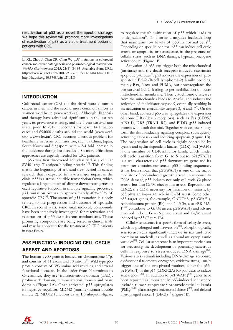
P53 FUNCTION: INDUCING CELL CYCLE ARREST AND APOPTOSISThe human TP53 gene is located on chromosome 17p, and consists of 11 exons and 10 introns[5]. Wild type p53 protein consists of 393 amino acid residues, and several functional domains. In the order from N-terminus to C-terminus, they are: transactivation domain (TAD), proline-rich domain, tetramerization domain and basic domain (Figure 1A). Once activated, p53 upregulates its negative regulator, MDM2 (murine/human double minute 2). MDM2 functions as an E3 ubiquitin-ligase,
to regulate the ubiquitination of p53 which leads to its degradation[6]. This forms a negative feedback loop that maintains low levels of p53 in normal cells[7]. Depending on specific context, p53 can induce cell cycle arrest, or apoptosis, or senescence, in the presence of cellular stress, such as DNA damage, hypoxia, oncogene activation, etc. (Figure 1B).
Activation of p53 can trigger both the mitochondrial (intrinsic) and the death-receptor-induced (extrinsic) apoptotic pathways[8]. p53 induces the expression of pro-apoptotic Bcl-2 (B-cell lymphoma-2) family proteins, mainly Bax, Noxa and PUMA, but downregulates the pro-survival Bcl-2, leading to permeabilization of outer mitochondrial membrane. Then cytochrome c releases from the mitochondria binds to Apaf-1, and induces the activation of the initiator caspase-9, eventually resulting in the activation of executioner caspase-3, -6 and -7[9]. On the other hand, activated p53 also upregulates the expression of some DRs (death receptors), such as Fas (CD95/APO-1), DR5 (TRAIL-R2), and PIDD (p53-induced protein with death domain). Together with caspase-8, they form the death-inducing signaling complex, subsequently activating caspase-3 and inducing apoptosis (Figure 1B). The progression of cell cycle is tightly controlled by cyclins and cyclin-dependent kinases (CDK). p21(WAF1) is one member of CDK inhibitor family, which hinder cell cycle transition from G1 to S phase. p21(WAF1) is a well-characterized p53-downstream gene and its promoter contains consensus p53-binding sequences. It has been shown that p21(WAF1) is one of the major mediator of p53-induced growth arrest. In response to DNA damage, p53 induces not only cell cycle G1 phase arrest, but also G2/M checkpoint arrest. Repression of CDC2, the CDK necessary for initiation of mitosis, by p53 plays an important role in G2/M arrest. Some other p53 target genes, for example, GADD45, p21(WAF1), retinoblastoma protein (Rb), and 14-3-3σ, also cRRIMA-1MET contribute to G2/M arrest. p21(WAF1) and Rb are involved in both G1 to S phase arrest and G2/M arrest induced by p53 (Figure 1B).
Cellular senescence is a specific form of cell cycle arrest, which is prolonged and irreversible[10]. Morphologically, senescence cells significantly increase in size and have prominent nucleoli, as well as abundant cytoplasmic vacuoles[11]. Cellular senescence is an important mechanism for preventing the development of potentially cancerous cells in response to stress-induced DNA damage[12]. Various stress stimuli including DNA-damage response, dysfunctional telomeres, oncogenes, oxidative stress, usually trigger one of the two pivotal routines, either the p53-p21(WAF1) or the p16 (CDKN2A)-Rb pathways to induce senescence[11,13]. In addition to p21(WAF1)[14], genes have been reported as important in p53-induced senescence include tumor suppressor promyelocytic leukemia (PML)[15,16], plasminogen activator inhibitor-1[17], and deleted in esophageal cancer 1 (DEC1)[18] (Figure 1B).
85 January 7, 2015|Volume 21|Issue 1|WJG|www.wjgnet.com
Li XL et al . p53 mutation in CRC
REGULATORS OF P53 ACTIVITY AND THEIR IMPLICATIONS IN CRCActivating transcription factor 3 Activating transcription factor 3 (ATF3) is one of the p53 target genes and involved in the complicated process of cellular stress response[19,20]. In addition, ATF3 also acts as a co-transcripition factor for p53 achieving ma-ximal induction of DR5 expression upon DNA damage in CRC[21]. DR5 is a trans-membrane TNF (tumor necrosis factor) receptor containing a death domain, which binds to the ligand TRAIL (tumor necrosis factor-related apoptosis-inducing ligand), and triggers cell death by activating the extrinsic apoptotic pathway[22]. Ectopic expression of ATF3 suppresses colon tumor growth and metastasisin mouse xenografts[23]. Post-translational modification of ATF3 by SUMO (small ubiquitin-related modifier) plays a negative role in the regulation of p53 activity[24]. ATF3 was also found to be bound to mutant p53, inactiving its oncogenic potential[25]. Of note, SUMO-1, a member of the SUMO protein family, involves a variety of biologically distinct functions
through SUMO attachment of target proteins[26]. Over-expression of SUMO-1 causes the accumulation of sumoylated p53 proteins in colon cancer cells, which leads to more frequent metastasis[27].
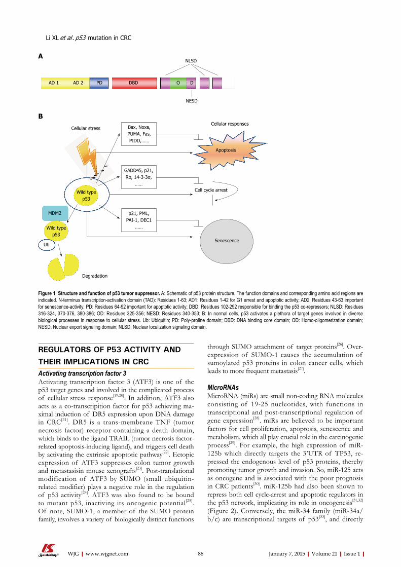
MicroRNAsMicroRNA (miRs) are small non-coding RNA molecules consisting of 19-25 nucleotides, with functions in transcriptional and post-transcriptional regulation of gene expression[28]. miRs are believed to be important factors for cell proliferation, apoptosis, senescence and metabolism, which all play crucial role in the carcinogenic process[29]. For example, the high expression of miR-125b which directly targets the 3’UTR of TP53, re-pressed the endogenous level of p53 proteins, thereby promoting tumor growth and invasion. So, miR-125 acts as oncogene and is associated with the poor prognosis in CRC patients[30]. miR-125b had also been shown to repress both cell cycle-arrest and apoptotic regulators in the p53 network, implicating its role in oncogenesis[31,32] (Figure 2). Conversely, the miR-34 family (miR-34a/b/c) are transcriptional targets of p53[33], and directly
86 January 7, 2015|Volume 21|Issue 1|WJG|www.wjgnet.com
Figure 1 Structure and function of p53 tumor suppressor. A: Schematic of p53 protein structure. The function domains and corresponding amino acid regions are indicated. N-terminus transcription-activation domain (TAD): Residues 1-63; AD1: Residues 1-42 for G1 arrest and apoptotic activity; AD2: Residues 43-63 important for senescence-activity; PD: Residues 64-92 important for apoptotic activity; DBD: Residues 102-292 responsible for binding the p53 co-repressors; NLSD: Residues 316-324, 370-376, 380-386; OD: Residues 325-356; NESD: Residues 340-353; B: In normal cells, p53 activates a plethora of target genes involved in diverse biological processes in response to cellular stress. Ub: Ubiquitin; PD: Poly-proline domain; DBD: DNA binding core domain; OD: Homo-oligomerization domain; NESD: Nuclear export signaling domain; NLSD: Nuclear localization signaling domain.
AD 1 AD 2 PD DBD O D
NESD
NLSD
Cellular stress Bax, Noxa, PUMA, Fas, PIDD,……
Wild type p53
MDM2
Wild type p53
Ub
Degradation
Senescence
GADD45, p21, Rb, 14-3-3σ,
……
p21, PML, PAI-1, DEC1
……
Cell cycle arrest
Apoptosis
Cellular responses
A
B
Li XL et al . p53 mutation in CRC
87 January 7, 2015|Volume 21|Issue 1|WJG|www.wjgnet.com
tumorous pathological process[41]. p53 mutation in CRC occurs in 34% of the proximal colon tumors, and in 45% of the distal colorectal tumors[8,42]. Majority of these mutations occur in exon 5 to 8 (DNA binding doman), and mainly in some hotspot codons, such as 175, 245, 248, 273 and 282, comprising of G to A, C to T transition and leading to the substitution of a single amino acid in p53 protein[41,42] (Table 1). Such substitutions most commonly cluster in the DNA binding domain, causing the disruption of specific DNA binding and sequential transactivation[7,42].
Different types of p53 mutations play a pivotal role in determining the biologic behavior of CRC, such as invasive depth, metastatic site and even the prognosis of patients. p53 mutations are associated with lymphatic invasion in proximal colon cancer, and show significant correlation with both lymphatic and vascular invasion in distal CRC[42] (Table 2). CRC patients with mutant p53 appear more chemo-resistance and have poorer prognosis than those with wild-type p53[43]. In a TP53 colorectal cancer international collaborative study, it was observed that patients with mutant p53 in exon 5 had worse outcome for proximal colon cancer[42] and inactivating mutation of p53 occurred more frequent in advanced stage tumors and were negatively associated with survival[44] (Table 2).
PHARMACOLOGICAL REACTIVATION OF P53 AS CANCER THERAPYResults from a large number of studies have unequivocally evidence demonstrated that mutant p53 not only plays a pivotal role in the transformation of CRC, but also
suppresses a range of Wnt and epithelial-mesenchymal transition (EMT) genes[34-37]. Thus, part of p53 tumor suppressor function is due to its inhibition of Wnt pathway and EMT transition through miR-34 and loss of this inhibition could trigger the proliferation and tissue-invasion of CRC cells[34,35] (Figure 2).
P53 MUTATION IN CRCDevelopment of CRC is a multi-factorial and multi-stage process involving the activation of oncogenes and inactiva-tion of tumor suppressor genes. Confirmed by numerous studies, p53 is a key tumor suppressor gene and is one of the most important elements of our body’s anticancer defense[38]. It is generally known that the progression of CRC follows mutations of the APC, K-Ras, and p53 genes[39]. p53 is the most commonly mutated gene in human cancers[40]. It is thought that p53 mutations play a critical role in the adenoma-carcinoma transition during
miRs p53
miR-125b p53miR-34 family p53
p53 protein
Wnt pathwayEMT transition
Cell proliferation Tumor growth Tissue invasive
Figure 2 Schematic representation of miRNAs regulating p53 pathway and subsequent tumorigenesis.
Table 1 Common, high frequency of p53 missense alterations in colorectal cancer
Exon Codon Codon change Nucleotide change
Amino acid change
5 175 CGC→CAC G→A Arg→His7 245 GGC→AGC G→A Gly→Ser7 245 GGC→GAC G→A Gly→Asp7 248 CGG→TGG C→T Arg→Trp7 248 CGG→CAG G→A Arg→Gln8 273 CGT→TGT C→T Arg→Cys8 273 CGT→CAT G→A Arg→His8 282 CGG→TGG C→T Arg→Trp
Data selected from UMD TP53 mutation database (http://p53.fr).
Li XL et al . p53 mutation in CRC
88 January 7, 2015|Volume 21|Issue 1|WJG|www.wjgnet.com
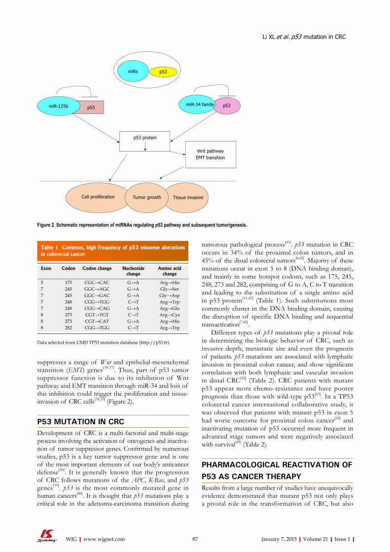
contributes to the aggressiveness and invasiveness of CRC. It is not surprising that manipulation of the p53 pathway has attracted interest soon after the discovery of p53 gene. Although reintroduction of wild type p53 by gene therapy appears a straightforward and logical choice, this approached is impeded by the technical challenge of efficient gene delivery and safety issues inherent in the use of viral vectors[45]. In recent years, we witness an array of small molecule inhibitors modulating the p53 pathway being developed (Figure 3). Some of these compounds have been tested as potential therapeutic agents in CRC.
Modulation of wild-type p53 activity via inhibiting MDM2-p53 interactionMDM2 protein, the E3 ubiquitin protein ligase, binds to the amino-terminal of p53, and ubiquitylates p53, leading to its proteasomal degradiaiton; this inhibits its suppressive function in cancer cells[46]. Pharmacological inhibitors of MDM2 have already been extensively researched for their anti-cancer activities through stabilization of p53 protein[47-49]. Activation of p53
without DNA damage should be a great advantage, compared to many traditional chemotherapeutic agents[47].
MIs (MDM2 Inhibitors): In recent years, a number of MIs that disrupt the MDM2-p53 interaction have been discovered. The spiro oxindole MI-43 is one of these specific MDM2 antagonists that cause p53 accumulation and lead to the induction of target genes, e.g., p21, Puma, and Noxa[50]. In colon cancer cells, cell cycle arrest and apoptosis were induced by MI-43 in a p53-dependent manner [51]. MI-219 is an improved MDM2-p53 inhibitor with improved pharmacokinetic profile and higher binding affinity to MDM2. MI-219 showed potent efficacy as a single agent in inducing apoptosis in HCT-116 colon cancer cell line. Furthermore, the combination of MI-219 with chemotherapeutic drug, Oxaliplatin, achieved high synergism in p53-mediated apoptotic response[52]. Nutlins: Nutlins are cis-imidazoline analogs, which occupy the binding pocket of MDM2, thus disrupting
Table 2 Summary of major conclusions on the importance of p53 in colorectal cancer development
Ref. Major conclusions
Taketani et al[21] p53 partners with ATF3 in maximal induction of DR5 uponDNA damage
Wei et al[25] ATF3 binds mutant p53 and inhibits its oncogenic functionNishida et al[30] High expression of miRNA-125b predicts poor survival in CRC. miRNA-125 decreases p53 expression Kim et al[34,35] Loss of p53 de-represses Wnt pathway and EMT transition through miRNA-34López et al[41] p53 mutations occur in 54% of sporadic CRCRusso et al[42] p53 mutations correlate with the site, biologic behaviour and outcome of CRCIacopetta et al[44] p53 mutations that lose transactivational ability are more common in advanced CRC and associated with poor survival
EMT: Epithelial-mesenchymal transition; CRC: Colorectal cancer.
Cell cycle arrest
Senescence
Apoptosis
MI-43nutlin-3
RITA
PRIMA-1PRIMA-1MET
NSC176372
Maslinic acidepicatechin gallate
a-lipoic acidquinacrine
MDM2
Mutantp53
Wildtype p53
Figure 3 Small molecule compounds pharmacologically reactivating of p53 function. MI43 and Nutlin-3 bound to MDM2 blocking MDM2-p53 interaction. RITA bound to p53 interfering MDM2-p53 interaction. a-Lipoic acid increased p53 protein stability and its apoptotic effect. Quinacrine induced the autophagy-associated cell death in a p53-dependent manner. NSC17632 activated p53-like activatity dependent on p73. PRIMA-1/PRIMA-1MET restored mutant p53 to exert apoptotic effect. Maslinic acid and Epicatechin gallate as plant extraction modulated the expression of p53 and its target genes in p53-dependent apoptotic and cell cycle arrest pathway.
Li XL et al . p53 mutation in CRC
p73
89 January 7, 2015|Volume 21|Issue 1|WJG|www.wjgnet.com
MDM2-p53 interaction. Nutlins were first discovered using biochemical screening strategy by Vassilev and colleagues in Roche in 2004[53]. Among them, Nutlin-3 (R1772) has been widely tested in a variety of cancers in vitro, in mouse xenografts bearing human tumors, as well as clinical trials in human subjects[54]. Nutlin-3 was observed to act as MDM2 antagonist, stabilize p53 and activate p53 target genes in CRC cells expressing wild-type p53. MDMX, another member of MDM protein family, shares a similar amino acid sequence and structural organization with MDM2. Although both MDM2 and MDMX negatively regulate p53, the relative abundance of MDM2 and MDMX level influences cancer cells response to Nutlin-3. Cancer cells overexpressing MDM2 are sensitive to Nutlin-3, in contrast, cancer cells overexpressing MDMX are resistant to Nutlin-3 due to its inability to block p53-MDMX interaction[55]. Nutlin-3a, but not the aftermentioned RIAT (reactivation of p53 and induction of tumor cell apoptosis), has been shown to specifically downregulate a5 integrin in p53 wild type colon cancer[56]. These findings are useful in patient selection in a clinical trial aiming to evaluate Nutlin-3 against CRC. Nutlin may offer clinical benefits for CRC bearing high expression MDM2 or a5 integrin.
Cancer cells often acquire secondary resistance after a prolonged exposure of single agent, so it is clinically desirable to treatment the cancer patients with combination therapy. Nutlin has been tested in combination with other drugs in CRC. Tumor necrosis factor (TNF)-related apoptosis-inducing ligand (TRAIL) is one of the DNA damage-inducible p53 target gene[57]. Notably, TRAIL induces cell death mainly through the induction of extrinsic apoptosis pathway, while Nutlin works predominately through inducing the intrinsic apoptosis pathway. Combination of Nutlin-3 and TRAIL synergistically enhances cell death in human p53 wild type sarcoma HOS cells and colon cancer HCT116 cells owing to the simultaneous engagement of intrinsic and extrinsic apoptosis pathways[58]. Furthermore, Nutlin-3 treatment increases DR5 expression on both mRNA and protein levels[58,59]. Controlled, concomitant release of Nutlin-3 and Doxil, the liposomal preparation of doxorubicin, by novel drug engineering, leads to synergistic anti-proliferative effect and induction of cell death in CRC cells carrying both wild-type and mutant p53[60]. Combination treatment with Nutlin-3 and Inauhzin, a SIRT1 (Sirtuin 1) inhibitor in colon and lung cancer cell lines, is able to enhances their apoptotic effect in a p53-dependent manner[61]. It is also noteworthy that Nutlin-3 can mediate the phosphorylation of p53 at key DNA-damage-specific serine residues (Ser15, 20 and 37) and initiate the DNA damage signaling pathway which resulted in cell cycle arrest in p53-independent manner[62]. Currently, Nutlin-3 has already been evaluated in phase I clinical trial to treat patients suffering from hematologic neoplasms[63]. Taken together, Nutlin-3 may be a helpful addition to our armamentarium combating CRC, particularly used in conjunction with other drugs.
RITA: RITA was identified from National Cancer Institute library compound Challenge set for its ability to inhibit the proliferation of HCT-116 (p53 wild type) much more than its p53 null counterpart[64]. RITA has been shown to suppress colon cancer growth in a mouse xenofgraft model. Mechanically, this compound directly binds to p53 rather than MDM2, and induces a conformational change in p53, which interfered with the p53-MDM2 interaction, and p53 ubiquitination, resulting in p53 accumulation and cellular apoptosis[64,65]. The study carried out by Di Marzo et al[66] implicated that RITA also reactivated mutant p53 function in malignant mesothelioma. Whether RITA is also effective in CRC cells harboring mutant p53 would merit further investigation.
Activation of p53-like activity via other p53 family members, p67 and p73In addition to p53, the p53 family includes two other members, p63 and p73[67]. They encode proteins with significant sequence homology and functional similarity with p53. A derivative of the cytotoxic plant alkaloid ellipticine, NSC176327 induced potent killing in CRC cells regardless of p53 status. Further experiments revealed that NSC176327 treatment increased the expression of p73, p21 and DR5, while knockdown of p73 in p53 null cells rendered these cells resistant to this drug treatment[68]. The notion that p73 is also a drug target in CRC is reinforced by other studies. Ray et al[69] reported that MDM2 inhibitors, like Nutlin-3 , could also disrupt the MDM2-p73 binding, and induce the expression of apoptotic proteins such as Noxa, PUMA and cell cycle arrest protein p21 in CRC cells lacking of functional p53[70]. Securinine, a widely used alkaloid, was identified to promote p73-dependent apoptosis in p53-deficent CRC cells[71]. In conclusion, these results shed new light on the induction of p73 as a therapeutic option in CRC patients with either mutant p53 or p53 null.
Reactivation of mutant p53It has been long recognized that mutant p53 protein not only abrogates the tumor suppressor function, but also gain novel oncogenic function, which promotes a more aggressive, metastatic cancer phenotype. However, it is until recently that promising compounds that specifically targeting this type of mutant oncogenic p53 proteins have been developed. Aiming to screen compounds that specifically targeting mutant p53, Bykov et al[72] discovered one compound 2,2-bis(hydroxymethyl)-1-azabicyclo[2,2,2]octan -3-one, which inhibited the growth of Saos-2-His-273 cells, a Tet-off mutant p53 cell line. This compound was named PRIMA-1 (p53-reactivation and induction of massive apoptosis-1, APR-017)[72]. Late, its methylated form, RRIMA-1MET (APR-246) which is more efficient, was developed by the same group[73]. PRIMA-1 restores the sequence-specific DNA binding region via forming adducts with thiols in mutant p53 and activating several p53 target genes, promoting apoptosis
Li XL et al . p53 mutation in CRC
90 January 7, 2015|Volume 21|Issue 1|WJG|www.wjgnet.com
in human cancer cells with mutant p53[74]. The initial consideration was that these two compounds had potent effects on p53-mutant cells, compared to cells with wild-type p53. However, emerging evidence demonstrated that unfolded mutant p53 and unfolded wild-type p53 could also be refolded by PRIMA-1 and PRIMA-1MET[74,75]. So PRIMA-1 and PRIMA-1MET may induce apoptosis in cancer cells carrying either wild-type p53 or mutant p53. Among the class of small molecules that can selectively induce apoptosis in cancer cells with mutant p53, PRIMA-1MET is the first drug which has already advanced to a phase Ⅰ/Ⅱ clinical trial for hematologic malignancies and prostate cancer[76,77]. However, there is little investigation about the ability of PRIMA-1MET to induce apoptosis and inhibit tumor growth in different CRC cell lines with different p53 status, thus, more studies are necessary to intensively explore RRIMA-1MET as a novel therapeutic strategy in CRC.
Natural agents extracted from plants Recently, the anticancer function of agents extracted from nature plants is attracting some attention. The mechanisms implicated have been uncovered constantly.
Maslinic acid: Maslinic acid (MA) is a natural triterpene from Olea europaea, and possesses potent anticancer property aganist CRC cells. Exposure to MA induced the expression of JNK (c-Jun NH2-terminal kinase), p53, and increased the mitochondrial apoptotic signaling molecules, resulting in cell cycle arrest and apoptosis[78,79]. In p53-deficient CRC cells, apoptosis could also be induced by MA without requiring the mitochondrial pathway[80].
Epicatechin gallate: Experimental and epidemiological evidences reveal that dietary polyphenolic plant-derived compounds have anti-proliferative and anti-invasive activity in cancers of gastrointestinal tract, lung, skin, prostate and breast[81-83]. Epicatechin gallate (ECG) is one of the most important compounds of polyphenols found in green tea, which stimulated the expression of p53, p21, and MAPKs (mitogen-activated protein kinases) in CRC cells, leading to cell cycle arrest at G0/G1-S phase in a time-dependent manner[82]. Furthermore, ECG could inhibit the degradation of p53 protein and RNA that contributed to the stabilization of p53.
Other compoundsp53 proteins can be targeted for proteasomal degradation in both normal and cancer cells. a - Lipoic acid (a-LA) is the most common drug worldwide to treat diabetic polyneuropathy. Yoo and colleagues had shown a-LA inhibited proliferation and induced apoptosis in colon cancer cells via preventing p53 degradation. Specifically, a-LA treatment downregulated ribosomal protein p90S6K (RPS6KA4) which was confirmed to inhibit p53 function. Furthermore, a-LA exerted an inhibitory effect on the nuclear translocation of nuclear factor-κB
(NF-κB), which played an important role in regulating RPS6KA4 gene expression[84].
FUTURE PERSPECTIVESThere is no doubt that reactivation and restoration of p53 function have great potential as a novel therapeutic strategy in CRC. However, the majority of molecules that lead to cell cycle arrest and apoptosis in CRC cells, has only been tested in cell lines and animal models, and has yet to enter in clinical trials. In addition, it is clear that mutant p53 promotes various oncogenic events. Nevertheless, the critical mechanisms are still not completely understood. The issue that different mutations might affect p53 function differently makes small molecule inhibitors targeting mutant p53 more complicated to assess in a clinical trial. This theme needs to be explored further. Importantly, resistance to treatments and poor prognosis for CRC patients with new p53 mutations will require the continuing development of new agent targeting these novel mutations. Riding on the last 30 years of intensive research in p53 area, this is now the time to harvest the fruits from this body of work and translate our knowledge of p53 into clinical practice for CRC patients.
REFERENCES1 Moghimi-Dehkordi B, Safaee A. An overview of colorectal
cancer survival rates and prognosis in Asia. World J Gastrointest Oncol 2012; 4: 71-75 [PMID: 22532879 DOI: 10.4251/wjgo.v4.i4.71]
2 Farnebo M , Bykov VJ, Wiman KG. The p53 tumor suppressor: a master regulator of diverse cellular processes and therapeutic target in cancer. Biochem Biophys Res Commun 2010; 396: 85-89 [PMID: 20494116 DOI: 10.1016/j.bbrc.2010.02.152]
3 Tan TH, Wallis J, Levine AJ. Identification of the p53 protein domain involved in formation of the simian virus 40 large T-antigen-p53 protein complex. J Virol 1986; 59: 574-583 [PMID: 3016321]
4 Takayama T, Miyanishi K, Hayashi T, Sato Y, Niitsu Y. Colorectal cancer: genetics of development and metastasis. J Gastroenterol 2006; 41: 185-192 [PMID: 16699851 DOI: 10.1007/s00535-006-1801-6]
5 Saha MN , Qiu L, Chang H. Targeting p53 by small molecules in hematological malignancies. J Hematol Oncol 2013; 6: 23 [PMID: 23531342 DOI: 10.1186/1756-8722-6-23]
6 Li Q, Lozano G. Molecular pathways: targeting Mdm2 and Mdm4 in cancer therapy. Clin Cancer Res 2013; 19: 34-41 [PMID: 23262034 DOI: 10.1158/1078-0432.CCR-12-0053]
7 Zandi R , Selivanova G, Christensen CL, Gerds TA, Willumsen BM, Poulsen HS. PRIMA-1Met/APR-246 induces apoptosis and tumor growth delay in small cell lung cancer expressing mutant p53. Clin Cancer Res 2011; 17: 2830-2841 [PMID: 21415220 DOI: 10.1158/1078-0432.CCR-10-3168]
8 Ryan KM, Phillips AC, Vousden KH. Regulation and function of the p53 tumor suppressor protein. Curr Opin Cell Biol 2001; 13: 332-337 [PMID: 11343904]
9 Shen J, Vakifahmetoglu H, Stridh H, Zhivotovsky B, Wiman KG. PRIMA-1MET induces mitochondrial apoptosis through activation of caspase-2. Oncogene 2008; 27: 6571-6580 [PMID: 18663359 DOI: 10.1038/onc.2008.249]
10 Salama R, Sadaie M, Hoare M, Narita M. Cellular senescence
Li XL et al . p53 mutation in CRC
91 January 7, 2015|Volume 21|Issue 1|WJG|www.wjgnet.com
and its effector programs. Genes Dev 2014; 28: 99-114 [PMID: 24449267 DOI: 10.1101/gad.235184.113]
11 Campisi J, d’Adda di Fagagna F. Cellular senescence: when bad things happen to good cells. Nat Rev Mol Cell Biol 2007; 8: 729-740 [PMID: 17667954 DOI: 10.1038/nrm2233]
12 Mallette FA, Ferbeyre G. The DNA damage signaling pathway connects oncogenic stress to cellular senescence. Cell Cycle 2007; 6: 1831-1836 [PMID: 17671427]
13 Shay JW, Pereira-Smith OM, Wright WE. A role for both RB and p53 in the regulation of human cellular senescence. Exp Cell Res 1991; 196: 33-39 [PMID: 1652450]
14 Noda A, Ning Y, Venable SF, Pereira-Smith OM, Smith JR. Cloning of senescent cell-derived inhibitors of DNA synthesis using an expression screen. Exp Cell Res 1994; 211: 90-98 [PMID: 8125163 DOI: 10.1006/excr.1994.1063]
15 Ferbeyre G, de Stanchina E, Querido E, Baptiste N, Prives C, Lowe SW. PML is induced by oncogenic ras and promotes premature senescence. Genes Dev 2000; 14: 2015-2027 [PMID: 10950866]
16 Pearson M, Carbone R, Sebastiani C, Cioce M, Fagioli M, Saito S, Higashimoto Y, Appella E, Minucci S, Pandolfi PP, Pelicci PG. PML regulates p53 acetylation and premature senescence induced by oncogenic Ras. Nature 2000; 406: 207-210 [PMID: 10910364 DOI: 10.1038/35018127]
17 Kortlever RM, Higgins PJ, Bernards R. Plasminogen activator inhibitor-1 is a critical downstream target of p53 in the induction of replicative senescence. Nat Cell Biol 2006; 8: 877-884 [PMID: 16862142 DOI: 10.1038/ncb1448]
18 Qian Y, Zhang J, Yan B, Chen X. DEC1, a basic helix-loop-helix transcription factor and a novel target gene of the p53 family, mediates p53-dependent premature senescence. J Biol Chem 2008; 283: 2896-2905 [PMID: 18025081 DOI: 10.1074/jbc.M708624200]
19 Zhang C, Gao C, Kawauchi J, Hashimoto Y, Tsuchida N, Kitajima S. Transcriptional activation of the human stress-inducible transcriptional repressor ATF3 gene promoter by p53. Biochem Biophys Res Commun 2002; 297: 1302-1310 [PMID: 12372430]
20 Kannan K, Amariglio N, Rechavi G, Jakob-Hirsch J, Kela I, Kaminski N, Getz G, Domany E, Givol D. DNA microarrays identification of primary and secondary target genes regulated by p53. Oncogene 2001; 20: 2225-2234 [PMID: 11402317 DOI: 10.1038/sj.onc.1204319]
21 Taketani K, Kawauchi J, Tanaka-Okamoto M, Ishizaki H, Tanaka Y, Sakai T, Miyoshi J, Maehara Y, Kitajima S. Key role of ATF3 in p53-dependent DR5 induction upon DNA damage of human colon cancer cells. Oncogene 2012; 31: 2210-2221 [PMID: 21927023 DOI: 10.1038/onc.2011.397]
22 MacFarlane M, Ahmad M, Srinivasula SM, Fernandes-Alnemri T, Cohen GM, Alnemri ES. Identification and molecular cloning of two novel receptors for the cytotoxic ligand TRAIL. J Biol Chem 1997; 272: 25417-25420 [PMID: 9325248]
23 Hackl C, Lang SA, Moser C, Mori A, Fichtner-Feigl S, Hellerbrand C, Dietmeier W, Schlitt HJ, Geissler EK, Stoeltzing O. Activating transcription factor-3 (ATF3) functions as a tumor suppressor in colon cancer and is up-regulated upon heat-shock protein 90 (Hsp90) inhibition. BMC Cancer 2010; 10: 668 [PMID: 21129190 DOI: 10.1186/1471-2407-10-668]
24 Wang CM, Brennan VC, Gutierrez NM, Wang X, Wang L, Yang WH. SUMOylation of ATF3 alters its transcriptional activity on regulation of TP53 gene. J Cell Biochem 2013; 114: 589-598 [PMID: 22991139 DOI: 10.1002/jcb.24396]
25 Wei S, Wang H, Lu C, Malmut S, Zhang J, Ren S, Yu G, Wang W, Tang DD, Yan C. The activating transcription factor 3 protein suppresses the oncogenic function of mutant p53 proteins. J Biol Chem 2014; 289: 8947-8959 [PMID: 24554706 DOI: 10.1074/jbc.M113.503755]
26 Barry J, Lock RB. Small ubiquitin-related modifier-1:
Wrestling with protein regulation. Int J Biochem Cell Biol 2011; 43: 37-40 [PMID: 20932933 DOI: 10.1016/j.biocel.2010.09.022]
27 Zhang H, Kuai X, Ji Z, Li Z, Shi R. Over-expression of small ubiquitin-related modifier-1 and sumoylated p53 in colon cancer. Cell Biochem Biophys 2013; 67: 1081-1087 [PMID: 23640307 DOI: 10.1007/s12013-013-9612-x]
28 Bi CL, Chng WJ. miRNA deregulation in multiple myeloma. Chin Med J (Engl) 2011; 124: 3164-3169 [PMID: 22040573]
29 Ye JJ, Cao J. MicroRNAs in colorectal cancer as markers and targets: Recent advances. World J Gastroenterol 2014; 20: 4288-4299 [PMID: 24764666 DOI: 10.3748/wjg.v20.i15.4288]
30 Nishida N, Yokobori T, Mimori K, Sudo T, Tanaka F, Shibata K, Ishii H, Doki Y, Kuwano H, Mori M. MicroRNA miR-125b is a prognostic marker in human colorectal cancer. Int J Oncol 2011; 38: 1437-1443 [PMID: 21399871 DOI: 10.3892/ijo.2011.969]
31 Le MT, Shyh-Chang N, Khaw SL, Chin L, Teh C, Tay J, O’Day E, Korzh V, Yang H, Lal A, Lieberman J, Lodish HF, Lim B. Conserved regulation of p53 network dosage by microRNA-125b occurs through evolving miRNA-target gene pairs. PLoS Genet 2011; 7: e1002242 [PMID: 21935352 DOI: 10.1371/journal.pgen.1002242]
32 Kumar M, Lu Z, Takwi AA, Chen W, Callander NS, Ramos KS, Young KH, Li Y. Negative regulation of the tumor suppressor p53 gene by microRNAs. Oncogene 2011; 30: 843-853 [PMID: 20935678 DOI: 10.1038/onc.2010.457]
33 He L, He X, Lim LP, de Stanchina E, Xuan Z, Liang Y, Xue W, Zender L, Magnus J, Ridzon D, Jackson AL, Linsley PS, Chen C, Lowe SW, Cleary MA, Hannon GJ. A microRNA component of the p53 tumour suppressor network. Nature 2007; 447: 1130-1134 [PMID: 17554337 DOI: 10.1038/nature05939]
34 Kim NH, Kim HS, Kim NG, Lee I, Choi HS, Li XY, Kang SE, Cha SY, Ryu JK, Na JM, Park C, Kim K, Lee S, Gumbiner BM, Yook JI, Weiss SJ. p53 and microRNA-34 are suppressors of canonical Wnt signaling. Sci Signal 2011; 4: ra71 [PMID: 22045851 DOI: 10.1126/scisignal.2001744]
35 Kim NH, Cha YH, Kang SE, Lee Y, Lee I, Cha SY, Ryu JK, Na JM, Park C, Yoon HG, Park GJ, Yook JI, Kim HS. p53 regulates nuclear GSK-3 levels through miR-34-mediated Axin2 suppression in colorectal cancer cells. Cell Cycle 2013; 12: 1578-1587 [PMID: 23624843 DOI: 10.4161/cc.24739]
36 Cha YH , Kim NH, Park C, Lee I, Kim HS, Yook JI. MiRNA-34 intrinsically links p53 tumor suppressor and Wnt signaling. Cell Cycle 2012; 11: 1273-1281 [PMID: 22421157 DOI: 10.4161/cc.19618]
37 Siemens H, Jackstadt R, Hünten S, Kaller M, Menssen A, Götz U, Hermeking H. miR-34 and SNAIL form a double-negative feedback loop to regulate epithelial-mesenchymal transitions. Cell Cycle 2011; 10: 4256-4271 [PMID: 22134354 DOI: 10.4161/cc.10.24.18552]
38 Levine AJ, Oren M. The first 30 years of p53: growing ever more complex. Nat Rev Cancer 2009; 9: 749-758 [PMID: 19776744 DOI: 10.1038/nrc2723]
39 Cottu PH, Muzeau F, Estreicher A, Fléjou JF, Iggo R, Thomas G, Hamelin R. Inverse correlation between RER+ status and p53 mutation in colorectal cancer cell lines. Oncogene 1996; 13: 2727-2730 [PMID: 9000147]
40 Kandoth C, McLellan MD, Vandin F, Ye K, Niu B, Lu C, Xie M, Zhang Q, McMichael JF, Wyczalkowski MA, Leiserson MD, Miller CA, Welch JS, Walter MJ, Wendl MC, Ley TJ, Wilson RK, Raphael BJ, Ding L. Mutational landscape and significance across 12 major cancer types. Nature 2013; 502: 333-339 [PMID: 24132290 DOI: 10.1038/nature12634]
41 López I, P Oliveira L, Tucci P, Alvarez-Valín F, A Coudry R, Marín M. Different mutation profiles associated to P53 accumulation in colorectal cancer. Gene 2012; 499: 81-87 [PMID: 22373952 DOI: 10.1016/j.gene.2012.02.011]
42 Russo A, Bazan V, Iacopetta B, Kerr D, Soussi T, Gebbia N. The TP53 colorectal cancer international collaborative
Li XL et al . p53 mutation in CRC
92 January 7, 2015|Volume 21|Issue 1|WJG|www.wjgnet.com
study on the prognostic and predictive significance of p53 mutation: influence of tumor site, type of mutation, and adjuvant treatment. J Clin Oncol 2005; 23: 7518-7528 [PMID: 16172461 DOI: 10.1200/JCO.2005.00.471]
43 Iacopetta B. TP53 mutation in colorectal cancer. Hum Mutat 2003; 21: 271-276 [PMID: 12619112 DOI: 10.1002/humu.10175]
44 Iacopetta B, Russo A, Bazan V, Dardanoni G, Gebbia N, Soussi T, Kerr D, Elsaleh H, Soong R, Kandioler D, Janschek E, Kappel S, Lung M, Leung CS, Ko JM, Yuen S, Ho J, Leung SY, Crapez E, Duffour J, Ychou M, Leahy DT, O’Donoghue DP, Agnese V, Cascio S, Di Fede G, Chieco-Bianchi L, Bertorelle R, Belluco C, Giaretti W, Castagnola P, Ricevuto E, Ficorella C, Bosari S, Arizzi CD, Miyaki M, Onda M, Kampman E, Diergaarde B, Royds J, Lothe RA, Diep CB, Meling GI, Ostrowski J, Trzeciak L, Guzinska-Ustymowicz K, Zalewski B, Capellá GM, Moreno V, Peinado MA, Lönnroth C, Lundholm K, Sun XF, Jansson A, Bouzourene H, Hsieh LL, Tang R, Smith DR, Allen-Mersh TG, Khan ZA, Shorthouse AJ, Silverman ML, Kato S, Ishioka C. Functional categories of TP53 mutation in colorectal cancer: results of an International Collaborative Study. Ann Oncol 2006; 17: 842-847 [PMID: 16524972 DOI: 10.1093/annonc/mdl035]
45 Lane DP, Cheok CF, Lain S. p53-based cancer therapy. Cold Spring Harb Perspect Biol 2010; 2: a001222 [PMID: 20463003 DOI: 10.1101/cshperspect.a001222]
46 Micel LN, Tentler JJ, Smith PG, Eckhardt GS. Role of ubiquitin ligases and the proteasome in oncogenesis: novel targets for anticancer therapies. J Clin Oncol 2013; 31: 1231-1238 [PMID: 23358974 DOI: 10.1200/JCO.2012.44.0958]
47 Rigatti MJ, Verma R, Belinsky GS, Rosenberg DW, Giardina C. Pharmacological inhibition of Mdm2 triggers growth arrest and promotes DNA breakage in mouse colon tumors and human colon cancer cells. Mol Carcinog 2012; 51: 363-378 [PMID: 21557332 DOI: 10.1002/mc.20795]
48 Patel S, Player MR. Small-molecule inhibitors of the p53-HDM2 interaction for the treatment of cancer. Expert Opin Investig Drugs 2008; 17: 1865-1882 [PMID: 19012502 DOI: 10.1517/13543780802493366]
49 Vassilev LT. MDM2 inhibitors for cancer therapy. Trends Mol Med 2007; 13: 23-31 [PMID: 17126603 DOI: 10.1016/j.molmed.2006.11.002]
50 Sun SH, Zheng M, Ding K, Wang S, Sun Y. A small molecule that disrupts Mdm2-p53 binding activates p53, induces apoptosis and sensitizes lung cancer cells to chemotherapy. Cancer Biol Ther 2008; 7: 845-852 [PMID: 18340116]
51 Shangary S, Ding K, Qiu S, Nikolovska-Coleska Z, Bauer JA, Liu M, Wang G, Lu Y, McEachern D, Bernard D, Bradford CR, Carey TE, Wang S. Reactivation of p53 by a specific MDM2 antagonist (MI-43) leads to p21-mediated cell cycle arrest and selective cell death in colon cancer. Mol Cancer Ther 2008; 7: 1533-1542 [PMID: 18566224 DOI: 10.1158/1535-7163.MCT-08-0140]
52 Azmi AS, Banerjee S, Ali S, Wang Z, Bao B, Beck FW, Maitah M, Choi M, Shields TF, Philip PA, Sarkar FH, Mohammad RM. Network modeling of MDM2 inhibitor-oxaliplatin combination reveals biological synergy in wt-p53 solid tumors. Oncotarget 2011; 2: 378-392 [PMID: 21623005]
53 Vassilev LT, Vu BT, Graves B, Carvajal D, Podlaski F, Filipovic Z, Kong N, Kammlott U, Lukacs C, Klein C, Fotouhi N, Liu EA. In vivo activation of the p53 pathway by small-molecule antagonists of MDM2. Science 2004; 303: 844-848 [PMID: 14704432 DOI: 10.1126/science.1092472]
54 Shen H, Maki CG. Pharmacologic activation of p53 by small-molecule MDM2 antagonists. Curr Pharm Des 2011; 17: 560-568 [PMID: 21391906]
55 Patton JT, Mayo LD, Singhi AD, Gudkov AV, Stark GR, Jackson MW. Levels of HdmX expression dictate the sensitivity of normal and transformed cells to Nutlin-3. Cancer Res 2006; 66: 3169-3176 [PMID: 16540668 DOI:
10.1158/0008-5472.CAN-05-3832]56 Janouskova H, Ray AM, Noulet F, Lelong-Rebel I, Choulier
L, Schaffner F, Lehmann M, Martin S, Teisinger J, Dontenwill M. Activation of p53 pathway by Nutlin-3a inhibits the expression of the therapeutic target α5 integrin in colon cancer cells. Cancer Lett 2013; 336: 307-318 [PMID: 23523610 DOI: 10.1016/j.canlet.2013.03.018]
57 Wu GS, Burns TF, McDonald ER, Jiang W, Meng R, Krantz ID, Kao G, Gan DD, Zhou JY, Muschel R, Hamilton SR, Spinner NB, Markowitz S, Wu G, el-Deiry WS. KILLER/DR5 is a DNA damage-inducible p53-regulated death receptor gene. Nat Genet 1997; 17: 141-143 [PMID: 9326928 DOI: 10.1038/ng1097-141]
58 Hori T, Kondo T, Kanamori M, Tabuchi Y, Ogawa R, Zhao QL, Ahmed K, Yasuda T, Seki S, Suzuki K, Kimura T. Nutlin-3 enhances tumor necrosis factor-related apoptosis-inducing ligand (TRAIL)-induced apoptosis through up-regulation of death receptor 5 (DR5) in human sarcoma HOS cells and human colon cancer HCT116 cells. Cancer Lett 2010; 287: 98-108 [PMID: 19577358 DOI: 10.1016/j.canlet.2009.06.002]
59 Meijer A, Kruyt FA, van der Zee AG, Hollema H, Le P, ten Hoor KA, Groothuis GM, Quax WJ, de Vries EG, de Jong S. Nutlin-3 preferentially sensitises wild-type p53-expressing cancer cells to DR5-selective TRAIL over rhTRAIL. Br J Cancer 2013; 109: 2685-2695 [PMID: 24136147 DOI: 10.1038/bjc.2013.636]
60 Nadler-Milbauer M, Apter L, Haupt Y, Haupt S, Barenholz Y, Minko T, Rubinstein A. Synchronized release of Doxil and Nutlin-3 by remote degradation of polysaccharide matrices and its possible use in the local treatment of colorectal cancer. J Drug Target 2011; 19: 859-873 [PMID: 22082104 DOI: 10.3109/1061186X.2011.622401]
61 Zhang Y, Zhang Q, Zeng SX, Zhang Y, Mayo LD, Lu H. Inauhzin and Nutlin3 synergistically activate p53 and suppress tumor growth. Cancer Biol Ther 2012; 13: 915-924 [PMID: 22785205 DOI: 10.4161/cbt.20844]
62 Valentine JM, Kumar S, Moumen A. A p53-independent role for the MDM2 antagonist Nutlin-3 in DNA damage response initiation. BMC Cancer 2011; 11: 79 [PMID: 21338495 DOI: 10.1186/1471-2407-11-79]
63 Chen F, Wang W, El-Deiry WS. Current strategies to target p53 in cancer. Biochem Pharmacol 2010; 80: 724-730 [PMID: 20450892 DOI: 10.1016/j.bcp.2010.04.031]
64 Issaeva N, Bozko P, Enge M, Protopopova M, Verhoef LG, Masucci M, Pramanik A, Selivanova G. Small molecule RITA binds to p53, blocks p53-HDM-2 interaction and activates p53 function in tumors. Nat Med 2004; 10: 1321-1328 [PMID: 15558054 DOI: 10.1038/nm1146]
65 Essmann F, Schulze-Osthoff K. Translational approaches targeting the p53 pathway for anti-cancer therapy. Br J Pharmacol 2012; 165: 328-344 [PMID: 21718309 DOI: 10.1111/j.1476-5381.2011.01570.x]
66 Di Marzo D, Forte IM, Indovina P, Di Gennaro E, Rizzo V, Giorgi F, Mattioli E, Iannuzzi CA, Budillon A, Giordano A, Pentimalli F. Pharmacological targeting of p53 through RITA is an effective antitumoral strategy for malignant pleural mesothelioma. Cell Cycle 2014; 13: 652-665 [PMID: 24345738 DOI: 10.4161/cc.27546]
67 Damia G, Broggini M. Cell cycle checkpoint proteins and cellular response to treatment by anticancer agents. Cell Cycle 2004; 3: 46-50 [PMID: 14657665]
68 Lu C, Wang W, El-Deiry WS. Non-genotoxic anti-neoplastic effects of ellipticine derivative NSC176327 in p53-deficient human colon carcinoma cells involve stimulation of p73. Cancer Biol Ther 2008; 7: 2039-2046 [PMID: 19106635]
69 Ray RM, Bhattacharya S, Johnson LR. Mdm2 inhibition induces apoptosis in p53 deficient human colon cancer cells by activating p73- and E2F1-mediated expression of PUMA and Siva-1. Apoptosis 2011; 16: 35-44 [PMID: 20812030 DOI:
Li XL et al . p53 mutation in CRC
93 January 7, 2015|Volume 21|Issue 1|WJG|www.wjgnet.com
10.1007/s10495-010-0538-0]70 Hong B, Prabhu VV, Zhang S, van den Heuvel AP, Dicker
DT, Kopelovich L, El-Deiry WS. Prodigiosin rescues deficient p53 signaling and antitumor effects via upregulating p73 and disrupting its interaction with mutant p53. Cancer Res 2014; 74: 1153-1165 [PMID: 24247721 DOI: 10.1158/0008-5472.CAN-13-0955]
71 Rana S, Gupta K, Gomez J, Matsuyama S, Chakrabarti A, Agarwal ML, Agarwal A, Agarwal MK, Wald DN. Securinine induces p73-dependent apoptosis preferentially in p53-deficient colon cancer cells. FASEB J 2010; 24: 2126-2134 [PMID: 20133503 DOI: 10.1096/fj.09-148999]
72 Bykov VJ, Issaeva N, Shilov A, Hultcrantz M, Pugacheva E, Chumakov P, Bergman J, Wiman KG, Selivanova G. Restoration of the tumor suppressor function to mutant p53 by a low-molecular-weight compound. Nat Med 2002; 8: 282-288 [PMID: 11875500 DOI: 10.1038/nm0302-282]
73 Bykov VJ, Zache N, Stridh H, Westman J, Bergman J, Selivanova G, Wiman KG. PRIMA-1(MET) synergizes with cisplatin to induce tumor cell apoptosis. Oncogene 2005; 24: 3484-3491 [PMID: 15735745 DOI: 10.1038/sj.onc.1208419]
74 Lambert JM, Gorzov P, Veprintsev DB, Söderqvist M, Segerbäck D, Bergman J, Fersht AR, Hainaut P, Wiman KG, Bykov VJ. PRIMA-1 reactivates mutant p53 by covalent binding to the core domain. Cancer Cell 2009; 15: 376-388 [PMID: 19411067 DOI: 10.1016/j.ccr.2009.03.003]
75 Rieber M, Strasberg-Rieber M. Hypoxia, Mn-SOD and H(2)O(2) regulate p53 reactivation and PRIMA-1 toxicity irrespective of p53 status in human breast cancer cells. Biochem Pharmacol 2012; 84: 1563-1570 [PMID: 22982566 DOI: 10.1016/j.bcp.2012.09.003]
76 Cheok CF, Verma CS, Baselga J, Lane DP. Translating p53 into the clinic. Nat Rev Clin Oncol 2011; 8: 25-37 [PMID: 20975744 DOI: 10.1038/nrclinonc.2010.174]
77 Lehmann S, Bykov VJ, Ali D, Andrén O, Cherif H, Tidefelt U, Uggla B, Yachnin J, Juliusson G, Moshfegh A, Paul C, Wiman KG, Andersson PO. Targeting p53 in vivo: a first-in-human study with p53-targeting compound APR-246 in
refractory hematologic malignancies and prostate cancer. J Clin Oncol 2012; 30: 3633-3639 [PMID: 22965953 DOI: 10.1200/JCO.2011.40.7783]
78 Rufino-Palomares EE, Reyes-Zurita FJ, García-Salguero L, Mokhtari K, Medina PP, Lupiáñez JA, Peragón J. Maslinic acid, a triterpenic anti-tumoural agent, interferes with cytoskeleton protein expression in HT29 human colon-cancer cells. J Proteomics 2013; 83: 15-25 [PMID: 23499989 DOI: 10.1016/j.jprot.2013.02.031]
79 Reyes-Zurita FJ, Pachón-Peña G, Lizárraga D, Rufino-Palomares EE, Cascante M, Lupiáñez JA. The natural triterpene maslinic acid induces apoptosis in HT29 colon cancer cells by a JNK-p53-dependent mechanism. BMC Cancer 2011; 11: 154 [PMID: 21524306 DOI: 10.1186/1471-2407-11-154]
80 Reyes-Zurita FJ, Rufino-Palomares EE, Medina PP, Leticia García-Salguero E, Peragón J, Cascante M, Lupiáñez JA. Antitumour activity on extrinsic apoptotic targets of the triterpenoid maslinic acid in p53-deficient Caco-2 adenocarcinoma cells. Biochimie 2013; 95: 2157-2167 [PMID: 23973282 DOI: 10.1016/j.biochi.2013.08.017]
81 Baek SJ, Kim JS, Jackson FR, Eling TE, McEntee MF, Lee SH. Epicatechin gallate-induced expression of NAG-1 is associated with growth inhibition and apoptosis in colon cancer cells. Carcinogenesis 2004; 25: 2425-2432 [PMID: 15308587 DOI: 10.1093/carcin/bgh255]
82 Cordero-Herrera I, Martín MA, Bravo L, Goya L, Ramos S. Epicatechin gallate induces cell death via p53 activation and stimulation of p38 and JNK in human colon cancer SW480 cells. Nutr Cancer 2013; 65: 718-728 [PMID: 23859040 DOI: 10.1080/01635581.2013.795981]
83 Sánchez-Tena S, Alcarraz-Vizán G, Marín S, Torres JL, Cascante M. Epicatechin gallate impairs colon cancer cell metabolic productivity. J Agric Food Chem 2013; 61: 4310-4317 [PMID: 23594085 DOI: 10.1021/jf3052785]
84 Yoo TH, Lee JH, Chun HS, Chi SG. α-Lipoic acid prevents p53 degradation in colon cancer cells by blocking NF-κB induction of RPS6KA4. Anticancer Drugs 2013; 24: 555-565 [PMID: 23599020 DOI: 10.1097/CAD.0b013e32836181eb]
P- Reviewer: Gao CM, Lakatos PL, Moussata D S- Editor: Ma YJ L- Editor: A E- Editor: Wang CH
Li XL et al . p53 mutation in CRC

© 2015 Baishideng Publishing Group Inc. All rights reserved.
Published by Baishideng Publishing Group Inc8226 Regency Drive, Pleasanton, CA 94588, USA
Telephone: +1-925-223-8242Fax: +1-925-223-8243
E-mail: [email protected] Desk: http://www.wjgnet.com/esps/helpdesk.aspx
http://www.wjgnet.com
I S S N 1 0 0 7 - 9 3 2 7
9 7 7 1 0 07 9 3 2 0 45
0 1