Embed Size (px)
Citation preview
Therapeutics, Targets, and Chemical Biology
p53 Loss in MYC-Driven Neuroblastoma Leadsto Metabolic Adaptations SupportingRadioresistanceOrli Yogev1, Karen Barker1, Arti Sikka2, Gilberto S. Almeida1,3, Albert Hallsworth1,Laura M. Smith1, Yann Jamin3, Ruth Ruddle4, Alexander Koers1, Hannah T.Webber1,Florence I. Raynaud4, Sergey Popov4,5, Chris Jones4,5, Kevin Petrie1,Simon P. Robinson3, Hector C. Keun2, and Louis Chesler1
Abstract
Neuroblastoma is the most common childhood extracranialsolid tumor. In high-risk cases, many of which are characterizedby amplification ofMYCN, outcome remains poor. Mutations inthe p53 (TP53) tumor suppressor are rare at diagnosis, butevidence suggests that p53 function is often impaired inrelapsed, treatment-resistant disease. To address the role ofp53 loss of function in the development and pathogenesis ofhigh-risk neuroblastoma, we generated a MYCN-driven geneti-cally engineered mouse model in which the tamoxifen-induciblep53ERTAM fusion protein was expressed from a knock-in allele(Th-MYCN/Trp53KI). We observed no significant differences intumor-free survival between Th-MYCN mice heterozygous forTrp53KI (n ¼ 188) and Th-MYCN mice with wild-type p53 (n ¼101). Conversely, the survival of Th-MYCN/Trp53KI/KI micelacking functional p53 (n ¼ 60) was greatly reduced. We foundthat Th-MYCN/Trp53KI/KI tumors were resistant to ionizing
radiation (IR), as expected. However, restoration of functionalp53ERTAM reinstated sensitivity to IR in only 50% of Th-MYCN/Trp53KI/KI tumors, indicating the acquisition of additional resis-tance mechanisms. Gene expression and metabolic analysesindicated that the principal acquired mechanism of resistanceto IR in the absence of functional p53 was metabolic adaptationin response to chronic oxidative stress. Tumors exhibitedincreased antioxidant metabolites and upregulation of glutathi-one S-transferase pathway genes, including Gstp1 and Gstz1,which are associated with poor outcome in human neuroblas-toma. Accordingly, glutathione depletion by buthionine sulfox-imine together with restoration of p53 activity resensitizedtumors to IR. Our findings highlight the complex pathwaysoperating in relapsed neuroblastomas and the need for combi-nation therapies that target the diverse resistance mechanisms atplay. Cancer Res; 76(10); 3025–35. �2016 AACR.
Introductionp53 is a critical tumor suppressor that performs a diverse range
of functions, including induction of apoptosis, senescence, andDNA repair in response to genotoxic stress. Deregulation of itsactivity is associatedwith tumor initiation andprogression aswellas resistance to therapy. Recent in vivo evidence indicates that therole of p53 as a tumor suppressor is independent of its canonicalrole as a cell cycle, senescence, and proapoptosis regulator, and
may be due to other p53-dependent activities such as mainte-nance of DNA stability and metabolic adaptation (1). In the lastdecade, a role for p53 inmetabolic adaptation has been identified(2, 3), often activated by metabolic stress induced by low oxygenlevels or nutrient scarcity (3). This leads to changes in severalmetabolic mechanisms affecting energy homeostasis, such asglycolysis, downregulation of reactive oxygen species (ROS),oxidative phosphorylation (OXPHOS), b-oxidation, and gluco-neogenesis (4). However, to date, there is no direct evidencedemonstrating an advantage in tumor survival or growth in vivodue to metabolic alterations induced by p53 loss of function.
Neuroblastoma is the most common extracranial pediatricsolid tumor, and amplification of the MYCN gene is a predictorof high-risk disease. Treatment options for high-risk patientsinclude intensive cytotoxic chemotherapy and surgical resection,myeloablative autologous stem cell transplantation, radiothera-py, and intensive multimodal therapy. Although most high-riskpatients initially respond to therapy, a majority of these patientswill relapse with treatment-resistant disease. Approximately 50%of relapsed tumors are associated with p53 loss of function,mainly through alterations to the p53 pathway. In contrast withother MYC-driven cancers, such as medulloblastoma (5) andlymphoma, mutation of genes in the p53 pathway is rare atdiagnosis (6), implying that selection for p53 pathway deficiencyoccurs following treatment. Additional mechanisms such as epi-genetic alterations and metabolic adaptation have also been
1Division of Clinical Studies,The Institute of Cancer Research, London,United Kingdom. 2Department of Surgery and Cancer, Imperial Col-lege, London, United Kingdom. 3Division of Radiotherapy and Imag-ing,The Institute of Cancer Research, London, United Kingdom. 4Divi-sion of Cancer Therapeutics, The Institute of Cancer Research, Lon-don, United Kingdom. 5Department of Molecular Pathology,The Insti-tute of Cancer Research, London, United Kingdom.
Note: Supplementary data for this article are available at Cancer ResearchOnline (http://cancerres.aacrjournals.org/).
Current address for K. Petrie: School of Natural Sciences, University of Stirling,Stirling, United Kingdom.
Corresponding Author: Louis Chesler, The Institute of Cancer Research, 15Cotswold Road, Sutton, Surrey SM2 5NG, United Kingdom. Phone:02087224176; E-mail: [email protected]
doi: 10.1158/0008-5472.CAN-15-1939
�2016 American Association for Cancer Research.
CancerResearch
www.aacrjournals.org 3025
on July 5, 2019. © 2016 American Association for Cancer Research. cancerres.aacrjournals.org Downloaded from
Published OnlineFirst March 29, 2016; DOI: 10.1158/0008-5472.CAN-15-1939
implicated in aggressive neuroblastoma (7–11). To better under-stand the role of p53 in the development of high-risk therapy-resistant neuroblastoma, we generated a MYCN-driven genetical-ly engineered mouse (GEM) model with inducible p53 loss offunction.
Materials and MethodsIn vivo studies
All experimental protocols were monitored and approved byThe Institute of Cancer Research Animal Welfare and EthicalReview Body, in compliance with the UK Home Office Animals(Scientific Procedures) Act 1986, the United Kingdom NationalCancer Research Institute guidelines for the welfare of animals incancer research (12), and the ARRIVE (Animal Research: Report-ing of In Vivo Experiments) guidelines (13). Th-MYCN mice(129�1/SvJ-Tg(Th-MYCN)41Waw/Nci) have been described pre-viously (14). The Trp53KI/KI mice were kindly provided by G.I.Evan (15) and crossed with Th-MYCN animals into a backgroundof the 129�1/SvJ-Tg(TH-MYCN)41Waw/Nci (for more than 15generations). Transgenic Th-MYCN/Trp53KI animals with palpa-ble tumor were allocated to treatment or control groups. Resto-ration of wild-type (WT) p53 was achieved by administration ofchow-containing tamoxifen at 400 mg/kg to provide a daily doseof approximately 64mg/kg. Animalsweremonitored twice aweekby palpation for detectable tumors (>5 mm) and were sacrificedupondetection of a tumor larger than 15mmdiameter.Micewereallowed access to sterilized food and water ad libitum.
In vivo imagingMulti-slice 1H MRI was performed on a 7T horizontal bore
microimaging system (Bruker Instruments) using a 3-cm birdcagecoil. Anesthesia was induced using a gaseous mixture of 2%isoflurane/oxygen (v/v). Core body temperature was maintainedbywarmair blown through themagnet bore. T2-weighted coronaland transverse images were acquired from 20 contiguous 1-mm-thick slices through the mouse abdomen, using a rapid acquisi-tion with refocused echoes (RARE) sequence with 4 averages of128 phase encoding steps over a 3 � 3 cm field of view, an echotime (TE) of 36 ms, a repetition time (TR) of 4.5 seconds, and aRARE factor of 8. The resolutionwas0.234�0.234mm/pixel, andthe acquisition time was 3 minutes 24 seconds. Volumetric datawere analyzed using in-house software (ImageView; ref. 16).
Immunoblot analysisTotal proteinwas extracted from equal size tumor pieces in SDS
sample buffer using Precellys24. The following antibodies wereused to detect protein levels (all fromCell Signaling Technology):phospho-p53 (9284), cleaved caspase-3 (CC3) (9661), caspase-3(9665), p-ATM (5883), phospho-ATR (2853), phospho-CHK2(2661), GAPDH (8884), g-H2AX (9718), and tubulin (2128).Also used were antibodies against p19ARF (Novus Biologicals; 5-c3-1) and horseradish peroxidase (HRP)–conjugated-p53(R&D System; HAF1355). Proteins were detected using HRP-conjugated anti-mouse and anti-rabbit antibodies (Dako), ECLUltra (TMA-6; Lumigen) and imaged using a Fujifilm LAS-4000system.
ImmunohistochemistryTumors were processed using a ASP300S tissue processor
(Leica) according to the manufacturer's instructions. Sections
were deparaffinized and rehydrated through Histo-Clear andgraded alcohol series, rinsed for 5 minutes in tap water, boiledfor 5 minutes in 1% citric buffer, and left to cool to roomtemperature. Endogenous enzyme activity was blocked by 1%H2O2 for 20 minutes followed by three washes in ddH2O. Formouse antibodies, we used theM.O.Mkit (Vector BMK-2202), forrabbit, the following protocol was used: blocking for >1 hour inPBS 0.01% Triton, 5% BSA, 1st antibody was incubated at roomtemperature overnight 1:100 to 1:500, washed in TBS, 2nd Bio-tinylated Anti Rabbit was incubated at room temperature for >2hours (1:500), 3rd avidin HRP-conjugated (A2664; 1:1,000) 1hour at room temperature. Antibody solution: PBS 0.01% Tritonand 1%BSA. Sections were stained for 1 to 5minutes using VectorImm PACT DAB (SK-4105). We used the following antibodies:CD31 (ab28364), CC3 (Cell Signaling Technology; 9661), neu-rofilament-L (NF-L; Cell Signaling Technology; 2837), and Tuj-1(R&D Systems; BAM1195). Sections were counterstained withhematoxylin and eosin (H&E). Immunofluorescence stainingwasperformed using Invitrogen Alexa Fluor 488 goat anti-rabbit.Images were captured by confocal microscopy (LSM700, LSMT-PMT) and processed by ZEN2012 (Zeiss) software.
Real-time PCRTotal RNA was isolated from tumor tissue using the miRNAeasy
mini Kit (Qiagen) and cDNA prepared using Superscript II ReverseTranscriptase (Life Technologies).QuantitativePCRwasperformedin triplicateusingTaqmanGeneExpressionMix(LifeTechnologies)and gene-specific primers for Cdkn1a (Mm04205640), Puma(Mm00519268), Noxa-1 (Mm00549172), Bax (Mm00432051),and Actb (Mm00607939; Life Technologies). Relative expressionwas calculated according to the DDCt relative quantification meth-od against the average expression of control tumors.
HypoxiaTumor hypoxia was assessed as previously described (16, 17).
Briefly, tumor-bearing mice received 60 mg/kg i.p. of the hypoxiamarker pimonidazole hydrochloride (Hypoxyprobe). After 45minutes, tumors were rapidly excised, snap-frozen, and storedin liquid nitrogen. Sections were either stained using Hypoxy-probe-1 plus FITC-conjugated mouse monoclonal antibodies(1:100) or HRP-conjugated rabbit anti-FITC (Hypoxyprobe-1Plus Kit).
Quantitation of ROSFreshly excised tumor samples (�20 mg) were incubated with
10 mmol/L CellROXOxidative Stress (Molecular probes; C10444)for 1 hour. Subsequently, single cells were washed twice with PBSand fixedwith 4%PFA for 1 hour, washed in PBS, and acquired byFACS (BD-LSRII) using FACSDiva software (BD Biosciences).Sample were analyzed by FlowJo (FlowJo LLC).
Glutathione assayGlutathione was measured in fresh tumor samples using the
GSH-Glo glutathione assay (Promega). Luciferase levels werenormalized to tissue mass.
Statistical analysisSamples were compared using one-way ANOVA or two-way
ANOVA as indicated and post-hoc Bonferroni multiple compari-son tests using GraphPad Prism; P values < 0.05 were consideredas statistically significant.
Yogev et al.
Cancer Res; 76(10) May 15, 2016 Cancer Research3026
on July 5, 2019. © 2016 American Association for Cancer Research. cancerres.aacrjournals.org Downloaded from
Published OnlineFirst March 29, 2016; DOI: 10.1158/0008-5472.CAN-15-1939

ResultsTh-MYCN transgenic mice display a normal p53 response
During tumorigenesis, the expression or activity of checkpointregulators, including p53, is often suppressed. We sought toestablish whether this is also the case in our GEM model ofMYCN-driven neuroblastoma (Th-MYCN). To this end, we testedthe levels of the p53 upstream regulator p19Arf in matched tumorand spleen tissues and found similar protein expression levels(Supplementary Fig. S1A), suggesting that the p53 pathway isintact. We also examined p53 activation following stress byexposing Th-MYCN tumor-bearing mice to 5 Gy ionizingradiation (IR). The p53 pathway was induced by IR as evidencedby increased levels of transcriptionally active p53 phosphorylatedat serine 15 (p-p53Ser15) in tumors, spleen, and thymus (Supple-mentary Fig. S1B), as well as an increase in the p53 target geneCdkn1a (p21Cip1/Waf1; Supplementary Fig. S1C). Furthermore, wefound tumor-specific upregulation of the proapoptotic gene Bbc3(Puma), but not the proapoptotic genes Pmaip1 (Noxa) or Bax(Supplementary Fig. S1C and S1D). Both the cyclin-dependentkinase inhibitor p21 and PUMAarewell-characterized effectors ofp53-mediated growth arrest and apoptosis. Consistent with this,apoptosis was enhanced as evidenced by the presence of CC3 inthe tumor and the thymus (Supplementary Figs. S1B and S2).Induction of DNA damage was demonstrated by the presence ofphosphorylated histone gamma-H2AX (g-H2AX), primarily inthe spleen and the gut (Supplementary Fig. S2). Taken together,these results suggest that Th-MYCNmice are representative of p53WT human neuroblastoma.
p53 loss of function leads to increased tumor penetrance in Th-MYCN hemizygous mice
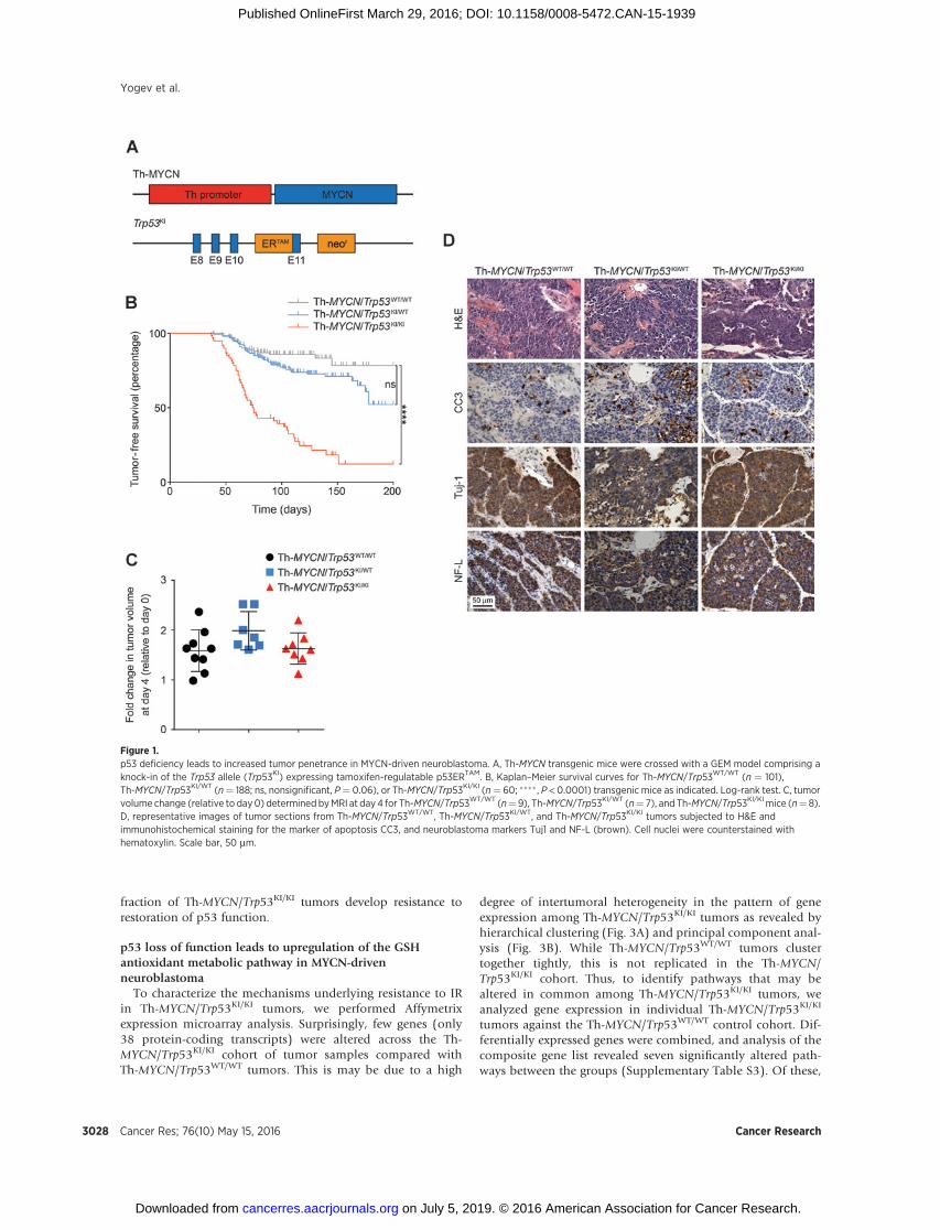
In order to evaluate p53 loss of function in neuroblastoma, wecrossed Th-MYCN mice (in which expression of a human MYCNtransgene is directed by a rat tyrosine hydroxylase (Th)promoter toneural crest cells during early development; ref. 14) with a GEMmodel conditionally deficient for functional p53 (Fig. 1A; ref. 15).Here, the endogenous Trp53 gene is replacedwith a knock-in allele(Trp53KI) encoding a 4-hydroxytamoxifen (4-OHT)–regulatablep53ERTAM fusion protein. In the presence of 4-OHT (a metaboliteof tamoxifen, Tam), the hormone-binding domain of the estrogenreceptor (ER) is released from its inhibitory conformation, andp53ERTAM is translocated to the nucleus. Homozygous Th-MYCNtransgenicmice display 100%penetrance (18), developing tumorswithin 7 to 8 weeks of birth, whereas only 16% of hemizygous Th-MYCN transgenic mice with WT Trp53 (Th-MYCN/Trp53WT/WT)developed tumors by 21 weeks of age with a median latency of 62days (Fig. 1B; Supplementary Table S1). Thus, penetrance andlatency in the Th-MYCN GEMmodel are dependent on transgenedosage, and in this study, only heterozygous Th-MYCNmice wereused. While Th-MYCN mice heterozygous for MYCN and Trp53KI
(Th-MYCN/Trp53KI/WT) displayed a moderate increase in pene-trance to 27% with no significant decrease in tumor-free survival,this increased dramatically to 75% for mice homozygous forTrp53KI (Th-MYCN/Trp53KI/KI). No littermates lacking Th-MYCN,either heterozygous or homozygous for Trp53KI, developed neu-roblastomas by 200 days. While Trp53KI homozygosity increasedpenetrance, this had no effect on either latency or tumor growthrate as measured by volumetric MRI (Fig. 1C).
To test whether the increased tumor incidence in Th-MYCN/Trp53KI/WT mice was due to a mutation in the WT Trp53 allele, we
sequenced exons 5 to 9 (corresponding to the p53 DNA-bindingdomain) in Th-MYCN/Trp53WT/WT (n¼ 16), Th-MYCN/Trp53KI/WT
(n¼12), andTh-MYCN/Trp53KI/KI (n¼11) tumors.We foundonlyone mutation arising in the Th-MYCN/Trp53KI/WT cohort (Supple-mentary Table S2), supporting the notion that increased tumorpenetrance in Th-MYCN/Trp53KI/WT mice is not caused by inacti-vationof the remainingWTTrp53allele.Wealso examinedwhetherpostnatal Tam-induced restoration of functional p53 could affecttumor penetrance, latency, and growth rate. We administered Tamto tumor-bearing mice aged 50 to 80 days as well as to 30-day oldmice, a timepoint atwhich tumors are not yet detectable.We foundthat restoration of functional p53ERTAM had no effect on tumorgrowth rate or penetrance in Th-MYCN/Trp53KI/KI mice (Supple-mentary Fig. S3A and S3B).
Pathologic investigations in Th-MYCN/Trp53KI transgenicmicerevealed that they developed paravertebral, thoracic, or abdom-inal solid tumors, consistent with a parasympathetic origin incommonwith the Th-MYCNGEMmodel. Immunohistochemicalanalysis revealed that all Th-MYCN/Trp53KI tumors stained pos-itive for the neuroblastoma markers Tuj-1 and NF-L (Fig. 1D).Furthermore, in agreement with the parasympathetic origin of theTh-MYCN tumors (19), histopathologic analysis revealed anincrease in the percentage of pups positive for neuroblast hyper-plasia in the Th-MYCN/Trp53KI/KI compared with Th-MYCN/Trp53WT/WT (Supplementary Fig. S4). Thus, our results suggestedthat p53 loss of function interacts with aberrant expression ofMYCN at an early stage of neuroblastoma tumorigenesis. How-ever, p53 deficiency did not affect latency or tumor growth rate.
MYCN-driven neuroblastomas deficient for p53 are resistant toapoptosis induced by IR
Given that p53 pathway alterations are associated withrelapsed/treatment-resistant neuroblastoma, we tested whetherp53 deficiency conferred an antiapoptotic advantage to tumors inTh-MYCN/Trp53KI/KI mice. External beam radiotherapy is widelyused in neuroblastoma treatment following induction chemo-therapy and surgery, although a period of remission is oftenfollowed by subsequent relapse in high-risk cases (20). To exam-ine a role for p53deficiency in treatment-resistant neuroblastoma,we exposed Th-MYCN/Trp53WT/WT and Th-MYCN/Trp53KI/KI
tumor-bearingmice to IR in the presence or absence of tamoxifen.In Th-MYCN/Trp53WT/WT tumors, 5 Gy IR induced apoptosis 5.5hours after IR as measured by the presence of pyknotic nuclei in90%to95%of the cells (Fig. 2A). By contrast, Th-MYCN/Trp53KI/KI
tumors were found to be resistant to IR-induced apoptosis. Thisresistancewasmaintained in some individual animals upon Tam-induced restoration of functional p53ERTAM with responses rang-ing from IR resistance to complete response (Fig. 2A). IR alsofailed to significantly improve survival in Th-MYCN/Trp53KI/KI
allografts, providing evidence for intrinsic resistance to IR in thesetumors (Fig. 2B). While the degree of activation of p53ERTAM wassimilar (as measured by nuclear p-p53Ser15 and induction ofCdkn1a expression) after IR and Tam administration, this did notcorrelate with induction of apoptosis. Furthermore, tumors thatshowed a similar degree of p53 activation displayed differences inapoptotic response (Fig. 2C and D). Pharmacokinetic analysis(LC-MS/MS) of Tam levels supported these findings with resultsshowing a significant correlation between levels of Tam and 4-OHT but no apoptotic response (Fig. 2E). Collectively, theseresults suggest that p53 loss of function confers an antiapoptoticadvantage to tumors in response to cytotoxic IR, and that a
Metabolic Adaptation in High-Risk Neuroblastoma Lacking p53
www.aacrjournals.org Cancer Res; 76(10) May 15, 2016 3027
on July 5, 2019. © 2016 American Association for Cancer Research. cancerres.aacrjournals.org Downloaded from
Published OnlineFirst March 29, 2016; DOI: 10.1158/0008-5472.CAN-15-1939
fraction of Th-MYCN/Trp53KI/KI tumors develop resistance torestoration of p53 function.
p53 loss of function leads to upregulation of the GSHantioxidant metabolic pathway in MYCN-drivenneuroblastoma
To characterize the mechanisms underlying resistance to IRin Th-MYCN/Trp53KI/KI tumors, we performed Affymetrixexpression microarray analysis. Surprisingly, few genes (only38 protein-coding transcripts) were altered across the Th-MYCN/Trp53KI/KI cohort of tumor samples compared withTh-MYCN/Trp53WT/WT tumors. This is may be due to a high
degree of intertumoral heterogeneity in the pattern of geneexpression among Th-MYCN/Trp53KI/KI tumors as revealed byhierarchical clustering (Fig. 3A) and principal component anal-ysis (Fig. 3B). While Th-MYCN/Trp53WT/WT tumors clustertogether tightly, this is not replicated in the Th-MYCN/Trp53KI/KI cohort. Thus, to identify pathways that may bealtered in common among Th-MYCN/Trp53KI/KI tumors, weanalyzed gene expression in individual Th-MYCN/Trp53KI/KI
tumors against the Th-MYCN/Trp53WT/WT control cohort. Dif-ferentially expressed genes were combined, and analysis of thecomposite gene list revealed seven significantly altered path-ways between the groups (Supplementary Table S3). Of these,
Figure 1.p53 deficiency leads to increased tumor penetrance in MYCN-driven neuroblastoma. A, Th-MYCN transgenic mice were crossed with a GEM model comprising aknock-in of the Trp53 allele (Trp53KI) expressing tamoxifen-regulatable p53ERTAM. B, Kaplan–Meier survival curves for Th-MYCN/Trp53WT/WT (n ¼ 101),Th-MYCN/Trp53KI/WT (n¼ 188; ns, nonsignificant, P¼ 0.06), or Th-MYCN/Trp53KI/KI (n¼ 60; ���� , P < 0.0001) transgenic mice as indicated. Log-rank test. C, tumorvolume change (relative to day0) determined byMRI at day4 for Th-MYCN/Trp53WT/WT (n¼ 9), Th-MYCN/Trp53KI/WT (n¼ 7), and Th-MYCN/Trp53KI/KI mice (n¼ 8).D, representative images of tumor sections from Th-MYCN/Trp53WT/WT, Th-MYCN/Trp53KI/WT, and Th-MYCN/Trp53KI/KI tumors subjected to H&E andimmunohistochemical staining for the marker of apoptosis CC3, and neuroblastoma markers Tuj1 and NF-L (brown). Cell nuclei were counterstained withhematoxylin. Scale bar, 50 mm.
Yogev et al.
Cancer Res; 76(10) May 15, 2016 Cancer Research3028
on July 5, 2019. © 2016 American Association for Cancer Research. cancerres.aacrjournals.org Downloaded from
Published OnlineFirst March 29, 2016; DOI: 10.1158/0008-5472.CAN-15-1939
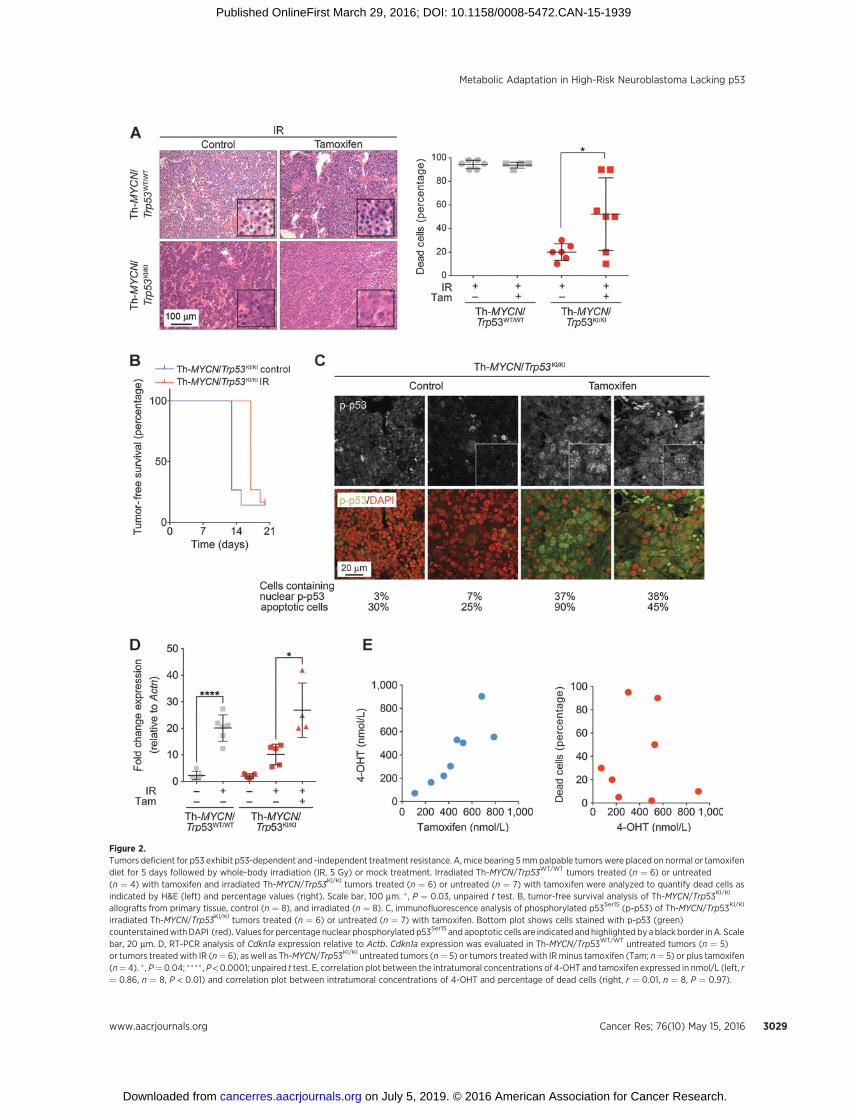
Figure 2.Tumors deficient for p53 exhibit p53-dependent and -independent treatment resistance. A, mice bearing 5mmpalpable tumorswere placed on normal or tamoxifendiet for 5 days followed by whole-body irradiation (IR, 5 Gy) or mock treatment. Irradiated Th-MYCN/Trp53WT/WT tumors treated (n ¼ 6) or untreated(n ¼ 4) with tamoxifen and irradiated Th-MYCN/Trp53KI/KI tumors treated (n ¼ 6) or untreated (n ¼ 7) with tamoxifen were analyzed to quantify dead cells asindicated by H&E (left) and percentage values (right). Scale bar, 100 mm. � , P ¼ 0.03, unpaired t test. B, tumor-free survival analysis of Th-MYCN/Trp53KI/KI
allografts from primary tissue, control (n ¼ 8), and irradiated (n ¼ 8). C, immunofluorescence analysis of phosphorylated p53Ser15 (p-p53) of Th-MYCN/Trp53KI/KI
irradiated Th-MYCN/Trp53KI/KI tumors treated (n ¼ 6) or untreated (n ¼ 7) with tamoxifen. Bottom plot shows cells stained with p-p53 (green)counterstainedwithDAPI (red). Values for percentage nuclear phosphorylated p53Ser15 and apoptotic cells are indicated andhighlighted by ablack border inA. Scalebar, 20 mm. D, RT-PCR analysis of Cdkn1a expression relative to Actb. Cdkn1a expression was evaluated in Th-MYCN/Trp53WT/WT untreated tumors (n ¼ 5)or tumors treated with IR (n¼ 6), as well as Th-MYCN/Trp53KI/KI untreated tumors (n¼ 5) or tumors treated with IRminus tamoxifen (Tam; n¼ 5) or plus tamoxifen(n¼ 4). � , P¼ 0.04; ���� , P < 0.0001; unpaired t test. E, correlation plot between the intratumoral concentrations of 4-OHT and tamoxifen expressed in nmol/L (left, r¼ 0.86, n ¼ 8, P < 0.01) and correlation plot between intratumoral concentrations of 4-OHT and percentage of dead cells (right, r ¼ 0.01, n ¼ 8, P ¼ 0.97).
Metabolic Adaptation in High-Risk Neuroblastoma Lacking p53
www.aacrjournals.org Cancer Res; 76(10) May 15, 2016 3029
on July 5, 2019. © 2016 American Association for Cancer Research. cancerres.aacrjournals.org Downloaded from
Published OnlineFirst March 29, 2016; DOI: 10.1158/0008-5472.CAN-15-1939
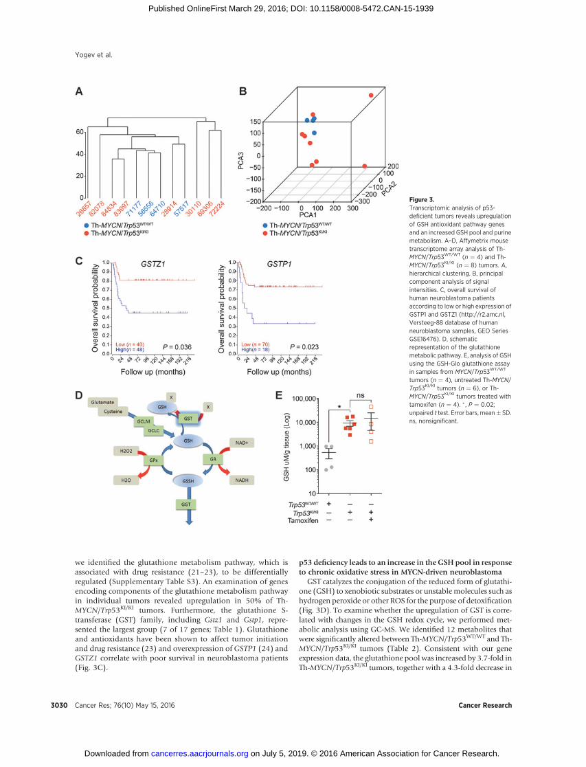
we identified the glutathione metabolism pathway, which isassociated with drug resistance (21–23), to be differentiallyregulated (Supplementary Table S3). An examination of genesencoding components of the glutathione metabolism pathwayin individual tumors revealed upregulation in 50% of Th-MYCN/Trp53KI/KI tumors. Furthermore, the glutathione S-transferase (GST) family, including Gstz1 and Gstp1, repre-sented the largest group (7 of 17 genes; Table 1). Glutathioneand antioxidants have been shown to affect tumor initiationand drug resistance (23) and overexpression of GSTP1 (24) andGSTZ1 correlate with poor survival in neuroblastoma patients(Fig. 3C).
p53 deficiency leads to an increase in the GSH pool in responseto chronic oxidative stress in MYCN-driven neuroblastoma
GST catalyzes the conjugation of the reduced form of glutathi-one (GSH) to xenobiotic substrates or unstable molecules such ashydrogen peroxide or other ROS for the purpose of detoxification(Fig. 3D). To examine whether the upregulation of GST is corre-lated with changes in the GSH redox cycle, we performed met-abolic analysis using GC-MS. We identified 12 metabolites thatwere significantly altered between Th-MYCN/Trp53WT/WT and Th-MYCN/Trp53KI/KI tumors (Table 2). Consistent with our geneexpression data, the glutathione pool was increased by 3.7-fold inTh-MYCN/Trp53KI/KI tumors, together with a 4.3-fold decrease in
Figure 3.Transcriptomic analysis of p53-deficient tumors reveals upregulationof GSH antioxidant pathway genesand an increased GSH pool and purinemetabolism. A–D, Affymetrix mousetranscriptome array analysis of Th-MYCN/Trp53WT/WT (n ¼ 4) and Th-MYCN/Trp53KI/KI (n ¼ 8) tumors. A,hierarchical clustering. B, principalcomponent analysis of signalintensities. C, overall survival ofhuman neuroblastoma patientsaccording to low or high expression ofGSTP1 and GSTZ1 (http://r2.amc.nl,Versteeg-88 database of humanneuroblastoma samples, GEO SeriesGSE16476). D, schematicrepresentation of the glutathionemetabolic pathway. E, analysis of GSHusing the GSH-Glo glutathione assayin samples from MYCN/Trp53WT/WT
tumors (n ¼ 4), untreated Th-MYCN/Trp53KI/KI tumors (n ¼ 6), or Th-MYCN/Trp53KI/KI tumors treated withtamoxifen (n ¼ 4). � , P ¼ 0.02;unpaired t test. Error bars, mean� SD.ns, nonsignificant.
Yogev et al.
Cancer Res; 76(10) May 15, 2016 Cancer Research3030
on July 5, 2019. © 2016 American Association for Cancer Research. cancerres.aacrjournals.org Downloaded from
Published OnlineFirst March 29, 2016; DOI: 10.1158/0008-5472.CAN-15-1939

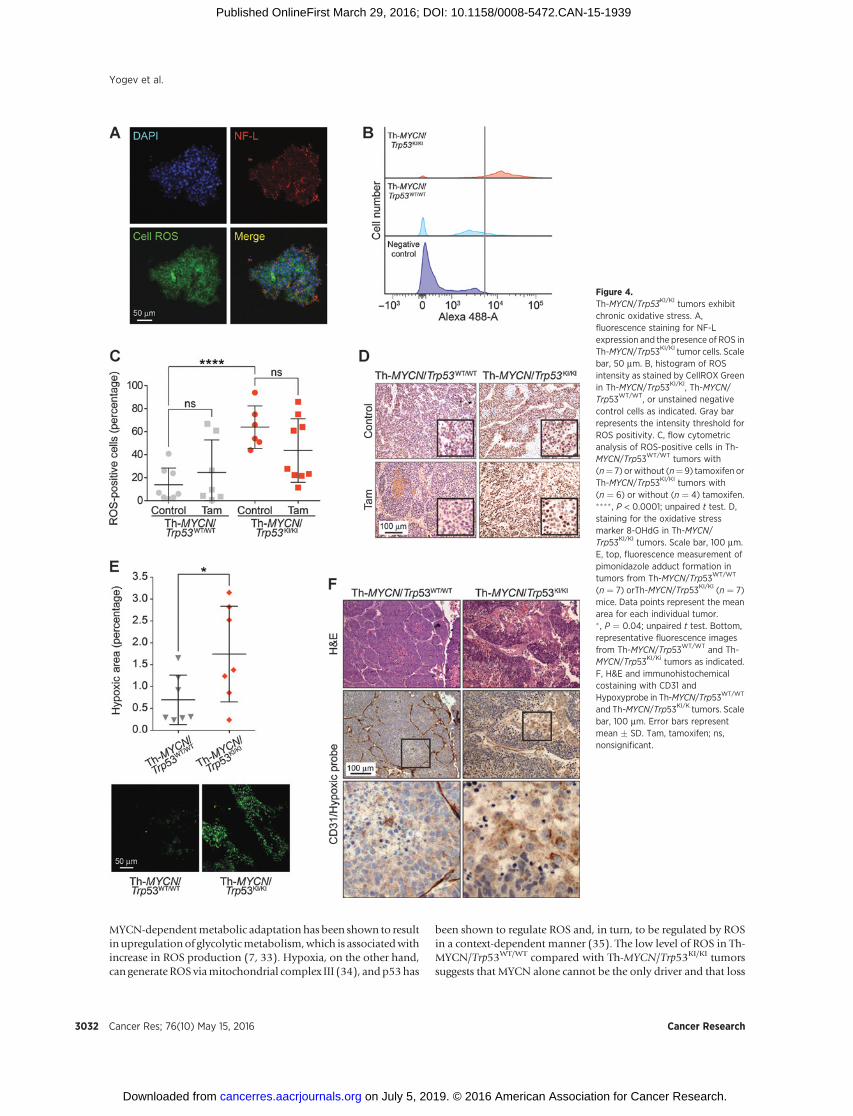
glycine, a glutathione precursor (Table 2). The increase in GSH inTh-MYCN/Trp53KI/KI tumors was confirmed using the GSH-Gloglutathione assay (Fig. 3E). Interestingly, p53 restoration did notreverse this metabolic adaptation, with high GSH levels main-tained in Th-MYCN/Trp53KI/KI tumors. Upregulation of the GSHantioxidant metabolic pathway in p53-deficient MYCN-drivenneuroblastoma suggested an increased requirement for detoxifi-cation of GST substrates, such as derivatives of ROS that areimplicated in tumorigenesis, disease progression, and drug-induced resistance (25). Furthermore, c-MYC overexpression andp53 loss of function have previously been shown to be associatedwith an alteration in ROS levels (2, 26–28). Consistent with theseresults, we found a significant increase in ROS-positive cells in Th-MYCN/Trp53KI/KI tumors (mean of 64%) compared with Th-MYCN/Trp53WT/WT tumors (mean of 14%; Fig. 4A–C). Further-more, in agreement with the data shown in Fig. 2, restoration ofp53ERTAM activity reduced the percentage of ROS-positive cells inapproximately 50% of tumors (Fig. 4C). This suggests thatenhanced ROS levels may be regulated (either directly or indi-rectly) by p53 in a fraction of Th-MYCN/Trp53KI/KI tumors. Asnoted above, this was not associated with a decrease in tumorgrowth or improved overall survival (Supplementary Fig. S3).Evidence for thepresenceof increasedoxidative stress inTh-MYCN/Trp53KI/KI tumors was supported by the finding of enhancedstaining for 8-hydroxyguanosine (8-OHdG), a modified base thatoccurs in DNA due to attack by hydroxyl radicals (Fig. 4D; ref. 29).ROS levels can also be increased by hypoxia and staining with thehypoxia marker pimonidazole, which revealed that Th-MYCN/Trp53KI/KI tumors contained increasedperinecrotic hypoxic regions(Fig. 4E and F). Increased ROS has been reported to lead to damageto enzymes, membranes, and DNA (29, 30), but despite enhanced8-OHdG staining (Fig. 4D), we found no significant changes in thelevels of DNA damage markers (p-CHEK2 and g-H2AX) betweenuntreated Th-MYCN/Trp53WT/WT and Th-MYCN/Trp53KI/KI tumors(Supplementary Fig. S5).
Our results suggest that resistance to IR in Th-MYCN/Trp53KI/KI
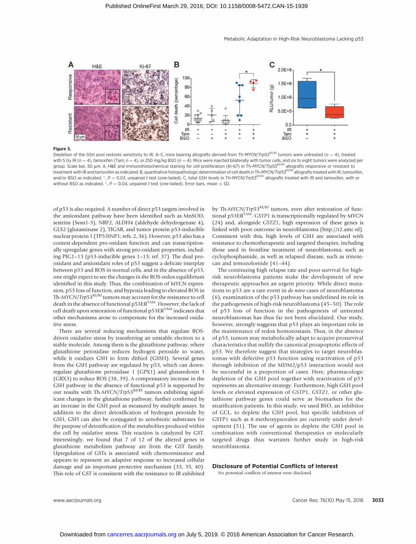
mice may be due to metabolic adaption to chronic oxidativestress through upregulation of GST pathway genes andincreased levels of antioxidant metabolites. This implied thatthe depletion of the GSH pool could resensitize IR-resistant
p53-deficient neuroblastoma cells. Thus, we treated tumorswith buthionine sulfoximine (BSO), an inhibitor of GSHsynthesis that targets glutamate cysteine ligase (GCL), the firstenzyme of the cellular GSH biosynthetic pathway (Fig. 3D). Weimplanted Th-MYCN/Trp53KI/KI primary tumor cells subcuta-neously into 129�1/SvJ-Tg Th-MYCN–negative mice. Consis-tent with our findings in the Th-MYCN/Trp53KI/KI GEM modelof spontaneous neuroblastoma, 50% of the allografted tumorsin which p53 activity was restored by the addition of Tamdisplayed resistance to 5 Gy IR and a higher Ki-67 proliferativeindex (Fig. 5A). However, importantly, this resistance wasabolished in the presence of BSO (250 mg/kg; Fig. 5B and C).
DiscussionIn this study, we have demonstrated that p53 deficiency com-
bines with aberrant expression of MYCN to drive neuroblasthyperplasia and increased neuroblastoma penetrance. Restora-tion of functional p53ERTAM prior to tumors reaching detectablesize or during later stages failed to alter tumor growth or pene-trance. These findings suggest that p53 has an important role inthe early stages of neuroblastoma tumorigenesis in Th-MYCNtransgenic mice. Furthermore, p53 loss of function gives rise totumors that can acquire growth and survival mechanisms that areresistant to the reintroduction of functional p53. This is in starkcontrast with a medulloblastoma GEM model driven by expres-sion ofMYCNandp53deficiency, GTML/Trp53KI/KI (GTML;Glt1–tTA/TRE–MYCN–Luc), where restoration of functional p53ERTAM
by Tam led to increased survival and inhibition of tumor growth(5). Other transgenicmodels of p53 loss of function do, however,develop tumors that display partial resistance to the restoration offunctional p53ERTAM including lymphoma where a requirementfor induction of the oncogenic signaling sensor p19ARF wasidentified (31). In addition, p53 restoration in K-Ras–inducednon-small cell lung cancer failed to induce tumor regression, butdiminished the proportion of high-grade tumors (32).
Recent results have suggested that the outcome of p53 resto-ration in established tumors is highly context-dependent. There isnow evidence that the tumor suppressor function of p53 may bedue to maintenance of genomic stability as well as metabolic andoxidative balance rather than simply its canonical role as atranscriptional activator of cell-cycle inhibitory and proapoptoticeffectors, such as p21, PUMA, and NOXA (1, 3). Consistent withthese findings, we found that tumors with p53 loss of functiondisplayed elevated ROS. Three characteristics of Th-MYCN/Trp53KI/KI tumors could contribute to the increased levels ofROS: MYCN expression, hypoxia, and p53 loss of function.
Table 2. Analysis of metabolites in p53-deficient tumors
Metabolite KIKI:WT/WT Ratio P value
Alanine 0.480 1.23E�02Valine 0.496 3.26E�02Threonine 0.544 3.90E�02Glycine 0.233 2.38E�02Succinate 0.631 4.47E�02PEP 0.538 3.52E�02Hypotaurine 0.382 4.50E�02Hypoxanthine 1.924 2.81E�023-GP 0.531 4.33E�02L-ascorbic acid 0.397 3.18E�02Ribose-5-phosphate 0.651 3.13E�02GSH pool 3.761 1.66E�02
Table 1. Altered expression of genes in the glutathione pathway amongindividual Th-MYCN/Trp53KI/KI tumors versus Th-MYCN/Trp53WT/WT tumors
SampleGene 82078 83997 84834 28914 69306 72224 26857
Gstm1 þ þGstm7 þ þGstm2 þGstm4 þGstp1 þ þ þGstt1 þGstz þMgst3 þGstm3 þ þRrm2 þ þAnpepGclc þGpx3 þ þ þGstt2 þGstk1 þMgst1 þMgsta3 þNOTE: þ, altered expression of genes among individual Th-MYCN/Trp53KI/KI.
Metabolic Adaptation in High-Risk Neuroblastoma Lacking p53
www.aacrjournals.org Cancer Res; 76(10) May 15, 2016 3031
on July 5, 2019. © 2016 American Association for Cancer Research. cancerres.aacrjournals.org Downloaded from
Published OnlineFirst March 29, 2016; DOI: 10.1158/0008-5472.CAN-15-1939
MYCN-dependentmetabolic adaptation has been shown to resultin upregulationof glycolyticmetabolism,which is associatedwithincrease in ROS production (7, 33). Hypoxia, on the other hand,can generate ROS viamitochondrial complex III (34), andp53has
been shown to regulate ROS and, in turn, to be regulated by ROSin a context-dependent manner (35). The low level of ROS in Th-MYCN/Trp53WT/WT compared with Th-MYCN/Trp53KI/KI tumorssuggests that MYCN alone cannot be the only driver and that loss
Figure 4.Th-MYCN/Trp53KI/KI tumors exhibitchronic oxidative stress. A,fluorescence staining for NF-Lexpression and the presence of ROS inTh-MYCN/Trp53KI/KI tumor cells. Scalebar, 50 mm. B, histogram of ROSintensity as stained by CellROX Greenin Th-MYCN/Trp53KI/KI, Th-MYCN/Trp53WT/WT, or unstained negativecontrol cells as indicated. Gray barrepresents the intensity threshold forROS positivity. C, flow cytometricanalysis of ROS-positive cells in Th-MYCN/Trp53WT/WT tumors with(n¼ 7) orwithout (n¼9) tamoxifen orTh-MYCN/Trp53KI/KI tumors with(n ¼ 6) or without (n ¼ 4) tamoxifen.���� , P < 0.0001; unpaired t test. D,staining for the oxidative stressmarker 8-OHdG in Th-MYCN/Trp53KI/KI tumors. Scale bar, 100 mm.E, top, fluorescence measurement ofpimonidazole adduct formation intumors from Th-MYCN/Trp53WT/WT
(n ¼ 7) orTh-MYCN/Trp53KI/KI (n ¼ 7)mice. Data points represent the meanarea for each individual tumor.� , P ¼ 0.04; unpaired t test. Bottom,representative fluorescence imagesfrom Th-MYCN/Trp53WT/WT and Th-MYCN/Trp53KI/Ki tumors as indicated.F, H&E and immunohistochemicalcostaining with CD31 andHypoxyprobe in Th-MYCN/Trp53WT/WT
and Th-MYCN/Trp53KI/K tumors. Scalebar, 100 mm. Error bars representmean � SD. Tam, tamoxifen; ns,nonsignificant.
Yogev et al.
Cancer Res; 76(10) May 15, 2016 Cancer Research3032
on July 5, 2019. © 2016 American Association for Cancer Research. cancerres.aacrjournals.org Downloaded from
Published OnlineFirst March 29, 2016; DOI: 10.1158/0008-5472.CAN-15-1939
of p53 is also required. A number of direct p53 targets involved inthe antioxidant pathway have been identified such as MnSOD,sestrins (Sesn1-3), NRF2, ALDH4 (aldehyde dehydrogenase 4),GLS2 (glutaminase 2), TIGAR, and tumor protein p53-induciblenuclear protein 1 (TP53INP1; refs. 2, 36).However, p53 also has acontext-dependent pro-oxidant function and can transcription-ally upregulate genes with strong pro-oxidant properties, includ-ing PIG1–13 (p53-inducible genes 1–13; ref. 37). The dual pro-oxidant and antioxidant roles of p53 suggest a delicate interplaybetween p53 and ROS in normal cells, and in the absence of p53,onemight expect to see the changes in the ROS-redox equilibriumidentified in this study. Thus, the combination of MYCN expres-sion, p53 loss of function, and hypoxia leading to elevated ROS inTh-MYCN/Trp53KI/KI tumorsmay account for the resistance to celldeath in the absence of functional p53ERTAM.However, the lack ofcell death upon restoration of functional p53ERTAM indicates thatother mechanisms arose to compensate for the increased oxida-tive stress.
There are several reducing mechanisms that regulate ROS-driven oxidative stress by transferring an unstable electron to astable molecule. Among them is the glutathione pathway, whereglutathione peroxidase reduces hydrogen peroxide to water,while it oxidizes GSH to form dithiol (GSSH). Several genesfrom the GSH pathway are regulated by p53, which can down-regulate glutathione peroxidase 1 (GPX1) and glutaredoxin 3(GRX3) to reduce ROS (38, 39). A compensatory increase in theGSH pathway in the absence of functional p53 is supported byour results with Th-MYCN/Trp53KI/KI tumors exhibiting signif-icant changes in the glutathione pathway, further confirmed byan increase in the GSH pool as measured by multiple assays. Inaddition to the direct detoxification of hydrogen peroxide byGSH, GSH can also be conjugated to xenobiotic substrates forthe purpose of detoxification of the metabolites produced withinthe cell by oxidative stress. This reaction is catalyzed by GST.Interestingly, we found that 7 of 12 of the altered genes inglutathione metabolism pathway are from the GST family.Upregulation of GSTs is associated with chemoresistance andappears to represent an adaptive response to increased cellulardamage and an important protective mechanism (33, 35, 40).This role of GST is consistent with the resistance to IR exhibited
by Th-MYCN/Trp53KI/KI tumors, even after restoration of func-tional p53ERTAM. GSTP1 is transcriptionally regulated by MYCN(24) and, alongside GSTZ1, high expression of these genes islinked with poor outcome in neuroblastoma (http://r2.amc.nl).Consistent with this, high levels of GSH are associated withresistance to chemotherapeutic and targeted therapies, includingthose used in frontline treatment of neuroblastoma, such ascyclophosphamide, as well as relapsed disease, such as irinote-can and temozolomide (41–44).
The continuing high relapse rate and poor survival for high-risk neuroblastoma patients make the development of newtherapeutic approaches an urgent priority. While direct muta-tions to p53 are a rare event in de novo cases of neuroblastoma(6), examination of the p53 pathway has underlined its role inthe pathogenesis of high-risk neuroblastoma (45–50). The roleof p53 loss of function in the pathogenesis of untreatedneuroblastomas has thus far not been elucidated. Our study,however, strongly suggests that p53 plays an important role inthe maintenance of redox homoeostasis. Thus, in the absenceof p53, tumors may metabolically adapt to acquire prosurvivalcharacteristics that nullify the canonical proapoptotic effects ofp53. We therefore suggest that strategies to target neuroblas-tomas with defective p53 function using reactivation of p53through inhibition of the MDM2/p53 interaction would notbe successful in a proportion of cases. Here, pharmacologicdepletion of the GSH pool together with reactivation of p53represents an alternative strategy. Furthermore, high GSH poollevels or elevated expression of GSTP1, GSTZ1, or other glu-tathione pathway genes could serve as biomarkers for thestratification patients. In this study, we used BSO, an inhibitorof GCL, to deplete the GSH pool, but specific inhibitors ofGSTP1 such as 8-methoxypsoralen are currently under devel-opment (51). The use of agents to deplete the GSH pool incombination with conventional therapeutics or molecularlytargeted drugs thus warrants further study in high-riskneuroblastoma.
Disclosure of Potential Conflicts of InterestNo potential conflicts of interest were disclosed.
Figure 5.Depletion of the GSH pool restores sensitivity to IR. A–C, mice bearing allografts derived from Th-MYCN/Trp53KI/KI tumors were untreated (n ¼ 4), treatedwith 5 Gy IR (n¼ 4), tamoxifen (Tam; n¼ 4), or 250 mg/kg BSO (n¼ 4). Mice were injected bilaterally with tumor cells, and six to eight tumors were analyzed pergroup. Scale bar, 50 mm. A, H&E and immunohistochemical staining for cell proliferation (Ki-67) in Th-MYCN/Trp53KI/KI allografts responsive or resistant totreatmentwith IR and tamoxifen as indicated. B, quantitative histopathologic determination of cell death in Th-MYCN/Trp53KI/KI allografts treatedwith IR, tamoxifen,and/or BSO as indicated. � , P ¼ 0.03, unpaired t test (one-tailed). C, total GSH levels in Th-MYCN/Trp53KI/KI allografts treated with IR and tamoxifen, with orwithout BSO as indicated. � , P ¼ 0.04, unpaired t test (one-tailed). Error bars, mean � SD.
www.aacrjournals.org Cancer Res; 76(10) May 15, 2016 3033
Metabolic Adaptation in High-Risk Neuroblastoma Lacking p53
on July 5, 2019. © 2016 American Association for Cancer Research. cancerres.aacrjournals.org Downloaded from
Published OnlineFirst March 29, 2016; DOI: 10.1158/0008-5472.CAN-15-1939
Authors' ContributionsConception and design: O. Yogev, L. CheslerDevelopment of methodology: O. Yogev, A. Hallsworth, Y. Jamin, R. Ruddle,F.I. Raynaud, L. CheslerAcquisition of data (provided animals, acquired and managed patients,provided facilities, etc.): O. Yogev, K. Barker, A. Sikka, G.S. Almeida,A. Hallsworth, L.M. Smith, Y. Jamin, R. Ruddle, A. Koers, H.T. Webber,F.I. Raynaud, H.C. Keun, L. CheslerAnalysis and interpretation of data (e.g., statistical analysis, biostatistics,computational analysis):O. Yogev, A. Sikka, G.S. Almeida, Y. Jamin, R. Ruddle,S. Popov, C. Jones, K. Petrie, S.P. Robinson, H.C. Keun, L. CheslerWriting, review, and/or revision of the manuscript: O. Yogev, K. Barker,G.S. Almeida, A. Hallsworth, Y. Jamin, F.I. Raynaud, K. Petrie, S.P. Robinson,H.C. Keun, L. CheslerAdministrative, technical, or material support (i.e., reporting or organizingdata, constructing databases):O. Yogev, K. Barker, A. Hallsworth, L.M. Smith,L. CheslerStudy supervision: S.P. Robinson, L. Chesler
AcknowledgmentsThe authors thank Alan Mackay, Pawan Poudel, and Anguraj Sadanan-
dam for valuable advice on bioinformatic analysis; Elizabeth Want and
Volker Behrends for advice on mass-spectrometry analysis. They also thankSue Eccles and Eitan Shaulian for critical reading of the article.
Grant SupportThis study was supported by grants from The Neuroblastoma Society (O.
Yogev and K. Barker), MRC funding (MR/J015938/1 to A. Sikka), The FelixWhite Cancer Charity (A. Hallsworth), Cancer Research UK and EPSRC to theCancer Imaging Centre at the Institute of Cancer Research (ICR) and The RoyalMarsden Hospital, in association with the MRC and Department of Health(England; C1060/A10334 and C1060/A16464), The Wellcome Trust(091763Z/10/Z), an EPSRC Platform Grant (EP/H046526/1), NHS fundingto theNIHRBiomedical ResearchCentre at The RoyalMarsden and the ICR, anda Paul O'Gorman Postdoctoral Fellowship funded by Children with Cancer UK(Y. Jamin).
The costs of publication of this article were defrayed in part by thepayment of page charges. This article must therefore be hereby markedadvertisement in accordance with 18 U.S.C. Section 1734 solely to indicatethis fact.
Received July 20, 2015; revised December 31, 2015; accepted February 9,2016; published OnlineFirst March 29, 2016.
References1. Valente LJ,GrayDH,Michalak EM, Pinon-Hofbauer J, EgleA, Scott CL, et al.
p53 efficiently suppresses tumor development in the complete absence ofits cell-cycle inhibitory and proapoptotic effectors p21, Puma, and Noxa.Cell Rep 2013;3:1339–45.
2. Puzio-Kuter AM. The role of p53 in metabolic regulation. Genes Cancer2011;2:385–91.
3. Vousden KH, Ryan KM. p53 and metabolism. Nat Rev Cancer 2009;9:691–700.
4. Berkers CR, Maddocks OD, Cheung EC, Mor I, Vousden KH. Metabolicregulation by p53 family members. Cell Metab 2013;18:617–33.
5. Hill RM, Kuijper S, Lindsey JC, Petrie K, Schwalbe EC, Barker K, et al.Combined MYC and P53 defects emerge at medulloblastoma relapse anddefine rapidly progressive, therapeutically targetable disease. Cancer Cell2015;27:72–84.
6. Pugh TJ, Morozova O, Attiyeh EF, Asgharzadeh S, Wei JS, Auclair D, et al.The genetic landscape of high-risk neuroblastoma. Nat Genet 2013;45:279–84.
7. Qing G, Li B, Vu A, Skuli N, Walton ZE, Liu X, et al. ATF4 regulates MYC-mediated neuroblastoma cell death upon glutamine deprivation. CancerCell 2012;22:631–44.
8. Rapizzi E, Ercolino T, Fucci R, Zampetti B, Felici R, Guasti D, et al. Succinatedehydrogenase subunit B mutations modify human neuroblastoma cellmetabolism and proliferation. Horm Cancer 2014;5:174–84.
9. Das S, Bryan K, Buckley PG, Piskareva O, Bray IM, Foley N, et al. Modu-lation of neuroblastoma disease pathogenesis by an extensive network ofepigenetically regulated microRNAs. Oncogene 2013;32:2927–36.
10. Wang C, Liu Z, Woo CW, Li Z, Wang L, Wei JS, et al. EZH2 Mediatesepigenetic silencing of neuroblastoma suppressor genes CASZ1, CLU,RUNX3, and NGFR. Cancer Res 2012;72:315–24.
11. Fukuoka H, Takahashi Y. The role of genetic and epigenetic changes inpituitary tumorigenesis. Neurol Med Chir 2014;54:943–57.
12. Workman P, Aboagye EO, Balkwill F, Balmain A, Bruder G, Chaplin DJ,et al. Guidelines for the welfare and use of animals in cancer research. Br JCancer 2010;102:1555–77.
13. Kilkenny C, Browne WJ, Cuthill IC, Emerson M, Altman DG. Improvingbioscience research reporting: The ARRIVE guidelines for reporting animalresearch. PLoS Biol 2010;8:e1000412.
14. Weiss WA, Aldape K, Mohapatra G, Feuerstein BG, Bishop JM. Targetedexpression of MYCN causes neuroblastoma in transgenic mice. EMBO J1997;16:2985–95.
15. Christophorou MA, Martin-Zanca D, Soucek L, Lawlor ER, Brown-SwigartL, Verschuren EW, et al. Temporal dissection of p53 function in vitro and invivo. Nat Genet 2005;37:718–26.
16. Jamin Y, Tucker ER, Poon E, Popov S, Vaughan L, Boult JK, et al. Evaluationof clinically translatableMR imaging biomarkers of therapeutic response inthe TH-MYCN transgenic mouse model of neuroblastoma. Radiology2013;266:130–40.
17. Boult JK, Walker-Samuel S, Jamin Y, Leiper JM, Whitley GS, Robinson SP.Active site mutant dimethylarginine dimethylaminohydrolase 1 expres-sion confers an intermediate tumour phenotype in C6 gliomas. J Pathol2011;225:344–52.
18. Rasmuson A, Segerstrom L, Nethander M, Finnman J, Elfman LH, Javan-mardi N, et al. Tumor development, growth characteristics and spectrumofgenetic aberrations in the TH-MYCN mouse model of neuroblastoma.PLoS One 2012;7:e51297.
19. Hansford LM, Thomas WD, Keating JM, Burkhart CA, Peaston AE, NorrisMD, et al. Mechanisms of embryonal tumor initiation: distinct roles forMycN expression and MYCN amplification. Proc Natl Acad Sci U S A2004;101:12664–9.
20. Matthay KK, Villablanca JG, Seeger RC, Stram DO, Harris RE, Ramsay NK,et al. Treatment of high-risk neuroblastoma with intensive chemotherapy,radiotherapy, autologous bone marrow transplantation, and 13-cis-reti-noic acid. Children's Cancer Group. N Engl J Med 1999;341:1165–73.
21. Townsend DM, Tew KD. The role of glutathione-S-transferase in anti-cancer drug resistance. Oncogene 2003;22:7369–75.
22. Tew KD. Glutathione-associated enzymes in anticancer drug resistance.Cancer Res 1994;54:4313–20.
23. Traverso N, Ricciarelli R, Nitti M, Marengo B, Furfaro AL, Pronzato MA,et al. Role of glutathione in cancer progression and chemoresistance. OxidMed Cell Longev 2013;2013:972913.
24. Fletcher JI, Gherardi S,Murray J, Burkhart CA, Russell A, Valli E, et al.N-Mycregulates expression of the detoxifying enzyme glutathione transferaseGSTP1, a marker of poor outcome in neuroblastoma. Cancer Res 2012;72:845–53.
25. Sosa V,Moline T, Somoza R, Paciucci R, KondohH,ME LL.Oxidative stressand cancer: An overview. Ageing Res Rev 2013;12:376–90.
26. Graves JA, Metukuri M, Scott D, Rothermund K, Prochownik EV. Regula-tion of reactive oxygen species homeostasis by peroxiredoxins and c-Myc.J Biol Chem 2009;284:6520–9.
27. Ostrakhovitch EA, Cherian MG. Role of p53 and reactive oxygen species inapoptotic response to copper and zinc in epithelial breast cancer cells.Apoptosis 2005;10:111–21.
28. Vafa O, WadeM, Kern S, Beeche M, Pandita TK, Hampton GM, et al. c-Myccan induce DNA damage, increase reactive oxygen species, and mitigatep53 function: a mechanism for oncogene-induced genetic instability. MolCell 2002;9:1031–44.
Cancer Res; 76(10) May 15, 2016 Cancer Research3034
Yogev et al.
on July 5, 2019. © 2016 American Association for Cancer Research. cancerres.aacrjournals.org Downloaded from
Published OnlineFirst March 29, 2016; DOI: 10.1158/0008-5472.CAN-15-1939
29. Ziech D, Franco R, Pappa A, Panayiotidis MI. Reactive oxygen species(ROS)–induced genetic and epigenetic alterations in human carcinogen-esis. Mutat Res 2011;711:167–73.
30. Cooke MS, Evans MD, Dizdaroglu M, Lunec J. Oxidative DNA damage:Mechanisms, mutation, and disease. FASEB J 2003;17:1195–214.
31. Martins CP, Brown-Swigart L, EvanGI.Modeling the therapeutic efficacy ofp53 restoration in tumors. Cell 2006;127:1323–34.
32. Junttila MR, Karnezis AN, Garcia D, Madriles F, Kortlever RM, Rostker F,et al. Selective activation of p53-mediated tumour suppression in high-grade tumours. Nature 2010;468:567–71.
33. Sabharwal SS, Schumacker PT. Mitochondrial ROS in cancer: Initiators,amplifiers or an Achilles' heel?Nat Rev Cancer 2014;14:709–21.
34. Guzy RD, Hoyos B, Robin E, Chen H, Liu L, Mansfield KD, et al. Mito-chondrial complex III is required for hypoxia-inducedROSproduction andcellular oxygen sensing. Cell Metab 2005;1:401–8.
35. Maillet A, Pervaiz S. Redox regulation of p53, redox effectors regulated byp53: A subtle balance. Antioxid Redox Signal 2012;16:1285–94.
36. Budanov AV. The role of tumor suppressor p53 in the antioxidant defenseand metabolism. Subcell Biochem 2014;85:337–58.
37. Polyak K, Xia Y, Zweier JL, Kinzler KW, Vogelstein B. A model for p53-induced apoptosis. Nature 1997;389:300–5.
38. Brynczka C, Labhart P, Merrick BA. NGF-mediated transcriptionaltargets of p53 in PC12 neuronal differentiation. BMC Genomics 2007;8:139.
39. Tan M, Li S, Swaroop M, Guan K, Oberley LW, Sun Y. Transcriptionalactivation of the human glutathione peroxidase promoter by p53. J BiolChem 1999;274:12061–6.
40. Waypa GB, Marks JD, Guzy R, Mungai PT, Schriewer J, Dokic D, et al.Hypoxia triggers subcellular compartmental redox signaling in vascularsmooth muscle cells. Circ Res 2010;106:526–35.
41. Rocha CR, Garcia CC, Vieira DB, Quinet A, de Andrade-Lima LC,Munford V, et al. Glutathione depletion sensitizes cisplatin- and temo-
zolomide-resistant glioma cells in vitro and in vivo. Cell Death Dis2015;6:e1727.
42. Estrela JM, Ortega A, Obrador E. Glutathione in cancer biology andtherapy. Crit Rev Clin Lab Sci 2006;43:143–81.
43. Montero AJ, Jassem J. Cellular redox pathways as a therapeutic target in thetreatment of cancer. Drugs 2011;71:1385–96.
44. St-Coeur PD, Poitras JJ, Cuperlovic-Culf M, Touaibia M, Morin PJ. Inves-tigating a signature of temozolomide resistance in GBM cell lines usingmetabolomics. J Neurooncol 2015;125:91–102.
45. Tweddle DA, Pearson AD, Haber M, Norris MD, Xue C, Flemming C, et al.The p53 pathway and its inactivation in neuroblastoma. Cancer Lett2003;197:93–8.
46. Tweddle DA, Malcolm AJ, Cole M, Pearson AD, Lunec J. p53 cellularlocalization and function in neuroblastoma: Evidence for defective G(1)arrest despite WAF1 induction in MYCN-amplified cells. Am J Pathol2001;158:2067–77.
47. Kim E, Shohet J. Targeted molecular therapy for neuroblastoma: The ARF/MDM2/p53 axis. J Natl Cancer Inst 2009;101:1527–9.
48. Wolff A, Technau A, Ihling C, Technau-Ihling K, Erber R, Bosch FX, et al.Evidence that wild-type p53 in neuroblastoma cells is in a conformationrefractory to integration into the transcriptional complex. Oncogene2001;20:1307–17.
49. Gamble LD, Kees UR, Tweddle DA, Lunec J. MYCN sensitizes neuroblas-toma to the MDM2-p53 antagonists Nutlin-3 and MI-63. Oncogene2012;31:752–63.
50. Xue C, Haber M, Flemming C, Marshall GM, Lock RB, MacKenzie KL, et al.p53 determinesmultidrug sensitivity of childhood neuroblastoma. CancerRes 2007;67:10351–60.
51. de Oliveira DM, de Farias MT, Teles AL, Dos Santos Junior MC, deCerqueira MD, Lima RM, et al. 8-Methoxypsoralen is a competitiveinhibitor of glutathione S-transferase P1-1. Front Cell Neurosci 2014;8:308.
www.aacrjournals.org Cancer Res; 76(10) May 15, 2016 3035
Metabolic Adaptation in High-Risk Neuroblastoma Lacking p53
on July 5, 2019. © 2016 American Association for Cancer Research. cancerres.aacrjournals.org Downloaded from
Published OnlineFirst March 29, 2016; DOI: 10.1158/0008-5472.CAN-15-1939

2016;76:3025-3035. Published OnlineFirst March 29, 2016.Cancer Res Orli Yogev, Karen Barker, Arti Sikka, et al. Adaptations Supporting Radioresistancep53 Loss in MYC-Driven Neuroblastoma Leads to Metabolic
Updated version
10.1158/0008-5472.CAN-15-1939doi:
Access the most recent version of this article at:
Material
Supplementary
http://cancerres.aacrjournals.org/content/suppl/2017/02/06/0008-5472.CAN-15-1939.DC2
Access the most recent supplemental material at:
Cited articles
http://cancerres.aacrjournals.org/content/76/10/3025.full#ref-list-1
This article cites 51 articles, 9 of which you can access for free at:
E-mail alerts related to this article or journal.Sign up to receive free email-alerts
Subscriptions
Reprints and
To order reprints of this article or to subscribe to the journal, contact the AACR Publications Department at
Permissions
Rightslink site. Click on "Request Permissions" which will take you to the Copyright Clearance Center's (CCC)
.http://cancerres.aacrjournals.org/content/76/10/3025To request permission to re-use all or part of this article, use this link
on July 5, 2019. © 2016 American Association for Cancer Research. cancerres.aacrjournals.org Downloaded from
Published OnlineFirst March 29, 2016; DOI: 10.1158/0008-5472.CAN-15-1939