Embed Size (px)
Citation preview
Asian Pacific Journal of Cancer Prevention, Vol 13, 2012 5439
DOI:http://dx.doi.org/10.7314/APJCP.2012.13.11.5439P53 and PCNA and HPV Infection in Laryngeal Lesions in Xinjiang
Asian Pacific J Cancer Prev, 13 (11), 5439-5444
Introduction
Laryngeal cancer is one of the most common head and neck malignancy, and approximately 159,000 new cases are diagnosed annually worldwide (Parkin et al., 2005; Marioni et al., 2006; Marur and Forastiere, 2008). Squamous cell carcinoma represents most of laryngeal cancer, accounting for more than 95% of primary laryngeal tumors, and is more common in males (sex ratio 7:1) (Parkin et al., 2005; Ji et al., 2008). Despite efforts over the last 20 years in laryngeal squamous cell carcinoma (LSCC) have been made, and some factors such as smoking, alcohol, occupational, and environmental factors have been implicated as important etiologic agents in the pathogenesis of LSCC, the complex pathogenesis is still unclear and the clinical prognosis remains poor (Parkin et al., 2001; Marioni et al., 2006). Therefore, there is a great need to explore and clarify the pathogenesis of LSCC, expect to find some key biomarkers that can help clinical treatment. Human papillomavirus (HPV) is a small double-
1Department of ENT, the First Affiliated Hospital of Xinjiang Medical University, 2Department of General Surgery, the People’s Hospital of Xinjiang Uygur Autonomous Region, 3Department of ENT, the Second Affiliated Hospital of Xinjiang Medical University, Urumqi, China &Equal contributors *For correspondence: [email protected]
Abstract
Objective: To explore the correlation of human papillomavious (HPV) infection with expression of p53 and proliferating cell nuclear antigen (PCNA) in patients with different ethnicity in Xinjiang, China. Methods: 166 biopsy specimens from 83 laryngeal squamous cell carcinomas (LSCC), 63 laryngeal papillomas (LP), and 20 laryngeal inflammatory polyps (LIP) were included in this study. HPV infection was determined by polymerase chain reaction (PCR) using specific types of HPV primers. Expression of p53 and PCNA was assessed using immunohistostaining. Results: The frequency of HPV 6/11 was higher in LP (33.3%) than in LSCC (9.6%) (P <0.0005), whereas the frequency of HPV 16/18 was higher in LSCC (37.3 %) than in LP (6.3%) (P < 0.0005). Patients of the Han ethnic group with LSCC had a higher infection rate with HPV 6/11 or HPV 6/11 and HPV 16/18 coinfection than those of Uygur and Kazak ethnicity (P <0.05). Overexpression of p53 and PCNA were higher in LSCC (62.7%, 57.8%) than in LP (38%, 33.3%) (P <0.005, and P <0.005, respectively). That of p53 was not associated with lymph-node metastases and clinical stages, but overexpression of PCNA closely correlated with clinical stage. Conclusions: These results strongly implicate HPV6/11 infection in the carcinogenesis of LSCC and LP, respectively. There was a higher coincidence of increased malignancy of laryngeal tumors with overexpression of p53 and PCNA. Overexpression of p53 may serve as an early risk marker for malignant transformation in HPV infected cells while the overexpression of PCNA may serve as a late marker for progression of LSCC.
Keywords: p53 - PCNA - human papillomavious - laryngeal epitheliopapillomatous lesions
RESEARCH ARTICLE
P53 and PCNA is Positively Correlated with HPV Infection in Laryngeal Epitheliopapillomatous Lesions in Patiets with Different Ethnic Backgrounds in XinjiangJie Sun1&, Ju Xiong2&, Yan Zhen3, Zhao-Lun Chen1, Hua Zhang1*
stranded DNA virus that is capable of infecting cutaneous and mucosal epithelium, resulting in a variety of cancers, including LSCC (Yao, 1992; Scheffner et al., 1993; Grace Nirmala and Narendhirakannan, 2011; Lin et al., 2011; El-Naggar and Westra, 2012). HPV has been demonstrated to be a promoter in multistep of carcinogenesis process In LSCC (Koskinen et al., 2007), and HPV-mediated tumorigenesis is mainly due to the activities of two viral oncoproteins: E6 and E7. HPVE6 and E7 are able to induce the degradation of p53 by binding to the ubiquitin ligase E6AP, inhibiting p53-dependent signaling upon stress stimuli, and contributing to LSCC development (Pruneri et al., 1997; Du et al., 2003). The p53 tumor suppressor gene has also been implicated in many functions such as inhibition of cell proliferation, DNA repair, cellular differentiation and apoptosis (Gao et al., 2000; Maxwell and Davis, 2000). Among the most frequent alterations in human laryngeal carcinomas is point mutation of p53 gene which is thought to play a role in an early stage in the development of laryngeal carcinoma (Horibe et al., 2000). An association
Jie Sun et al
Asian Pacific Journal of Cancer Prevention, Vol 13, 20125440
between an HPV infection and a mutation of p53 has been identified in both laryngeal carcinoma and laryngeal papilloma (Xie et al., 1997; Stasikowska-Kanicka et al., 2011). The expression of HPV alone is insufficient to induce malignant progression, and the additional genetic alterations are necessary for malignant progression in the setting of virus-induced changes of cell proliferation. HPV infection also can lead to uncontrolled cell proliferation which can be detected by a specific marker for proliferation cell nuclear antigen (PCNA) which is a nuclear antigen present in the G1 and S phases of the cellular cycle. (Demeter et al., 1994; Azzimonti et al., 2004). Meantime, PCNA expression can be modulated by wild or mutated p53 protein (Micozkadioglu et al., 2008; Sarafoleanu et al., 2009). P53 protein expression is positively related to proliferative activity in premalignant and malignant lesions of the larynx. Therefore, studies attempting to correlate the incidence of HPV infection with expression of p53 and PCNA have been performed to evaluate the carcinogenic role and to seek useful biomarkers with laryngeal malignant and pre-neoplastic lesions. In this study, we evaluated the expression of HPV, p53, and PCNA in laryngeal malignant and pre-neoplastic lesions of various racial patients in northwest China, analyzed the potential association of these molecules with tumor stage, and investigated their predictive value on laryngeal lesions.
Materials and Methods
General data A total of 166 biopsies and laryngectomy specimens were obtained from the first Teaching Hospital of Xinjiang Medical University from 1980 to 1997. This study was conducted in accordance with the declaration of Helsinki. This study was approved by the Ethics Committee of the First Affiliated Hospital of Xinjiang Medical University. Written informed consent was obtained from all participants. Biopsy specimens included 83 laryngeal squamous cell carcinomas (LSCC), 63 laryngeal papillomas, and 20 laryngeal inflammatory polyps (LIP) from patients which had received neither radiotherapy nor chemotherapy. The patients were came from three racial groups: 92 Han, 70 Uygur, and 4 Kazakh. There were 117 males and 49 females, with an average age of 58.7 years in LSCC, 25.6 years LP, and 37.8 years in LIP. Specimens were fixed in10% buffered formalin and embedded in paraffin. Haematoxylineosin stained sections were used for pathological evaluation. Initial pathological diagnoses were reviewed and reaffirmed prior to the study. The clinical staging of LSCC was based on the tumor-node metastasis (TNM) classification of malignant tumors of UICC (Hall et al., 2009).
DNA extraction and polymerase chain reaction One-5um-thick section of paraffin-embedded, unstaind tissue were was collected in a microfuge tube, suspended in 100 to 200ul of digestion buffer (200 la g/ml proteinase K in 5mM Tris buffer, pH 7.6 and in EDTA), incubated with digestion buffer overnight at 37°C, and then inactivated
with proteinase K for 10 min at 95°C. The samples were processed using the QIAamp DNA mini kit (Qiangen, Hidlden, Germany) protocol and stored in distilled water. To ensure integrity of the DNA, an aliquto 5ul of DNA sample was amplified in a polymerase chain reaction volume of specific primers. The specific primers for HPV 16/18 (294 bp) and HPV 6/11 (438 bp) were purchased from the Medical University Biotech Co. Reactions were performed in 100ul of PCR mixture containing 200 um of each dNTP (dATP, dCTP, dGTP, dTTP), 2 units of the Taxi DNA polymerase (Beckman Co, USA), 50mM KCL, 10 mM Tris (pH 8.3), 4mM MgCI2, and 10 pmol of each primer. After the other components were heat- inactivated for 5 rain at 95°C the Taq DNA polymerase was added. The solution was overlaid with 100ul of paraffin oil and 40 cycles of amplification were completed with an amplifier (MJ Research Co, USA). The cycling parameters were 1 rain at 95°C, 1 min at 55°C, and 2 rains at 72°C. Each amplification of PCR included one negative HPV control sample and one reaction mixture without DNA. For each PCR reaction, 10ul aliquot was tasted on a 15% agarose electrophoresis gel containing ethidiun brommide.
Immunohistochemistry Immunohistochemistry was carried out as described previously using the Avidin-Biotin-Complex (ABC) Vectastain Kit (Vector Laboratories, Burlingame, CA, USA) according to the mamufacturer’s instructions (Schena et al., 1995). Paraffin sections, 4um-thick, were mounted onto glass slides and allowed to dry over three hours at 55°C. Paraffin was extracted from sections with xylene, followed by rehydration in graded ethanol. Endogenous peroxidase was blocked with 3% H2O2 at room temperature (RT) for 20 minutes. Antigen retrieval was performed by microwave treatment in citrate buffer (pH 6.0) for 15min, and then cooled at RT. The sections were then blocked by 5% bovine serum albumin (BSA, Sigma-Aldrich, Inc.) at room temperature for 1h and primary antibody at 4°C overnight. The sections were subsequently placed in the detection system of the Envision (Dako, carpinteria, CA, USA). At last, slides were counterstained with haematoxylin, dehydrated with graded ethanol, and permanently cover slipped. Slides were washed in PBS (pH 7.4) after every step but not after incubation with 5% BSA. The primary antibodies used were mouse monoclonal anti-human p53 (1:100, Dako, Carpinteria, CA, USA) and the mouse monoclonal anti-human PCNA (1:200, Dako, Carpinteria, CA, USA). Positive controls sections of esophageal squamous cell carcinoma previously known to express p53 and PCNA were stained and negative controls the primary antibodies were substituted with non-immune mouse immunoglobulin. Three pathologists independently evaluated the slides. For judgment of p53 and PCNA expression, cells were considered positive if their nuclei had reddish-brown staining. A semiquantitative scoring system was used to assess the intensity and incidence of positive-stained cells, and was graded as follows: 0, no stanining; 1, 1~24% positive cells; 2, 25%-74% positive cells, and 3, ≥ 75% positive cells.
Asian Pacific Journal of Cancer Prevention, Vol 13, 2012 5441
DOI:http://dx.doi.org/10.7314/APJCP.2012.13.11.5439P53 and PCNA and HPV Infection in Laryngeal Lesions in Xinjiang
0
25.0
50.0
75.0
100.0
New
ly d
iagn
osed
with
out
trea
tmen
t
New
ly d
iagn
osed
with
tre
atm
ent
Pers
iste
nce
or r
ecur
renc
e
Rem
issi
on
Non
e
Chem
othe
rapy
Radi
othe
rapy
Conc
urre
nt c
hem
orad
iatio
n
10.3
0
12.8
30.025.0
20.310.16.3
51.7
75.051.1
30.031.354.2
46.856.3
27.625.033.130.031.3
23.738.0
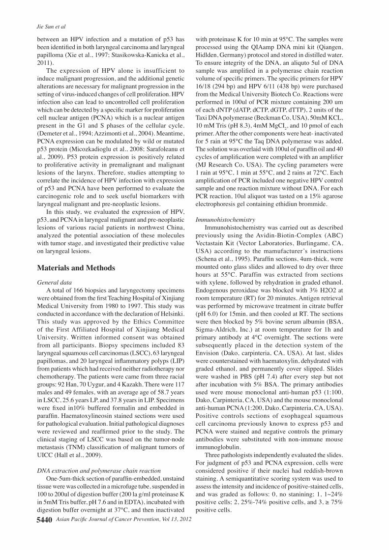
31.3Figure 1. Detection of HPV DNA with PCR. Using primers specific of HPV16/ l8 and agarose gel electrophoresis DNA fragment of approximately 300 bp was detected in laryngeal papillomas (LSCC) (A); HPV6/11and agarose gel electrophoresis DNA fragment of approximately 500 bp was detected in laryngeal papillomas (LP) (B). M: DNA size marker; 1: positive control; 2: negative control; 3 and 4: tissue samples
Table 1. Incidence of HPV Subtypes 6/11and 16/18 Detection in LSCC, LP, and LIP LSCC LP LIP
+ - + - + -
HPV 6/11 (number) 8 75 21 42 0 20HPV 16/18 (number) 31 52 4 59 0 20HPV6/11&16/18 (number) 3 80 0 0 0 20
Table 2. Incidence of HPV 6/11 and 16/18 in Various Ethic Patients with LSCC, LP, and LIP LSCC LP LIP
U+K H U+K H U+K H
HPV6/11* 0/31 8/52 9/33 12/30 0/10 0/10HPV16/18* 9/31 22/52 1/33 3/30 0/10 0/10HPV6/11&16/18* 0/31 3/52 0/33 0/30 0/10 0/10
*positive/total number; U, Uygur group; K, kazak group; H, Han group
Statistics analysis Statistical analysis was performed using SPSS 16.0 software (SPSS Inc., IL, and USA). The Student t test was used to compare quantitative variables. The Chi-square test using was applied to compare qualitative variables; correlations between continuous variables were evaluated using Chi-square test and Rank sum test. A value of P< 0.05 was considered significant.
Results
Detection of HPV Specific bands of HPV 16/18 and HPV 6/11 (approximately 300 bp and 500 bp) were noted in LSCC and LP samples respectively (Figure 1A, B), no detection was found in LIP. The frequency of HPV infection varied with the subtypes of HPV in laryngeal carcinomas, appaloosas, and polyps was showed in Table 1, HPV 6/11 were detected in 21/63 (33.3%) of the LP, 8/83(9.6%) of the LSCC, and none of the 20 LIP (P <0.0005). The frequency of HPV 16 /18 was higher significantly in 31/83 (37.3%) of the LSCC than in 4/63 (6.3%) of the LP (6.3%), and none of the 20 LIP (P <0.0001). Co-infection with HPV 6/11 and 16/18 was revealed in 3/83 (3.6%) of the LSCC as compared with 0/63 of the LP and 0/20 of the LIP respectively (P <0.05, P <0.0001, respectively). The frequency of HPV detection in various racial patients is presented in Table 2. HPV 6/11 were detected in 8/52 (15.4%) in Han group with LSCC, while (0/31) none detection in Kazak and Uygur group; 12/30(40%) in Han group with LP, 9/33(27.3) in other two ethic groups. The frequency of HPV16/18 were detected in 22/52 (42.3%) in
Han group with LSCC, while 9/31(29.3%) were detected in Kazak and Uygur group; 3/30 (10%) in Han group with LP, 1/33(3.0%) in other two ethic groups. coinfection of HPV6/11with HPV16/18 were only detected 3/52(5.8%) in Han group, ethnic groups. The frequency of HPV 16 /18 was higher significantly in LSCC than in LP, and LIP (P <0.0001). Co-infection HPV 6/11 with 16/18 was revealed in LSCC as compared with LP, and LIP respectively (P <0.05, P <0.0001, respectively). HPV 6/11 and HPV 6 /11with 16/18 co-infetcion group were detected more in the Han racial group than in the other two racial groups of Uygur or Kazak (P <0.05). However, no significant statistical differences were determined in the frequency of HPV 16/18 among the various racial groups (P >0.05).
Expression of p53 and PCNA The immunostaining of p53 and PCNA were reddish-brown granules in the nuclei of positive cells. The
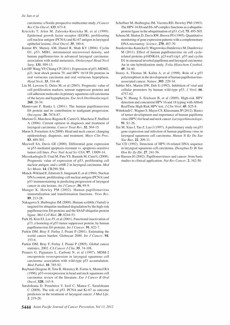
Figure 2. Expression of p53 and PCNA with Immunostaining. The positive reaction for p53 was indicated by reddish-bream color and located in the nuclei, diffuse staining pattern was evident throughout the carcinomatous nests in LSCC, X 400 (A); positive reaction for p53 was identified only in scattered squamous cells within the basal and parabasal layers in LP. X 400. (B): Expression of PCNA were detected in the nuclei of cells and appeared to be reddish-brown color in LSCC (C), X 400
Jie Sun et al
Asian Pacific Journal of Cancer Prevention, Vol 13, 20125442
expression of p53 in LSCC were throughout cancerous cell nests with a diffuse distribution pattern, which in LP tended to be restricted predominantly in the basal epithelial cells and rarely in the intermediate or superficial layer cells (Figure 2A and 2B). Immunostaining for PCNA indicated a diffuse pattern in LSCC and LP (Figure 2C). A significantly higher of p53 overexpression was observed in LSCC 52/83 (62.7%) than in LP 24/63 (38.1%), and 0/20(0%) in LIP (P <0.005, P <0.005, respectively). The overexpression of PCNA was higher significantly in LSCC 48/83 (57.8%) than in LP 21/63 (33.3%), and 0/20(0%) in LIP (P <0.005, P <0.005, respectively). The scales of p53 and PCNA expression were showed in Table 3. The scales score of P53 and PCNA in LSCC was significantly higher than those in LP (P <0.05, respectively) with rank sum test. We compared the expression of p53 and PCNA in various racial groups, the results showed no significant difference among the racial groups of Hah, Urgyr, and Kazak. Nevertheless, we further compared the clinical staging of LSCC with the expression of P53 and PCNA, as showed in Table 4. The clinical staging of LSCC was associated with the overexpression of PCNA, but not with p53, and the expression of PCNA was higher in clinical stage IV than in stage I in LSCC (P <0.05). No association of p53 and PCNA expression with lymph node metastases was found.
Discussion
HPV infection is an important risk factor in the development of LSCC (Almadori et al., 2001; Kumar et al., 2004). Evidence have suggested that high risk HPV subtypes 16 and 18 are associated mainly with laryngeal carcinoma whereas low risk HPV subtypes 6 and 11 correlated usually with laryngeal papillomas, and HPV 30 and 33 have been detected in a few laryngeal carcinomas (zur Hausen, 2002; Fakhry et al., 2008; Tang et al., 2009). Furthermore, HPV can act as a promoter or a synergistic
factor cooperating with chemical factors (smoking, alcohol, etc) in the multistep of carcinogenesis of laryngeal carcinomas and papillomas (Wittekindt et al., 2012).
Our data add evidence supporting correlations of HPV 16/18 with LSCC and HPV 6/11 with LP. The combined results confirm the observations of others that high risk HPV 16/18 and low risk HPV 6/11 play a distinct role in the development of laryngeal carcinomas and papillomas, respectively. Moreover, some association of low risk HPV 6/11 with laryngeal carcinoma and of high risk HPV 16/18 with laryngeal polyp was found in the present study. Since in the present study all biopsy specimens were selected strictly and reviewed prior to PCR, it seems unlikely that findings of HPV 6/11 in LSCC could be due to contamination with peripheral normal or hyperplastic tissue. Also it unlike that detection of 16/18 in LP was due to undetected cancerous nests. Therefore, it is reasonable to conclude that the induction of malignant or benign tumors of the larynx is not always clearly correlated with “high risk” or “low risk” HPV genetic factor.
The mechanism remains unclear whereby HPV induces a malignant transformation in infected cells. Proposed mechanisms include inactivation of p53 by its binding to high risk HPV-18 E6 or HPV-16 E7 or by its binding to MDM 2 gene products (Park et al., 2001). The binding of p53 with HPV 18-E6 results in inactivation and degradation of p53 via the ubiquitin-mediate pathway thereby interfering with the tumor suppressor function of p53 and ultimately leading to oncogenesis (Mantovani and Banks, 2001). In addition, HPV-18 E6 not only inhibits p53-suppression of transformed cell growth, but also, through ubiquitin mediated degradation, inhibits the induction of p53-mediated apoptosis. The suppression of these two different actions of p53, by its binding to HPV 18 E6, i.e. the suppression of cell proliferation and induction of apoptosis allows uncontrolled cell proliferation (Freedman and Levine, 1998; Nakagawa and Huibregtse, 2000). Furthermore, the susceptibility of two polymorphic forms of wild-type p53 to E6-mediated degradation in vivo may contribute to the HPV-associated carcinogenesis. Storey et al. have shown that p53 degradation by HPV-16 E6 and HPV-18 E6 is more effective in the arginine form of p53 than in the proline form of p53 (Storey et al., 1998). Our data demonstrate a higher and overexpression of p53. In patients with LP, an intermediate association between HPV detection and overexpression of p53 was found. In LIP, neither HPV detection nor p53 overexpression was found. These results indicate that the interaction between HPV infection and p53 may play a role in the development of malignant or benign tumors of the larynx. Many studies assessed overexpression of p53 as a useful prognosticator for LSCC or as a potentially useful marker in the management of patients at for premalignant lesions of the larynx Molecular markers in laryngeal squamous cell carcinoma: Towards an integrated clinicobiological approach. In most laryngeal carcinomas showed no close correlation of p53 overexpression with clinical stage, lymph-node metastasis, or patient survival (Raybaud-Diogene et al., 1996). In the present study, neither clinical staging nor lymph-node metastasis was correlated with overexpression of p53 protein in LSCC. It seems that p53
Table 3. The Scales of p53 and PCNA Expression in LSCC, LP, and LIP with Immunostaining P53 (staingning scales) PCNA (stainining scales) 0 1+ 2+ 3+ 0 1+ 2+ 3+
LSCC (number) 31 20 9 23* 35 8 16 24*LP (number) 39 16 7 1 42 11 7 3LIP (number) 20 0 0 0 20 0 0 0
*Semiquntitative scores for p53 and PCNA in the LSCC signifcantly higher than those in the LP (P<0.05 by rank sum test)
Table 4. Corrlelation of Clinical Staging and Expression of p53 and PCNA in Variorus Racial Patients with LSCC, LP, LIP Clinical Staging P53 PCNA positive negative positive negative
I 13 12 10 15II 14 7 11 10III 12 7 12 7IV 13 5 15 3

Asian Pacific Journal of Cancer Prevention, Vol 13, 2012 5443
DOI:http://dx.doi.org/10.7314/APJCP.2012.13.11.5439P53 and PCNA and HPV Infection in Laryngeal Lesions in Xinjiang
overexpression cannot serve as a useful prognosticator in patients with laryngeal carcinoma. However, p53 overexpression in laryngeal papilloma may indicate, at least, minimal p53 mutation. For this reason, p53 overexpression may serve as a risk indicator for malignant transformation in the HPV infected cell. In the present study, no correlation between p53 expression and the ethnic group of patients was found. These results indicate that cytogenetic alteration may be a common pathway in human laryngeal carcinogenesis without influence of ethnic differences.
The expression of PCNA may be modulated by either wild or mutated p53 protein (Subler et al., 1992). Positive correlation of p53 overexpression and high proliferative activity as demonstrated by PCNA immnunoreactivity, has been detected in laryngeal squamous cell carcinomas and laryngeal preneoplastic lesions (Krecicki et al., 1999). A high incidence of PCNA was found in the in situ lesions of laryngeal squamous cell carcinoma (Munck-Wikland et al., 1994). In HPV associated tumors, HPV E6 proteins have a unique role in inhibiting p53-mediated apoptosis via ubiquitin mediated degradation. Therefore, it is conceivable that in the HPV 16/18-infected LSCC, HPV 16/18 infection may lead to uncontrolled cell proliferation as indicated by a specific marker of cell proliferation (Munger and Howley, 2002). Combination of overexpression of PCNA and p53, as well as detection of nuclear DNA alteration, may predict the progression of the in situ lesions of laryngeal carcinoma to invasive lesions. Recently it has been suggested that PCNA expression correlated with survival of patients with LSCC, and thus PCNA may be a useful biomarker for LSCC prognosis. In some studies overexpression of PCNA correlated well with lymph node metastasis of laryngeal carcinoma (Dong et al., 2000; Liu et al., 2003). In the present study, the frequency of PCNA overexpression of LSCC was higher significantly in stage IV than in stage I, stage II, and stage III of LSCC, which suggested that the proliferating rate of squamous cancer cells increases gradually with the tumor progressing. The frequency of PCNA overexpression was higher in patients with lymph node metastasis than in patients without node metastasis, however no statistical significance was determined between the two groups of patients.
In conclusion, this report presents evidence demonstrating a close correlation of HPV16/18 or co-infection of HPV16/18 with 6/11 in LSCC, and of HPV 6/11 infection in LP. The expression of HPV 6/11 and co-infection of HPV6/11 with 16 /18 were higher significantly in the Han racial group patients than in the Uygur and Kazak racial group patients. Furthermore, overexpression of p53 and PCNA was higher significantly in patients with LSCC than in patients with LP. Correlation of PCNA overexpression with clinical stage was observed in LSCC. These results suggest that HPV infection, overexpression of p53, and PCNA may contribute to the multistep processes of carcinogenesis of laryngeal carcinomas and papillomas. Overexpression of p53 may be used as an early marker for monitoring malignant transformation, whereas overexpression of PCNA as a late marker for malignant progression.
Acknowledgements
The authors would like to thank Drs. Jun Zhang and Sydeney Ellis, Center for Evaluation and Research, U. S. Food and Drug Administration, for editing the manuscript. We are also indebted to Prof. Qian Xue, Department of statistics, Xinjiang Medical University ,China, for his assistance in statistical analyses .We are also thank Prof. Wang Changhong, Department of Pharmacy, Second Teaching Hospital of Xinjiang Medical University, China, for his assistance in statistical analyses.
References
Almadori G, Cadoni G, Cattani P, et al (2001). Human papillomavirus infection and epidermal growth factor receptor expression in primary laryngeal squamous cell carcinoma. Clin Cancer Res, 7, 3988-93.
Azzimonti B, Pagano M, Mondini M, et al (2004). Altered patterns of the interferon-inducible gene IFI16 expression in head and neck squamous cell carcinoma: immunohistochemical study including correlation with retinoblastoma protein, human papillomavirus infection and proliferation index. Histopathology, 45, 560-72.
Demeter LM, Stoler MH, Broker TR, Chow LT (1994). Induction of proliferating cell nuclear antigen in differentiated keratinocytes of human papillomavirus-infected lesions. Hum Pathol, 25, 343-8.
Dong Y, Sui L, Tai Y, et al (2000). Prognostic significance of cyclin E overexpression in laryngeal squamous cell carcinomas. Clin Cancer Res, 6, 4253-8.
Du J, Chen GG, Vlantis AC, et al (2003). The nuclear localization of NFkappaB and p53 is positively correlated with HPV16 E7 level in laryngeal squamous cell carcinoma. J Histochem Cytochem, 51, 533-9.
El-Naggar AK, Westra WH (2012). p16 expression as a surrogate marker for HPV-related oropharyngeal carcinoma: a guide for interpretative relevance and consistency. Head Neck, 34, 459-61.
Fakhry C, Westra WH, Li S, et al (2008). Improved survival of patients with human papillomavirus-positive head and neck squamous cell carcinoma in a prospective clinical trial. J Natl Cancer Inst, 100, 261-9.
Freedman DA, Levine AJ (1998). Nuclear export is required for degradation of endogenous p53 by MDM2 and human papillomavirus E6. Mol Cell Biol, 18, 7288-93.
Gao Y, Ferguson DO, Xie W, et al (2000). Interplay of p53 and DNA-repair protein XRCC4 in tumorigenesis, genomic stability and development. Nature, 404, 897-900.
Grace Nirmala J, Narendhirakannan RT (2011). Detection and genotyping of high-risk HPV and evaluation of anti-oxidant status in cervical carcinoma patients in Tamil Nadu State, India--a case control study. Asian Pac J Cancer Prev, 12, 2689-95.
Hall SF, Groome PA, Irish J, O’Sullivan B (2009). TNM-based stage groupings in head and neck cancer: application in cancer of the hypopharynx. Head Neck, 31, 1-8.
Horibe Y, Murakami M, Komori K, Imaeda Y, Kasahara M (2000). Expression of topoisomerase II alpha, Ki-67 and p53 in early stage laryngeal carcinomas not featuring vocal cord fixation. Apmis, 108, 689-96.
Ji W, Guan C, Pan Z (2008). Analysis of curative effects on laryngeal carcinoma patients in the northeast region of China. Acta Otolaryngol, 128, 574-7.
Koskinen WJ, Brondbo K, Mellin Dahlstrand H, et al (2007). Alcohol, smoking and human papillomavirus in laryngeal
Jie Sun et al
Asian Pacific Journal of Cancer Prevention, Vol 13, 20125444
carcinoma: a Nordic prospective multicenter study. J Cancer Res Clin Oncol, 133, 673-8.
Krecicki T, Jelen M, Zalesska-Krecicka M, et al (1999). Epidermal growth factor receptor (EGFR), proliferating cell nuclear antigen (PCNA) and Ki-67 antigen in laryngeal epithelial lesions. Oral Oncol, 35, 180-6.
Kumar RV, Shenoy AM, Daniel R, Shah KV (2004). Cyclin D1, p53, MIB1, intratumoral microvessel density, and human papillomavirus in advanced laryngeal carcinoma: association with nodal metastasis. Otolaryngol Head Neck Surg, 131, 509-13.
Lin HP, Wang YP, Chiang CP (2011). Expression of p53, MDM2, p21, heat shock protein 70, and HPV 16/18 E6 proteins in oral verrucous carcinoma and oral verrucous hyperplasia. Head Neck, 33, 334-40.
Liu M, Lawson G, Delos M, et al (2003). Prognostic value of cell proliferation markers, tumour suppressor proteins and cell adhesion molecules in primary squamous cell carcinoma of the larynx and hypopharynx. Eur Arch Otorhinolaryngol, 260, 28-34.
Mantovani F, Banks L (2001). The human papillomavirus E6 protein and its contribution to malignant progression. Oncogene, 20, 7874-87.
Marioni G, Marchese-Ragona R, Cartei G, Marchese F, Staffieri A (2006). Current opinion in diagnosis and treatment of laryngeal carcinoma. Cancer Treat Rev, 32, 504-15.
Marur S, Forastiere AA (2008). Head and neck cancer: changing epidemiology, diagnosis, and treatment. Mayo Clin Proc, 83, 489-501.
Maxwell SA, Davis GE (2000). Differential gene expression in p53-mediated apoptosis-resistant vs. apoptosis-sensitive tumor cell lines. Proc Natl Acad Sci USA, 97, 13009-14.
Micozkadioglu D, Unal M, Pata YS, Basturk M, Cinel L (2008). Prognostic value of expression of p53, proliferating cell nuclear antigen, and c-erbB-2 in laryngeal carcinoma. Med Sci Monit, 14, CR299-304.
Munck-Wikland E, Edstrom S, Jungmark E, et al (1994). Nuclear DNA content, proliferating-cell nuclear antigen (PCNA) and p53 immunostaining in predicting progression of laryngeal cancer in situ lesions. Int J Cancer, 56, 95-9.
Munger K, Howley PM (2002). Human papillomavirus immortalization and transformation functions. Virus Res, 89, 213-28.
Nakagawa S, Huibregtse JM (2000). Human scribble (Vartul) is targeted for ubiquitin-mediated degradation by the high-risk papillomavirus E6 proteins and the E6AP ubiquitin-protein ligase. Mol Cell Biol, 20, 8244-53.
Park JS, Kim EJ, Lee JY, et al (2001). Functional inactivation of p73, a homolog of p53 tumor suppressor protein, by human papillomavirus E6 proteins. Int J Cancer, 91, 822-7.
Parkin DM, Bray F, Ferlay J, Pisani P (2001). Estimating the world cancer burden: Globocan 2000. Int J Cancer, 94, 153-6.
Parkin DM, Bray F, Ferlay J, Pisani P (2005). Global cancer statistics, 2002. CA Cancer J Clin, 55, 74-108.
Pruneri G, Pignataro L, Carboni N, et al (1997). MDM-2 oncoprotein overexpression in laryngeal squamous cell carcinoma: association with wild-type p53 accumulation. Mod Pathol, 10, 785-92.
Raybaud-Diogene H, Tetu B, Morency R, Fortin A, Monteil RA (1996). p53 overexpression in head and neck squamous cell carcinoma: review of the literature. Eur J Cancer B Oral Oncol, 32B, 143-9.
Sarafoleanu D, Postelnicu V, Iosif C, Manea C, Sarafoleanu C (2009). The role of p53, PCNA and Ki-67 as outcome predictors in the treatment of laryngeal cancer. J Med Life, 2, 219-26.
Scheffner M, Huibregtse JM, Vierstra RD, Howley PM (1993). The HPV-16 E6 and E6-AP complex functions as a ubiquitin-protein ligase in the ubiquitination of p53. Cell, 75, 495-505.
Schena M, Shalon D, Davis RW, Brown PO (1995). Quantitative monitoring of gene expression patterns with a complementary DNA microarray. Science, 270, 467-70.
Stasikowska-Kanicka O, Wagrowska-Danilewicz M, Danilewicz M (2011). Effect of human papillomavirus on cell cycle-related proteins p16INK4A, p21waf1/cip1, p53 and cyclin D1 in sinonasal inverted papilloma and laryngeal carcinoma. An in situ hybridization study. Folia Histochem Cytobiol, 49, 34-40.
Storey A, Thomas M, Kalita A, et al (1998). Role of a p53 polymorphism in the development of human papillomavirus-associated cancer. Nature, 393, 229-34.
Subler MA, Martin DW, Deb S (1992). Inhibition of viral and cellular promoters by human wild-type p53. J Virol, 66, 4757-62.
Tang N, Huang S, Erickson B, et al (2009). High-risk HPV detection and concurrent HPV 16 and 18 typing with Abbott RealTime High Risk HPV test. J Clin Virol, 45, S25-8.
Wittekindt C, Wagner S, Mayer CS, Klussmann JP (2012). Basics of tumor development and importance of human papilloma virus (HPV) for head and neck cancer. Laryngorhinootologie, 91, S1-26.
Xie M, Xiao J, Tao Z, Luo J (1997). A preliminary study on p53 gene expression and infection of human papilloma virus in laryngeal squamous cell carcinoma. Hunan Yi Ke Da Xue Xue Bao, 22, 209-11.
Yao CD (1992). Detection of HPV-16-related DNA sequence in laryngeal squamous cell carcinoma. Zhonghua Er Bi Yan Hou Ke Za Zhi, 27, 241-56.
zur Hausen H (2002). Papillomaviruses and cancer: from basic studies to clinical application. Nat Rev Cancer, 2, 342-50.


![Theranostics · Web viewThus, several molecular markers, such as TP53 mutations [9, 10], P53/P16 immunohistochemistry [11], HPV genotyping [12, 13], gene expression [14], and loss](https://img.pdfslide.us/doc/110x75/60e49b4976d2144a4809da27/theranostics-web-view-thus-several-molecular-markers-such-as-tp53-mutations-9.jpg)