Embed Size (px)
Citation preview
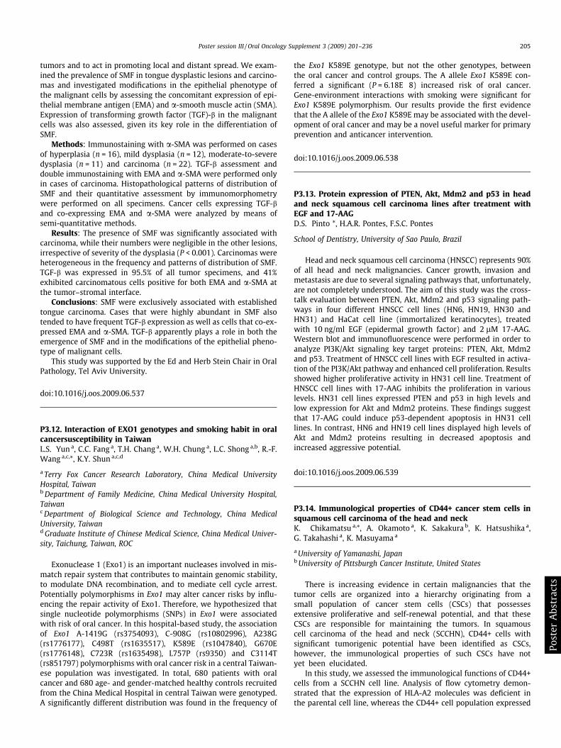
tumors and to act in promoting local and distant spread. We exam-ined the prevalence of SMF in tongue dysplastic lesions and carcino-mas and investigated modifications in the epithelial phenotype ofthe malignant cells by assessing the concomitant expression of epi-thelial membrane antigen (EMA) and a-smooth muscle actin (SMA).Expression of transforming growth factor (TGF)-b in the malignantcells was also assessed, given its key role in the differentiation ofSMF.
Methods: Immunostaining with a-SMA was performed on casesof hyperplasia (n = 16), mild dysplasia (n = 12), moderate-to-severedysplasia (n = 11) and carcinoma (n = 22). TGF-b assessment anddouble immunostaining with EMA and a-SMA were performed onlyin cases of carcinoma. Histopathological patterns of distribution ofSMF and their quantitative assessment by immunomorphometrywere performed on all specimens. Cancer cells expressing TGF-band co-expressing EMA and a-SMA were analyzed by means ofsemi-quantitative methods.
Results: The presence of SMF was significantly associated withcarcinoma, while their numbers were negligible in the other lesions,irrespective of severity of the dysplasia (P < 0.001). Carcinomas wereheterogeneous in the frequency and patterns of distribution of SMF.TGF-b was expressed in 95.5% of all tumor specimens, and 41%exhibited carcinomatous cells positive for both EMA and a-SMA atthe tumor–stromal interface.
Conclusions: SMF were exclusively associated with establishedtongue carcinoma. Cases that were highly abundant in SMF alsotended to have frequent TGF-b expression as well as cells that co-ex-pressed EMA and a-SMA. TGF-b apparently plays a role in both theemergence of SMF and in the modifications of the epithelial pheno-type of malignant cells.
This study was supported by the Ed and Herb Stein Chair in OralPathology, Tel Aviv University.
doi:10.1016/j.oos.2009.06.537
P3.12. Interaction of EXO1 genotypes and smoking habit in oralcancersusceptibility in TaiwanL.S. Yun a, C.C. Fang a, T.H. Chang a, W.H. Chung a, L.C. Shong a,b, R.-F.Wang a,c,*, K.Y. Shun a,c,d
a Terry Fox Cancer Research Laboratory, China Medical UniversityHospital, Taiwanb Department of Family Medicine, China Medical University Hospital,Taiwanc Department of Biological Science and Technology, China MedicalUniversity, Taiwand Graduate Institute of Chinese Medical Science, China Medical Univer-sity, Taichung, Taiwan, ROC
Exonuclease 1 (Exo1) is an important nucleases involved in mis-match repair system that contributes to maintain genomic stability,to modulate DNA recombination, and to mediate cell cycle arrest.Potentially polymorphisms in Exo1 may alter cancer risks by influ-encing the repair activity of Exo1. Therefore, we hypothesized thatsingle nucleotide polymorphisms (SNPs) in Exo1 were associatedwith risk of oral cancer. In this hospital-based study, the associationof Exo1 A-1419G (rs3754093), C-908G (rs10802996), A238G(rs1776177), C498T (rs1635517), K589E (rs1047840), G670E(rs1776148), C723R (rs1635498), L757P (rs9350) and C3114T(rs851797) polymorphisms with oral cancer risk in a central Taiwan-ese population was investigated. In total, 680 patients with oralcancer and 680 age- and gender-matched healthy controls recruitedfrom the China Medical Hospital in central Taiwan were genotyped.A significantly different distribution was found in the frequency of
the Exo1 K589E genotype, but not the other genotypes, betweenthe oral cancer and control groups. The A allele Exo1 K589E con-ferred a significant (P = 6.18E�8) increased risk of oral cancer.Gene-environment interactions with smoking were significant forExo1 K589E polymorphism. Our results provide the first evidencethat the A allele of the Exo1 K589E may be associated with the devel-opment of oral cancer and may be a novel useful marker for primaryprevention and anticancer intervention.
doi:10.1016/j.oos.2009.06.538
P3.13. Protein expression of PTEN, Akt, Mdm2 and p53 in headand neck squamous cell carcinoma lines after treatment withEGF and 17-AAGD.S. Pinto *, H.A.R. Pontes, F.S.C. Pontes
School of Dentistry, University of Sao Paulo, Brazil
Head and neck squamous cell carcinoma (HNSCC) represents 90%of all head and neck malignancies. Cancer growth, invasion andmetastasis are due to several signaling pathways that, unfortunately,are not completely understood. The aim of this study was the cross-talk evaluation between PTEN, Akt, Mdm2 and p53 signaling path-ways in four different HNSCC cell lines (HN6, HN19, HN30 andHN31) and HaCat cell line (immortalized keratinocytes), treatedwith 10 ng/ml EGF (epidermal growth factor) and 2 lM 17-AAG.Western blot and immunofluorescence were performed in order toanalyze PI3K/Akt signaling key target proteins: PTEN, Akt, Mdm2and p53. Treatment of HNSCC cell lines with EGF resulted in activa-tion of the PI3K/Akt pathway and enhanced cell proliferation. Resultsshowed higher proliferative activity in HN31 cell line. Treatment ofHNSCC cell lines with 17-AAG inhibits the proliferation in variouslevels. HN31 cell lines expressed PTEN and p53 in high levels andlow expression for Akt and Mdm2 proteins. These findings suggestthat 17-AAG could induce p53-dependent apoptosis in HN31 celllines. In contrast, HN6 and HN19 cell lines displayed high levels ofAkt and Mdm2 proteins resulting in decreased apoptosis andincreased aggressive potential.
doi:10.1016/j.oos.2009.06.539
P3.14. Immunological properties of CD44+ cancer stem cells insquamous cell carcinoma of the head and neckK. Chikamatsu a,*, A. Okamoto a, K. Sakakura b, K. Hatsushika a,G. Takahashi a, K. Masuyama a
a University of Yamanashi, Japanb University of Pittsburgh Cancer Institute, United States
There is increasing evidence in certain malignancies that thetumor cells are organized into a hierarchy originating from asmall population of cancer stem cells (CSCs) that possessesextensive proliferative and self-renewal potential, and that theseCSCs are responsible for maintaining the tumors. In squamouscell carcinoma of the head and neck (SCCHN), CD44+ cells withsignificant tumorigenic potential have been identified as CSCs,however, the immunological properties of such CSCs have notyet been elucidated.
In this study, we assessed the immunological functions of CD44+cells from a SCCHN cell line. Analysis of flow cytometry demon-strated that the expression of HLA-A2 molecules was deficient inthe parental cell line, whereas the CD44+ cell population expressed
Poster session III / Oral Oncology Supplement 3 (2009) 201–236 205
Com
mit
tee
List
ings
Wel
com
eIA
OO
Prog
ram
Key
not
eB
ios.
Key
not
eA
bs.
Pan
.Dis
c.&
Sym
p.A
bs.
Ora
lsLi
stPo
ster
List
Ora
lA
bstr
acts
Post
erA
bstr
acts


![Human exonuclease 1 (EXO1) activity characterization and its … · 2017-10-04 · EXO1 to the DNA [8], while the I-domain exhibits multiple cysteine and glutamate residues that are](https://img.pdfslide.us/doc/110x75/5f8ce5575c41787f96248c61/human-exonuclease-1-exo1-activity-characterization-and-its-2017-10-04-exo1-to.jpg)


![CaseReport Habit Breaking Appliance for Multiple Corrections · Habit Breaking Appliance for Multiple Corrections ... removable habit breaking appliances [15, 16]. Hence, habit breaking](https://img.pdfslide.us/doc/110x75/5f15893424a8522d646af1b7/casereport-habit-breaking-appliance-for-multiple-corrections-habit-breaking-appliance.jpg)