Embed Size (px)
Citation preview
J ENDOVASC THER2006;13:377–388
377
Q 2006 by the INTERNATIONAL SOCIETY OF ENDOVASCULAR SPECIALISTS Available at www.jevt.org
lFELLOWS’ COMPETITION, FIRST PLACE, LABORATORY SCIENCE l
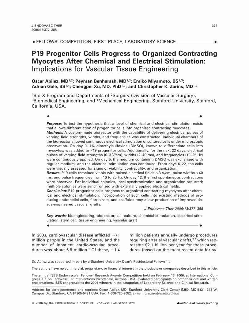
P19 Progenitor Cells Progress to Organized ContractingMyocytes After Chemical and Electrical Stimulation:Implications for Vascular Tissue Engineering
Oscar Abilez, MD1,2; Peyman Benharash, MD1,2; Emiko Miyamoto, BS1,3;
Adrian Gale, BS1,4; Chengpei Xu, MD, PhD1,2; and Christopher K. Zarins, MD1,2
1Bio-X Program and Departments of 2Surgery (Division of Vascular Surgery),3Biomedical Engineering, and 4Mechanical Engineering, Stanford University, Stanford,California, USA.
l l
Purpose: To test the hypothesis that a level of chemical and electrical stimulation existsthat allows differentiation of progenitor cells into organized contracting myocytes.Methods: A custom-made bioreactor with the capability of delivering electrical pulses ofvarying field strengths, widths, and frequencies was constructed. Individual chambers ofthe bioreactor allowed continuous electrical stimulation of cultured cells under microscopicobservation. On day 0, 1% dimethylsulfoxide (DMSO), known to differentiate cells intomyocytes, was added to P19 progenitor cells. Additionally, for the next 22 days, electricalpulses of varying field strengths (0–3 V/cm), widths (2–40 ms), and frequencies (10–25 Hz)were continuously applied. On day 5, the medium containing DMSO was exchanged withregular medium, and the electrical stimulation was continued. From days 6–22, the cellswere visually assessed for signs of viability, contractility, and organization.Results: P19 cells remained viable with pulsed electrical fields ,3 V/cm, pulse widths ,40ms, and pulse frequencies from 10 to 25 Hz. On day 12, the first spontaneous contractionswere observed. For individual colonies, local synchronization and organization occurred;multiple colonies were synchronized with externally applied electrical fields.Conclusion: P19 progenitor cells progress to organized contracting myocytes after chem-ical and electrical stimulation. Incorporation of such cells into existing methods of pro-ducing endothelial cells, fibroblasts, and scaffolds may allow production of improved tis-sue-engineered vascular grafts.
J Endovasc Ther 2006;13:377–388
Key words: bioengineering, bioreactor, cell culture, chemical stimulation, electrical stim-ulation, stem cell, tissue engineering, vascular graftl l
Dr. Abilez was supported in part by a Stanford University Dean’s Postdoctoral Fellowship.
The authors have no commercial, proprietary, or financial interest in the products or companies described in this article.
The annual ISES Endovascular Fellows’ Research Awards Competition held on February 13, 2006, at International Con-gress XIX on Endovascular Interventions (Scottsdale, Arizona, USA) evaluated participants on both their oral and writtenpresentations. ISES congratulates the 2006 winners in the categories of Laboratory Science and Clinical Research.
Address for correspondence and reprints: Oscar Abilez, MD, Stanford University Clark Center E350, MC 5431, 318 W.Campus Dr., Stanford, CA 94305-5431 USA. Fax: 1-650-725-9082; E-mail: [email protected]
In 2003, cardiovascular disease afflicted ;71million people in the United States, and thenumber of inpatient cardiovascular proce-dures was about 6.8 million.1 Of these, ;1.4
million patients annually undergo proceduresrequiring arterial vascular grafts,2,3 which rep-resents $2.1 billion per year for these proce-dures (based on the most recent data for av-

378 P19 PROGENITOR-DERIVED MYOCYTESAbilez et al.
J ENDOVASC THER2006;13:377–388
erage cost per procedure). Vascular grafts arecurrently used as bypass grafts, endovasculargrafts, and interposition grafts.1,3,4 However,the currently available grafts have been lim-ited by variable patency rates, material avail-ability, and immunological rejection.5–7
In attempts to address these limitationsover the last 20 years, experimental humanand animal tissue-engineered vascular grafts(TEVG) have been assembled from endothe-lial cells (EC), smooth muscle cells (SMC), andfibroblast cells (FC)8–12; these experimentalTEVGs have demonstrated favorablestrengths and patency rates. However, theirmain drawback has been immunological re-jection during in vivo testing.8,10,13 The crea-tion of a TEVG from autologous stem cellswould potentially address these shortcom-ings and, furthermore, could potentially serveas the vascular source for other tissue-engi-neered materials, such as lung, heart, liver, orbone tissue.14–22
Of the several stem cell types that exist, themouse embryonic stem cell (mESC) is wellcharacterized, readily available, and has norestrictions on its use.23 Furthermore, groupshave reported differentiating mESC into ECand SMC; in addition, FC derived from mouseembryos are commercially available.24–29
However, the subsequent in vitro assembly ofthese cell types into 3-layered blood vesselshas not yet been reported. In addition, it is notentirely known how various stimuli affectstem cell differentiation into these cell types.Furthermore, the differentiation of stem/pro-genitor cells into myocytes for use in vasculartissue engineering has been ill-defined todate. Myocytes must exhibit both functionalorganization and contractility in order to serveas components for tissue-engineered vascu-lar grafts. Recently, groups have demonstrat-ed the salutary effects of electrical stimulationon primary myocyte organization and stemcell differentiation.30–33
Our purpose was to test the hypothesis thata level of chemical and electrical stimulationexists that allows differentiation of progenitorcells into organized contracting myocytes. Totest our hypothesis, we applied these stimu-lation signals to P19 cells, a stem cell line de-rived from a mouse embryonal carcinoma
that is known to have the potential to differ-entiate into myocytes.34–38
METHODS
Complete Medium
A complete medium was prepared from Min-imal Essential Medium Alpha (a-MEM) with ri-bonucleosides and deoxynucleosides (Invitro-gen, Carlsbad, CA, USA) supplemented with7.5% calf bovine serum (American Type Cul-ture Collection [ATCC], Manassas, VA, USA)and 2.5% fetal bovine serum (GIBCO, Carls-bad, CA, USA). Next, penicillin-streptomycin(GIBCO) diluted from a 1003 concentration ofstock solution was added to the above mix-ture to obtain a final concentration of 13 inthe complete medium. Finally, b-mercapto-ethanol was added to a final concentration of0.1 mM.
Cell Culture
A 1-mL vial of frozen P19 mouse embryonalcarcinoma stem cells (ATCC #CRL-1825) wasthawed in a 378C water bath. The cells werethen re-suspended in 9 mL of new completemedium in a 15-mL tube. The tube was spunin a Clinical 200 centrifuge (VWR, West Ches-ter, PA, USA) at 300g (corresponding to 1750rpm) for 3 minutes. The medium was then as-pirated, leaving the pellet of cells in the tube.Next, 5 mL of new fresh complete mediumwas added to the tube, and the clumped cellswere then dissociated by pipetting up anddown. The dissociated cells and new mediumwere then transferred into a T-25 tissue-cul-ture flask (Becton Dickinson Biosciences, Bed-ford, MA, USA), which was placed in a 378Cincubator (Fisher Scientific Isotemp, Hamp-ton, NH, USA) with 5% CO2. No feeder layerwas used.
To ensure that they were healthy and con-tinuing to grow, the cells were observed onthe second day of culture with a DM-IL (LeicaMicrosystems USA, Bannockburn, IL, USA) orTS-100F (Nikon USA, Melville, NY, USA) mi-croscope (magnification from 403 to 4003with Hoffman modulation contrast and phasecontrast optics). On the third day, the cellswere fed. The original medium (usually dark

J ENDOVASC THER2006;13:377–388
P19 PROGENITOR-DERIVED MYOCYTESAbilez et al.
379
Figure 1l (A) Electrical stimulation was accom-plished with a custom-made electric cell pulser. (B)The pulser delivered square waves of various volt-age amplitude, pulse width, and pulse frequency.(C) The electronic circuit design. Op Amp: opera-tional amplifier, FET: field effect transistor, VDC:voltage direct current, V1: positive voltage, V2:negative voltage, Sync-OUT: output synchroniza-tion from timing chip.
yellow, indicating active cellular metabolism)was removed with a glass pipette connectedto a vacuum. Care was taken not to aspiratethe attached cells. Next, 5 mL of new freshcomplete medium were added to the cells,and then the flask was placed back in the in-cubator.
On the fourth day, the cells were generallysplit into 10 parts, with 9 parts frozen for fu-ture use and 1 part propagated in culture. Tosplit the cells, the medium from the flask wasremoved. Then, 1000 mL of trypsin (GIBCO)was added to the T-25 flask to detach the cellsfrom the bottom, and the flask was incubatedat 378C for 5 minutes in 5% CO2. Next, the 900mL of trypsin and cells were transferred intoa 15-mL tube, to which was added 9.1 mL offreezing medium [95% complete medium, 5%dimethylsulfoxide (DMSO; Sigma-Aldrich, St.Louis, MO, USA)] to inactivate the trypsin andbring the total volume to 10 mL. Pipetting thecells up and down in each tube broke apartany cell clumps. The 10 mL of freezing me-dium/cells were distributed in 1-mL aliquotsto 10 cryotubes, which were placed in a2808C freezer overnight and then transferredto a 21808C liquid nitrogen tank the followingday. To the 100 mL of trypsin and cells re-maining in the T-25 flask, 4.9 mL of fresh com-plete medium was added, inactivating thetrypsin and bringing the total volume back to5 mL. The flask was then re-incubated at 378Cin 5% CO2.
Electric Cell Pulser
A custom-made cell pulser (Fig. 1A) was de-signed with 4 channels to simultaneouslystimulate the P-19 cells in 4 separate biore-actors. Each channel could deliver a squarewave pulse (Fig. 1B) of varying voltage am-plitude (1–10 V), width (0.5–125 ms), and fre-quency (0.6–300 Hz). Due to technical limita-tions, the minimum frequency obtained forthese experiments was 10 Hz.
The electronic circuit design of the cell puls-er (Fig. 1C) included an LM 556 timing chip(Jameco Electronics, Belmont, CA, USA) tocoordinate the manual pulse width and fre-quency adjustment. This chip also allowedcomputer control of the pulse width and fre-quency via 2 operational amplifiers (Jameco
Electronics). The voltage amplitude adjust-ment was achieved with an LM 317 voltageregulator (Jameco Electronics). A field effecttransistor (Jameco Electronics) was used inan open collector configuration. A triple-out-put power supply (model CPS 250; Tektronix,Beaverton, OR, USA) was used to provide 15-volt direct current to both the timing chip andthe voltage regulator. Finally, to observe theoutput from the timing chip on a digital stor-

380 P19 PROGENITOR-DERIVED MYOCYTESAbilez et al.
J ENDOVASC THER2006;13:377–388
Figure 2l(A) Electrical stimulation was delivered via a custom-made 4-well bioreactor. (B,
C) The experimental setup consisted of 4 bioreactors placed in an incubator. (D) The biore-actors, which were powered with an adjustable power supply, were connected to the electriccell pulser (placed on top of the incubator).
age oscilloscope (model VC-6025; Hitachi, To-kyo, Japan), a synchronization channel wasadded.
Bioreactor
Off-the-shelf items were used to assemblethe individual bioreactors (Fig. 2A), includinga 4-well Lab-Tek Chamber-Slide system (NalgeNunc, Rochester, NY, USA) in which thechamber was made of polypropylene and theslide of Permanox. Using a standard drill-press fitted with a 1/64-inch drill bit, one holewas drilled at each end of every well (8 holestotal). Into each hole was placed ;1 cm of99% pure gold wire (Sigma-Aldrich) to serveas the electrodes for electrical stimulation.The outside ends of the gold electrodes wereconnected 1 cm apart to flat ribbon computerwire (Jameco Electronics) via gold-plated
connectors (Jameco Electronics) and attachedto the chamber with Loctite Five-Minute ep-oxy (Loctite-Henkel, Rocky Hill, CT, USA). Ap-plied voltages from the electric cell pulserwere divided by the 1-cm distance separatingthe electrodes to obtain field strengths in V/cm.
Four bioreactors were used for all chemicaland electrical stimulation experiments. Thebioreactors were incubated at 378C in 5% CO2
while they were connected to the electric cellpulser and power supply (Fig. 2B–D). A dataacquisition system consisting of National In-struments cFP-2000 control module hardwareand LabView 7.1 software (National Instru-ments, Austin, TX, USA) was used to controlthe pulse width and frequency of the electriccell pulser. The hardware was directly con-nected to the cell pulser via BNC (Bayonet NutCoupling) connectors.

J ENDOVASC THER2006;13:377–388
P19 PROGENITOR-DERIVED MYOCYTESAbilez et al.
381
Figure 3lSchematic of the experimental design.
l l
TABLE 1
Electrical Stimulation Parameters for the 4 Bioreactors
Bioreactor
1 2 3 4
Pulse width, ms 2 30 35 40Field strength, V/cm 0, 1, 2, 3 0, 1, 2, 3 0, 1, 2, 3 0, 1, 2, 3Pulse frequency, Hz 20 20 25 10l l
To observe the daily activity in the biore-actors, a DM-IL (Leica Microsystems USA) in-verted microscope fitted with 103 ocularsand 43, 103, 203, and 403 objectives wasused to provide magnifications of 403, 1003,2003, and 4003. Attached to the microscopewas a Retiga 2000R high-speed digital CCDcamera (QImaging, Burnaby, BC, Canada) ca-pable of taking single frames and/or video-quality movies (30 frames/s).
Chemical and Electrical Stimulation
The experimental design for chemical andelectrical stimulation is shown in Figure 3. Onday 27, P19 cells were thawed, grown, andsplit as outlined above. On day 0, the P19 cellswere washed 3 times with phosphate-buff-ered saline (PBS, pH 7.4) and then transferredfrom the complete medium to differentiationmedium containing 1% DMSO. This medium,known to differentiate cells into myocytes,was used to chemically stimulate the P19 cellsfor 5 days.
Additionally, for the next 22 days, electricalpulses of varying field strengths (0–3 V/cm),
widths (2–40 ms), and frequencies (10–25 Hz)were continuously applied (Table 1). On day5, the medium containing DMSO was ex-changed with complete medium (containingno DMSO), and the electrical stimulation wascontinued. From days 6 to 22, the cells werevisually assessed for signs of viability, con-tractility, and organization. Spontaneouslycontracting P19-derived myocyte colonieswere counted daily by 1 observer and weredocumented with the image acquisition sys-tem. Finally, either the differentiation mediumor complete medium was renewed every 3days.
Electrical Synchronization
Electrical synchronization (pacing) was per-formed on day 22 of culture on P19-derivedmyocytes and myocyte colonies in Bioreactor1 only because it demonstrated the mostspontaneously contracting myocytes, whichwere also noted to be asynchronously con-tracting. The 4-channel pulser was discon-nected, and an identical single-channel pulserwas connected to the flat ribbon computer

382 P19 PROGENITOR-DERIVED MYOCYTESAbilez et al.
J ENDOVASC THER2006;13:377–388
Figure 4l (A) Two P19-derived myocyte colonies.(B) The 2 colonies (shaded areas) were electricallysynchronized and their contractions were mea-sured along the lines.
wire bearing each pair of gold electrodesfrom a given well of the bioreactor. The sin-gle-channel pulser delivered the synchroni-zation signals, which consisted of squarewave pulses having widths of either 2 ms or10 to 100 ms (in 10-ms increments). Pulsefield strengths from 0 to 10 V/cm were appliedin increments of 2.5 V. Pulse frequency wasset at a constant 2 Hz (corresponding to 120contractions per minute).
As the different pulse parameters were ap-plied, the myocytes were visually monitoredvia microscopy and were assessed for syn-chronization capture, which was defined ascoordinated contractions of all myocytes atthe applied frequency of 2 Hz. At baseline, themyocyte contraction rate ranged from zero(corresponding to no visually detectable con-tractions) to a maximum of 1.3 Hz (corre-sponding to 80 contractions per minute).
Synchronization was documented with 200-frame movies obtained at 20 frames/s usingQCapture Pro 5.1 software (QImaging) oper-ating on a custom-made computer equippedwith a 3.4-GHz Pentium 4 processor, 2 GBRAM, and a 300-GB hard drive for storage.The movie was taken before, during, and aftersynchronized contractions, then deconvolut-ed into individual frames using Vision Assis-tant 7.1 software (National Instruments).Next, using the same software, the first frame(Fig. 4A) of the movie was used to create an
edge detection algorithm by drawing 1 line oneach colony such that each line overlappedwith 2 edges of each colony (Fig. 4B). The dis-placement of the colony edges with respectto the overlapping lines could then be deter-mined for each frame. The displacements cor-responded to contractions in the directions ofthe arrows shown in Figure 4B. The edge de-tection algorithm was applied to all theframes in an automated fashion, and the re-sulting displacements were recorded in a Mi-crosoft Excel file (Microsoft Corp, Redmond,WA, USA) for further analysis.
Statistical Analysis
Correlation coefficients were calculated forthe electrical synchronization experiment us-ing Microsoft Excel. Correlation of contrac-tions between 2 separate P19-derived myo-cyte colonies was determined before, during,and after the application of a synchronizingelectrical stimulus. Significance of correlationwas determined by using the following rela-tion
n 2 2t 5 r
2!1 2 r
where t represents the statistical significanceat n22 degrees of freedom, n is the samplesize, and r is the calculated correlation coef-ficient. P,0.05 was taken to be statisticallysignificant.
RESULTS
Chemical and Electrical Stimulation
Figure 5 shows a representative set of P19progenitor cells exposed both to chemicaland electrical stimulation. Over the course ofthe 22-day experiment, cell viability, as as-sessed by cell morphology, was inverselyproportional to pulse width and field strengthand had no apparent dependence on pulsefrequency.
Bioreactor 1 was exposed to 1% DMSO for5 days and to electrical stimulation of pulsewidth 2 ms; field strengths of 0, 1, 2, and 3 V/cm; and a pulse frequency of 20 Hz. Through-out the experiment, the cells in all the wellsof this bioreactor were uniform in size, at-

J ENDOVASC THER2006;13:377–388
P19 PROGENITOR-DERIVED MYOCYTESAbilez et al.
383
Figure 5lThese images show the qualitative analysis of the P19 progenitor cells exposedto chemical stimulation with 1% DMSO and electrical pulses of increasing pulse widths andfield strengths. Over the course of the 22-day experiment, cell viability, as assessed by cellmorphology, was inversely proportional to pulse width and field strength.
tached to the bottom of the wells, and did notshow any nuclear or cytoplasmic changes.
Bioreactor 2 was exposed to 1% DMSO for5 days and to electrical stimulation of pulsewidth 30 ms; field strengths of 0, 1, 2, and 3V/cm; and a pulse frequency also of 20 Hz. Asthe experiment progressed, the cells exposedto field strengths of 2 and 3 V/cm demonstrat-ed nuclear condensation and cytoplasmicfragmentation; by day 22, they appeared non-viable. In addition, these same cells graduallylost their ability to adhere to the bottom of thewells. The cells exposed to 0 and 1 V/cm ap-peared healthy but did not exhibit any spon-taneous contractions.
Bioreactor 3 was exposed to 1% DMSO for5 days and to electrical stimulation of 35-mspulse width; field strengths of 0, 1, 2, and 3 V/cm; and a pulse frequency of 25 Hz. As theexperiment progressed, the cells exposed tofield strengths of 1, 2, and 3 V/cm also dem-
onstrated nuclear condensation, cytoplasmicfragmentation, and an inability to attach. Byday 22, the cells exposed to 2 and 3 V/cm ap-peared non-viable; the cell suspension wasdark. The cells exposed to 0 and 1 V/cmshowed some healthy cells.
Bioreactor 4 was exposed to 1% DMSO for5 days and to electrical stimulation of pulsewidth 40 ms; field strengths of 0, 1, 2, and 3V/cm; and a pulse frequency of 10 Hz. Only 2days into the experiment, the cells exposedto field strengths of 1, 2, and 3 V/cm demon-strated nuclear condensation, cytoplasmicfragmentation, and the inability to attach. Byday 22, all the cells except those exposed to0 V/cm appeared non-viable and had turneda dark brown color and were not identifiable.
Spontaneously contracting P19-derivedmyocyte colonies (Movie 1, Fig. 6) appearedin Bioreactor 1 in all wells on day 12. Thenumber of colonies were greatest in the cells

384 P19 PROGENITOR-DERIVED MYOCYTESAbilez et al.
J ENDOVASC THER2006;13:377–388
Figure 6lGraph showing the number of spontaneously contracting P19-derived myocytecolonies after chemical and electrical stimulation of P19 cells in Bioreactor 1. All cells wereexposed to 1% DMSO for 5 days and to the electrical parameters shown.
l l
TABLE 2
Electrical Synchronization Results
PulseWidth, ms
PulseFrequency, Hz
Field Strength,V/cm
Capture?(Y/N)
2, 10–40 2 0 N2.5 N5 N7.5 N
10 N50–100 2 0 N
2.5 N5 N7.5 Y
10 Yl l
exposed to field strengths of 1 and 2 V/cm;these cells reached their maximum numberon days 15 and 18, respectively. Since the col-onies were counted by only 1 observer, nostatistical results could be reported.
Electrical Synchronization
For pulse widths ,40 ms, capture could notbe achieved at any field strength (Table 2).Additionally, at field strengths #5 V/cm, cap-ture also could not be achieved with anypulse width. The threshold for capture oc-curred for signals having field strengths of 7.5and 10 V/cm, pulse widths 50 to 100 ms, anda frequency of 2 Hz. Cells uniformly exposedto these parameters could be synchronized(Movie 2, Fig. 7), but this was performed foronly a few minutes; long-term synchroniza-tion was reserved for future experiments. Thecorrelation coefficient of contractions be-tween the colonies before electrical synchro-nization was 20.6, which was not statisticallysignificant. In contrast, the correlation coeffi-
cient of contractions between the coloniesduring synchronization was statistically sig-nificant (0.6, p,0.001), verifying synchroniza-tion. Even after synchronization, the correla-tion coefficient of contractions between thecolonies was statistically significant (0.5,p,0.001), which may be a positive by-productof prior synchronization.

J ENDOVASC THER2006;13:377–388
P19 PROGENITOR-DERIVED MYOCYTESAbilez et al.
385
Figure 7lP19-derived myocyte colony contractions before, during, and after electrical syn-chronization. #: correlation coefficient 20.6 (p5NS); *: correlation coefficient 0.6, p,0.001; 1:correlation coefficient 0.5, p,0.001.
DISCUSSION
In this study we have shown the effects ofchemical and electrical stimulation on pro-genitor cell differentiation and organization.The results presented here will provide a gen-eral direction for future experiments usingchemical and electrical stimulation as differ-entiation signals.
Chemical and Electrical Stimulation
For years, chemical and electrical stimulihave been noted in the early embryo.39 Theeffects of electrical stimulation on myocyteorganization30–32 and stem cell differentia-tion33 have recently been described. The workof Radisic et al.30 demonstrated that myocytesexhibit structural, ultra-structural, and func-tional changes upon prolonged electricalstimulation. However, the goal of their workwas to demonstrate these changes in primarymyocytes and not in progenitor-derived myo-cytes. Also, in light of Deisseroth’s descriptionof neuronal stem cell differentiation with elec-trical stimulation,33 our results expand on theuse of electrical stimulation on stem cells toderive myocytes.
Although we have demonstrated the effectsof simultaneous application of chemical andelectrical stimulation, the consequences of
applying the individual stimuli at variousstages of differentiation are yet to be deter-mined. Creating a layer of myocytes with ar-chitectural and electrical organization is a crit-ical step toward production of functionalengineered vascular grafts. The application ofchemical and electrical signals to a multidi-mensional scaffold and assembly of differentcell types may serve to generate more phys-iological vascular organization.
Electrical Synchronization
To our knowledge, synchronization of stemcell–derived myocytes using external pacinghas not been previously reported. The abilityto synchronize multiple colonies with an ex-ternal field yields insights into the electro-physiological response of these myocytes. Al-though we did not study the effects oflong-term synchronization, one could envi-sion its beneficial effects with regards to cell-cell communication and structural and ultra-structural organization as suggested by thework of Radisic et al.30,31 Altering the rate ofthe synchronization signal may allow gener-ation of myocytes with more of a smoothmuscle phenotype through differential ex-pression of various types of ion channels.

386 P19 PROGENITOR-DERIVED MYOCYTESAbilez et al.
J ENDOVASC THER2006;13:377–388
This will also need to be investigated in futurestudies.
Other Stimulation
Mechanical forces have been shown to af-fect organization of cell cultures and directlyinfluence blood vessel physiology.40–44 Com-bining these effects with chemical and elec-trical stimulation will ultimately provide amore realistic niche for stem cell differentia-tion and organization. A by-product of electri-cal stimulation appears to be generation offree radicals through hydrolysis, an issue notaddressed in the current study. Application offlow to cell cultures under electrical stimula-tion may not only aid in cellular organization,but would also mitigate the deleterious ef-fects of free radicals by continuously remov-ing them from the local environment.
Clearly, manipulation of other stimuli, suchas oxygen tension, pH, and the concentrationof growth factors (e.g., vascular-endothelialgrowth factor and transforming growth fac-tor-beta), will influence differentiation andsubsequent proliferation of stem cells. Thesestimuli, which have been studied individuallyin great detail,45–47 need to be investigated incombination with mechanical, electrical, andother chemical stimuli.
Limitations
One of the shortcomings of this study wasthe use of a single measurement to quantifythe number of spontaneously contractingP19-derived myocyte colonies, thus limitingthe statistical analysis of this particular part ofthe experiment. In addition, cell viability wasdetermined by morphological changes, suchas nuclear condensation, cytoplasmic frag-mentation, and lack of adherence. While thechanges were apparent to us, our descrip-tions are qualitative in nature and do not re-flect the quantitative differences between cellpopulations. Use of Annexin-V immunocyto-chemistry and propidium iodide staining toquantify degrees of apoptosis and necrosis,respectively, would obviate this point and willbe employed in the future.
Finally, our study used a mixed populationof undifferentiated and differentiated P19
cells prior to exposing them to the chemicaland electrical stimulation. The presence of al-ready differentiated cells probably led to over-all lower yields of differentiated myocytes;however, this must be confirmed in futurestudies.
Conclusion
P19 progenitor cells progress to organizedcontracting myocytes after chemical and elec-trical stimulation. We will use the methodsand results from this study to design addi-tional electrical stimulation experiments withthe goal of differentiating other progenitorcells into organized myocytes. Incorporationof such cells into existing methods of produc-ing endothelial cells, fibroblasts, and scaf-folds may allow production of improved tis-sue-engineered vascular grafts.
Acknowledgments: The authors would like to thank RitaWedell, Maria Martinez, Shyla Barker, Deepa Basava, andvarious members of the Bio-X Program for their input andassistance in this work.
REFERENCES
1. American Heart Association. Heart Disease andStroke Statistics–2006 Update. Circulation.2006;113:e85.
2. Langer R, Vacanti JP. Tissue engineering. Sci-ence. 1993;260:920–926.
3. Vacanti JP, Langer R. Tissue engineering: thedesign and fabrication of living replacementdevices for surgical reconstruction and trans-plantation. Lancet. 1999;354:SI32–SI34.
4. Weitz JI, Byrne J, Clagett GP, et al. Diagnosisand treatment of chronic arterial insufficiencyof the lower extremities: a critical review. Cir-culation. 1996;94:3026–3049.
5. Quinones-Baldrich WJ, Busuttil RW, Baker JD,et al. Is the preferential use of polytetrafluoro-ethylene grafts for femoropopliteal bypass jus-tified? J Vasc Surg. 1988;8:219–228.
6. Pevec WC, Darling RC, L’Italien GJ, et al. Fem-oropopliteal reconstruction with knitted, non-velour Dacron versus expanded polytetraflu-oroethylene. J Vasc Surg. 1992;16:60–65.
7. Hamada Y, Kawachi K, Yamamoto T, et al. Ef-fect of coronary artery bypass grafting on na-tive coronary artery stenosis. Comparison ofinternal thoracic artery and saphenous veingrafts. J Cardiovasc Surg (Torino). 2001;42:159–164.

J ENDOVASC THER2006;13:377–388
P19 PROGENITOR-DERIVED MYOCYTESAbilez et al.
387
8. Weinberg CB, Bell E. A blood-vessel modelconstructed from collagen and cultured vas-cular cells. Science. 1986;231:397–400.
9. L’Heureux N, Germain L, Labbe R, et al. In vitroconstruction of a human blood vessel from cul-tured vascular cells: a morphologic study. JVasc Surg. 1993;17:499–509.
10. Niklason LE, Gao J, Abbott WM, et al. Func-tional arteries grown in vitro. Science. 1999;284:489–493.
11. Huynh T, Abraham G, Murray J, et al. Remod-eling of an acellular collagen graft into a phys-iologically responsive neovessel. Nat Biotech-nol. 1999;17:1083–1086.
12. Hoerstrup SP, Zund G, Sodian R, et al. Tissueengineering of small caliber vascular grafts.Eur J Cardiothoracic Surg. 2001;20:164–169.
13. L’Heureux N, Paquet S, Labbe R, et al. A com-pletely biological tissue-engineered humanblood vessel. FASEB J. 1998;12:47–56.
14. Nerem RM, Seliktar D. Vascular tissue engi-neering. Annu Rev Biomed Eng. 2001;3:225–243.
15. Zandstra PW, Nagy A. Stem cell bioengineer-ing. Annu Rev Biomed Eng. 2001;3:275–305.
16. MacNeill BD, Pomerantseva I, Lowe HC, et al.Toward a new blood vessel. Vasc Med. 2002;7:241–246.
17. Rafii S, Lyden D. Therapeutic stem and progen-itor cell transplantation for organ vasculariza-tion and regeneration. Nat Med. 2003;9:702–712.
18. Matsumoto K, Yoshitomi H, Rossant J, et al.Liver organogenesis promoted by endothelialcells prior to vascular function. Science. 2001;294:559–563.
19. Lammert E, Cleaver O, Melton D. Induction ofpancreatic differentiation by signals fromblood vessels. Science. 2001;294:564–567.
20. Rezai N, Podor TJ, McManus BM. Bone mar-row cells in the repair and modulation of heartand blood vessels: emerging opportunities innative and engineered tissue and biomechani-cal materials. Artif Organs. 2004;28:142–151.
21. Merchant AM, Flake AW. Surgeons and stemcells: a pragmatic perspective on shifting par-adigms. Surgery. 2004;136:975–980.
22. Kannan RY, Salacinski HJ, Sales K, et al. Theroles of tissue engineering and vascularisationin the development of micro-vascular net-works: a review. Biomaterials. 2005;26:1857–1875.
23. Evans MJ, Kaufman MH. Establishment in cul-ture of pluripotential cells from mouse embry-os. Nature. 1981;292:154–156.
24. Risau W, Sariola H, Zerwes HG, et al. Vascu-
logenesis and angiogenesis in embryonic-stem-cell-derived embryoid bodies. Develop-ment. 1988;102:471–478.
25. Hirashima M, Kataoka H, Nishikawa S, et al.Maturation of embryonic stem cells into en-dothelial cells in an in vitro model of vasculo-genesis. Blood. 1999;93:1253–1263.
26. Yamashita J, Itoh H, Hirashima M, et al. Flk1-positive cells derived from embryonic stemcells serve as vascular progenitors. Nature.2000;408:92–96.
27. Dinsmore J, Ratliff J, Deacon T, et al. Embry-onic stem cells differentiated in vitro as a novelsource of cells for transplantation. Cell Trans-plant. 1996;5:131–143.
28. Drab M, Haller H, Bychkov R, et al. From toti-potent embryonic stem cells to spontaneouslycontracting smooth muscle cells: a retinoicacid and db-cAMP in vitro differentiation mod-el. FASEB J. 1997;11:905–915.
29. Qiu H, Fujimori Y, Kai S, et al. Establishment ofmouse embryonic fibroblast cell lines that pro-mote ex vivo expansion of human cord bloodCD341 hematopoietic progenitors. J Hema-tother Stem Cell Res. 2003;12:39–46.
30. Radisic M, Park H, Shing H, et al. Functionalassembly of engineered myocardium by elec-trical stimulation of cardiac myocytes culturedon scaffolds. Proc Natl Acad Sci U S A. 2004;101:18129–18134.
31. Radisic M, Yang L, Boublik J, et al. Mediumperfusion enables engineering of compact andcontractile cardiac tissue. Am J Physiol HeartCirc Physiol. 2004;286:H507–H516.
32. Pedrotty DM, Koh J, Davis BH, et al. Engineer-ing skeletal myoblasts: roles of three-dimen-sional culture and electrical stimulation. Am JPhysiol Heart Circ Physiol. 2005;288:H1620–H1626.
33. Deisseroth K, Singla S, Toda H, et al. Excita-tion-neurogenesis coupling in adult neuralstem/progenitor cells. Neuron. 2004;42:535–552.
34. McBurney MW, Jones-Villeneuve EM, EdwardsMK, et al. Control of muscle and neuronal dif-ferentiation in a cultured embryonal carcinomacell line. Nature. 1982;299:165.
35. Edwards MK, Harris JF, McBurney MW. In-duced muscle differentiation in an embryonalcarcinoma cell line. Mol Cell Biol. 1983;3:2280–2286.
36. McBurney MW. P19 embryonal carcinomacells. Int J Dev Biol. 1993;37:135–140.
37. Moore JC, Spijker R, Martens AC, et al. AP19Cl6 GFP reporter line to quantify cardio-

388 P19 PROGENITOR-DERIVED MYOCYTESAbilez et al.
J ENDOVASC THER2006;13:377–388
myocyte differentiation of stem cells. Int J DevBiol. 2004;48:47–55.
38. Choi SC, Yoon J, Shim WJ, et al. 5-azacytidineinduces cardiac differentiation of P19 embry-onic stem cells. Exp Mol Med. 2004;36:515–523.
39. Nuccitelli R. Endogenous ionic currents and DCelectric fields in multicellular animal tissues.Bioelectromagnetics. 1992; Suppl 1:147–157.
40. Barbee KA. Role of subcellular shear-stress dis-tributions in endothelial cell mechanotransduc-tion. Ann Biomed Eng. 2002;30:472–482.
41. Yamamoto K, Takahashi T, Asahara T, et al. Pro-liferation, differentiation, and tube formationby endothelial progenitor cells in response toshear stress. J Appl Physiol. 2003;95:2081–2088.
42. Yamamoto K, Sokabe T, Watabe T, et al. Fluidshear stress induces differentiation of Flk-1-positive embryonic stem cells into vascular en-dothelial cells in vitro. Am J Physiol Heart CircPhysiol. 2005;288:H1915–24.
43. Chiu JJ, Chen LJ, Lee PL, et al. Shear stress
inhibits adhesion molecule expression in vas-cular endothelial cells induced by coculturewith smooth muscle cells. Blood. 2003;101:2667–2674.
44. Braddon LG, Karoyli D, Harrison DG, et al.Maintenance of a functional endothelial cellmonolayer on a fibroblast/polymer substrateunder physiologically relevant shear stressconditions. Tissue Eng. 2002;8:695–708.
45. Gassmann M, Fandrey J, Bichet S, et al. Oxy-gen supply and oxygen-dependent gene ex-pression in differentiating embryonic stemcells. Proc Natl Acad Sci U S A. 1996;93:2867–2872.
46. Carmeliet P, Dor Y, Herbert JM, et al. Role ofHIF-1 alpha in hypoxia-mediated apoptosis,cell proliferation and tumour angiogenesis. Na-ture. 1998;394:485.
47. Hirashima M, Ogawa M, Nishikawa S, et al. Achemically defined culture of VEGFR21 cellsderived from embryonic stem cells reveals therole of VEGFR1 in tuning the threshold forVEGF in developing endothelial cells. Blood.2003;101:2261–2267.


![[P19 pg 1] - AZUSA BOOKS](https://img.pdfslide.us/doc/110x75/61afcd53fa033653dc636bb8/p19-pg-1-azusa-books.jpg)