Embed Size (px)
Citation preview
Biochimica et Biophysica Acta 932 (1988) 325-334 325 Elsevier
BBA 42713
Phosphorescence from the primary electron donor in Rhodobacter sphaeroides and Rhodopseudomonas viridis reaction centers
Larry Takiff and Steven G. Boxer Department of Chemistry, Stanford Unioersity, Stanford, CA (U.S.A.)
(Received 21 August 1987)
Key words: Triplet state donor; Phosphorescence; Reaction center; ( Rb. sphaeroides); (Rps. viridis)
Phosphorescence has been detected from the dimeric primary electron donor in Rhodobacter sphaeroides and Rhodopseudomonas viridis reaction centers. For Rb. sphaeroides the emission maximum occurs at 1318 + 2 nm at 20 K and is nearly independent of temperature. The emission linewidth (full width at half maximum) is 240 + 20 cm-! at 20 K and increases to 580 + 40 cm-i at 280 K. The phosphorescence lifetime is identical to that of the triplet state of the primary electron donor measured by ground state recovery or triplet-triplet absorption kinetics; likewise, the same magnetic-field effect is observed for the triplet state quantum yield by phosphorescence and absorption measurements. A comparison of the difference between the fluorescence and phosphorescence maxima of the special pair (3424 cm -1) and of pure monomeric bacteriochlurophyU a (4600 cm - t ) and the observation that the phosphorescence linewidth for the special pair in the reaction center is smaller than the fluorescence iinewidth suggest that the triplet state has weak charge-transfer character. For Rps. vb'idis the emission maximum occurs at 1497 4- 2 um and the energy difference between the maxima of the fluorescence and phosphorescence is 3130 cm- t, compared to 4210 cm-t for pure monomeric bacteriochlorophyii b.
Introduction
The triplet state of the primary electron donor in reaction centers from photosynthetic bacteria and green plants is formed by charge recombina- tion when secondary electron transfer is blocked by prior reduction or removal of quinone (Fig. 1) [1]. In bacterial reaction centers the excited singlet
Abbreviations: BCbl, bacteriochlorophyll; EDTA, ethylene- diaminetetracetic acid; EPR, electron paramagnetic resonance; FWHM, full width at half maximum; Q, quinone; Qy, absorp- tion band for lowest energy singlet electronic state; PVA, poly(vinyl alcohol); P, primary electron donor; 1p, singlet-ex- cited state of P; 3p, triplet-excited state of P; I, primary electron acceptor; LDAO, lauryldimethylamine N-oxide.
Correspondence: S.G. Boxer, Department of Chemistry, Stan- ford University, Stanford, CA 94305, U.S.A.
state of the pr imary electron donor or special pair, Xp, transfers an electron in 3-5 ps to the initial electron acceptor, I, to form the radical-ion pair, P t I ~ [2,3], which recombines in blocked reaction centers to form the ground or excited triplet state of P. In quinone-depleted reaction centers from Rhodobacter sphaeroides, R-26 mutant, the quan- t u m yield of 3p formation is about 0.3 at room temperature and about 0.9 at 20 K [4-6]; the 3p lifetime is about 30 ~ts at room temperature and 150 ~s at 20 K. Until now, the triplet state has been detected optically by triplet-triplet absorp- tion (T n ~ T1) or by recovery of the special pair ground state absorption; transitions among the magnetic sublevels of 3p have been extensively studied by EPR [7] and optically detected mag- netic resonance [8] spectroscopies. Much less is known about the triplet state of Rps. viridis, but it
0005-2728/88/$03.50 © 1988 Elsevier Science Publishers B.V. (Biomedical Division)
326
1p
h~,
1 kl
l(p+. I - ) ~, 3(p.+ I 7)
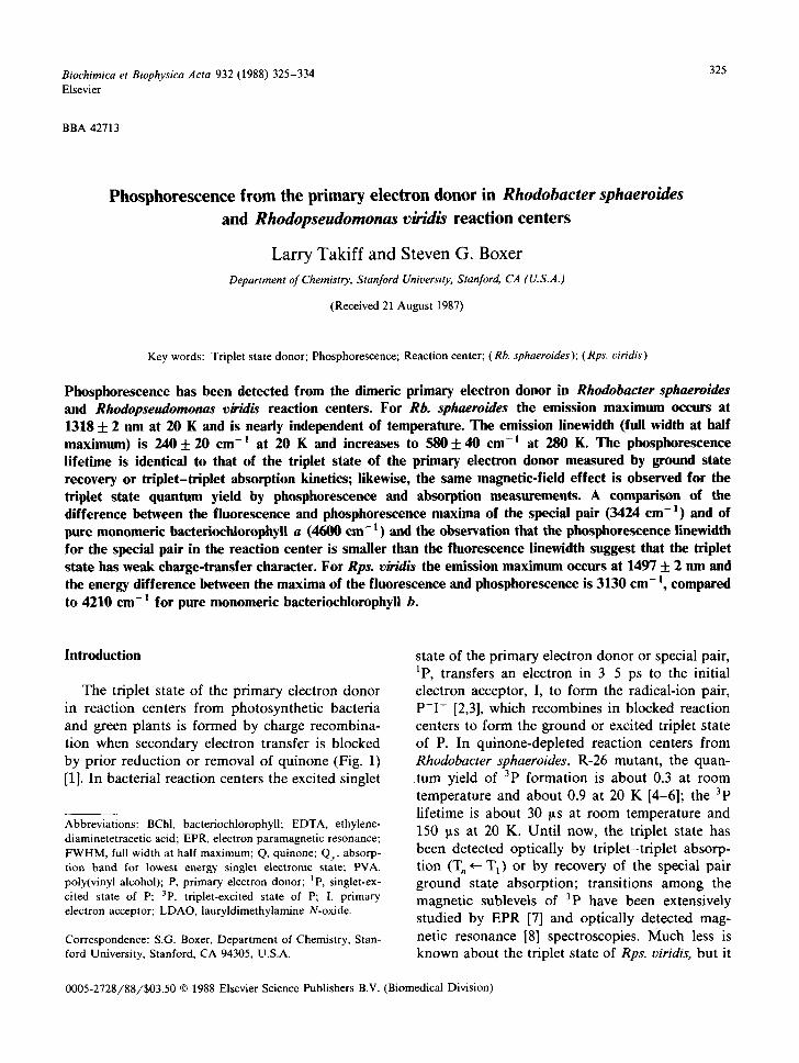
P 1 Fig. 1. Reaction scheme describing the intermediates, kinetics and energetics for the initial photochemistry in Q-depleted reaction centers from Rb. sphaeroides. P is the primary electron donor, consisting of two bacteriochlorophylls; I is the initial
electron acceptor which is a bacteriopheophytin.
appears to be similar te that of Rb. sphaeroides. Direct absorption from the ground state (T 1 ~- S 0) or phosphorescent emission (T 1 ~ So) have not been observed either from reaction centers or pure bacteriochlorophylls, presumably because of the very low cross-section for excitation, the very low emission quantum yield, and the difficulty of working in a wavelength region further into the infrared than can be detected with conventional photomultiplier tubes. Recent advances in near infra-red detectors, notably ultra-high sensitivity cooled germanium detectors, now make it possible to make measurements in the 1000-1700 nm re- gion with high sensitivity. Using such a device, we have been able to detect phosphorescence from the primary electron donor in Rb. sphaeroides and Rps. viridis reaction centers and from pure bacteriochlorophylls a and b (Takiff, L. and Boxer, S.G., unpublished data). A preliminary account of this work was presented earlier [9].
Although the triplet state is not formed in appreciable yield in functioning photosynthetic reaction centers, its properties in blocked reaction centers have proven to be to very useful for under- standing the mechanism of the initial charge sep- aration steps. Until now only the absolute energy of the singlet state was known. The energy of the 3p state places a lower limit on the energy of the initial charge-separated product state, P-~I~; the energy of PeI ~ provides the fundamental in- formation which is needed to analyze the energet- ics of photoconversion in photosynthesis. For Rb. sphaeroides the free energy of the 3p state has been estimated by measuring the temperature de-
pendence of the amplitude of the delayed fluores- cence component whose lifetime is comparable with that of the triplet state [10]. Delayed fluores- cence measurements are very valuable for estimat- ing the energies of a variety of reactive in- termediates [11,12], but there has been no inde- pendent check of their reliability. Furthermore, this method is based on several assumptions, mak- ing an independent measurement of the P+I ~ en- ergy desirable. We have shown that the energy of P*I ~ can also be obtained by measuring activated reformation of P*I ~ from 3p (Ref. 13; see also Goldstein, R.A., Takiff, L. and Boxer, S.G., un- pubhshed data). Although the energy difference between P+I ~ and 3p can be estimated quite accu- rately from such measurements, the absolute en- ergy of P t I ~ obtained by this approach is only as good as the value for energy of 3p. Finally, the nature of the 3p state itself has been the subject of much discussion, especially in comparison with the properties of the first singlet-excited state and radical cation of P. In particular, the relative contributions of charge-transfer and exciton inter- actions in the excited states of dimeric P have been the subject of several theoretical analyses [14-17]. As shown in the following sections, the phosphorescence spectrum of 3p provides infor- mation which bears on each of these issues.
Experimental
QA-containing reaction centers of the R-26 mutant of Rb. sphaeroides were prepared by standard methods [18]; quinone depletion was performed on an ion-exchange column [19]. Reac- tion centers in 10 mM Tris-HC1 (pH 8.0) 0.1% lauryldimethylamine N-oxide, 0.1 mM EDTA were embedded in poly(vinyl alcohol) films (PVA, Al- drich, average M r 125000). Films used for phos- phorescence experiments each had an absorbance of about 2 at 803 nm and two or three were stacked (reabsorption is not an issue in these measurements); those used for fluorescence or transient absorption were about one-tenth as con- centrated. Q-depleted reaction centers in films were determined to be more than 99% Q-depleted by transient absorption kinetics; Q-containing re- action centers in films were more than 90% Q g - containing. Aqueous samples were prepared using
327
0.1% Triton X-100 or sodium cholate as the deter- gent, 67% (v/v) glycerol, and were deoxygenated by repeated cycles of partial evacuation followed by addition of Ar. About 1 mg solid sodium dithionite was then added and the samples were transferred anaerobically to a quartz cuvette which was sealed under vacuum.
Rps. viridis reaction centers were prepared by a procedure similar to that for Rb. sphaeroides ex- cept that a sucrose density gradient centrifugation replaced the second extraction with LDAO (Middendorf, D., personal communication). The reaction centers were exchanged into deuterium oxide with 10 mM Tris-HCl (pH 8.0), 0.1% LDAO, 0.1 mM EDTA, diluted to 67% glycerol-d3, and deoxygenated by partial evacuation followed by backfilling with argon. The quinone was pre-re- duced by cooling the sample to 77 K in the presence of 20 mM sodium ascorbate (pH 8.0), while under cw illumination from a xenon lamp (approx. 200 mW/cm 2, absorbance at 830 nm about 3 in a 3 nun quartz cuvette held in an optical dewar). Quantitative quinone reduction and triplet formation under these conditions was veri- fied by transient absorption spectroscopy.
Transient absorption kinetics were measured by monitoring the change in absorption at 868 nm for Rb. sphaeroides or 752.5 nm for Rps. viridis after subsaturating 8 ns excitation flashes (532 nm for Rb. sphaeroides, 608 nm for Rps. viridis, 10 Hz) with a time resolution of about 1 ~ts. Triplet lifetimes and relative triplet quantum yields were obtained by fitting the decay data to a single exponential plus a baseline.
A time-resolved luminescence spectrometer was built around an ultra-sensitive liquid nitrogen cooled germanium photodiode detector (Model 403L, ADC Corp., Fresno, CA). Film samples were squeezed against a gold-coated copper sam- ple holder with a quartz fiat. The copper sample holders were attached to the cold finger of a closed-cycle helium refrigerator whose tempera- ture could be varied between 20 and 280 K. Aqueous samples in quartz cuvettes were held at 77 K in a liquid nitrogen optical dewar. Phos- phorescence was observed by exciting samples with about 80% saturating 8 ns light pulses at 532 nm for Rb. sphaeroides or 608 nm for Rps. viridis from a Q-switched Nd : YAG or YAG-pumped dye laser
operating at 10 Hz. For some experiments, the dye laser output was Raman shifted with high pressure H 2 gas to 850-900 nm. Bandpass filters (RT-830, Hoya Corp.) allowed only the first Stokes-Raman lines to excite the sample. A spherical mirror focussed luminescence from the sample onto the entrance slit of a monochromator equipped with a 1 ~m blazed, 600 groove/mm grating (bandwidth 19 nm, linewidth measurements were corrected for this bandwidth). The output of the monochroma- tor was focussed through an 850 nm long-pass filter onto the detector. The optics were aligned using the prompt fluorescence of reaction centers. The output of the detector (time constant, approx. 1 ms) was processed with a boxcar averager (gate duration, 1 ms) and digitized in a minicomputer.
In order to discriminate the red edge of the relatively intense fluorescence (completely de- cayed by about 30 ns [11]) from the more slowly decaying and very weak phosphorescence (lifetime on the order of 100 ~s, vide infra), a light chopper was mounted in front of the monochromator en- trance slit. The synchronous output signal from the chopper was divided in frequency to about 10 Hz, delayed by an adjustable time, and used to trigger the pulsed laser and the boxcar. By adjust- ing the delay, the time between the laser pulse and when the chopper wheel unblocked the detection system could be varied. To record phosphores- cence spectra, this delay was set so that the laser pulse (and the prompt fluorescence) occurred just prior to the unblocking of the slit by the chopper wheel. To measure luminescence kinetics, the de- lay was increased further in set amounts, and the luminescence signal was recorded at each delay time value *
The method used to measure the luminescence decay kinetics can be modeled as follows. The edge of the chopper wheel opening is taken to reach the monochromator slit in the variable delay time, ~'d, relative to the laser flash, and uncovers it linearly in time T u (the slit remains uncovered during the entire subsequent decay). The detector puts out a signal whose height is proportional to the time integral of the intensity of the lumines-
* While Ge detectors with a faster time response are available, they are not sensitive enough to detect the very weak luminescence described in this paper.
328
cence that strikes it; the boxcar output signal, S, is proportional to the height of the detector signal. Then:
(Z? Z? S = A -- e - k ' d t + d t + B T u + .r u
= .4 ( l _ e _ k ¢ u ) e _ k ¢ a + B k2.ru
where A and B are constants, and k is the luminescence decay rate constant. The signal mea- sured in this manner should be a single exponen- tial to a baseline when plotted against the variable delay time T d, and the decay rate of this signal is the decay rate of the luminescence. An important requirement of this method is that the chopper speed be sufficiently high that the wheel moves from completely occluding to completely exposing the detection system before the luminescence has decayed away. The chopper used for these experi- ments (PAR Model 192) operated at 100 rps with an effective radius of 10 cm and could be swept past a 2.5 mm slit in about 40 I~s, which was thus the system's time response for this size slit.
The response of the detection system varied with wavelength. This response was corrected by measuring the spectrum of a standard lamp which was treated as a blackbody radiation source, and by dividing all spectra by the system response curve. The correction was small in the region in which the phosphorescence was observed. The germanium detector has a high cross section for absorption of gamma rays; these appear as ran- dom spikes in the spectrum that decay with the time constant of the boxcar. Since they are easily identified, they were removed from the spectra manually. The magnetic field effect on the luminescence intensity was measured by setting the monochromator (the bandwidth was 38 nm for these measurements) to the signal peak and aver- aging the luminescence signal for 128 s. The field was then set to zero and the measurement was repeated; dividing the signal measured at a given field by that measured at zero field yielded a relative luminescence intensity at that field. Aver- ages and standard deviations of four repetitions at each field were calculated. A small correction was made for the baseline signal recorded far to the
red of the phosphorescence peak. Magnetic fields of up to 300 G were applied with a Helmholtz coil mounted outside the refrigerator.
Results
Rhodobacter sphaeroides A luminescence signal was detected to the red
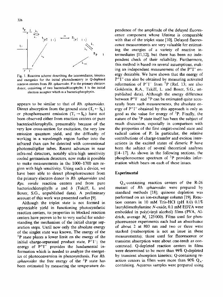
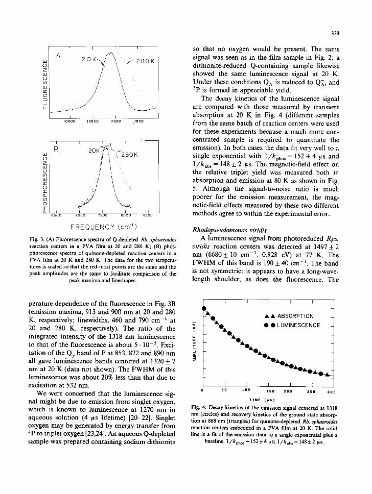
of the fluorescence for quinone-depleted Rb. sphaeroides reaction centers. The observed signal at 20 K is shown in Fig. 2; it has an emission maximum of 1318 + 2 nm (7589 + 10 cm -1, 0.941 eV) and full width at half maximum (FWHM) of 240 + 20 cm -1. Also shown in Fig. 2 is the ab- sence of luminescence in this wavelength region from a quinone-containing sample under identical conditions. For the latter sample 3p is not formed because electron transfer from I ~ to QA is much faster than charge recombination between P* and I ~. The luminescence signal for Q-depleted reac- tion centers was measured as a function of tem- perature; spectra at 20 and 280 K are compared in Fig. 3B. The luminescence signal is much smaller at higher temperatures because the quantum yield of formation of 3p is lower, the triplet lifetime is considerably shorter, and the band broadens. As seen in Fig. 3B, the peak maximum of the luminescence does not change appreciably with temperature (1307 + 4 nm at 280 K), while the luminescence linewidth (FWHM) increases from 240 + 20 cm -1 at 20 K to 580 + 40 cm -1 at 280 K. It is also seen that the line broadening is not symmetric. This result is compared with the tem-
,,,,oo , ,s,oo nm,
> -
z_ I I I
7000 7500 8000
FREQUENCY (cm -I) Fig. 2. Luminescence spectrum of quinone-depleted Rb. sphaeroides reaction centers in a PVA film at 20 K compared with that of a comparably concentrated quinone-containing
reaction center film taken under identical conditions.
W (..) Z (_.) (.0 I.J r'r" 0 ED _1 I J_
I I I I
L I I I I 0 0 0 0 1 0 5 0 0 I 1 0 0 0 1 1 5 0 0
Z IJ
CO Ld n ~ 0 q-
O_ CO 0 -I- O_
I I I I I
B i '~;''ii~ K
I I I I I 6500 7000 7500 8000 8500
FREQUENCY (cm-') Fig. 3. (A) Fluorescence spectra of Q-depleted Rb. sphaeroides reaction centers in a PVA film at 20 and 280 K; (B) phos- phorescence spectra of quinone-depleted reaction centers in a PVA film at 20 K and 280 K. The data for the two tempera- tures is scaled so that the red-most points are the same and the peak amplitudes are the same to facilitate comparison of the
peak maxima and lineshapes.
329
so that no oxygen would be present. The same signal was seen as in the film sample in Fig. 2; a dithionite-reduced Q-containing sample likewise showed the same luminescence signal at 20 K. Under these conditions QA is reduced to QA, and 3p is formed in appreciable yield.
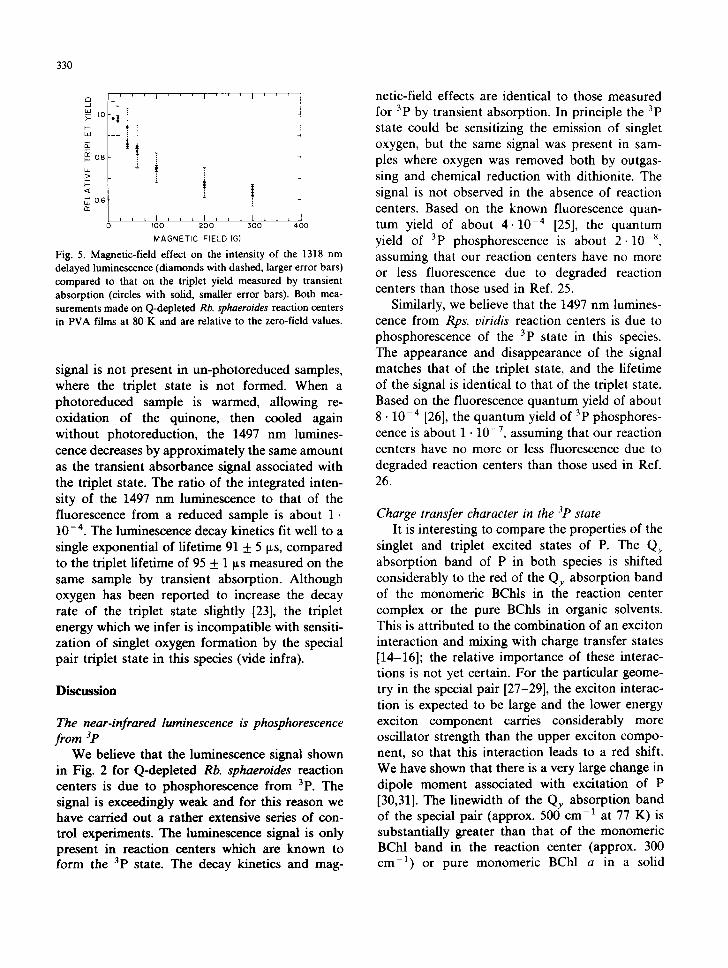
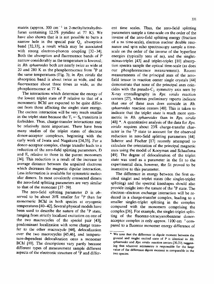
The decay kinetics of the luminescence signal are compared with those measured by transient absorption at 20 K in Fig. 4 (different samples from the same batch of reaction centers were used for these experiments because a much more con- centrated sample is required to quantitate the emission). In both cases the data fit very well to a single exponential with l / / kphos = 152 _+ 4 ~ts and l / kab s = 148 _+ 2 ~S. The magnetic-field effect on the relative triplet yield was measured both in absorption and emission at 80 K as shown in Fig. 5. Although the signal-to-noise ratio is much poorer for the emission measurement, the mag- netic-field effects measured by these two different methods agree to within the experimental error.
Rhodopseudomonas oiridis A luminescence signal from photoreduced Rps.
viridis reaction centers was detected at 1497 _+ 2 nm (6680_+ 10 cm -1, 0.828 eV) at 77 K. The F W H M of this band is 190 _+ 40 cm -1. The band is not symmetric: it appears to have a long-wave- length shoulder, as does the fluorescence. The
perature dependence of the fluorescence in Fig. 3B (emission maxima, 913 and 900 nm at 20 and 280 K, respectively; linewidths, 460 and 790 cm -1 at 20 and 280 K, respectively). The ratio of the integrated intensity of the 1318 nm luminescence to that of the fluorescence is about 5 • 10-5. Exci- tation of the Qy band of P at 853, 872 and 890 nm all gave luminescence bands centered at 1320 + 2 nm at 20 K (data not shown). The FWHM of this luminescence was about 20% less than that due to excitation at 532 nm.
We were concerned that the luminescence sig- nal might be due to emission from singlet oxygen, which is known to luminescence at 1270 nm in aqueous solution (4 ~ts lifetime) [20-22]. Singlet oxygen may be generated by energy transfer from 3p to triplet oxygen [23,24]. An aqueous Q-depleted sample was prepared containing sodium dithionite
l I I I I I t
- ~ • • ABSORPTION _
I [ I I 1 5 0 1 0 0 1 5 0 2 0 0 2 5 0 3 0 0
T t M E ( p s )
Fig. 4. Decay kinetics of the emission signal centered at 1318. n m (circles) and recovery kinetics of the ground state absorp- tion at 868 n m (triangles) for quinone-depleted Rb. sphaeroides reaction centers embedded in a PVA film at 20 K. The solid line is a fit of the emission data to a single exponential plus a
baseline. 1/kpho~ = 152 + 4 ~s; 1 / k . b ~ = 148 + 2 ~s.
330
i i i I '
, o
- i b-
~ 0 . 6
i L L 0
I ' f I
T
!
I * , , I , ~ , I J I 0 0 2 0 0 3 0 0
MAGNETIC F IELD(G)
L i 4OO
Fig. 5. Magnetic-field effect on the intensity of the 1318 nm delayed luminescence (diamonds with dashed, larger error bars) compared to that on the triplet yield measured by transient absorption (circles with solid, smaller error bars). Both mea- surements made on Q-depleted Rb. sphaeroides reaction centers in PVA films at 80 K and are relative to the zero-field values.
signal is not present in un-photoreduced samples, where the triplet state is not formed. When a photoreduced sample is warmed, allowing re- oxidation of the quinone, then cooled again without photoreduction, the 1497 nm lumines- cence decreases by approximately the same amount as the transient absorbance signal associated with the triplet state. The ratio of the integrated inten- sity of the 1497 nm luminescence to that of the fluorescence from a reduced sample is about 1. 10 -4. The luminescence decay kinetics fit well to a single exponential of lifetime 91 + 5 I~s, compared to the triplet lifetime of 95 + 1 ~ts measured on the same sample by transient absorption. Although oxygen has been reported to increase the decay rate of the triplet state slightly [23], the triplet energy which we infer is incompatible with sensiti- zation of singlet oxygen formation by the special pair triplet state in this species (vide infra).
D i s c u s s i o n
The near-infrared luminescence is phosphorescence from ~P
We believe that the luminescence signal shown in Fig. 2 for Q-depleted Rb. sphaeroides reaction centers is due to phosphorescence from 3p. The signal is exceedingly weak and for this reason we have carried out a rather extensive series of con- trol experiments. The luminescence signal is only present in reaction centers which are known to form the 3p state. The decay kinetics and mag-
netic-field effects are identical to those measured for 3p by transient absorption. In principle the 3p state could be sensitizing the emission of singlet oxygen, but the same signal was present in sam- ples where oxygen was removed both by outgas- sing and chemical reduction with dithionite. The signal is not observed in the absence of reaction centers. Based on the known fluorescence quan- tum yield of about 4 . 1 0 - 4 [25], the quantum yield of 3p phosphorescence is about 2 -10 -~, assuming that our reaction centers have no more or less fluorescence due to degraded reaction centers than those used in Ref. 25.
Similarly, we believe that the 1497 nm lumines- cence from Rps. oiridis reaction centers is due to phosphorescence of the 3p state in this species. The appearance and disappearance of the signal matches that of the triplet state, and the lifetime of the signal is identical to that of the triplet state. Based on the fluorescence quantum yield of about 8 • 10 - 4 [26], the quantum yield of 3p phosphores- cence is about 1 • 10 -7, assuming that our reaction centers have no more or less fluorescence due to degraded reaction centers than those used in Ref. 26.
Charge transfer character in the 31) state It is interesting to compare the properties of the
singlet and triplet excited states of P. The Qy absorption band of P in both species is shifted considerably to the red of the Q y absorption band of the monomeric BChls in the reaction center complex or the pure BChls in organic solvents. This is attributed to the combination of an exciton interaction and mixing with charge transfer states [14-16]; the relative importance of these interac- tions is not yet certain. For the particular geome- try in the special pair [27-29], the exciton interac- tion is expected to be large and the lower energy exciton component carries considerably more oscillator strength than the upper exciton compo- nent, so that this interaction leads to a red shift. We have shown that there is a very large change in dipole moment associated with excitation of P [30,31]. The linewidth of the Qy absorption band of the special pair (approx. 500 cm-a at 77 K) is substantially greater than that of the monomeric BChl band in the reaction center (approx. 300 cm -1) or pure monomeric BChl a in a solid
331
matrix (approx. 300 cm-1 in 2-methyltetrahydro- furan containing 12.5% pyridine at 77 K). We have also shown that it is not possible to burn a narrow hole in the special pair Qy absorption band [32,33], a result which may be associated with strong electron-phonon coupling [32-34]. Both the absorption and fluorescence bands of P narrow considerably as the temperature is lowered; in Rb. sphaeroides both are nearly twice as wide at 20 and 280 K as the phosphorescence from 3p at the same temperatures (Fig. 3); in Rps. viridis the absorption band is about twice as wide, and the fluorescence about three times as wide, as the phosphorescence at 77 K.
The interactions which determine the energy of the lowest triplet state of P relative to that of a monomeric BChl are expected to be quite differ- ent from those affecting the singlet state energy. The exciton interaction will be very much smaller in the triplet state because the T~ ~ S O transition is forbidden. Thus, charge-transfer interactions may be relatively more important. There have been many studies of the triplet states of electron donor-acceptor complexes, beginning with the early work of Iwata and Nagakura [34,35]. For a donor-acceptor complex, charge transfer leads to a reduction of the zero-field splitting parameters, D and E, relative to those in the parent monomers [36]. This reduction is a result of the increase in average distance between the unpaired electrons which decreases the magnetic dipolar interaction. Less information is available for symmetric molec- ular dimers. In most covalently connected dimers the zero-field splitting parameters are very similar to that of the monomer [37-39].
The zero-field splitting parameter D is ob- served to be about 20% smaller for 3p than for monomeric BChl in both species at cryogenic temperatures [40-42]. Several physical models have been used to describe the nature of the 3p state, ranging from strictly localized excitation on one of the two macrocycles of the special pair [43], predominant localization with some charge trans- fer to the other macrocycle [44], delocalization over the two macrocycles [45,46], and tempera- ture-dependent delocalization onto a monomer BChl [10]. The descriptions vary partly because different types of measurement sample different aspects of the electronic structure of 3p and differ-
ent time scales. Thus, the zero-field splitting parameters sample a time-scale on the order of the inverse of the zero-field splitting energy (fraction of a ns time-scale), electron nuclear double reso- nance and spin echo spectroscopy sample a time- scale on the order of the inverse of the hyperfine energies (typically tens of ns), and the singlet- minus-triplet [43] and triplet-triplet [10] absorp- tion spectra sample the optical time-scale (as does our phosphorescence measurement). Recent measurements of the principal axes of the zero- field tensor in reaction center single crystals [44] demonstrate that none of the principal axes coin- cides with the pseudo-C 2 symmetry axis seen by X-ray crystallography in Rps. viridis reaction centers [27], whereas preliminary reports indicate that one of these axes does coincide in Rb. sphaeroides reaction centers [48]. This is taken to indicate that the triplet state is much more sym- metric in Rb. sphaeroides than in Rps. viridis [48] *. A quantitative analysis of the data for Rps. viridis requires about 23% charge-transfer char- acter in the 3p state to account for the observed reduction in zero-field splitting parameters [44]. Scherer and Fischer [17] recently attempted to calculate the orientation of the principal magnetic axes using the model of Kooyman and Schaafsma [49]. The degree of delocalization of the triplet state was used as a parameter in the fit to the experimental data, however, the fit proved to be insensitive to this parameter.
The difference in energy between the first ex- cited singlet and triplet states (the singlet-triplet splitting) and the spectral lineshapes should also provide insight into the nature of the 3p state. The electron-electron exchange interaction will be re- duced in a charge-transfer complex, leading to a smaller singlet-triplet splitting in the complex compared with the monomers comprising the complex [36]. For example, the singlet-triplet split- ting of the fluorene-tetracyanobenzene donor- acceptor complex is only approx. 1100 cm-~ com- pared to a fluorene monomer energy difference of
* We note that the difference in dipole moment between the ground and singlet excited state of P is identical in Rb. sphaeroides and Rps. viridis reaction centers [30,31], suggest- ing that whatever asymmetry is responsible for the large value of the difference dipole moment is comparable in the two species.
332
9300 cm-1; the reduction in zero-field splitting parameters also indicates substantial charge-trans- fer character, estimated to be about 70% [50]. It has also been found that the phosphorescence lines of complexes possessing a substantial degree of charge-transfer character are considerably broader than those whose excited states are rela- tively more neutral [36]. This is rationalized by the much larger interaction with the solvent for a polar excited state, a result which is well known for polar singlet excited states (e.g., the l p state).
We find that the energy difference between the maxima of the fluorescence and phosphorescence of P is 3290 cm-1 for Rb. sphaeroides at 20 K and 3130 cm -1 for Rps. viridis at 77 K. These values are not identical to the singlet-triplet splitting because they are not corrected for possible dif- ferences in the Stokes shift between absorbance and fluorescence vs. T 1 ~ S O absorption and phos- phorescence. However, we expect such differences to be small (up to 200 cm-1). These energy dif- ferences are each about 30% smaller than those measured for the pure monomers which comprise P: 4580 cm -1 for bacteriochlorophyll a, 4210 cm -1 for bacteriochlorophyll b (six-coordinate complexes ) (Takiff, L. and Boxer, S.G., unpub- lished data), or chlorophyll a (approx. 4700 cm- 1) [51,52]. This reduction in singlet-triplet splitting is consistent with some charge-transfer character in the 3p state. The reduction is very similar for the two species, suggesting a similar degree of charge- transfer in each. This observation contradicts the hypothesis mentioned earlier that the triplet is much more localized in Rps. viridis than in Rb. sphaeroides reaction centers [44].
We also observe, however, that the 3p phos- phorescence linewidth is substantially narrower in both species than the singlet absorption or fluores- cence (Fig. 3) which suggests a substantially weaker interaction of the triplet state than the singlet state with the environment. Interestingly, the Rb. sphaeroides phosphorescence linewidth is quite similar to that for pure BChl a, whose singiet absorption, fluorescence and phosphorescence linewidths are all about 300 cm -1 (measured in 2-methyltetrahydrofuran/pyridine at 77 K) (Takiff, L. and Boxer, S.G., unpublished data). The phosphorescence linewidth of Rps. oiridis is only about half of that measured for pure BChl b.
Temperature and excitation wavelength dependence in Rb. sphaeroides
In contrast to the absorption and fluorescence of P, we find that the 3p phosphorescence maxi- mum does not change appreciably with tempera- ture in Rb. sphaeroides (Fig. 3). The energy dif- ference between the maxima of fluorescence and phosphorescence increases by only 160 cm -1 be- tween 20 and 280 K. Thus, it is very unlikely that the degree of delocalization of this state is strongly temperature dependent [10,47]. The origin of the change of the absorption and fluorescence with temperature is not fully understood; it may reflect strong electron-phonon coupling in 1p [53], a re- sult which we and others have argued is consistent with photochemical holeburning data [32-34]. The much smaller shift in the phosphorescence with temperature thus argues for much weaker elec- tron-phonon coupling in the triplet state than in the singlet state. Finally, we note that a small (20%) phosphorescence line narrowing was ob- served when P was excited directly in its Qy absorption band compared with higher energy ex- citation. Phosphorescence line narrowing has been reported following direct excitation of the triplet state of simple aromatic molecules [54], but is quite rare following excitation of the singlet state [55]. The existence of some degree of correlation between the site energies of the singiet and triplet states was suggested by Den Blanken and Hoff [43] in absorbance-detected magnetic resonance studies. They concluded that there is a small inho- mogeneous broadening of both the singlet state and the zero-field splitting values in the triplet state, that these were at least partly due to the same mechanism and, thus, might be correlated.
Charge-separation energetics in Rb. sphaeroides Based on the discussion above concerning the
degree of charge-transfer character in the 3p state, it is likely that the Stokes shift for the triplet state is at most as large as that for the singlet state and is probably considerably smaller *. Adding one half of the approx. 570 cm -1 Stokes shift in the
* In principle it should be possible to measure the phos- phorescence excitation spectrum; however, the absorption cross section is expected to be incredibly small, and it is difficult to obtain tunable high intensity excitation light in the 1000-1400 nm range.
singlet state (quinone-depleted reaction centers in PVA at 77 K) to the energy of the phosphores- cence maximum (7589 cm -1) gives a triplet state energy of 7874 cm-1 or 0.98 eV; if the Stokes shift were 150 cm -1 (as it is for the singlet excited state of pure BChl a), the value is 7664 cm -1 (0.95 eV). This energy agrees reasonably well with that estimated by Parson and Shuvalov from delayed fluorescence measurements (approx. 8300 cm -1 [10]).
We have recently determined the standard free energy difference between the 3p state and the radical pair states in Rb. sphaeroides reaction centers (Ref. 13; see also Goldstein, R.A., Takiff, L. and Boxer, S.G., unpublished data). Based on data taken at a magnetic field of 135 kG, we obtained a standard free-energy difference at room temperature of 1360 + 40 cm -1. In combination with the 3p free energy of 7500 cm -~ (the energy of about 7730 cm-1 minus kT In 3 = 230 cm-1 to account for the spin multiplicity of the triplet state) determined here and the ~P energy of 11220 cm-1 (from absorption and fluorescence measure- ments), we conclude that the free energy of the P*I ~ state is 9090 cm -1 and the standard free energy change for the initial charge separation step in Rb. sphaeroides is 2130 cm -1 (0.264 eV).
Acknowledgements
We wish to thank Louis Wang and Eric Miller of the ADC Corporation, Fresno, CA, for gener- ously lending us the germanium detector used for these experiments, and PAR for the loan of the high-speed chopper. This work was supported in part by a grant from the National Science Foun- dation (DMB8607799) and a Presidential Young Investigator Award to S.G.B.
References
1 Dutton, P.L., Leigh, J.S. and Reed, D.W. (1973) Biochim. Biophys. Acta 292, 654-664.
2 Woodbury, N.W., Becker, M., Middendorf, D. and Parson, W.W. (1985) Biochemistry 24, 7516-7521.
3 Breton, J., Martin, J.L., Migus, A., Antonetti, A. and Oszag, A. (1986) Proc. Natl. Acad. Sei. USA 83, 5121-5125.
4 Parson, W.W., Clayton, R.K. and Cogdeil, R.J. (1985) Biochim. Biophys. Acta 38, 265-278.
5 Chidsey, C.E.D., Kirmaier, C., Holten, D. and Boxer, S.G. (1984) Biochim. Biophys. Acta 766, 424-437.
333
6 Schenck, C.C., Mathis, P. and Lutz, M. (1984) Photochem. Photobiol. 39, 407-417.
7 Hoff, A.J. (1979) Physics Rep. 54, 75-200. 8 Hoff, A.J. (1982) in Triplet State ODMR Spectroscopy
(Clarke, R.H., ed.), pp. 367-425, John Wiley and Sons, New York.
9 Takiff, L. and Boxer, S.G. (1987) Photochem. Photobiol. 45, 61S (abstract).
10 Shuvalov, V.A. and Parson, W.W. (1981) Proc. Natl. Acad. Sci. USA 78, 957-961.
11 Woodbury, N.W. and Parson, W.W. (1984) Biochim. Bio- phys. Acta 767, 345-361.
12 Arata, H. and Parson, W.W. (1981) Biochim. Biophys. Acta 638, 201-209.
13 Chidsey, C.E.D., Takiff, L., Goldstein, R.A. and Boxer, S.G. (1985) Proc. Natl. Acad. Sci. USA 82, 6850-6854.
14 Warshel, A. and Parson, W.W. (1987) J. Am. Chem. Soc., 6143-6152.
15 Parson, W.W. and Warshel, A. (1987) J. Am. Chem. Soc., 6152-6163.
16 Knapp, E.W., Fischer, S.F., Zinth, W., Sander, M., Kaiser, W., Deisenhofer, J. and Michel, H. (1985) Proc. Natl. Acad. Sci. USA 82, 8463-8467.
17 Scherer, P.O.J. and Fisher, S.F. (1987) Chem. Phys. Lett. 136, 431-435.
18 Schenck, C.C., Biankenship, R.E. and Parson, W.W. (1982) Biochim. Biophys. Acta 680, 44-49.
19 Okamura, M.Y., Issacson, R.A. and Feher, G. (1975) Proc. Natl. Acad. Sci. USA 72, 3491-3495.
20 Krasnofsky, A.A., Jr. (1976) Biofizika 21,748-749. 21 Rogers, M.A.J. (1983) J. Am. Chem. Soc. 105, 6201-6205. 22 Hall, R.D. and Chignell, C.F. (1987) Photochem. Photo-
biol. 45, 459-464. 23 Shuvalov, V.A. and Parson, W.W. (1981) Biochim. Biophys.
Acta 638, 50-59. 24 Boucher, F., Van der Rest, M. and Gingras, G. (1977)
Biochim. Biophys. Acta 461, 339-357. 25 Zankel, K.L., Reed, D.W. and Clayton, R.K. (1968) Proc.
Natl. Acad. Sci. USA 61, 1243-1249. 26 Horber, J.K.H., Gobel, W., Ogrodnik, A., Michei-Beyerle,
M.E. and Cogdell, R.J. (1986) FEBS Lett. 198, 268-272. 27 Deisenhofer, J., Epp, O., Miki, K., Huber, R. and Michel,
H. (1984) J. Mol. Biol. 180, 385-398. 28 Chang, C.H., Tiede, D., Tang, J., Smith, U., Norris, J. and
Schiffer, M. (1986) FEBS Lett. 205, 82-86. 29 Allen, J.P., Feher, G., Yeates, T.O., Komiya, H. and Rees,
D.C. (1987) Proc. Natl. Acad. Sci. USA 84, 5730-5734. 30 Lockhart, D.J. and Boxer, S.G. (1987) Biochemistry 26,
664-668. 31 Lockhart, D.J. and Boxer, S.G. (1987) Proc. Natl. Acad.
Sci. U.S.A., in press. 32 Boxer, S.G., Lockhart, D.J. and Middendorf, T.R. (1986)
Chem. Phys. Lett. 123, 476-482. 33 Boxer, S.G., Middendorf, T.R. and Lockhart, D.J. (1986)
FEBS Lett. 200, 237-241. 34 Hayes, J.M. and Small, G.J. (1986) J. Phys. Chem. 90,
4928-4931. 35 Iwata, S., Tanaka, J. and Nagakura, S. (1967) J. Chem.
Phys. 47, 2203-2209.
334
36 Nagakura, S. (1975) in Excited States (Lim, E., ed.), Vol. 2, pp. 321-383, Academic Press, New York.
37 Schweitzer, D., Colpa, J.P., Behnke, J., Hausser, K.H., Haenel, M. and Staab, H.A. (1975) Chem. Phys. 11, 373-384.
38 Clarke, R.H., Hobart, D.R. and Leenstra, W.R. (1979) J. Am. Chem. Soc. 101, 2416-2423.
39 Bucks, R.R. and Boxer, S.G. (1982) J. Am. Chem. Soc. 104, 340-343.
40 Leigh, J.S., Jr. and Dutton, P.L. (1974) Biochim. Biophys. Acta 357, 67-77.
41 Levanon, H. and Norris, J.R. (1978) Chem. Rev. 78, 185-198.
42 Den Blanken, H.J., Jongenelis, A.P.J.M. and Hoff, A.J. (1983) Biochim. Biophys. Acta 725, 472-482.
43 Den Blanken, H.J. and Hoff, A.J. (1982) Biochim. Biophys. Acta 681, 365-374.
44 Norris, J.R., Lin, C.P. and Budil, D.E. (1987) J. Chem. Soc. Faraday Trans. 83, 12-27.
45 Lendzian, F., van Willigen, H., Sastry, S., Mobius, K., Scheer, H. and Feick, R. (1985) Chem. Phys. Lett. 118, 145-150.
46 De Groot, A., Evelo, R., Hoff, A.J., De Beer, R. and Scheer, H. (1985) Chem. Phys. Lett. 118, 48-54.
47 Hoff, A.J. and Proskuryakov, 1.I. (1985) Chem. Phys. Lett. 115, 303-310.
48 Norris, J.R., Budil, D.E., Chang, C. and Schiffer, M. (1987) Photochem. Photobiol. 45, 275 (abstract).
49 Kooyman, R.P.H. and Schaafsma, T.J. (1980) J. Mol. Struct. 60, 373-380.
50 Miihle, W., Krzystek, J., Von Schutz, J.U., Wolf, H.C., Stigler, R.D. and Stezowski, J.J. (1986) Chem. Phys. 108, 1-13.
51 Mau, A.W.H. and Puza, M. (1977) Photochem. Photobiol. 25, 601-603.
52 Mauring, K., Suisalu, A.L., Avarmaa, R.A. and Kras- novskii, A.A., Jr. (1980) Doklady Akademii Nauk SSSR, 251,729-731.
53 Scherer, P.O.J., Fischer, S.F., HiSrber, J.K.H., Michel-Be- yerle, M.E. and Michel, H. (1985) in Antennas and Reac- tion Centers of Photosynthetic Bacteria (Michel-Beyerle, M.E., ed.), pp. 131-137, Springer-Verlag, Berlin.
54 Williamson, R.L. and Kwiram, A.L. (1979) J. Phys. Chem. 83, 3393-23397.
55 Suter, G.W., Wild, U.F. and Hotzwarth, A.R. (1986) Chem. Phys. 102, 205-214.
56 Van Egmond, J., Kohler, B.E. and Chan, I.Y. (1975) Chem. Phys. Lett. 34, 423-426.