Embed Size (px)
Citation preview

1401
CHAPTER
108Oxygen Therapy, Respiratory Care, and MonitoringVittoria Comellini, lara Pisani, and stefano naVa
InTROduCTIOn
Supplemental oxygen is one of the most widely used therapies for people admitted to the hospital and it is used across the whole range of specialties. Although oxygen therapy is used as a medical treatment in both acute and chronic setting, it is often administered improperly. The major problems consist in the fact that about this topic there are strongly held beliefs, but very few randomized controlled trials and frequently health care professionals receive conflicting advice about oxygen use from different “experts” during their clinical training and dur-ing their careers, and many are confused about the oxygen’s prescription and use. This chapter will give a comprehensive and practical overview of oxygen pathophysiologic effects, current modalities for its delivery, and monitoring in the criti-cal care setting. In order to give a more complete view about the management of patients with acute respiratory failure (ARF), we will also discuss the other respiratory monitoring techniques in the intensive care unit (ICU) and in specific cir-cumstances like during noninvasive ventilation (NIV).
Oxygen TheRapy
Oxygen makes up 21% of the atmosphere we breathe; in the air it exists in a diatomic molecular form (O2, molecular weight [MW] 16 g/mol) that is the state in which we admin-ister oxygen to our patients as a respiratory gas, either pure oxygen or in mixtures with air or helium (Heliox). While the gaseous state is most clinically relevant, oxygen can be found in liquid and solid states as well, under appropriate conditions.
Oxygen was not discovered as a separate gas until the late 18th century, when the Swedish apothecary, Karl W. Scheele in 1772, made a series of experiments with mercuric oxide and potassium nitrate and, heating these two elements, obtained a gas that caused candles to burn more brightly; he did not, however, rush to publication. In the same years, the English amateur chemist Joseph Priestley liberated oxygen by intensely heating mercurius calcinatus (mercuric oxide) placed over liquid mercury in a closed vessel; he called this new gas “dephlogisticated air” (1).
The first notice about the use of oxygen as a remedy in dis-ease dates back to 1870 (2). Although oxygen life-supporting role was understood, it took about 150 years for the gas to be used in a proper way: during the first 150 years after this discovery, the therapeutic use of oxygen was sporadic, erratic, and controversial.
Currently, oxygen is administered for three main indica-tions, but only one is evidence-based. First, oxygen is given to correct hypoxemia as there is good evidence that severe
hypoxemia is harmful. Second, oxygen is administered to ill patients in case they might become hypoxemic. Recent evi-dence suggests that this practice may actually place patients at increased risk of developing the toxic effects mediated by reactive oxygen species (ROS) (3), leading to tissue damage and absorption atelectasis. Third, a very high proportion of medical oxygen is administered because most clinicians believe that oxygen can alleviate breathlessness. However, there is no evidence that oxygen holds this beneficial effect in nonhypox-emic patients and there is evidence of lack of effectiveness in nonhypoxemic breathless patients with chronic obstructive pulmonary disease (COPD) and advanced cancer (4,5).
paThOphysIOlOgy
Respiration is the process that involves the exchange of oxygen and carbon dioxide (CO2) between the environment and a liv-ing organism. Oxygen is indispensable for the aerobic metabo-lism of cells, so it is essential for humans to maintain a safe level of this gas in the bloodstream. Several mechanisms exist to regulate breathing in such a way that it is maintained within quite a narrow range. Inside the lungs, oxygen passes from inspired air into the bloodstream, and its diffusion depends on alveolar oxygen pressure. Most of the oxygen in the blood is bound to a carrying protein contained in red blood cells and called hemoglobin, whereas, normally, only a small amount is dissolved in the blood itself. Under normal conditions, almost all of the oxygen-carrying capacity of hemoglobin in the blood is used when the oxygen saturation (SaO2) is in the normal range, 95% to 98%. Therefore, giving supplementary oxygen to a healthy young person will increase the saturation level only slightly from about 97% to 99% or a maximum of 100%, producing only a very small increase in amount of oxygen made available to the tissues.
Hypoxemia is the result of respiratory failure, a condition that leads to inadequate oxygen delivery (DO2) to the tissues (partial respiratory failure) and/or to inadequate removal of carbon dioxide (global respiratory failure). The most common form of hypoxemia occurs when there is sufficient oxygen-carrying capacity—in patients with a normal level of hemoglobin—but insufficient oxygen uptake in the lungs. This can be the result of poor ventilation of areas of lung or abnor-malities of gas exchange during illnesses such as pneumonia. This form of hypoxemia is the easiest to treat with oxygen therapy; on the contrary oxygen therapy is less effective in other situations, including anemia where there is a low car-rying capacity or intoxications where the carrying capacity of hemoglobin has been reduced by a toxic substance. For exam-ple, carbon monoxide (CO), by combining with hemoglobin
LWBK1580_C108_p1401-1409.indd 1401 29/07/17 11:54 AM

1402 SECTion 11 resPiratory DisorDers
to form carboxyhemoglobin (HbCO), blocks oxygen binding to hemoglobin despite having a normal level of oxygen in the lungs and in the blood.
Oxygen therapy increases alveolar oxygen (PAO2) and is, therefore, effective only when alveolar capillary units have some functional ventilation. In turn, it is ineffective if there is a pure shunt, such as pulmonary arteriovenous malformations, where mixed venous blood does not pass through an alveolar capillary unit. There will be, in this situation, a small overall increase in PaO2 due to an increase in dissolved oxygen in the pulmonary venous blood from ventilated alveolar capillary units, which is minor compared with the oxygen carried by hemoglobin. Despite this, there is good evidence that breath-hold times can be increased by breathing oxygen (6,7). The same principles are used to preoxygenate, actually denitrogenate, patients before intubation during anesthesia; it is thought that the additional breath-hold time is produced not by the mar-ginal increase in blood oxygen levels but by the increased reser-voir of oxygen in the lungs after breathing oxygen-enriched air. In poorly ventilated alveoli (i.e., low ventilation/perfusion ratio [V/Q]), PAO2 will be low and increasing FiO2 will increase PAO2 and therefore PaO2 (7). When there is a diffusion barrier due to increased alveolar capillary membrane thickness, such as in fibrotic lung disease (6), increasing PAO2 will augment the rate of diffusion across the alveolar capillary membrane by increasing the concentration gradient.
TReaTMenT
Oxygen represents the first line of treatment for hypoxemia in those patients who are not breathless, aiming to achieve normal or near-normal oxygen saturation. For critically ill patients with an oxygen saturation below the target, after having ascertained that the airway is clear, oxygen should be administered as soon as possible and the flow rates should be adjusted to keep the oxygen saturation (SpO2) in the target range, or at least above 90% (8,9). Indeed when prescribing oxygen therapy, the health care professional should indicate the target SpO2 range rather than prescribing a fixed dose of oxygen or FiO2. The clinician may indicate a starting dose, the device and the flow rate, but there needs to be an agreed-on system for adjusting the oxygen dose upward or downward according to the patient’s needs. It is important to consider that every requirement for an increased dose of oxygen is an indication for urgent clinical reassessment of the patient, repeating arterial blood gas (ABG) measurements in most instances. The SpO2 should be monitored continuously until the patient is stable and, as soon as ABG measurements are available, the patient’s further treatment should be guided by the results of this test.
Some subjects, especially those older than 70 years, may have SpO2 measurements below 94% and yet do not require oxygen therapy if clinically stable. Despite this, a reduction of more than 3% in a patient’s usual SpO2, even if it remains within the target range, may be the first evidence of an acute ill-ness. It is known that oxygenation is reduced in the supine posi-tion, so fully conscious hypoxemic patients should ideally be allowed to maintain the most upright posture possible, or the most comfortable posture for the patient, unless there are con-traindications (e.g., skeletal or spinal trauma, severe hypoten-sion). A wide variation in SpO2 characterizes sleep: all healthy
subjects in all age groups routinely have transient dips overnight with a mean saturation nadir of 90.4% (10). These decrements should be interpreted with caution, monitoring the subject for few minutes in order to determine whether the hypoxemia is sustained or just a transient, normal nocturnal dip.
There are several medical emergencies in which patients are likely to suffer from hypoxemia, including cardiac arrest, major trauma, shock, major sepsis, anaphylaxis, major pulmo-nary hemorrhage, massive hemoptysis, major head injury, and near-drowning. In all these conditions initial treatment should involve high-concentration oxygen, aiming at an SpO2 of 94% to 98% pending availability of satisfactory ABG measure-ments or until the airway is secured by endotracheal intuba-tion. High-concentration oxygen is additionally recommended during resuscitation and in case of CO poisoning, taking into account that in this condition patients have a normal level of PaO2 but a greatly reduced level of oxygen bound to hemo-globin because this has been displaced by the CO (11). Pulse oximetry cannot screen for CO exposure, as it does not dif-ferentiate carboxyhemoglobin from oxyhemoglobin and ABG analysis will show an apparently normal SaO2. The blood car-boxyhemoglobin level must be measured to assess the degree of CO poisoning. The half-life of carboxyhemoglobin in a patient breathing room air is approximately 300 minutes; this decreases to 90 minutes with high-concentration oxygen via a reservoir mask. The most important treatment for a patient with CO poisoning is, therefore, to give high-dose oxygen via a reservoir mask. Comatose patients or those with severe mental impairment should be endotracheally intubated and ventilated with 100% oxygen. The role of hyperbaric oxygen remains controversial (12,13).
Patients who present serious illness but are not critical or greatly hypoxic—pneumonia, acute asthma, lung cancer, interstitial lung disease, pneumothorax, pleural effusion, pul-monary embolism, acute heart failure with hypoxemia, stroke with hypoxemia, labor with hypoxemia, postoperative breath-lessness or hypoxemia, or severe anemia with breathlessness—can be treated with medium-dose oxygen therapy with a target SpO2 range of 94% to 98%. Generally, breathless and hypox-emic patients do not have a firm diagnosis at the time of pre-sentation to the hospital. For most acutely hypoxemic patients whose medical problem is not yet diagnosed, an SpO2 range of 94% to 98% will avoid the potential risks associated with hypoxemia or hyperoxia. Aiming for an SpO2 in the normal range will also have the effect of using the lowest effective FiO2, avoiding risks such as absorption atelectasis and V/Q mismatch. The priority for such patients is to make a specific diagnosis as early as possible and to institute specific treat-ment for the underlying condition. Early ABG measurement is mandatory in the management of patients with sudden unex-plained hypoxemia.
Patients with chronic lung disease may tolerate a low SpO2 chronically when clinically stable; however, these resting oxygen levels may not be adequate for tissue oxygenation during acute illness or in those conditions where oxygen demand increases. For those patients with COPD (14–22) or other known risk factors that can predispose to hypercapnic respiratory failure with acidosis—morbid obesity, chest wall deformities, or neuromuscular disorders, cystic fibrosis, severe lung scarring from old tuberculosis, overdose of respiratory depressant drugs—a target SpO2 range of 88% to 92% is suggested pending the availability of ABG results. For this
LWBK1580_C108_p1401-1409.indd 1402 29/07/17 11:54 AM
CHAPTER 108 oxygen therapy, respiratory Care, and monitoring 1403
subgroup of patients, it is recommended that treatment is carefully titrated with ABG measurements in order to prevent episodes of hypercapnic respiratory failure, using, if neces-sary, noninvasive or invasive mechanical ventilation. Nonin-vasive ventilation is recommended for patients with COPD with hypercapnia and a pH less than 7.35 despite 1 hour of standard medical treatment (23,24).
On the contrary, there are no published trials supporting the use of oxygen to relieve breathlessness in nonhypoxemic patients, and there is evidence from randomized studies that oxygen does not relieve breathlessness compared with air in nonhypoxemic patients with COPD who are breathless following exertion (25).
Oxygen Delivery Devices
Oxygen can be delivered to spontaneously breathing patients through a very wide range of devices that are described in the following. But first we note the main method of storage and provision of oxygen. Compressed gas can be contained at a very high pressure into cylinders that come in an array of sizes and, hence, capacity ranging from small portable cylinders for individual use to large ones suitable for hospital use. Addition-ally, oxygen exists in a liquid form, which is obtained from atmospheric oxygen by fractional distillation and is stored in pressure tanks; it has to be evaporated into gas before use. Finally, oxygen can be obtained from the concentrators.
Returning to delivery devices for oxygen therapy, they are typically classified into two groups: variable flow or fixed-flow equipment. The term variable flow relates to the fact that as the patient’s respiratory pattern changes, delivered oxygen is diluted with room air. This results in a widely inconsistent and fluctuating FiO2. In fact, despite some commonly published figures for delivered FiO2 at given flow rates, the actual FiO2 delivered to the patient by various devices is neither precise nor predictable. Variable flow devices include nasal catheter, nasal cannulae, transtracheal oxygen catheter, and various oxygen masks. Fixed-flow equipment, on the other hand, provides the entire patient’s inspired gas with a precisely controlled FiO2; when applied appropriately, the FiO2 delivered to the patient is therefore constant, regardless of ventilatory pattern. Fixed-flow devices include air-entrainment masks, large-volume aerosol systems, and large-volume humidifier systems.
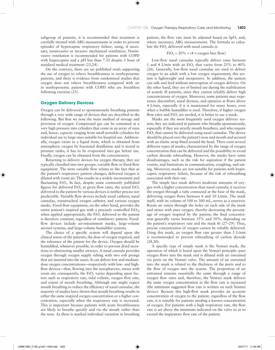
The choice of a specific system will depend upon the clinical status of the patients, the dose of oxygen required, and the tolerance of the patient for the device. Oxygen should be humidified, whenever possible, in order to prevent dried secre-tions to obstructing smaller airways. A nasal cannula provides oxygen through oxygen supply tubing with two soft prongs that are inserted into the nares. It can deliver low and medium-dose oxygen concentrations—respectively with low- and high-flow devices—that, flowing into the nasopharynx, mixes with room air; consequently, the FiO2 varies depending upon fac-tors such as respiratory rate, tidal volume, oxygen flow rate, and extent of mouth breathing. Although one might expect mouth breathing to reduce the efficiency of nasal cannulae, the majority of studies have shown that mouth breathing results in either the same inspired oxygen concentration or a higher con-centration, especially when the respiratory rate is increased. This is important because patients with acute breathlessness are likely to breathe quickly and via the mouth rather than the nose. As there is marked individual variation in breathing
pattern, the flow rate must be adjusted based on SpO2 and, where necessary, ABG measurements. The formula to calcu-late the FiO2 delivered with nasal cannula is:
FiO2 = 20% + (4 × oxygen liter flow)
Low-flow nasal cannulae typically deliver rates between 1 and 4 L/min with an FiO2 that varies from 25% to 40% (26). Generally, low-flow nasal cannulae are used to deliver oxygen to an adult with a low oxygen requirement; this sys-tem is lightweight and inexpensive. In addition, the patient can talk and feed without interruption of oxygen delivery. On the other hand, they are of limited use during the stabilization of acutely ill patients, since they cannot reliably deliver high concentrations of oxygen. Moreover, some patients may expe-rience discomfort, nasal dryness, and epistaxis at flows above 4 L/min, especially if it is maintained for many hours, even when a bubble humidifier is used. Therefore, if higher oxygen flow rates and FiO2 are needed, it is better to use a mask.
Masks are the most frequently used oxygen delivery sys-tem; they are indicated in patients who breathe spontaneously, especially if they are strictly mouth breathers, and who require FiO2 that cannot be delivered using nasal cannulae. The device should be placed over the patient’s nose and mouth and secured with an elastic strap fitted around the head. There exist several different types of masks, characterized by the range of oxygen concentration that can be delivered and the possibility to avoid carbon dioxide rebreathing. However, the masks have some disadvantages, such as the risk for aspiration if the patient vomits, and limitations in communication, coughing, and eat-ing. Moreover, masks are not suitable for patients with hyper-capnic respiratory failure, because of the risk of rebreathing associated with their use.
The simple face mask delivers moderate amounts of oxy-gen with a higher concentration than nasal cannula; it receives the oxygen through a tube connected at the base of the mask, delivering oxygen flows between 6 and 10 L/min. The mask itself, with its volume of 100 to 300 mL, serves as a reservoir. Room air enters through the holes on each side of the mask and mixes with pure oxygen, thereby decreasing the percent-age of oxygen inspired by the patient; the final concentra-tion generally varies between 35% and 50%, depending on the patient’s respiratory rate and the mask fit (27–29). Thus, precise concentration of oxygen cannot be reliably delivered. Using this mask, an oxygen flow rate greater than 5 L/min is recommended to prevent rebreathing of carbon dioxide (28,30).
A specific type of simple mask is the Venturi mask, the operation of which is based upon the Venturi principle: pure oxygen flows into the mask and is diluted with air entrained via ports on the Venturi valve. The amount of air entrained into the mask is related to the thickness of the ports and to the flow of oxygen into the system. The proportion of air entrained remains essentially the same through a range of oxygen flow rates and, therefore, the Venturi mask delivers the same oxygen concentration as the flow rate is increased (the minimum suggested flow rate is written on each Venturi device). Because this high-flow mask provides an accurate concentration of oxygen to the patient, regardless of the flow rate, it is suitable for patients needing a known concentration of oxygen. For patients with a high respiratory rate, the flow rate is set above the minimum indicated on the valve so as to exceed the inspiratory flow rate of the patient.
LWBK1580_C108_p1401-1409.indd 1403 29/07/17 11:54 AM
1404 SECTion 11 resPiratory DisorDers
A simple mask connected to a reservoir is defined a par-tial rebreathing mask. This kind of mask needs oxygen flow rates of between 10 and 12 L/min and delivers an oxygen con-centration ranging between 50% and 60% (28,31). During inspiration, air is drawn predominantly from the fresh oxy-gen contained in the reservoir. Despite the fact that this com-partment contains some exhaled gases as well, gases in the reservoir are oxygen rich because the early exhaled air that flows into the reservoir from respiratory dead space is oxygen rich and contains little carbon dioxide (31,32). To avoid this commingling of pure oxygen flow with exhaled gas, and to avoid the risk of rebreathing, it can be used a nonrebreathing face mask with reservoir and one-way valves. This system is composed of one-way valves over the exhalation ports that allow the egress of expired gases during exhalation and pre-vent room air from entering the mask during inspiration (33). Another one-way valve is located between the mask and the reservoir in order to prevent flow of exhaled gases into the res-ervoir. With this mask, oxygen should flow into the reservoir at 8 to 15 L/min and, in any case, the flow should be adjusted to prevent the collapse of reservoir. Notwithstanding the fact that, with this aid, an FiO2 of up to 95% can be achieved (34), the real oxygen concentration inhaled by the patient is variable and not accurately predictable, depending on the flow rate and the breathing pattern.
High-Flow Nasal Cannulae (HFNC)
All the conventional high-flow oxygen delivery systems dis-cussed above are not well tolerated by the patients due to discomfort, obtrusiveness, and insufficient heating and humid-ification of the inspired gases (35–37). To obviate these disad-vantages, effort has been spent over the past two decades to develop and validate an alternative oxygen delivery system: the high-flow nasal cannulae (HFNC). This device provides a gas mixture, with an FiO2 ranging from 0.21 to nearly 1.0 to nares via nasal prolongs. The presence of an oxygen blender allows the desired FiO2 to be set and delivered, adjusting it indepen-dently from the flow rate, which can reach a maximum of 60 L/min. In addition, the gas mixture can be warmed to body temperature and saturated with water via an inline humidifier. Several studies showed that the use of HFNC was associated with a greater comfort and tolerance, lower dyspnea, lower dryness of upper airways, and lower desiccation of secre-tions, compared to conventional face masks (38–40). One of the consequences of inhaling a warmed and fully humidified gas mixture is the maintenance of the integrity of mucociliary function, consequently rendering secretions easier to mobilize. This produces a decrease in the work imposed on muscles for expectoration. Another beneficial effect of this device consists in its ability of generate a flow rate that exceeds the patient’s peak inspiratory flow rate, in most cases, guaranteeing that there is less room air mixed with the inspired gas, and the desired FiO2 is more reliably delivered to the patient (41,42). This is particularly useful for patients in respiratory distress, who often have inspiratory flow rates that exceed those that can be delivered by standard systems (33). Patients with respi-ratory distress can also benefit from the continuous wash out of carbon dioxide from the anatomic dead space (43,44), occur-ring with HFNC, especially at higher flow rates (45). Over the last 10 years, multiple studies have shown that HFNC gener-ate a positive pressure inside the nasopharynx and esophagus
during both inspiration and expiration (46,47). Perhaps as a consequence, HFNC may reduce the work of breathing by providing inspiratory assistance, which increases the tidal vol-ume, reduces respiratory rate, and counterbalances autoPEEP, especially in COPD patients (48–50). Although HFNC has been proposed as a management tool for various conditions, including hypoxemic respiratory failure, cardiogenic pulmo-nary edema, as well as to prevent respiratory failure in post-operative and postextubation patients, the data available for adult applications of HFNC are limited (51).
For patients who have a tracheostomy or a transtracheal fistula, oxygen can be delivered through a tracheostomy mask or a transtracheal catheter. These devices deliver high-flow oxygen directly into trachea, promoting gas exchange; indeed they can reduce the work of breathing and augment carbon dioxide removal. When oxygen is given in this way for a prolonged period, humidification and suctions to remove the mucus from the airways are indicated.
RespIRaTORy CaRe and MOnITORIng In The ICu
Respiratory monitoring in critically ill patients is mandatory to detect inadequate oxygenation, acid–base imbalance and tissue hypoperfusion at an early stage. The correct use and interpretation of data from bedside assessment can improve patient outcomes. We describe here the potential clinical value of advanced respiratory monitoring technologies, according with a recent published consensus (52).
Monitoring Systems
Gas Exchange
The diagnosis of hypoxemia is essentially based on two parameters: oxygen saturation and arterial oxygen tension. The amount of oxygen carried in the blood is often expressed in terms of oxygen saturation of circulating hemoglobin. This is what is meant by “oxygen saturation level” (SpO2, SaO2), defined as one of the five vital signs. If the measurement is checked using pulse oximeter, rather than via an ABG mea-surement (SaO2), it is called the SpO2: for adults aged less than 70 years, the normal range is approximately 95% to 98% at sea level, and it declines gradually within this age range (53). The mean SpO2 may be lower in older people; however, it is difficult to dissociate the effects of advancing age from the effects of the disease which are common in old age (54,55). If the oxygen saturation is measured directly from an arterial blood sample, it is called the SaO2 (56–58).
Alternatively, it is possible to measure the oxygen tension of the blood (PaO2), known as the “partial pressure of oxygen.” This measurement can be expressed in kilopascals (kPa, normal range 12.0 to 14.6 kPa) or in millimeters of mercury (normal range 90 to 110 mmHg for young adults) and it is usually obtained from an arterial specimen. More rarely, it is obtained from arteriolized earlobe blood. In both cases, it is preferable to use local anesthesia to obtain these specimens, except in emergencies or if the patient is unconscious or anesthetized. The presence of a normal saturation measured by SpO2 does not exclude the need for performing ABG analysis, especially among critically ill patients. Indeed SpO2 can be normal in
LWBK1580_C108_p1401-1409.indd 1404 29/07/17 11:54 AM

CHAPTER 108 oxygen therapy, respiratory Care, and monitoring 1405
a patient with an alteration of pH or arterial carbon diox-ide tension (PaCO2) or with anemia. Therefore, ABG analy-sis should be evaluated early in all critically ill patients, in all subjects having an unexpected or inappropriate SpO2, in any case of increasing breathlessness in a patient with previously stable hypoxemia, in a stable patient who deteriorates and requires a significantly increased FiO2 to maintain a constant SpO2, in any patient with risk factors for hypercapnic respira-tory failure who develops acute breathlessness, deteriorating oxygen saturation or drowsiness, or other symptoms of CO2 retention. Additionally, ABG analysis should be performed in breathless patients who are thought to be at risk of metabolic conditions such as diabetic ketoacidosis or metabolic acido-sis due to renal failure, in acutely breathless or critically ill patients with poor peripheral circulation in whom a reliable oximetry signal cannot be obtained, or in the presence of any other evidence that would indicate that ABG blood gas results would be useful in the patient’s management, for example, an unexpected change in “track and trigger” systems such as a sudden rise of several units in the medical early warning score (MEWS) or an unexpected fall in SpO2 of 3% or more, even if within the target range (59).
Before evaluating the oxygenation of a patient, it’s impor-tant to record the FiO2 and the PaO2/FiO2 ratio; the latter is a quick and simple parameter that refers to the ratio of arterial oxygen partial pressure to fractional inspired oxygen. Accord-ing to the Berlin definition of acute respiratory distress syn-drome (ARDS; see Chapter 109), patients are categorized into three different categories (mild, moderate, or severe), based on the PaO2/FIO2 ratio (60). Although it has limitations, the PaO2/FIO2 ratio is a very useful clinical parameter used for predicting outcome and response to therapy in patients with ARDS (61).
As mentioned above, when assessing the level of oxygen-ation of critically ill patients with respiratory failure, PaO2 and SaO2 (or SpO2) are the main determinants and are most fre-quently used; however, there are many conditions where they may not provide sufficient information about the adequacy of oxygen delivery (DO2). Indeed this parameter relies on many determinants, such as passive oxygen diffusion into the arterial blood, but also on oxygen-carrying capacity of blood, ade-quate cardiac output, and local control of blood flow to the organs. Global DO2 is the product of cardiac output (CO) and arterial oxygen content (CaO2); CaO2 is, in turn, the product of SaO2, hemoglobin concentration and a constant reflecting hemoglobin–oxygen binding capacity (6). In the case of V/Q abnormalities, anemia, or low CO, the PA–aO2 gradient or the PaO2/FiO2 ratio may be more sensitive indicators and correlate better with the adequacy of DO2.
Devices for Titration of Oxygen Flow
Over the past several years, devices have been developed that automatically adjust—in a closed-loop manner—oxygen flow to spontaneously breathing patients, maintaining a set oxy-genation target, such as the O2 flow regulator (62) and the free O2 (63). It is recognized that the oxygen flow administered to the patient is not always optimal; patient’s needs vary dur-ing the day, and even in healthy people, episodes of oxygen desaturation may occur during daily activities or during sleep. These drops in saturation occur more frequently in the criti-cally ill patient and in those affected by COPD (64), inducing complications such as reduction in exercise tolerance (65),
pulmonary hypertension, right heart failure, polycythemia, and increased mortality (66). Some studies have found that oxygen therapy is not optimally prescribed in COPD patients; that the commonly prescribed, fixed dose, usually titrated at rest, may not meet the patient’s needs during an exacerbation, inducing both hypoxemia and hyperoxia, with their atten-dant complications (67–70). It would be preferable, therefore, to tailor the oxygen flow to the actual changing need of the patient, and to titrate the flow rate based upon an SpO2 sig-nal to maintain a target saturation, for example, a target of 93% or more (71). This adjustment in oxygen flow minimizes episodes of desaturation, avoids excessive administration that may produce respiratory acidosis, and maintains stable SpO2 at all activity levels.
Transcutaneous Arterial Carbon Dioxide Pressure
Transcutaneous carbon dioxide monitoring (PtcCO2) is a noninvasive method used to estimate the PaCO2. It is able to give an electrochemical measurement of PaCO2 by warming the skin and inducing hyperperfusion (72). Although there is a good concordance between arterial and transcutaneous PaCO2 values (73), the role of PtcCO2 in the adult intensive care setting remains unclear. Major issues include the need for frequent recalibration and the mismatch in presence of hyperoxia, hypoperfusion, improper electrode placement, and tissue edema. Additionally, skin breakdown and tissue loss can occur, especially if the probe is left in place for long time (74).
Volumetric Capnography
Volumetric capnography is a noninvasive technique that examines the CO2 concentration in exhaled air as a function of expired volume by using infrared light. As described for the first time by Aitken and Clarke–Kennedy (75), the capnogram curve represents one respiratory cycle and has several compo-nents: the first part of the waveform (phase I) represents the CO2 from the airway (dead space), the second part (phase II) is a sharp rise (beginning of exhalation) that coincides with the transition between airway and alveolar gas. The highest por-tion of this segment is called the end-tidal CO2 and represents the maximum pressure of CO2 at the end of breath; normally it is between 35 and 40 mmHg. The phase III corresponds to the CO2 eliminated from the alveoli (plateau line). The homo-geneity of the gas distribution and alveolar ventilation are the major determinants of the capnogram’s shape.
In addition, dead space can be deduced noninvasively using volumetric capnography (76–78). It has, thus, been useful in the monitoring of various diseases, particularly when V/Q mismatch is present, such as in pulmonary thromboembolism (79), interstitial lung disease, and acute lung injury, as well as in patients undergoing mechanical ventilation (52,80). How-ever, this technique needs a complex equipment which repre-sents a limitation of its use in clinical practice (52).
Respiratory System Mechanics
The respiratory system includes both passive (lung and the chest wall) and active structures (respiratory muscles); these components have both elastic and resistive properties. The elasticity is defined as a driving pressure variation (∆P) over a change in volume (∆V). Resistance is expressed as the
LWBK1580_C108_p1401-1409.indd 1405 29/07/17 11:54 AM

1406 SECTion 11 resPiratory DisorDers
ratio between the pressure change and the gas flow (V. ) varia-
tion (∆P/∆V. ). It is not possible to separate the chest wall and
the lung from the respiratory muscles, and so the respiratory mechanics properties can be evaluated only when the respira-tory muscle activity is quiescent (patient deeply sedated or par-alyzed). During passive mechanical ventilation, compliance is calculated as the ratio between VT and plateau pressure (Pplat) minus positive end-expiratory pressure (PEEP). On the other hand, the difference between peak or maximal pressure (PMAX) and plateau pressure (Pplat) over a constant V
. define the resis-
tance (81,82).During volume-controlled ventilation, with a constant
flow, it is possible to calculate both the resistive forces and the compliance of the respiratory system by performing easily bedside end-inspiratory and end-expiratory occlusion maneu-vers. Briefly, after the end-inspiratory occlusion technique, the rapid pressure drop from PMAX to Pplat represents the pres-sure required to move the flow through the airways and the endotracheal tube (ETT). The following slow drop until a pla-teau depends mainly on the elastic properties of the system. With the end-expiratory occlusion, clinicians can evaluate the values for intrinsic (auto) PEEP and total PEEP. Thus, com-pliance and resistance measurements provided by ventilators performing end-inspiratory and end-expiratory pauses, as well as the interpretation of P/V curves with the identification of the lower (LIP) and upper (UIP) inflection points, furnish use-ful information to optimize mechanical ventilation manage-ment according with the underlying diseases.
Esophageal and Transpulmonary Pressure
The esophageal pressure (Pes) is an acceptable surrogate of pleural pressure and can be measured by using an esophageal balloon catheter. Thanks to this correlation, it is possible to estimate the transpulmonary pressure (PTP), the pressure needed to distend the lungs, as the difference between the airway pressure (Paw) and the pleural pressure (Ppl); it is also possible to measure the gastric pressure (Pga) and, conse-quently, the transdiaphragmatic pressure (Pdi), which results from the difference between Pga and Pes, by adding a gastric balloon.
Recently, a Pleural Pressure Working Group (PLUG) reviewed the newest information on esophageal pressure mon-itoring in patients with respiratory failure (83). Currently, the measurement of Pes in mechanically ventilated patients is often performed in an attempt to detect and treat patient/ventilator asynchrony, to measure work of breathing and to guide wean-ing, and to optimize mechanical ventilation in patients with ARDS. ARDS is characterized by a decrease in respiratory system compliance due to the presence of alveolar and inter-stitial fluid, and the loss of surfactant. The standard of care for mechanically ventilated patients with ARDS includes low tidal volume (Vt = 6 mL/kg of predicted body weight) and low plateau pressure (Pplat ≤30 cm H2O) (84). PEEP is also applied in order to maintain open air spaces and to prevent the cyclic opening and closing of alveoli that produces the ventilator-induced lung injury (85,86). As previously mentioned, clini-cians optimize ventilator support by monitoring the airway pressure, assuming that the Paw reflects the PTP. On the other hand, because the Paw represents the sum of pressure across the lung and the chest wall, in case of chest compliance reduction, as one would see with obese patients, as well as in patients
with edema or with abdominal distension, it does not always correspond to the pressure seen by the lung (87).
Therefore, the Pes has been proposed to calculate the end-expiratory PTP and to set PEEP so as to prevent the cyclic lung recruitment and derecruitment that are present when Ppl is greater than Paw at the end of expiration (88). As shown by the EPVent study (89), the strategy that uses Pes to adjust PEEP in order to achieve end-expiratory PTP between 0 and 10 cm H2O (the esophageal pressure–guided group) significantly improves oxygenation and compliance compared with the control group in which PEEP was set according to the patient’s PaO2 and FiO2 (gas exchange–based PEEP group). Based upon these considerations, the use of the Pes to optimize ventilator setting in ARDS patients seems more useful to avoid both under- and over-lung distension compared to the use of airway pressure as guide.
The Procedure
Although advances in electronic and computer technology over the last decades have generated several ventilators with additional pressure transducers that provide Pes measurement at the patient’s bedside, the most commonly used procedure is performed via esophageal and gastric balloons that are now commercially available. The passage of the catheters through the nasal cavity may cause pain, irritation, cough, and some-times vomiting. Thus, initial preparation of the awake patient includes the local nebulization of lidocaine and a mild dose of sedative, if the patient is particularly anxious. Placing the patient into the semi-recumbent position is the best approach to the insertion of the balloon (90). Usually two operators are required for the operation, the first one dedicated to extend the neck of the patient, with the second one very slowly introducing the catheter into the nares, preferably the one appearing to be unoccluded or less occluded.
The esophageal balloon should be first pushed into the stomach and moved backward step by step until it passes through the esophageal sphincter where the waveform during inspiration will begin to become negative. The balloon is then positioned roughly in the middle third of the esophagus where the “occlusion test” is performed (91). The gastric catheter’s position is checked by pushing over the patient’s abdomen to see a clear positive pressure swing. Once the catheter is in place, it needs to be fixed with tape at the nostril, and 1 mL of air is then inserted with a syringe and then slowly removed until about 0.3 to 0.4 mL remain (92–96).
Despite being relatively well standardized, the technique for measuring respiratory mechanics requires special attention to avoid errors and complications. Patient should be fasting to reduce the risk of vomiting. Additionally, disorders of the mouth, pharynx—tumors, cervical spine disease, or pharyn-goesophageal diverticulum—and esophageal pathologies—tumor, strictures secondary to radiation or chemical burns, medications or ulcers, Schatzki’s ring, or foreign bodies—may represent relative and absolute contraindications to the exami-nation (92). Clinicians must pay careful attention to patients with pre-existing cardiac dysrhythmias, as catheter placement may increase the risk of complication, as well as of a vagal reflex syndrome. Finally, neuromuscular problems—stroke, Parkinson’s disease, Huntington’s disease, multiple sclero-sis, myasthenia gravis, muscular dystrophy, polymyositis, amyotrophic lateral sclerosis or scleroderma—may also make balloon passage difficult (92).
LWBK1580_C108_p1401-1409.indd 1406 29/07/17 11:54 AM

CHAPTER 108 oxygen therapy, respiratory Care, and monitoring 1407
Lungs Volumes
Direct Measurement of End-Expiratory Lung Volume
The lung volume at the end of passive expiration is defined functional residual capacity (FRC). The term end-expiratory lung volume (EELV) is the more precise term to describe the lung volume when PEEP is applied during mechanical venti-lation. The measurement of FRC and EELV is useful for the management of patients with ARDS that present a reduction in lung volume. To calculate the EELV we can use several methods. The closed dilution technique requires the calcula-tion of a known soluble gas volume (helium or methane) in expired breath. Recently, bedside EELV measurement using nitrogen or O2 and CO2 sensors in ICU ventilators—so-called washout/wash-in techniques—have been proposed in clini-cal practice (52). EELV estimation may help to set PEEP and monitor alveolar recruitment maneuver responses in patients with ARDS when combined with compliance measures (97).
Lung Ultrasound
Over the past decade, the use of lung ultrasound (LUS), sup-ported by emerging data, has increased in different clinical settings. The International Consensus Conference provided recommendations and indications for LUS in order to stan-dardize this technique (98). Actually, LUS is considered an accurate tool for bedside respiratory monitoring in the ICUs. In fact, LUS allows detection of pleural effusion, differen-tiation of consolidation potentially secondary to pulmonary embolism, pneumonia, or atelectasis, and as a tool to rule out pneumothorax and diaphragmatic dysfunction. Moreover, thoracic ultrasound is used as a tool to guide thoracentesis and for placement of thoracostomy tubes or central venous lines. LUS is also able to monitor the effects of therapy in various acute lung diseases, including acute pulmonary edema, ARDS, community-acquired pneumonia, and ventilator-associated pneumonia (98,99). Thoracic ultrasound has some advantages compared to the chest radiography and CT scan, including portability and the ability to perform real-time imaging. In addition, LUS does not expose the patient to ionizing radia-tion. The major limitations include the need for staff training and the inability to give precise information in case of deep lesions without consolidation/effusion and/or paravertebral lesions, along with technical challenges in obese or edematous patients (100).
Respiratory Monitoring During Noninvasive Ventilation
NIV is a useful therapeutic modality for many patients and its use in the ICU has increased in the last two decades. As suc-cess with NIV depends on several factors, monitoring during NIV should be directed to identify and, if possible, resolving the issues that are associated with failure of this mode of ven-tilation, in order to improve patient’s outcomes. As noted by Olzyman et al. (101), the possible causes of immediate NIV failure—that is, failure within minutes to less than 1 hour of initiation of NIV—are a weak cough reflex and/or excessive secretions, severe neurologic impairment, patient–ventilator asynchrony, and intolerance and agitation. Thus, an accurate evaluation of vital signs, respiratory muscle function, cough effectiveness, and degree of consciousness is mandatory prior
to initiation if NIV. In addition, unstable patients with ARF should be closely monitored with ABG analysis, both to estab-lish the baseline severity and to avoid delay in endotracheal intubation in case of worsening gas exchange.
Patient–ventilator interaction is an important issue that ICU staff should take into account when using NIV. In fact, high inspiratory ventilator pressures and the presence of air leaks are major determinants of NIV asynchronies (52,102). Although recent technologic advances—for instance, new NIV algorithms and modes such as neurally adjusted venti-lator assistance (NAVA)—have been designed to reduce the occurrence of patient–ventilator asynchrony (103–105), bed-side methods, including evaluation of spontaneous versus ventilator-delivered breaths and accessory muscle use should be always considered initially. Additionally, air leaks can be identified by following the inspired and expired tidal volume values, and the presence of autoPEEP can be expected when the flow does not reach zero at the end of expiration. These simple measures, in association with flow and pressure wave-form observations on the ventilator screen, should be used by physicians to detect “gross” patient–ventilator mismatching. In patients receiving NIV for acute COPD exacerbation, opti-mization of the ventilator settings by analyzing flow and pres-sure waveforms on the screen (“optimized ventilation”) is a more efficient manner to set the ventilator than by considering only numerical data (“standard ventilation”) in terms of gas exchange and NIV success (106).
Finally, NIV interface intolerance is another important reason of failure when attempting to utilize NIV for an acute episode. Consequently, sedation should be considered part of the strategy aimed at improving NIV acceptance, mainly related to minimizing interface discomfort (101,107).
In conclusion, it is advisable for every clinician prescrib-ing NIV to have an experience in clinical, pharmacologic, and technologic competences as well as in monitoring skills in order to improve patient outcomes and to avoid delays in endotracheal intubation, especially in patient with de novo hypoxemic respiratory failure (108).
• Oxygen therapy should be handled carefully in criti-cally ill patients according to the underline cause of respiratory failure.
• Oxygen therapy can be delivered via different devices. The choice of the most appropriate system should be made based upon patient’s clinical status, level of required oxygen supply, and patient’s tolerance and comfort. The use of HFNC is associated with a greater comfort and tolerance, lower dyspnea, lower dryness of upper airways, compared to standard oxygen. How-ever, the data available for adult applications of HFNC are limited.
• Critically ill patients should be closely monitored in order to detect the early onset of acute respiratory dis-tress and failure to maintaining adequate saturation, to avoid oxygen toxicity and its side effects. On the other hand, respiratory monitoring is essential to iden-tify the causes of the disease involved and the effects of treatment.
Key points
LWBK1580_C108_p1401-1409.indd 1407 29/07/17 11:54 AM

1408 SECTion 11 resPiratory DisorDers
References 1. Priestley J. Experiments and Observations on Different Kinds of Air.
London, J. Johnson. 1775; revised 1790. 2. Smith AH. Oxygen Gas as a Remedy in Disease. New York, NY: D.
Appleton & Company; 1870. 3. Bryan CL, Jenkinson SG. Oxygen toxicity. Clin Chest Med. 1988;9:141–152. 4. O’Neill B, Mahon JM, Bradley J. Short-burst oxygen therapy in chronic
obstructive pulmonary disease. Respir Med. 2006;100:1129–1138. 5. Clemens KE, Klaschik E. Symptomatic therapy of dyspnea with strong
opioids and its effect on ventilation in palliative care patients. J Pain Symp-tom Manag. 2007;33:473–481.
6. Marks B, Mitchell DG, Simelaro JP. Breath-holding in healthy and pulmo-nary compromised patients: effects of hyperventilation and oxygen inspi-ration. J Magn Reson Imaging. 1997;7:595–597.
7. McCarthy RM, Shea S, Deshpande VS, et al. Coronary MR angiography: true FISP imaging improved by prolonging breath holds with preoxygen-ation in healthy volunteers. Radiology. 2003;227:283–288.
8. Bowton DL, Scuderi PE, Haponik EF. The incidence and effect on outcome of hypoxemia in hospitalised medical patients. Am J Med. 1994;97:38–46.
9. Slutsky AS. Consensus conference on mechanical ventilation. Part I. European Society of Intensive Care Medicine, the ACCP and the SCCM. Intensive Care Med. 1994;20:64–79.
10. Gries RE, Brooks LJ. Normal oxyhemoglobin saturation during sleep: how low does it go? Chest. 1996;110:1489–1492.
11. Kao LW, Nanagas KA. Toxicity associated with carbon monoxide. Clin Lab Med. 2006;26:99–125.
12. Juurlink DN, Buckley NA, Stanbrook MB, et al. Hyperbaric oxygen for carbon monoxide poisoning. Cochrane Database Syst Rev. 2005;(1): CD002041.
13. Weaver LK, Valentine KJ, Hopkins RO. Carbon monoxide poisoning: risk factors for cognitive sequelae and the role of hyperbaric oxygen. Am J Respir Crit Care Med. 2007;176:491–497.
14. Plant PK, Owen JL, Elliott MW. One year period prevalence study of respiratory acidosis in acute exacerbations of COPD: implications for the provision of noninvasive ventilation and oxygen administration. Thorax. 2000;55:550–554.
15. Warrel DA, Edwards RHT, Godfrey S, et al. Effect of controlled oxygen therapy on arterial blood gases in acute respiratory failure. BMJ. 1970; 2:452–455.
16. Campbell EJM. The management of acute respiratory failure in chronic bronchitis and emphysema. Am Rev Respir Dis. 1967;96:26–39.
17. Lopez-Majano V, Dutton RE. Regulation of respiration during oxygen breathing in COLD. Am Rev Respir Dis. 1973;108:232–240.
18. Aubier M, Murciano D, Milic Emili J, et al. Effects of the administration of O2 on ventilation and blood gases in patients with chronic obstructive pulmonary disease during acute respiratory failure. Am Rev Respir Dis. 1980;122:747–754.
19. Johnson JE, Peacock MD, Hayes JA, et al. Forced expiratory flow is reduced by 100% oxygen in patients with chronic obstructive pulmonary disease. South Med J. 1995;88:443–449.
20. Refsum HE. Relationship between state of consciousness and arterial hypoxaemia and hypercapnia in patients with pulmonary insufficiency, breathing air. Clin Sci. 1963;25:361–367.
21. Joosten SA, Koh MS, Bu X, et al. The effects of oxygen therapy in patients presenting to an emergency department with exacerbation of chronic obstructive pulmonary disease. Med J Aust. 2007;186:235–238.
22. Campbell EJ. A method of controlled oxygen administration which reduces the risk of carbon-dioxide retention. Lancet. 1960;2:12–24.
23. National Institute for Clinical Excellence (NICE). Chronic obstructive pul-monary disease. NICE Guideline CG12. Available at: http://www.nice.org.uk/guidance/CG12/niceguidance/pdf/English. 2004.
24. British Thoracic Society Standards of Care Committee. Non-invasive ventilation in acute respiratory failure. Thorax. 2002;57:192–211.
25. Abernethy AP, McDonald CF, Frith PA, et al. Effect of palliative oxy-gen versus room air in relief of breathlessness in patients with refractory dyspnoea: a double-blind, randomised controlled trial. Lancet. 2010; 376(9743):784–793.
26. Wettstein RB, Shelledy DC, Peters JI. Delivered oxygen concentrations using low-flow and high-flow nasal cannulas. Respir Care. 2005;50:604–609.
27. Bateman NT, Leach RM. ABC of oxygen: acute oxygen therapy. BMJ. 1998;317:798–801
28. Myers TR, American Association for Respiratory Care (AARC). AARC Clinical Practice Guideline: selection of an oxygen delivery device for neonatal and pediatric patients—2002 revision & update. Respir Care. 2002;47:707–716.
29. Milross J, Young IH, Donnelly P. The oxygen delivery characteristics of the Hudson Oxy-one face mask. Anaesth Intensive Care. 1989;17:180–184.
30. Jensen AG, Johnson A, Sandstedt S. Rebreathing during oxygen treatment with face mask: the effect of oxygen flow rates on ventilation. Acta Anaes-thesiol Scand. 1991;35:289–292.
31. King BR, King C, Coates WC. Critical procedures. In: Gausche-Hill M, Fuchs S, Yamamoto L, eds. APLS: The Pediatric Emergency Medicine Resource. 4th ed. Sudbury, MA: Jones and Bartlett; 2004:686.
32. Ludwig S. Resuscitation: pediatric basic and advanced life support. In: Fleisher GR, Ludwig S, eds. Textbook of Pediatric Emergency Medicine. Baltimore, MD: Williams & Wilkins; 1993:3.
33. Scarfone RJ. Airway adjuncts, oxygen delivery, and suctioning of the upper airway. In: Henretig FM, King C, eds. Textbook of Pediatric Emergency Medicine Procedures. Philadelphia, PA: Lippincott Williams & Wilkins, 1997:101.
34. Boumphrey SM, Morris EA, Kinsella SM. 100% inspired oxygen from a Hudson mask-a realistic goal? Resuscitation. 2003;57:69–72.
35. Kallstrom TJ. AARC Clinical Practice Guideline: oxygen therapy for adults in the acute care facility—2002 revision & update. Respir Care. 2002:47:717–720.
36. Shapiro BA, Kacmarek RM, Cane RD, et al. Clinical Application of Respi-ratory Care. 4th ed. St. Louis, MA: Mosby-Year Book; 1991.
37. Chanques G, Constantin JM, Sauter M, et al. Discomfort associated with underhumidified high-flow oxygen therapy in critically ill patients. Inten-sive Care Med. 2009:35:996–1003.
38. Roca O, Riera J, Torres F, et al. High-flow oxygen therapy in acute respira-tory failure. Respir Care. 2010:55(4):408–413.
39. Tiruvoipati R, Lewis D, Haji K, et al. High-flow nasal oxygen vs. high-flow face mask: a randomized crossover trial in extubated patients. J Crit Care. 2010:25:463–468.
40. Rittayamai N, Tscheikuna J, Rujiwit P. High-flow nasal oxygen versus conventional oxygen therapy after endotracheal extubation: a randomized crossover physiologic study. Respir Care. 2014; 59(4):485–490.
41. Sim MAB, Dean P, Kinsella J, et al. Performance of oxygen delivery devices when the breathing pattern of respiratory failure is simulated. Anaesthesia. 2008:63:938–940.
42. Wagstaff TA, Soni N. Performance of six types of oxygen delivery devices at varying respiratory rates. Anaesthesia. 2007:62:492–503.
43. Sztrymf B, Messika J, Bertrans F, et al. Beneficial effects of humidified high flow oxygen in critical care patients: a prospective pilot study. Intensive Care Med. 2011:37:1780–1786.
44. Lenglet H, Sztrymf B, Leroy C, et al. Humidified high flow nasal oxygen during respiratory failure in the emergency department: feasibility and efficacy. Respir Care. 2012:57(11):1873–1878.
45. Frizzola M, Miller TL, Rodriguez ME, et al. High-flow nasal cannula: impact on oxygenation and ventilation in an acute lung injury model. Pediatr Pulmonol. 2011:46(1):67–74.
46. Hasan RA, Habib RH. Effects of flow-rate and airleak at the nares and mouth opening on positive distending pressure delivery using commercially available high-flow nasal cannula systems: a lung model study. Pediatr Crit Care Med. 2011:12(1):e29–e33.
47. Parke RL, Ecclestone ML, McGuinness SP. The effects of flow on airway pressure during nasal high-flow oxygen therapy. Respir Care. 2011:56(8): 1151–1155.
48. Braunlich J, Beyer D, Mai D, et al. Effects of nasal high flow on ventilation in volunteers, COPD and idiopathic pulmonary fibrosis patients. Respira-tion. 2013;85(4):319–325.
49. Mundel T, Feng S, Tatkov S, et al. Mechanisms of nasal high-flow on ventilation during wakefulness and sleep. J Appl Physiol. 2013:114: 1058–1065.
50. Corley A, Caruana LR, Barnett AG, et al. Oxygen delivery through high-flow nasal cannulae increase end-expiratory lung volume and reduce respi-ratory rate in post-cardiac surgical patients. Br J Anaesth. 2011:107(6): 998–1004.
51. Spoletini G, Alotaibi M, Blasi F, et al. Heated humidified high-flow nasal oxygen in adults: mechanism of action and clinical implications. Chest. 2015;148(1):253–261.
52. Brochard L, Martin GS, Blanch L, et al. Clinical review: respiratory monitoring in the ICU—a consensus of 16. Crit Care. 2012;16(2):219.
53. Crapo RO, Jensen RL, Hegewald M, et al. Arterial blood gas reference values for sea level and an altitude of 1,400 meters. Am J Respir Crit Care Med. 1999;160(5 Pt 1):1525–1531.
54. Hardie JA, Vollmer WM, Buist AS, et al. Reference values for arterial blood gases in the elderly. Chest. 2004;125:2053–2060.
55. Blom H, Mulder M, Verweij W. Arterial oxygen tension and saturation in hospital patients: effect of age and activity. BMJ. 1988;297:720–721.
LWBK1580_C108_p1401-1409.indd 1408 29/07/17 11:54 AM

CHAPTER 108 oxygen therapy, respiratory Care, and monitoring 1409
56. Wilson AT, Channer KS. Hypoxaemia and supplemental oxygen therapy in the first 24 hours after myocardial infarction: the role of pulse oximetry. J R Coll Physicians Lond. 1997;31:657–661.
57. Levin KP, Hanusa BH, Rotondi A, et al. Arterial blood gas and pulse oximetry in initial management of patients with community-acquired pneumonia. J Gen Intern Med. 2001;16:590–598.
58. Moed BR, Boyd DW, Andring RE. Clinically inapparent hypoxemia after skeletal injury: the use of the pulse oximeter as a screening method. Clin Orthop. 1993;293:269–273.
59. O’Driscoll BR, Howard LS, Davison AG. BTS, guidelines for emergency oxygen use in adult patients. Thorax. 2008;63:vi1–vi68.
60. Ferguson ND, Fan E, Camporota L, et al. The Berlin definition of ARDS: an expanded rationale, justification, and supplementary material. Intensive Care Med. 2012;38:1573–1582.
61. Villar J, Fernández RL, Ambrós A, et al. Acute Lung Injury Epidemiology and Natural History Network: a clinical classification of the acute respira-tory distress syndrome for predicting outcome and guiding medical therapy. Crit Care Med. 2015;43(2):346–353.
62. Cirio S, Nava S. Pilot study of a new device to titrate oxygen flow in hypoxic patients on long-term oxygen therapy. Respir Care. 2011;56(4):429–434.
63. Lellouche F, L’her E. Automated oxygen flow titration to maintain con-stant oxygenation. Respir Care. 2012;57(8):1254–1262.
64. Kim V, Benditt JO, Wise RA, et al. Oxygen therapy in chronic obstructive pulmonary disease. Proc Am Thorac Soc. 2008; 5(4):513–518.
65. O’Donnell DE, Bain DJ, Webb KA. Factors contributing to relief of exer-tional breathlessness during hyperoxia in chronic airflow limitation. Am J Respir Crit Care Med. 1997;155(2):530–535.
66. Boushy SF, Thompson HK Jr, North LB, et al. Prognosis in chronic obstruc-tive pulmonary disease. Am Rev Respir Dis. 1973;108(6):1373–1383.
67. Aubier M, Murciano D, Milic-Emili J, et al. Effects of the administration of O2 on ventilation and blood gases in patients with chronic obstructive pulmonary disease during acute respiratory failure. Am Rev Respir Dis. 1980;122(5):747–754.
68. Sassoon CS, Hassell KT, Mahutte CK. Hyperoxic-induced hypercapnia in stable chronic obstructive pulmonary disease. Am Rev Respir Dis. 1987; 135(4):907–911.
69. Plant PK, Owen JL, Elliott MW. One year period prevalence study of respiratory acidosis in acute exacerbations of COPD: implications for the provision of non-invasive ventilation and oxygen administration. Thorax. 2000;55(7):550–554.
70. Hale KE, Gavin C, O’Driscoll BR. Audit of oxygen use in emergency ambulances and in a hospital emergency department. Emerg Med J. 2008; 25(11):773–776.
71. Carone M, Patessio A, Appendini L, et al. Comparison of invasive and non-invasive saturation monitoring in prescribing oxygen during exercise in COPD patients. Eur Respir J. 1997;10(2):446–451.
72. Monaco F, Nickerson BG, McQuitty JC. Continuous transcutaneous oxygenand carbon dioxide monitoring in the pediatric ICU. Crit Care Med. 1982;10:765–766.
73. Rodriguez P, Lellouche F, Aboab J, et al. Transcutaneous arterial carbon dioxide pressure monitoring in critically ill adult patients. Intensive Care Med. 2006;32(2):309–312
74. Coates BM, Chaize R, Goodman DM, et al. Performance of capnometry in non-intubated infants in the pediatric intensive care unit. BMC Pediatr. 2014;14:163.
75. Aitken RS, Clark-Kennedy AE. On the fluctuation in the composition of the alveolar air during the respiratory cycle in muscular exercise. J Physiol. 1928;65(4):389–411.
76. Wolff G, Brunner JX. Series dead space volume assessed as the mean value of a distribution function. Int J Clin Monit Comput. 1984;1(3): 177–181.
77. Tusman G, Sipmann FS, Borges JB, et al. Validation of Bohr dead space measured by volumetric capnography. Intensive Care Med. 2011;37(5): 870–874.
78. Fletcher R, Jonson B, Cumming G, et al. The concept of deadspace with special reference to the single breath test for carbon dioxide. Br J Anaesth. 1981;53(1):77–88.
79. Verschuren F, Heinonen E, Clause D, et al: Volumetric capnography as a bedside monitoring of thrombolysis in major pulmonary embolism. Inten-sive Care Med. 2004;30:2129–2132.
80. Kelly AM, Klim S. Agreement between arterial and transcutaneous PCO2 in patients undergoing non-invasive ventilation. Respir Med. 2011;105: 226–229.
81. Lu Q, Rouby JJ. Measurement of pressure-volume curves in patients on mechanical ventilation: methods and significance. Crit Care. 2000;4(2): 91–100.
82. Harris RS. Pressure-volume curves of the respiratory system. Respir Care. 2005;50(1):78–98.
83. Akoumianaki E, Maggiore SM, Valenza F, et al. The application of esoph-ageal pressure measurement in patients with respiratory failure. Am J Respir Crit Care Med. 2014;189(5):520–531.
84. The Acute Respiratory Distress Syndrome Network. Ventilation with lower tidal volumes as compared with traditional tidal volumes for acute lung injury and the acute respiratory distress syndrome. N Engl J Med. 2000;342(18):1301–1308.
85. Gattinoni L, Carlesso E, Cressoni M. Selecting the ‘right’ positive end-expiratory pressure level. Curr Opin Crit Care. 2015;21(1):50–57
86. Chiumello D, Guerin C. Understanding the setting of PEEP from esophageal pressure in patients with ARDS. Intensive Care Med. 2015;41(8):1465–1467.
87. Chiumello D, Guerin C. Understanding the setting of PEEP from esophageal pressure in patients with ARDS. Intensive Care Med. 2015;41(8):1465–1467.
88. Brochard L. Measurement of esophageal pressure at bedside: pros and cons. Curr Opin Crit Care. 2014;20(1):39–46.
89. Talmor D, Sarge T, Malhotra A, et al. Mechanical ventilation guided by esoph-ageal pressure in acute lung injury. N Engl J Med. 2008;359:2095–2104.
90. Baydur A, Cha EJ, Sassoon CS. Validation of esophageal balloon technique at different lung volumes and postures. J Appl Physiol. 1987;62:315–321.
91. Benditt JO. Esophageal and gastric pressure measurements. Respir Care. 2005;50(1):68–75.
92. Zin WA, Milic-Emili J. Esophageal pressure measurement. In: Hamid Q, Shannon J, Martin J, eds. Physiologic Basis of Respiratory Disease. New York, NY: BC Decker Inc; 2005:639–647.
93. Polese G, Serra A, Rossi A. Respiratory mechanics in the intensive care unit. Eur Respir Mon. 2005;31:195–206.
94. Gappa M, Colin AA, Goetz I, et al. ERS/ATS Task Force on Standards for Infant Respiratory Function Testing. Passive respiratory mechanics: the occlusion techniques. Eur Respir J. 2001;17(1):141–148.
95. Benditt JO. Esophageal and gastric pressure measurements. Respir Care. 2005;50(1):68–75.
96. Hedenstierna G. Esophageal pressure: benefit and limitations. Minerva Anestesiol. 2012;78(8):959–966.
97. Dellamonica J, Lerolle N, Sargentini C, et al. PEEP-induced changes in lung volume in acute respiratory distress syndrome: two methods to esti-mate alveolar recruitment. Intensive Care Med. 2011;37:1595–1604.
98. Volpicelli G, Elbarbary M, Blaivas M, et al. International evidence based recommendations for point-of-care lung ultrasound. Intensive Care Med. 2012;38:577–591.
99. Bouhemad B, Zhang M, Lu Q, et al. Clinical review: bedside lung ultra-sound in critical care practice. Crit Care. 2007;11:205.
100. Via G, Storti E, Gulati G, et al. Lung ultrasound in the ICU: from diag-nostic instrument to respiratory monitoring tool. Minerva Anestesiol. 2012;78:1282–1296.
101. Ozyilmaz E, Ugurlu AO, Nava S. Timing of noninvasive ventilation failure: causes, risk factors, and potential remedies. BMC Pulm Med. 2014;14:19.
102. Vignaux L, Vargas F, Roeseler J, et al. Patient-ventilator asynchrony dur-ing non-invasive ventilation for acute respiratory failure: a multicenter study. Intensive Care Med. 2009;35:840–846.
103. Schmidt M, Dres M, Raux M, et al. Neurally adjusted ventilatory assist improves patient-ventilator interaction during postextubation prophylac-tic noninvasive ventilation. Crit Care Med. 2012;40(6):1738–1744.
104. Cammarota G, Olivieri C, Costa R, et al. Noninvasive ventilation through a helmet in postextubation hypoxemic patients: physiologic comparison between neurally adjusted ventilatory assist and pressure support ventila-tion. Intensive Care Med. 2011;37(12):1943–1950.
105. Doorduin J, Sinderby CA, Beck J, et al. Automated patient-ventilator interaction analysis during neurally adjusted non-invasive ventilation and pressure support ventilation in chronic obstructive pulmonary disease. Crit Care. 2014;18(5):550.
106. Di Marco F, Centanni S, Bellone A, et al. Optimization of ventilator setting by flow and pressure waveforms analysis during noninvasive ventilation for acute exacerbations of COPD: a multicentric randomized controlled trial. Crit Care. 2011;15:R283.
107. Longrois D, Conti G, Mantz J, et al. Sedation in non-invasive ventilation: do we know what to do (and why)? Multidisc Respir Med. 2014;9:56.
108. Demoule A, Girou E, Richard JC, et al. Benefits and risks of success or failure of noninvasive ventilation. Intensive Care Med. 2006;32(11):1756–1765.
LWBK1580_C108_p1401-1409.indd 1409 29/07/17 11:54 AM