Embed Size (px)
Citation preview

Oxidative stress causesalveolar bone loss inmetabolic syndrome modelmice with type 2 diabetes
T. Ohnishi1, K. Bandow1,K. Kakimoto1, M. Machigashira2,T. Matsuyama2, T. Matsuguchi11Division of Biochemistry and MolecularDentistry, Department of DevelopmentalMedicine, Kagoshima University, GraduateSchool of Medical and Dental Sciences,Kagoshima, Japan and 2Department ofPeriodontology, Kagoshima University,Graduate School of Medical and DentalSciences, Kagoshima, Japan
The metabolic syndrome is character-
ized by a cluster of related cardiovas-
cular metabolic risk factors, including
various combinations of glucose toler-
ance, hypertension and hyperlipemia.
The metabolic syndrome was initially
described as a disorder of insulin
resistance (1), which is closely associ-
ated with type 2 diabetes, and is also
called the insulin resistance syndrome
by the European Group for the
Study of Insulin Resistance (2). The
Ohnishi T, Bandow K, Kakimoto K, Machigashira M, Matsuyama T, Matsuguchi T.
Oxidative stress causes alveolar bone loss in metabolic syndrome model mice with
type 2 diabetes. J Periodont Res 2009; 44: 43–51. � 2008 The Authors. Journal
compilation � 2008 Blackwell Munksgaard
Background and Objective: Alveolar bone loss is caused by a host response to
periodontal pathogens, and its progression is often enhanced by systemic condi-
tions such as insulin resistance.Alveolar bone dehiscence has been observed in
KK-Ay mice, which are metabolic syndrome model mice with type 2 diabetes. The
aim of this study was to investigate inducements responsible for alveolar bone
dehiscence in the KK-Ay mice.
Material and Methods: The expression of endothelial nitric oxide synthase in the
mandibles of mice was detected using immunohistochemical staining and the
reverse transcription–polymerase chain reaction. After administration of
N-acetylcysteine, an antioxidant, to KK-Ay mice, alveolar bone loss and the
expression of endothelial nitric oxide synthase protein in gingival keratinocytes
and of hydrogen peroxide concentrations in plasma, were analyzed. The effect of
hydrogen peroxide on endothelial nitric oxide synthase expression in keratinocytes
was examined using cultured keratinocytes.
Results: The expression of endothelial nitric oxide synthase was decreased in
gingival keratinocytes from KK-Ay mice compared with gingival keratinocytes
from control mice. Administration of N-acetylcysteine to the mice restored
endothelial nitric oxide synthase expression in the gingival keratinocytes, sup-
pressed the alveolar bone loss and decreased the hydrogen peroxide concentrations
in plasma without the improvement of obesity or diabetes. In vitro, stimulation
with hydrogen peroxide decreased the expression level of endothelial nitric oxide
synthase in cultured keratinocytes, which was restored by the addition of N-ace-
tylcysteine.
Conclusion: Reactive oxygen species, such as hydrogen peroxide, are responsible
for the alveolar bone loss accompanied by decreased endothelial nitric oxide syn-
thase expression in KK-Ay mice. Therefore, we propose a working hypothesis that
the generation of oxidative stress is an underlying systemic condition that enhances
alveolar bone loss in periodontitis occurring as a complication of diabetes.
Tetsuya Matsuguchi, Division of Biochemistryand Molecular Dentistry, Department ofDevelopmental Medicine, Kagoshima University,Graduate School of Medical and DentalSciences, 35-1 Sakuragaoka 8, Kagoshima,JapanTel: +81 99 275 6131Fax: +81 99 275 6138e-mail: [email protected]
Key words: diabetes; metabolic syndrome;endothelial nitric oxide synthase; reactiveoxygen species
Accepted for publication October 16, 2007
J Periodont Res 2009; 44: 43–51All rights reserved
� 2008 The Authors.Journal compilation � 2008 Blackwell Munksgaard
JOURNAL OF PERIODONTAL RESEARCH
doi:10.1111/j.1600-0765.2007.01060.x

development of obesity generally pre-
cedes the onset of insulin resistance in a
person with the metabolic syndrome.
KK-Ay mice, established as a type 2
diabetes mouse model accompanied by
obesity (3), are considered to be an
appropriate mouse model of the syn-
drome because the mice develop insulin
resistance and cardiovascular risk fac-
tors such as hypertension, hyperlipe-
mia and glucose tolerance (4,5).
The metabolic syndrome is associ-
ated with oxidative stress in various
tissues. The accumulation of fat in
adipose tissue increases the generation
of oxidative stress (6). The mechani-
cal stress to vascular cells, which is
caused by hypertension, stimulates
superoxide generation by nicotinamide
adenine dinucleotide phosphate-
oxidase through protein kinase C acti-
vation (7). Activity of protein kinase C
is also augmented by increased diacyl-
glycerol levels in many tissues from
diabetic model animals, and neu-
trophils from diabetic patients produce
more superoxide than neutrophils from
normal subjects (8,9). Simultaneously,
a high glucose condition occasionally
increases the generation of mitochon-
drial superoxide (10).
Periodontal disease, which affects at
least 50% of adults worldwide, occa-
sionally develops as one of the com-
plications of diabetes. The relationship
between diabetes and periodontal dis-
ease has been evaluated in numerous
reports. Patients with poor glycemic
control have an increased risk for
periodontitis (11,12). Periodontitis is
caused by a host response stimulated
by pathogenic microflora �dental pla-
que� on the tooth (13). The presence of
the dental plaque as a pathogen is
requisite for the development of perio-
dontitis, and the host response to oral
bacteria destroys the periodontal tis-
sues, including alveolar bone (14,15).
When periodontitis develops, the local
production of interleukin-8 and gran-
ulocyte–macrophage colony-stimulat-
ing factor recruits and activates
neutrophils, leading to the increased
production of reactive oxygen species
in the periodontal tissues (16–18). On
the other hand, the direct relationship
between reactive oxygen species and
bone metabolism was described in a
study reporting that antioxidant ame-
liorates ovariectomy-induced bone loss
in normal mice (19).
In the present study, we performed
histological analyses of the periodontal
tissues from a metabolic syndrome
model mouse strain, KK-Ay, and
found alveolar bone loss in the man-
dibles. We then examined the protein
expression of endothelial nitric oxide
synthase in the periodontal tissues of
the KK-Ay mice, as deficiency of
endothelial nitric oxide synthase has
been reported to cause a defect in bone
formation in vivo, suggesting that
endothelial nitric oxide synthase regu-
lates bone formation (20,21). We found
significantly decreased levels of endo-
thelial nitric oxide synthase in the gin-
gival epithelium of KK-Ay mice, which
was restored by the administration of
an antioxidant, N-acetylcysteine,
in vivo. As the production of nitric
oxide is reported to be regulated by the
expression level of endothelial nitric
oxide synthase under high glucose
conditions (22), we examined the effect
of N-acetylcysteine administration on
the expression of endothelial nitric
oxide synthase in the gingiva of KK-Ay
mice. In consequence, we proposed a
hypothesis that that alveolar bone loss
in a mouse model of the metabolic
syndrome is caused by oxidative stress
through the decreased expression of
endothelial nitric oxide synthase in the
gingival epithelium.
Material and methods
Animals
Seven-week-old male KK-Ay and
C57BL/6 mice were purchased from
Clea Japan Inc. (Tokyo, Japan). These
mice were housed in a room under
controlled temperature (23 ± 2�C)and had free access to water and nor-
mal food (CRF-1) (Charles River
Laboratory Japan, Yokohama, Japan),
with or without 1.2% N-acetylcysteine,
until the age of 20 wk. Four to six mice
were used in each group for the fol-
lowing experiments. Glucose levels in
urine were semiquantitatively analyzed
using Pretest� (Wako Pure Chemical
Industry, Osaka, Japan). The concen-
tration of glucose in mouse sera was
measured using glucose� (Trinder;
Sigma, St Louis, MO, USA). Mandi-
bles were dissected from male C57BL/6
and KK-Ay mice of 7 and 20 wk of
age, and treated with 4 M guanidium
hydrochloridefor 10 d to remove soft
tissues or fixed with 3.3% formalde-
hyde for 2 d. Animal experiments were
conducted in accordance with the
institutional guidelines for the care and
use of laboratory animals. Plasma lev-
els of hydrogen peroxide were mea-
sured using an Amplex Red hydrogen
peroxide assay kit (Invitrogen Corp.,
Carlsbad, CA, USA) (6).
Immunohistochemistry
Dissected mandibles from control and
KK-Ay mice were fixed and decalcified
with 0.4 M EDTA, containing 1%
formaldehyde, for 3 wk. Sections were
prepared from paraffin-embedded
blocks. After the treatment with or
without proteinase K at 37�C for
20 min to retrieve the epitope, sections
were blocked with 1% bovine serum
albumin for 1 h at room temperature
and then incubated with 2 lg/mL of
antibody to endothelial nitric oxide
synthase (C-2) (Santa Cruz Biotech-
nology, Santa Cruz, CA, USA) in 1%
bovine serum albumin-containing
phosphate-buffered saline without
Ca2+ and Mg2+ [PBS())] for 24 h at
4�C. After three washes with PBS()),the sections were incubated with anti-
rabbit IgG in 1% bovine serum albu-
min-containing PBS()) for 24 h at 4�C.After a further three washes, color
development was performed with
diaminobentizine. Counterstaining was
performed using hematoxylin and
eosin.
Reverse transcription–polymerasechain reaction
Total RNA was extracted from the
gingiva dissected from the mandibles
of KK-Ay and control mice at 20 wk of
age. Reverse transcription was per-
formed using oligo dT, as previously
described (23). Subsequent amplifica-
tion for the detection of mouse endo-
thelial nitric oxide synthase mRNA
was carried out using the polymerase
chain reaction (PCR) over 35 cycles of
44 Ohnishi et al.
95�C for 30 s, 55�C for 30 s and 72�Cfor 60 s using endothelial nitric oxide
synthase primers that were designed
based on the sequence of cDNA for
mice endothelial nitric oxide synthase
(NM008713). The primers used were
5¢-CCTTCCGCTACCAGCCAGA-3¢and 5¢-CAGAGATCTTCACTGCAT-
TGGCTA-3¢, generating a 105-bp
fragment (24). b-Actin was also
amplified as an internal control over 25
cycles under the conditions described
previously (23). The primers used were
5¢-GTGGGCCGCTCTAGGCACCA-
A-3¢ and 5¢-CTCTTTGATGTCACG-
CACGATTTC-3¢ (25). The PCR
products were analyzed by 1.8% (w/v)
agarose-gel electrophoresis, and the
intensities of the bands were analyzed
using BIO-PROFIL (Vilber Lourmat,
Marne-La-Vallee, France). The rela-
tive values of endothelial nitric oxide
synthase mRNA by reverse transcrip-
tion-PCR (RT-PCR) were normalized
with respect to b-actin mRNA.
Stimulation of Pam 212 cells byhydrogen peroxide
Pam 212 cells, a mouse keratinocyte cell
line from the American Type Culture
Collection (Manassas, VA, USA), were
cultured in Dulbecco�s modified Eagle�sminimal essential medium, containing
10% fetal calf serum, and then stimu-
lated with various concentrations of
hydrogen peroxide for 5, 12, or 24 h.
Proteins were extracted from the stim-
ulated cells using PLC lysis buffer
(10mM Tris-HCl, pH 7.5, 5mM EGTA,
150mM NaCl, 1% Triton X-100, 10%
glycerol and 1mM sodium vanadate)
containing protease inhibitors.
Assessment of alveolar bone loss bydigital histomorphometry
Mouse mandibles were treated with
4 M guanidine hydrochloride for 1 wk
to remove the soft tissue. Measurement
of the alveolar bone loss areas located
on a proximal root of the first molar
was performed using a stereomicro-
scope with a digital camera system
(SMZ-2T and digital sight DS-L;
Nikon Corp., Tokyo, Japan) and a
periodontal probe as an indicator of
length. The mandible bone images
were captured under ·17 magnifica-
tion. We determined two apical posi-
tions of interdental septa between the
first and the second molars, and inter-
radicular septa between a buccal
proximal root and a buccal mesial root
of the first molar, of mandibular bone.
The two apical positions were con-
nected by a straight line. Then, we
drew a curved line on the edge of the
alveolar bone at the position of first
molar. We determined dimensions of
the area framed by the straight line and
the curved line on the edge. More
specifically, alveolar bone loss was
determined as a triangular area located
on the proximal root of the first molar,
which is surrounded by the alveolar
bone between the first and second
molars of the lower jaw, and inter-
radicular bone between the buccal
distal and buccal mesial roots.
Western blot analysis
Gingiva and brain (as a positive con-
trol) were removed from C57BL/6 mice
and lysed by homogenization in PLC
buffer. Extracts were separated by
sodium dodecyl sulfate-polyacrylamide
gel electrophoresis and subjected to
western blotting analysis with antibody
to endothelial nitric oxide synthase.
Results
Alveolar bone loss of mandibles fromKK-Ay mice.
We maintained KK-Ay and control
mice until 20 wk of age on a normal
food diet. The mean weight of KK-Ay
mice was 45 ± 2.0 g (standard devia-
tion), which was approximately 1.5
times higher than the mean weight of
control mice. We dissected mandibles
from these mice and removed soft
tissues using guanidium hydrochloride.
Bone dehiscence was apparently
observed in alveolar bones from all the
KK-Ay mice examined, whereas the
location of molars and the size of
mandible bones of KK-Ay mice were
similar to those of their normal coun-
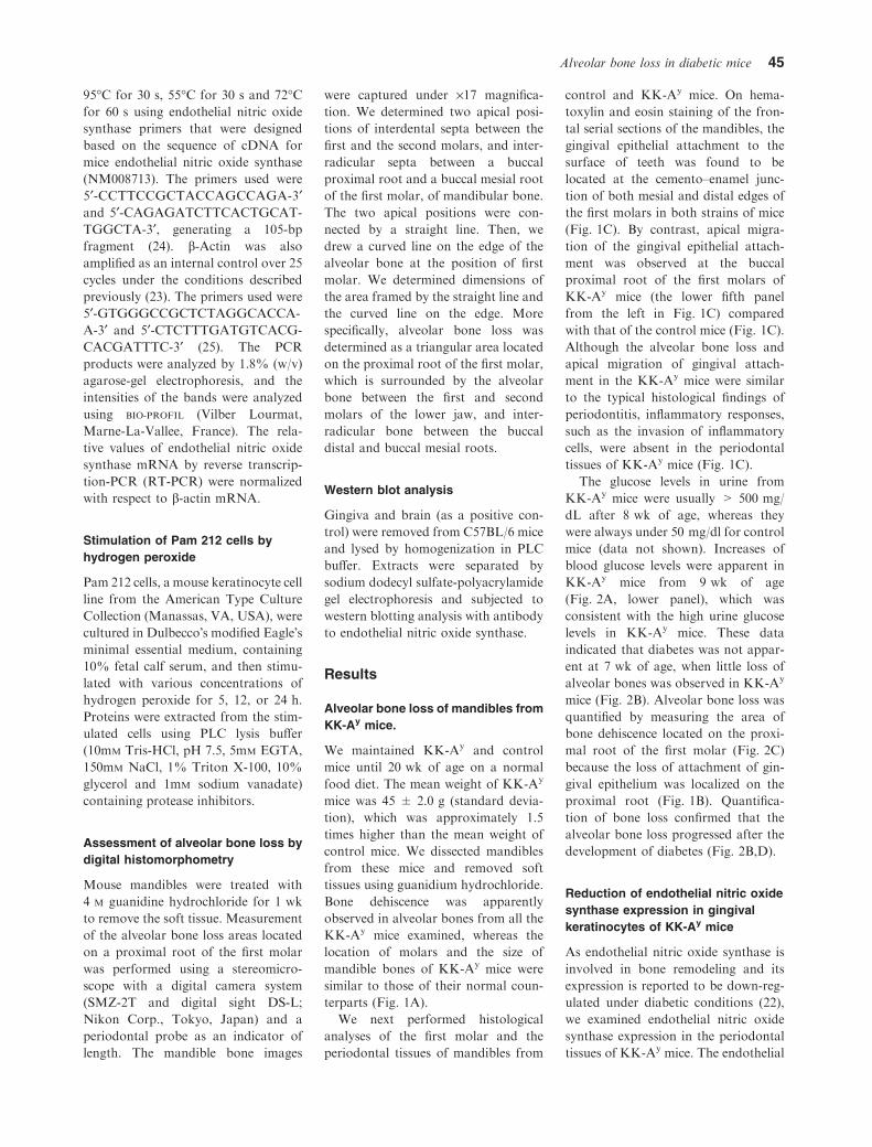
terparts (Fig. 1A).
We next performed histological
analyses of the first molar and the
periodontal tissues of mandibles from
control and KK-Ay mice. On hema-
toxylin and eosin staining of the fron-
tal serial sections of the mandibles, the
gingival epithelial attachment to the
surface of teeth was found to be
located at the cemento–enamel junc-
tion of both mesial and distal edges of
the first molars in both strains of mice
(Fig. 1C). By contrast, apical migra-
tion of the gingival epithelial attach-
ment was observed at the buccal
proximal root of the first molars of
KK-Ay mice (the lower fifth panel
from the left in Fig. 1C) compared
with that of the control mice (Fig. 1C).
Although the alveolar bone loss and
apical migration of gingival attach-
ment in the KK-Ay mice were similar
to the typical histological findings of
periodontitis, inflammatory responses,
such as the invasion of inflammatory
cells, were absent in the periodontal
tissues of KK-Ay mice (Fig. 1C).
The glucose levels in urine from
KK-Ay mice were usually > 500 mg/
dL after 8 wk of age, whereas they
were always under 50 mg/dl for control
mice (data not shown). Increases of
blood glucose levels were apparent in
KK-Ay mice from 9 wk of age
(Fig. 2A, lower panel), which was
consistent with the high urine glucose
levels in KK-Ay mice. These data
indicated that diabetes was not appar-
ent at 7 wk of age, when little loss of
alveolar bones was observed in KK-Ay
mice (Fig. 2B). Alveolar bone loss was
quantified by measuring the area of
bone dehiscence located on the proxi-
mal root of the first molar (Fig. 2C)
because the loss of attachment of gin-
gival epithelium was localized on the
proximal root (Fig. 1B). Quantifica-
tion of bone loss confirmed that the
alveolar bone loss progressed after the
development of diabetes (Fig. 2B,D).
Reduction of endothelial nitric oxidesynthase expression in gingivalkeratinocytes of KK-Ay mice
As endothelial nitric oxide synthase is
involved in bone remodeling and its
expression is reported to be down-reg-
ulated under diabetic conditions (22),
we examined endothelial nitric oxide
synthase expression in the periodontal
tissues of KK-Ay mice. The endothelial
Alveolar bone loss in diabetic mice 45
nitric oxide synthase protein expression
level in gingival keratinocytes ofKK-Ay
mice was significantly lower than in the
gingival keratinocytes of control mice,
whereas endothelial nitric oxide syn-
thase was expressed at similar levels in
the endothelial cells of both mouse
strains (Fig. 3A–D). In order to confirm
this finding,we then performed partially
quantitative analysis of endothelial
nitric oxide synthase mRNA in gingiva
of both mouse strains by RT-PCR
(Fig. 3E). The endothelial nitric oxide
synthase mRNA expression levels in
gingival specimens (lanes 6 and 8) from
KK-Ay mice were higher than those in
the other specimens from KK-Ay mice.
The gingival specimens probably con-
tain other cell types, such as endothelial
cells, in which endothelial nitric oxide
synthase expression was similar
between KK-Ay and control mice.
Although RNA samples were isolated
from gingival tissues, including endo-
thelial cells, we still found a statistically
significant decrease of endothelial nitric
oxide synthasemRNA expression in the
gingivae from KK-Ay mice compared
with those from control mice (Fig. 3F).
Inhibition of endothelial nitric oxidesynthase expression in keratinocytesby reactive oxygen species
Reactive oxygen species play a causal
role in insulin resistance (26). The
production of reactive oxygen species,
such as superoxide and hydrogen
peroxide, was reported to increase in
type 2 diabetic model mice (6). To
explore the possible involvement of
reactive oxygen species in the regula-
tion of endothelial nitric oxide syn-
thase expression in keratinocytes, we
stimulated a mouse keratinocyte cell
line, Pam 212 with hydrogen peroxide.
We detected a 135-kDa molecule as
endothelial nitric oxide synthase in
mouse brain and gingiva extracts, and
in keratinocytes, by western blot
analysis followed by probing with the
endothelial nitric oxide synthase anti-
body. A 90-kDa molecule in kerati-
nocytes appeared to be nonspecifically
recognized by the endothelial nitric
oxide synthase antibody. We found
that hydrogen peroxide significantly
decreased endothelial nitric oxide
synthase expression in a time-depen-
dent manner (Fig. 4). The maximum
reduction (26%) was observed at 8 h
and the expression level remained
decreased at least until 24 h after
the hydrogen peroxide stimulation
(Fig 4).
Pretreatment with an antioxidant,
N-acetylcysteine, abrogated the dec-
rease of endothelial nitric oxide syn-
thase by hydrogen peroxide (Fig. 4C).
To examine if the antioxidant restored
the endothelial nitric oxide synthase
expression in gingival keratinocytes of
KK-Ay mice in vivo, we maintained
KK-Ay and control mice on an N-ace-
tylcysteine -containing diet from 8 wk
of age. After 12 wk of the N-acetyl-
cysteine-containing diet, the lower jaws
were extracted and the decalcified
sections were stained with endothelial
nitric oxide synthase antibody. The
administration of N-acetylcysteine-
containing food restored endothelial
nitric oxide synthase expression in
gingival keratinocytes (Fig. 5). In
addition, we measured hydrogen per-
oxide levels in plasma to confirm
reduction of the reactive oxygen species
level in vivo by the administration of
N-acetylcysteine. The levels of hydro-
gen peroxide in the plasma of KK-Ay
mice were significantly higher than in
control mice and were decreased by the
administration of N-acetylcysteine-
containing food. (Fig. 6A).
A
C
B
Control
1 2 3 4 5 6 7
Mesial Distal
Mesial Distal
Mesial Distal
KK-Ay
1 2 3 4 5 6 7
Fig. 1. Periodontal tissues in diabetic model mice. (A) Mandibles were extracted from male
control and KK-Ay mice at 20 wk of age, and the soft tissues were removed. Bars indicate the
length of 1 mm. (B) Location of the histological section of periodontal tissues in (C). (C) The
extracted mandibles were decalcified with 0.5 M EDTA. Serial sections of the first molars and
the periodontal tissues from control (upper panel) and KK-Ay mice (lower panel) were
stained with hematoxylin and eosin. Arrowheads indicate the cemento–enamel junction.
46 Ohnishi et al.

Recovery of alveolar bone loss inKK-Ay mice fed an N-acetylcysteine-containing diet
We then examined the effect of an
N-acetylcysteine-containing diet on the
alveolar bone loss in KK-Ay mice. The
defiscence of alveolar bone decreased
in KK-Ay mice given an N-acetylcys-
teine-containing diet compared with
the normal-diet counterparts. The
bone loss areas in KK-Ay mice were
decreased by approximately 30% as a
result of eating N-acetylcysteine-con-
taining food, which was still higher
than those of control mice (Fig. 6A,B).
N-Acetylcysteine not only has an anti-
oxidant effect, but also an inhibitory
effect on nuclear factor-jB, which is
associated with osteoblast differentia-
tion. According to these results, we
presented a working hypothesis that
reactive oxygen species are associated
with alveolar bone loss.
Discussion
In the present study, alveolar bone
loss was examined in mandibles from
metabolic syndrome model mice with
type 2 diabetes. The bone loss pro-
gressed after the development of dia-
betes in the mice, at about 8 wk of
age, and the mice also showed a
decrease of endothelial nitric oxide
synthase in the gingival keratinocytes.
Mice fed a diet containing N-acetyl-
cysteine, an antioxidant, restored the
bone loss. However, N-acetylcysteine
is also reported to be an inhibitor for
nuclear factors such as nuclear factor-
jB (27). Nuclear factor-jB is acti-
vated during osteoclast differentiation
by stimulation with receptor activator
of nuclear factor-jB ligand. The pre-
sent study could not disregard the
possibility that inhibition of nuclear
factor-jB activity by N-acetylcysteine
inhibited the progression of alveolar
bone loss. Therefore, we proposed a
working hypothesis that alveolar bone
dehiscence was implicated in the gen-
eration of reactive oxygen species.
A B
C
D
40
Control (7 wk)
Control
20 wk7 wkAge
KK-Ay (7 wk) KK-Ay (20 wk)
Control (20 wk)
30
20
20
10
8
6
4
3
0
Alv
eola
r bo
ne lo
ss (
arbi
trar
y un
it)
Age (wk)
Bod
y w
eigh
t (g
)
10
100
700
600
500
400
300
200
100Blo
od g
luco
se (
mg/
dL)
0
0
20Age (wk)
100
KK-Ay
p > 0.1
p < 0.01
Fig. 2. Progress of alveolar bone loss and increase of blood glucose levels. (A) Body weights
(upper panel) and blood glucose levels (lower panel) of control mice and KK-Ay mice were
measured at the time-points indicated. (B) Mandibles were extracted from male control or
KK-Ay mice at 7 and 20 wk of age, and the soft tissues were removed. Bars indicate the
length of 1 mm. (C) The edge of alveolar bone is indicated as a black line and the purple area
defines the alveolar bone loss. (D) Alveolar bone loss at the proximal areas of the first molars
was measured in control and KK-Ay mice at 7 and 20 wk of age, as described in the Material
and methods (results are given as means ± standard deviation, n = 4).
A
B
C
D
E
F
Control
eNOS1 2 3 4 5 6 7 8
p < 0.05
9 10
2
0
4
eNO
S ex
pres
sion
(arb
itrar
y un
it)
6
8
10
Actin
KK-Ay
Control KK-Ay
Fig. 3. Endothelial nitric oxide synthase
expression in gingiva from control or dia-
betic mice. Mandibles were extracted from
male control (A, C) or KK-Ay (B, D) mice
at 20 wk of age. After treatment with (C, D)
or without (A, B) proteinase K, the man-
dibles were decalcified with 0.5 M EDTA.
The sections were stained with endothelial
nitric oxide synthase antibody, as described
in the Material and methods. Arrows indi-
cate endothelial cells. (A and B, magnifica-
tion ·64; C and D, magnification ·320.) (E)Total RNA extracted from individual
periodontal tissues of control (lanes 1–5) or
KK-Ay (lanes 6–10) mice was reverse tran-
scribed and amplified using the polymerase
chain reaction (PCR). The reverse tran-
scription-PCR products were subjected to
electrophoresis and stained with ethidium
bromide. (F) The intensities of the bands of
endothelial nitric oxide synthase in (E) were
analyzed using BIO-PROFIL, and relative val-
ues of endothelial nitric oxide synthase
mRNA were normalized with respect to
those for b-actin. eNOS, endothelial nitric
oxide synthase.
Alveolar bone loss in diabetic mice 47

Previous studies on endothelial ni-
tric oxide synthasedeficient mice have
revealed that endothelial nitric oxide
synthase regulates bone formation by
activating osteoblasts (20,21). Consis-
tently, RT-PCR and immunohisto-
chemical staining showed the
expression of endothelial nitric oxide
synthase in bone cells among three
isoforms of NOS (28). However, nei-
ther the detailed quantification nor the
physiological significance of endothe-
lial nitric oxide synthase expression in
osteoblasts has been clarified. In this
study, we detected endothelial nitric
oxide synthase protein in Pam 212, a
mouse keratinocytic cell line, but not in
MC3T3-E1, a mouse osteoblastic cell
line, by western blot analysis (data not
shown). These observations suggested
that the expression of endothelial nitric
oxide synthase in osteoblasts may be
lower than in other cell types, such as
keratinocytes. Thus, it is currently
uncertain whether the bone defect in
endothelial nitric oxide synthase
knockout mice is a result of the loss of
endothelial nitric oxide synthase in
osteoblasts or other cell types.
Studies on endothelial nitric oxide
synthase-deficient mice have revealed
that endothelial nitric oxide synthase
plays an important role in the preven-
tion of ovariectomy-induced bone loss
(20,21). The administration of N-ace-
tylcysteine was reported to abolish
ovariectomy-induced bone loss (19). In
the present study, we found that
N-acetylcysteine restored endothelial
nitric oxide synthase expression in the
keratinocytes of diabetic mice, accom-
panied with the reversal of alveolar
bone loss. While the role of gingival
keratinocytes in bone formation is
unclear, considering these obser-
vations, the alveolar bone defect of
diabetic mice may be associated with
reduction of endothelial nitric oxide
synthase expression in the adjacent
gingival keratinocytes. In our present
study, we also found that treatment
with hydrogen peroxide decreased
endothelial nitric oxide synthase
expression in keratinocytes. Moreover,
we found that N-acetylcysteine did not
affect the ascorbic acid-induced activ-
ity of alkaline phosphatase and calci-
fication of MC3T3-E1 cells (data not
A
B
C
197 kDa
eNOS
eNOS
eNOS
Pam21
2
Gingiva
Brain
-
-
-
126 kDa
Time (h) 0 0.5 2
Control NAC
8 13 25
Time (h) 0 4 12 40 12
81 kDa
Fig. 4. Effect of hydrogen peroxide on endothelial nitric oxide synthase expression in
keratinocytes. (A) Extracts from mouse gingiva and brain, accompanied by Pam 212 cell
lysate (50 lg of each), were separated on a 7.5% polyacrylamide gel and subjected to
immunoblotting with endothelial nitric oxide synthase antibody. (B, C) Pam 212 cells were
cultured in Dulbecco�s modified Eagle�s minimal essential medium containing 10% fetal calf
serum and then stimulated with 0.2 mM hydrogen peroxide in the presence (C) or absence (B,
C) of 5 mM N-acetylcysteine for the indicated time. Aliquots of 200 lg of protein in cell
lysates were analyzed by western blotting and detection with anti-endothelial nitric oxide
synthase. eNOS, endothelial nitric oxide synthase; NAC, N-acetylcysteine.
Control KK-Ay KK-Ay
Normal food Normal food NAC food
Fig. 5. Endothelial nitric oxide synthase expression in gingiva from control or diabetic mice.
Control or KK-Ay mice at 8 wk of age were maintained on a normal diet or on a diet of
N-acetylcysteine–containing food for 12 wk. Mandibles were extracted from male control
and KK-Ay mice, and then decalcified in 0.5 M EDTA. The sections were stained with
endothelial nitric oxide synthase antibody (upper panels) or nonimmunized rabbit IgG (lower
panels), as described in the Material and methods. D, dentin; eNOS, endothelial nitric oxide
synthase; NAC, N-acetylcysteine.
48 Ohnishi et al.

shown) suggesting that N-acetylcyste-
ine may not directly stimulate the
differentiation of osteoblasts. Consid-
ering the abovementioned importance
of endothelial nitric oxide synthase in
cell types other than osteoblasts, it is
possible that the inhibitory effect of
N-acetylcysteine on bone loss occurs
through restoring the endothelial nitric
oxide synthase expression in cell types
other than osteoblasts.
It is of note that the form of the
alveolar bone defect in KK-Ay mice
was sphenoid-shaped, which is distinct
from the bone defects induced by gen-
eral periodontitis. The alveolar bone
dehiscence means that a buccal bone
defect is present at the locus adjacent
to dental roots, which results in an
occlusal force on the alveolar bone. In
fact, occlusal wear was observed in the
molar of lower jaws from the diabetic
mice (Fig. 1A). Occlusal forces do not
initiate periodontitis, but modify and
progress the attachment loss and alve-
olar bone loss (29,30). In this study, we
did not observe distinct inflammatory
responses, such as the invasion of
inflammatory cells, in the periodontal
tissues of KK-Ay mice. The remodeling
of alveolar bone, which responds to
occlusal force in KK-Ay mice, might be
reduced compared with that in control
mice, because the bone remodeling in
diabetic animals is reported to be
decreased (31).
The expression of endothelial nitric
oxide synthase in gingiva has been
reported to be indirectly involved in
alveolar bone-induced bone remodel-
ing, leading to tooth movements (32).
In the present study, the expression of
endothelial nitric oxide synthase in the
gingival tissues of orthodontically
treated teeth was significantly greater
than that of control teeth. In other
reports, administration of a general
inhibitor of nitric oxide synthase, but
not of an indicible nitric oxide syn-
thase-specific inhibitor, reduced the
tooth movement by an orthodontic
force (33,34), suggesting that the
activity of endothelial nitric oxide
synthase affects alveolar bone remod-
eling. These reports, along with our
immunohistochemical staining results,
in which endothelial nitric oxide
A
B
C
Normal food
Control
12
Alv
eola
r bo
ne lo
ss (
arbi
trar
y un
it)
10
8
6
4
4
5
3
Plas
ma
H2O
2 (n
mol
MD
A/m
L)
2
1
0
2
0
KK-Ay
Control KK-Ay
Control KK-Ay
NAC food
Normal food
p < 0.01 p < 0.01
p < 0.05
NAC food
Normal foodp < 0.01
NAC food
Fig. 6. Effect of an N-acetylcysteine–containing diet on alveolar bone loss and plasma
hydrogen peroxide levels in diabetic mice. (A) Control or KK-Ay mice at 8 wk of age were
maintained on a diet of normal or N-acetylcysteine-supplemented food for 12 wk. The
hydrogen peroxide levels in plasma of KK-Ay and control mice were measured (A).
Mandibles were extracted from male control (upper panel) and KK-Ay (lower panel) mice
at 20 wk of age, and the soft tissues were removed. The bars indicate the length of 1 mm.
The edge of alveolar bone is indicated as a black line and the purple area defines the
alveolar bone loss. (B, C) Alveolar bone loss at the proximal areas of the first molars in
control or KK-Ay mice was measured as described in the Material and methods
(means ± standard deviation, n = 6). H2O2, hydrogen peroxide; MDA, malondialdehyde;
NAC, N-acetylcysteine.
Alveolar bone loss in diabetic mice 49
synthase expression is localized on the
gingival epithelium, indicate the possi-
bility that alveolar bone remodeling is
affected by endothelial nitric oxide
synthase activity in periodontal tissues
such as gingiva.
Reduced expression of endothelial
nitric oxide synthase has been reported
in skin keratinocytes of other type 2
diabetic model mice with obesity, and
is implicated as a mechanism for the
impaired healing of diabetic patients
(22). Our present data have indicated
that the reduced endothelial nitric
oxide synthase expression in keratino-
cytes occurs as a consequence of
increased reactive oxygen species gen-
eration. It should be noted, however,
that other mechanisms are conceivable
for the reduced endothelial nitric oxide
synthase expression. One possible
candidate is tumor necrosis factor-a.Tumor necrosis factor-a, whose
expression increases in type 2 diabetes,
has been reported to down-regulate
endothelial nitric oxide synthase
expression (35,36). However, the plas-
ma levels of tumor necrosis factor-a in
control and KK-Ay mice were lower
than 50 pg/mL and there was no sig-
nificant difference between the tumor
necrosis factor-a levels in these mice
(data not shown). The second possible
candidate is adiponectin. The expres-
sion of adiponectin, which up-regulates
endothelial nitric oxide synthase
expression, has been reported to be
decreased in the obese KK-Ay mice
(37). The third possible mechanism
involves decreased insulin action.
Insulin increases the expression of
endothelial nitric oxide synthase so that
endothelial nitric oxide synthase is
produced at short intervals in vivo (38).
Thus, decreased levels of insulin may
lead to the reduced endothelial nitric
oxide synthase expression observed in
the KK-Ay mice with type 2 diabetes.
Reactive oxygen species are known
to be one of the reasons for various
complications of type 2 diabetes,
including periodontitis. In the present
study we found that an antioxidant,
N-acetylcysteine, may exert an inhibi-
tory effect on bone loss. N-Acetylcys-
teine is known be a popular supplement
and beneficial in conditions character-
ized by an oxidative stress (39). How-
ever, N-acetylcysteine is also known to
be an inhibitor for nuclear factor-jB,which is associated with osteoclast
differentiation. Because the target
molecules of N-acetylcysteine are still
obscure, further investigation is neces-
sary to prove that reactive oxygen spe-
cies are associated with the alveolar
bone in the diabetes model mice.
Acknowledgements
This work was supported by a grant
from the Ministry of Education, Cul-
ture, Sports, Science and Technology
of Japan (Grants 17591943, 14571767
and 19390474).
References
1. Reaven GM. Banting lecture 1988. Role
of insulin resistance in human disease.
Diabetes 1988;37:1595–1607.
2. Balkau B, Charles M. European group for
the study of insulin resistance (EGIR).
Comment on the provisional report of the
WHO consultation [letter]. Diabet Med
1999;16:442–443.
3. Nishimura M. Breeding of mice strains for
diabetes mellitus. Exp Anim 1969;18:147–
157.
4. Iwatsuka H, Shino A, Suzuoki Z. General
survey of diabetic features of yellow KK
mice. Endocrinol Jpn 1970;17:23–35.
5. Aizawa-Abe M, Ogawa Y, Masuzaki H
et al. Pathophysiological role of leptin in
obesity-related hypertension. J Clin Invest
2000;105:1243–1252.
6. Furukawa S, Fujita T, Shimabukuro M
et al. Increased oxidative stress in obesity
and its impact on metabolic syndrome.
J Clin Invest 2004;114:1752–1761.
7. Touyz RM, Schiffrin EL. Reactive oxygen
species in vascular biology: implications in
hypertension. Histochem Cell Biol
2004;122:339–352.
8. Karima M, Kantarci A, Ohira T et al.
Enhanced superoxide release and elevated
protein kinase C activity in neutrophils
from diabetic patients: association with
periodontitis. J Leukoc Biol 2005;78:862–
870.
9. Koya D, King GL. Protein kinase C
activation and the development of dia-
betic complications. Diabetes 1998;47:
859–866.
10. Brownlee M. Biochemistry and molecular
cell biology of diabetic complications.
Nature 2001;414:813–820.
11. Shlossman M, Knowler WC, Pettitt DJ,
Genco RJ. Type 2 diabetes mellitus and
periodontal disease. J Am Dent Assoc
1990;121:532–536.
12. Tsai C, Hayes C, Taylor GW. Glycemic
control of type 2 diabetes and severe
periodontal disease in the US adult pop-
ulation. Community Dent Oral Epidemiol
2002;30:182–192.
13. Pihlstrom BL, Michalowicz BS, Johnson
NW. Periodontal diseases. Lancet
2005;366:1809–1820.
14. Williams RC. Periodontal disease. N Engl
J Med 1990;322:373–382.
15. Genco RJ. Host responses in periodontal
diseases: current concepts. J Periodontol
1992;63:338–355.
16. Guarnieri C, Zucchelli G, Bernardi F,
Scheda M, Valentini AF, Calandriello M.
Enhanced superoxide production with no
change of the antioxidant activity in gin-
gival fluid of patients with chronic adult
periodontitis. Free Radic Res Commun
1991;15:11–16.
17. Tsai CC, Ho YP, Chen CC. Levels of
interleukin-1 beta and interleukin-8 in
gingival crevicular fluids in adult
periodontitis. J Periodontol 1995;66:852–
859.
18. Ren Y, Maltha JC, Van�t Hof MA, Von
Den Hoff JW, Kuijpers-Jagtman AM,
Zhang D. Cytokine levels in crevicular
fluid are less responsive to orthodontic
force in adults than in juveniles. J Clin
Periodontol 2002;29:757–762.
19. Lean JM, Davies JT, Fuller K et al.
A crucial role for thiol antioxidants in
estrogen-deficiency bone loss. J Clin Invest
2003;112:915–923.
20. Aguirre J, Buttery L, O�Shaughnessy M
et al. Endothelial nitric oxide synthase
gene-deficient mice demonstrate marked
retardation in postnatal bone formation,
reduced bone volume, and defects in
osteoblast maturation and activity. Am J
Pathol 2001;158:247–257.
21. Armour KE, Armour KJ, Gallagher ME
et al. Defective bone formation and ana-
bolic response to exogenous estrogen in
mice with targeted disruption of endothe-
lial nitric oxide synthase. Endocrinology
2001;142:760–766.
22. Stallmeyer B, Anhold M, Wetzler C,
Kahlina K, Pfeilschifter J, Frank S.
Regulation of eNOS in normal and dia-
betes-impaired skin repair: implications
for tissue regeneration. Nitric Oxide
2002;6:168–177.
23. Ohnishi T, Suwa M, Oyama T, Arakaki
N, Torii M, Daikuhara Y. Prostaglandin
E2 predominantly induces production of
hepatocyte growth factor/scatter factor in
human dental pulp in acute inflammation.
J Dent Res 2000;79:748–755.
24. Chu Y, Heistad DD, Knudtson KL,
Lamping KG, Faraci FM. Quantification
of mRNA for endothelial NO synthase in
mouse blood vessels by real-time poly-
merase chain reaction. Arterioscler
Thromb Vasc Biol 2002;22:611–616.
50 Ohnishi et al.

25. Tokunaga K, Taniguchi H, Yoda K,
Shimizu M, Sakiyama S. Nucleotide
sequence of a full-length cDNA for mouse
cytoskeletal b-actin mRNA. Nucleic Acids
Res 1986;14:2829.
26. Houstis N, Rosen ED, Lander ES. Reac-
tive oxygen species have a causal role in
multiple forms of insulin resistance. Nat-
ure 2006;440:944–948.
27. Allen RG, Tresini M. Oxidative stress and
gene regulation. Free Radic Biol Med
2000;28:463–499.
28. Helfrich MH, Evans DE, Grabowski PS,
Pollock JS, Ohshima H, Ralston SH.
Expression of nitric oxide synthase iso-
forms in bone and bone cell cultures.
J Bone Miner Res 1997;12:1108–1115.
29. Gher ME. Changing concepts. The effects
of occlusion on periodontitis. Dent Clin
North Am 1998;42:285–299.
30. Serio FG, Hawley CE. Periodontal trau-
ma and mobility. Diagnosis and treatment
planning. Dent Clin North Am 1999;43:37–
44.
31. Liu Z, Aronson J, Wahl EC et al. A novel
rat model for the study of deficits in bone
formation in type-2 diabetes. Acta Orthop
2007;78:46–55.
32. D�Attillio M, Di Maio F, D�Arcangela C
et al. Gingival endothelial and inducible
nitric oxide synthase levels during ortho-
dontic treatment: a cross-sectional study.
Angle Orthod 2004;74:851–858.
33. Shirazi M, Nilforoushan D, Alghasi H,
Dehpour AR. The role of nitric oxide in
orthodontic tooth movement in rats.
Angle Orthod 2002;72:211–215.
34. Hayashi K, Igarashi K, Miyoshi K,
Shinoda H, Mitani H. Involvement of
nitric oxide in orthodontic tooth move-
ment in rats. Am J Orthod Dentofacial
Orthop 2002;122:306–309.
35. Nilsson J, Jovinge S, Niemann A, Rene-
land R, Lithell H. Relation between plas-
ma tumor necrosis factor-alpha and
insulin sensitivity in elderly men with
non-insulin-dependent diabetes mellitus.
Arterioscler Thromb Vasc Biol 1998;18:
1199–1202.
36. Anderson HD, Rahmutula D, Gardner
DG. Tumor necrosis factor-alpha inhibits
endothelial nitric-oxide synthase gene
promoter activity in bovine aortic endo-
thelial cells. J Biol Chem 2004;279:963–
969.
37. Hattori Y, Suzuki M, Hattori S, Kasai K.
Globular adiponectin upregulates nitric
oxide synthaseproduction in vascular
endothelial cells. Diabetologia 2003;
46:1543–1549.
38. Kuboki K, Jiang ZY, Takahara N et al.
Regulation of endothelial constitutive ni-
tric oxide synthase gene expression in
endothelial cells and in vivo: a specific
vascular action of insulin. Circulation
2000;101:676–681.
39. Kelly GS. Clinical applications of N-ace-
tylcysteine. Altern Med Rev 1998;3:114–
127.
Alveolar bone loss in diabetic mice 51