Embed Size (px)
Citation preview

Louisiana State UniversityLSU Digital Commons
LSU Master's Theses Graduate School
2013
Oxidative stress based response of a transcriptionalregulator, OhrR, from Burkholderia thailandensisAnuja PandeLouisiana State University and Agricultural and Mechanical College, [email protected]
Follow this and additional works at: https://digitalcommons.lsu.edu/gradschool_theses
This Thesis is brought to you for free and open access by the Graduate School at LSU Digital Commons. It has been accepted for inclusion in LSUMaster's Theses by an authorized graduate school editor of LSU Digital Commons. For more information, please contact [email protected].
Recommended CitationPande, Anuja, "Oxidative stress based response of a transcriptional regulator, OhrR, from Burkholderia thailandensis" (2013). LSUMaster's Theses. 2895.https://digitalcommons.lsu.edu/gradschool_theses/2895

OXIDATIVE STRESS BASED RESPONSE OF A TRANSCRIPTIONAL REGULATOR, OhrR, FROM BURKHOLDERIA THAILANDENSIS.
A Thesis
Submitted to the Graduate Faculty of the Louisiana State University and
Agricultural and Mechanical College in partial fulfillment of the requirement for degree of
Master of Science
in
The Department of Biological Sciences
by Anuja Rajiv Pande
B.Sc, University of Mumbai, 2007 M.Sc, SVKM’s NMIMS University, 2011
December 2013

ii
Dedicated to,
My Parents, Aai and Baba
And
My advisor, Dr. Anne Grove.
iii
ACKNOWLEDGEMENTS
The ride’s been a bumpy one, but in the end as they say, ‘it’s all worth it’! The study
couldn’t have reached its completion, and successfully, without the unconditional love
and support of people closely associated with me.
First and foremost I would like to thank God for blessing me with inner peace,
strength and positive environment to live and work in, which made this achievement
possible. My advisor, Dr. Anne Grove, for her incessant encouragement and
enthusiasm to do science, ‘have fun’ with it and think out of the box. It is only
because of her that research now has a new meaning in my life, something I am
passionate about and ready to pour my heart into. She totally personifies the clichéd
statement of being a ‘Friend, Philosopher, Guide’! My committee members Dr. Marcia
Newcomer and Dr. Yong-Hwan Lee, for their valuable time, suggestions, challenging
questions and guidance during my research project.
I would like to thank all my friends at the Grove Lab, Dinesh, Smitha, Ambuj, Ashish,
Kavitha, Hao, Khoa, Arvind, Tiffany, Brian, Daniel, Emmanuela and Dante for their
technical support and a constant fun-filled environment in lab. With a special mention
of Dinesh for his invaluable help in making me understand several concepts and all
the brainstorming sessions we would have at untimely hours and Smitha for being a
patient listener and bearing with all my emotional outbursts! 5th floor Choppin Hall
members, especially Alexandria for helping me with Gel Filtration analysis and
members from Dr. Newcomers lab Matt, Sunayana, Cody, and Erin for being kind
and helpful at several occasions.
Genomics and Protein facilities at LSU, for allowing me to use their RT-PCR and
Circular Dichroism machines.
I truly feel blessed to have such a wonderful family, my parents (Mr. Rajiv Pande and
Mrs. Sharmila Pande), my sister (Rutuja Pande), my husband (Sandeep Pathak) and
my Da (Santonu Ghosh), who have been my pillar of strength throughout my life and

iv
have been a constant source of motivation throughout this project. My parents
endless love and constant words of encouragement, untimely wake up calls,
invaluable suggestions and advice from them, my sister backing me up and
reminding me of my dreams, Da’s energy and never say die attitude and my
husbands unconditional love and thoughts on looking at research as a whole learning
process, have all been instrumental in this accomplishment. I would also like to thank
my in-laws for their love and support.
I am extremely grateful to my previous advisors Dr. Krutika Desai (Masters) for her
Dr. Shalini Rai (Bachelors) for their love and inspirational words and my mentors Dr.
Sagarika Damle, Dr. Purvi Bhatt, Dr. Rashmikant Mohile, Dr. Preeti Mohile, Dr. Rajiv
Joshi and Mr. Benedict Mascarenhas for their kind words and contributions in my
scientific career.
Last but not the least my wonderful friends here and in India for being so caring, and
at times parent-like strict with me. This journey would not have been a complete nor a
special one without them, my close friends Ami, Richie, An’s, Venks, Braj, and
Jacobo for always being there for me and being my strength! Asawari for her
untimely scientific inputs and helping me a lot in preparing for my defence
presentation. Awesomest room-mates ever Shabs, Pri, Deepti, and Jeed for making
this place a home away from home for me, bearing with my tantrums, literally living
my life with me and most importantly being my food providers to keep me going! I
would also like to take this opportunity and thank all my dear friends Nidhi, Saroj,
Prat, Tulip, Sudha, Shafi and Anindita for being so loving, and inspiring at all times.

v
TABLE OF CONTENTS
Acknowledgments ........................................................................................................... iii
List of Tables ................................................................................................................... vi
List of Figures ................................................................................................................ vii
Abstract ......................................................................................................................... viii
Introduction ......................................................................................................................1
Materials and Methods ................................................................................................... 13
Results and Discussions ................................................................................................ 21
References ..................................................................................................................... 41
Vita ................................................................................................................................. 46
vi
LIST OF TABLES
1. Comparison of melting temperatures (Tm) of oxidized and reduced OhrR. ................ 33

vii
LIST OF FIGURES
1. General architecture of a typical MarR homologue. .................................................2
2. Schematic representation of a MarR gene locus ......................................................6
3. Purified OhrR resolved on 12% SDS-PAGE with its CD spectra ............................... 20
4. Glutaraldehyde crosslinking and gel filtration analysis of OhrR ................................. 22
5. Effect of oxidants on OhrR as resolved in 12% SDS-PAGE ...................................... 24
6. Modeled structure of B. thailandensis OhrR............................................................... 25
7. Surface rendering of the modeled B. thailandensis OhrR .......................................... 26
8. Metal treated samples of OhrR resolved in 12% SDS-PAGE .................................... 29
9. Reversibility of OhrR oxidation as observed on SDS-PAGE ...................................... 30
10. Thermal melts depictIng fluorescence emission spectra of OhrR ............................ 32
11. OhrR treated with low concentration of oxidants resolved on 12% SDS-PAGE ....... 33
12. Promoter region of ohr ............................................................................................. 35
13. EMSA showing titration of OhrR protein with labeled ohrO-l (long) DNA ................. 35
14. Plot indicating half maximal saturation of ohrO-l DNA ............................................. 36
15. Competitive EMSA ................................................................................................... 36
16. Comparative EMSA ................................................................................................. 37
17. Proposed Mechanism of OhrR binding DNA………………………………...……..………39
viii
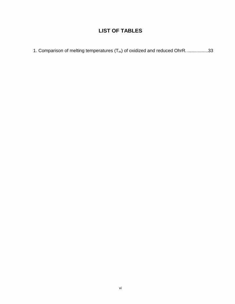
ABSTRACT
Being ubiquitous in nature, bacteria are often faced by environmental stresses. The
predominant form of stress is oxidative stress, which prevails during host invasion. As
one of the host’s protective mechanisms, bacteria are bombarded by organic and
inorganic oxidants. Several bacterial antioxidant systems are potent enough to
scavenge host-derived reactive oxygen species, helping in bacterial survival. Broadly,
they are categorized into those specific for inorganic or organic oxidants. Although a
lot has been studied about the former, the latter still remains uncharacterized in
several organisms.
Burkholderia thailandensis is a soil dwelling bacterium, continually under stress from
organic exudates released from plants in addition to other biotic compounds. Besides
sharing about 80% genetic homology with its pathogenic homologues B.
pseudomallei and B. mallei, the oxidative environment (rich in organic oxidants) it
thrives in makes it an interesting model organism for studying the oxidant sensor-
responder proteins it possesses and their mechanism of action.
With an emphasis on such proteins, belonging to the MarR family, OhrR (organic
hydroperoxide reductase regulator) protein from B. thailandensis was characterized
using in vitro assays. The gene was cloned from B. thailandensis genomic DNA and
protein expressed in E. coli. The protein was found to exist as a homodimer. It

ix
formed a series of reversible, oligomeric species on being treated with hydrogen
peroxide, cumene hydroperoxide and tert-butyl hydroperoxide as was observed from
SDS-PAGE. The reduced form of the protein was observed to be relatively
thermostable (Tm of 63.5 °C), with an appreciable thermal instability observed in
OhrR treated with higher concentration of oxidants (5 mM). DNA binding assays
revealed specificity of B. thailandensis for the promoter region of ohr, although the
DNA length to which it bound made a difference in the stability of protein-DNA
complexes formed and detected using EMSA.
Since OhrR is predicted to control production of organic hydroperoxide reductase,
which is important for detoxifying organic hydroperoxides, the initial characterization
and oxidant response of B. thailandensis OhrR, determined using in vitro assays,
thus indicate its probable role as a crucial protein among the B. thailandensis protein
machinery, aiding in bacterial survival.
1
INTRODUCTION
Oxidative Stress
Oxygen (O2) is essential for most organisms living on planet Earth. In addition to
being the most abundant element by mass in the Earth’s crust, 21% of the total
volume of air we breathe constitutes oxygen. Except for certain anaerobic and aero-
tolerant single-celled organisms, all animals, plants and bacteria require oxygen for
carrying out metabolic activities efficiently.
Originally, oxygen was not a part of the Earth’s atmosphere. It was only with the
evolution of photosynthetic organisms that oxygen began to be formed and
replenished in the atmosphere. For a cell’s metabolic activities, most of the oxygen
from air is useful as found in its diatomic form. However, being chemically reactive,
molecular oxygen can dissipate into several unstable species called ‘free radicals’
that are extremely harmful [1, 2].
A free radical is any elemental species that contains one or more unpaired electrons.
This makes it highly unstable and therefore very reactive. They stabilize themselves
by reacting with other substrate molecules, usually biomolecules, thereby modifying
these molecules and in some cases even rendering them inactive.
Reactive oxygen species (ROS) are free radical species generated from
molecular oxygen. They are important for normal functioning of living systems
because they play a crucial role in several cellular processes such as apoptosis,
cell-proliferation and differentiation [3], in the immune system as defense against
invading pathogens[4], in transcriptional regulation [5] are involved in the electron
transport chain and in fact are byproducts of aerobic metabolism [6].

2
However, on the flipside, they are also one of the major causative factors of
neurodegenerative and cardiovascular disorders, skin disorders, diabetes and
can even lead to cancer progression [2, 7].
Under normal conditions, cells maintain a steady level of free radicals. They do
so by balancing the amount of free radicals generated with the cells own
enzymatic and non-enzymatic antioxidant (free radical scavengers) levels. This is
the sole reason why structural biomolecules such as proteins, lipids and nucleic
acids are still stable even in aerobic environments [6]. The problem arises when
this balance is disturbed, and the system is not able to proportionally detoxify the
amount of free radicals produced. Biological systems are continually challenged
with a burst of free radicals either exogenously (due to exposure to solar radiations,
xenobiotics, or elevated oxygen levels) and/or endogenously (in phagocytes and/or
host’s defense response, unorthodox oxidative metabolic cycles, or depleted
antioxidants). Such a condition is called ‘oxidative stress’ [2, 7].
As a result of their ubiquitous nature, prokaryotic organisms are most prevalently
exposed to a variety of oxidative environmental conditions. Other than normal
circumstances such as aerobic respiration wherein bacteria are exposed to ROS,
another common scenario where bacterial cells are exposed to a burst of oxide
radicals is when an invading pathogen encounters the primary line of immune
response of the plant or animal host. Elevated levels of these free radicals can
damage proteins and lipids in bacterial membranes, in addition to interacting with
cellular proteins and nucleic acids, ultimately resulting in cell death [8]. Being
non-specific in nature, free-radical production also results in host tissue damage.
3
The expanse of a bacterial infection therefore depends both on the antioxidant
response mechanism of the bacteria to the host-derived ROS, and the extent of
host tissue damage [9].
With their rapidly evolving antioxidant systems, bacteria are increasingly
becoming resistant to oxidant responses in the process causing host tissue
damage, making bacterial oxidative stress responses therefore a leading cause
of concern for the scientific world.
Multiple Antibiotic Resistance Regulators (MarRs)
Prokaryotes dwell in diverse surroundings varying in pH, osmolarity, temperature,
and/or nutrient availability, with each condition capable of being highly stressful
for the organism [10]. It is intriguing how the bacteria have been and even now
are still evolving to deal with each stress condition, having such a basic cellular
and metabolic machinery.
In order to survive and proliferate within the host, among the vast array of
proteins bacteria possess to deal with stress, are a family of proteins conferring
the bacteria antibiotic resistance. These proteins are transcriptional regulators
which sense the oxidative environment, and regulate associated expression of
genes involved directly or indirectly as antioxidants (enzymatic or non-
enzymatic). Thus, it is the antioxidative response of the bacterium that confers it
full virulence [9].
First identified in multidrug resistant strains of Escherichia coli [11] such family of
proteins that help bacteria sense and respond to stressful environmental
challenges are the proteins belonging to multiple antibiotic resistance regulator
4
(MarR) protein family [12]. With more than 12,000 MarR like proteins annotated
as produced in bacterial and archaeal species, they seem to be highly prevalent
and therefore biologically important. Till date, MarRs have been shown to
function as transcriptional regulators and to control the expression of several
genes encoding proteins involved in essential cellular processes such as certain
metabolic pathways, enzymatic or non-enzymatic antioxidation, end-product
degradation and/or export of toxic substances (byproducts of metabolism, drugs,
antibiotics, organic solvents and oxidative agents found in household detergents
and disinfecting agents) [13, 14] and in some organisms even virulence [15, 16].
Structure of a MarR protein
As shown in the first crystal structure of a MarR protein isolated from E. coli,
proteins belonging to the MarR family exist as dimers and possess features
common to DNA binding proteins. They usually adopt a triangular shaped
structure (Figure 1) with each subunit composed of six α-helices and three β-
strands assuming an α1 – α6, β1 - β3 topology. The N- terminal region of the
protein has been found to be involved in protein-protein interactions while it is the
C-terminal region, which is actually involved in DNA binding [17, 18]. The
structure of each monomeric unit consists of a DNA-binding domain with a
characteristic winged helix-turn-helix (wHTH) motif, and an extensive inter-
subunit dimerization interface [19]. The terminal residues of both the N- and C-
terminal regions form a hydrophobic dimerization interface stabilized by
hydrophobic interactions and hydrogen bonding. Just like in humans, where an

5
elbow directs the movement of our forearm, the dimerization domain is
responsible for flexibility of the DNA binding domains.
Figure 1. General architecture of a typical MarR homologue with chain A (cyan) and chain B (multicolored) showing different domains. Helices 3 (magenta) and 4 (brown) form the DNA binding domain; helices 1 (red), 5 (orange) and 6 (purple) form the dimerization domain; helix 2 (dark red) lying perpendicular to helix 5 and helix 5 form the connection link between the DNA binding domain and the top part of the MarR structure. Figures 1, 6 and 7 were drawn using Pymol software (www.pymol.org).
The hydrophobic residues in the dimerization domain also help maintain a spatial
arrangement between the DNA binding lobes so that each lobe can work
independently of the other and ensure better DNA binding [20].
DNA binding of MarRs
MarR homologs bind to their cognate DNA sequence as a dimer [11]. The wHTH
DNA binding motif (conserved for DNA binding in MarR homologs across all
domains of life) has a profound specificity for double-stranded DNA sequences
containing inverted repeats, which may or may not be completely palindromic.
Dimerization
domain
DNA
binding
domain

6
Based on previous structural and biochemical studies, the wHTH recognition
motif of the DNA binding domain contacts the DNA just as a bird claw clutching
onto an object. In almost all MarRs, the recognition helix of the DNA binding
domain interlocks with the major groove of DNA first, followed by the secondary
association of the winged region with the adjacent minor groove ensuring tight
binding [21]. The positively charged residues lining the winged region play a
crucial role in DNA binding [22]. Studies also indicate the role of the wing in
multimer formation via protein-protein interactions [23].
Figure 2. Schematic representation of a MarR gene locus. The green arrow represents the gene coding for a MarR protein while the blue arrow represents the gene that is being regulated. Panel on the left shows the MarR protein repressing gene expression. Panel on the right shows derepression of the gene under regulation
The MarR homologs are encoded by gene loci mostly including two divergent
genes, of which one gene encodes for the MarR homolog itself and the other for
the gene it is regulating. The intergenic gene sequence separating the two genes
contains the MarR-specific cognate DNA. This allows them to regulate both
genes, thereby also functioning as auto-regulators [24].
MarRs mainly function as transcriptional repressors [25] although (some/few) of
them are also known to behave as transcriptional activators or sometimes both,
depending on the dynamics involved in DNA binding [26]. What governs their
behavior as gene activators or repressors is the positioning of MarR homologs
onto or upstream of the promoter site. Transcriptional repression is achieved by
x x

7
the MarR protein specifically binding the intergenic DNA sequence, with the
sequence usually overlapping promoter region of a gene, thus causing the MarR
protein to occupy the transcription start site of the gene (the binding site for RNA
polymerase). This can be achieved only when the protein is bound to the DNA in
its reduced or unmodified conformation [19, 26] Gene activation or derepression
is achieved by the transcriptional factor coming off or remaining loosely bound to
the DNA. This helps in making room for the RNA polymerase complex to bind
onto the promoter site and continue transcription and therefore gene expression.
DNA dissociation is typically a consequence of the MarR protein conformation
being changed or modified by an external stimulus, either in the form of binding
of a small molecule ligand or oxidation of sensitive cysteine residues [27-30].
Organic Hydroperoxide Reductase Regulator (OhrR)
Depending on the environment they are inhabiting, bacteria are always facing
harsh oxidative environments, yet are able to survive by virtue of the antioxidative
machinery they possess. In addition to several other effectors released in an
oxidative stress condition, the major types of ROS generated include superoxide,
hydrogen peroxide and organic hydroperoxides. Research till date shows that
prokaryotes have an array of transcriptional regulators such as SoxR, OxyR, PerR
and OhrR (to name a few), which can directly sense harmful concentrations of ROS
and upregulate the expression of genes encoding proteins involved in detoxification
[25, 30-33].
In most organisms, the inorganic peroxide species, produced in an oxidative
stress condition (superoxides and hydrogen peroxides), are effectively sensed

8
and detoxified by enzymes encoded and regulated by the SoxRS and OxyR
regulons [33]. On the other hand, not much is still clear about proteins
responsible for degrading organic hydroperoxides.
Organic hydroperoxides are considered a most toxic form of peroxides, since
they not only react with lipid molecules in bacterial membranes and affect
membrane fluidity, but their degradation results in generation of more reactive
byproducts such as acrolein and malondialdehyde, which can form adducts with
proteins and DNA [34] . To prevent their toxic effects, two systems specific for
organic hydroperoxide detoxification have been characterized in bacteria, one is
alkyl hydroperoxide reductase enzyme (AhpC), a member of the peroxiredoxin
family [35], and the other is organic hydroperoxide reductase enzyme (Ohr) [30].
The former is less specific for organic peroxides as it contributes to the reduction
of organic hydroperoxides to lesser toxic alcohols in addition to degrading
hydrogen peroxides generated endogenously as part of aerobic respiration [36].
Ohr, on the other hand, only participates in the reduction of organic
hydroperoxides to alcohols, in a thiol-dependent peroxidase manner and does not
respond to any other peroxides. The Ohr protein is regulated by organic
hydroperoxide reductase regulator (OhrR), a member of the MarR family,
conserved amongst all Gram-negative and Gram-positive bacteria [25, 26, 35].
Initially studied in Xanthomonas campestris, Ohr is known to be highly specific for
organic hydroperoxides and the ohr gene is uniquely upregulated in vivo only in
the presence of such compounds [25, 30] .The oxidant sensing mechanism of
OhrR is facilitated by a family-wide conserved N-terminal active cysteine residue,
9
which acts as a redox sensor [35, 37, 38] In its reduced form, OhrR protein can
bind to its cognate DNA sequence as a clamp (as mentioned before) with the
wHTH binding motif facilitating DNA binding. Thus, mostly, in the reduced form,
OhrR behaves as a transcriptional repressor [25, 26, 39]. It is only when it is
exposed to oxidation, that the OhrR protein changes its confirmation, with the
OhrR DNA binding motif undergoing a rotation and dissociating from its cognate
DNA (overlapping the ohr promoter region) causing consequent ohr gene
induction [37].
Sequence analysis divides all OhrR proteins into two classes based on the
presence/absence of another active cysteine residue. In the first class of proteins
with multiple cysteines, the presence of oxidants oxidizes the primary cysteine
residue to sulphenic acid, an event that ultimately mediates conformational
modification of the protein by intersubunit disulfide bond formation with a
neighboring cysteine residue [37]. In proteins with a single cysteine, such as
Bacillus subtitlis (Bs) OhrR and Streptomyces coelicolor (Sc) OhrR, the cysteine
residue gets oxidized to a sulphenic acid derivative, which does modify the
protein and causes weak DNA binding, but is not sufficient to result in ohr gene
expression [26, 35]. The latter is achieved only when the sulphenic acid
intermediate forms a mixed disulphide bond with another intra-subunit thiol group
[38]. In some organisms, such as S. coelicolor, loosely bound OhrR protein in fact
behaves as an activator of ohrR [26]. This indicates that although being of the
same kind, OhrR protein isolated from different bacteria can exhibit varied DNA
10
binding properties depending on their amino acid content. So far, only few OhrR
proteins have been characterized in detail.
Burkholderia thailandensis
Walter H. Burkholder, was the first to describe one of the species of Burkholderia
when he first isolated the bacteria from bulb rots caused in rice plants in New
York [40]. Although initially placed in the genus Pseudomonas, they are now
categorized under the genus Burkholderia (since 1992) and contain more than 30
different species. Most of the bacteria belonging to this group are opportunistic
pathogens, Gram-negative, motile, and obligatory aerobes. They have a large-
sized genome with a high degree of plasticity and flexibility that makes survival in
varied ecological and stress environments possible, including different kinds of
hosts [41]. Additionally, they also have an ability to degrade many chemical
compounds [42], which makes some species ecologically and biotechnologically
important. However, the degree of pathogenicity associated with others can
probably make their ecological benefit debatable.
As a result of their ubiquitous nature, bacteria belonging to the Burkholderia spp.
have been known to contaminate inanimate objects and therefore are a primary
reason for nosocomial infections in hospitals. Their ability to degrade organic and
inorganic compounds worsens the case even more, since this property makes
disinfectants ineffective against them [43]. Recent studies have shown their
prevalence in immunocompromised individuals and those suffering from cystic
fibrosis, essentially in patients using various medical devices [44]. Infection with
B. cenocepacia is particularly common among cystic fibrosis patients.

11
Of the several species of Burkholderia known and studied, of unique interest are
the two species – B. mallei (Bm) and B. pseudomallei (Bp) - listed as category B
potential biowarfare agents by the US Centers for Disease Control and
Prevention (CDC). They are considered severe health hazards especially to
humans due to their aerosol mode of transmission, difficulty in diagnosis and
treatment as a result of syngergistic infectivity with other disorders, and combined
resistance to antibiotics and disinfectants They have been shown to cause
glanders (in horses) and melioidosis (in humans), respectively [45], with a high
rate of transfection between the animal and human hosts. Genome comparison
between Burkholderia spp. shed light on another non-pathogenic species of
Burkholderia sharing approximately 80% homology with pathogenic Bm and Bp
strains, Burkholderia thailandensis. It is a non-fermenting, Gram-negative bacilli,
essentially non-pathogenic for higher organisms and therefore an ideal model to
study proteins responsible for rendering Bm and Bp strains virulent and resistant
to antibiotics and disinfectants.
Being a natural inhabitant of soil, B. thailandensis is exposed to a variety of
oxidants, with a majority of them being organic hydroperoxides present in soil
from plant exudates, and antibiotic compounds (fertilizers and pesticides) and
other ROS generated as a consequence of oxidative stress. As mentioned
earlier, organic hydroperoxides are considered to be the most toxic of all the ROS
produced. Although studies have been carried out to understand the role of
proteins involved in drug export and peroxide degradation, within the
Burkholderia species, not much is yet known about organic hydroperoxide

12
sensitive proteins and how they sense these organic peroxides and trigger their
degradation, aiding in bacterial survival.
To better understand this, I decided to study the oxidative stress response of a
transcriptional regulator, OhrR, from B. thailandensis, predicted to regulate the
expression of an organic hydroperoxide reductase (ohr) gene, expected to
encode an Ohr protein responsible for degrading these organic hydroperoxides.

13
MATERIALS AND METHODS
Cloning and Purification of OhrR
Burkholderia thailandensis E264 bacterial strain was purchased from ATCC®
(700388D-5™). For extraction of genomic DNA, the bacteria were grown overnight at
37°C in Luria Bertani (LB) broth and DNA was isolated as described (Current
Protocols in Molecular Biology). The gene encoding B. thailandensis OhrR
(BTH_II0598) was amplified from genomic DNA using forward primer
5'-CTTACCGAAAATCTCCATATGAACGACTCG-3' and reverse primer
5'- CGGACTGGTTCGAACGCCGG-3' (restriction sites underlined). The 453 bp PCR
product obtained was then cloned into the NdeI-HindIII restriction sites of pET28b
expression vector (Novagen), such that sequence encoding an N-terminal His6-Tag
preceeded the gene. The constructed recombinant plasmid was then transformed
into E. coli Top10 cells (Invitrogen), confirmed to be correct by DNA sequencing and
then re-transformed into E. coli BL21 (DE3)pLysS cells for protein expression.
Protein synthesis was initiated by picking up a single colony from a freshly streaked
E. coli BL21 (DE3)pLysS plate, and growing it overnight at 250 rpm (37°C) in LB
broth containing 30 µg/mL kanamycin. For overexpression, the overnight culture was
diluted 1:200 times with LB broth containing 30 µg/mL kanamycin and grown for
about 2 hours at 250 rpm (37°C) until the O.D.600 reached about 0.6. Over-expression
of protein was induced with 0.5 mM isopropyl-β-D-1-thiogalactopyranoside (IPTG) for
2 hours. The induced cultures were then cooled down on ice, pelleted and stored at -
80°C. For protein extraction, the cells were thawed on ice for about an hour and
resuspended in 12 mL ice-cold lysis buffer [300 mM NaCl, 50 mM sodium phosphate

14
buffer (pH 8.0), 5% glycerol, 300 µg/mL lysozyme, 0.05% Triton-X 100, 2 mM β-
mercaptoethanol, 1 mM ethylene diamine tetraacetic acid (EDTA), 1 mM
phenylmethylsulphonyl fluoride (PMSF) and 5 mM imidazole]. After incubating the
cells in lysis buffer for about 1 hour on ice, the cells were disrupted using a sonicator
(3 sets of 7 short pulses over a period of 2 minutes.). This solution was then
centrifuged at 9000 rpm for 1 hour. The supernatant was then loaded onto a HIS-
Select Nickel Affinity column (Sigma), previously equilibrated with equilibration buffer
[300 mM NaCl, 50 mM sodium phosphate buffer (pH 8.0), 5% glycerol]. Further, the
column was washed using wash buffer [300 mM NaCl, 50 mM sodium phosphate
buffer (pH 8.0), 5% glycerol, 10 mM imidazole, 2 mM β-mercaptoethanol]. The
protein was then eluted using elution buffer with gradient concentrations of imidazole
in increasing order [300 mM NaCl, 50 mM sodium phosphate buffer (pH 8.0), 5%
glycerol, 2 mM β-mercaptoethanol with 15 mM-50 mM-250 mM imidazole]. After
confirming the presence of OhrR protein and its purity using SDS-PAGE (12%),
specific peak fractions were pooled and dialysed against 1L dialysis buffer [300 mM
NaCl, 50 mM sodium phosphate buffer (pH 8.0), 5% glycerol, 2 mM β-
mercaptoethanol] for about 12 hours (or overnight). To obtain a higher concentration
protein, the dialysed protein was then collected and concentrated using a Millipore
concentration column (Centriprep Centrifugal Filter Unit with Ultracel-10 membrane).
The concentration of OhrR protein was determined spectrophotometrically at 562 nm,
using the BCA protein assay (Pierce). Purity of the protein was confirmed again using
SDS-PAGE (12%) followed by staining of the gels using Coomassie Brilliant Blue
stain, and protein fractions were stored at -80oC. On an average, depending upon

15
how long before was the protein stored and purified, prior reduction of OhrR protein
may be required before experimentation. 10 µM OhrR protein can be reduced with 1-
20 mM DTT approximately.
Gel Filtration
The oligomeric nature of OhrR protein was primarily studied using Gel Filtration.
OhrR was easily oxidized in the presence of air. Hence, its oligomeric nature in
reduced and oxidized states was determined. For this purpose, a Superose 12
10/300 GL (GE Healthcare) column (10x300 mm) was pre-equilibrated and eluted
with gel filtration buffer [150 mM NaCl, 50 mM sodium phosphate buffer (pH 8.0), 2%
glycerol]. As a means of comparison, gel filtration markers (Biorad) were used to
create a standard curve. The markers used were bovine serum albumin (66.0 KDa),
ovalbumin (44.0 KDa), myoglobin (17.0 KDa) and vitamin B12 (1350 Da). The
equation Kav = (VE-VO)/(VT-VO) was used to calculate the Kaverage (Kav) of a protein.
In this equation, VE, VO and VT represent the retention volume of the protein, void
volume of the column and the geometric bed volume of the column respectively.
Circular Dichroism Spectroscopy
This technique was used to estimate the secondary structure composition of OhrR
protein. A Jasco J-815 circular dichroism spectrometer (Jasco Inc.) was used to
measure the far-UV circular dichroism spectrum of 10 µM OhrR in CD buffer [20 mM
NaCl, 12.5 mM sodium phosphate buffer (pH 8.0), 2.5% glycerol, 0.5 mM β-
mercaptoethanol] at 20°C. Spectrometric readings were conducted at data pitch
points of 1 nm in triplicates using a quartz cuvette with 0.1 cm path length. Secondary
structure composition was calculated using the K2D programme from the Dichroweb
16
DichroWeb [46]. The goodness of fit for the CD spectrum obtained was determined
from the NRMSD value of 0.08 with a maximum error of 0.182.
Effect of oxidants on OhrR
To understand the effect of organic and inorganic oxidizing agents on OhrR in vitro,
5 µM protein (reduced) was treated with increasing concentrations of hydrogen
peroxide, cumene hydroperoxide and tertiary butyl hydroperoxide for 15 minutes at
room temperature. Air-oxidized OhrR and protein sample treated with dithiothretol
(DTT) were used as controls. The reactions were terminated by adding sample
buffer without any reducing agent (DTT/β-mercaptoethanol) making up the total
volume to 10 µL. Protein samples were then boiled and subjected to electrophoresis
on a 12% SDS-polyacrylamide gel and observed by staining the gels using
Coomassie Brilliant blue stain [15].
Effect of transition metals on OhrR
To understand the effect of transition metals on B. thailandensis OhrR in vitro, 10 µM
protein (reduced) was treated with increasing concentrations of Cu (II), Co (II), or
Zn (II) for 15 minutes at room temperature. Air-oxidized OhrR and protein sample
treated with dithiothretol (DTT) were used as controls. The reactions were terminated
by adding sample buffer without any reducing agent (DTT/β-mercaptoethanol)
making up the total volume to 10 µL. Protein samples were then boiled and subjected
to electrophoresis on a 12% SDS-polyacrylamide gel and observed by staining the
gels using Coomassie Brilliant blue stain [47].

17
Reversibility assay to study restoration of reduced OhrR
This assay was mainly carried out to assess if the reduced state of OhrR can be
restored after oxidation, in order to understand the reversible nature of disulfide
bonds formed. For this experiment, 5 µM protein (reduced) was treated with 100 µM
of hydrogen peroxide, cumene hydroperoxide and tert-butyl hydroperoxide separately
for 15 minutes. Air-oxidized OhrR and protein sample treated with dithiothretol (DTT)
were used as controls. The samples to be restored were reduced with 50 mM DTT
and incubated at room temperature for 15 minutes. The reactions were terminated by
adding sample buffer without any reducing agent (DTT/β-mercaptoethanol) making
up the total volume to 10 µL. Protein samples were then boiled and subjected to
electrophoresis on a 12% SDS-polyacrylamide gel and observed by staining the gels
using Coomassie Brilliant blue stain [15].
Glutaraldehyde crosslinking
Protein and glutaraldehyde were combined in buffers free from amines. A 1%
glutaraldehyde solution was freshly prepared in 50 mM sodium phosphate buffer (pH
8.0). OhrR protein treated with dithiothretol (DTT) was used as a control. For
glutaraldehyde treatment, 10 µM protein was treated with increasing concentrations
of glutaraldehyde and incubated for 20 minutes at room temperature. The reaction
was terminated by adding sample buffer without any reducing agent (DTT/β-
mercaptoethanol). Protein samples were then subjected to electrophoresis on a 12%
SDS-polyacrylamide gel and observed by staining the gels using Coomassie Brilliant
blue stain.

18
DNA Binding Assays
The operator DNA present in the promoter region of B. thailandensis ohr (BTH_II0597)
was amplified from B. thailandensis E264 genomic DNA using primers OhrR-F
(5'-GTCCTTCATTCGAAGAATCGCGCCCGCG-3') and OhrR-R (5'-
CGGTGGAAATATAGCGTGCCAATAATTAGTGTG-3'). The resulting 73 bp operator
DNA (ohrO-l) constituted a part of the DNA sequence between adjacent OhrR and
ohr genes. It was selected based on its similarity with the DNA binding sites of known
OhrR proteins. The PCR product was gel purified and solubilized in TE’ buffer.
Phenol-chloroform extraction can also be carried out after gel extraction for higher
yield of the product. Two pmoles of the PCR product was used for 5’-end labeling
with 32P-ATP and T4-polynucleotide kinase for EMSA. In order to narrow down the
specific DNA binding site, a shorter DNA sequence 32 bp in length (ohrO-s) was
commercially synthesized (Operon) and purified using denaturing gels as described
[48]. Phenol-chloroform extraction of the oligonucleotides was then carried out
followed by solubilizing the oligos in TE’ buffer. The respective forward and reverse
oligos were annealed overnight from a temperature range of 90°C to room
temperature (with the temperatre changing at a slow rate). Two pmoles of ds oligos
were then used for 5’-end labeling with 32P-ATP and T4-polynucleotide kinase for
EMSA.
DNA binding of OhrR was studied using electrophoretic mobility shift assays (EMSA).
To determine the half maximal saturation of OhrR protein, EMSA using 8% (w/v)
polyacrylamide gel (39:1 acrylamide:bisacrylamide) in 0.5X Tris-Borate-EDTA (TBE)
buffer was carried out. 32P-labeled ohrR-l DNA (0.1 nM) was incubated with

19
increasing concentrations of OhrR protein in binding buffer [25 mM Tris-Cl (pH 8.0),
50 mM NaCl, 0.1 mM disodium EDTA, 5 mM dithiothretol (DTT), 0.05% Brij58, 50
µg/mL bovine serum albumin (BSA) and 0.8% glycerol], and non-specific DNA (2.0
nM/reaction, linearized pET28b) at room temperature (25°C) for 1 hour. After the gel
was pre-run for 30 minutes at 10V cm-1 in 0.5X TBE buffer at room temperature, the
samples were loaded onto the gel and run at 10V cm-1 for 1 hour. The gel was then
dried and exposed to phosphor screens and visualized using a Storm 840
phosphorimager (GE healthcare). ImageQuant 5.1 software was used to analyse the
data.
Site-specific DNA binding of OhrR protein was studied using a competition assay in
which 32P labeled ohrO-l DNA was competed against increasing concentrations of
unlabeled ohrO-l (competitor 1) and ohrO-s (competitor 2). OhrR protein
concentration was maintained at 2.0 nM based on the half maximal saturation value
of the protein. Non-specific DNA (1.125 nM, linearized pET28b) was added in each
reaction. The rest of the EMSA conditions and gel development protocol were
maintained same as before.
EMSA to determine DNA binding affinity of OhrR to ohrO-l and ohrO-s (maintaining
common EMSA conditions) was carried out by titrating increasing concentrations of
OhrR protein with 32P-labeled ohrO-l and ohrO-s DNA (0.05 nM). The same range of
protein concentrations were used for titrating both DNA for better comparison. Non-
specific DNA (0.84 nM, linearized pUC18) was added in each reaction. The rest of
the EMSA conditions and gel development protocol was followed same as before.
20
Thermal Stability Assay
The thermal stability of OhrR protein, in various oxidative environments, was
measured over a temperature range of 5°C – 94°C. For this purpose, a 96 well
reaction plate was used. All reactions were assembled on ice. Depending on the
number of conditions to be tested, 6 µM OhrR protein was added to a 1X thermal
stability assay (TSA) buffer [20 µM Tris (pH 8.0), 20 mM NaCl] mixed with a
reference fluorescent dye 5X SYPRO orange (Invitrogen) and distributed in the wells.
Autoclaved distilled water was used to make up the volume to 50 µL [49].
The OhrR protein was treated with increasing concentrations of hydrogen peroxide,
cumene hydroperoxide and tertiary butyl hydroperoxide. An additional reaction
contained OhrR protein treated with 100 µM Cu (II) solution. Air oxidized protein
sample of OhrR and reduced fraction of OhrR protein were also included as controls.
Respective blanks for each treatment included the TSA buffer (1X), SYPRO Orange
dye (5X), specific treatment conditions (oxidants/reductant) at their respective
concentrations and distilled water.
After combining all reagents, the plate was immediately placed for fluorescence
emission measurement over a temperature range of 5°C – 94°C in 1°C increments
for 10 seconds using an Applied Biosystems 7500 Real-Time PCR system. SYBR
green filter was used for detection. Total fluorescence yield obtained from each
sample was corrected by subtracting the measured fluorescence obtained from the
respective blanks. The sigmoidal part of the melting curve was fit to a four-parameter
sigmoidal equation using Sigma Plot 9. The experiment was performed in triplicates
and at three independent times for accurate measurements.

21
RESULTS AND DISCUSSIONS
Characterization of OhrR
Figure 3A. Purified OhrR resolved as a monomer in 12% SDS-PAGE gel. Lane 1, molecular weight marker (NEB; Mw indicated on the left); Lane 2, Purified OhrR. B. Far-UV CD spectra of purified OhrR (reduced state).
The OhrR gene was amplified from the genomic DNA of Burkholderia thailandensis
and cloned into an expression plasmid pET28b and expressed in E. coli BL21(DE3)
cells [26, 39]. Being a small protein and easily soluble, OhrR was well-expressed in
E. coli BL 21(DE3) cells and purified to apparent homogeneity using a Ni-affinity
column. SDS-PAGE was carried out to determine the purity and molecular weight of
OhrR protein. The protein was found to be more than 90% pure and resolved on the
gel close to 20.0 kDa (Figure 3A), which was consistent with the calculated
monomeric weight of recombinant OhrR protein (19.17 kDa). Far-UV circular
dichroism spectra showed that the secondary structure composition of OhrR was
about 54% α-helices, 12% β-sheets and 34% random coils (Figure 3B), based on the
secondary structure composition estimated by K2D algorithm (Dichroweb) [46]. This
was found to be similar to a typical MarR protein containing 58% α-helices, 12% β-
sheets and 34% random coils [27].
A B

22
Size exclusion chromatography separates protein molecules based on their
oligomeric size and shape, in their native state (as purified). Crystallographic studies
of MarR proteins [31, 50] reveal that proteins belonging to this family mainly exist as
dimers. OhrR proteins function as oxidant sensors, and they change their
conformation on being exposed to oxidants [39, 50]. The conserved cysteine
residues in these proteins are responsible for sensing the oxidative environment and
changing the protein conformation into numerous higher oligomeric states. In order to
determine if the same holds true for OhrR protein obtained from B. thailandensis,
both the reduced and oxidized samples of purified OhrR protein were run on a size-
exclusion column. It was observed that the reduced and oxidized fractions of OhrR
protein, both eluted at approximately 34.0 kDa (Figure 4B) from the gel filtration
column, which was close to the expected molecular weight of the dimeric
recombinant OhrR protein (38.34 kDa, as calculated from its DNA sequence).
Figure 4. Glutaraldehyde crosslinking of OhrR monomeric units, where ‘M’ represents monomeric OhrR species,‘D’ dimeric, ‘P’ pentameric, ‘H’ hexameric and ‘De’ decameric. Lane 1, molecular weight marker (NEB); Lanes 2-5, 3.8 µg OhrR protein; Lane 2, OhrR (reduced); Lanes 3-5, OhrR treated with 0.2%, 0.4% and 0.6% glutaraldehyde, respectively. B. Gel filtration analysis of OhrR. The standard curve was generated by plotting the Kaverage of molecular weight standards (diamonds) as a function of Log10 (MW). The Kaverage of reduced and oxidized OhrR are shown as a faint grey square and grey triangle respectively.
A B

23
The fact that reduced OhrR protein also eluted close to its dimeric molecular weight
led us to the conclusion that conserved cysteine residues in OhrR protein from B.
thailandensis might be responsible for sensing oxidants and introducing a
conformational change in the protein, but are not involved in oligomerising the OhrR
monomer units into dimers, indicating non-covalent bonding between monomer units.
Based on the desired function, protein-protein interactions could be long-term, stable
(eg: requiring association prior to performing a function), or temporary (eg: interacting
with each other only momentarily as a catalyst). Some proteins also have a tendency
to simply aggregate or form oligomers under various oxidative conditions. Chemical
crosslinking can be used to understand protein-protein interactions in vitro. The
technique involves formation of a covalent bond between two residues within a
protein, yielding dimeric species if intermolecular bonds from each unit are introduced
on reaction with an artificial chemical crosslinker [51].
In order to confirm the gel filtration results in understanding the oligomerization
nature of OhrR, chemical crosslinking of OhrR monomer units was carried out using
glutaraldehyde as the chemical cross-linker; this reagent crosslinks lysine residues.
SDS-PAGE results revealed the complete conversion of untreated OhrR sample from
its monomeric state to a mixed population of very few monomers (M) and
predominantly higher oligomeric species of OhrR including dimers (D), unresolved
pentamers/hexamers (P/H), and decamers (De). Treatment with 0.2% glutaraldehyde
for 15 minutes was sufficient to convert the monomeric OhrR protein into multimers.
The untreated OhrR protein (reduced) fraction showed the presence of some dimeric
species, as a consequence of air oxidation. This experiment thus was observed to be

24
consistent with gel filtration results, showing OhrR from B. thailandensis to exist as a
dimer or associate as a multimer of dimers under specific conditions.
Effects of various oxidative environments on OhrR from B.thailandensis
Based on literature, ‘Ohr-OhrR’ system is the second most popular system studied
for organic hydroperoxide scavenging in Gram positive and Gram negative bacteria
[26, 30]. By virtue of their ubiquitous nature, bacteria are exposed to a variety of
oxidants in the event of oxidative stress. Being a soil bacterium, B. thailandensis, is
constantly bombarded with several organic and inorganic oxidants. Organic
hydroperoxides, known to be more toxic of the lot, have yet not been well
characterised in Burkholderia spp. hence cumene hydroperoxide and tert-butyl
hydroperoxides were chosen. A recent study published by Peeters et al [52] showed
a 30-40 fold upregulation of the ohr gene expression along with a 2.5 fold increase in
the OhrR gene expression in response to hydrogen peroxide in B. cenocepacia cells.
This was a new phenomenon to be observed for a protein thought to be specific for
organic hydroperoxide sensing. Being a part of the same genus, we chose to include
hydrogen peroxide as one of the organic oxidants to be studied to probably have an
effect on OhrR from B. thailandensis.
The effect of oxidants on OhrR protein was studied by treating the reduced fraction of
OhrR protein (0.95 µg) with various organic and inorganic oxidants (10 µM – 5 mM)
and resolving them in an SDS-PAGE gel. Though not completely converted to
multimeric species, an obvious shift in the oligomeric nature of the protein was
observed from its monomeric form (reduced) to its multimeric states with increasing
oxidant concentration.

25
Figure 5. Oxidised and reduced samples of OhrR resolved in non-reducing conditions on 12% SDS-PAGE gel, where ‘M’ represents monomeric OhrR species and ‘D’ dimeric. Lane 1, molecular weight marker (NEB); Lanes 2-15 contain 0.95 µg OhrR protein (initially reduced with DTT). Lanes 2-5 represent OhrR protein treated with 10 µM, 100 µM, 1 mM and 5 mM respectively of hydrogen peroxide; Lanes 6-9 represent OhrR protein treated with 10 µM, 100 µM, 1 mM and 5 mM respectively of cumene hydroperoxide; Lanes 10-13 represent OhrR protein treated with 10 µM, 100 µM, 1 mM and 5 mM respectively of tert-butyl hydroperoxide; Lane 14 – OhrR protein (reduced) Lane 15 – air oxidized OhrR protein
Treatment with all the three oxidants resulted in formation of dimeric species at 10
µM oxidant concentrations. Hydrogen peroxide caused formation of OhrR multimers
at 1 mM and higher concentration, while cumene hyperoxide and tert-butyl
hydroperoxide caused multimers to form at 100 µM concentration.
In comparison to the organic hydroperoxides, hydrogen peroxide caused relatively
more multimer formation with a significant disappearance of the monomer band at
the highest oxidant concentration (5 mM) probably indicating a global oxidative
damage to the protein, caused as a result of small-sized oxidative radicals produced
Higher oligomeric
states
D
M

26
by hydrogen peroxide. On the other hand, cumene hydroperoxide and tert-butyl
hydroperoxide showed a higher population of dimeric species as compared to
multimers. This could possibly be as a result of local, site-specific oxidation of
cysteines by large-sized organic oxidant molecules in comparison to the inorganic
molecules. The probable model of OhrR (Figure 6) shows two cysteines (one from
each monomeric chain) symmetrically disposed on either side of the central axis.
In the case of hydrogen peroxide and tert-butyl hydroperoxide, a mixed population of
OhrR oxidation products were observed ranging from monomers and dimers to
multimers. The dimer band was observed appearing as a doublet, probably because
of the type either one of the following possible inter-subunit disulfide formations a)
One or both of the Cys121 involved in inter-molecular bonding with the respective
Cys16 of the opposing subunit or, b) Cys16 of both the subunits (lying near the
central axis of the structure) involved in inter-molecular bonding. Dimers associated
by a single disulfide linkage might migrate slower on the gel as compared to those
associated with more than one disulfide bond, which would separate faster on the gel
by virtue of a more rigid structure. Hence, the doublet.
On the other hand, at higher oxidant concentrations, multimeric species might have
been formed via multiple inter-molecular disulfide bonding between one or both of the
protruding cysteines (Cys121) with one or both of the equivalent cysteines (Cys121)
of neighboring protein molecules (chain- like assembly) or a due to a combination of
an inter-subunit disulfide bond (either Cys16-cys16 or Cys121-Cys16) within a
protein molecule and a simultaneous chain-like intersubunit disulfide bond between

27
Figure 6. Modeled structure of B. thailandensis OhrR (Swiss-model server) with one subunit colored purple with cysteines marked in cyan and the other subunit yellow with cysteines marked in blue.
Figure 7A. Surface rendering of the modeled B. thailandensis OhrR with Cys121 (green) shown protruding out. B. Dorsal view of the modeled B. thailandensis OhrR with Cys121 (green) shown protruding out and Cys16 (yellow) buried in the central axial pocket.
the free Cys121 of the same protein molecule with Cys121 of the neighbor. In
contrast, cumene hydroperoxide-treated OhrR samples showed only faint traces of
B
B

28
the fast-moving dimer band, instead showing a gradual increase in the slow moving
dimeric OhrR band intensity, indicating an increase in dimer species associated via
single disulfide linkage. This could probably be occurring as a result of preferred
oxidation of Cys16 by cumene hydroperoxide in the process resulting in asymmetric
inter-molecular disulfide linkage between Cys16 and Cys121 on either of the two
sides of the OhrR structure (relative to the central axis). Absence of multimers
suggests preferred oxidation of buried cysteines by cumene hydroperoxide resulting
in only specific dimers. At higher oxidant concentrations, some intermediary,
incompletely oxidized conformations of OhrR were also observed migrating faster
than the monomeric band of the protein. Although intra-molecular disulfide bonding
might be rare as a result of the two cysteines being quite far apart from each other
(34.49°A), the faster moving band near the monomer might be a consequence of
such an event. Disulfide linkage confers a rigid structure upon the protein making it
move faster on the gel. The untreated OhrR protein (reduced) fraction showed the
presence of some dimeric species, as a consequence of air oxidation.
Overall it was observed that oxidation of OhrR with both organic or inorganic oxidants
resulted in protein dimerization. For cumene hydroperoxide, dimeric species were the
primary oxidation product, on the other hand, hydrogen peroxide and tert-butyl
hydroperoxide yielded additional oxidation products. This might probably be due to
the specificity of OhrR protein to organic hydroperoxides (OHPs) especially cumene
hydroperoxide by virtue of its highly hydrophobic nature and small size in comparison
with tert-butyl hydroperoxide. Structural studies on OhrR isolated from X. campestris,
reveals the presence of an OHP binding pocket lined by hydrophobic residues (valine

29
methionine and proline) and a small hollow cleft just enough in size for cumene
hydroperoxide to fit [50].
Metals as oxidants.
As compared to other elements, transition metals occur at a relatively lower
concentration in the living system, yet they play a crucial role in various metabolic
and signaling pathways. Transition metals can exist in several oxidation states,
making them useful catalysts for oxido-reductive. Although very little has yet been
studied about metalloregulatory MarR proteins, with the exception of two studies
carried out in the past – one of the AdcR protein from S. pneumonia and the other
published recently showing a MarR regulator responding to copper [47, 53], we
thought it would be interesting to study the metal binding nature of OhrR from B.
thailandensis, if any.
By virtue of their electronic distribution and redox properties, transition metals play a
mediatory role in catalyzing free radical reactions. The problem arises when their
cellular concentration becomes uncontrolled, and they start catalyzing unwanted free
radical reactions. Their reactive property allows their escaping easy from the cell’s
own homeostasis control [54]. Thus, transition metals are prime players in oxidative
stress.
To test the effect of metals on OhrR, 10 µM OhrR protein was treated with 1µM - 90
µM of Cu (II), CO (II), and Zn (II) and resolved on an SDS-PAGE gel. It was observed
30
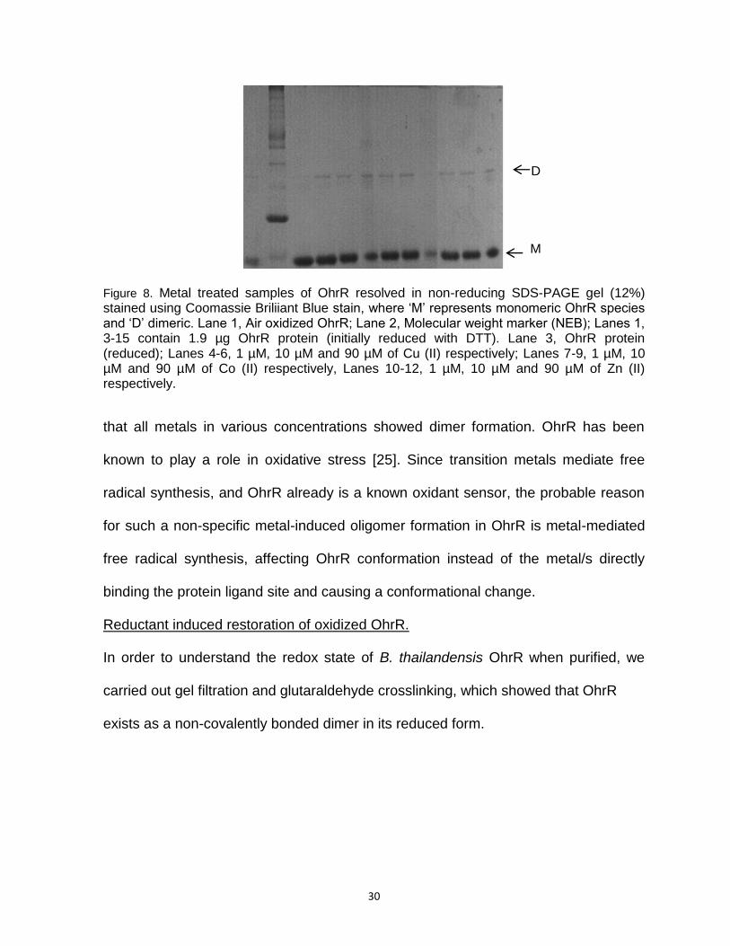
Figure 8. Metal treated samples of OhrR resolved in non-reducing SDS-PAGE gel (12%) stained using Coomassie Briliiant Blue stain, where ‘M’ represents monomeric OhrR species and ‘D’ dimeric. Lane 1, Air oxidized OhrR; Lane 2, Molecular weight marker (NEB); Lanes 1, 3-15 contain 1.9 µg OhrR protein (initially reduced with DTT). Lane 3, OhrR protein (reduced); Lanes 4-6, 1 µM, 10 µM and 90 µM of Cu (II) respectively; Lanes 7-9, 1 µM, 10 µM and 90 µM of Co (II) respectively, Lanes 10-12, 1 µM, 10 µM and 90 µM of Zn (II) respectively.
that all metals in various concentrations showed dimer formation. OhrR has been
known to play a role in oxidative stress [25]. Since transition metals mediate free
radical synthesis, and OhrR already is a known oxidant sensor, the probable reason
for such a non-specific metal-induced oligomer formation in OhrR is metal-mediated
free radical synthesis, affecting OhrR conformation instead of the metal/s directly
binding the protein ligand site and causing a conformational change.
Reductant induced restoration of oxidized OhrR.
In order to understand the redox state of B. thailandensis OhrR when purified, we
carried out gel filtration and glutaraldehyde crosslinking, which showed that OhrR
exists as a non-covalently bonded dimer in its reduced form.
M
D
31
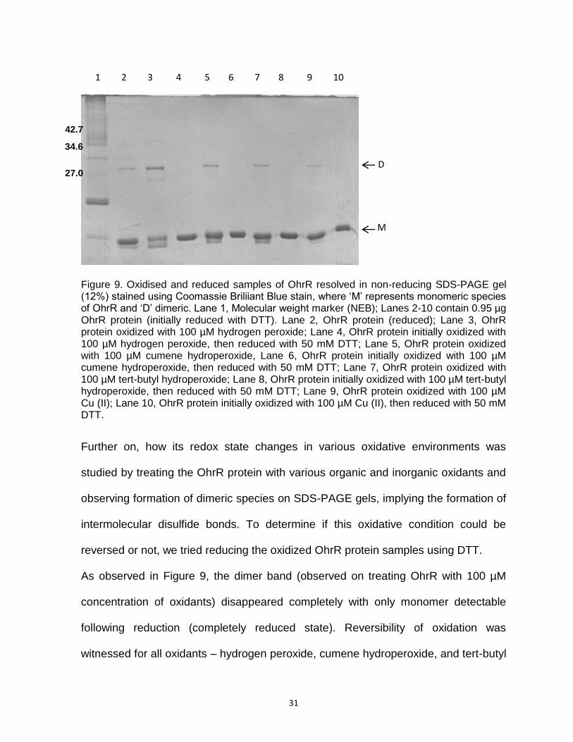
Figure 9. Oxidised and reduced samples of OhrR resolved in non-reducing SDS-PAGE gel (12%) stained using Coomassie Briliiant Blue stain, where ‘M’ represents monomeric species of OhrR and ‘D’ dimeric. Lane 1, Molecular weight marker (NEB); Lanes 2-10 contain 0.95 µg OhrR protein (initially reduced with DTT). Lane 2, OhrR protein (reduced); Lane 3, OhrR protein oxidized with 100 µM hydrogen peroxide; Lane 4, OhrR protein initially oxidized with 100 µM hydrogen peroxide, then reduced with 50 mM DTT; Lane 5, OhrR protein oxidized with 100 µM cumene hydroperoxide, Lane 6, OhrR protein initially oxidized with 100 µM cumene hydroperoxide, then reduced with 50 mM DTT; Lane 7, OhrR protein oxidized with 100 µM tert-butyl hydroperoxide; Lane 8, OhrR protein initially oxidized with 100 µM tert-butyl hydroperoxide, then reduced with 50 mM DTT; Lane 9, OhrR protein oxidized with 100 µM Cu (II); Lane 10, OhrR protein initially oxidized with 100 µM Cu (II), then reduced with 50 mM DTT.
Further on, how its redox state changes in various oxidative environments was
studied by treating the OhrR protein with various organic and inorganic oxidants and
observing formation of dimeric species on SDS-PAGE gels, implying the formation of
intermolecular disulfide bonds. To determine if this oxidative condition could be
reversed or not, we tried reducing the oxidized OhrR protein samples using DTT.
As observed in Figure 9, the dimer band (observed on treating OhrR with 100 µM
concentration of oxidants) disappeared completely with only monomer detectable
following reduction (completely reduced state). Reversibility of oxidation was
witnessed for all oxidants – hydrogen peroxide, cumene hydroperoxide, and tert-butyl
1 2 3 4 5 6 7 8 9 10
M
D
42.7
34.6
27.0

32
hydroperoxide. The untreated OhrR protein (reduced) fraction showed the presence
of some dimeric species, as a consequence of air oxidation. Since a much higher
concentration of DTT was used for reduction, the shift in the oligomeric state of OhrR
from dimer to a monomer was rapid. A gradient reduction of oxidized OhrR samples
using DTT could be performed in order to study which of the two dimer conformations
(relaxed or rigid) is converted to monomeric state first and at what DTT
concentrations. This could shine some light on the stability of the various disulfide
bonds formed, and probable placement of cysteine residues involved in bonding [15].
Thermal Stability of Reduced and Oxidised forms of OhrR
Thermal stability of OhrR protein, in various oxidative conditions, was determined
using thermal shift assay. The thermal stability profile was measured using
fluorescence spectroscopy. The fluorescent dye used for the purpose, Sypro Orange,
is hydrophobic in nature. As the protein unfolds as a function of temperature, the
buried hydrophobic residues in the protein get exposed, to which the dye Sypro
Orange (whose fluorescence is normally quenched in aqueous solutions by water)
binds (via hydrophobic interactions), resulting in an increase in fluorescence.
According to the thermal stability results analysed using Sigma Plot 9 software,
purified OhrR (reduced state) was observed to be quite stable with a Tm of 63.5 ±
0.4°C (Figure 10, table 1). This was consistent with other MarR homologues, showing
relatively high thermal stability [27, 55]. In comparison to reduced OhrR, air-oxidized
OhrR and OhrR treated with low concentrations (100 µM) of organic and inorganic
oxidants, contained only a small percentage of dimer as revealed by the SDS-PAGE
gels (Figure 11), but a significant fraction of faster migrating monomer.

33
Figure 10. Thermal melts depict fluorescence emission of reduced vs oxidized OhrR, treated with low concentration of oxidants (left panel) and oxidized OhrR, treated with high concentration of oxidants (right panel).
A probable reason for this could be the significant content of non-covalently linked
(reduced) OhrR molecules still co-existing with the partly oxidized OhrR protein (also
evident in Figure 11), resulting in the measurement of thermal stability of a mixed
population of species.
Figure 11. OhrR protein fractions used for assessing thermal stability resolved on SDS-PAGE gel.Oxidant treated samples of OhrR resolved in non-reducing SDS-PAGE gel (12%) stained using Coomassie Briliiant Blue stain, where ‘M’ represents monomeric species of OhrR and ‘D’ dimeric. Lane 1, Molecular weight marker (NEB); Lanes 2-8 contain 0.12 µg OhrR protein (initially reduced with DTT); Lanes 3-4, OhrR protein treated with 1 µM hydrogen peroxide, and tert-butyl hydroperoxide respectively; Lanes 5-6, OhrR protein treated with 10 µM hydrogen peroxide, and tert-butyl hydroperoxide respectively; Lanes 7-8,
Higher oligomeric
states.
D
M
1 2 3 4 5 6 7 8
34
OhrR protein treated with 100 µM hydrogen peroxide and tert-butyl hydroperoxide respectively; Lane 11, Air oxidized OhrR protein.
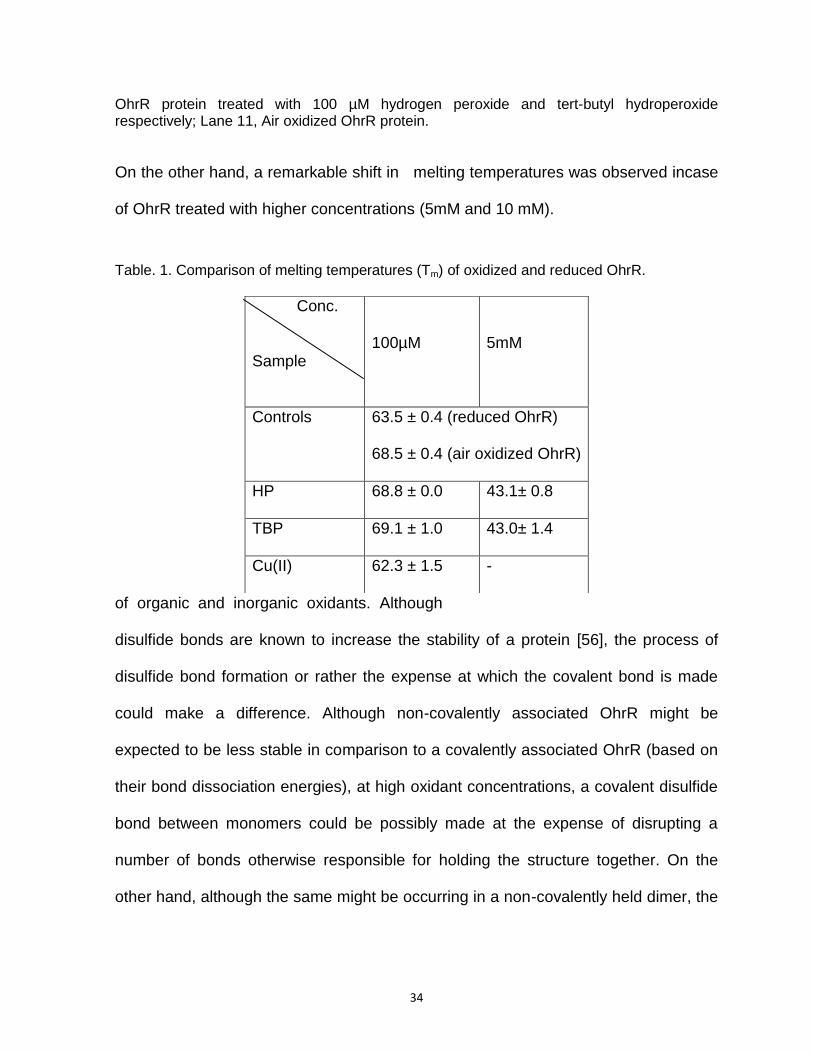
On the other hand, a remarkable shift in melting temperatures was observed incase
of OhrR treated with higher concentrations (5mM and 10 mM).
Table. 1. Comparison of melting temperatures (Tm) of oxidized and reduced OhrR.
of organic and inorganic oxidants. Although
disulfide bonds are known to increase the stability of a protein [56], the process of
disulfide bond formation or rather the expense at which the covalent bond is made
could make a difference. Although non-covalently associated OhrR might be
expected to be less stable in comparison to a covalently associated OhrR (based on
their bond dissociation energies), at high oxidant concentrations, a covalent disulfide
bond between monomers could be possibly made at the expense of disrupting a
number of bonds otherwise responsible for holding the structure together. On the
other hand, although the same might be occurring in a non-covalently held dimer, the
Conc.
Sample
100µM
5mM
Controls 63.5 ± 0.4 (reduced OhrR)
68.5 ± 0.4 (air oxidized OhrR)
HP 68.8 ± 0.0 43.1± 0.8
TBP 69.1 ± 1.0 43.0± 1.4
Cu(II) 62.3 ± 1.5 -

35
event might be accompanied by other associative bonds holding the structure
together adding overall stability to the protein.
Cu (II) treated OhrR also showed a decrease in thermal stability, probably for the
same reason as high concentration of oxidants. Transition metals generate free
radicals as byproducts predominantly forming hydroxyl radicals, which on account of
their small size, reactivity and non-specific mode of action could be responsible in
forming non-specific disulfide linkages just like hydrogen peroxide, ultimately
resulting in decreased thermal stability.
DNA binding properties of B. thailandensis OhrR
Previous studies suggest most of the proteins belonging to the MarR family have high
specificity for palindromic sequences lying in the intergenic region between divergent
genes coding for the MarR protein and the gene whose expression the former
regulates [Inoka 15, 27]. Although the DNA binding sites for OhrR proteins vary in
different bacteria, a conserved core DNA binding region has been identified in the ohr
promoter region and found to be rich in inverted ‘AATT’ repeats [25, 57, 58].
5’ CCTTCGTCCTTCATTCGAAGAATCGCGCCCGCGAAAATTAATTTGCACACTAATT
ohrO-l
ATTGGCACGCTATATTTCCACCGTGGCCGGGCATTGTGATTCGTCGAGGCCCATTCCTC
Figure 12. Promoter region of ohr. Conserved OhrR DNA binding site in bold. 73 bp OhrO-l DNA sequence (underlined), 32 bp ohrO-s DNA sequence (selected in red; the oligo was synthesized with the last base changed from T to G to maintain experimental stability).
Based on this knowledge, DNA interaction studies of B. thailandensis OhrR were
carried out using purified OhrR (reduced state) and a 73 bp long operator DNA
ohrO-s

36
(containing the hypothesized OhrR binding site) named ohrO-l, lying in the ohr
promoter region of the B. thailandensis genome.
Figure 13. EMSA showing binding of OhrR protein to ohrO-long DNA (73 bp). Lanes 2-15 represent labeled 73 bp DNA titrated with increasing concentrations of reduced OhrR protein (10 pM – 2.0 µM). 1.6 nM linearized non-specific plasmid DNA (pUC18) was added per reaction. Reaction in Lane 1 contains free DNA. Three detectable complexes (C1, C2, C3) and free DNA (F) are marked as arrows.
Figure 14. Plot indicating half maximal saturation of 73 bp long DNA sequence to OhrR.
OhrR specifically bound to labeled ohrO-l forming three clearly visible complexes as
seen in Figure 13. The binding was of high affinity as evidenced by the first OhrR-
ohr-l complex (C1) appearing at about 1 nM protein concentration (Figure13) in
1 2 3 4 5 6 7 8 9 10 11 12 13 14 15
C3
C2
C3
F
37
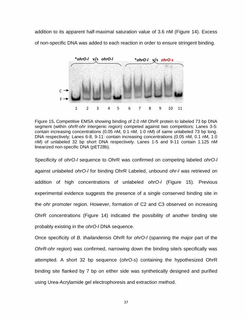
addition to its apparent half-maximal saturation value of 3.6 nM (Figure 14). Excess
of non-specific DNA was added to each reaction in order to ensure stringent binding.
Figure 15. Competitive EMSA showing binding of 2.0 nM OhrR protein to labeled 73 bp DNA segment (within ohrR-ohr intergenic region) competed against two competitors; Lanes 3-5: contain increasing concentrations (0.05 nM, 0.1 nM, 1.0 nM) of same unlabeled 73 bp long. DNA respectively; Lanes 6-8, 9-11: contain increasing concentrations (0.05 nM, 0.1 nM, 1.0 nM) of unlabeled 32 bp short DNA respectively. Lanes 1-5 and 9-11 contain 1.125 nM linearized non-specific DNA (pET28b).
Specificity of ohrO-l sequence to OhrR was confirmed on competing labeled ohrO-l
against unlabeled ohrO-l for binding OhrR Labeled, unbound ohr-l was retrieved on
addition of high concentrations of unlabeled ohrO-l (Figure 15). Previous
experimental evidence suggests the presence of a single conserved binding site in
the ohr promoter region. However, formation of C2 and C3 observed on increasing
OhrR concentrations (Figure 14) indicated the possibility of another binding site
probably existing in the ohrO-l DNA sequence.
Once specificity of B. thailandensis OhrR for ohrO-l (spanning the major part of the
OhrR-ohr region) was confirmed, narrowing down the binding site/s specifically was
attempted. A short 32 bp sequence (ohrO-s) containing the hypothesized OhrR
binding site flanked by 7 bp on either side was synthetically designed and purified
using Urea-Acrylamide gel electrophoresis and extraction method.
*ohrO-l v/s ohrO-l *ohrO-l v/s ohrO-s
1 2 3 4 5 6 7 8 9 10 11
F
C

38
Figure 16. Comparative EMSA showing a contrast in DNA binding of OhrR to the 73 bp labeled long DNA (Lanes 1-7, 0.05 nM) and 32 bp labeled short DNA (Lanes 8-14, 0.05 nM). Titrations of OhrR protein in increasing protein concentrations (0.1 nM – 1.0 µM) against the specific labeled DNA have been shown. Lanes 1 and 8 contain free DNA. Lanes 1-14 contain 0.84 nM non-specific linearized plasmid DNA per reaction. Three detectable complexes (C1, C2, C3) are marked as arrows.
To confirm the specificity of B. thailandensis OhrR to ohrO-s, labeled ohrO-l was
competed against ohrO-s for binding OhrR. Unlabeled ohrO-s competed very well
with labeled ohrO-l with almost complete retrieval of the latter at higher
concentrations of the former (Figure 15). The highest concentration of unlabeled
ohrO-s (1.0 nM) in fact competed against labeled ohrO-l to the same degree as
unlabeled ohrO-l (1.0 nM), suggesting that the conserved 18 bp DNA sequence
(hypothesized DNA binding site) is the primary binding site for OhrR.
Although the specificity of OhrR for ohrO-s was indicated by competition assay, it
was surprising to observe no protein-DNA complex formed on titrating labeled ohrO-s
with increasing concentrations of OhrR protein (Figure 16), probably a consequence
of unstable complexes formed non-detectable by EMSA.
From the EMSA results obtained, the probable reason for unstable complex
formation between OhrR and ohrO-s could be the DNA length. Although highly
specific (with only one conserved DNA binding site being present and not much extra
1 2 3 4 5 6 7 8 9 10 11 12 13 14
F
F
C3 C2
C1

39
DNA), ohrO-s is probably too short to hold two protein molecules in a stable state to
be detected in an EMSA (Figure 16). When the same binding site is lengthened with
extra base pairs on either side to generate the 73 bp DNA (ohrO-l) it shows better
binding to OhrR demonstrated as three detectable protein-DNA complexes (Figures
14 and 16). An argument could be raised with the smeary complex band being non-
specific, however, with an excess of linearized non-specific plasmid DNA being
present in the reaction, that seems unlikely. The ohrO-s site, being highly palindromic
and similar to other OhrR binding sites, is probably a more preferred binding site for
OhrR. What might be happening is that when the protein concentration is low, OhrR
binds the ohrO-s site due to preferred binding, but the short length of DNA might only
be able to accommodate one protein molecule, appearing as unstable smeary C1
bands (Figure 17).
As the protein concentration increases and more protein molecules are available to
stabilize primarily weakly bonded protein, C1 bands are seen to be disappearing and
transitioning into a relatively stable tight complex band C2. The gradual transition of
Figure 17. Proposed mechanism of OhrR binding DNA.
smeary faint bands of C1 into C3 via C2 as protein concentration increases (Figure
14) supports this interpretation. This could also mean that the binding of OhrR dimer
onto DNA is not ribosome like, but like inverted cups attached end to end in chains,
where one protein molecule uses the already bound protein molecule as the anchor.
DNA

40
At very high concentrations, a third complex appears indicating another lower affinity
binding site occurring in the promoter region of ohr OhrR-ohr intergenic region, which
is not surprising as sequence analysis of the promoter reveals another ‘AT’ rich site
with a characteristic ‘AATT’ repeat, which might be the probable second binding site.
These interpretations need to be confirmed by footprinting.

41
REFERENCES
1. Bruyninckx, W.J., H.S. Mason, and S.A. Morse, Are physiological oxygen concentrations mutagenic? Nature, 1978. 274(5671): p. 606-7.
2. Halliwell, B., Reactive species and antioxidants. Redox biology is a
fundamental theme of aerobic life. Plant Physiol, 2006. 141(2): p. 312-22. 3. Veal, E.A., A.M. Day, and B.A. Morgan, Hydrogen peroxide sensing and
signaling. Mol Cell, 2007. 26(1): p. 1-14. 4. Carlyon, J.A. and E. Fikrig, Mechanisms of evasion of neutrophil killing by
Anaplasma phagocytophilum. Curr Opin Hematol, 2006. 13(1): p. 28-33. 5. Paget, M.S. and M.J. Buttner, Thiol-based regulatory switches. Annu Rev
Genet, 2003. 37: p. 91-121. 6. Imlay, J.A., Pathways of oxidative damage. Annu Rev Microbiol, 2003. 57: p.
395-418. 7. de Gruijl, F.R., Skin cancer and solar UV radiation. Eur J Cancer, 1999.
35(14): p. 2003-9. 8. Imlay, J.A., Cellular defenses against superoxide and hydrogen peroxide.
Annu Rev Biochem, 2008. 77: p. 755-76. 9. Reverchon, S., et al., Characterization of indigoidine biosynthetic genes in
Erwinia chrysanthemi and role of this blue pigment in pathogenicity. J Bacteriol, 2002. 184(3): p. 654-65.
10. Tezel, G., et al., Oxidative stress and the regulation of complement activation
in human glaucoma. Invest Ophthalmol Vis Sci, 2010. 51(10): p. 5071-82. 11. George, A.M. and S.B. Levy, Gene in the major cotransduction gap of the
Escherichia coli K-12 linkage map required for the expression of chromosomal resistance to tetracycline and other antibiotics. J Bacteriol, 1983. 155(2): p. 541-8.
12. Ellison, D.W. and V.L. Miller, Regulation of virulence by members of the
MarR/SlyA family. Curr Opin Microbiol, 2006. 9(2): p. 153-9. 13. Ariza, R.R., et al., Repressor mutations in the marRAB operon that activate
oxidative stress genes and multiple antibiotic resistance in Escherichia coli. J Bacteriol, 1994. 176(1): p. 143-8.
42
14. Alekshun, M.N. and S.B. Levy, The mar regulon: multiple resistance to antibiotics and other toxic chemicals. Trends Microbiol, 1999. 7(10): p. 410-3.
15. Clair, G., et al., OhrRA functions as a redox-responsive system controlling
toxinogenesis in Bacillus cereus. Journal of Proteomics, (0). 16. Wilkinson, S.P. and A. Grove, Negative cooperativity of uric acid binding to the
transcriptional regulator HucR from Deinococcus radiodurans. J Mol Biol, 2005. 350(4): p. 617-30.
17. Alekshun, M.N., Y.S. Kim, and S.B. Levy, Mutational analysis of MarR, the
negative regulator of marRAB expression in Escherichia coli, suggests the presence of two regions required for DNA binding. Mol Microbiol, 2000. 35(6): p. 1394-404.
18. Linde, H.J., et al., In vivo increase in resistance to ciprofloxacin in Escherichia
coli associated with deletion of the C-terminal part of MarR. Antimicrob Agents Chemother, 2000. 44(7): p. 1865-8.
19. Alekshun, M.N., et al., The crystal structure of MarR, a regulator of multiple
antibiotic resistance, at 2.3 A resolution. Nat Struct Biol, 2001. 8(8): p. 710-4. 20. Andresen, C., et al., Critical biophysical properties in the Pseudomonas
aeruginosa efflux gene regulator MexR are targeted by mutations conferring multidrug resistance. Protein Sci, 2010. 19(4): p. 680-92.
21. Clark, K.L., et al., Co-crystal structure of the HNF-3/fork head DNA-recognition
motif resembles histone H5. Nature, 1993. 364(6436): p. 412-20. 22. Ramsay, G.D. and M.R. Eftink, Analysis of multidimensional spectroscopic
data to monitor unfolding of proteins. Methods Enzymol, 1994. 240: p. 615-45. 23. Perera, I.C., et al., Mechanism for attenuation of DNA binding by MarR family
transcriptional regulators by small molecule ligands. J Mol Biol, 2009. 390(5): p. 1019-29.
24. Rouanet, C., et al., Definition of a consensus DNA-binding site for PecS, a
global regulator of virulence gene expression in Erwinia chrysanthemi and identification of new members of the PecS regulon. J Biol Chem, 2004. 279(29): p. 30158-67.
25. Mongkolsuk, S., et al., The repressor for an organic peroxide-inducible operon
is uniquely regulated at multiple levels. Mol Microbiol, 2002. 44(3): p. 793-802.
43
26. Oh, S.Y., J.H. Shin, and J.H. Roe, Dual role of OhrR as a repressor and an activator in response to organic hydroperoxides in Streptomyces coelicolor. J Bacteriol, 2007. 189(17): p. 6284-92.
27. Wilkinson, S.P. and A. Grove, HucR, a novel uric acid-responsive member of
the MarR family of transcriptional regulators from Deinococcus radiodurans. J Biol Chem, 2004. 279(49): p. 51442-50.
28. Wilkinson, S.P. and A. Grove, Ligand-responsive transcriptional regulation by
members of the MarR family of winged helix proteins. Curr Issues Mol Biol, 2006. 8(1): p. 51-62.
29. Brugarolas, P., et al., The oxidation-sensing regulator (MosR) is a new redox-
dependent transcription factor in Mycobacterium tuberculosis. J Biol Chem, 2012. 287(45): p. 37703-12.
30. Mongkolsuk, S., et al., Identification and characterization of a new organic
hydroperoxide resistance (ohr) gene with a novel pattern of oxidative stress regulation from Xanthomonas campestris pv. phaseoli. J Bacteriol, 1998. 180(10): p. 2636-43.
31. Hong, M., et al., Structure of an OhrR-ohrA operator complex reveals the DNA
binding mechanism of the MarR family. Mol Cell, 2005. 20(1): p. 131-41. 32. Chen, H., et al., The Pseudomonas aeruginosa multidrug efflux regulator
MexR uses an oxidation-sensing mechanism. Proc Natl Acad Sci U S A, 2008. 105(36): p. 13586-91.
33. Greenberg, J.T., et al., Positive control of a global antioxidant defense regulon
activated by superoxide-generating agents in Escherichia coli. Proc Natl Acad Sci U S A, 1990. 87(16): p. 6181-5.
34. Uemi, M., et al., Generation of cholesterol carboxyaldehyde by the reaction of
singlet molecular oxygen [O2 (1Delta(g))] as well as ozone with cholesterol. Chem Res Toxicol, 2009. 22(5): p. 875-84.
35. Mongkolsuk, S. and J.D. Helmann, Regulation of inducible peroxide stress
responses. Mol Microbiol, 2002. 45(1): p. 9-15. 36. Seaver, L.C. and J.A. Imlay, Alkyl hydroperoxide reductase is the primary
scavenger of endogenous hydrogen peroxide in Escherichia coli. J Bacteriol, 2001. 183(24): p. 7173-81.
37. Panmanee, W., et al., Novel organic hydroperoxide-sensing and responding
mechanisms for OhrR, a major bacterial sensor and regulator of organic hydroperoxide stress. J Bacteriol, 2006. 188(4): p. 1389-95.
44
38. Lee, J.W., S. Soonsanga, and J.D. Helmann, A complex thiolate switch
regulates the Bacillus subtilis organic peroxide sensor OhrR. Proc Natl Acad Sci U S A, 2007. 104(21): p. 8743-8.
39. Atichartpongkul, S., et al., Analyses of the regulatory mechanism and
physiological roles of Pseudomonas aeruginosa OhrR, a transcription regulator and a sensor of organic hydroperoxides. J Bacteriol, 2010. 192(8): p. 2093-101.
40. Starr, M.P., BACTERIA AS PLANT PATHOGENS. Annual Review of
Microbiology, 1959. 13: p. 211-238. 41. Chain, P.S., et al., Burkholderia xenovorans LB400 harbors a multi-replicon,
9.73-Mbp genome shaped for versatility. Proc Natl Acad Sci U S A, 2006. 103(42): p. 15280-7.
42. Fries, M.R., L.J. Forney, and J.M. Tiedje, Phenol- and toluene-degrading
microbial populations from an aquifer in which successful trichloroethene cometabolism occurred. Appl Environ Microbiol, 1997. 63(4): p. 1523-30.
43. Alvarez-Lerma, F., et al., Moisturizing body milk as a reservoir of Burkholderia
cepacia: outbreak of nosocomial infection in a multidisciplinary intensive care unit. Crit Care, 2008. 12(1): p. R10.
44. Baldwin, A., et al., Environmental Burkholderia cepacia complex isolates in
human infections. Emerg Infect Dis, 2007. 13(3): p. 458-61. 45. Dance, D.A., Melioidosis as an emerging global problem. Acta Trop, 2000.
74(2-3): p. 115-9. 46. Whitmore, L. and B.A. Wallace, Protein secondary structure analyses from
circular dichroism spectroscopy: methods and reference databases. Biopolymers, 2008. 89(5): p. 392-400.
47. Hao, Z., et al., The multiple antibiotic resistance regulator MarR is a copper
sensor in Escherichia coli. Nat Chem Biol, 2013. advance online publication. 48. Smith, A.J., DNA sequence analysis by primed synthesis. Methods Enzymol,
1980. 65(1): p. 560-80. 49. Huang, H. and A. Grove, The transcriptional regulator TamR from
Streptomyces coelicolor controls a key step in central metabolism during oxidative stress. Mol Microbiol, 2013. 87(6): p. 1151-66.
45
50. Newberry, K.J., et al., Structural mechanism of organic hydroperoxide induction of the transcription regulator OhrR. Mol Cell, 2007. 28(4): p. 652-64.
51. Fadouloglou, V.E., M. Kokkinidis, and N.M. Glykos, Determination of protein
oligomerization state: two approaches based on glutaraldehyde crosslinking. Anal Biochem, 2008. 373(2): p. 404-6.
52. Peeters, E., et al., Transcriptional response of Burkholderia cenocepacia J2315 sessile cells to treatments with high doses of hydrogen peroxide and sodium hypochlorite. BMC Genomics, 2010. 11: p. 90.
53. Guerra, A.J., C.E. Dann, 3rd, and D.P. Giedroc, Crystal structure of the zinc-
dependent MarR family transcriptional regulator AdcR in the Zn(II)-bound state. J Am Chem Soc, 2011. 133(49): p. 19614-7.
54. Kogan, V., et al., Carbon-carbon and carbon-nitrogen coupling reactions
catalyzed by palladium nanoparticles derived from a palladium substituted Keggin-type polyoxometalate. Org Lett, 2002. 4(20): p. 3529-32.
55. Perera, I.C. and A. Grove, Urate is a ligand for the transcriptional regulator
PecS. J Mol Biol, 2010. 402(3): p. 539-51. 56. Bulleid, N.J. and L. Ellgaard, Multiple ways to make disulfides. Trends
Biochem Sci, 2011. 36(9): p. 485-92. 57. Chuchue, T., et al., ohrR and ohr are the primary sensor/regulator and
protective genes against organic hydroperoxide stress in Agrobacterium tumefaciens. J Bacteriol, 2006. 188(3): p. 842-51.
58. Fuangthong, M., et al., OhrR is a repressor of ohrA, a key organic
hydroperoxide resistance determinant in Bacillus subtilis. J Bacteriol, 2001. 183(14): p. 4134-41.

46
VITA
Anuja Rajiv Pande, was born in Mumbai, India. Having completed her
primary education in Mumbai and partly in Dubai, she completed her
undergraduate studies in Life Sciences and Biochemistry, from the
University of Mumbai, in the year 2007. During her bachelors tenure, in
addition to academic studies, she also had the opportunity of being a
part of individual research projects at the collegiate and national levels.
In 2011, she completed her Masters in Biological Sciences, from SVKM’s
NMIMS University, School of Science and then moved to United States
to pursue her second Masters degree at the Department of Biological
Sciences at Louisiana State University, Baton Rouge.
In the Spring of 2012, she joined Dr. Anne Grove’s lab to study the MarR
family of transcriptional regulators responsive to organic hydroperoxides,
as part of her Masters programme. Upon completion of her degree she
wishes to pursue her doctorate under the guidance of Dr. Anne Grove
and carry the research element further.