Embed Size (px)
Citation preview
Full Terms & Conditions of access and use can be found athttp://www.tandfonline.com/action/journalInformation?journalCode=iebm20
Download by: [University of California, Berkeley] Date: 24 August 2016, At: 15:39
Electromagnetic Biology and Medicine
ISSN: 1536-8378 (Print) 1536-8386 (Online) Journal homepage: http://www.tandfonline.com/loi/iebm20
Oxidative mechanisms of biological activity of low-intensity radiofrequency radiation
Igor Yakymenko, Olexandr Tsybulin, Evgeniy Sidorik, Diane Henshel, OlgaKyrylenko & Sergiy Kyrylenko
To cite this article: Igor Yakymenko, Olexandr Tsybulin, Evgeniy Sidorik, Diane Henshel,Olga Kyrylenko & Sergiy Kyrylenko (2016) Oxidative mechanisms of biological activity of low-intensity radiofrequency radiation, Electromagnetic Biology and Medicine, 35:2, 186-202, DOI:10.3109/15368378.2015.1043557
To link to this article: http://dx.doi.org/10.3109/15368378.2015.1043557
Published online: 07 Jul 2015.
Submit your article to this journal
Article views: 2348
View related articles
View Crossmark data
Citing articles: 11 View citing articles
REVIEW ARTICLE
Oxidative mechanisms of biological activity of low-intensity radiofrequencyradiationIgor Yakymenkoa, Olexandr Tsybulinb, Evgeniy Sidorika, Diane Henshelc, Olga Kyrylenkod, and Sergiy Kyrylenkoe
aInstitute of Experimental Pathology, Oncology and Radiobiology, National Academy of Sciences of Ukraine, Kyiv, Ukraine; bDepartment ofBiophysics, Bila Tserkva National Agrarian University, Bila Tserkva, Ukraine; cSchool of Public and Environmental Affairs, Indiana UniversityBloomington, Bloomington, IN, USA; dA.I. Virtanen Institute, University of Eastern Finland, Kuopio, Finland; eDepartment of Structural andFunctional Biology, University of Campinas, Campinas, Brazil
ABSTRACTThis review aims to cover experimental data on oxidative effects of low-intensity radiofrequencyradiation (RFR) in living cells. Analysis of the currently available peer-reviewed scientific literaturereveals molecular effects induced by low-intensity RFR in living cells; this includes significantactivation of key pathways generating reactive oxygen species (ROS), activation of peroxidation,oxidative damage of DNA and changes in the activity of antioxidant enzymes. It indicates thatamong 100 currently available peer-reviewed studies dealing with oxidative effects of low-intensity RFR, in general, 93 confirmed that RFR induces oxidative effects in biological systems.A wide pathogenic potential of the induced ROS and their involvement in cell signaling pathwaysexplains a range of biological/health effects of low-intensity RFR, which include both cancer andnon-cancer pathologies. In conclusion, our analysis demonstrates that low-intensity RFR is anexpressive oxidative agent for living cells with a high pathogenic potential and that the oxidativestress induced by RFR exposure should be recognized as one of the primary mechanisms of thebiological activity of this kind of radiation.
ARTICLE HISTORYReceived 10 January 2015Accepted 12 April 2015Published online 7 July 2015
KEYWORDSCellular signaling; cancer;free radicals; oxidativestress; radiofrequencyradiation; reactive oxygenspecies
Introduction
Intensive development of wireless technologies duringthe last decades led to a dramatic increase of back-ground radiofrequency radiation (RFR) in the humanenvironment. Thus, the level of indoor backgroundRFR in industrialized countries increased 5,000-foldfrom 1985 to 2005 (Maes, 2005). Such significant envir-onmental changes may have a serious impact onhuman biology and health. As a proof of such impact,a series of epidemiological studies on the increased riskof tumorigenesis in “heavy” users of wireless telephonyexists (Hardell et al., 2007, 2011; Sadetzki et al., 2008;Sato et al., 2011). Some studies indicate that long-termRFR exposure in humans can cause various non-cancerdisorders, e.g., headache, fatigue, depression, tinnitus,skin irritation, hormonal disorders and other condi-tions (Abdel-Rassoul et al., 2007; Buchner & Eger,2011; Chu et al., 2011; Johansson, 2006; Santini et al.,2002; Yakymenko et al., 2011). In addition, convincingstudies on hazardous effects of RFR in human germcells have been published (Agarwal et al., 2009; DeIuliis et al., 2009).
All abovementioned studies dealt with the effects oflow-intensity RFR. This means that the intensity ofradiation was far below observable thermal effects inbiological tissues, and far below safety limits of theInternational Commissions on Non-IonizingRadiation Protection (ICNIRP) (ICNIRP, 1998). Todate, molecular mechanisms of non-thermal effects ofRFR are still a bottleneck in the research on the biolo-gical/health effects of low-intensity RFR, althoughrecently many studies have been carried out on meta-bolic changes in living cells under low-intensity RFR,and comprehensive reviews were published (Belyaev,2010; Consales et al., 2012; Desai et al., 2009;Yakymenko et al., 2011). In the present work, we ana-lyze the results of molecular effects of low-intensityRFR in living cells and model systems, with a specialemphasis on oxidative effects and free radical mechan-isms. It might seem paradoxical that, despite beingnon-ionizing, RFR can induce significant activation offree radical processes and overproduction of reactiveoxygen species (ROS) in living cells. We believe that theanalysis of recent findings will allow recognition of a
CONTACT Igor Yakymenko [email protected] Laboratory of Biophysics, Institute of Experimental Pathology, Oncology and Radiobiology of NASof Ukraine, Vasylkivska str. 45, Kyiv, 03022 Ukraine.
ELECTROMAGNETIC BIOLOGY AND MEDICINE2016, VOL. 35, NO. 2, 186–202http://dx.doi.org/10.3109/15368378.2015.1043557
© 2016 Taylor & Francis
general picture of the potential health effects of alreadyubiquitous and ever-increasing RFR.
Radiofrequency radiation
RFR is a part of electromagnetic spectrum with fre-quencies from 30 kHz to 300 GHz. RFR is classified asnon-ionizing, which means that it does not carry suffi-cient energy for ionization of atoms and molecules. Apart of RFR with the highest frequencies (300MHz to300 GHz) is referred to as microwaves (MWs). MW isRFR with the highest energy, which can potentiallygenerate the highest thermal effects in the absorbingmatter.
The main indexes of RFR are (i) frequency (Hz); (ii)intensity or power density (PD) of radiation (W/m2 orµW/cm2); (iii) its modulated or non-modulated nature;and (iv) continuous or discontinuous pattern of radia-tion. For the absorbed RFR energy, a parameter ofspecific absorption rate (SAR) is used (W/kg). Themost common digital standard of RFR for mobile com-munication is still GSM (Global System for Mobilecommunication), which utilizes frequencies at about850, 900, 1800 and 1900MHz. This radiation is fre-quency modulated, with channel rotation frequency of217 Hz, and belongs to the radiation of the pulsedmode (Hyland, 2000).
As to the international safety limits, the ICNIRPrecommendations restrict intensity of RFR to 450–1000 µW/cm2 (depending on the frequency of radia-tion) and the SAR value to 2W/kg, as calculated forhuman heads and torsos (ICNIRP, 1998). Theseindexes were adopted by ICNIRP based on the beha-vioral response of laboratory rats, which were exposedto gradually increased intensities of RFR to determinethe point at which the animals became thermally dis-tressed (Gandhi et al., 2012).
Low-intensity RFR is referred to as radiation withintensities which do not induce significant thermaleffects in biological tissues. Accordingly, any intensityof RFR under the ICNIRP limits can be referred to aslow-intensity. In this paper we will analyze only theeffects of low-intensity RFR.
Physical/biophysical effects of low-intensityRFR in living cells
RFR, especially MW, can produce thermal effects inmatter due to interaction with charged particles,including free electrons, ions or polar molecules, indu-cing their oscillations in electromagnetic field. Thethermal effect of MW can be seen when warmingfood in the microwave. The effect strongly depends
on the intensity of radiation and is mostly negligibleunder low-intensity RFR conditions. On the otherhand, energy of RFR/MW is insufficient not only forthe ionization of molecules, but even for activation oforbital electrons. Hence, RFR was often assessed as afactor producing only thermal effects. Nevertheless,evident biological effects of low-intensity RFR pro-moted research on physical mechanisms of non-ther-mal biological effects of this kind of radiation.
A biophysical model of a forced-vibration of freeions on the surface of a cell membrane due to externaloscillating electromagnetic field (EMF) was proposed(Panagopoulos et al., 2000, 2002). According to theauthors, this vibration of electric charges can causedisruption of the cellular electrochemical balance andfunctions.
A “moving charge interaction” model was proposedfor low-frequency EMF (Blank and Soo, 2001). Theauthors explained activation of genes and synthesis ofstress proteins under EMF exposure due to interactionof the field with moving electrons in DNA (Blank andSoo, 2001; Goodman and Blank, 2002). They alsodemonstrated that EMF increased electron transferrates in cytochrome oxidase and accelerated chargesin the Na,K-ATPase reaction. Moreover, they demon-strated acceleration of the oscillating Belousov–Zhabotinski reaction in homogeneous solutions due tothe application of low-frequency EMF (Blank and Soo,2003).
An ability of low-strength magnetic fields to triggeronset- and offset-evoked potentials was demonstrated(Marino et al., 2009). Effectiveness of a rapid magneticstimulus (0.2 ms) has led the authors to a conclusion ondirect interaction between the field and ion channels inplasma membrane. A plausible mechanism of overpro-duction of free radicals in living cell due to electronspin flipping in confined free radical pairs in magneticfield of RFR was proposed (Georgiou, 2010).
A significant effect of low-intensity RFR on ferritin,an iron cage protein present in most living organismsfrom bacteria to humans, was revealed (Céspedes andUeno, 2009). Exposure of ferritin solution to low-inten-sity RFR significantly, up to threefold, reduced ironchelation with ferrozine. The authors explained thatmagnetic field of RFR plays a principle role in theobserved effect, and that this effect is strongly non-thermal. The non-thermal mechanism of the interac-tion of RFR magnetic fields with ferritin is supposedlymediated by an inner super-paramagnetic nanoparticle(9H2O × 5Fe2O3 with up to 4500 iron ions), which is anatural phenomenon intrinsic to the cells. It results inreduction of input of iron chelates into the ferritin cage.The authors underlined the potential role of ferritin
ELECTROMAGNETIC BIOLOGY AND MEDICINE 187
malfunction for oxidative processes in living cell due tothe participation of Fe2+ ions in the Fenton reaction,which produces hydroxyl radicals. In this respect, it isinteresting to point to the results of an in vitro studywith RFR exposure of rat lymphocytes treated by ironions (Zmyślony et al., 2004). Although RFR exposure(930MHz) did not induce detectable intracellular ROSoverproduction, the same exposure in the presence ofFeCl2 in the lymphocyte suspensions induced a signifi-cant overproduction of ROS.
Another set of studies indicates on a possibility ofchanges in protein conformation under RFR exposure.Thus, low-intensity 2.45MHz RFR accelerated confor-mational changes in β-lactoglobulin through excitationof so-called collective intrinsic modes in the protein(Bohr and Bohr, 2000a, 2000b), which suggests a prin-cipal ability of RFR to modulate the non-random col-lective movements of entire protein domains. Similarly,a frequency-dependent effect on intrinsic flexibility ininsulin structure due to applied oscillating electric fieldwas demonstrated (Budi et al., 2007). Moreover, macro-molecular structure of cytoskeleton was significantlyaltered in fibroblasts of Chinese hamster after the expo-sure to modulated RFR of the GSM standard (Pavicicand Trosic, 2010). Thus, a 3 h exposure of fibroblasts tomodulated RFR (975MHz) led to significant changes inthe structure of microtubules and actin microfilaments,which have polar cytoskeleton structures, while non-polar vimentin filaments reportedly stayed unchanged.Taking into account an extensive regulatory potentialof cytoskeleton on cell homeostasis, these data couldobviously add to the nature of the biological effectsof RFR.
It was shown that ornithine decarboxylase (ODC)can significantly change its activity under low-intensityRFR exposure (Byus et al., 1988; Hoyto et al., 2007;Litovitz et al., 1993, 1997; Paulraj et al., 1999).
In addition, so-called “calcium effects” under RFRexposure in living cells have been demonstrated (Duttaet al., 1989; Paulraj et al., 1999; Rao et al., 2008), whichinclude a significant increase in intracellular Ca2+ spik-ing. Taking into account that calcium is a ubiquitousregulator of cellular metabolism, these data point to apossibility that non-thermal RFR can activate multipleCa2+-dependent signaling cascades.
Finally, an ability of low-intensity MW to dissociatewater molecules was demonstrated in model experi-ments years ago (Vaks et al., 1994). In these experi-ments, MW of 10 GHz with radiated power 30 mWproduced a significant level of H2O2 in deionizedwater (and also in MgSO4 solution) under stable tem-perature conditions. According to the authors, a kineticexcitation of liquid water associates C(H2O) upon the
absorption of MW leads to subsequent viscous lossesdue to friction between moving clusters of water mole-cules. It results in partial irreversible decomposition ofwater, including breaks of intramolecular bonds (H–OH) due to a mechanochemical reaction, and genera-tion of H•; OH•; H+ and OH− groups. Among these, thehydroxyl radical (OH•) is the most aggressive form ofROS, which can break any chemical bond in surround-ing molecules (Halliwell, 2007). The authors assessedthat this type of mechanochemical transformation inwater could be responsible for 10−4–10−8 relative partsof the total MW energy absorbed. Given the fact thatthe water molecules are ubiquitous in living cells, evena subtle chance for dissociation of water moleculesunder low-intensity RFR exposure could have a pro-found effect on tissue homeostasis. It is of note herethat one OH• radical can initiate irreversible peroxida-tion of many hundreds of macromolecules, e.g. lipidmolecules (Halliwell, 1991). Taken together, these datashow that non-thermal RFR can be absorbed by parti-cular charges, molecules and cellular structures, and inthis way can potentially induce substantial modulatoryeffects in living cell.
Generation of reactive oxygen species underRFR exposure in living cells
NADH oxidase of cellular membrane was suggested asa primary mediator of RFR interaction with living cells(Friedman et al., 2007). Using purified membranesfrom HeLa cells, the authors experimentally provedthat the exposure to RFR of 875MHz, 200 µW/cm2
for 5 or 10 min significantly, almost threefold, increasedthe activity of NADH oxidase. NADH oxidases aremembrane-associated enzymes that catalyze one-elec-tron reduction of oxygen into superoxide radical usingNADH as a donor of electron, thus producing powerfulROS. This enzyme has been traditionally known due toits role in induction of oxidative burst in phagocytes asa part of immune response. Yet, later the existence ofnon-phagocytic NAD(P)H oxidases was revealed invarious types of cells, including fibroblasts, vascularand cardiac cells (Griendling et al., 2000). Obviously,the presence of superoxide-generating enzyme in manytypes of non-phagocytic cells points to the considerableregulatory roles of ROS in living cells. On the otherhand, an ability of low-intensity RFR to modulate theactivity of the NADH oxidase automatically makes thisfactor a notable and potentially dangerous effector ofcell metabolism. Notably, the authors pointed out thatthe acceptor of RFR is different from the peroxide-generating NADPH oxidases, which are also found inplasma membranes (Low et al., 2012).
188 I. YAKYMENKO ET AL.

The other powerful source of ROS in cells is mito-chondrial electron transport chain (ETC), which cangenerate superoxide due to breakdowns in electrontransport (Inoue et al., 2003). It was demonstratedthat generation of ROS by mitochondrial pathway canbe activated under RFR exposure in human spermato-zoa (De Iuliis et al., 2009). The authors revealed a dose-dependent effect of 1.8 GHz RFR exposure on ROSproduction in spermatozoa, particularly in their mito-chondria. The significantly increased level of total ROSin spermatozoa was detected under RFR with SAR = 1W/kg, which is below the safety limits accepted inmany countries. It was demonstrated recently in ourlaboratory that the exposure of quail embryos in ovo toextremely low-intensity RFR (GSM 900MHz, 0.25 µW/cm2) during the initial days of embryogenesis resultedin a robust overproduction of superoxide and nitrogenoxide radicals in mitochondria of embryonic cells(Burlaka et al., 2013). It is not clear yet which particularpart of ETC is responsible for the interaction with RFR.To date, three possible sites of generation of superoxidein ETC have been shown: the ETC complex I (Inoueet al., 2003), complex II (Liu et al., 2002), and complexIII (Guzy and Schumacker, 2006). A significant inversecorrelation between mitochondrial membrane potentialand ROS levels in living cell was found (Wang et al.,2003). As the authors underlined, such a relationshipcould be due to two mutually interconnected phenom-ena: ROS causing damage to the mitochondrial mem-brane, and the damaged mitochondrial membranecausing increased ROS production.
In addition to the well-established role of the mito-chondria in energy metabolism, regulation of cell deathis a second major function of these organelles. This, inturn, is linked to their role as the powerful intracellularsource of ROS. Mitochondria-generated ROS play animportant role in the release of cytochrome c and otherpro-apoptotic proteins, which can trigger caspase acti-vation and apoptosis (Ott et al., 2007). A few reportsindicate on activation of apoptosis due to low-intensityRFR exposure. In human epidermoid cancer KB cells,1950MHz RFR induced time-dependent apoptosis(45% after 3 h) that is paralleled by 2.5-fold decreaseof the expression of ras and Raf-1 and of the activity ofras and Erk-1/2 (Caraglia et al., 2005). Primary culturedneurons and astrocytes exposed to GSM 1900MHzRFR for 2 h demonstrated up-regulation of caspase-2,caspase-6 and Asc (apoptosis associated speck-like pro-tein containing a card) (Zhao et al., 2007). Up-regula-tion in neurons occurred in both “on” and “stand-by”modes, but in astrocytes only in the “on” mode. Weshould underline that, in that study an extremely highbiological sensitivity to RFR was demonstrated, as a cell
phone in the “stand-by” position emits negligibly low-intensity of radiation (up to hundredths µW/cm2).
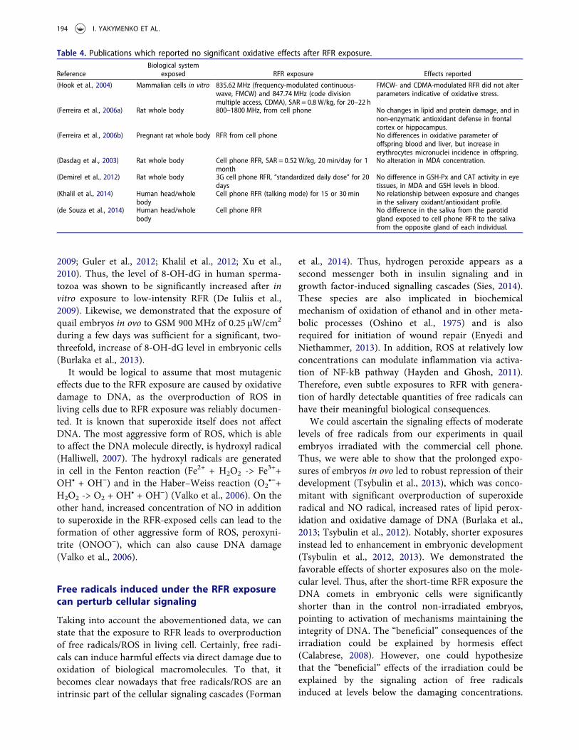
Based on the analysis of available literature data, weidentified altogether 100 experimental studies in biolo-gical models which investigated oxidative stress due tolow-intensity RFR exposures. From these 100 articles,93 studies (93%) demonstrated significant oxidativeeffects induced by low-intensity RFR exposure(Table 1–3), while 7 studies (7%) demonstrated theabsence of significant changes (Table 4). The totalnumber includes 18 in vitro studies, 73 studies in ani-mals, 3 studies in plants and 6 studies in humans.Majority of the research was done on laboratory rats(58 studies, with 54 positive results), while 4 studies outof 6 in humans were positive. From the in vitro studies,17 were positive (94.4%), including 2 studies on humanspermatozoa and 2 studies on human blood cells.
Most of the studies utilized RFR exposure in MWrange, including a use of commercial or trial cellphones as sources of radiation. The power densities ofRFR applied in positive studies varied from 0.1 µW/cm2
(Oksay et al., 2014) to 680 µW/cm2 (Jelodar et al., 2013)and SAR values varied from 3 µW/kg (Burlaka et al.,2013) to the ICNIRP recommended limit of 2W/kg(Naziroglu et al., 2012a; Xu et al., 2010). Exposuretimes in positive studies varied from 5min (Friedmanet al., 2007) to 12.5 years, 29.6 h/month (Hamzanyet al., 2013).
The most often used indexes of oxidative stressanalyzed in the studies were ROS production, levels oflipid peroxidation (LPO)/malondialdehyde (MDA),protein oxidation (PO), nitric oxides (NOx), glu-tathione (GSH), activity of antioxidant enzymes (super-oxide dismutase (SOD), catalase (CAT), glutathioneperoxidase (GSH-Px)). It is important that some studiesdirectly pointed to induction of free radicals (super-oxide radical, NO) as a primary reaction of living cellsto RFR exposure (Burlaka et al., 2013; Friedman et al.,2007). As we pointed out earlier, direct activation ofNADH oxidase (Friedman et al., 2007) and the mito-chondrial pathway of superoxide overproduction(Burlaka et al., 2013; De Iuliis et al., 2009) have beenexperimentally proven. Besides, a significant overpro-duction of nitrogen oxide was revealed in some studies(Avci et al., 2012; Bilgici et al., 2013; Burlaka et al.,2013), although it is unclear whether an induction ofexpression of NO-synthases or direct activation of theenzyme took place. It is however clear that significantlyincreased levels of these free radical species (superoxideand nitrogen oxide) in cells due to RFR exposure resultin an activation of peroxidation and repression of activ-ities of key antioxidant enzymes. It is indicative thatmany studies demonstrated effectiveness of different
ELECTROMAGNETIC BIOLOGY AND MEDICINE 189

antioxidants to override oxidative stress caused by RFRexposure. Such effects have been reported for melato-nin (Ayata et al., 2004; Lai and Singh, 1997; Oktemet al., 2005; Ozguner et al., 2006; Sokolovic et al., 2008),vitamin E and C (Jelodar et al., 2013; Oral et al., 2006),caffeic acid phenethyl ester (Ozguner et al., 2006), sele-nium, L-carnitine (Turker et al., 2011) and garlic (Avciet al., 2012; Bilgici et al., 2013).
It is worthwhile to emphasize a strict non-thermalcharacter of ROS overproduction under RFR exposuredescribed in the cited reports. As low as 0.1 µW/cm2
intensity of RFR and absorbed energy (specific absorp-tion rate, SAR) of 0.3 µW/kg were demonstrated to beeffective in inducing significant oxidative stress in liv-ing cells (Burlaka et al., 2013; Oksay et al., 2014). Thisobservation is particularly important as the moderninternational safety limits on RFR exposure are basedsolely on the thermal effects of radiation and onlyrestrict RFR intensity to 450–1000 µW/cm2 and SARto 2W/kg (ICNIRP, 1998). Moreover, studies wherehigh (thermal) intensities of RFR have been used
could not reveal oxidative effects (Hong et al., 2012;Kang et al., 2013; Luukkonen et al., 2009), which mightpoint to the variety of molecular mechanisms for dif-ferent radiation intensities.
Taken together, the analysis of the contemporaryscientific literature on the biological effects of RFRpersuasively proves that the exposure to low-intensityRFR in living cells leads to generation of significantlevels of ROS and results in a significant oxidativestress.
Oxidative damage of DNA under RFR exposure
To date more than hundred papers have been pub-lished on mutagenic effects of RFR and most of themrevealed significant effects (Ruediger, 2009). There is asubstantial number of studies which demonstrated theformation of micronuclei (Garaj-Vrhovac et al., 1992;Tice et al., 2002; Zotti-Martelli et al., 2005) or structuralanomalies of metaphase chromosomes (Garson et al.,1991; Kerbacher et al., 1990; Maes et al., 2000) in living
Table 1. Publications which reported positive findings on oxidative stress caused by RFR exposure of cells in vitro.Reference Biological system exposed RFR exposure Statistically significant effects reported*
(Agarwal et al., 2009) Human spermatozoa Cell phone RFR, in talk mode, for 1 h Increase in reactive oxygen species (ROS) level, decrease insperm motility and viability.
(Campisi et al., 2010) Rat astroglial cells 900 MHz (continuous or modulated),electric field 10 V/m, for5; 10; 20 min
Increase in ROS levels and DNA fragmentation afterexposure to modulated RFR for 20 min.
(De Iuliis et al., 2009) Human spermatozoa 1.8 GHz, SAR = 0.4–27.5 W/kg Increased amounts of ROS.(Friedman et al., 2007) HeLa membranes 875 MHz, 200 µW/cm2, for 5 and 10 min Increased NADH oxidase activity.(Hou et al., 2014) Mouse embryonic
fibroblasts (NIH/3T3)1800-MHz GSM-talk mode RFR, SAR = 2W/kg, intermittent exposure (5 min on/10 min off) for 0.5–8 h
Increased intracellular ROS levels.
(Kahya et al., 2014) Cancer cell cultures 900 MHz RFR, SAR = 0.36 W/kg, for 1 h Induced apoptosis effects through oxidative stress,selenium counteracted the effects of RFR exposure.
(Lantow et al., 2006a) Human blood cells Continuous wave or GSM signal,SAR =2W/kg, for 30 or 45 min of continuousor 5 min ON, 5 min OFF
After continuous or intermittent GSMsignal a different ROSproduction was detected in human monocytes comparedto sham.
(Lantow et al., 2006b) Human Mono Mac 6 andK562 cells
Continuous wave, GSM speaking only,GSM hearing only, GSM talk, SARs of0.5, 1.0, 1.5 and 2.0 W/kg.
The GSM-DTX signal at 2 W/kg produced difference in freeradical production compared to sham.
(Liu et al., 2013b) GC-2 cells 1800 MHz, SAR = 1; 2 W/kg,5 min ON,10 min OFF for 24 h
In the 2 W/kg exposed cultures, the level of ROS wasincreased.
(Lu et al., 2012) Human bloodmononuclear cells
900 MHz, SAR = 0.4 W/kg, for 1–8 h The increased level of apoptosis induced through themitochondrial pathway and mediated by activating ROSand caspase-3.
(Marjanovic et al., 2014) V79 cells 1800 MHz, SAR = 1.6 W/kg, for 10, 30and 60 min
ROS level increased after 10 min of exposure. Decrease inROS level after 30-min treatment indicating antioxidantdefense mechanism activation.
(Naziroglu et al., 2012b) HL-60 cells 2450 MHz, pulsed, SAR = 0.1–2.5 W/kg,for 1; 2; 12 or 24 h
Lipid peroxide (LPO) levels were increased at all exposuretimes.
(Ni et al., 2013) Human lens epithelial cells 1800 MHz, SAR = 2; 3; 4 W/kg The ROS and malondialdehyde (MDA) levels wereincreased.
(Pilla, 2012) Neuronal cells and humanfibroblasts
27.12 MHz, pulsed, electric field 41 V/m,2 min prior to lipopolysaccharideadministration or for 15 min
Increased level of nitric oxide (NO).
(Sefidbakht et al., 2014) HEK293T cells 940 MHz, SAR = 0.09 W/kg, for 15, 30,45, 60 and 90 min
ROS generation increased in the 30 min exposed cells. Asharp rise in catalase (CAT) and superoxide dismutase(SOD) activity and elevation of glutathione (GSH) duringthe 45 min exposure.
(Xu et al., 2010) Primary cultured neurons 1800 MHz, pulsed, SAR = 2 W/kg,for 24 h
An increase in the levels of8-hydroxy-2'-deoxyguanosine(8-OH-dG).
(Zmyślony et al., 2004) Rat lymphocytes 930 MHz, PD of 500 µW/cm2, SAR = 1.5W/kg, for 5 and 15 min
Intracellular ROS level increased in exposed FeCl2 treatedcells compared with unexposed FeCl2 treated cells.
*All effects were statistically significant (at least p < 0.05) as compared to control or sham exposed groups.
190 I. YAKYMENKO ET AL.
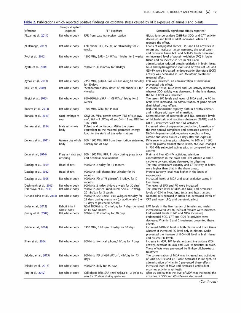
Table 2. Publications which reported positive findings on oxidative stress caused by RFR exposure of animals and plants.
ReferenceBiological system
exposed RFR exposure Statistically significant effects reported*
(Akbari et al., 2014) Rat whole body RFR from base transceiver station Glutathione peroxidase (GSH-Px), SOD, and CAT activitydecreased and level of MDA increased. Vitamin Creduced the effect.
(Al-Damegh, 2012) Rat whole body Cell phone RFR, 15, 30, or 60 min/day for 2weeks
Levels of conjugated dienes, LPO and CAT activities inserum and testicular tissue increased, the total serumand testicular tissue GSH and GSH-Px levels decreased.
(Avci et al., 2012) Rat whole body 1800 MHz, SAR = 0.4 W/kg, 1 h/day for 3 weeks An increased level of protein oxidation (PO) in braintissue and an increase in serum NO. Garlicadministration reduced protein oxidation in brain tissue.
(Ayata et al., 2004) Rat whole body 900 MHz, 30 min/day for 10 days MDA and hydroxyproline levels and activities of CAT andGSH-Px were increased, andsuperoxide dismutase (SOD)activity was decreased in skin. Melatonin treatmentreversed effect.
(Aynali et al., 2013) Rat whole body 2450 MHz, pulsed, SAR = 0.143 W/kg,60 min/dayfor 30 days
LPO was increased, an administration of melatoninprevented this effect.
(Balci et al., 2007) Rat whole body “Standardized daily dose” of cell phoneRFR for4 weeks
In corneal tissue, MDA level and CAT activity increased,whereas SOD activity was decreased. In the lens tissues,the MDA level was increased.
(Bilgici et al., 2013) Rat whole body 850–950 MHz,SAR = 1.08 W/kg,1 h/day for 3weeks
The serum NO levels and levels of MDA and the PO inbrain were increased. An administration of garlic extractdiminished these effects.
(Bodera et al., 2013) Rat whole body 1800 MHz, GSM, for 15 min Reduced antioxidant capacity both in healthy animalsand in those with paw inflammation.
(Burlaka et al., 2013) Quail embryo inovo
GSM 900 MHz, power density (PD) of 0.25 µW/cm2, SAR = 3 µW/kg, 48 sec ON - 12 sec OFF, for158–360 h
Overproduction of superoxide and NO, increased levelsof thiobarbituric acid reactive substances (TBARS) and 8-OH-dG, decreased SOD and CAT activities.
(Burlaka et al., 2014) Male rat wholebody
Pulsed and continuous MWin the dosesequivalent to the maximal permitted energyload for the staffs of the radar stations
Increased rates of superoxide production, formation ofthe iron-nitrosyl complexes and decreased activity ofNADH-ubiquinone oxidoreductase complex in liver,cardiac and aorta tissues 28 days after the exposure.
(Cenesiz et al., 2011) Guinea pig wholebody
900; 1800 MHz RFR from base station antennas,4 h/day for 20 days
Difference in guinea pigs subjected to 900 and 1800MHz for plasma oxidant status levels. NO level changedin 900 MHz subjected guinea pigs, as compared to thecontrol.
(Cetin et al., 2014) Pregnant rats andoffspring
900; 1800 MHz RFR, 1 h/day during pregnancyand neonatal development
Brain and liver GSH-Px activities, seleniumconcentrations in the brain and liver vitamin A and β-carotene concentrations decreased in offspring.
(Dasdag et al., 2009) Head of rats 900 MHz, 2 h/day for 10 months The total antioxidant capacity and CATactivity in brainswere higher than that in the sham group.
(Dasdag et al., 2012) Head of rats 900 MHz, cell-phones-like, 2 h/day for 10months
Protein carbonyl level was higher in the brain ofexposedrats.
(Dasdag et al., 2008) Rat whole body 900 MHz, PD of 78 µW/cm2, 2 h/days for10months.
Increased levels of MDA and total oxidative status inliver tissue.
(Deshmukh et al., 2013) Rat whole body 900 MHz, 2 h/day, 5 days a week for 30 days The levels of LPO and PO were increased.(Esmekaya et al., 2011) Rat whole body 900 MHz, pulsed, modulated, SAR = 1.2 W/kg,
20 min/day for 3 weeksThe increased level of MDA and NOx, and decreasedlevels of GSH in liver, lung, testis and heart tissues.
(Furtado-Filho et al., 2014) Rat whole body 950 MHz, SAR = 0.01–0.88 W/kg,30 min/day for21 days during pregnancy (or additionally 6 or15 days of postnatal period)
Neonatal rats exposed in utero had decreased levels ofCAT and lower LPO, and genotoxic effect.
(Guler et al., 2012) Rabbit infantwhole body
GSM 1800 MHz, 15 min/day for 7 days (females)or 14 days (males)
LPO levels in the liver tissues of females and malesincreased,liver 8-OH-dG levels of females were increased.
(Guney et al., 2007) Rat whole body 900 MHz, 30 min/day for 30 days Endometrial levels of NO and MDA increased,endometrial SOD, CAT and GSH-Px activities weredecreased.Vitamin E and C treatment prevented theseeffects.
(Gürler et al., 2014) Rat whole body 2450 MHz, 3.68 V/m, 1 h/day for 30 days Increased 8-OH-dG level in both plasma and brain tissuewhereas it increased PO level only in plasma. Garlicprevented the increase of 8-OH-dG level in brain tissueand plasma PO levels.
(Ilhan et al., 2004) Rat whole body 900 MHz, from cell phone,1 h/day for 7 days Increase in MDA, NO levels, andxanthine oxidase (XO)activity, decrease in SOD and GSH-Px activities in brain.These effects were prevented by Ginkgo bilobaextracttreatment.
(Jelodar, et al., 2013) Rat whole body 900 MHz, PD of 680 µW/cm2, 4 h/day for 45days,
The concentration of MDA was increased and activitiesof SOD, GSH-Px and CAT were decreased in rat eyes. Anadministration of vitamin C prevented these effects.
(Jelodar et al., 2013) Rat whole body 900 MHz, daily for 45 days Increased level of MDA and decreased antioxidantenzymes activity in rat testis.
(Jing et al., 2012) Rat whole body Cell phone RFR, SAR = 0.9 W/kg,3 x 10; 30 or 60min for 20 days during gestation
After 30 and 60 min the level of MDA was increased, theactivities of SOD and GSH-Pxwere decreased.
(Continued )
ELECTROMAGNETIC BIOLOGY AND MEDICINE 191
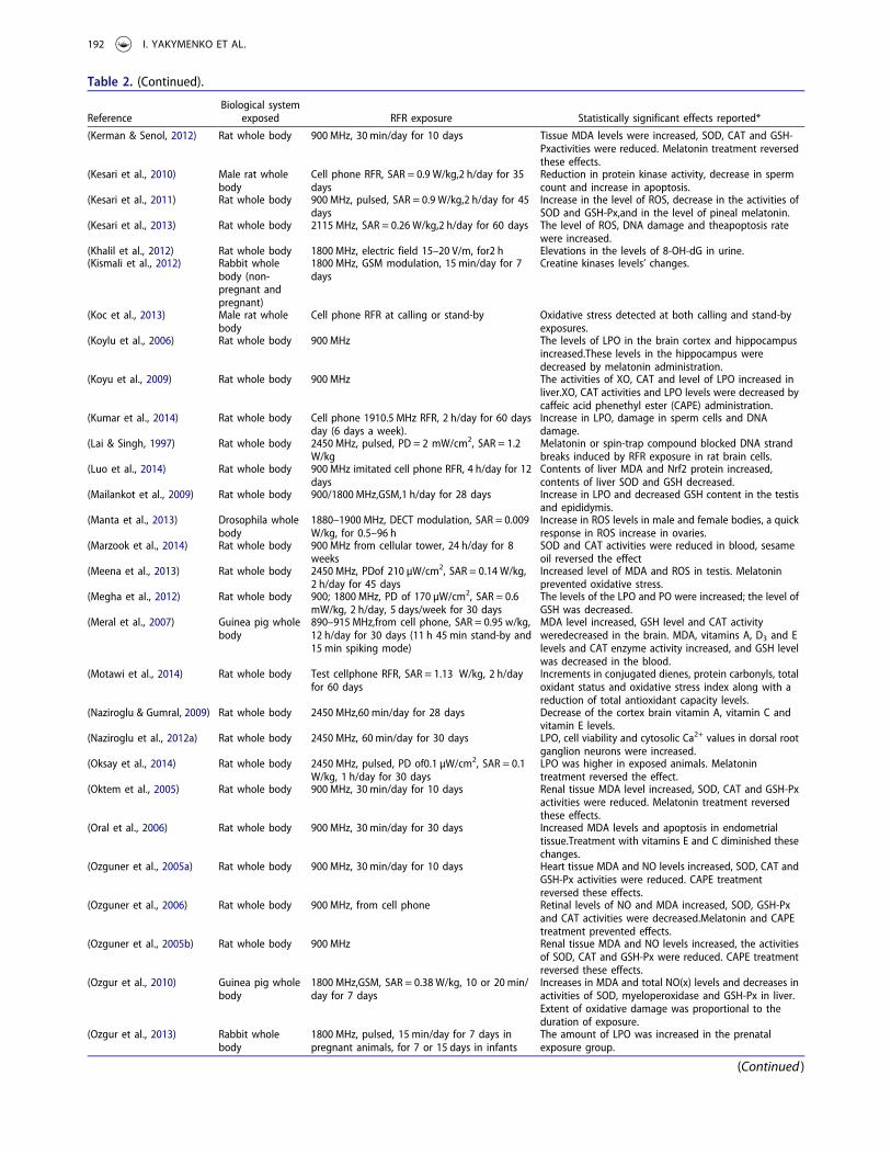
Table 2. (Continued).
ReferenceBiological system
exposed RFR exposure Statistically significant effects reported*
(Kerman & Senol, 2012) Rat whole body 900 MHz, 30 min/day for 10 days Tissue MDA levels were increased, SOD, CAT and GSH-Pxactivities were reduced. Melatonin treatment reversedthese effects.
(Kesari et al., 2010) Male rat wholebody
Cell phone RFR, SAR = 0.9 W/kg,2 h/day for 35days
Reduction in protein kinase activity, decrease in spermcount and increase in apoptosis.
(Kesari et al., 2011) Rat whole body 900 MHz, pulsed, SAR = 0.9 W/kg,2 h/day for 45days
Increase in the level of ROS, decrease in the activities ofSOD and GSH-Px,and in the level of pineal melatonin.
(Kesari et al., 2013) Rat whole body 2115 MHz, SAR = 0.26 W/kg,2 h/day for 60 days The level of ROS, DNA damage and theapoptosis ratewere increased.
(Khalil et al., 2012) Rat whole body 1800 MHz, electric field 15–20 V/m, for2 h Elevations in the levels of 8-OH-dG in urine.(Kismali et al., 2012) Rabbit whole
body (non-pregnant andpregnant)
1800 MHz, GSM modulation, 15 min/day for 7days
Creatine kinases levels’ changes.
(Koc et al., 2013) Male rat wholebody
Cell phone RFR at calling or stand-by Oxidative stress detected at both calling and stand-byexposures.
(Koylu et al., 2006) Rat whole body 900 MHz The levels of LPO in the brain cortex and hippocampusincreased.These levels in the hippocampus weredecreased by melatonin administration.
(Koyu et al., 2009) Rat whole body 900 MHz The activities of XO, CAT and level of LPO increased inliver.XO, CAT activities and LPO levels were decreased bycaffeic acid phenethyl ester (CAPE) administration.
(Kumar et al., 2014) Rat whole body Cell phone 1910.5 MHz RFR, 2 h/day for 60 daysday (6 days a week).
Increase in LPO, damage in sperm cells and DNAdamage.
(Lai & Singh, 1997) Rat whole body 2450 MHz, pulsed, PD = 2 mW/cm2, SAR = 1.2W/kg
Melatonin or spin-trap compound blocked DNA strandbreaks induced by RFR exposure in rat brain cells.
(Luo et al., 2014) Rat whole body 900 MHz imitated cell phone RFR, 4 h/day for 12days
Contents of liver MDA and Nrf2 protein increased,contents of liver SOD and GSH decreased.
(Mailankot et al., 2009) Rat whole body 900/1800 MHz,GSM,1 h/day for 28 days Increase in LPO and decreased GSH content in the testisand epididymis.
(Manta et al., 2013) Drosophila wholebody
1880–1900 MHz, DECT modulation, SAR = 0.009W/kg, for 0.5–96 h
Increase in ROS levels in male and female bodies, a quickresponse in ROS increase in ovaries.
(Marzook et al., 2014) Rat whole body 900 MHz from cellular tower, 24 h/day for 8weeks
SOD and CAT activities were reduced in blood, sesameoil reversed the effect
(Meena et al., 2013) Rat whole body 2450 MHz, PDof 210 µW/cm2, SAR = 0.14 W/kg,2 h/day for 45 days
Increased level of MDA and ROS in testis. Melatoninprevented oxidative stress.
(Megha et al., 2012) Rat whole body 900; 1800 MHz, PD of 170 µW/cm2, SAR = 0.6mW/kg, 2 h/day, 5 days/week for 30 days
The levels of the LPO and PO were increased; the level ofGSH was decreased.
(Meral et al., 2007) Guinea pig wholebody
890–915 MHz,from cell phone, SAR = 0.95 w/kg,12 h/day for 30 days (11 h 45 min stand-by and15 min spiking mode)
MDA level increased, GSH level and CAT activityweredecreased in the brain. MDA, vitamins A, D3 and Elevels and CAT enzyme activity increased, and GSH levelwas decreased in the blood.
(Motawi et al., 2014) Rat whole body Test cellphone RFR, SAR = 1.13 W/kg, 2 h/dayfor 60 days
Increments in conjugated dienes, protein carbonyls, totaloxidant status and oxidative stress index along with areduction of total antioxidant capacity levels.
(Naziroglu & Gumral, 2009) Rat whole body 2450 MHz,60 min/day for 28 days Decrease of the cortex brain vitamin A, vitamin C andvitamin E levels.
(Naziroglu et al., 2012a) Rat whole body 2450 MHz, 60 min/day for 30 days LPO, cell viability and cytosolic Ca2+ values in dorsal rootganglion neurons were increased.
(Oksay et al., 2014) Rat whole body 2450 MHz, pulsed, PD of0.1 µW/cm2, SAR = 0.1W/kg, 1 h/day for 30 days
LPO was higher in exposed animals. Melatonintreatment reversed the effect.
(Oktem et al., 2005) Rat whole body 900 MHz, 30 min/day for 10 days Renal tissue MDA level increased, SOD, CAT and GSH-Pxactivities were reduced. Melatonin treatment reversedthese effects.
(Oral et al., 2006) Rat whole body 900 MHz, 30 min/day for 30 days Increased MDA levels and apoptosis in endometrialtissue.Treatment with vitamins E and C diminished thesechanges.
(Ozguner et al., 2005a) Rat whole body 900 MHz, 30 min/day for 10 days Heart tissue MDA and NO levels increased, SOD, CAT andGSH-Px activities were reduced. CAPE treatmentreversed these effects.
(Ozguner et al., 2006) Rat whole body 900 MHz, from cell phone Retinal levels of NO and MDA increased, SOD, GSH-Pxand CAT activities were decreased.Melatonin and CAPEtreatment prevented effects.
(Ozguner et al., 2005b) Rat whole body 900 MHz Renal tissue MDA and NO levels increased, the activitiesof SOD, CAT and GSH-Px were reduced. CAPE treatmentreversed these effects.
(Ozgur et al., 2010) Guinea pig wholebody
1800 MHz,GSM, SAR = 0.38 W/kg, 10 or 20 min/day for 7 days
Increases in MDA and total NO(x) levels and decreases inactivities of SOD, myeloperoxidase and GSH-Px in liver.Extent of oxidative damage was proportional to theduration of exposure.
(Ozgur et al., 2013) Rabbit wholebody
1800 MHz, pulsed, 15 min/day for 7 days inpregnant animals, for 7 or 15 days in infants
The amount of LPO was increased in the prenatalexposure group.
(Continued )
192 I. YAKYMENKO ET AL.
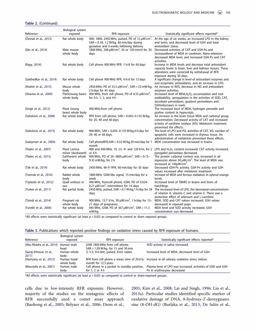
cells due to low-intensity RFR exposure. However,majority of the studies on the mutagenic effects ofRFR successfully used a comet assay approach(Baohong et al., 2005; Belyaev et al., 2006; Diem et al.,
2005; Kim et al., 2008; Lai and Singh, 1996; Liu et al.,2013a). Particular studies identified specific marker ofoxidative damage of DNA, 8-hydroxy-2'-deoxyguano-sine (8-OH-dG) (Burlaka et al., 2013; De Iuliis et al.,
Table 2. (Continued).
ReferenceBiological system
exposed RFR exposure Statistically significant effects reported*
(Özorak et al., 2013) Rat whole body 900; 1800; 2450 MHz, pulsed, PD of 12 µW/cm2.SAR = 0.18; 1.2 W/kg, 60 min/day duringgestation and 6 weeks following delivery
At the age of six weeks, an increased LPO in the kidneyand testis, and decreased level of GSH and totalantioxidant status.
(Qin et al., 2014) Male mousewhole body
1800 MHz, 208 µW/cm2, 30 or 120 min/d for 30days
Decreased activities of CAT and GSH-Px andincreasedlevel of MDA in cerebrum. Nano-seleniumdecreased MDA level, and increased GSH-Px and CATactivities.
(Ragy, 2014) Rat whole body Cell phone 900 MHz RFR, 1 h/d for 60 days Increase in MDA levels and decrease total antioxidantcapacity levels in brain, liver and kidneys tissues. Thesealterations were corrected by withdrawal of RFRexposure during 30 days.
(Saikhedkar et al., 2014) Rat whole body Cell phone 900 MHz RFR, 4 h/d for 15 days A significant change in level of antioxidant enzymes andnon-enzymatic antioxidants, and an increase in LPO.
(Shahin et al., 2013) Mouse wholebody
2450 MHz, PD of 33.5 µW/cm2, SAR = 23 mW/kg,2 h/day for 45 days
An increase in ROS, decrease in NO and antioxidantenzymes activities.
(Sharma et al., 2009) Plant(mung bean)whole body
900 MHz, from cell phone, PD of 8.55 µW/cm2;for 0.5; 1; 2, and 4 h
Increased level of MDA,H2O2 accumulation and rootoxidizability, upregulation in the activities of SOD, CAT,ascorbate peroxidases, guaiacol peroxidases andGSHreductases in roots.
(Singh et al., 2012) Plant (mungbean) whole body
900 MHz,from cell phone The increased level of MDA, hydrogen peroxide andproline content in hypocotyls.
(Sokolovic et al., 2008) Rat whole body RFR from cell phone, SAR = 0.043–0.135 W/kg,for 20, 40 and 60 days
An increase in the brain tissue MDA and carbonyl groupconcentration. Decreased activity of CAT and increasedactivity of xanthine oxidase (XO). Melatonin treatmentprevented the effects.
(Sokolovic et al., 2013) Rat whole body 900 MHz, SAR = 0,043–0.135 W/kg,4 h/day for29; 40 or 60 days,
The level of LPO and PO, activities of CAT, XO, number ofapoptotic cells were increased in thymus tissue. Anadministration of melatonin prevented these effects.
(Suleyman et al., 2004) Rat whole body Cell phoneRFR,SAR = 0.52 W/kg,20 min/day for 1month
MDA concentration was increased in brains.
(Tkalec et al., 2007) Plant Lemnaminor (duckweed)
400 and 900 MHz, 10, 23, 41 and 120 V/m, for 2or 4 h
LPO and H2O2 content increased: CAT activity increased,pyrogallol peroxidase decreased.
(Tkalec et al., 2013) Earthworm wholebody
900 MHz, PD of 30–3800 µW/cm2, SAR = 0.13–9.33 mW/kg, for 2 h
The protein carbonyl content was increased in allexposures above 30 µWc/m2. The level of MDA wasincreased at 140µW/cm2.
(Tök et al., 2014) Rat whole body 2450 MHz, Wi-Fi RFR, 60 min/day for 30 days Decreased GSH-Px activity. GSH-Px activity and GSHvalues increased after melatonin treatment.
(Tomruk et al., 2010) Rabbit wholebody
1800 MHz, GSM-like signal, 15 min/day for aweek
Increase of MDA and ferrous oxidation in xylenol orangelevels.
(Tsybulin et al., 2012) Quail embryo inovo
900 MHz, fromcell phone, GSM, PD of 0.024–0.21 µW/cm2, intermittent for 14 days
Increased level of TBARS in brains and livers ofhatchlings.
(Turker et al., 2011) Rat partial body 2450 MHz, pulsed, SAR = 0.1 W/kg,1 h/day for 28days
The increased level of LPO, the decreased concentrationsof vitamin A, vitamin C and vitamin E. There was aprotective effect of selenium and L-carnitine.
(Türedi et al., 2014) Pregnant ratwhole body
900 MHz, 13.7 V/m, 50 µW/cm2, 1 h/day for 13–21 days of pregnancy
MDA, SOD and CAT values increased, GSH valuesdecreased in exposed pups.
(Yurekli et al., 2006) Rat whole body 945 MHz, GSM, PD of 367 µW/cm2, SAR = 11.3mW/kg
MDA level and SOD activity increased, GSHconcentration was decreased.
*All effects were statistically significant (at least p < 0.05) as compared to control or sham exposed groups.
Table 3. Publications which reported positive findings on oxidative stress caused by RFR exposure of humans.
ReferenceBiological system
exposed RFR exposure Statistically significant effects reported*
(Abu Khadra et al., 2014) Human malehead
GSM 1800 MHz from cell phone,SAR = 1.09 W/kg, for 15 and 30 min
SOD activity in saliva increased.
(Garaj-Vrhovac et al.,2011)
Human wholebody
3; 5.5; 9.4 GHz, pulsed, from radars Increased level of MDA, decreased level of GSH.
(Hamzany et al., 2013) Human head/whole body
RFR from cell phone a mean time of 29.6 h/month for 12.5 years
Increase in all salivary oxidative stress indices.
(Moustafa et al., 2001) Human malebody
Cell phone in a pocket in standby position,for 1; 2 or 4 h
Plasma level of LPO was increased, activities of SOD and GSH-Px in erythrocytes decreased.
*All effects were statistically significant (at least p < 0.05) as compared to control or sham-exposed groups.
ELECTROMAGNETIC BIOLOGY AND MEDICINE 193
2009; Guler et al., 2012; Khalil et al., 2012; Xu et al.,2010). Thus, the level of 8-OH-dG in human sperma-tozoa was shown to be significantly increased after invitro exposure to low-intensity RFR (De Iuliis et al.,2009). Likewise, we demonstrated that the exposure ofquail embryos in ovo to GSM 900MHz of 0.25 µW/cm2
during a few days was sufficient for a significant, two-threefold, increase of 8-OH-dG level in embryonic cells(Burlaka et al., 2013).
It would be logical to assume that most mutageniceffects due to the RFR exposure are caused by oxidativedamage to DNA, as the overproduction of ROS inliving cells due to RFR exposure was reliably documen-ted. It is known that superoxide itself does not affectDNA. The most aggressive form of ROS, which is ableto affect the DNA molecule directly, is hydroxyl radical(Halliwell, 2007). The hydroxyl radicals are generatedin cell in the Fenton reaction (Fe2+ + H2O2 -> Fe3++OH• + OH−) and in the Haber–Weiss reaction (O2
•−+H2O2 -> O2 + OH• + OH−) (Valko et al., 2006). On theother hand, increased concentration of NO in additionto superoxide in the RFR-exposed cells can lead to theformation of other aggressive form of ROS, peroxyni-trite (ONOO−), which can also cause DNA damage(Valko et al., 2006).
Free radicals induced under the RFR exposurecan perturb cellular signaling
Taking into account the abovementioned data, we canstate that the exposure to RFR leads to overproductionof free radicals/ROS in living cell. Certainly, free radi-cals can induce harmful effects via direct damage due tooxidation of biological macromolecules. To that, itbecomes clear nowadays that free radicals/ROS are anintrinsic part of the cellular signaling cascades (Forman
et al., 2014). Thus, hydrogen peroxide appears as asecond messenger both in insulin signaling and ingrowth factor-induced signalling cascades (Sies, 2014).These species are also implicated in biochemicalmechanism of oxidation of ethanol and in other meta-bolic processes (Oshino et al., 1975) and is alsorequired for initiation of wound repair (Enyedi andNiethammer, 2013). In addition, ROS at relatively lowconcentrations can modulate inflammation via activa-tion of NF-kB pathway (Hayden and Ghosh, 2011).Therefore, even subtle exposures to RFR with genera-tion of hardly detectable quantities of free radicals canhave their meaningful biological consequences.
We could ascertain the signaling effects of moderatelevels of free radicals from our experiments in quailembryos irradiated with the commercial cell phone.Thus, we were able to show that the prolonged expo-sures of embryos in ovo led to robust repression of theirdevelopment (Tsybulin et al., 2013), which was conco-mitant with significant overproduction of superoxideradical and NO radical, increased rates of lipid perox-idation and oxidative damage of DNA (Burlaka et al.,2013; Tsybulin et al., 2012). Notably, shorter exposuresinstead led to enhancement in embryonic development(Tsybulin et al., 2012, 2013). We demonstrated thefavorable effects of shorter exposures also on the mole-cular level. Thus, after the short-time RFR exposure theDNA comets in embryonic cells were significantlyshorter than in the control non-irradiated embryos,pointing to activation of mechanisms maintaining theintegrity of DNA. The “beneficial” consequences of theirradiation could be explained by hormesis effect(Calabrese, 2008). However, one could hypothesizethat the “beneficial” effects of the irradiation could beexplained by the signaling action of free radicalsinduced at levels below the damaging concentrations.
Table 4. Publications which reported no significant oxidative effects after RFR exposure.
ReferenceBiological system
exposed RFR exposure Effects reported
(Hook et al., 2004) Mammalian cells in vitro 835.62 MHz (frequency-modulated continuous-wave, FMCW) and 847.74 MHz (code divisionmultiple access, CDMA), SAR = 0.8 W/kg, for 20–22 h
FMCW- and CDMA-modulated RFR did not alterparameters indicative of oxidative stress.
(Ferreira et al., 2006a) Rat whole body 800–1800 MHz, from cell phone No changes in lipid and protein damage, and innon-enzymatic antioxidant defense in frontalcortex or hippocampus.
(Ferreira et al., 2006b) Pregnant rat whole body RFR from cell phone No differences in oxidative parameter ofoffspring blood and liver, but increase inerythrocytes micronuclei incidence in offspring.
(Dasdag et al., 2003) Rat whole body Cell phone RFR, SAR = 0.52 W/kg, 20 min/day for 1month
No alteration in MDA concentration.
(Demirel et al., 2012) Rat whole body 3G cell phone RFR, “standardized daily dose” for 20days
No difference in GSH-Px and CAT activity in eyetissues, in MDA and GSH levels in blood.
(Khalil et al., 2014) Human head/wholebody
Cell phone RFR (talking mode) for 15 or 30 min No relationship between exposure and changesin the salivary oxidant/antioxidant profile.
(de Souza et al., 2014) Human head/wholebody
Cell phone RFR No difference in the saliva from the parotidgland exposed to cell phone RFR to the salivafrom the opposite gland of each individual.
194 I. YAKYMENKO ET AL.
Obviously, any seemingly beneficial effect of externalenvironmental impact should be treated with cautionand possibly minimized before careful evaluation of thelong-term consequences. Altogether, this gives a clearwarning of the adverse health effects of low-intensityRFR, which could be evoked both by the direct oxida-tive damage and by disturbed cellular signaling.
Oxidative effects and non-cancer health effectsof RFR
A new medical condition, so-called electrohypersensi-tivity (EHS), in which people suffer due to RFR expo-sure, has been described (Johansson, 2006). Typically,these persons suffer from skin- and mucosa-relatedsymptoms (itching, smarting, pain, heat sensation), orheart and nervous system disorders after exposure tocomputer monitors, cell phones and other electromag-netic devices. This disorder is growing continuously:starting from 0.06% of the total population in 1985,this category now includes as much as 9–11% of theEuropean population (Hallberg and Oberfeld, 2006). InSweden, for example, EHS has become an officiallyrecognized health impairment.
To that, a high percentage, up to 18–43% of youngpeople, has recently been described to be suffering fromheadache/earache during or after cell phone conversa-tions (Chu et al., 2011; Yakymenko et al., 2011).Likewise, a number of psychophysical and preclinicaldisorders including fatigue, irritation, headache, sleepdisorders, hormonal imbalances were detected in highpercent of people living nearby cell phone base trans-ceiver stations (Buchner and Eger, 2011; Santini et al.,2002).
An allergy reaction to RFR in humans has beenconfirmed by a significant increase in the level ofmast cells in skin of persons under exposure to electro-magnetic devices (Johansson et al., 2001). Likewise,higher level of degranulated mast cells in dermis ofEHS persons has been detected (Johansson, 2006). Inturn, the activated mast cells can release histamine andother mediators of such reactions which include allergichypersensitivity, itching, dermatoses, etc. Importantly,an implication of ROS in allergic reactions is ratherclear nowadays. For example, in case of airway allergicinflammation, the lung cells generate superoxide innanomolar concentrations following antigen challenges(Nagata, 2005). Then, mast cells generate ROS follow-ing aggregation of FcɛRI, a high-affinity IgE receptor(Okayama, 2005). In addition, pollen NADPH oxidasesrapidly increase the level of ROS in lung epithelium(Boldogh et al., 2005); and removal of pollen NADPHoxidases from the challenge material reduced antigen-
induced allergic airway inflammation. Thus, it seemsplausible that EHS-like conditions can be attributed atleast partially to ROS overproduction in cells due toRFR exposures.
Oxidative effects and potential carcinogenicityof RFR
During recent years, a number of epidemiological stu-dies indicated a significant increase in incidence ofvarious types of tumors among long-term or “heavy”users of cellular phones (Yakymenko et al., 2011).Briefly, reports pointed to the increased risk in braintumors (Cardis et al., 2010; Hardell and Carlberg, 2009;Hardell et al., 2007), acoustic neuroma (Hardell et al.,2005; Sato et al., 2011), tumors of parotid glands(Sadetzki et al., 2008), seminomas (Hardell et al.,2007), melanomas (Hardell et al., 2011) and lympho-mas (Hardell et al., 2005) in these cohorts of people. Tothat, a significant increase in tumor incidence amongpeople living nearby cellular base transceiver stationswas also reported (Eger et al., 2004; Wolf and Wolf,2007). Similarly, experimental evidences of cancerexpansion in rodents caused by long-term low-intensityRFR exposure were published (Chou et al., 1992;Repacholi et al., 1997; Szmigielski et al., 1982; Toleret al., 1997). To that, activation of ODC was detected inRFR-exposed cells (Hoyto et al., 2007). ODC isinvolved in processes of cell growth and differentiation,and its activity is increased in tumor cells. Althoughoverexpression of ODC is not sufficient for tumori-genic transformation, an increased activity of thisenzyme was shown to promote the development oftumors from pre-tumor cells (Clifford et al., 1995).
Significant overproduction of ROS leads to oxidativestress in living cells, induces oxidative damage of DNAand can cause malignant transformation (Halliwell andWhiteman, 2004; Valko et al., 2007). It is known that inaddition to mutagenic effects, ROS play a role as asecond messenger for intracellular signaling cascadeswhich can also induce oncogenic transformation(Valko et al., 2006). Earlier we hypothesized (Burlakaet al., 2013) that low-intensity RFR exposure leads todysfunctions of mitochondria, which result in overpro-duction of superoxide and NO, and subsequently toROS-mediated mutagenesis. To that, it is well estab-lished that oxidative stress is associated with carcino-genesis; for instance, the oxidative stress elicited byMembrane-Type 1 Matrix Metalloproteinase is impli-cated in both the pathogenesis and progression ofprostate cancer (Nguyen et al., 2011). Similarly, a pro-gressive elevation in mitochondrial ROS production(chronic ROS) under both hypoxia and/or low glucose,
ELECTROMAGNETIC BIOLOGY AND MEDICINE 195

which leads to stabilization of cells via increasedHIF-2alpha expression, can eventually result in malig-nant transformation (Ralph et al., 2010). These data,together with the strong experimental evidences onactivation of NADH oxidase under RFR exposure(Friedman et al., 2007) suggest that low-intensity RFRis a multifactorial stress factor for living cell, significantfeature of which is oxidative effects and potential car-cinogenicity as a result.
Conclusions
The analysis of modern data on biological effects oflow-intensity RFR leads to a firm conclusion that thisphysical agent is a powerful oxidative stressor for livingcell. The oxidative efficiency of RFR can be mediatedvia changes in activities of key ROS-generating systems,including mitochondria and non-phagocytic NADHoxidases, via direct effects on water molecules, and viainduction of conformation changes in biologicallyimportant macromolecules. In turn, a broad biologicalpotential of ROS and other free radicals, including boththeir mutagenic effects and their signaling regulatorypotential, makes RFR a potentially hazardous factor forhuman health. We suggest minimizing the intensityand time of RFR exposures, and taking a precautionaryapproach towards wireless technologies in everydayhuman life.
Acknowledgments
The authors are grateful to the unknown referees for thevaluable comments on the first version of the manuscript.
Declaration of interest
The authors declare no conflicts of interest. This studywas supported by National Academy of Sciences ofUkraine (I.Y., E.S.) and by University of Campinas viaPPVE (Programa Professor Visitante do Exterior),Brazil (S.K.).
References
Abdel-Rassoul, G., El-Fateh, O. A., Salem, M. A., et al. (2007).Neurobehavioral effects among inhabitants around mobilephone base stations. Neurotoxicology 28:434–440.
Abu Khadra, K. M., Khalil, A. M., Abu Samak, M., et al.(2014). Evaluation of selected biochemical parameters inthe saliva of young males using mobile phones.Electromagn. Biol. Med. 32:72–76.
Agarwal, A., Desai, N. R., Makker, K., et al. (2009). Effects ofradiofrequency electromagnetic waves (RF-EMW) from
cellular phones on human ejaculated semen: An in vitropilot study. Fertil. Steril. 92:1318–1325.
Akbari, A., Jelodar, G., Nazifi, S. (2014). Vitamin C protectsrat cerebellum and encephalon from oxidative stress fol-lowing exposure to radiofrequency wave generated by BTSantenna mobile. Toxicol. Mechanisms Methods 24:347–352.
Al-Damegh, M. A. (2012). Rat testicular impairment inducedby electromagnetic radiation from a conventional cellulartelephone and the protective effects of the antioxidantsvitamins C and E. Clinics 67:785–792.
Avci, B., Akar, A., Bilgici, B., et al. (2012). Oxidative stressinduced by 1.8 GHz radio frequency electromagneticradiation and effects of garlic extract in rats. Int. J.Radiat. Biol. 88:799–805.
Ayata, A., Mollaoglu, H., Yilmaz, H. R., et al. (2004).Oxidative stress-mediated skin damage in an experimentalmobile phone model can be prevented by melatonin. J.Dermatol. 31:878–883.
Aynali, G., Naziroglu, M., Celik, O., et al. (2013). Modulationof wireless (2.45 GHz)-induced oxidative toxicity in laryn-gotracheal mucosa of rat by melatonin. Eur. Arch. Oto-Rhino-Laryngol. 270:1695–1700.
Balci, M., Devrim, E., Durak, I. (2007). Effects of mobilephones on oxidant/antioxidant balance in cornea andlens of rats. Curr. Eye Res. 32:21–25.
Baohong, W., Jiliang, H., Lifen, J., et al. (2005). Studying thesynergistic damage effects induced by 1.8 GHz radiofre-quency field radiation (RFR) with four chemical mutagenson human lymphocyte DNA using comet assay in vitro.Mutat. Res. 578:149–157.
Belyaev, I. (2010). Dependence of non-thermal biologicaleffects of microwaves on physical and biological variables:Implications for reproducibility and safety standards. Eur.J. Oncol. Library 5:187–217.
Belyaev, I. Y., Koch, C. B., Terenius, O., et al. (2006).Exposure of rat brain to 915MHz GSM microwavesinduces changes in gene expression but not doublestranded DNA breaks or effects on chromatin conforma-tion. Bioelectromagnetics 27:295–306.
Bilgici, B., Akar, A., Avci, B., et al. (2013). Effect of 900MHzradiofrequency radiation on oxidative stress in rat brainand serum. Electromagn. Biol. Med. 32:20–29.
Blank, M., Soo, L. (2001). Electromagnetic acceleration ofelectron transfer reactions. J. Cell Biochem. 81:278–283.
Blank, M., Soo, L. (2003). Electromagnetic acceleration ofthe Belousov–Zhabotinski reaction. Bioelectrochemistry61:93–97.
Bodera, P., Stankiewicz, W., Zawada, K., et al. (2013).Changes in antioxidant capacity of blood due to mutualaction of electromagnetic field (1800MHz) and opioiddrug (tramadol) in animal model of persistent inflamma-tory state. Pharmacol. Rep. 65:421–428.
Bohr, H., Bohr, J. (2000a). Microwave-enhanced foldingand denaturation of globular proteins. Phys. Rev. E61:4310–4314.
Bohr, H., Bohr, J. (2000b). Microwave enhanced kineticsobserved in ORD studies of a protein. Bioelectromagnetics21:68–72.
Boldogh, I., Bacsi, A., Choudhury, B. K., et al. (2005). ROSgenerated by pollen NADPH oxidase provide a signal thataugments antigen-induced allergic airway inflammation. J.Clin. Investig. 115:2169–2179.
196 I. YAKYMENKO ET AL.

Buchner, K., Eger, H. (2011). [Changes of clinically importantneurotransmitters under the influence of modulated RFfields—A long-term study under real-life conditions].Umwelt -Medizin-Gesellschaft 24:44–57.
Budi, A., Legge, F. S., Treutlein, H., et al. (2007). Effect offrequency on insulin response to electric field stress. J.Phys. Chem. B. 111:5748–5756.
Burlaka, A., Selyuk, M., Gafurov, M., et al. (2014). Changesin mitochondrial functioning with electromagneticradiation of ultra high frequency as revealed by electronparamagnetic resonance methods. Int. J. Radiat. Biol.90:357–362.
Burlaka, A., Tsybulin, O., Sidorik, E., et al. (2013).Overproduction of free radical species in embryonal cellsexposed to low intensity radiofrequency radiation. Exp.Oncol. 35:219–225.
Byus, C. V., Kartun, K., Pieper, S., et al. (1988). Increasedornithine decarboxylase activity in cultured cells exposedto low energy modulated microwave fields and phorbolester tumor promoters. Cancer Res. 48:4222–4226.
Calabrese, E. J. (2008). Hormesis: Why it is important totoxicology and toxicologists. Environ. Toxicol. Chem.27:1451–1474.
Campisi, A., Gulino, M., Acquaviva, R., et al. (2010). Reactiveoxygen species levels and DNA fragmentation on astro-cytes in primary culture after acute exposure to low inten-sity microwave electromagnetic field. Neurosci. Lett.473:52–55.
Caraglia, M., Marra, M., Mancinelli, F., et al. (2005).Electromagnetic fields at mobile phone frequency induceapoptosis and inactivation of the multi-chaperone complexin human epidermoid cancer cells. J. Cell. Physiol.204:539–548.
Cardis, E., Deltour, I., Vrijheid, M., et al. (2010). Braintumour risk in relation to mobile telephone use: Resultsof the INTERPHONE international case-control study. Int.J. Epidemiol. 39:675–694.
Cenesis, M., Atakisi, O., Akar, A., et al. (2011). Effects of 900and 1800MHz electromagnetic field application on elec-trocardiogram, nitric oxide, total antioxidant capacity,total oxidant capacity, total protein, albumin and globulinlevels in guinea pigs. Kafkas Üniv. Vet. Fakültesi Dergisi17:357–362.
Céspedes, O., Ueno, S. (2009). Effects of radio frequencymagnetic fields on iron release from cage proteins.Bioelectromagnetics 30:336–342.
Cetin, H., Naziroglu, M., Celik, Ö., et al. (2014). Liver anti-oxidant stores protect the brain from electromagneticradiation (900 and 1800MHz)-induced oxidative stress inrats during pregnancy and the development of offspring. J.Matern.-Fetal Neonat. Med. 72:1915–1921.
Chou, C. K., Guy, A. W., Kunz, L. L., et al. (1992). Long-term, low-level microwave irradiation of rats.Bioelectromagnetics 13:469–496.
Chu, M. K., Song, H. G., Kim, C., et al. (2011). Clinicalfeatures of headache associated with mobile phone use: Across-sectional study in university students. BMC Neurol.11:115.
Clifford, A., Morgan, D., Yuspa, S. H., et al. (1995). Role ofornithine decarboxylase in epidermal tumorigenesis.Cancer Res. 55:1680–1686.
Consales, C., Merla, C., Marino, C., et al. (2012).Electromagnetic fields, oxidative stress, and neurodegen-eration. Int. J. Cell Biol. 2012:683897.
Dasdag, S., Akdag, M. Z., Kizil, G., et al. (2012). Effect of 900MHz radio frequency radiation on beta amyloid protein,protein carbonyl, and malondialdehyde in the brain.Electromagn. Biol. Med. 31:67–74.
Dasdag, S., Akdag, M. Z., Ulukaya, E., et al. (2009). Effect ofmobile phone exposure on apoptotic glial cells and statusof oxidative stress in rat brain. Electromagn. Biol. Med.28:342–354.
Dasdag, S., Bilgin, H., Akdag, M. Z., et al. (2008). Effect oflong term mobile phone exposure on oxidative-antioxida-tive processes and nitric oxide in rats. Biotechnol.Biotechnol. Equip. 22:992–997.
Dasdag, S., Zulkuf Akdag, M., Aksen, F., et al. (2003). Wholebody exposure of rats to microwaves emitted from a cellphone does not affect the testes. Bioelectromagnetics24:182–188.
De Iuliis, G. N., Newey, R. J., King, B. V., et al. (2009). Mobilephone radiation induces reactive oxygen species produc-tion and DNA damage in human spermatozoa in vitro.PLoS One 4:e6446.
de Souza, F. T., Silva, J. F., Ferreira, E. F., et al. (2014). Cellphone use and parotid salivary gland alterations: No mole-cular evidence. Cancer Epidemiol. Biomarkers Prevent.23:1428–1431.
Demirel, S., Doganay, S., Turkoz, Y., et al. (2012). Effects ofthird generation mobile phone-emitted electromagneticradiation on oxidative stress parameters in eye tissue andblood of rats. Cutan. Ocul. Toxicol. 31:89–94.
Desai, N. R., Kesari, K. K., Agarwal, A. (2009).Pathophysiology of cell phone radiation: Oxidative stressand carcinogenesis with focus on male reproductive sys-tem. Reprod. Biol. Endocrinol. 7:114.
Deshmukh, P. S., Banerjee, B. D., Abegaonkar, M. P., et al.(2013). Effect of low level microwave radiation exposureon cognitive function and oxidative stress in rats. Indian J.Biochem. Biophys. 50:114–119.
Diem, E., Schwarz, C., Adlkofer, F., et al. (2005). Non-ther-mal DNA breakage by mobile-phone radiation (1800MHz) in human fibroblasts and in transformed GFSH-R17 rat granulosa cells in vitro. Mutat. Res. 583:178–183.
Dutta, S. K., Ghosh, B., Blackman, C. F. (1989).Radiofrequency radiation-induced calcium ion effluxenhancement from human and other neuroblastoma cellsin culture. Bioelectromagnetics 10:197–202.
Eger, H., Hagen, K., Lucas, B., et al. (2004). [Influence of theproximity of mobile phone base stations on the incidenceof cancer]. Environ. Med. Soc. 17:273–356.
Enyedi, B., Niethammer, P. (2013). H2O2: A chemoattractant?Methods Enzymol. 528:237–255.
Esmekaya, M. A., Ozer, C., Seyhan, N. (2011). 900MHzpulse-modulated radiofrequency radiation induces oxida-tive stress on heart, lung, testis and liver tissues. Gen.Physiol. Biophys. 30:84–89.
Ferreira, A. R., Bonatto, F., de Bittencourt Pasquali, M. A.,et al. (2006a). Oxidative stress effects on the central ner-vous system of rats after acute exposure to ultra highfrequency electromagnetic fields. Bioelectromagnetics27:487–493.
ELECTROMAGNETIC BIOLOGY AND MEDICINE 197
Ferreira, A. R., Knakievicz, T., Pasquali, M. A., et al. (2006b).Ultra high frequency-electromagnetic field irradiation dur-ing pregnancy leads to an increase in erythrocytes micro-nuclei incidence in rat offspring. Life Sci. 80:43–50.
Forman, H. J., Ursini, F., Maiorino, M. (2014). An overview ofmechanisms of redox signaling. J. Mol. Cell Cardiol. 73:2–9.
Friedman, J., Kraus, S., Hauptman, Y., et al. (2007).Mechanism of short-term ERK activation by electromag-netic fields at mobile phone frequencies. Biochem. J.405:559–568.
Furtado-Filho, O. V., Borba, J. B., Dallegrave, A., et al. (2014).Effect of 950MHz UHF electromagnetic radiation on bio-markers of oxidative damage, metabolism of UFA andantioxidants in the livers of young rats of different ages.Int. J. Radiat. Biol. 90:159–168.
Gandhi, O. P., Morgan, L. L., de Salles, A. A., et al. (2012).Exposure limits: The underestimation of absorbed cellphone radiation, especially in children. Electromagn. Biol.Med. 31:34–51.
Garaj-Vrhovac, V., Fucic, A., Horvat, D. (1992). The correla-tion between the frequency of micronuclei and specificchromosome aberrations in human lymphocytes exposedto microwave radiation in vitro. Mutat. Res. 281:181–186.
Garaj-Vrhovac, V., Gajski, G., Pažanin, S., et al. (2011).Assessment of cytogenetic damage and oxidative stress inpersonnel occupationally exposed to the pulsed microwaveradiation of marine radar equipment. Int. J. Hyg. Environ.Health. 214:59–65.
Garson, O. M., McRobert, T. L., Campbell, L. J., et al. (1991).A chromosomal study of workers with long-term exposureto radio-frequency radiation. Med. J. Austral. 155:289–292.
Georgiou, C. D. (2010). Oxidative stress-induced biologicaldamage by low-level EMFs: Mechanism of free radical pairelectron spin-polarization and biochemical amplification.Eur. J. Oncol. 5:63–113.
Goodman, R., Blank, M. (2002). Insights into electromagneticinteraction mechanisms. J. Cell Physiol. 192:16–22.
Griendling, K. K., Sorescu, D., Ushio-Fukai, M. (2000). NAD(P)H oxidase: Role in cardiovascular biology and disease.Circ. Res. 86:494–501.
Guler, G., Tomruk, A., Ozgur, E., et al. (2012). The effect ofradiofrequency radiation on DNA and lipid damage infemale and male infant rabbits. Int. J. Radiat. Biol.88:367–373.
Guney, M., Ozguner, F., Oral, B., et al. (2007). 900MHzradiofrequency-induced histopathologic changes and oxi-dative stress in rat endometrium: Protection by vitamins Eand C. Toxicol. Ind. Health 23:411–420.
Gürler, H. Ş., Bilgici, B., Akar, A. K., et al. (2014). IncreasedDNA oxidation (8-OHdG) and protein oxidation (AOPP) bylow level electromagnetic field (2.45 GHz) in rat brain andprotective effect of garlic. Int. J. Radiat. Biol. 90:892–896.
Guzy, R. D., Schumacker, P. T. (2006). Oxygen sensing bymitochondria at complex III: The paradox of increasedreactive oxygen species during hypoxia. Exp. Physiol.91:807–819.
Hallberg, O., Oberfeld, G. (2006). Letter to the editor: Will weall become electrosensitive? Electromagn. Biol. Med.25:189–191.
Halliwell, B. (1991). Reactive oxygen species in living systems:Source, biochemistry, and role in human disease. Am. J.Med. 91:14S–22S.
Halliwell, B. (2007). Biochemistry of oxidative stress.Biochem. Soc. Trans. 35:1147–1150.
Halliwell, B., Whiteman, M. (2004). Measuring reactive spe-cies and oxidative damage in vivo and in cell culture: Howshould you do it and what do the results mean? Br. J.Pharmacol. 142:231–255.
Hamzany, Y., Feinmesser, R., Shpitzer, T., et al. (2013). Ishuman saliva an indicator of the adverse health effectsof using mobile phones? Antioxid .Redox. Signal.18:622–627.
Hardell, L., Carlberg, M. (2009). Mobile phones, cordlessphones and the risk for brain tumours. Int. J. Oncol.35:5–17.
Hardell, L., Carlberg, M., Hansson Mild, K. (2005). Case-control study on cellular and cordless telephones and therisk for acoustic neuroma or meningioma in patients diag-nosed 2000–2003. Neuroepidemiology 25:120–128.
Hardell, L., Carlberg, M., Hansson Mild, K., et al. (2011).Case-control study on the use of mobile and cordlessphones and the risk for malignant melanoma in the headand neck region. Pathophysiology 18:325–333.
Hardell, L., Carlberg, M., Ohlson, C. G., et al. (2007). Use ofcellular and cordless telephones and risk of testicular can-cer. Int. J. Androl. 30:115–122.
Hardell, L., Carlberg, M., Soderqvist, F., et al. (2007). Long-term use of cellular phones and brain tumours: Increasedrisk associated with use for > or = 0 years. Occup.Environ. Med. 64:626–632.
Hardell, L., Eriksson, M., Carlberg, M., et al. (2005). Use ofcellular or cordless telephones and the risk for non-Hodgkin's lymphoma. Int. Arch. Occup. Environ. Health78:625–632.
Hayden, M. S., Ghosh, S. (2011). NF-kappa B in immuno-biology. Cell Res. 21:223–244.
Hong, M. N., Kim, B. C., Ko, Y. G., et al. (2012). Effects of837 and 1950MHz radiofrequency radiation exposurealone or combined on oxidative stress in MCF10A cells.Bioelectromagnetics 33:604–611.
Hook, G. J., Spitz, D. R., Sim, J. E., et al. (2004). Evaluation ofparameters of oxidative stress after in vitro exposure toFMCW- and CDMA-modulated radiofrequency radiationfields. Radiat. Res. 162:497–504.
Hou, Q., Wang, M., Wu, S., et al. (2014). Oxidative changes andapoptosis induced by 1800-MHz electromagnetic radiationin NIH/3T3 cells. Electromagn. Biol. Med. 34:85–92.
Hoyto, A., Juutilainen, J., Naarala, J. (2007). Ornithine dec-arboxylase activity is affected in primary astrocytes but notin secondary cell lines exposed to 872MHz RF radiation.Int. J. Radiat. Biol. 83:367–374.
Hyland, G. J. (2000). Physics and biology of mobile tele-phony. Lancet 356:1833–1836.
ICNIRP. (1998). Guidelines for limiting exposure to time-varying elecrtic, magnetic and electromagnetic fields (up to300 GHz). Health Phys. 74:494–522.
Ilhan, A., Gurel, A., Armutcu, F., et al. (2004). Ginkgo bilobaprevents mobile phone-induced oxidative stress in ratbrain. Clin. Chim. Acta. 340:153–162.
Inoue, M., Sato, E. F., Nishikawa, M., et al. (2003).Mitochondrial generation of reactive oxygen species andits role in aerobic life. Curr. Med. Chem. 10:2495–2505.
Jelodar, G., Akbari, A., Nazifi, S. (2013). The prophylacticeffect of vitamin C on oxidative stress indexes in rat eyes
198 I. YAKYMENKO ET AL.

following exposure to radiofrequency wave generated by aBTS antenna model. Int. J. Radiat. Biol. 89:128–131.
Jelodar, G., Nazifi, S., Akbari, A. (2013). The prophylacticeffect of vitamin C on induced oxidative stress in rat testisfollowing exposure to 900 MHz radio frequency wavegenerated by a BTS antenna model. Electromagn. Biol.Med. 32:409–416.
Jing, J., Yuhua, Z., Xiao-qian, Y., et al. (2012). The influenceof microwave radiation from cellular phone on fetal ratbrain. Electromagn. Biol. Med. 31:57–66.
Johansson, O. (2006). Electrohypersensitivity: State-of-the-artof a functional impairment. Electromagn. Biol. Med.25:245–258.
Johansson, O., Gangi, S., Liang, Y., et al. (2001). Cutaneousmast cells are altered in normal healthy volunteers sittingin front of ordinary TVs/PCs – results from open-fieldprovocation experiments. J. Cutan. Pathol. 28:513–519.
Kahya, M. C., Nazıroğlu, M., Çiğ, B. (2014). Selenium reducesmobile phone (900MHz)-induced oxidative stress, mito-chondrial function, and apoptosis in breast cancer cells.Biol. Trace Elem. Res. 160:285–293.
Kang, K. A., Lee, H. C., Lee, J. J., et al. (2013). Effects ofcombined radiofrequency radiation exposure on levels ofreactive oxygen species in neuronal cells. J. Radiat. Res.(Published online):rrt116.
Kerbacher, J. J., Meltz, M. L., Erwin, D. N. (1990). Influenceof radiofrequency radiation on chromosome aberrations inCHO cells and its interaction with DNA-damaging agents.Radiat. Res. 123:311–319.
Kerman, M., Senol, N. (2012). Oxidative stress in hippocam-pus induced by 900MHz electromagnetic field emittingmobile phone: Protection by melatonin. Biomed. Res..23:147–151.
Kesari, K. K., Kumar, S., Behari, J. (2010). Mobile phoneusage and male infertility in Wistar rats. Indian J. Exp.Biol. 48:987–992.
Kesari, K. K., Kumar, S., Behari, J. (2011). 900-MHz micro-wave radiation promotes oxidation in rat brain. [ResearchSupport, Non-U.S. Gov't]. Electromagn. Biol. Med. 30:219–234.
Kesari, K. K., Meena, R., Nirala, J., et al. (2013). Effect of 3Gcell phone exposure with computer controlled 2-D steppermotor on non-thermal activation of the hsp27/p38MAPKstress pathway in rat brain. Cell Biochem. Biophys. 68:347–358.
Khalil, A. M., Abu Khadra, K. M., Aljaberi, A. M., et al.(2014). Assessment of oxidant/antioxidant status in salivaof cell phone users. Electromagn. Biol. Med. 32:92–97.
Khalil, A. M., Gagaa, M. H., Alshamali, A. M. (2012). 8-Oxo-7, 8-dihydro-2'-deoxyguanosine as a biomarker of DNAdamage by mobile phone radiation. Hum. Exp. Toxicol.31:734–740.
Kim, J. Y., Hong, S. Y., Lee, Y. M., et al. (2008). In vitroassessment of clastogenicity of mobile-phone radiation(835MHz) using the alkaline comet assay and chromoso-mal aberration test. [Research Support, Non-U.S. Gov't].Environ. Toxicol.. 23:319–327.
Kismali, G., Ozgur, E., Guler, G., et al. (2012). The influenceof 1800MHz GSM-like signals on blood chemistry andoxidative stress in non-pregnant and pregnant rabbits.Int. J. Radiat. Biol. 88:414–419.
Koc, A., Unal, D., Cimentepe, E. (2013). The effects of anti-oxidants on testicular apoptosis and oxidative stress pro-duced by cell phones. Turk. J. Med. Sci. 43:131–137.
Koylu, H., Mollaoglu, H., Ozguner, F., et al. (2006).Melatonin modulates 900Mhz microwave-induced lipidperoxidation changes in rat brain. Toxicol. Ind. Health22:211–216.
Koyu, A., Ozguner, F., Yilmaz, H., et al. (2009). The protec-tive effect of caffeic acid phenethyl ester (CAPE) on oxi-dative stress in rat liver exposed to the 900MHzelectromagnetic field. Toxicol. Ind. Health 25:429–434.
Kumar, S., Nirala, J. P., Behari, J., et al. (2014). Effect ofelectromagnetic irradiation produced by 3G mobilephone on male rat reproductive system in a simulatedscenario. Indian J. Exp. Biol. 52:890–897.
Lai, H., Singh, N. P. (1996). Single- and double-strand DNAbreaks in rat brain cells after acute exposure to radiofre-quency electromagnetic radiation. Int. J. Radiat. Biol.69:513–521.
Lai, H., Singh, N. P. (1997). Melatonin and a spin-trapcompound block radiofrequency electromagnetic radia-tion-induced DNA strand breaks in rat brain cells.Bioelectromagnetics 18:446–454.
Lantow, M., Lupke, M., Frahm, J., et al. (2006a). ROS releaseand Hsp70 expression after exposure to 1,800MHz radio-frequency electromagnetic fields in primary human mono-cytes and lymphocytes. Radiat. Environ. Biophys. 45:55–62.
Lantow, M., Schuderer, J., Hartwig, C., et al. (2006b). Freeradical release and HSP70 expression in two humanimmune-relevant cell lines after exposure to 1800MHzradiofrequency radiation. Radiat. Res. 165:88–94.
Litovitz, T. A., Krause, D., Penafiel, M., et al. (1993). The roleof coherence time in the effect of microwaves on ornithinedecarboxylase activity. Bioelectromagnetics 14:395–403.
Litovitz, T. A., Penafiel, L. M., Farrel, J. M., et al. (1997).Bioeffects induced by exposure to microwaves are miti-gated by superposition of ELF noise. Bioelectromagnetics18:422–430.
Liu, C., Duan, W., Xu, S., et al. (2013a). Exposure to 1800MHz radiofrequency electromagnetic radiation inducesoxidative DNA base damage in a mouse spermatocyte-derived cell line. Toxicol. Lett. 218:2–9.
Liu, C., Gao, P., Xu, S.-C., et al. (2013b). Mobile phoneradiation induces mode-dependent DNA damage in amouse spermatocyte-derived cell line: A protective role ofmelatonin. Int J Radiat Biol. 89:993–1001.
Liu, Y., Fiskum, G., Schubert, D. (2002). Generation of reac-tive oxygen species by the mitochondrial electron transportchain. J. Neurochem. 80:780–787.
Low, H., Crane, F. L., Morre, D. J. (2012). Putting together aplasma membrane NADH oxidase: a tale of three labora-tories. Int. J. Biochem. Cell Biol. 44:1834–1838.
Lu, Y. S., Huang, B. T., Huang, Y. X. (2012). Reactive oxygenspecies formation and apoptosis in human peripheralblood mononuclear cell induced by 900MHz mobilephone radiation. Oxid. Med. Cell Longev. 2012:740280.
Luo, Y.-p., Ma, H.-R., Chen, J.-W., et al. (2014). [Effect ofAmerican Ginseng Capsule on the liver oxidative injuryand the Nrf2 protein expression in rats exposed by elec-tromagnetic radiation of frequency of cell phone].Zhongguo Zhong xi yi jie he za zhi Zhongguo Zhongxiyi
ELECTROMAGNETIC BIOLOGY AND MEDICINE 199
jiehe zazhi =Chin. J. Integr. Tradit. Western Med. 34:575–580.
Luukkonen, J., Hakulinen, P., Maki-Paakkanen, J., et al.(2009). Enhancement of chemically induced reactive oxy-gen species production and DNA damage in human SH-SY5Y neuroblastoma cells by 872MHz radiofrequencyradiation. Mutat. Res. 662:54–58.
Maes, A., Collier, M., Verschaeve, L. (2000). Cytogeneticinvestigations on microwaves emitted by a 455.7 MHz carphone. Folia Biol. 46:175–180.
Maes, W. (2005). [Stress Caused by Electromagnetic Fieldsand Radiation]. Neubeuern, Germany: IBN.
Mailankot, M., Kunnath, A. P., Jayalekshmi, H., et al. (2009).Radio frequency electromagnetic radiation (RF-EMR)from GSM (0.9/1.8GHz) mobile phones induces oxidativestress and reduces sperm motility in rats. Clinics 64:561–565.
Manta, A. K., Stravopodis, D. J., Papassideri, I. S., et al.(2013). Reactive oxygen species elevation and recovery inDrosophila bodies and ovaries following short-term andlong-term exposure to DECT base EMF. Electromagn.Biol. Med. 33:118–131.
Marino, A. A., Carrubba, S., Frilot, C., et al. (2009). Evidencethat transduction of electromagnetic field is mediated by aforce receptor. Neurosci. Lett. 452:119–123.
Marjanovic, A. M., Pavicic, I., Trosic, I. (2014). Cell oxida-tion–reduction imbalance after modulated radiofrequencyradiation. Electromagn. Biol. Med. (Published online).13:1–6.
Marzook, E. A., Abd El Moneim, A. E., Elhadary, A. A.(2014). Protective role of sesame oil against mobile basestation-induced oxidative stress. J. Radiat. Res. Appl. Sci.7:1–6.
Meena, R., Kumari, K., Kumar, J., et al. (2013).Therapeutic approaches of melatonin in microwaveradiations-induced oxidative stress-mediated toxicity onmale fertility pattern of Wistar rats. Electromagn. Biol.Med. 33:81–91.
Megha, K., Deshmukh, P. S., Banerjee, B. D., et al. (2012).Microwave radiation induced oxidative stress, cognitiveimpairment and inflammation in brain of Fischer rats.Indian J. Exp. Biol. 50:889–896.
Meral, I., Mert, H., Mert, N., et al. (2007). Effects of 900-MHzelectromagnetic field emitted from cellular phone on brainoxidative stress and some vitamin levels of guinea pigs.Brain Res. 1169:120–124.
Motawi, T., Darwish, H., Moustafa, Y., et al. (2014).Biochemical modifications and neuronal damage in brainof young and adult rats after long-term exposure to mobilephone radiations. Cell Biochem. Biophys. 70:845–855.
Moustafa, Y. M., Moustafa, R. M., Belacy, A., et al. (2001).Effects of acute exposure to the radiofrequency fields ofcellular phones on plasma lipid peroxide and antioxidaseactivities in human erythrocytes. J. Pharm. Biomed. Anal.26:605–608.
Nagata, M. (2005). Inflammatory cells and oxygen radicals.Curr. Drug Targets 4:503–504.
Naziroglu, M., Celik, O., Ozgul, C., et al. (2012a). Melatoninmodulates wireless (2.45 GHz)-induced oxidative injurythrough TRPM2 and voltage gated Ca(2+) channels inbrain and dorsal root ganglion in rat. Physiol. Behav.105:683–692.
Naziroglu, M., Cig, B., Dogan, S., et al. (2012b). 2.45-Gzwireless devices induce oxidative stress and proliferationthrough cytosolic Ca(2)(+) influx in human leukemia can-cer cells. Int. J. Radiat. Biol. 88:449–456.
Naziroglu, M., Gumral, N. (2009). Modulator effects of L-carnitine and selenium on wireless devices (2.45 GHz)-induced oxidative stress and electroencephalographyrecords in brain of rat. Int. J. Radiat. Biol. 85:680–689.
Nguyen, H. L., Zucker, S., Zarrabi, K., et al. (2011). Oxidativestress and prostate cancer progression are elicited by mem-brane-type 1 matrix metalloproteinase. Mol. Cancer Res.9:1305–1318.
Ni, S., Yu, Y., Zhang, Y., et al. (2013). Study of oxidativestress in human lens epithelial cells exposed to 1.8 GHzradiofrequency fields. PLoS One. 8:e72370.
Okayama, Y. (2005). Oxidative stress in allergic and inflam-matory skin diseases. Curr. Drug Targets 4:517–519.
Oksay, T., Naziroğlu, M., Doğan, S., et al. (2014). Protectiveeffects of melatonin against oxidative injury in rat testisinduced by wireless (2.45 GHz) devices. Andrologia 46:65–72.
Oktem, F., Ozguner, F., Mollaoglu, H., et al. (2005). Oxidativedamage in the kidney induced by 900-MHz-emittedmobile phone: Protection by melatonin. Arch. Med. Res.36:350–355.
Oral, B., Guney, M., Ozguner, F., et al. (2006). Endometrialapoptosis induced by a 900-MHz mobile phone: Preventiveeffects of vitamins E and C. Adv. Ther. 23:957–973.
Oshino, N., Jamieson, D., Sugano, T., et al. (1975). Opticalmeasurement of catalase-hydrogen peroxide intermediate(compound-i) in liver of anesthetized rats and its implica-tion to hydrogen-peroxide production in situ. Biochem. J.146:67–77.
Ott, M., Gogvadze, V., Orrenius, S., et al. (2007).Mitochondria, oxidative stress and cell death. Apoptosis12:913–922.
Ozguner, F., Altinbas, A., Ozaydin, M., et al. (2005a). Mobilephone-induced myocardial oxidative stress: Protection by anovel antioxidant agent caffeic acid phenethyl ester.Toxicol. Ind. Health. 21:223–230.
Ozguner, F., Bardak, Y., Comlekci, S. (2006). Protectiveeffects of melatonin and caffeic acid phenethyl esteragainst retinal oxidative stress in long-term use of mobilephone: A comparative study. Mol. Cell Biochem. 282:83–88.
Ozguner, F., Oktem, F., Ayata, A., et al. (2005b). A novelantioxidant agent caffeic acid phenethyl ester preventslong-term mobile phone exposure-induced renal impair-ment in rat. Prognostic value of malondialdehyde, N-acetyl-beta-D-glucosaminidase and nitric oxide determina-tion. Mol. Cell Biochem. 277:73–80.
Ozgur, E., Guler, G., Seyhan, N. (2010). Mobile phone radia-tion-induced free radical damage in the liver is inhibitedby the antioxidants N-acetyl cysteine and epigallocatechin-gallate. Int. J. Radiat. Biol. 86:935–945.
Ozgur, E., Kismali, G., Guler, G., et al. (2013). Effects ofprenatal and postnatal exposure to gsm-like radiofre-quency on blood chemistry and oxidative stress in infantrabbits, an experimental study. Cell Biochem. Biophys.67:743–751.
Özorak, A., Nazıroğlu, M., Çelik, Ö., et al. (2013). Wi-Fi(2.45 GHz)- and mobile phone (900 and 1800MHz)-
200 I. YAKYMENKO ET AL.

induced risks on oxidative stress and elements in kidneyand testis of rats during pregnancy and the development ofoffspring. Biol. Trace Elem. Res. 156:221–229.
Panagopoulos, D. J., Karabarbounis, A., Margaritis, L. H.(2002). Mechanism for action of electromagnetic fieldson cells. Biochem. Biophys. Res. Commun. 298:95–102.
Panagopoulos, D. J., Messini, N., Karabarbounis, A., et al.(2000). A mechanism for action of oscillating electricfields on cells. Biochem. Biophys. Res. Commun. 272:634–640.
Paulraj, R., Behari, J., Rao, A. R. (1999). Effect of amplitudemodulated RF radiation on calcium ion efflux and ODCactivity in chronically exposed rat brain. Indian J.Biochem. Biophys. 36:337–340.
Pavicic, I., Trosic, I. (2010). Interaction of GSM modulatedRF radiation and macromolecular cytoskeleton structures.Paper presented at the 6th International Workshop onBiological Effects of Electromagnetic Fields.
Pilla, A. A. (2012). Electromagnetic fields instantaneouslymodulate nitric oxide signaling in challenged biologicalsystems. Biochem. Biophys. Res. Commun. 426:330–333.
Qin, F., Yuan, H., Nie, J., et al. (2014). [Effects of nano-selenium on cognition performance of mice exposed in1800MHz radiofrequency fields]. Wei sheng yan jiu = J.Hygiene Res. 43:16–21.
Ragy, M. M. (2014). Effect of exposure and withdrawal of900-MHz-electromagnetic waves on brain, kidney andliver oxidative stress and some biochemical parametersin male rats. Electromagn. Biol. Med. (Publishedonline).:1–6.
Ralph, S. J., Rodríguez-Enríquez, S., Neuzil, J., et al. (2010).The causes of cancer revisited: “Mitochondrial malig-nancy” and ROS-induced oncogenic transformation –Why mitochondria are targets for cancer therapy. Mol.Aspects Med. 31:145–170.
Rao, V. S., Titushkin, I. A., Moros, E. G., et al. (2008).Nonthermal effects of radiofrequency-field exposure oncalcium dynamics in stem cell-derived neuronal cells:Elucidation of calcium pathways. Radiat. Res. 169:319–329.
Repacholi, M. H., Basten, A., Gebski, V., et al. (1997).Lymphomas in E mu-Pim1 transgenic mice exposed topulsed 900MHZ electromagnetic fields. Radiat. Res.147:631–640.
Ruediger, H. W. (2009). Genotoxic effects of radiofrequencyelectromagnetic fields. Pathophysiology 16:89–102.
Sadetzki, S., Chetrit, A., Jarus-Hakak, A., et al. (2008).Cellular phone use and risk of benign and malignantparotid gland tumors – A nationwide case-control study.Am. J. Epidemiol. 167:457–467.
Saikhedkar, N., Bhatnagar, M., Jain, A., et al. (2014). Effectsof mobile phone radiation (900MHz radiofrequency) onstructure and functions of rat brain. Neurol. Res. 36:1072–1079.
Santini, R., Santini, P., Danze, J. M., et al. (2002). Study of thehealth of people living in the vicinity of mobile phone basestations: 1. Influences of distance and sex. Pathol. Biol.50:369–373.
Sato, Y., Akiba, S., Kubo, O., et al. (2011). A case-case studyof mobile phone use and acoustic neuroma risk in Japan.Bioelectromagnetics 32:85–93.
Sefidbakht, Y., Moosavi-Movahedi, A. A., Hosseinkhani, S.,et al. (2014). Effects of 940MHz EMF on bioluminescence
and oxidative response of stable luciferase producing HEKcells. Photochem. Photobiol. Sci. 13:1082–1092.
Shahin, S., Singh, V. P., Shukla, R. K., et al. (2013). 2.45 GHzmicrowave irradiation-induced oxidative stress affectsimplantation or pregnancy in mice, Mus musculus. Appl.Biochem. Biotechnol. 169:1727–1751.
Sharma, V. P., Singh, H. P., Kohli, R. K., et al. (2009). Mobilephone radiation inhibits Vigna radiata (mung bean) rootgrowth by inducing oxidative stress. Sci. Total Environ.407:5543–5547.
Sies, H. (2014). Role of metabolic H2O2 generation: Redoxsignalling and oxidative stress. J. Biol. Chem. 289:8735–8741.
Singh, H. P., Sharma, V. P., Batish, D. R., et al. (2012). Cellphone electromagnetic field radiations affect rhizogenesisthrough impairment of biochemical processes. Environ.Monitor. Assess. 184:1813–1821.
Sokolovic, D., Djindjic, B., Nikolic, J., et al. (2008). Melatoninreduces oxidative stress induced by chronic exposure ofmicrowave radiation from mobile phones in rat brain. J.Radiat. Res. (Tokyo). 49:579–586.
Sokolovic, D., Djordjevic, B., Kocic, G., et al. (2013).Melatonin protects rat thymus against oxidative stresscaused by exposure to microwaves and modulates prolif-eration/apoptosis of thymocytes. Gen. Physiol. Biophys.32:79–90.
Suleyman, D., M. Zulkuf, A., Feyzan, A., et al. (2004). Does900MHZ GSM mobile phone exposure affect rat brain?Electromagn. Biol. Med.. 23:201–214.
Szmigielski, S., Szudzinski, A., Pietraszek, A., et al. (1982).Accelerated development of spontaneous and benzopyr-ene-induced skin cancer in mice exposed to 2450-MHzmicrowave radiation. Bioelectromagnetics 3:179–191.
Tice, R. R., Hook, G. G., Donner, M., et al. (2002).Genotoxicity of radiofrequency signals. I. Investigation ofDNA damage and micronuclei induction in culturedhuman blood cells. Bioelectromagnetics 23:113–126.
Tkalec, M., Malaric, K., Pevalek-Kozlina, B. (2007). Exposureto radiofrequency radiation induces oxidative stress induckweed Lemna minor L. Sci. Total. Environ. 388:78–89.
Tkalec, M., Stambuk, A., Srut, M., et al. (2013). Oxidative andgenotoxic effects of 900MHz electromagnetic fields in theearthworm Eisenia fetida. Ecotoxicol. Environ. Saf. 90:7–12.
Tök, L., Nazıroğlu, M., Doğan, S., et al. (2014). Effects ofmelatonin on Wi-Fi-induced oxidative stress in lens ofrats. Indian J. Ophthalmol. 62:12–15.
Toler, J. C., Shelton, W. W., Frei, M. R., et al. (1997). Long-term,low-level exposure of mice prone to mammary tumors to 435MHz radiofrequency radiation. Radiat. Res. 148:227–234.
Tomruk, A., Guler, G., Dincel, A. S. (2010). The influence of1800MHz GSM-like signals on hepatic oxidative DNA andlipid damage in nonpregnant, pregnant, and newly bornrabbits. Cell. Biochem. Biophys. 56:39–47.
Tsybulin, O., Sidorik, E., Brieieva, O., et al. (2013). GSM900MHz cellular phone radiation can either stimulate ordepress early embryogenesis in Japanese quails depend-ing on the duration of exposure. Int. J. Radiat. Biol.89:756–763.
Tsybulin, O., Sidorik, E., Kyrylenko, S., et al. (2012). GSM900MHz microwave radiation affects embryo develop-ment of Japanese quails. Electromagn. Biol. Med. 31:75–86.
Türedi, S., Hancı, H., Topal, Z., et al. (2014). The effects ofprenatal exposure to a 900-MHz electromagnetic field on
ELECTROMAGNETIC BIOLOGY AND MEDICINE 201
the 21-day-old male rat heart. Electromagn. Biol. Med.(Published online).1–8.
Turker, Y., Naziroglu, M., Gumral, N., et al. (2011). Seleniumand L-carnitine reduce oxidative stress in the heart of ratinduced by 2.45-GHz radiation from wireless devices. Biol.Trace Elem. Res. 143:1640–1650.
Vaks, V. L., Domrachev, G. A., Rodygin, Y. L., et al. (1994).Dissociation of water by microwave radiation. Radiophys.Quant. Electron. 37:85–88.
Valko, M., Leibfritz, D., Moncol, J., et al. (2007). Free radicalsand antioxidants in normal physiological functions andhuman disease. Int. J. Biochem. Cell Biol. 39:44–84.
Valko, M., Rhodes, C. J., Moncol, J., et al. (2006). Freeradicals, metals and antioxidants in oxidative stress-induced cancer. Chem. Biol. Interact. 160:1–40.
Wang, X., Sharma, R. K., Gupta, A., et al. (2003). Alterationsin mitochondria membrane potential and oxidative stressin infertile men: A prospective observational study. Fertil.Steril. 80:844–850.
Wolf, R., Wolf, D. (2007). Increased incidence of cancer neara cell-phone transmitted station. In F. Columbus (Ed.),Trends in Cancer Prevention New York: Nova SciencePublishers, Inc. pp. 1–8.
Xu, S., Zhou, Z., Zhang, L., et al. (2010). Exposure to 1800MHz radiofrequency radiation induces oxidative damage
to mitochondrial DNA in primary cultured neurons.Brain. Res. 1311:189–196.
Yakymenko, I., Sidorik, E., Kyrylenko, S., et al. (2011). Long-term exposure to microwave radiation provokes cancergrowth: Evidences from radars and mobile communicationsystems. Exp. Oncol. 33:62–70.
Yakymenko, I., Sidorik, E., Tsybulin, O., et al. (2011).Potential risks of microwaves from mobile phones foryouth health. Environ. Health 56:48–51.
Yurekli, A. I., Ozkan, M., Kalkan, T., et al. (2006). GSM basestation electromagnetic radiation and oxidative stress inrats. Electromagn. Biol. Med. 25:177–188.
Zhao, T. Y., Zou, S. P., Knapp, P. E. (2007). Exposure tocell phone radiation up-regulates apoptosis genes inprimary cultures of neurons and astrocytes. Neurosci.Lett. 412:34–38.
Zmyślony, M., Politanski, P., Rajkowska, E., et al. (2004).Acute exposure to 930 MHz CW electromagnetic radia-tion in vitro affects reactive oxygen species level in ratlymphocytes treated by iron ions. Bioelectromagnetics25:324–328.
Zotti-Martelli, L., Peccatori, M., Maggini, V., et al. (2005).Individual responsiveness to induction of micronuclei inhuman lymphocytes after exposure in vitro to 1800-MHzmicrowave radiation. Mutat. Res. 582:42–52.
202 I. YAKYMENKO ET AL.